2015-05-25 release
Multiple improvements to multiple systems, highlights including enteropneust subdivisions and representation of trigeminal placodes
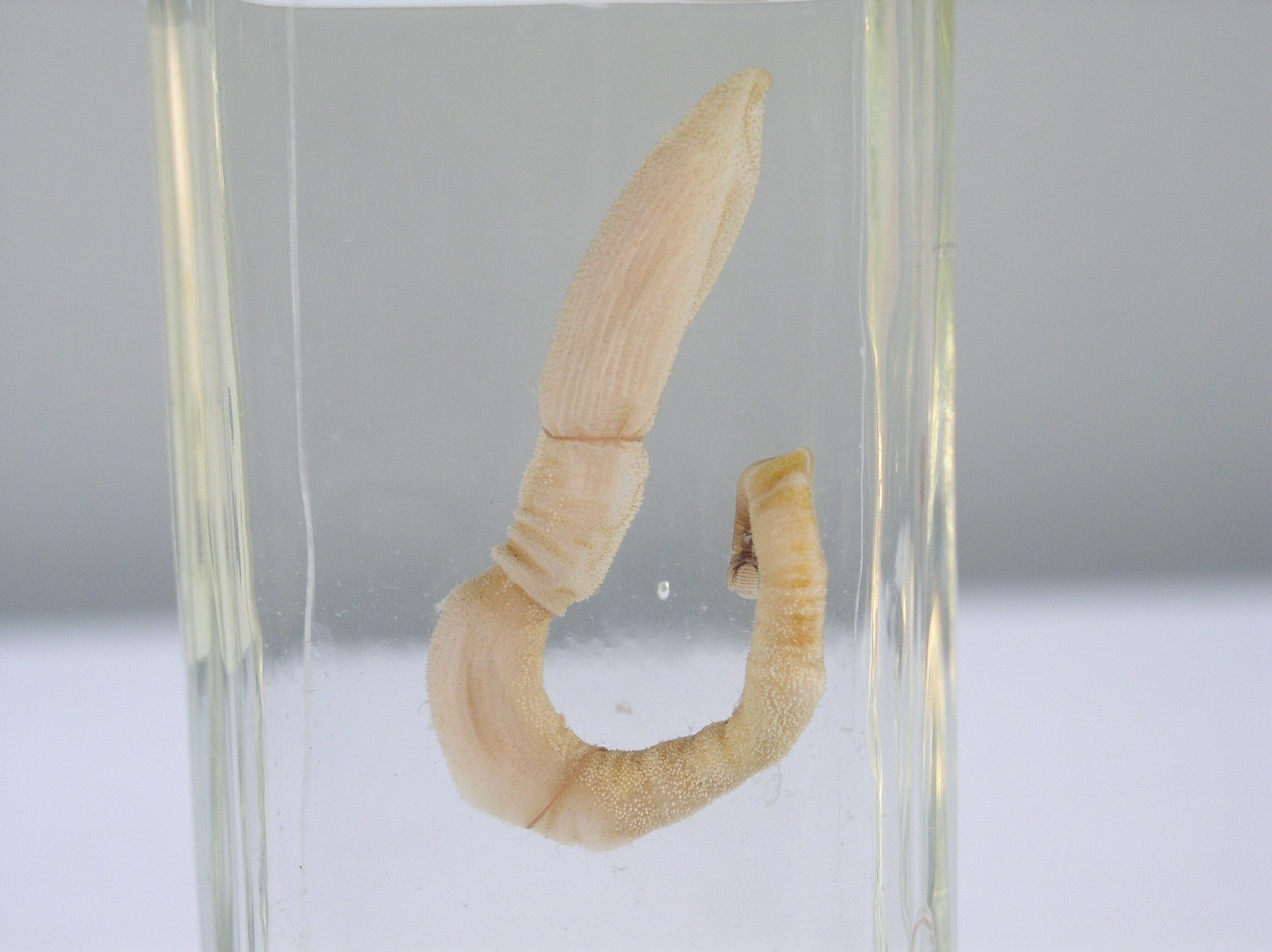
This release incorporates a first pass at organism subdivisions for the acorn worm (enteropneust). We would like to bud this off into an external ontology that is federated with the core Uberon. For more information, see 692
We also clarified the distinctinction between trigeminal and profundal placodes and the structures they give rise to. For more information, see 693, and the following diagram:

Ontology Diff Report
- invertebrate biology
- muscle
- Redefining some musculature classes in terms of attachments.
- added note to suprahyoid. 697 RDruzinsky
- merging ‘skeletal muscle of tongue’ into ‘tongue muscle’ as all tongue muscles are skeletal (RDruzinsky). 331
- obsoleting tongue musculature (trivially paralleled ‘tongue muscle’)
- neuro
- placodes
- Syns and develops from for profundal and trigeminal placodes, nerves etc. Issue 693 ANiknejad mellybelly
- obsoleting AEO-derived epithelial placode and merging with ectodermal placode
- adding df to pre-placodal ectoderm for all neurogenic placodes
- urogenital
- gonadal/genital ridge fixes
- comparative biology
- adding taxon comments to otolith courtesy of SomeCallMeDave
- skeletal
- carapace plate syns
- added definitions for ribs, clarifying that homology of serial elements not implied
- vasculature
- bridging axioms
- Moved some constraints into pending axioms, to avoid incoherent combined ontologies. Fixes 694 balhoff
- fixing non 1:1 emapa xrefs, thanks for tfhayamizu for clarifications
- Fixed some FMA xrefs. Issue 683 drseb
- fixed wikipedia depiction images; we always now use the full image, not a thumb
- Added more Wikipedia xrefs
- Other
Original Ontology
- IRI: http://purl.obolibrary.org/obo/uberon.owl
- VersionIRI: http://purl.obolibrary.org/obo/uberon/releases/2015-04-23/uberon.owl
New Ontology
- IRI: http://purl.obolibrary.org/obo/uberon.owl
- VersionIRI: http://purl.obolibrary.org/obo/uberon/releases/2015-05-25/uberon.owl
Report for classes
Class objects lost from source: 2
Class objects new in target: 165
New Class : pectoral fin base
- pectoral fin base label pectoral fin base
- pectoral fin base SubClassOf part of some pectoral fin
- pectoral fin base created by WD
- pectoral fin base definition Subdivision of organism that is the basal region of the pectoral fin. [PHENOSCAPE:WD].
- pectoral fin base SubClassOf paired limb/fin segment
New Class : right inguinal part of abdomen
- right inguinal part of abdomen SubClassOf in right side of some abdomen
- right inguinal part of abdomen database cross reference http://www.snomedbrowser.com/Codes/Details/362724003
- right inguinal part of abdomen has exact synonym right inguinal region { database cross reference=FMA:24036 }
- right inguinal part of abdomen database cross reference http://ncicb.nci.nih.gov/xml/owl/EVS/Right_Inguinal_Region
- right inguinal part of abdomen database cross reference http://linkedlifedata.com/resource/umls/id/C0230318
- right inguinal part of abdomen id UBERON:0035505
- right inguinal part of abdomen label right inguinal part of abdomen
- right inguinal part of abdomen has exact synonym right iliac fossa viewed surgically { database cross reference=FMA:24036 }
- right inguinal part of abdomen EquivalentTo inguinal part of abdomen and in right side of some abdomen
- right inguinal part of abdomen has exact synonym right iliac region { database cross reference=FMA:24036 }
- right inguinal part of abdomen database cross reference FMA:24036
- right inguinal part of abdomen SubClassOf inguinal part of abdomen
- right inguinal part of abdomen SubClassOf lateral structure
- right inguinal part of abdomen has obo namespace uberon
New Class : branch of posterior cerebral artery
- branch of posterior cerebral artery SubClassOf systemic artery
- branch of posterior cerebral artery SubClassOf head blood vessel
- branch of posterior cerebral artery SubClassOf branching part of some posterior cerebral artery
- branch of posterior cerebral artery has exact synonym posterior cerebral arterial branch { database cross reference=FMA:50586 }
- branch of posterior cerebral artery database cross reference http://linkedlifedata.com/resource/umls/id/C0923792
- branch of posterior cerebral artery database cross reference http://ncicb.nci.nih.gov/xml/owl/EVS/Posterior_Cerebral_Artery_Branch
- branch of posterior cerebral artery has obo namespace uberon
- branch of posterior cerebral artery label branch of posterior cerebral artery
- branch of posterior cerebral artery id UBERON:0035508
- branch of posterior cerebral artery database cross reference http://www.snomedbrowser.com/Codes/Details/314305001
- branch of posterior cerebral artery EquivalentTo artery and branching part of some posterior cerebral artery
- branch of posterior cerebral artery database cross reference FMA:50586
New Class : unencapsulated nerve ending
- unencapsulated nerve ending has obo namespace uberon
- unencapsulated nerve ending id UBERON:0035501
- unencapsulated nerve ending database cross reference FMA:84005
- unencapsulated nerve ending database cross reference http://linkedlifedata.com/resource/umls/id/C0228107
- unencapsulated nerve ending label unencapsulated nerve ending
- unencapsulated nerve ending SubClassOf tactile mechanoreceptor
- unencapsulated nerve ending definition Free nerve endings are widely distributed throughout the body, and are found as branches of unmyelinated, or lightly myelinated fibres grouped in bundles beneath the epithelium. As they penetrate the epithelium, they lose their myelin, and branch among the epithelial cells. Branches of one nerve may cover a wide area and overlap the territories of other nerves. The free nerve endings detect pain, touch, pressure and temperature, and are associated with C fibres. { database cross reference=ncithesaurus:Free_Nerve_Ending }
- unencapsulated nerve ending database cross reference http://ncicb.nci.nih.gov/xml/owl/EVS/Free_Nerve_Ending
- unencapsulated nerve ending SubClassOf part of some dermis
- unencapsulated nerve ending has exact synonym free nerve ending { database cross reference=FMA:84005 }
- unencapsulated nerve ending database cross reference http://www.snomedbrowser.com/Codes/Details/38005009
New Class : special sense organ system
- special sense organ system database cross reference FMA:7189
- special sense organ system label special sense organ system
- special sense organ system SubClassOf organ system subdivision
- special sense organ system id UBERON:0035514
- special sense organ system has obo namespace uberon
- special sense organ system database cross reference http://linkedlifedata.com/resource/umls/id/C0228069
- special sense organ system database cross reference http://ncicb.nci.nih.gov/xml/owl/EVS/Special_Sense_Organ_System
- special sense organ system database cross reference http://www.snomedbrowser.com/Codes/Details/276152001
New Class : left common iliac artery
- left common iliac artery SubClassOf lateral structure
- left common iliac artery has obo namespace uberon
- left common iliac artery database cross reference http://linkedlifedata.com/resource/umls/id/C0226363
- left common iliac artery database cross reference http://ncicb.nci.nih.gov/xml/owl/EVS/Left_Common_Iliac_Artery
- left common iliac artery SubClassOf in left side of some multicellular organism
- left common iliac artery id UBERON:0035529
- left common iliac artery label left common iliac artery
- left common iliac artery has exact synonym trunk of left common iliac arterial tree { database cross reference=FMA:14766 }
- left common iliac artery database cross reference FMA:14766
- left common iliac artery database cross reference http://www.snomedbrowser.com/Codes/Details/283493001
- left common iliac artery SubClassOf common iliac artery
- left common iliac artery EquivalentTo common iliac artery and in left side of some multicellular organism
New Class : superficial fibular nerve
- superficial fibular nerve has obo namespace uberon
- superficial fibular nerve database cross reference http://en.wikipedia.org/wiki/Superficial_fibular_nerve
- superficial fibular nerve label superficial fibular nerve
- superficial fibular nerve database cross reference http://linkedlifedata.com/resource/umls/id/C0228952
- superficial fibular nerve has exact synonym superficial peroneal nerve { database cross reference=FMA:44699 }
- superficial fibular nerve database cross reference http://www.snomedbrowser.com/Codes/Details/181080008
- superficial fibular nerve SubClassOf branching part of some common fibular nerve
- superficial fibular nerve id UBERON:0035526
- superficial fibular nerve SubClassOf leg nerve
- superficial fibular nerve definition A branch of the common peroneal nerve. It innervates the surface of the calf and foot. { database cross reference=ncithesaurus:Superficial_Peroneal_Nerve }
- superficial fibular nerve SubClassOf innervates some hindlimb
- superficial fibular nerve database cross reference http://ncicb.nci.nih.gov/xml/owl/EVS/Superficial_Peroneal_Nerve
- superficial fibular nerve database cross reference FMA:44699
New Class : anterior mediastinal lymph node
- anterior mediastinal lymph node definition A lymph node located in the superior mediastinum that collects lymph from the thymus, the pericardium, and the right side of the heart. { database cross reference=ncithesaurus:Anterior_Mediastinal_Lymph_Node }
- anterior mediastinal lymph node database cross reference FMA:12775
- anterior mediastinal lymph node has exact synonym anterior mediastinal node { database cross reference=FMA:12775 }
- anterior mediastinal lymph node database cross reference http://ncicb.nci.nih.gov/xml/owl/EVS/Anterior_Mediastinal_Lymph_Node
- anterior mediastinal lymph node SubClassOf mediastinal lymph node
- anterior mediastinal lymph node id UBERON:0035520
- anterior mediastinal lymph node label anterior mediastinal lymph node
- anterior mediastinal lymph node database cross reference http://www.snomedbrowser.com/Codes/Details/245335005
- anterior mediastinal lymph node has obo namespace uberon
- anterior mediastinal lymph node database cross reference http://linkedlifedata.com/resource/umls/id/C0229758
New Class : anterior surface of prostate
- anterior surface of prostate SubClassOf bounding layer of some prostate gland
- anterior surface of prostate database cross reference FMA:19591
- anterior surface of prostate label anterior surface of prostate
- anterior surface of prostate has exact synonym anterior surface of prostate gland { database cross reference=FMA:19591 }
- anterior surface of prostate SubClassOf in anterior side of some prostate gland
- anterior surface of prostate database cross reference http://linkedlifedata.com/resource/umls/id/C0227960
- anterior surface of prostate database cross reference http://www.snomedbrowser.com/Codes/Details/279696001
- anterior surface of prostate EquivalentTo anatomical surface and in anterior side of some prostate gland and bounding layer of some prostate gland
- anterior surface of prostate database cross reference http://ncicb.nci.nih.gov/xml/owl/EVS/Anterior_Surface_of_the_Prostate
- anterior surface of prostate has exact synonym facies anterior (prostatae) { database cross reference=FMA:19591 }
- anterior surface of prostate has obo namespace uberon
- anterior surface of prostate definition The aspect of the prostate facing the pubic symphysis. { database cross reference=ncithesaurus:Anterior_Surface_of_the_Prostate }
- anterior surface of prostate id UBERON:0035523
- anterior surface of prostate SubClassOf surface of prostate
New Class : esophageal artery
- esophageal artery definition Any of several arteries that arise from the aorta and supply blood to the esophagus. { database cross reference=ncithesaurus:Esophageal_Artery }
- esophageal artery SubClassOf systemic artery
- esophageal artery EquivalentTo artery and supplies some esophagus
- esophageal artery database cross reference http://ncicb.nci.nih.gov/xml/owl/EVS/Esophageal_Artery
- esophageal artery database cross reference http://www.snomedbrowser.com/Codes/Details/56548006
- esophageal artery SubClassOf thoracic segment blood vessel
- esophageal artery has exact synonym aortic esophageal artery { database cross reference=FMA:4149 }
- esophageal artery id UBERON:0035539
- esophageal artery SubClassOf branching part of some thoracic aorta
- esophageal artery label esophageal artery
- esophageal artery has obo namespace uberon
- esophageal artery database cross reference FMA:4149
- esophageal artery database cross reference http://linkedlifedata.com/resource/umls/id/C0226294
- esophageal artery has exact synonym oesophageal artery { database cross reference=FMA:4149 }
- esophageal artery SubClassOf supplies some esophagus
New Class : body of gallbladder
- body of gallbladder label body of gallbladder
- body of gallbladder database cross reference http://ncicb.nci.nih.gov/xml/owl/EVS/Gallbladder_Body
- body of gallbladder has exact synonym gallbladder body { database cross reference=FMA:14537 }
- body of gallbladder has obo namespace uberon
- body of gallbladder database cross reference http://linkedlifedata.com/resource/umls/id/C0227545
- body of gallbladder SubClassOf endoderm-derived structure
- body of gallbladder SubClassOf zone of organ
- body of gallbladder database cross reference http://en.wikipedia.org/wiki/Body_of_gallbladder
- body of gallbladder definition The middle portion of the gallbladder which is distal to the gallbladder neck and proximal to the gallbladder fundus. { database cross reference=ncithesaurus:Gallbladder_Body }
- body of gallbladder SubClassOf part of some gall bladder
- body of gallbladder database cross reference http://www.snomedbrowser.com/Codes/Details/245392006
- body of gallbladder database cross reference FMA:14537
- body of gallbladder id UBERON:0035536
New Class : deep middle cerebral vein
- deep middle cerebral vein database cross reference FMA:51309
- deep middle cerebral vein database cross reference http://en.wikipedia.org/wiki/Deep_middle_cerebral_vein
- deep middle cerebral vein label deep middle cerebral vein
- deep middle cerebral vein database cross reference http://linkedlifedata.com/resource/umls/id/C0226586
- deep middle cerebral vein has obo namespace uberon
- deep middle cerebral vein database cross reference http://www.snomedbrowser.com/Codes/Details/279272001
- deep middle cerebral vein SubClassOf tributary of some basal vein
- deep middle cerebral vein SubClassOf cerebral vein
- deep middle cerebral vein id UBERON:0035532
- deep middle cerebral vein database cross reference http://ncicb.nci.nih.gov/xml/owl/EVS/Deep_Middle_Cerebral_Vein
- deep middle cerebral vein definition The blood vessel that receives deoxygenated blood from the insula and gyri and drains into the basal vein of Rosenthal deep in the lateral sulcus. { database cross reference=ncithesaurus:Deep_Middle_Cerebral_Vein }
New Class : basal vein
- basal vein has exact synonym basal vein of rosenthal { database cross reference=FMA:50990 }
- basal vein has related synonym inferior striate vein { database cross reference=http://en.wikipedia.org/wiki/Basal_vein }
- basal vein SubClassOf deep cerebral vein
- basal vein database cross reference http://en.wikipedia.org/wiki/Basal_vein
- basal vein has related synonym basal veins { database cross reference=http://en.wikipedia.org/wiki/Basal_vein }
- basal vein database cross reference http://www.snomedbrowser.com/Codes/Details/244398001
- basal vein database cross reference FMA:50990
- basal vein definition The basal vein is formed at the anterior perforated substance by the union of (a) a small anterior cerebral vein which accompanies the anterior cerebral artery and supplies the medial surface of the frontal lobe by the fronto-basal vein. (b) the deep middle cerebral vein (deep Sylvian vein), which receives tributaries from the insula and neighboring gyri, and runs in the lower part of the lateral cerebral fissure, and (c) the inferior striate veins, which leave the corpus striatum through the anterior perforated substance. The basal vein passes backward around the cerebral peduncle, and ends in the internal cerebral vein; it receives tributaries from the interpeduncular fossa, the inferior horn of the lateral ventricle, the hippocampal gyrus, and the mid-brain. { database cross reference=http://en.wikipedia.org/wiki/Basal_vein }
- basal vein label basal vein
- basal vein has obo namespace uberon
- basal vein has related synonym inferior striate veins { database cross reference=http://en.wikipedia.org/wiki/Basal_vein }
- basal vein has exact synonym rosenthal’s vein { database cross reference=FMA:50990 }
- basal vein id UBERON:0035530
New Class : lower inner quadrant of breast
- lower inner quadrant of breast database cross reference http://www.snomedbrowser.com/Codes/Details/181135009
- lower inner quadrant of breast database cross reference FMA:61377
- lower inner quadrant of breast SubClassOf part of some breast
- lower inner quadrant of breast id UBERON:0035477
- lower inner quadrant of breast definition The quarter of the breast which is inferior and medial. { database cross reference=ncithesaurus:Lower-inner_Quadrant_of_the_Breast }
- lower inner quadrant of breast has obo namespace uberon
- lower inner quadrant of breast has exact synonym lower inner quadrant of female breast { database cross reference=FMA:61377 }
- lower inner quadrant of breast SubClassOf organism subdivision
- lower inner quadrant of breast label lower inner quadrant of breast
- lower inner quadrant of breast database cross reference http://ncicb.nci.nih.gov/xml/owl/EVS/Lower-inner_Quadrant_of_the_Breast
- lower inner quadrant of breast database cross reference http://linkedlifedata.com/resource/umls/id/C0222597
New Class : right subcostal vein
- right subcostal vein definition A vein that extends along the bottom of the twelfth rib on the right side of the body. { database cross reference=ncithesaurus:Right_Subcostal_Vein }
- right subcostal vein has exact synonym vena subcostalis dextra { database cross reference=FMA:4844 }
- right subcostal vein has obo namespace uberon
- right subcostal vein id UBERON:0035474
- right subcostal vein database cross reference http://ncicb.nci.nih.gov/xml/owl/EVS/Right_Subcostal_Vein
- right subcostal vein label right subcostal vein
- right subcostal vein SubClassOf lateral structure
- right subcostal vein database cross reference FMA:4844
- right subcostal vein database cross reference http://www.snomedbrowser.com/Codes/Details/361638003
- right subcostal vein SubClassOf in right side of some multicellular organism
- right subcostal vein database cross reference http://linkedlifedata.com/resource/umls/id/C0501189
- right subcostal vein EquivalentTo subcostal vein and in right side of some multicellular organism
- right subcostal vein SubClassOf subcostal vein
New Class : posterior surface of kidney
- posterior surface of kidney id UBERON:0035471
- posterior surface of kidney label posterior surface of kidney
- posterior surface of kidney database cross reference http://ncicb.nci.nih.gov/xml/owl/EVS/Kidney_Posterior_Surface
- posterior surface of kidney database cross reference http://linkedlifedata.com/resource/umls/id/C0227610
- posterior surface of kidney EquivalentTo anatomical surface and in posterior side of some kidney and bounding layer of some kidney
- posterior surface of kidney database cross reference http://www.snomedbrowser.com/Codes/Details/279376000
- posterior surface of kidney SubClassOf in posterior side of some kidney
- posterior surface of kidney SubClassOf bounding layer of some kidney
- posterior surface of kidney database cross reference FMA:15590
- posterior surface of kidney has obo namespace uberon
- posterior surface of kidney SubClassOf anatomical surface
- posterior surface of kidney has exact synonym facies posterior (Ren) { database cross reference=FMA:15590 }
New Class : costodiaphragmatic recess
- costodiaphragmatic recess database cross reference http://en.wikipedia.org/wiki/Costodiaphragmatic_recess
- costodiaphragmatic recess has obo namespace uberon
- costodiaphragmatic recess database cross reference FMA:11355
- costodiaphragmatic recess label costodiaphragmatic recess
- costodiaphragmatic recess SubClassOf zone of organ
- costodiaphragmatic recess has exact synonym costophrenic angle { database cross reference=FMA:11355 }
- costodiaphragmatic recess SubClassOf part of some diaphragm
- costodiaphragmatic recess id UBERON:0035468
- costodiaphragmatic recess database cross reference http://linkedlifedata.com/resource/umls/id/C0230151
- costodiaphragmatic recess database cross reference http://www.snomedbrowser.com/Codes/Details/46297007
- costodiaphragmatic recess SubClassOf mesoderm-derived structure
- costodiaphragmatic recess database cross reference http://ncicb.nci.nih.gov/xml/owl/EVS/Costophrenic_Angle
- costodiaphragmatic recess definition The angled junction where the diaphragm meets the chest wall. { database cross reference=ncithesaurus:Costophrenic_Angle }
New Class : endometrial cavity
- endometrial cavity SubClassOf lumen of some body of uterus
- endometrial cavity database cross reference http://en.wikipedia.org/wiki/Uterine_cavity
- endometrial cavity has exact synonym endometrial cavity { database cross reference=FMA:18076 }
- endometrial cavity database cross reference FMA:18076
- endometrial cavity SubClassOf part of some uterine lumen
- endometrial cavity database cross reference http://linkedlifedata.com/resource/umls/id/C0227844
- endometrial cavity label endometrial cavity
- endometrial cavity SubClassOf adjacent to some endometrium
- endometrial cavity database cross reference http://www.snomedbrowser.com/Codes/Details/362257002
- endometrial cavity database cross reference http://ncicb.nci.nih.gov/xml/owl/EVS/Endometrial_Cavity
- endometrial cavity has exact synonym endometrial lumen
- endometrial cavity has obo namespace uberon
- endometrial cavity SubClassOf anatomical cavity
- endometrial cavity id UBERON:0035465
- endometrial cavity has exact synonym intrauterine cavity { database cross reference=FMA:18076 }
- endometrial cavity has exact synonym cavity of body of uterus { database cross reference=FMA:18076 }
- endometrial cavity definition A space inside the uterus lined by a layer of mucus membranes called the endometrium. { database cross reference=ncithesaurus:Endometrial_Cavity }
- endometrial cavity EquivalentTo anatomical cavity and adjacent to some endometrium and lumen of some body of uterus
New Class : anterior parietal artery
- anterior parietal artery label anterior parietal artery
- anterior parietal artery SubClassOf branching part of some middle cerebral artery
- anterior parietal artery has obo namespace uberon
- anterior parietal artery database cross reference FMA:50485
- anterior parietal artery definition The artery that delivers blood to the anterior portion of the parietal lobe. { database cross reference=ncithesaurus:Anterior_Parietal_Artery }
- anterior parietal artery database cross reference http://ncicb.nci.nih.gov/xml/owl/EVS/Anterior_Parietal_Artery
- anterior parietal artery database cross reference http://www.snomedbrowser.com/Codes/Details/78499006
- anterior parietal artery database cross reference http://linkedlifedata.com/resource/umls/id/C0226226
- anterior parietal artery id UBERON:0035462
- anterior parietal artery SubClassOf branch of middle cerebral artery
New Class : hilum of lymph node
- hilum of lymph node database cross reference http://www.snomedbrowser.com/Codes/Details/327052008
- hilum of lymph node database cross reference http://ncicb.nci.nih.gov/xml/owl/EVS/Lymph_Node_Hilum
- hilum of lymph node id UBERON:0035495
- hilum of lymph node EquivalentTo hilum and part of some lymph node
- hilum of lymph node database cross reference http://linkedlifedata.com/resource/umls/id/C0229703
- hilum of lymph node SubClassOf part of some lymph node
- hilum of lymph node definition The concave side of the lymph node. { database cross reference=ncithesaurus:Lymph_Node_Hilum }
- hilum of lymph node SubClassOf hilum
- hilum of lymph node label hilum of lymph node
- hilum of lymph node has exact synonym lymph node hilum { database cross reference=FMA:62842 }
- hilum of lymph node database cross reference FMA:62842
- hilum of lymph node has obo namespace uberon
New Class : gastrophrenic ligament
- gastrophrenic ligament SubClassOf connects some diaphragm
- gastrophrenic ligament SubClassOf connects some greater curvature of stomach
- gastrophrenic ligament SubClassOf part of some greater omentum
- gastrophrenic ligament label gastrophrenic ligament
- gastrophrenic ligament SubClassOf mesoderm-derived structure
- gastrophrenic ligament database cross reference FMA:16518
- gastrophrenic ligament SubClassOf abdomen element
- gastrophrenic ligament SubClassOf nonskeletal ligament
- gastrophrenic ligament database cross reference http://linkedlifedata.com/resource/umls/id/C0230231
- gastrophrenic ligament database cross reference http://ncicb.nci.nih.gov/xml/owl/EVS/Gastrophrenic_Ligament
- gastrophrenic ligament definition the portion of the greater omentum that extends from the greater curvature of the stomach to the inferior surface of the diaphragm { database cross reference=http://medical-dictionary.thefreedictionary.com/gastrophrenic+ligament }
- gastrophrenic ligament has obo namespace uberon
- gastrophrenic ligament database cross reference http://www.snomedbrowser.com/Codes/Details/260725009
- gastrophrenic ligament EquivalentTo nonskeletal ligament and part of some greater omentum and connects some diaphragm and connects some greater curvature of stomach
- gastrophrenic ligament id UBERON:0035498
New Class : inferior hypophysial artery
- inferior hypophysial artery database cross reference FMA:49846
- inferior hypophysial artery SubClassOf supplies some pituitary gland
- inferior hypophysial artery label inferior hypophysial artery
- inferior hypophysial artery database cross reference http://www.snomedbrowser.com/Codes/Details/303428003
- inferior hypophysial artery database cross reference http://en.wikipedia.org/wiki/Inferior_hypophysial_artery
- inferior hypophysial artery SubClassOf hypophysial artery
- inferior hypophysial artery SubClassOf branching part of some internal carotid artery
- inferior hypophysial artery database cross reference http://ncicb.nci.nih.gov/xml/owl/EVS/Inferior_Hypophyseal_Artery
- inferior hypophysial artery has exact synonym inferior hypophyseal artery { database cross reference=FMA:49846 }
- inferior hypophysial artery SubClassOf structure with developmental contribution from neural crest
- inferior hypophysial artery SubClassOf head blood vessel
- inferior hypophysial artery SubClassOf mixed endoderm/mesoderm-derived structure
- inferior hypophysial artery id UBERON:0035492
- inferior hypophysial artery database cross reference http://linkedlifedata.com/resource/umls/id/C1261083
- inferior hypophysial artery has obo namespace uberon
- inferior hypophysial artery definition The inferior hypophysial artery is an artery supplying the pituitary gland. It is a branch of the cavernous carotid artery (internal carotid artery). { database cross reference=http://en.wikipedia.org/wiki/Inferior_hypophysial_artery,Wikipedia:Inferior_hypophysial_artery }
- inferior hypophysial artery SubClassOf systemic artery
New Class : left suprarenal vein
- left suprarenal vein database cross reference http://ncicb.nci.nih.gov/xml/owl/EVS/Left_Suprarenal_Vein
- left suprarenal vein has obo namespace uberon
- left suprarenal vein definition A vein that drains blood from the left adrenal gland into the left renal artery. { database cross reference=ncithesaurus:Left_Suprarenal_Vein }
- left suprarenal vein SubClassOf lateral structure
- left suprarenal vein database cross reference http://www.snomedbrowser.com/Codes/Details/85087001
- left suprarenal vein database cross reference FMA:14349
- left suprarenal vein database cross reference http://linkedlifedata.com/resource/umls/id/C0226709
- left suprarenal vein label left suprarenal vein
- left suprarenal vein SubClassOf in left side of some multicellular organism
- left suprarenal vein database cross reference http://en.wikipedia.org/wiki/Suprarenal_veins
- left suprarenal vein SubClassOf suprarenal vein
- left suprarenal vein EquivalentTo suprarenal vein and in left side of some multicellular organism
- left suprarenal vein id UBERON:0035483
New Class : branch of basilar artery
- branch of basilar artery EquivalentTo artery and branching part of some basilar artery
- branch of basilar artery SubClassOf branching part of some basilar artery
- branch of basilar artery SubClassOf systemic artery
- branch of basilar artery database cross reference http://www.snomedbrowser.com/Codes/Details/360537008
- branch of basilar artery database cross reference http://linkedlifedata.com/resource/umls/id/C1185381
- branch of basilar artery label branch of basilar artery
- branch of basilar artery SubClassOf head blood vessel
- branch of basilar artery has obo namespace uberon
- branch of basilar artery database cross reference FMA:76269
- branch of basilar artery database cross reference http://ncicb.nci.nih.gov/xml/owl/EVS/Basilar_Artery_Branch
- branch of basilar artery id UBERON:0035489
- branch of basilar artery has exact synonym basilar arterial branch { database cross reference=FMA:76269 }
New Class : surface of prostate
- surface of prostate definition The external portion of the prostate including the anterior, inferolateral, lateral and posterior surfaces. { database cross reference=ncithesaurus:Surface_of_the_Prostate }
- surface of prostate SubClassOf anatomical surface
- surface of prostate database cross reference FMA:19590
- surface of prostate has obo namespace uberon
- surface of prostate label surface of prostate
- surface of prostate database cross reference http://linkedlifedata.com/resource/umls/id/C0426735
- surface of prostate has exact synonym surface of prostate gland { database cross reference=FMA:19590 }
- surface of prostate database cross reference http://ncicb.nci.nih.gov/xml/owl/EVS/Surface_of_the_Prostate
- surface of prostate id UBERON:0035480
- surface of prostate EquivalentTo anatomical surface and bounding layer of some prostate gland
- surface of prostate SubClassOf bounding layer of some prostate gland
- surface of prostate has exact synonym prostatic surface { database cross reference=FMA:19590 }
- surface of prostate database cross reference http://www.snomedbrowser.com/Codes/Details/279694003
New Class : right suprarenal vein
- right suprarenal vein has exact synonym vena suprarenalis (adrenalis) dextra { database cross reference=FMA:14343 }
- right suprarenal vein database cross reference http://ncicb.nci.nih.gov/xml/owl/EVS/Right_Suprarenal_Vein
- right suprarenal vein database cross reference FMA:14343
- right suprarenal vein label right suprarenal vein
- right suprarenal vein id UBERON:0035435
- right suprarenal vein SubClassOf in right side of some multicellular organism
- right suprarenal vein database cross reference http://www.snomedbrowser.com/Codes/Details/69354005
- right suprarenal vein SubClassOf suprarenal vein
- right suprarenal vein has obo namespace uberon
- right suprarenal vein SubClassOf lateral structure
- right suprarenal vein database cross reference http://linkedlifedata.com/resource/umls/id/C0226721
- right suprarenal vein EquivalentTo suprarenal vein and in right side of some multicellular organism
- right suprarenal vein definition A vein that drains blood from the right adrenal gland into the inferior vena cava. { database cross reference=ncithesaurus:Right_Suprarenal_Vein }
New Class : mediastinal pleura
- mediastinal pleura SubClassOf zone of organ
- mediastinal pleura SubClassOf part of some parietal pleura
- mediastinal pleura SubClassOf mixed endoderm/mesoderm-derived structure
- mediastinal pleura has exact synonym pars mediastinalis (pleurae) { database cross reference=FMA:9736 }
- mediastinal pleura database cross reference FMA:9736
- mediastinal pleura database cross reference http://ncicb.nci.nih.gov/xml/owl/EVS/Mediastinal_Pleura
- mediastinal pleura database cross reference http://en.wikipedia.org/wiki/Mediastinal_pleura
- mediastinal pleura database cross reference http://linkedlifedata.com/resource/umls/id/C0225789
- mediastinal pleura SubClassOf bounding layer of some mediastinum
- mediastinal pleura label mediastinal pleura
- mediastinal pleura has obo namespace uberon
- mediastinal pleura id UBERON:0035431
- mediastinal pleura definition The parietal pleura that lines the mediastinum. { database cross reference=ncithesaurus:Mediastinal_Pleura }
- mediastinal pleura database cross reference http://www.snomedbrowser.com/Codes/Details/362002008
- mediastinal pleura has exact synonym mediastinal part of parietal pleura { database cross reference=FMA:9736 }
New Class : mucoid tissue
- mucoid tissue has exact synonym mucous tissue { database cross reference=FMA:20111 }
- mucoid tissue label mucoid tissue
- mucoid tissue SubClassOf loose connective tissue
- mucoid tissue database cross reference http://ncicb.nci.nih.gov/xml/owl/EVS/Mucous_Connective_Tissue
- mucoid tissue database cross reference http://www.snomedbrowser.com/Codes/Details/61115003
- mucoid tissue has obo namespace uberon
- mucoid tissue id UBERON:0035438
- mucoid tissue database cross reference FMA:20111
- mucoid tissue external ontology notes ncit restricts to umbilical cord { external ontology=ncit }
- mucoid tissue has exact synonym mucous connective tissue { database cross reference=FMA:20111 }
- mucoid tissue definition Loose connective tissue, the intercellular matrix of which consists predominantly of mucoid ground substance. Examples: Mucoid tissue of umbilical cord, mucoid tissue of vitreous body, mucoid tissue of nucleus pulposus. { database cross reference=FMA:20111 }
- mucoid tissue database cross reference http://linkedlifedata.com/resource/umls/id/C0230987
New Class : circumflex branch of left coronary artery
- circumflex branch of left coronary artery database cross reference http://linkedlifedata.com/resource/umls/id/C0226037
- circumflex branch of left coronary artery database cross reference http://en.wikipedia.org/wiki/Circumflex_branch_of_left_coronary_artery
- circumflex branch of left coronary artery database cross reference http://ncicb.nci.nih.gov/xml/owl/EVS/Circumflex_Branch_of_the_Left_Coronary_Artery
- circumflex branch of left coronary artery id UBERON:0035422
- circumflex branch of left coronary artery has obo namespace uberon
- circumflex branch of left coronary artery database cross reference http://www.snomedbrowser.com/Codes/Details/362036002
- circumflex branch of left coronary artery label circumflex branch of left coronary artery
- circumflex branch of left coronary artery SubClassOf part of some left coronary artery
- circumflex branch of left coronary artery SubClassOf branching part of some left coronary artery
- circumflex branch of left coronary artery has exact synonym circumflex coronary artery { database cross reference=FMA:3895 }
- circumflex branch of left coronary artery database cross reference FMA:3895
- circumflex branch of left coronary artery SubClassOf branch of left coronary artery
- circumflex branch of left coronary artery definition Branch of left coronary artery which runs perpendicular to the anterior interventricular branch of the left coronary artery on the left side of the interventricular sulcus and supplies the left side of the heart. { database cross reference=FMA:3895 }
- circumflex branch of left coronary artery has exact synonym left circumflex branch of left coronary artery { database cross reference=FMA:3895 }
New Class : postcapillary venule
- postcapillary venule database cross reference http://linkedlifedata.com/resource/umls/id/C0226505
- postcapillary venule id UBERON:0035428
- postcapillary venule database cross reference http://ncicb.nci.nih.gov/xml/owl/EVS/Postcapillary_Venule
- postcapillary venule database cross reference FMA:63191
- postcapillary venule SubClassOf venule
- postcapillary venule label postcapillary venule
- postcapillary venule database cross reference http://www.snomedbrowser.com/Codes/Details/6216007
- postcapillary venule has obo namespace uberon
New Class : falx cerebelli
- falx cerebelli SubClassOf mesoderm-derived structure
- falx cerebelli SubClassOf structure with developmental contribution from neural crest
- falx cerebelli definition A small triangular process of dura matter beginning at the internal occipital crest just beneath the tentorium and projecting forward. { database cross reference=ncithesaurus:Falx_Cerebelli }
- falx cerebelli SubClassOf organ part
- falx cerebelli has obo namespace uberon
- falx cerebelli database cross reference http://ncicb.nci.nih.gov/xml/owl/EVS/Falx_Cerebelli
- falx cerebelli label falx cerebelli
- falx cerebelli database cross reference http://www.snomedbrowser.com/Codes/Details/280380009
- falx cerebelli database cross reference FMA:83974
- falx cerebelli has exact synonym cerebellar falx { database cross reference=FMA:83974 }
- falx cerebelli database cross reference http://en.wikipedia.org/wiki/Falx_cerebelli
- falx cerebelli SubClassOf part of some meningeal dura mater
- falx cerebelli database cross reference http://linkedlifedata.com/resource/umls/id/C0228122
- falx cerebelli SubClassOf part of some dura mater
- falx cerebelli SubClassOf ectoderm-derived structure
- falx cerebelli id UBERON:0035425
New Class : cervical part of esophagus
- cervical part of esophagus label cervical part of esophagus
- cervical part of esophagus SubClassOf part of some esophagus
- cervical part of esophagus has exact synonym cervical esophagus { database cross reference=FMA:9395 }
- cervical part of esophagus SubClassOf subdivision of tube
- cervical part of esophagus database cross reference http://linkedlifedata.com/resource/umls/id/C0227186
- cervical part of esophagus has exact synonym pars cervicalis (oesophagus) { database cross reference=FMA:9395 }
- cervical part of esophagus database cross reference FMA:9395
- cervical part of esophagus has exact synonym cervical part of oesophagus { database cross reference=FMA:9395 }
- cervical part of esophagus database cross reference http://ncicb.nci.nih.gov/xml/owl/EVS/Cervical_Esophagus
- cervical part of esophagus definition Clinical esophageal segment composed of skeletal muscle. It corresponds to the superior part of the upper third topographic segment of the esophagus. { database cross reference=ncithesaurus:Cervical_Esophagus }
- cervical part of esophagus has obo namespace uberon
- cervical part of esophagus database cross reference http://www.snomedbrowser.com/Codes/Details/245405004
- cervical part of esophagus id UBERON:0035450
New Class : laryngeal ventricle
- laryngeal ventricle has exact synonym ventricle of larynx { database cross reference=FMA:64171 }
- laryngeal ventricle database cross reference http://www.snomedbrowser.com/Codes/Details/361949007
- laryngeal ventricle database cross reference http://linkedlifedata.com/resource/umls/id/C0225566
- laryngeal ventricle id UBERON:0035453
- laryngeal ventricle has exact synonym ventricle of Morgagni { database cross reference=FMA:64171 }
- laryngeal ventricle database cross reference FMA:64171
- laryngeal ventricle SubClassOf part of some larynx
- laryngeal ventricle database cross reference http://ncicb.nci.nih.gov/xml/owl/EVS/Laryngeal_Ventricle
- laryngeal ventricle SubClassOf endoderm-derived structure
- laryngeal ventricle label laryngeal ventricle
- laryngeal ventricle has obo namespace uberon
- laryngeal ventricle SubClassOf zone of organ
New Class : apex of prostate
- apex of prostate database cross reference http://linkedlifedata.com/resource/umls/id/C0227959
- apex of prostate SubClassOf part of some prostate gland
- apex of prostate database cross reference FMA:19594
- apex of prostate database cross reference http://ncicb.nci.nih.gov/xml/owl/EVS/Apex_of_the_Prostate
- apex of prostate has exact synonym apex of prostate gland { database cross reference=FMA:19594 }
- apex of prostate has exact synonym prostatic apex { database cross reference=FMA:19594 }
- apex of prostate label apex of prostate
- apex of prostate SubClassOf anatomical point
- apex of prostate id UBERON:0035441
- apex of prostate definition The lowest part of the prostate gland adjacent to the superior fascia of the urogenital diaphragm. { database cross reference=ncithesaurus:Apex_of_the_Prostate }
- apex of prostate database cross reference http://www.snomedbrowser.com/Codes/Details/279703006
- apex of prostate has obo namespace uberon
New Class : urogenital diaphragm
- urogenital diaphragm database cross reference http://ncicb.nci.nih.gov/xml/owl/EVS/Triangular_Ligament
- urogenital diaphragm editor note consider obsoletion
- urogenital diaphragm SubClassOf ligament
- urogenital diaphragm label urogenital diaphragm
- urogenital diaphragm database cross reference http://linkedlifedata.com/resource/umls/id/C0230241
- urogenital diaphragm database cross reference http://en.wikipedia.org/wiki/Urogenital_diaphragm
- urogenital diaphragm SubClassOf abdominal segment element
- urogenital diaphragm SubClassOf part of some perineum
- urogenital diaphragm id UBERON:0035445
- urogenital diaphragm definition muscular components of the deep perineal pouch. { database cross reference=https://en.wikipedia.org/wiki/Urogenital_diaphragm }
- urogenital diaphragm has obo namespace uberon
New Class : triangular ligament of liver
- triangular ligament of liver database cross reference http://www.snomedbrowser.com/Codes/Details/279968005
- triangular ligament of liver label triangular ligament of liver
- triangular ligament of liver SubClassOf ligament
- triangular ligament of liver has obo namespace uberon
- triangular ligament of liver database cross reference FMA:15821
- triangular ligament of liver SubClassOf digestive system element
- triangular ligament of liver SubClassOf part of some liver
- triangular ligament of liver has broad synonym triangular ligament { database cross reference=FMA:15821 }
- triangular ligament of liver SubClassOf endoderm-derived structure
- triangular ligament of liver id UBERON:0035444
- triangular ligament of liver SubClassOf abdomen element
New Class : cartilage of external acoustic meatus
- cartilage of external acoustic meatus id UBERON:0035606
- cartilage of external acoustic meatus has exact synonym cartilage of acoustic meatus { database cross reference=FMA:61298 }
- cartilage of external acoustic meatus has exact synonym cartilage of auditory canal { database cross reference=FMA:61298 }
- cartilage of external acoustic meatus label cartilage of external acoustic meatus
- cartilage of external acoustic meatus has obo namespace uberon
- cartilage of external acoustic meatus EquivalentTo zone of organ and part of some external acoustic meatus and part of some cartilage of external ear
- cartilage of external acoustic meatus SubClassOf endoderm-derived structure
- cartilage of external acoustic meatus SubClassOf part of some cartilage of external ear
- cartilage of external acoustic meatus SubClassOf ectoderm-derived structure
- cartilage of external acoustic meatus has exact synonym cartilago meatus acustici { database cross reference=FMA:TA }
- cartilage of external acoustic meatus SubClassOf zone of organ
- cartilage of external acoustic meatus has exact synonym external acoustic meatus cartilage { database cross reference=FMA:61298 }
- cartilage of external acoustic meatus database cross reference FMA:61298
- cartilage of external acoustic meatus SubClassOf part of some external acoustic meatus
New Class : enteropneust trunk
- enteropneust trunk definition Posteriormost of the three body regions of an enteropneust. { database cross reference=https://en.wikipedia.org/wiki/Acorn_worm , database cross reference=UBERON:cjm }
- enteropneust trunk has obo namespace uberon
- enteropneust trunk id UBERON:0035605
- enteropneust trunk label enteropneust trunk
- enteropneust trunk SubClassOf organism subdivision
New Class : accessory nerve cord of dorsal region
- accessory nerve cord of dorsal region has narrow synonym dorsal nerve cord { database cross reference=WBbt:0006750 }
- accessory nerve cord of dorsal region has related synonym dorsal cord { database cross reference=WBbt:0006750 }
- accessory nerve cord of dorsal region label accessory nerve cord of dorsal region
- accessory nerve cord of dorsal region has obo namespace uberon
- accessory nerve cord of dorsal region SubClassOf central nervous system cell part cluster
- accessory nerve cord of dorsal region id UBERON:0035607
- accessory nerve cord of dorsal region SubClassOf intersects midsagittal plane of some dorsum
- accessory nerve cord of dorsal region definition Any longitudinally extending condensed cluster of neurons and processes that runs along the dorsal mid-line of the animal that is not the most prominent nerve cord. { database cross reference=http://en.wikipedia.org/wiki/Dorsal_nerve_cord , database cross reference=UBERON:cjm }
- accessory nerve cord of dorsal region database cross reference WBbt:0006750
New Class : collar nerve cord
- collar nerve cord definition A nerve tract found in marine worms that is located in the collar body region (three body regions in marine worms: proboscis, collar (extending into tentacle-fringed arms) and trunk) { database cross reference=http://www.ncbi.nlm.nih.gov/pubmed/25903626 , database cross reference=Bgee:AN }
- collar nerve cord has obo namespace uberon
- collar nerve cord id UBERON:0035602
- collar nerve cord SubClassOf primary nerve cord
- collar nerve cord label collar nerve cord
- collar nerve cord SubClassOf part of some enteropneust collar
New Class : maxillomandibular part of trigeminal ganglion complex
- maxillomandibular part of trigeminal ganglion complex has related synonym maxillomandibular part of trigeminal ganglion
- maxillomandibular part of trigeminal ganglion complex has narrow synonym ophthalmic ganglion { has synonym type=taxonomic disambiguation }
- maxillomandibular part of trigeminal ganglion complex SubClassOf cranial ganglion
- maxillomandibular part of trigeminal ganglion complex id UBERON:0035601
- maxillomandibular part of trigeminal ganglion complex SubClassOf part of some trigeminal ganglion
- maxillomandibular part of trigeminal ganglion complex SubClassOf immediate transformation of some maxillomandibular placode
- maxillomandibular part of trigeminal ganglion complex has obo namespace uberon
- maxillomandibular part of trigeminal ganglion complex has narrow synonym profundal ganglion { has synonym type=taxonomic disambiguation }
- maxillomandibular part of trigeminal ganglion complex SubClassOf neural crest-derived structure
- maxillomandibular part of trigeminal ganglion complex has related synonym maxillomandibular division of trigeminal ganglion
- maxillomandibular part of trigeminal ganglion complex label maxillomandibular part of trigeminal ganglion complex
New Class : enteropneust collar
- enteropneust collar label enteropneust collar
- enteropneust collar SubClassOf organism subdivision
- enteropneust collar id UBERON:0035604
- enteropneust collar has obo namespace uberon
- enteropneust collar definition Middle of the three body regions of an enteropneust. { database cross reference=https://en.wikipedia.org/wiki/Acorn_worm , database cross reference=UBERON:cjm }
New Class : enteropneust proboscis
- enteropneust proboscis id UBERON:0035603
- enteropneust proboscis has obo namespace uberon
- enteropneust proboscis definition Anteriormost of the three body regions of an enteropneust. { database cross reference=https://en.wikipedia.org/wiki/Acorn_worm , database cross reference=UBERON:cjm }
- enteropneust proboscis label enteropneust proboscis
- enteropneust proboscis SubClassOf organism subdivision
New Class : nucleus interstitio-pretectalis-subpretectalis
- nucleus interstitio-pretectalis-subpretectalis label nucleus interstitio-pretectalis-subpretectalis
- nucleus interstitio-pretectalis-subpretectalis SubClassOf pretectal nucleus
- nucleus interstitio-pretectalis-subpretectalis SubClassOf part of some subpretectal complex of Aves
- nucleus interstitio-pretectalis-subpretectalis has obo namespace uberon
- nucleus interstitio-pretectalis-subpretectalis id UBERON:0035590
New Class : medial spiriform nucleus
- medial spiriform nucleus has obo namespace uberon
- medial spiriform nucleus SubClassOf pretectal nucleus
- medial spiriform nucleus id UBERON:0035592
- medial spiriform nucleus label medial spiriform nucleus
New Class : lateral spiriform nucleus
- lateral spiriform nucleus has obo namespace uberon
- lateral spiriform nucleus id UBERON:0035591
- lateral spiriform nucleus SubClassOf pretectal nucleus
- lateral spiriform nucleus label lateral spiriform nucleus
New Class : maxillomandibular placode
- maxillomandibular placode has obo namespace uberon
- maxillomandibular placode SubClassOf part of some trigeminal placode complex
- maxillomandibular placode has exact synonym maxillomandibular lobe of trigeminal placode complex { database cross reference=NCBIBook:NBK53171 }
- maxillomandibular placode has broad synonym trigeminal placode { database cross reference=NCBIBook:NBK53171 }
- maxillomandibular placode label maxillomandibular placode
- maxillomandibular placode SubClassOf neurogenic placode
- maxillomandibular placode id UBERON:0035598
New Class : profundal placode
- profundal placode has exact synonym ophthalmic placode { database cross reference=http://www.ncbi.nlm.nih.gov/pubmed/22512454 }
- profundal placode has exact synonym ophthalmic lobe of trigeminal placode complex { database cross reference=NCBIBook:NBK53171 }
- profundal placode id UBERON:0035597
- profundal placode SubClassOf part of some trigeminal placode complex
- profundal placode has exact synonym ophthalmic lobe of trigeminal placode { database cross reference=NCBIBook:NBK53171 }
- profundal placode SubClassOf neurogenic placode
- profundal placode has exact synonym profundus placode { database cross reference=XAO:0004093 }
- profundal placode label profundal placode
- profundal placode has obo namespace uberon
- profundal placode database cross reference XAO:0004093
- profundal placode definition An embryonic structure positioned halfway between the prospective eye and ear, adjacent to the future midbrain-hindbrain boundary. The profundal and the trigeminal ganglia are separate distally but fused at their proximal end as they condense around NF stage 24. { database cross reference=http://www.ncbi.nlm.nih.gov/pubmed/21452441 , database cross reference=XAO:0004093 }
New Class : profundal part of trigeminal ganglion complex
- profundal part of trigeminal ganglion complex SubClassOf immediate transformation of some profundal placode
- profundal part of trigeminal ganglion complex has related synonym ophthalmic division of trigeminal ganglion
- profundal part of trigeminal ganglion complex SubClassOf part of some trigeminal ganglion
- profundal part of trigeminal ganglion complex has obo namespace uberon
- profundal part of trigeminal ganglion complex has narrow synonym ophthalmic ganglion { has synonym type=taxonomic disambiguation }
- profundal part of trigeminal ganglion complex has related synonym ophthalmic part of trigeminal ganglion
- profundal part of trigeminal ganglion complex SubClassOf neural crest-derived structure
- profundal part of trigeminal ganglion complex has narrow synonym profundal ganglion { has synonym type=taxonomic disambiguation }
- profundal part of trigeminal ganglion complex label profundal part of trigeminal ganglion complex
- profundal part of trigeminal ganglion complex SubClassOf cranial ganglion
- profundal part of trigeminal ganglion complex id UBERON:0035599
New Class : accessory optic system
- accessory optic system definition Subdivision of visual system that processes movements of images across retina and regulates the movement of eyes to keep the image stable. { database cross reference=ISBN10:0471888893 , database cross reference=UBERON:cjm }
- accessory optic system label accessory optic system
- accessory optic system id UBERON:0035594
- accessory optic system SubClassOf part of some visual system
- accessory optic system SubClassOf organ system subdivision
- accessory optic system has obo namespace uberon
New Class : nucleus circularis of pretectum
- nucleus circularis of pretectum id UBERON:0035593
- nucleus circularis of pretectum SubClassOf pretectal nucleus
- nucleus circularis of pretectum label nucleus circularis of pretectum
- nucleus circularis of pretectum has broad synonym nucleus circularis { database cross reference=ISBN10:0471888893 }
- nucleus circularis of pretectum has obo namespace uberon
New Class : circular nucleus of antherior hypothalamic nucleus
- circular nucleus of antherior hypothalamic nucleus has broad synonym nucleus circularis { database cross reference=MBA:432 , has synonym type=latin term }
- circular nucleus of antherior hypothalamic nucleus label circular nucleus of antherior hypothalamic nucleus
- circular nucleus of antherior hypothalamic nucleus id UBERON:0035596
- circular nucleus of antherior hypothalamic nucleus has related synonym NC { database cross reference=MBA:432 , has synonym type=abbreviation }
- circular nucleus of antherior hypothalamic nucleus comment group of magnocellular elements arranged in a ring around a capillary bed. The cells are predominantly monopolar, tightly packed, and are flattened at the outer border of the ring. The entire nucleus is surrounded or encapsulated by myelinated fibers, hypothesized to be an osmoreceptor[PMID:974791]
- circular nucleus of antherior hypothalamic nucleus database cross reference http://braininfo.rprc.washington.edu/centraldirectory.aspx?ID=1853
- circular nucleus of antherior hypothalamic nucleus definition A group of cells located in the central part of the anterior hypothalamic nucleus in the anterior hypothalamic region of the rat ( Swanson-2004 ) and the mouse ( Paxinos-2001 ). Functionally it belongs to the magnocellular neuroendocrine cell groups of the subcortical motor system ( Swanson-2004 ) { database cross reference=NeuroNames:1853 }
- circular nucleus of antherior hypothalamic nucleus database cross reference MBA:432
- circular nucleus of antherior hypothalamic nucleus has related synonym circular nucleus { database cross reference=NeuroNames:1853 }
- circular nucleus of antherior hypothalamic nucleus has obo namespace uberon
- circular nucleus of antherior hypothalamic nucleus SubClassOf hypothalamic nucleus
- circular nucleus of antherior hypothalamic nucleus SubClassOf part of some periventricular zone of hypothalamus
New Class : accessory optic tract
- accessory optic tract database cross reference http://braininfo.rprc.washington.edu/centraldirectory.aspx?ID=1189
- accessory optic tract SubClassOf part of some optic tract
- accessory optic tract database cross reference MBA:876
- accessory optic tract label accessory optic tract
- accessory optic tract has related synonym accessory optic fibers
- accessory optic tract EquivalentTo axon tract and part of some accessory optic system
- accessory optic tract SubClassOf part of some accessory optic system
- accessory optic tract id UBERON:0035595
- accessory optic tract has related synonym aot { database cross reference=MBA:876 , has synonym type=abbreviation }
- accessory optic tract has obo namespace uberon
- accessory optic tract SubClassOf tract of diencephalon
New Class : nucleus lentiformis of pretectum
- nucleus lentiformis of pretectum has obo namespace uberon
- nucleus lentiformis of pretectum present in taxon http://purl.obolibrary.org/obo/NCBITaxon_8504 { source=ISBN10:0471888893 }
- nucleus lentiformis of pretectum has exact synonym nucleus lentiformis mesencephali { database cross reference=ISBN10:0471888893 , has synonym type=latin term }
- nucleus lentiformis of pretectum comment Receives retinal input
- nucleus lentiformis of pretectum id UBERON:0035581
- nucleus lentiformis of pretectum label nucleus lentiformis of pretectum
- nucleus lentiformis of pretectum SubClassOf pretectal nucleus
- nucleus lentiformis of pretectum present in taxon http://purl.obolibrary.org/obo/NCBITaxon_8459 { source=ISBN10:0471888893 }
New Class : nucleus geniculatus of pretectum
- nucleus geniculatus of pretectum comment Receives retinal input
- nucleus geniculatus of pretectum label nucleus geniculatus of pretectum
- nucleus geniculatus of pretectum present in taxon http://purl.obolibrary.org/obo/NCBITaxon_8459 { source=ISBN10:0471888893 }
- nucleus geniculatus of pretectum has exact synonym nucleus geniculatus pretectalis { database cross reference=ISBN10:0471888893 , has synonym type=latin term }
- nucleus geniculatus of pretectum id UBERON:0035580
- nucleus geniculatus of pretectum has obo namespace uberon
- nucleus geniculatus of pretectum present in taxon http://purl.obolibrary.org/obo/NCBITaxon_8504 { source=ISBN10:0471888893 }
- nucleus geniculatus of pretectum SubClassOf pretectal nucleus
New Class : upper uroneural
- upper uroneural created by LJ
- upper uroneural definition Uroneurals that form the uppermost three uroneural series that overlap, and lie at an angle to, longer anterior ones. [PHENOSCAPE: Patterson and Rosen 1977]
- upper uroneural SubClassOf uroneural
- upper uroneural label upper uroneural
New Class : nucleus subpretectalis
- nucleus subpretectalis label nucleus subpretectalis
- nucleus subpretectalis SubClassOf pretectal nucleus
- nucleus subpretectalis has obo namespace uberon
- nucleus subpretectalis id UBERON:0035589
- nucleus subpretectalis SubClassOf part of some subpretectal complex of Aves
New Class : subpretectal complex of Aves
- subpretectal complex of Aves SubClassOf nuclear complex of neuraxis
- subpretectal complex of Aves SubClassOf part of some pretectal region
- subpretectal complex of Aves label subpretectal complex of Aves
- subpretectal complex of Aves definition A nuclear complex in the ventral pretectal region. { database cross reference=ISBN10:0471888893 }
- subpretectal complex of Aves SubClassOf brain gray matter
- subpretectal complex of Aves id UBERON:0035588
- subpretectal complex of Aves has obo namespace uberon
New Class : nucleus pretectalis diffusus
- nucleus pretectalis diffusus label nucleus pretectalis diffusus
- nucleus pretectalis diffusus has obo namespace uberon
- nucleus pretectalis diffusus SubClassOf pretectal nucleus
- nucleus pretectalis diffusus id UBERON:0035587
New Class : nucleus lentiformis mesencephali (Aves)
- nucleus lentiformis mesencephali (Aves) SubClassOf homologous to some nucleus of optic tract
- nucleus lentiformis mesencephali (Aves) definition A pretectal nucleus in Aves that receives heavy retinal input and is hypothesized to be homologous to the nucleus of the optic tract of Mammals. { database cross reference=ISBN10:0471888893 }
- nucleus lentiformis mesencephali (Aves) id UBERON:0035586
- nucleus lentiformis mesencephali (Aves) SubClassOf pretectal nucleus
- nucleus lentiformis mesencephali (Aves) has obo namespace uberon
- nucleus lentiformis mesencephali (Aves) label nucleus lentiformis mesencephali (Aves)
New Class : tectal gray nucleus (Testudines)
- tectal gray nucleus (Testudines) has related synonym tectal grey
- tectal gray nucleus (Testudines) label tectal gray nucleus (Testudines)
- tectal gray nucleus (Testudines) SubClassOf pretectal nucleus
- tectal gray nucleus (Testudines) has obo namespace uberon
- tectal gray nucleus (Testudines) has exact synonym griseum tectalis { database cross reference=ISBN10:0471888893 , has synonym type=latin term }
- tectal gray nucleus (Testudines) has related synonym tectal gray { database cross reference=ISBN10:0471888893 }
- tectal gray nucleus (Testudines) id UBERON:0035585
New Class : ventral pretectal nucleus (sauropsida)
- ventral pretectal nucleus (sauropsida) has obo namespace uberon
- ventral pretectal nucleus (sauropsida) has related synonym ventral pretectal nucleus { database cross reference=ISBN10:0471888893 }
- ventral pretectal nucleus (sauropsida) SubClassOf pretectal nucleus
- ventral pretectal nucleus (sauropsida) label ventral pretectal nucleus (sauropsida)
- ventral pretectal nucleus (sauropsida) present in taxon http://purl.obolibrary.org/obo/NCBITaxon_8504 { source=ISBN10:0471888893 }
- ventral pretectal nucleus (sauropsida) id UBERON:0035584
New Class : nucleus lentiformis thalamus
- nucleus lentiformis thalamus SubClassOf pretectal nucleus
- nucleus lentiformis thalamus label nucleus lentiformis thalamus
- nucleus lentiformis thalamus present in taxon http://purl.obolibrary.org/obo/NCBITaxon_8504 { source=ISBN10:0471888893 }
- nucleus lentiformis thalamus id UBERON:0035583
- nucleus lentiformis thalamus has related synonym nucleus lentiformis thalami { database cross reference=ISBN10:0471888893 , has synonym type=latin term }
- nucleus lentiformis thalamus has obo namespace uberon
New Class : nucleus posteriodorsalis of pretectum
- nucleus posteriodorsalis of pretectum present in taxon http://purl.obolibrary.org/obo/NCBITaxon_8504 { source=ISBN10:0471888893 }
- nucleus posteriodorsalis of pretectum present in taxon http://purl.obolibrary.org/obo/NCBITaxon_8459 { source=ISBN10:0471888893 }
- nucleus posteriodorsalis of pretectum id UBERON:0035582
- nucleus posteriodorsalis of pretectum comment Receives retinal input
- nucleus posteriodorsalis of pretectum has obo namespace uberon
- nucleus posteriodorsalis of pretectum label nucleus posteriodorsalis of pretectum
- nucleus posteriodorsalis of pretectum has exact synonym nucleus posteriodorsalis { database cross reference=ISBN10:0471888893 , has synonym type=latin term }
- nucleus posteriodorsalis of pretectum SubClassOf pretectal nucleus
New Class : nucleus praetectalis profundus
- nucleus praetectalis profundus SubClassOf pretectal nucleus
- nucleus praetectalis profundus has obo namespace uberon
- nucleus praetectalis profundus present in taxon http://purl.obolibrary.org/obo/NCBITaxon_8293
- nucleus praetectalis profundus id UBERON:0035572
- nucleus praetectalis profundus has exact synonym nucleus pratectalis profundus { has synonym type=latin term }
- nucleus praetectalis profundus label nucleus praetectalis profundus
- nucleus praetectalis profundus has exact synonym profundal pretectal nucleus
- nucleus praetectalis profundus definition A neural nucleus of the pretectal region that lies lateral to the posterior commissure and gives rise to widespread projections. { database cross reference=ISBN10:0471888893 }
New Class : dorsal pretectal periventricular nucleus
- dorsal pretectal periventricular nucleus label dorsal pretectal periventricular nucleus
- dorsal pretectal periventricular nucleus SubClassOf periventricular pretectal nucleus
- dorsal pretectal periventricular nucleus has obo namespace uberon
- dorsal pretectal periventricular nucleus has related synonym optic nucleus of posterior commissure { database cross reference=ISBN10:0471888893 }
- dorsal pretectal periventricular nucleus id UBERON:0035573
- dorsal pretectal periventricular nucleus has related synonym PPd { database cross reference=ISBN10:0471888893 , has synonym type=abbreviation }
New Class : ventral pretectal periventricular nucleus
- ventral pretectal periventricular nucleus id UBERON:0035574
- ventral pretectal periventricular nucleus SubClassOf periventricular pretectal nucleus
- ventral pretectal periventricular nucleus has obo namespace uberon
- ventral pretectal periventricular nucleus has related synonym PPv { database cross reference=ISBN10:0471888893 , has synonym type=abbreviation }
- ventral pretectal periventricular nucleus label ventral pretectal periventricular nucleus
New Class : paracommissural nucleus of solitary tract
- paracommissural nucleus of solitary tract label paracommissural nucleus of solitary tract
- paracommissural nucleus of solitary tract database cross reference FMA:77466
- paracommissural nucleus of solitary tract has obo namespace uberon
- paracommissural nucleus of solitary tract id UBERON:0035575
- paracommissural nucleus of solitary tract SubClassOf nucleus of solitary tract
New Class : paracommissural periventricular pretectal nucleus
- paracommissural periventricular pretectal nucleus id UBERON:0035577
- paracommissural periventricular pretectal nucleus database cross reference TAO:0000404
- paracommissural periventricular pretectal nucleus SubClassOf periventricular pretectal nucleus
- paracommissural periventricular pretectal nucleus has exact synonym paracommissural nucleus { database cross reference=ZFA:0000404 }
- paracommissural periventricular pretectal nucleus database cross reference ZFA:0000404
- paracommissural periventricular pretectal nucleus label paracommissural periventricular pretectal nucleus
- paracommissural periventricular pretectal nucleus has obo namespace uberon
New Class : tectothalamic tract
- tectothalamic tract has related synonym tectothalamic pathway { database cross reference=NeuroNames:1424 }
- tectothalamic tract has obo namespace uberon
- tectothalamic tract database cross reference http://braininfo.rprc.washington.edu/centraldirectory.aspx?ID=1424
- tectothalamic tract SubClassOf extends fibers into some superior colliculus
- tectothalamic tract has exact synonym tectothalamic fibers { database cross reference=NeuroNames:1424 }
- tectothalamic tract has related synonym ttp { database cross reference=MBA:357 , has synonym type=abbreviation }
- tectothalamic tract has related synonym tectothalamic tract { database cross reference=NeuroNames:1424 }
- tectothalamic tract has related synonym tectothalamic fibers { database cross reference=NeuroNames:1424 }
- tectothalamic tract SubClassOf tract of diencephalon
- tectothalamic tract SubClassOf part of some optic tract
- tectothalamic tract database cross reference MBA:357
- tectothalamic tract label tectothalamic tract
- tectothalamic tract has related synonym fibre tettotalamiche@it { database cross reference=NeuroNames:1424 }
- tectothalamic tract id UBERON:0035570
- tectothalamic tract EquivalentTo tract of brain and part of some optic tract and extends fibers into some superior colliculus
New Class : intermediate pretectal nucleus
- intermediate pretectal nucleus has obo namespace uberon
- intermediate pretectal nucleus SubClassOf pretectal nucleus
- intermediate pretectal nucleus id UBERON:0035562
- intermediate pretectal nucleus label intermediate pretectal nucleus
- intermediate pretectal nucleus has related synonym IPT { database cross reference=DMBA:16598 , has synonym type=abbreviation }
- intermediate pretectal nucleus database cross reference DMBA:16598
New Class : magnocellular superficial pretectal nucleus
- magnocellular superficial pretectal nucleus SubClassOf superficial pretectal nucleus
- magnocellular superficial pretectal nucleus database cross reference TAO:0000235
- magnocellular superficial pretectal nucleus external definition Diencephalic nucleus that is part of the superficial pretectum and is caudally contiguous with the caudal pretectal nucleus. Cells of the magnocellular superficial pretectal nucleus are slightly larger and more orderly around a central neuropil than those of the caudal pretectal nucleus. From Neuroanatomy of the Zebrafish Brain. { source=ZFA:0000235 }
- magnocellular superficial pretectal nucleus label magnocellular superficial pretectal nucleus
- magnocellular superficial pretectal nucleus database cross reference ZFA:0000235
- magnocellular superficial pretectal nucleus has exact synonym magnocellular superficial pretectal nuclei { database cross reference=ZFA:0000235 , has synonym type=plural term }
- magnocellular superficial pretectal nucleus has related synonym PSm { database cross reference=ISBN10:0471888893 , has synonym type=abbreviation }
- magnocellular superficial pretectal nucleus SubClassOf part of some superficial pretectum
- magnocellular superficial pretectal nucleus id UBERON:0035563
- magnocellular superficial pretectal nucleus SubClassOf diencephalic nucleus
- magnocellular superficial pretectal nucleus definition A nucleus that is part of the superficial pretectum and is characterized by large widely scattered cells. { database cross reference=ISBN10:0471888893 }
- magnocellular superficial pretectal nucleus has obo namespace uberon
New Class : obsolete fascia dentata
- obsolete fascia dentata comment This class has been retired. Based on usage and FMA, we have added fascia dentata as a synonym for dentate gyrus. (Maryann Martone)[NIFSTD]
- obsolete fascia dentata consider http://braininfo.rprc.washington.edu/centraldirectory.aspx?ID=180
- obsolete fascia dentata consider http://en.wikipedia.org/wiki/Fascia_dentata
- obsolete fascia dentata term replaced by UBERON:0001885
- obsolete fascia dentata has related synonym fascia dentata (crosby) { database cross reference=NIFSTD:birnlex_1191 }
- obsolete fascia dentata definition OBSOLETE: The fascia dentata is the earliest stage of the hippocampal circuit. Its primary input is the perforant path from the superficial layers of entorhinal cortex. Its principal neurons are tiny granule cells which give rise to unmyelinated axons called the mossy fibers which project to the hilus and CA3. The fascia dentata of the rat contains approximately 1,000,000 granule cells. It receives feedback connections from mossy cells in the hilus at distant levels in the septal and temporal directions. The fascia dentata and the hilus together make up the dentate gyrus. As with all regions of the hippocampus, the dentate gyrus also receives GABAergic and cholinergic input from the medial septum and the diagonal band of Broca. { database cross reference=http://en.wikipedia.org/wiki/Fascia_dentata }
- obsolete fascia dentata database cross reference http://linkedlifedata.com/resource/umls/id/C0152314
- obsolete fascia dentata consider BTO:0002615
- obsolete fascia dentata id UBERON:0035560
- obsolete fascia dentata has related synonym fascia dentata { database cross reference=NIFSTD:birnlex_1191 }
- obsolete fascia dentata deprecated true
- obsolete fascia dentata consider http://uri.neuinfo.org/nif/nifstd/birnlex_1191
- obsolete fascia dentata label obsolete fascia dentata
- obsolete fascia dentata editor note A quick perusal of definitions for fascia dentata suggests that it is sometimes used to refer to the entire dentate gyrus, in which case it should be a synonym, and sometimes to a specific cytoarchitectural field of dentate gyrus. More research is needed. In the meanwhile, I have not assigned it as a part of dentate gyrus, assigning instead the cytoarchitectural layers. (Maryann Martone)
- obsolete fascia dentata has related synonym fascia dentata hippocampi (shantha) { database cross reference=NIFSTD:birnlex_1191 }
- obsolete fascia dentata has obo namespace uberon
New Class : styliform element
- styliform element definition An unjointed, rod-like bony or cartilaginous structures that extend from distal limb joints in tetrapods. { database cross reference=http://dx.doi.org/10.1038/nature14423 , database cross reference=https://github.com/obophenotype/uberon/issues/695 }
- styliform element taxon notes in some mammals this is found in a cartilaginous state, and articulates with the lateral border of the carpus.
- styliform element SubClassOf part of some forelimb skeleton
- styliform element SubClassOf forelimb endochondral element
- styliform element label styliform element
- styliform element has obo namespace uberon
- styliform element id UBERON:0035561
- styliform element SubClassOf connected to some carpal skeleton
New Class : central pretectal nucleus
- central pretectal nucleus SubClassOf pretectal nucleus
- central pretectal nucleus has obo namespace uberon
- central pretectal nucleus SubClassOf part of some central pretectum
- central pretectal nucleus taxon notes Lateral to the posterior commissure in Squalimorphi due to lateral movements of neurons during pretectal development[ISBN10:0471888893]
- central pretectal nucleus database cross reference ZFA:0000635
- central pretectal nucleus database cross reference TAO:0000635
- central pretectal nucleus definition Any nucleus found in the central pretectum. { database cross reference=UBERON:cjm }
- central pretectal nucleus id UBERON:0035566
- central pretectal nucleus EquivalentTo neural nucleus and part of some central pretectum
- central pretectal nucleus present in taxon http://purl.obolibrary.org/obo/NCBITaxon_7898 { source=ISBN10:0471888893 }
- central pretectal nucleus has exact synonym nucleus praetectalis centralis { database cross reference=ZFA:0000635 , has synonym type=latin term }
- central pretectal nucleus external definition Diencephalic nucleus that is part of the central pretectum and is dorsal to the magnocellular superficial pretectal nucleus and the caudal pretectal nucleus. From Neuroanatomy of the Zebrafish Brain. { source=ZFA:0000635 }
- central pretectal nucleus label central pretectal nucleus
- central pretectal nucleus present in taxon http://purl.obolibrary.org/obo/NCBITaxon_118040 { source=ISBN10:0471888893 }
New Class : accessory pretectal nucleus
- accessory pretectal nucleus has obo namespace uberon
- accessory pretectal nucleus SubClassOf diencephalic nucleus
- accessory pretectal nucleus has exact synonym nucleus praetectalis accessorius { database cross reference=ZFA:0000714 , has synonym type=latin term }
- accessory pretectal nucleus has exact synonym APN { database cross reference=ZFA:0000714 }
- accessory pretectal nucleus id UBERON:0035567
- accessory pretectal nucleus definition Diencephalic nucleus that is part of the central pretectum and lies dorsolateral to the caudal pretectal nucleus. From Neuroanatomy of the Zebrafish Brain. { database cross reference=ZFA:0000714 }
- accessory pretectal nucleus label accessory pretectal nucleus
- accessory pretectal nucleus database cross reference TAO:0000714
- accessory pretectal nucleus SubClassOf central pretectal nucleus
- accessory pretectal nucleus database cross reference ZFA:0000714
- accessory pretectal nucleus SubClassOf part of some central pretectum
New Class : parvocellular superficial pretectal nucleus
- parvocellular superficial pretectal nucleus has exact synonym parvocellular superficial pretectal nuclei { database cross reference=ZFA:0000406 , has synonym type=plural term }
- parvocellular superficial pretectal nucleus database cross reference ZFA:0000406
- parvocellular superficial pretectal nucleus label parvocellular superficial pretectal nucleus
- parvocellular superficial pretectal nucleus has obo namespace uberon
- parvocellular superficial pretectal nucleus has related synonym PSp { database cross reference=ISBN10:0471888893 , has synonym type=abbreviation }
- parvocellular superficial pretectal nucleus SubClassOf superficial pretectal nucleus
- parvocellular superficial pretectal nucleus definition A nucleus that is part of the superficial pretectum and is characterized by small cells. { database cross reference=ISBN10:0471888893 , database cross reference=ZFA:0000406 }
- parvocellular superficial pretectal nucleus taxon notes in reedfishes and sturgeons this is the only nucleus in the superficial pretectal zone[ISBN10:0471888893]
- parvocellular superficial pretectal nucleus SubClassOf part of some superficial pretectum
- parvocellular superficial pretectal nucleus id UBERON:0035564
- parvocellular superficial pretectal nucleus database cross reference TAO:0000406
- parvocellular superficial pretectal nucleus has broad synonym superficial pretectal nucleus { database cross reference=ISBN10:0471888893 , database cross reference=NCBITaxon:27686 , database cross reference=NCBITaxon:7900 , has synonym type=taxonomic disambiguation }
- parvocellular superficial pretectal nucleus SubClassOf diencephalic nucleus
New Class : caudal pretectal nucleus
- caudal pretectal nucleus label caudal pretectal nucleus
- caudal pretectal nucleus database cross reference TAO:0000631
- caudal pretectal nucleus SubClassOf diencephalic nucleus
- caudal pretectal nucleus has related synonym posterior pretectal nucleus { database cross reference=ZFA:0000631 , has synonym type=indicates that a synonym is used in an inconsistent or confusing way, typically between species }
- caudal pretectal nucleus SubClassOf superficial pretectal nucleus
- caudal pretectal nucleus database cross reference ZFA:0000631
- caudal pretectal nucleus definition Diencephalic nucleus that is part of the superficial pretectum and is rostrally contiguous with the magnocellular superficial pretectal nucleus. Cells of the caudal pretectal nucleus are slightly smaller and less orderly around a central neuropil than those of the magnocellular superficial pretectal nucleus. From Neuroanatomy of the Zebrafish Brain. { database cross reference=ZFA:0000631 }
- caudal pretectal nucleus has related synonym nucleus glomerulosus
- caudal pretectal nucleus has obo namespace uberon
- caudal pretectal nucleus SubClassOf part of some superficial pretectum
- caudal pretectal nucleus present in taxon http://purl.obolibrary.org/obo/NCBITaxon_32455
- caudal pretectal nucleus has exact synonym caudal pretectal nuclei { database cross reference=ZFA:0000631 , has synonym type=plural term }
- caudal pretectal nucleus id UBERON:0035565
New Class : periventricular pretectal nucleus
- periventricular pretectal nucleus has exact synonym periventricular pretectum { database cross reference=ZFA:0000601 }
- periventricular pretectal nucleus database cross reference ZFA:0000601
- periventricular pretectal nucleus SubClassOf part of some pretectal region
- periventricular pretectal nucleus present in taxon http://purl.obolibrary.org/obo/NCBITaxon_118040 { src=ISBN10:0471888893 }
- periventricular pretectal nucleus comment Can be subdivided into dorsal and ventral[ISBN10:0471888893]
- periventricular pretectal nucleus SubClassOf pretectal nucleus
- periventricular pretectal nucleus SubClassOf part of some synencephalon
- periventricular pretectal nucleus database cross reference TAO:0000601
- periventricular pretectal nucleus SubClassOf diencephalic nucleus
- periventricular pretectal nucleus definition A nucleus medial to the central pretectal nucleus in the periventricular zone. { database cross reference=ISBN10:0471888893 }
- periventricular pretectal nucleus has obo namespace uberon
- periventricular pretectal nucleus id UBERON:0035569
- periventricular pretectal nucleus label periventricular pretectal nucleus
- periventricular pretectal nucleus has exact synonym pretectal periventricular nuclei { database cross reference=ZFA:0000601 , has synonym type=plural term }
New Class : superficial pretectal nucleus
- superficial pretectal nucleus id UBERON:0035568
- superficial pretectal nucleus SubClassOf part of some superficial pretectum
- superficial pretectal nucleus SubClassOf pretectal nucleus
- superficial pretectal nucleus definition Any nucleus found in the superficial pretectum. { database cross reference=UBERON:cjm }
- superficial pretectal nucleus taxon notes Lateral in Squalimorphi due to lateral movements of neurons during pretectal development[ISBN10:0471888893]
- superficial pretectal nucleus EquivalentTo neural nucleus and part of some superficial pretectum
- superficial pretectal nucleus has obo namespace uberon
- superficial pretectal nucleus label superficial pretectal nucleus
- superficial pretectal nucleus present in taxon http://purl.obolibrary.org/obo/NCBITaxon_118040 { src=ISBN10:0471888893 }
New Class : left cardiac chamber
- left cardiac chamber SubClassOf lateral structure
- left cardiac chamber database cross reference FMA:7166
- left cardiac chamber SubClassOf cardiac chamber
- left cardiac chamber label left cardiac chamber
- left cardiac chamber SubClassOf in left side of some heart
- left cardiac chamber id UBERON:0035553
- left cardiac chamber has obo namespace uberon
- left cardiac chamber definition Any chamber of the left side of the heart { database cross reference=UBERON:cjm }
- left cardiac chamber database cross reference http://www.snomedbrowser.com/Codes/Details/362008007
- left cardiac chamber EquivalentTo cardiac chamber and in left side of some heart
New Class : right cardiac chamber
- right cardiac chamber database cross reference FMA:7165
- right cardiac chamber database cross reference http://www.snomedbrowser.com/Codes/Details/362007002
- right cardiac chamber label right cardiac chamber
- right cardiac chamber definition Any chamber of the right side of the heart { database cross reference=UBERON:cjm }
- right cardiac chamber SubClassOf lateral structure
- right cardiac chamber SubClassOf cardiac chamber
- right cardiac chamber has obo namespace uberon
- right cardiac chamber id UBERON:0035554
- right cardiac chamber SubClassOf in right side of some heart
- right cardiac chamber EquivalentTo cardiac chamber and in right side of some heart
New Class : lateral line sense organ
- lateral line sense organ editor note ensure pit organ classifies here
- lateral line sense organ SubClassOf part of some lateral line system
- lateral line sense organ SubClassOf innervated by some lateral line nerve
- lateral line sense organ SubClassOf electroreceptor organ
- lateral line sense organ id UBERON:0035555
- lateral line sense organ has obo namespace uberon
- lateral line sense organ label lateral line sense organ
New Class : superficial vein
- superficial vein SubClassOf part of some superficial vasculature
- superficial vein label superficial vein
- superficial vein has related synonym superficial veins { database cross reference=http://en.wikipedia.org/wiki/Superficial_vein }
- superficial vein database cross reference http://www.snomedbrowser.com/Codes/Details/341382008
- superficial vein database cross reference http://ncicb.nci.nih.gov/xml/owl/EVS/Superficial_Vein
- superficial vein database cross reference http://linkedlifedata.com/resource/umls/id/C0226513
- superficial vein database cross reference http://en.wikipedia.org/wiki/Superficial_vein
- superficial vein definition Any of the veins carrying deoxygenated blood from the subcutaneous tissue layers. { database cross reference=ncithesaurus:Superficial_Vein }
- superficial vein id UBERON:0035550
- superficial vein SubClassOf vein
- superficial vein has obo namespace uberon
- superficial vein database cross reference FMA:76719
- superficial vein has related synonym superficial vessels { database cross reference=http://en.wikipedia.org/wiki/Superficial_vein }
- superficial vein has related synonym superfical vein { database cross reference=http://en.wikipedia.org/wiki/Superficial_vein }
- superficial vein EquivalentTo vein and part of some superficial vasculature
New Class : deep vasculature
- deep vasculature definition The part of the circulatory system that lies deep beneath the subcutaneous tissue layers away from the surface of the skin. { database cross reference=UBERON:cjm }
- deep vasculature id UBERON:0035551
- deep vasculature SubClassOf vasculature
- deep vasculature has exact synonym deep part of circulatory system
- deep vasculature has obo namespace uberon
- deep vasculature SubClassOf deep to some hypodermis
- deep vasculature label deep vasculature
- deep vasculature EquivalentTo vasculature and deep to some hypodermis
New Class : deep vein
- deep vein id UBERON:0035552
- deep vein has related synonym deep vessels { database cross reference=http://en.wikipedia.org/wiki/Deep_vein }
- deep vein SubClassOf vein
- deep vein database cross reference http://ncicb.nci.nih.gov/xml/owl/EVS/Deep_Vein
- deep vein database cross reference http://linkedlifedata.com/resource/umls/id/C0226514
- deep vein SubClassOf part of some deep vasculature
- deep vein label deep vein
- deep vein database cross reference http://www.snomedbrowser.com/Codes/Details/341484008
- deep vein EquivalentTo vein and part of some deep vasculature
- deep vein database cross reference http://en.wikipedia.org/wiki/Deep_vein
- deep vein definition A blood vessel carrying deoxygenated blood far beneath the skin usually accompanying an artery. { database cross reference=ncithesaurus:Deep_Vein }
- deep vein has obo namespace uberon
- deep vein database cross reference FMA:76718
- deep vein has related synonym deep veins { database cross reference=http://en.wikipedia.org/wiki/Deep_vein }
New Class : deep lymphatic vessel
- deep lymphatic vessel database cross reference FMA:52012
- deep lymphatic vessel has obo namespace uberon
- deep lymphatic vessel SubClassOf part of some deep vasculature
- deep lymphatic vessel database cross reference http://www.snomedbrowser.com/Codes/Details/14033005
- deep lymphatic vessel SubClassOf lymphatic vessel
- deep lymphatic vessel id UBERON:0035545
- deep lymphatic vessel label deep lymphatic vessel
- deep lymphatic vessel has exact synonym deep lymph vessel { database cross reference=FMA:52012 }
- deep lymphatic vessel database cross reference http://ncicb.nci.nih.gov/xml/owl/EVS/Deep_Lymphatic_Vessel
- deep lymphatic vessel database cross reference http://linkedlifedata.com/resource/umls/id/C1282746
- deep lymphatic vessel definition The tubules that carry lymph throughout the body that are in the interior of the body or limbs. { database cross reference=ncithesaurus:Deep_Lymphatic_Vessel }
- deep lymphatic vessel EquivalentTo lymphatic vessel and part of some deep vasculature
New Class : upper inner quadrant of breast
- upper inner quadrant of breast label upper inner quadrant of breast
- upper inner quadrant of breast definition The quarter of the breast which is superior and medial. { database cross reference=ncithesaurus:Upper-inner_Quadrant_of_the_Breast }
- upper inner quadrant of breast database cross reference http://linkedlifedata.com/resource/umls/id/C0222596
- upper inner quadrant of breast id UBERON:0035542
- upper inner quadrant of breast SubClassOf organism subdivision
- upper inner quadrant of breast SubClassOf part of some breast
- upper inner quadrant of breast database cross reference http://ncicb.nci.nih.gov/xml/owl/EVS/Upper-inner_Quadrant_of_the_Breast
- upper inner quadrant of breast database cross reference FMA:61375
- upper inner quadrant of breast has exact synonym upper inner quadrant of female breast { database cross reference=FMA:61375 }
- upper inner quadrant of breast has obo namespace uberon
- upper inner quadrant of breast database cross reference http://www.snomedbrowser.com/Codes/Details/181132007
New Class : superficial vasculature
- superficial vasculature has exact synonym superficial part of circulatory system
- superficial vasculature id UBERON:0035549
- superficial vasculature SubClassOf located in some hypodermis
- superficial vasculature EquivalentTo vasculature and located in some hypodermis
- superficial vasculature SubClassOf vasculature
- superficial vasculature has obo namespace uberon
- superficial vasculature definition The part of the circulatory system that lies within the subcutaneous tissue layers close to the surface of the skin. { database cross reference=UBERON:cjm }
- superficial vasculature label superficial vasculature
New Class : colic artery
- colic artery SubClassOf artery
- colic artery has obo namespace uberon
- colic artery EquivalentTo artery and supplies some colon
- colic artery database cross reference http://www.snomedbrowser.com/Codes/Details/77852007
- colic artery id UBERON:0035548
- colic artery label colic artery
- colic artery SubClassOf supplies some colon
New Class : posterior ciliary vein
- posterior ciliary vein has exact synonym vorticose vein { database cross reference=FMA:51796 }
- posterior ciliary vein label posterior ciliary vein
- posterior ciliary vein has related synonym vorticose vein { database cross reference=http://en.wikipedia.org/wiki/Vorticose_veins }
- posterior ciliary vein has related synonym vortex veins { database cross reference=http://en.wikipedia.org/wiki/Vorticose_veins }
- posterior ciliary vein has related synonym venae vorticasae { database cross reference=http://en.wikipedia.org/wiki/Vorticose_veins }
- posterior ciliary vein id UBERON:0035547
- posterior ciliary vein has related synonym vorticosae { database cross reference=http://en.wikipedia.org/wiki/Vorticose_veins }
- posterior ciliary vein database cross reference http://en.wikipedia.org/wiki/Vorticose_veins
- posterior ciliary vein has related synonym venea vorticosae { database cross reference=http://en.wikipedia.org/wiki/Vorticose_veins }
- posterior ciliary vein has obo namespace uberon
- posterior ciliary vein database cross reference FMA:51796
- posterior ciliary vein has related synonym posterior veins { database cross reference=http://en.wikipedia.org/wiki/Vorticose_veins }
- posterior ciliary vein database cross reference http://www.snomedbrowser.com/Codes/Details/361319002
- posterior ciliary vein SubClassOf uveal vein
- posterior ciliary vein database cross reference http://www.snomedbrowser.com/Codes/Details/368813004
- posterior ciliary vein definition The outer layer of the choroid (lamina vasculosa) consists, in part, of the larger branches of the short ciliary arteries which run forward between the veins, before they bend inward to end in the capillaries, but is formed principally of veins, named, from their arrangement, the vorticose veins. They converge to four or five equidistant trunks, which pierce the sclera about midway between the sclero-corneal junction and the entrance of the optic nerve. They drain uveal tract. { database cross reference=http://en.wikipedia.org/wiki/Vorticose_veins }
- posterior ciliary vein has related synonym venae vorticosae { database cross reference=http://en.wikipedia.org/wiki/Vorticose_veins }
- posterior ciliary vein has related synonym vortex vein { database cross reference=http://en.wikipedia.org/wiki/Vorticose_veins }
New Class : uveal vein
- uveal vein id UBERON:0035546
- uveal vein SubClassOf drains some uvea
- uveal vein EquivalentTo vein and drains some uvea
- uveal vein database cross reference FMA:51797
- uveal vein has exact synonym ciliary vein { database cross reference=FMA:51797 }
- uveal vein database cross reference http://www.snomedbrowser.com/Codes/Details/149683007
- uveal vein SubClassOf vein
- uveal vein has obo namespace uberon
- uveal vein label uveal vein
New Class : calcarine artery
- calcarine artery database cross reference http://linkedlifedata.com/resource/umls/id/C0226253
- calcarine artery database cross reference FMA:50655
- calcarine artery label calcarine artery
- calcarine artery SubClassOf artery
- calcarine artery database cross reference http://ncicb.nci.nih.gov/xml/owl/EVS/Calcarine_Artery
- calcarine artery id UBERON:0035183
- calcarine artery database cross reference http://www.snomedbrowser.com/Codes/Details/2703009
- calcarine artery definition An artery that branches from the medial occipital artery that aligns with the calcarine sulcus. { database cross reference=ncithesaurus:Calcarine_Artery }
- calcarine artery has exact synonym calcarine branch of medial occipital artery { database cross reference=FMA:50655 }
- calcarine artery has obo namespace uberon
New Class : sigmoid artery
- sigmoid artery has obo namespace uberon
- sigmoid artery id UBERON:0035180
- sigmoid artery database cross reference FMA:14830
- sigmoid artery SubClassOf branching part of some inferior mesenteric artery
- sigmoid artery database cross reference http://www.snomedbrowser.com/Codes/Details/265956005
- sigmoid artery database cross reference http://linkedlifedata.com/resource/umls/id/C0226330
- sigmoid artery database cross reference http://ncicb.nci.nih.gov/xml/owl/EVS/Sigmoid_Artery
- sigmoid artery definition The sigmoid arteries, two or three in number, run obliquely downward and to the left behind the peritoneum and in front of the Psoas major, ureter, and internal spermatic vessels. Their branches supply the lower part of the descending colon, the iliac colon, and the sigmoid or pelvic colon; anastomosing above with the left colic, and below with the superior hemorrhoidal artery. { database cross reference=http://en.wikipedia.org/wiki/Sigmoid_arteries,Wikipedia:Sigmoid_arteries }
- sigmoid artery has exact synonym inferior left colic artery { database cross reference=FMA:14830 }
- sigmoid artery label sigmoid artery
- sigmoid artery database cross reference http://en.wikipedia.org/wiki/Sigmoid_arteries
- sigmoid artery SubClassOf systemic artery
New Class : valve of foramen ovale
- valve of foramen ovale id UBERON:0035186
- valve of foramen ovale label valve of foramen ovale
- valve of foramen ovale SubClassOf anatomical conduit space
- valve of foramen ovale has obo namespace uberon
- valve of foramen ovale database cross reference http://www.snomedbrowser.com/Codes/Details/188958004
- valve of foramen ovale database cross reference FMA:75348
- valve of foramen ovale database cross reference http://linkedlifedata.com/resource/umls/id/C1519951
- valve of foramen ovale has exact synonym foramen ovale valve { database cross reference=FMA:75348 }
- valve of foramen ovale SubClassOf part of some foramen ovale of heart
- valve of foramen ovale database cross reference http://ncicb.nci.nih.gov/xml/owl/EVS/Valve_of_the_Oval_Foramen
New Class : plantar metatarsal artery
- plantar metatarsal artery database cross reference http://ncicb.nci.nih.gov/xml/owl/EVS/Plantar_Metatarsal_Artery
- plantar metatarsal artery SubClassOf systemic artery
- plantar metatarsal artery SubClassOf part of some posterior tibial artery
- plantar metatarsal artery database cross reference http://linkedlifedata.com/resource/umls/id/C0226480
- plantar metatarsal artery has obo namespace uberon
- plantar metatarsal artery id UBERON:0035195
- plantar metatarsal artery has exact synonym plantar metatarsal branch of plantar arch { database cross reference=FMA:43956 }
- plantar metatarsal artery SubClassOf hindlimb blood vessel
- plantar metatarsal artery database cross reference http://www.snomedbrowser.com/Codes/Details/244343006
- plantar metatarsal artery label plantar metatarsal artery
- plantar metatarsal artery database cross reference FMA:43956
- plantar metatarsal artery definition The plantar metatarsal arteries (digital branches) are four in number, and run forward between the metatarsal bones and in contact with the Interossei. Each divides into a pair of plantar digital arteries which supply the adjacent sides of the toes. Near their points of division each sends upward an anterior perforating branch to join the corresponding dorsal metatarsal artery. The first plantar metatarsal artery (arteria princeps hallucis) springs from the junction between the lateral plantar and deep plantar arteries and sends a digital branch to the medial side of the first toe. The digital branch for the lateral side of the fifth toe arise from the lateral plantar artery near the base of the fifth metatarsal bone. { database cross reference=http://en.wikipedia.org/wiki/Plantar_metatarsal_arteries,Wikipedia:Plantar_metatarsal_arteries }
- plantar metatarsal artery database cross reference http://en.wikipedia.org/wiki/Plantar_metatarsal_arteries
New Class : superficial lymphatic vessel
- superficial lymphatic vessel has exact synonym superficial lymph vessel { database cross reference=FMA:52011 }
- superficial lymphatic vessel id UBERON:0035198
- superficial lymphatic vessel SubClassOf part of some superficial vasculature
- superficial lymphatic vessel has obo namespace uberon
- superficial lymphatic vessel SubClassOf lymphatic vessel
- superficial lymphatic vessel database cross reference http://ncicb.nci.nih.gov/xml/owl/EVS/Superficial_Lymphatic_Vessel
- superficial lymphatic vessel label superficial lymphatic vessel
- superficial lymphatic vessel database cross reference FMA:52011
- superficial lymphatic vessel EquivalentTo lymphatic vessel and part of some superficial vasculature
- superficial lymphatic vessel database cross reference http://www.snomedbrowser.com/Codes/Details/20315002
- superficial lymphatic vessel database cross reference http://linkedlifedata.com/resource/umls/id/C1282745
- superficial lymphatic vessel definition The tubules that carry lymph throughout the body that are just beneath the surface of the skin. { database cross reference=ncithesaurus:Superficial_Lymphatic_Vessel }
New Class : posterior surface of prostate
- posterior surface of prostate SubClassOf bounding layer of some prostate gland
- posterior surface of prostate database cross reference http://linkedlifedata.com/resource/umls/id/C0227961
- posterior surface of prostate SubClassOf in posterior side of some prostate gland
- posterior surface of prostate database cross reference http://www.snomedbrowser.com/Codes/Details/279697005
- posterior surface of prostate database cross reference http://ncicb.nci.nih.gov/xml/owl/EVS/Posterior_Surface_of_the_Prostate
- posterior surface of prostate SubClassOf surface of prostate
- posterior surface of prostate id UBERON:0035165
- posterior surface of prostate label posterior surface of prostate
- posterior surface of prostate has exact synonym posterior surface of prostate gland { database cross reference=FMA:19592 }
- posterior surface of prostate database cross reference FMA:19592
- posterior surface of prostate EquivalentTo anatomical surface and in posterior side of some prostate gland and bounding layer of some prostate gland
- posterior surface of prostate has obo namespace uberon
- posterior surface of prostate has exact synonym facies posterior (prostatae) { database cross reference=FMA:19592 }
- posterior surface of prostate definition The flattened aspect of the prostate with a slight furrow facing the rectum and anus. { database cross reference=ncithesaurus:Posterior_Surface_of_the_Prostate }
New Class : infraclavicular region
- infraclavicular region database cross reference http://ncicb.nci.nih.gov/xml/owl/EVS/Infraclavicular_Region
- infraclavicular region has exact synonym infraclavicular region { database cross reference=FMA:61479 }
- infraclavicular region has exact synonym infraclavicular part of chest { database cross reference=FMA:61479 }
- infraclavicular region SubClassOf organism subdivision
- infraclavicular region database cross reference http://linkedlifedata.com/resource/umls/id/C0230108
- infraclavicular region label infraclavicular region
- infraclavicular region database cross reference FMA:61479
- infraclavicular region id UBERON:0035168
- infraclavicular region has obo namespace uberon
- infraclavicular region definition The region below the clavicle. { database cross reference=ncithesaurus:Infraclavicular_Region }
- infraclavicular region database cross reference http://www.snomedbrowser.com/Codes/Details/181036008
- infraclavicular region SubClassOf part of some chest
New Class : infraclavicular lymph node
- infraclavicular lymph node label infraclavicular lymph node
- infraclavicular lymph node database cross reference FMA:14193
- infraclavicular lymph node has obo namespace uberon
- infraclavicular lymph node id UBERON:0035162
- infraclavicular lymph node database cross reference http://ncicb.nci.nih.gov/xml/owl/EVS/Infraclavicular_Lymph_Node
- infraclavicular lymph node has exact synonym infraclavicular node { database cross reference=FMA:14193 }
- infraclavicular lymph node database cross reference http://en.wikipedia.org/wiki/Deltopectoral_lymph_nodes
- infraclavicular lymph node database cross reference http://www.snomedbrowser.com/Codes/Details/9659009
- infraclavicular lymph node SubClassOf superficial lymph node
- infraclavicular lymph node has exact synonym deltopectoral lymph node { database cross reference=FMA:14193 }
- infraclavicular lymph node has exact synonym nodus lymphaticus infraclavicularis axillae { database cross reference=FMA:14193 }
- infraclavicular lymph node has exact synonym deltopectoral node { database cross reference=FMA:14193 }
- infraclavicular lymph node definition A lymph node located in the area below the clavicle. { database cross reference=ncithesaurus:Infraclavicular_Lymph_Node }
- infraclavicular lymph node database cross reference http://linkedlifedata.com/resource/umls/id/C0229743
New Class : abdominal part of esophagus
- abdominal part of esophagus has exact synonym pars abdominalis (oesophagus) { database cross reference=FMA:9397 }
- abdominal part of esophagus SubClassOf subdivision of tube
- abdominal part of esophagus label abdominal part of esophagus
- abdominal part of esophagus database cross reference http://www.snomedbrowser.com/Codes/Details/245407007
- abdominal part of esophagus definition Clinical esophageal segment composed of smooth muscle. It corresponds to the inferior part of the lower third topographic segment of the esophagus. { database cross reference=ncithesaurus:Abdominal_Esophagus }
- abdominal part of esophagus SubClassOf part of some esophagus
- abdominal part of esophagus has exact synonym T11 part of esophagus { database cross reference=FMA:9397 }
- abdominal part of esophagus has obo namespace uberon
- abdominal part of esophagus has exact synonym abdominal part of oesophagus { database cross reference=FMA:9397 }
- abdominal part of esophagus database cross reference FMA:9397
- abdominal part of esophagus id UBERON:0035177
- abdominal part of esophagus has exact synonym abdominal esophagus { database cross reference=FMA:9397 }
- abdominal part of esophagus database cross reference http://ncicb.nci.nih.gov/xml/owl/EVS/Abdominal_Esophagus
- abdominal part of esophagus database cross reference http://linkedlifedata.com/resource/umls/id/C0227190
New Class : right ear
- right ear database cross reference http://www.snomedbrowser.com/Codes/Details/368570004
- right ear SubClassOf in right side of some head
- right ear EquivalentTo ear and in right side of some head
- right ear definition The organ of hearing located on the right side of the head. { database cross reference=ncithesaurus:Right_Ear }
- right ear SubClassOf lateral structure
- right ear SubClassOf ear
- right ear label right ear
- right ear id UBERON:0035174
- right ear database cross reference FMA:53641
- right ear database cross reference http://ncicb.nci.nih.gov/xml/owl/EVS/Right_Ear
- right ear database cross reference http://linkedlifedata.com/resource/umls/id/C0229298
- right ear has obo namespace uberon
New Class : obturator lymph node
- obturator lymph node has exact synonym obturator node { database cross reference=FMA:16656 }
- obturator lymph node has obo namespace uberon
- obturator lymph node database cross reference http://www.snomedbrowser.com/Codes/Details/245368003
- obturator lymph node label obturator lymph node
- obturator lymph node database cross reference FMA:16656
- obturator lymph node id UBERON:0035171
- obturator lymph node SubClassOf external iliac lymph node
- obturator lymph node database cross reference http://linkedlifedata.com/resource/umls/id/C0229822
- obturator lymph node database cross reference http://ncicb.nci.nih.gov/xml/owl/EVS/Obturator_Lymph_Node
New Class : entire surface of organism
- entire surface of organism database cross reference FMA:61695
- entire surface of organism EquivalentTo anatomical surface and bounding layer of some multicellular organism
- entire surface of organism has obo namespace uberon
- entire surface of organism definition Anatomical surface, which is the external surface of the whole body. Examples: There is only one body surface. { database cross reference=FMA:61695 }
- entire surface of organism has exact synonym surface of body { database cross reference=FMA:61695 }
- entire surface of organism database cross reference http://ncicb.nci.nih.gov/xml/owl/EVS/Body_Surface
- entire surface of organism SubClassOf anatomical surface
- entire surface of organism SubClassOf bounding layer of some multicellular organism
- entire surface of organism id UBERON:0035159
- entire surface of organism database cross reference http://linkedlifedata.com/resource/umls/id/C0489451
- entire surface of organism database cross reference http://www.snomedbrowser.com/Codes/Details/261060002
- entire surface of organism label entire surface of organism
New Class : posterior temporal artery
- posterior temporal artery has obo namespace uberon
- posterior temporal artery label posterior temporal artery
- posterior temporal artery database cross reference http://www.snomedbrowser.com/Codes/Details/23830002
- posterior temporal artery id UBERON:0035261
- posterior temporal artery SubClassOf temporal artery
- posterior temporal artery database cross reference http://linkedlifedata.com/resource/umls/id/C0226229
- posterior temporal artery database cross reference http://ncicb.nci.nih.gov/xml/owl/EVS/Posterior_Temporal_Artery
- posterior temporal artery database cross reference FMA:50458
- posterior temporal artery SubClassOf branching part of some middle cerebral artery
- posterior temporal artery SubClassOf branch of middle cerebral artery
New Class : neck of gallbladder
- neck of gallbladder SubClassOf part of some gall bladder
- neck of gallbladder definition The narrowest portion of the gallbladder and distal to the cystic duct. { database cross reference=ncithesaurus:Gallbladder_Neck }
- neck of gallbladder SubClassOf endoderm-derived structure
- neck of gallbladder database cross reference http://www.snomedbrowser.com/Codes/Details/245394007
- neck of gallbladder database cross reference FMA:14538
- neck of gallbladder id UBERON:0035267
- neck of gallbladder has exact synonym cervix of gallbladder { database cross reference=FMA:14538 }
- neck of gallbladder has exact synonym gallbladder neck { database cross reference=FMA:14538 }
- neck of gallbladder label neck of gallbladder
- neck of gallbladder database cross reference http://en.wikipedia.org/wiki/Neck_of_gallbladder
- neck of gallbladder has obo namespace uberon
- neck of gallbladder database cross reference http://ncicb.nci.nih.gov/xml/owl/EVS/Gallbladder_Neck
- neck of gallbladder database cross reference http://linkedlifedata.com/resource/umls/id/C0227546
- neck of gallbladder SubClassOf neck of organ
- neck of gallbladder EquivalentTo neck of organ and part of some gall bladder
New Class : superior thoracic artery
- superior thoracic artery definition The superior thoracic artery (highest thoracic artery) is a small vessel normally arising from the first division of the axillary artery, but may arise from the thoracoacromial artery, itself a branch of the second division of the axillary artery. Running forward and medialward along the upper border of the pectoralis minor, the superior thoracic artery passes between it and the pectoralis major to the side of the chest. It supplies branches to the first and second intercostal spaces as well as to the superior portion of serratus anterior. It anastomoses with the internal mammary and intercostal arteries. { database cross reference=http://en.wikipedia.org/wiki/Superior_thoracic_artery,Wikipedia:Superior_thoracic_artery }
- superior thoracic artery label superior thoracic artery
- superior thoracic artery database cross reference http://ncicb.nci.nih.gov/xml/owl/EVS/Superior_Thoracic_Artery
- superior thoracic artery database cross reference FMA:22668
- superior thoracic artery id UBERON:0035273
- superior thoracic artery database cross reference http://linkedlifedata.com/resource/umls/id/C0226419
- superior thoracic artery database cross reference http://www.snomedbrowser.com/Codes/Details/198713000
- superior thoracic artery SubClassOf branching part of some subclavian artery
- superior thoracic artery SubClassOf systemic artery
- superior thoracic artery has obo namespace uberon
- superior thoracic artery database cross reference http://en.wikipedia.org/wiki/Superior_thoracic_artery
- superior thoracic artery SubClassOf branching part of some axillary artery
New Class : roof of nasopharynx
- roof of nasopharynx database cross reference http://ncicb.nci.nih.gov/xml/owl/EVS/Superior_Wall_of_the_Nasopharynx
- roof of nasopharynx has exact synonym nasopharynx roof { database cross reference=FMA:54906 }
- roof of nasopharynx id UBERON:0035270
- roof of nasopharynx has exact synonym superior wall of nasopharynx { database cross reference=FMA:54906 }
- roof of nasopharynx label roof of nasopharynx
- roof of nasopharynx has obo namespace uberon
- roof of nasopharynx database cross reference http://linkedlifedata.com/resource/umls/id/C0225492
- roof of nasopharynx has exact synonym pharyngeal fornix { database cross reference=FMA:54906 }
- roof of nasopharynx has exact synonym vault of pharynx { database cross reference=FMA:54906 }
- roof of nasopharynx SubClassOf endoderm-derived structure
- roof of nasopharynx database cross reference FMA:54906
- roof of nasopharynx SubClassOf gut wall
- roof of nasopharynx SubClassOf part of some nasopharynx
- roof of nasopharynx has exact synonym superior nasopharynx { database cross reference=FMA:54906 }
- roof of nasopharynx database cross reference http://www.snomedbrowser.com/Codes/Details/179268001
- roof of nasopharynx has exact synonym roof of nasal part of pharynx { database cross reference=FMA:54906 }
New Class : supraclavicular lymph node
- supraclavicular lymph node database cross reference http://ncicb.nci.nih.gov/xml/owl/EVS/Supraclavicular_Lymph_Node
- supraclavicular lymph node database cross reference FMA:14192
- supraclavicular lymph node has exact synonym supraclavicular node { database cross reference=FMA:14192 }
- supraclavicular lymph node label supraclavicular lymph node
- supraclavicular lymph node database cross reference http://linkedlifedata.com/resource/umls/id/C0229730
- supraclavicular lymph node definition A lymph node which is located above the clavicle. { database cross reference=ncithesaurus:Supraclavicular_Lymph_Node }
- supraclavicular lymph node SubClassOf cervical lymph node
- supraclavicular lymph node has obo namespace uberon
- supraclavicular lymph node database cross reference http://en.wikipedia.org/wiki/Supraclavicular_lymph_nodes
- supraclavicular lymph node id UBERON:0035279
- supraclavicular lymph node database cross reference http://www.snomedbrowser.com/Codes/Details/245325004
New Class : epigastrium
- epigastrium has obo namespace uberon
- epigastrium database cross reference http://linkedlifedata.com/resource/umls/id/C0230185
- epigastrium has exact synonym epigastric fossa { database cross reference=FMA:20389 }
- epigastrium database cross reference http://ncicb.nci.nih.gov/xml/owl/EVS/Epigastric_Region
- epigastrium database cross reference http://en.wikipedia.org/wiki/Epigastrium
- epigastrium database cross reference http://www.snomedbrowser.com/Codes/Details/362693006
- epigastrium SubClassOf zone of organ
- epigastrium has exact synonym epigastric region { database cross reference=FMA:20389 }
- epigastrium definition The upper central region of the abdomen between the costal margins and the subcostal plane. { database cross reference=ncithesaurus:Epigastric_Region }
- epigastrium label epigastrium
- epigastrium database cross reference FMA:20389
- epigastrium SubClassOf part of some abdomen
- epigastrium has exact synonym epigastric part of abdomen { database cross reference=FMA:20389 }
- epigastrium id UBERON:0035276
New Class : lateral wall of oropharynx
- lateral wall of oropharynx database cross reference http://ncicb.nci.nih.gov/xml/owl/EVS/Lateral_Wall_of_the_Oropharynx
- lateral wall of oropharynx database cross reference http://linkedlifedata.com/resource/umls/id/C0227156
- lateral wall of oropharynx EquivalentTo anatomical wall and part of some oropharynx and in lateral side of some oropharynx
- lateral wall of oropharynx SubClassOf endoderm-derived structure
- lateral wall of oropharynx has obo namespace uberon
- lateral wall of oropharynx id UBERON:0035286
- lateral wall of oropharynx SubClassOf gut wall
- lateral wall of oropharynx label lateral wall of oropharynx
- lateral wall of oropharynx database cross reference http://www.snomedbrowser.com/Codes/Details/362125007
- lateral wall of oropharynx database cross reference FMA:55008
- lateral wall of oropharynx SubClassOf part of some oropharynx
- lateral wall of oropharynx SubClassOf in lateral side of some oropharynx
New Class : axillary tail of breast
- axillary tail of breast database cross reference FMA:58072
- axillary tail of breast SubClassOf organism subdivision
- axillary tail of breast database cross reference http://www.snomedbrowser.com/Codes/Details/181137001
- axillary tail of breast has exact synonym tail of Spence { database cross reference=FMA:58072 }
- axillary tail of breast label axillary tail of breast
- axillary tail of breast has exact synonym processus axillaris (Glandula mammaria) { database cross reference=FMA:58072 }
- axillary tail of breast has exact synonym axillary process { database cross reference=FMA:58072 }
- axillary tail of breast id UBERON:0035289
- axillary tail of breast has obo namespace uberon
- axillary tail of breast SubClassOf part of some breast
- axillary tail of breast definition An extension of the tissue of the breast which extends into the axilla. { database cross reference=http://en.wikipedia.org/wiki/Tail_of_Spence,Wikipedia:Tail_of_Spence }
- axillary tail of breast has exact synonym axillary tail of Spence { database cross reference=FMA:58072 }
- axillary tail of breast database cross reference http://ncicb.nci.nih.gov/xml/owl/EVS/Axillary_Tail_of_the_Breast
- axillary tail of breast database cross reference http://en.wikipedia.org/wiki/Tail_of_Spence
- axillary tail of breast has exact synonym axillary tail { database cross reference=FMA:58072 }
- axillary tail of breast database cross reference http://linkedlifedata.com/resource/umls/id/C0222611
New Class : left ear
- left ear database cross reference http://www.snomedbrowser.com/Codes/Details/368592000
- left ear SubClassOf ear
- left ear database cross reference http://linkedlifedata.com/resource/umls/id/C0229299
- left ear has obo namespace uberon
- left ear database cross reference http://ncicb.nci.nih.gov/xml/owl/EVS/Left_Ear
- left ear label left ear
- left ear id UBERON:0035295
- left ear definition The organ of hearing located on the left side of the head[ncit,modified]. { database cross reference=http://ncicb.nci.nih.gov/xml/owl/EVS/Left_Ear , database cross reference=UBERON:cjm }
- left ear database cross reference FMA:53642
- left ear SubClassOf in left side of some head
- left ear SubClassOf lateral structure
- left ear EquivalentTo ear and in left side of some head
New Class : branch of posterior tibial artery
- branch of posterior tibial artery database cross reference http://ncicb.nci.nih.gov/xml/owl/EVS/Posterior_Tibial_Artery_Branch
- branch of posterior tibial artery database cross reference FMA:69784
- branch of posterior tibial artery database cross reference http://linkedlifedata.com/resource/umls/id/C1181956
- branch of posterior tibial artery database cross reference http://www.snomedbrowser.com/Codes/Details/314363005
- branch of posterior tibial artery SubClassOf hindlimb blood vessel
- branch of posterior tibial artery label branch of posterior tibial artery
- branch of posterior tibial artery has obo namespace uberon
- branch of posterior tibial artery SubClassOf branching part of some posterior tibial artery
- branch of posterior tibial artery has exact synonym posterior tibial arterial branch { database cross reference=FMA:69784 }
- branch of posterior tibial artery id UBERON:0035292
- branch of posterior tibial artery EquivalentTo artery and branching part of some posterior tibial artery
- branch of posterior tibial artery SubClassOf systemic artery
New Class : tuberculum sellae
- tuberculum sellae database cross reference http://www.snomedbrowser.com/Codes/Details/139784008
- tuberculum sellae database cross reference http://linkedlifedata.com/resource/umls/id/C0230049
- tuberculum sellae SubClassOf neural crest-derived structure
- tuberculum sellae SubClassOf mesoderm-derived structure
- tuberculum sellae SubClassOf zone of organ
- tuberculum sellae database cross reference http://en.wikipedia.org/wiki/Tuberculum_sellae
- tuberculum sellae has obo namespace uberon
- tuberculum sellae SubClassOf part of some sella turcica
- tuberculum sellae database cross reference FMA:54719
- tuberculum sellae id UBERON:0035298
- tuberculum sellae database cross reference http://ncicb.nci.nih.gov/xml/owl/EVS/Tuberculum_Sellae
- tuberculum sellae label tuberculum sellae
- tuberculum sellae definition An elevated zone of the sella turcica behind the chiasmatic groove[WP,modified]. { database cross reference=http://en.wikipedia.org/wiki/Tuberculum_sellae }
New Class : deep fibular nerve
- deep fibular nerve database cross reference http://ncicb.nci.nih.gov/xml/owl/EVS/Deep_Peroneal_Nerve
- deep fibular nerve SubClassOf common fibular nerve
- deep fibular nerve id UBERON:0035207
- deep fibular nerve has obo namespace uberon
- deep fibular nerve database cross reference FMA:44771
- deep fibular nerve definition A branch of the common peroneal nerve. It innervates the ankle and toes. { database cross reference=ncithesaurus:Deep_Peroneal_Nerve }
- deep fibular nerve database cross reference http://www.snomedbrowser.com/Codes/Details/181081007
- deep fibular nerve database cross reference http://en.wikipedia.org/wiki/Deep_fibular_nerve
- deep fibular nerve has exact synonym deep peroneal nerve { database cross reference=FMA:44771 }
- deep fibular nerve label deep fibular nerve
- deep fibular nerve database cross reference http://linkedlifedata.com/resource/umls/id/C0228949
New Class : occipital lymph node
- occipital lymph node database cross reference http://www.snomedbrowser.com/Codes/Details/245326003
- occipital lymph node definition A lymph node located in the back of the head adjacent to the trapezius muscle. { database cross reference=ncithesaurus:Occipital_Lymph_Node }
- occipital lymph node database cross reference http://ncicb.nci.nih.gov/xml/owl/EVS/Occipital_Lymph_Node
- occipital lymph node label occipital lymph node
- occipital lymph node SubClassOf superficial lymph node
- occipital lymph node has exact synonym occipital node { database cross reference=FMA:61214 }
- occipital lymph node SubClassOf part of some head
- occipital lymph node database cross reference http://linkedlifedata.com/resource/umls/id/C0229711
- occipital lymph node has obo namespace uberon
- occipital lymph node database cross reference FMA:61214
- occipital lymph node database cross reference http://en.wikipedia.org/wiki/Occipital_lymph_nodes
- occipital lymph node SubClassOf lymph node of head
- occipital lymph node id UBERON:0035204
New Class : gastrocolic ligament
- gastrocolic ligament SubClassOf connects some transverse colon
- gastrocolic ligament definition A major portion of the greater omentum between the stomach and the transverse colon. { database cross reference=ncithesaurus:Gastrocolic_Ligament }
- gastrocolic ligament database cross reference http://linkedlifedata.com/resource/umls/id/C0230229
- gastrocolic ligament label gastrocolic ligament
- gastrocolic ligament id UBERON:0035201
- gastrocolic ligament SubClassOf connects some stomach
- gastrocolic ligament SubClassOf part of some greater omentum
- gastrocolic ligament SubClassOf fold of peritoneum
- gastrocolic ligament database cross reference FMA:16552
- gastrocolic ligament database cross reference http://ncicb.nci.nih.gov/xml/owl/EVS/Gastrocolic_Ligament
- gastrocolic ligament EquivalentTo fold of peritoneum and connects some stomach and connects some transverse colon
- gastrocolic ligament database cross reference http://www.snomedbrowser.com/Codes/Details/260539007
- gastrocolic ligament has obo namespace uberon
New Class : thoracic part of esophagus
- thoracic part of esophagus label thoracic part of esophagus
- thoracic part of esophagus definition Zone of esophagus located in the thorax. { database cross reference=FMA:9396 }
- thoracic part of esophagus has exact synonym thoracic part of oesophagus { database cross reference=FMA:9396 }
- thoracic part of esophagus database cross reference http://ncicb.nci.nih.gov/xml/owl/EVS/Thoracic_Esophagus
- thoracic part of esophagus database cross reference FMA:9396
- thoracic part of esophagus has exact synonym thoracic esophagus { database cross reference=FMA:9396 }
- thoracic part of esophagus database cross reference http://linkedlifedata.com/resource/umls/id/C0227188
- thoracic part of esophagus id UBERON:0035216
- thoracic part of esophagus database cross reference http://www.snomedbrowser.com/Codes/Details/245406003
- thoracic part of esophagus has obo namespace uberon
- thoracic part of esophagus SubClassOf part of some esophagus
- thoracic part of esophagus SubClassOf subdivision of tube
New Class : parasternal lymph node
- parasternal lymph node has exact synonym internal mammary lymph node { database cross reference=FMA:5849 }
- parasternal lymph node SubClassOf thoracic lymph node
- parasternal lymph node database cross reference FMA:5849
- parasternal lymph node has exact synonym nodus parasternale { database cross reference=FMA:5849 }
- parasternal lymph node database cross reference http://ncicb.nci.nih.gov/xml/owl/EVS/Internal_Mammary_Lymph_Node
- parasternal lymph node label parasternal lymph node
- parasternal lymph node database cross reference http://linkedlifedata.com/resource/umls/id/C0229755
- parasternal lymph node database cross reference http://en.wikipedia.org/wiki/Parasternal_lymph_nodes
- parasternal lymph node has exact synonym parasternal node { database cross reference=FMA:5849 }
- parasternal lymph node definition Any of the lymph nodes of the breast located under the ribcage, near the sternum. { database cross reference=ncithesaurus:Internal_Mammary_Lymph_Node }
- parasternal lymph node database cross reference http://www.snomedbrowser.com/Codes/Details/421997006
- parasternal lymph node id UBERON:0035219
- parasternal lymph node has obo namespace uberon
New Class : paracolic gutter
- paracolic gutter id UBERON:0035210
- paracolic gutter database cross reference http://linkedlifedata.com/resource/umls/id/C0230267
- paracolic gutter database cross reference http://en.wikipedia.org/wiki/Paracolic_gutters
- paracolic gutter SubClassOf sulcus
- paracolic gutter database cross reference http://ncicb.nci.nih.gov/xml/owl/EVS/Paracolic_Gutter
- paracolic gutter database cross reference http://www.snomedbrowser.com/Codes/Details/243980001
- paracolic gutter has obo namespace uberon
- paracolic gutter database cross reference FMA:14719
- paracolic gutter definition The paracolic gutters (paracolic sulci, paracolic recesses) are spaces between the colon and the abdominal wall. There are two paracolic gutters: The right lateral paracolic gutter. The left lateral paracolic gutter. These gutters are clinically important because they allow a passage for infectious fluids from different compartments of the abdomen. For example; fluid from an infected appendix can track up the right paracolic gutter to the hepatorenal recess. { database cross reference=http://en.wikipedia.org/wiki/Paracolic_gutters,Wikipedia:Paracolic_gutters }
- paracolic gutter label paracolic gutter
- paracolic gutter has exact synonym paracolic sulcus { database cross reference=FMA:14719 }
New Class : basal zone of heart
- basal zone of heart definition The superior portion of the heart located opposite to the apical portion. It is formed mainly by the left atrium. { database cross reference=ncithesaurus:Base_of_the_Heart }
- basal zone of heart SubClassOf part of some heart
- basal zone of heart label basal zone of heart
- basal zone of heart SubClassOf mesoderm-derived structure
- basal zone of heart has exact synonym base of heart { database cross reference=FMA:223286 }
- basal zone of heart has obo namespace uberon
- basal zone of heart database cross reference FMA:223286
- basal zone of heart database cross reference http://www.snomedbrowser.com/Codes/Details/277701000
- basal zone of heart SubClassOf zone of organ
- basal zone of heart external definition Zone of heart which has as its parts the left atrium and the inflow part of right atrium. { source=FMA:223286 }
- basal zone of heart id UBERON:0035213
- basal zone of heart database cross reference http://ncicb.nci.nih.gov/xml/owl/EVS/Base_of_the_Heart
- basal zone of heart database cross reference http://linkedlifedata.com/resource/umls/id/C0225810
- basal zone of heart has exact synonym anatomical base of heart { database cross reference=FMA:223286 }
- basal zone of heart database cross reference http://en.wikipedia.org/wiki/Base_of_the_heart
New Class : tonsillar fossa
- tonsillar fossa has exact synonym tonsillar bed { database cross reference=FMA:55041 }
- tonsillar fossa database cross reference FMA:55041
- tonsillar fossa database cross reference http://ncicb.nci.nih.gov/xml/owl/EVS/Tonsillar_Fossa
- tonsillar fossa has exact synonym tonsillar sinus { database cross reference=FMA:55041 }
- tonsillar fossa database cross reference http://en.wikipedia.org/wiki/Tonsillar_fossa
- tonsillar fossa has obo namespace uberon
- tonsillar fossa id UBERON:0035228
- tonsillar fossa SubClassOf part of some tonsil
- tonsillar fossa database cross reference http://linkedlifedata.com/resource/umls/id/C0229881
- tonsillar fossa label tonsillar fossa
- tonsillar fossa SubClassOf anatomical space
- tonsillar fossa definition Between the plica triangularis and the surface of the palatine tonsils is a space known as the tonsillar fossa (or tonsillar sinus); in many cases, however, this sinus is obliterated by its walls becoming adherent. { database cross reference=http://en.wikipedia.org/wiki/Tonsillar_fossa,Wikipedia:Tonsillar_fossa }
- tonsillar fossa database cross reference http://www.snomedbrowser.com/Codes/Details/362596009
New Class : anterior temporal artery
- anterior temporal artery SubClassOf branching part of some middle cerebral artery
- anterior temporal artery database cross reference http://www.snomedbrowser.com/Codes/Details/44310009
- anterior temporal artery id UBERON:0035225
- anterior temporal artery database cross reference http://ncicb.nci.nih.gov/xml/owl/EVS/Anterior_Temporal_Artery
- anterior temporal artery database cross reference http://linkedlifedata.com/resource/umls/id/C0226228
- anterior temporal artery database cross reference FMA:50382
- anterior temporal artery SubClassOf temporal artery
- anterior temporal artery label anterior temporal artery
- anterior temporal artery has obo namespace uberon
- anterior temporal artery SubClassOf branch of middle cerebral artery
- anterior temporal artery definition A branch of the middle cerebral artery that supplies blood to the anterior part of the temporal lobe. { database cross reference=ncithesaurus:Anterior_Temporal_Artery }
New Class : posterior parietal artery
- posterior parietal artery SubClassOf branch of middle cerebral artery
- posterior parietal artery database cross reference FMA:50488
- posterior parietal artery label posterior parietal artery
- posterior parietal artery database cross reference http://linkedlifedata.com/resource/umls/id/C0226227
- posterior parietal artery database cross reference http://ncicb.nci.nih.gov/xml/owl/EVS/Posterior_Parietal_Artery
- posterior parietal artery id UBERON:0035222
- posterior parietal artery has obo namespace uberon
- posterior parietal artery database cross reference http://www.snomedbrowser.com/Codes/Details/44894007
- posterior parietal artery SubClassOf branching part of some middle cerebral artery
New Class : medial circumflex femoral vein
- medial circumflex femoral vein database cross reference FMA:44325
- medial circumflex femoral vein definition A circumflex femoral vein that accompanies the medial circumflex femoral artery. { database cross reference=ncithesaurus:Medial_Femoral_Vein }
- medial circumflex femoral vein has obo namespace uberon
- medial circumflex femoral vein SubClassOf vein
- medial circumflex femoral vein database cross reference http://www.snomedbrowser.com/Codes/Details/35235007
- medial circumflex femoral vein database cross reference http://linkedlifedata.com/resource/umls/id/C1708952
- medial circumflex femoral vein id UBERON:0035234
- medial circumflex femoral vein SubClassOf hindlimb blood vessel
- medial circumflex femoral vein database cross reference http://ncicb.nci.nih.gov/xml/owl/EVS/Medial_Femoral_Vein
- medial circumflex femoral vein label medial circumflex femoral vein
- medial circumflex femoral vein SubClassOf tributary of some femoral vein
New Class : branch of internal carotid artery
- branch of internal carotid artery has exact synonym subdivision of internal carotid artery { database cross reference=FMA:70506 }
- branch of internal carotid artery SubClassOf branching part of some internal carotid artery
- branch of internal carotid artery database cross reference http://www.snomedbrowser.com/Codes/Details/360629008
- branch of internal carotid artery SubClassOf mixed endoderm/mesoderm-derived structure
- branch of internal carotid artery SubClassOf head blood vessel
- branch of internal carotid artery EquivalentTo carotid artery segment and branching part of some internal carotid artery
- branch of internal carotid artery SubClassOf structure with developmental contribution from neural crest
- branch of internal carotid artery database cross reference http://ncicb.nci.nih.gov/xml/owl/EVS/Internal_Carotid_Artery_Branch
- branch of internal carotid artery has obo namespace uberon
- branch of internal carotid artery database cross reference http://linkedlifedata.com/resource/umls/id/C1182571
- branch of internal carotid artery SubClassOf carotid artery segment
- branch of internal carotid artery database cross reference FMA:70506
- branch of internal carotid artery id UBERON:0035237
- branch of internal carotid artery label branch of internal carotid artery
- branch of internal carotid artery has exact synonym internal carotid arterial subdivision { database cross reference=FMA:70506 }
New Class : superficial middle cerebral vein
- superficial middle cerebral vein database cross reference http://en.wikipedia.org/wiki/Superficial_middle_cerebral_vein
- superficial middle cerebral vein label superficial middle cerebral vein
- superficial middle cerebral vein id UBERON:0035231
- superficial middle cerebral vein database cross reference http://linkedlifedata.com/resource/umls/id/C0039027
- superficial middle cerebral vein database cross reference http://www.snomedbrowser.com/Codes/Details/308729005
- superficial middle cerebral vein database cross reference FMA:50983
- superficial middle cerebral vein has exact synonym vein of Labbe { database cross reference=FMA:50983 }
- superficial middle cerebral vein SubClassOf cerebral vein
- superficial middle cerebral vein database cross reference http://ncicb.nci.nih.gov/xml/owl/EVS/Superficial_Middle_Cerebral_Vein
- superficial middle cerebral vein definition A vein that runs along the fissure of Sylvius to the cavernous sinus on the lateral surface of the brain and connects to the superior sagittal and transverse sinuses. { database cross reference=ncithesaurus:Superficial_Middle_Cerebral_Vein }
- superficial middle cerebral vein has obo namespace uberon
- superficial middle cerebral vein has exact synonym sylvian vein { database cross reference=FMA:50983 }
- superficial middle cerebral vein SubClassOf tributary of some cavernous sinus
New Class : posterior external jugular vein
- posterior external jugular vein label posterior external jugular vein
- posterior external jugular vein SubClassOf craniocervical region vein
- posterior external jugular vein database cross reference http://www.snomedbrowser.com/Codes/Details/59579009
- posterior external jugular vein has obo namespace uberon
- posterior external jugular vein database cross reference http://en.wikipedia.org/wiki/Posterior_external_jugular_vein
- posterior external jugular vein SubClassOf tributary of some external jugular vein
- posterior external jugular vein database cross reference FMA:14318
- posterior external jugular vein definition A vein that drains the superficial muscles and integument of the upper back portion of the neck into the external jugular vein. { database cross reference=ncithesaurus:Posterior_External_Jugular_Vein }
- posterior external jugular vein id UBERON:0035249
- posterior external jugular vein database cross reference http://linkedlifedata.com/resource/umls/id/C0226546
- posterior external jugular vein database cross reference http://ncicb.nci.nih.gov/xml/owl/EVS/Posterior_External_Jugular_Vein
- posterior external jugular vein SubClassOf neck blood vessel
New Class : anal sinus
- anal sinus label anal sinus
- anal sinus has obo namespace uberon
- anal sinus database cross reference http://www.snomedbrowser.com/Codes/Details/245447000
- anal sinus database cross reference http://linkedlifedata.com/resource/umls/id/C0227427
- anal sinus database cross reference FMA:15717
- anal sinus definition Furrows, with pouchlike recesses at the lower end, separating the rectal columns. { database cross reference=ncithesaurus:Anal_Sinus }
- anal sinus SubClassOf anatomical space
- anal sinus id UBERON:0035243
- anal sinus SubClassOf part of some anal canal
- anal sinus database cross reference http://en.wikipedia.org/wiki/Anal_sinuses
- anal sinus has exact synonym anal crypt { database cross reference=FMA:15717 }
- anal sinus database cross reference http://ncicb.nci.nih.gov/xml/owl/EVS/Anal_Sinus
New Class : posterior longitudinal ligament
- posterior longitudinal ligament label posterior longitudinal ligament
- posterior longitudinal ligament database cross reference http://ncicb.nci.nih.gov/xml/owl/EVS/Posterior_Longitudinal_Ligament
- posterior longitudinal ligament database cross reference FMA:31894
- posterior longitudinal ligament database cross reference http://www.snomedbrowser.com/Codes/Details/244603005
- posterior longitudinal ligament id UBERON:0035246
- posterior longitudinal ligament SubClassOf ligament of vertebral column
- posterior longitudinal ligament has obo namespace uberon
- posterior longitudinal ligament database cross reference http://linkedlifedata.com/resource/umls/id/C0206330
New Class : posterior wall of oropharynx
- posterior wall of oropharynx database cross reference http://linkedlifedata.com/resource/umls/id/C0227155
- posterior wall of oropharynx SubClassOf endoderm-derived structure
- posterior wall of oropharynx SubClassOf in posterior side of some oropharynx
- posterior wall of oropharynx SubClassOf part of some oropharynx
- posterior wall of oropharynx id UBERON:0035240
- posterior wall of oropharynx database cross reference http://ncicb.nci.nih.gov/xml/owl/EVS/Posterior_Wall_of_the_Oropharynx
- posterior wall of oropharynx EquivalentTo anatomical wall and part of some oropharynx and in posterior side of some oropharynx
- posterior wall of oropharynx label posterior wall of oropharynx
- posterior wall of oropharynx SubClassOf gut wall
- posterior wall of oropharynx database cross reference http://www.snomedbrowser.com/Codes/Details/276960004
- posterior wall of oropharynx database cross reference FMA:55032
- posterior wall of oropharynx has obo namespace uberon
New Class : left subcostal vein
- left subcostal vein label left subcostal vein
- left subcostal vein database cross reference http://ncicb.nci.nih.gov/xml/owl/EVS/Left_Subcostal_Vein
- left subcostal vein database cross reference FMA:4951
- left subcostal vein has obo namespace uberon
- left subcostal vein SubClassOf lateral structure
- left subcostal vein SubClassOf in left side of some multicellular organism
- left subcostal vein has exact synonym vena subcostalis sinistra { database cross reference=FMA:4951 }
- left subcostal vein id UBERON:0035252
- left subcostal vein definition A vein that extends along the bottom of the twelfth rib on the left side of the body. { database cross reference=ncithesaurus:Left_Subcostal_Vein }
- left subcostal vein database cross reference http://www.snomedbrowser.com/Codes/Details/361639006
- left subcostal vein EquivalentTo subcostal vein and in left side of some multicellular organism
- left subcostal vein database cross reference http://linkedlifedata.com/resource/umls/id/C0501289
- left subcostal vein SubClassOf subcostal vein
New Class : mons pubis
- mons pubis database cross reference http://en.wikipedia.org/wiki/Mons_pubis
- mons pubis label mons pubis
- mons pubis id UBERON:0035258
- mons pubis has exact synonym mons veneris { database cross reference=FMA:20218 }
- mons pubis SubClassOf organism subdivision
- mons pubis database cross reference http://linkedlifedata.com/resource/umls/id/C0230190
- mons pubis database cross reference FMA:20218
- mons pubis SubClassOf part of some mammalian vulva
- mons pubis has obo namespace uberon
- mons pubis SubClassOf reproductive structure
- mons pubis database cross reference http://www.snomedbrowser.com/Codes/Details/362695004
- mons pubis definition In human anatomy or in mammals in general, the mons pubis (Latin for ‘pubic mound’), also known as the mons veneris (Latin, mound of Venus) or simply the mons, is the adipose tissue lying above the pubic bone of adult women, anterior to the symphysis pubis. The mons pubis forms the anterior portion of the vulva, and limits the perineal region proximally and anteriorly. The size of the mons pubis varies with the general level of hormone and body fat. After puberty it is covered with pubic hair and enlarges. In human females this mound is made of fat and is supposed to be larger. It provides protection of the pubic bone during intercourse. In humans, the mons pubis divides into the labia majora (literally ‘larger lips’) on either side of the furrow, known as the cleft of venus, that surrounds the labia minora, clitoris, vaginal opening, and other structures of the vulval vestibule. The fatty tissue of the mons veneris is sensitive to estrogen, causing a distinct mound to form with the onset of puberty. This pushes the forward portion of the labia majora out and away from the pubic bone. { database cross reference=http://en.wikipedia.org/wiki/Mons_pubis,Wikipedia:Mons_pubis }
- mons pubis database cross reference http://ncicb.nci.nih.gov/xml/owl/EVS/Mons_Pubis
New Class : left inguinal part of abdomen
- left inguinal part of abdomen database cross reference http://ncicb.nci.nih.gov/xml/owl/EVS/Left_Inguinal_Region
- left inguinal part of abdomen SubClassOf inguinal part of abdomen
- left inguinal part of abdomen SubClassOf lateral structure
- left inguinal part of abdomen label left inguinal part of abdomen
- left inguinal part of abdomen has obo namespace uberon
- left inguinal part of abdomen has exact synonym left iliac region { database cross reference=FMA:24037 }
- left inguinal part of abdomen has exact synonym left iliac fossa viewed surgically { database cross reference=FMA:24037 }
- left inguinal part of abdomen database cross reference http://linkedlifedata.com/resource/umls/id/C0230321
- left inguinal part of abdomen has exact synonym left inguinal region { database cross reference=FMA:24037 }
- left inguinal part of abdomen database cross reference http://www.snomedbrowser.com/Codes/Details/362725002
- left inguinal part of abdomen SubClassOf in left side of some abdomen
- left inguinal part of abdomen id UBERON:0035410
- left inguinal part of abdomen database cross reference FMA:24037
New Class : anterior longitudinal ligament
- anterior longitudinal ligament SubClassOf ligament of vertebral column
- anterior longitudinal ligament database cross reference FMA:31893
- anterior longitudinal ligament label anterior longitudinal ligament
- anterior longitudinal ligament database cross reference http://ncicb.nci.nih.gov/xml/owl/EVS/Anterior_Longitudinal_Ligament
- anterior longitudinal ligament definition A ligament running the length of the anterior surface of the spinal column and thereby interconnects all the vertebral bodies. { database cross reference=ncithesaurus:Anterior_Longitudinal_Ligament }
- anterior longitudinal ligament database cross reference http://linkedlifedata.com/resource/umls/id/C0206329
- anterior longitudinal ligament id UBERON:0035419
- anterior longitudinal ligament has obo namespace uberon
- anterior longitudinal ligament database cross reference http://www.snomedbrowser.com/Codes/Details/244601007
New Class : diaphragma sellae
- diaphragma sellae database cross reference http://linkedlifedata.com/resource/umls/id/C0228123
- diaphragma sellae SubClassOf mesoderm-derived structure
- diaphragma sellae has obo namespace uberon
- diaphragma sellae has exact synonym sellar diaphragm { database cross reference=FMA:78540 }
- diaphragma sellae label diaphragma sellae
- diaphragma sellae SubClassOf zone of organ
- diaphragma sellae definition The diaphragma sellae or sellar diaphragm is the circular fold of dura mater that almost completely roofs the fossa hypophyseos in the sphenoid bone of the skull. It retains the pituitary gland in the fossa hypophyseos, with only the infundibulum of the pituitary gland passing through it. { database cross reference=http://en.wikipedia.org/wiki/Diaphragma_sellae,Wikipedia:Diaphragma_sellae }
- diaphragma sellae SubClassOf structure with developmental contribution from neural crest
- diaphragma sellae SubClassOf part of some meningeal dura mater
- diaphragma sellae database cross reference http://en.wikipedia.org/wiki/Diaphragma_sellae
- diaphragma sellae SubClassOf ectoderm-derived structure
- diaphragma sellae id UBERON:0035416
- diaphragma sellae database cross reference http://ncicb.nci.nih.gov/xml/owl/EVS/Diaphragma_Sellae
- diaphragma sellae database cross reference FMA:78540
- diaphragma sellae database cross reference http://www.snomedbrowser.com/Codes/Details/32967008
New Class : posterior wall of nasopharynx
- posterior wall of nasopharynx id UBERON:0035401
- posterior wall of nasopharynx database cross reference http://www.snomedbrowser.com/Codes/Details/361936002
- posterior wall of nasopharynx SubClassOf endoderm-derived structure
- posterior wall of nasopharynx has exact synonym posterior nasopharynx { database cross reference=FMA:54907 }
- posterior wall of nasopharynx has obo namespace uberon
- posterior wall of nasopharynx database cross reference http://ncicb.nci.nih.gov/xml/owl/EVS/Posterior_Wall_of_the_Nasopharynx
- posterior wall of nasopharynx database cross reference FMA:54907
- posterior wall of nasopharynx label posterior wall of nasopharynx
- posterior wall of nasopharynx SubClassOf gut wall
- posterior wall of nasopharynx database cross reference http://linkedlifedata.com/resource/umls/id/C0225484
- posterior wall of nasopharynx SubClassOf in posterior side of some nasopharynx
- posterior wall of nasopharynx SubClassOf part of some nasopharynx
- posterior wall of nasopharynx EquivalentTo anatomical wall and part of some nasopharynx and in posterior side of some nasopharynx
New Class : superior hypophysial artery
- superior hypophysial artery SubClassOf structure with developmental contribution from neural crest
- superior hypophysial artery SubClassOf hypophysial artery
- superior hypophysial artery database cross reference FMA:49849
- superior hypophysial artery definition The superior hypophysial artery is an artery supplying the pars tuberalis,the infundibulum of the pituitary gland and the median eminence. It is a branch of the posterior communicating artery which branches from the internal carotid artery in the circle of Willis.. { database cross reference=http://en.wikipedia.org/wiki/Superior_hypophysial_artery,Wikipedia:Superior_hypophysial_artery }
- superior hypophysial artery label superior hypophysial artery
- superior hypophysial artery SubClassOf supplies some pituitary gland
- superior hypophysial artery SubClassOf head blood vessel
- superior hypophysial artery SubClassOf systemic artery
- superior hypophysial artery SubClassOf mixed endoderm/mesoderm-derived structure
- superior hypophysial artery database cross reference http://linkedlifedata.com/resource/umls/id/C1261387
- superior hypophysial artery SubClassOf branching part of some internal carotid artery
- superior hypophysial artery database cross reference http://www.snomedbrowser.com/Codes/Details/303427008
- superior hypophysial artery database cross reference http://ncicb.nci.nih.gov/xml/owl/EVS/Superior_Hypophyseal_Artery
- superior hypophysial artery has exact synonym superior hypophyseal artery { database cross reference=FMA:49849 }
- superior hypophysial artery database cross reference http://en.wikipedia.org/wiki/Superior_hypophysial_artery
- superior hypophysial artery id UBERON:0035404
- superior hypophysial artery has obo namespace uberon
New Class : hypophysial artery
- hypophysial artery SubClassOf artery
- hypophysial artery SubClassOf supplies some pituitary gland
- hypophysial artery id UBERON:0035403
- hypophysial artery EquivalentTo artery and supplies some pituitary gland
- hypophysial artery label hypophysial artery
- hypophysial artery has obo namespace uberon
- hypophysial artery definition An artery that supplies the pituitary gland. { database cross reference=UBERON:cjm }
New Class : space of Mall
- space of Mall database cross reference http://linkedlifedata.com/resource/umls/id/C0227532
- space of Mall label space of Mall
- space of Mall database cross reference FMA:70118
- space of Mall has exact synonym mall space { database cross reference=FMA:70118 }
- space of Mall SubClassOf part of some alimentary part of gastrointestinal system
- space of Mall database cross reference http://www.snomedbrowser.com/Codes/Details/89510009
- space of Mall database cross reference http://ncicb.nci.nih.gov/xml/owl/EVS/Space_of_Mall
- space of Mall has obo namespace uberon
- space of Mall SubClassOf anatomical space
- space of Mall id UBERON:0035386
New Class : branch of anterior cerebral artery
- branch of anterior cerebral artery has obo namespace uberon
- branch of anterior cerebral artery database cross reference http://www.snomedbrowser.com/Codes/Details/314304002
- branch of anterior cerebral artery SubClassOf mixed endoderm/mesoderm-derived structure
- branch of anterior cerebral artery SubClassOf structure with developmental contribution from neural crest
- branch of anterior cerebral artery database cross reference http://linkedlifedata.com/resource/umls/id/C0923371
- branch of anterior cerebral artery SubClassOf systemic artery
- branch of anterior cerebral artery EquivalentTo artery and branching part of some anterior cerebral artery
- branch of anterior cerebral artery has exact synonym anterior cerebral arterial branch { database cross reference=FMA:50031 }
- branch of anterior cerebral artery database cross reference http://ncicb.nci.nih.gov/xml/owl/EVS/Anterior_Cerebral_Artery_Branch
- branch of anterior cerebral artery id UBERON:0035380
- branch of anterior cerebral artery label branch of anterior cerebral artery
- branch of anterior cerebral artery database cross reference FMA:50031
- branch of anterior cerebral artery SubClassOf head blood vessel
- branch of anterior cerebral artery definition An arterial branch arising from the anterior cerebral artery. It includes the callosomarginal artery, frontopolar artery, medial orbitofrontal artery, pericallosal artery, and recurrent artery of Heubner. { database cross reference=ncithesaurus:Anterior_Cerebral_Artery_Branch }
- branch of anterior cerebral artery SubClassOf branching part of some anterior cerebral artery
New Class : lateral wall of nasopharynx
- lateral wall of nasopharynx has obo namespace uberon
- lateral wall of nasopharynx database cross reference FMA:54908
- lateral wall of nasopharynx database cross reference http://ncicb.nci.nih.gov/xml/owl/EVS/Lateral_Wall_of_the_Nasopharynx
- lateral wall of nasopharynx SubClassOf endoderm-derived structure
- lateral wall of nasopharynx database cross reference http://www.snomedbrowser.com/Codes/Details/361937006
- lateral wall of nasopharynx EquivalentTo anatomical wall and part of some nasopharynx and in lateral side of some nasopharynx
- lateral wall of nasopharynx SubClassOf gut wall
- lateral wall of nasopharynx label lateral wall of nasopharynx
- lateral wall of nasopharynx id UBERON:0035383
- lateral wall of nasopharynx SubClassOf part of some nasopharynx
- lateral wall of nasopharynx SubClassOf in lateral side of some nasopharynx
- lateral wall of nasopharynx database cross reference http://linkedlifedata.com/resource/umls/id/C0225486
- lateral wall of nasopharynx has exact synonym lateral nasopharynx { database cross reference=FMA:54908 }
New Class : branch of external carotid artery
- branch of external carotid artery SubClassOf branching part of some external carotid artery
- branch of external carotid artery database cross reference http://linkedlifedata.com/resource/umls/id/C0447038
- branch of external carotid artery SubClassOf systemic artery
- branch of external carotid artery EquivalentTo artery and branching part of some external carotid artery
- branch of external carotid artery label branch of external carotid artery
- branch of external carotid artery database cross reference FMA:70505
- branch of external carotid artery SubClassOf mixed endoderm/mesoderm-derived structure
- branch of external carotid artery database cross reference http://ncicb.nci.nih.gov/xml/owl/EVS/External_Carotid_Artery_Branch
- branch of external carotid artery has exact synonym subdivision of external carotid artery { database cross reference=FMA:70505 }
- branch of external carotid artery has exact synonym external carotid arterial subdivision { database cross reference=FMA:70505 }
- branch of external carotid artery id UBERON:0035398
- branch of external carotid artery has obo namespace uberon
- branch of external carotid artery SubClassOf structure with developmental contribution from neural crest
- branch of external carotid artery database cross reference http://www.snomedbrowser.com/Codes/Details/244216003
New Class : branch of left coronary artery
- branch of left coronary artery id UBERON:0035395
- branch of left coronary artery database cross reference http://linkedlifedata.com/resource/umls/id/C0447055
- branch of left coronary artery database cross reference FMA:66328
- branch of left coronary artery SubClassOf heart blood vessel
- branch of left coronary artery has obo namespace uberon
- branch of left coronary artery database cross reference http://ncicb.nci.nih.gov/xml/owl/EVS/Left_Coronary_Artery_Branch
- branch of left coronary artery SubClassOf branching part of some left coronary artery
- branch of left coronary artery database cross reference http://www.snomedbrowser.com/Codes/Details/244248004
- branch of left coronary artery SubClassOf systemic artery
- branch of left coronary artery has exact synonym left coronary arterial branch { database cross reference=FMA:66328 }
- branch of left coronary artery label branch of left coronary artery
- branch of left coronary artery EquivalentTo artery and branching part of some left coronary artery
New Class : cystic vein
- cystic vein SubClassOf tributary of some superior mesenteric vein
- cystic vein database cross reference http://en.wikipedia.org/wiki/Cystic_vein
- cystic vein database cross reference FMA:15403
- cystic vein SubClassOf vein
- cystic vein SubClassOf part of some portal system
- cystic vein id UBERON:0035392
- cystic vein label cystic vein
- cystic vein database cross reference http://linkedlifedata.com/resource/umls/id/C0226741
- cystic vein has exact synonym cholecystic vein { database cross reference=FMA:15403 }
- cystic vein database cross reference http://ncicb.nci.nih.gov/xml/owl/EVS/Cystic_Vein
- cystic vein database cross reference http://www.snomedbrowser.com/Codes/Details/281059001
- cystic vein has obo namespace uberon
- cystic vein definition A blood vessel carrying deoxygenated blood from the gallbladder and the cystic duct into the right branch of the portal vein. { database cross reference=ncithesaurus:Cystic_Vein }
New Class : upper outer quadrant of breast
- upper outer quadrant of breast id UBERON:0035328
- upper outer quadrant of breast database cross reference http://linkedlifedata.com/resource/umls/id/C0222598
- upper outer quadrant of breast has exact synonym upper outer quadrant of female breast { database cross reference=FMA:61380 }
- upper outer quadrant of breast has obo namespace uberon
- upper outer quadrant of breast database cross reference http://www.snomedbrowser.com/Codes/Details/181134008
- upper outer quadrant of breast definition The quarter of the breast which is superior and lateral. { database cross reference=ncithesaurus:Upper-outer_Quadrant_of_the_Breast }
- upper outer quadrant of breast database cross reference FMA:61380
- upper outer quadrant of breast SubClassOf organism subdivision
- upper outer quadrant of breast label upper outer quadrant of breast
- upper outer quadrant of breast SubClassOf part of some breast
- upper outer quadrant of breast database cross reference http://ncicb.nci.nih.gov/xml/owl/EVS/Upper-outer_Quadrant_of_the_Breast
New Class : right common iliac artery
- right common iliac artery id UBERON:0035322
- right common iliac artery database cross reference http://linkedlifedata.com/resource/umls/id/C0226362
- right common iliac artery database cross reference http://www.snomedbrowser.com/Codes/Details/283392005
- right common iliac artery SubClassOf common iliac artery
- right common iliac artery database cross reference http://ncicb.nci.nih.gov/xml/owl/EVS/Right_Common_Iliac_Artery
- right common iliac artery label right common iliac artery
- right common iliac artery database cross reference FMA:14765
- right common iliac artery EquivalentTo common iliac artery and in right side of some multicellular organism
- right common iliac artery SubClassOf lateral structure
- right common iliac artery has exact synonym trunk of right common iliac arterial tree { database cross reference=FMA:14765 }
- right common iliac artery has obo namespace uberon
- right common iliac artery SubClassOf in right side of some multicellular organism
New Class : sphenoparietal sinus
- sphenoparietal sinus database cross reference http://linkedlifedata.com/resource/umls/id/C0226866
- sphenoparietal sinus label sphenoparietal sinus
- sphenoparietal sinus definition The cavernous sinus receives the superior ophthalmic vein through the superior orbital fissure, some of the cerebral veins, and also the small sphenoparietal sinus, which courses along the under surface of the small wing of the sphenoid. { database cross reference=http://en.wikipedia.org/wiki/Sphenoparietal_sinus,Wikipedia:Sphenoparietal_sinus }
- sphenoparietal sinus SubClassOf paired venous dural sinus
- sphenoparietal sinus id UBERON:0035338
- sphenoparietal sinus has obo namespace uberon
- sphenoparietal sinus database cross reference FMA:50776
- sphenoparietal sinus database cross reference http://en.wikipedia.org/wiki/Sphenoparietal_sinus
- sphenoparietal sinus database cross reference http://ncicb.nci.nih.gov/xml/owl/EVS/Sphenoparietal_Sinus
- sphenoparietal sinus database cross reference http://www.snomedbrowser.com/Codes/Details/279266001
New Class : base of prostate
- base of prostate database cross reference FMA:19595
- base of prostate SubClassOf zone of organ
- base of prostate has obo namespace uberon
- base of prostate database cross reference http://linkedlifedata.com/resource/umls/id/C0227958
- base of prostate has exact synonym base of prostate gland { database cross reference=FMA:19595 }
- base of prostate definition The uppermost part of the prostate gland adjacent to the bladder wall. { database cross reference=ncithesaurus:Base_of_the_Prostate }
- base of prostate database cross reference http://ncicb.nci.nih.gov/xml/owl/EVS/Base_of_the_Prostate
- base of prostate SubClassOf part of some prostate gland
- base of prostate id UBERON:0035331
- base of prostate SubClassOf mixed endoderm/mesoderm-derived structure
- base of prostate database cross reference http://www.snomedbrowser.com/Codes/Details/279702001
- base of prostate label base of prostate
- base of prostate has exact synonym prostatic base { database cross reference=FMA:19595 }
- base of prostate SubClassOf reproductive structure
New Class : branch of vertebral artery
- branch of vertebral artery label branch of vertebral artery
- branch of vertebral artery SubClassOf branching part of some vertebral artery
- branch of vertebral artery database cross reference http://ncicb.nci.nih.gov/xml/owl/EVS/Vertebral_Artery_Branch
- branch of vertebral artery EquivalentTo artery and branching part of some vertebral artery
- branch of vertebral artery database cross reference http://www.snomedbrowser.com/Codes/Details/360535000
- branch of vertebral artery has obo namespace uberon
- branch of vertebral artery SubClassOf systemic artery
- branch of vertebral artery id UBERON:0035307
- branch of vertebral artery has exact synonym vertebral arterial branch { database cross reference=FMA:76259 }
- branch of vertebral artery database cross reference FMA:76259
- branch of vertebral artery database cross reference http://linkedlifedata.com/resource/umls/id/C1185372
New Class : branch of ulnar artery
- branch of ulnar artery SubClassOf branching part of some ulnar artery
- branch of ulnar artery database cross reference FMA:22799
- branch of ulnar artery label branch of ulnar artery
- branch of ulnar artery has exact synonym ulnar arterial branch { database cross reference=FMA:22799 }
- branch of ulnar artery id UBERON:0035304
- branch of ulnar artery database cross reference http://linkedlifedata.com/resource/umls/id/C0816071
- branch of ulnar artery EquivalentTo artery and branching part of some ulnar artery
- branch of ulnar artery SubClassOf artery
- branch of ulnar artery has obo namespace uberon
- branch of ulnar artery database cross reference http://www.snomedbrowser.com/Codes/Details/360551008
- branch of ulnar artery database cross reference http://ncicb.nci.nih.gov/xml/owl/EVS/Ulnar_Artery_Branch
New Class : anterior median fissure of spinal cord
- anterior median fissure of spinal cord has obo namespace uberon
- anterior median fissure of spinal cord database cross reference FMA:83735
- anterior median fissure of spinal cord id UBERON:0035319
- anterior median fissure of spinal cord database cross reference http://en.wikipedia.org/wiki/Anterior_median_fissure_of_spinal_cord
- anterior median fissure of spinal cord label anterior median fissure of spinal cord
- anterior median fissure of spinal cord database cross reference http://www.snomedbrowser.com/Codes/Details/314225000
- anterior median fissure of spinal cord has exact synonym ventral median fissure of spinal cord { database cross reference=FMA:83735 }
- anterior median fissure of spinal cord database cross reference http://ncicb.nci.nih.gov/xml/owl/EVS/Anterior_Median_Fissure_of_Spinal_Cord
- anterior median fissure of spinal cord definition A fissure in the midline of the anterior surface of the spinal cord. { database cross reference=ncithesaurus:Anterior_Median_Fissure_of_Spinal_Cord }
- anterior median fissure of spinal cord database cross reference http://linkedlifedata.com/resource/umls/id/C1744548
- anterior median fissure of spinal cord SubClassOf subarachnoid fissure
New Class : prostatic capsule
- prostatic capsule database cross reference FMA:19583
- prostatic capsule label prostatic capsule
- prostatic capsule definition The membrane the surrounds the prostate gland. { database cross reference=ncithesaurus:Capsule_of_the_Prostate }
- prostatic capsule has exact synonym capsule of prostate { database cross reference=FMA:19583 }
- prostatic capsule has obo namespace uberon
- prostatic capsule SubClassOf mixed endoderm/mesoderm-derived structure
- prostatic capsule EquivalentTo capsule and part of some prostate gland
- prostatic capsule SubClassOf part of some prostate gland
- prostatic capsule database cross reference http://www.snomedbrowser.com/Codes/Details/279682007
- prostatic capsule database cross reference http://linkedlifedata.com/resource/umls/id/C0227963
- prostatic capsule database cross reference http://ncicb.nci.nih.gov/xml/owl/EVS/Capsule_of_the_Prostate
- prostatic capsule SubClassOf reproductive structure
- prostatic capsule has exact synonym capsule of prostate gland { database cross reference=FMA:19583 }
- prostatic capsule SubClassOf capsule
- prostatic capsule id UBERON:0035316
New Class : posterior wall of laryngopharynx
- posterior wall of laryngopharynx database cross reference http://www.snomedbrowser.com/Codes/Details/276966005
- posterior wall of laryngopharynx EquivalentTo anatomical wall and part of some hypopharynx and in posterior side of some hypopharynx
- posterior wall of laryngopharynx id UBERON:0035313
- posterior wall of laryngopharynx database cross reference FMA:55063
- posterior wall of laryngopharynx database cross reference http://ncicb.nci.nih.gov/xml/owl/EVS/Posterior_Wall_of_the_Hypopharynx
- posterior wall of laryngopharynx has exact synonym posterior wall of hypopharynx { database cross reference=FMA:55063 }
- posterior wall of laryngopharynx label posterior wall of laryngopharynx
- posterior wall of laryngopharynx SubClassOf part of some hypopharynx
- posterior wall of laryngopharynx SubClassOf endoderm-derived structure
- posterior wall of laryngopharynx SubClassOf gut wall
- posterior wall of laryngopharynx SubClassOf in posterior side of some hypopharynx
- posterior wall of laryngopharynx has obo namespace uberon
- posterior wall of laryngopharynx database cross reference http://linkedlifedata.com/resource/umls/id/C0227174
New Class : inferolateral surface of prostate
- inferolateral surface of prostate SubClassOf anatomical surface
- inferolateral surface of prostate database cross reference http://www.snomedbrowser.com/Codes/Details/279698000
- inferolateral surface of prostate label inferolateral surface of prostate
- inferolateral surface of prostate database cross reference http://ncicb.nci.nih.gov/xml/owl/EVS/Inferolateral_Surface_of_the_Prostate
- inferolateral surface of prostate has exact synonym facies inferolaterales (prostatae) { database cross reference=FMA:19596 }
- inferolateral surface of prostate database cross reference FMA:19596
- inferolateral surface of prostate SubClassOf part of some prostate gland
- inferolateral surface of prostate has exact synonym inferolateral surface of prostate gland { database cross reference=FMA:19596 }
- inferolateral surface of prostate has obo namespace uberon
- inferolateral surface of prostate definition The aspect of the prostate facing the pelvic diaphragm and pubis. { database cross reference=ncithesaurus:Inferolateral_Surface_of_the_Prostate }
- inferolateral surface of prostate id UBERON:0035310
- inferolateral surface of prostate database cross reference http://linkedlifedata.com/resource/umls/id/C0227962
New Class : lower outer quadrant of breast
- lower outer quadrant of breast SubClassOf organism subdivision
- lower outer quadrant of breast has obo namespace uberon
- lower outer quadrant of breast label lower outer quadrant of breast
- lower outer quadrant of breast database cross reference FMA:61376
- lower outer quadrant of breast has exact synonym lower outer quadrant of female breast { database cross reference=FMA:61376 }
- lower outer quadrant of breast definition The quarter of the breast which is inferior and lateral. { database cross reference=ncithesaurus:Lower-outer_Quadrant_of_the_Breast }
- lower outer quadrant of breast SubClassOf part of some breast
- lower outer quadrant of breast database cross reference http://linkedlifedata.com/resource/umls/id/C0222599
- lower outer quadrant of breast database cross reference http://ncicb.nci.nih.gov/xml/owl/EVS/Lower-outer_Quadrant_of_the_Breast
- lower outer quadrant of breast database cross reference http://www.snomedbrowser.com/Codes/Details/181136005
- lower outer quadrant of breast id UBERON:0035365
New Class : anterior surface of kidney
- anterior surface of kidney label anterior surface of kidney
- anterior surface of kidney database cross reference http://ncicb.nci.nih.gov/xml/owl/EVS/Anterior_Surface_of_the_Kidney
- anterior surface of kidney database cross reference http://linkedlifedata.com/resource/umls/id/C0227609
- anterior surface of kidney SubClassOf part of some kidney
- anterior surface of kidney database cross reference http://www.snomedbrowser.com/Codes/Details/279375001
- anterior surface of kidney definition The aspect of the kidney facing the abdominal cavity. { database cross reference=ncithesaurus:Anterior_Surface_of_the_Kidney }
- anterior surface of kidney SubClassOf anatomical surface
- anterior surface of kidney has obo namespace uberon
- anterior surface of kidney id UBERON:0035368
- anterior surface of kidney has exact synonym facies anterior (Ren) { database cross reference=FMA:15589 }
- anterior surface of kidney database cross reference FMA:15589
New Class : retroperitoneal lymph node
- retroperitoneal lymph node has obo namespace uberon
- retroperitoneal lymph node database cross reference http://linkedlifedata.com/resource/umls/id/C0229802
- retroperitoneal lymph node label retroperitoneal lymph node
- retroperitoneal lymph node database cross reference FMA:16694
- retroperitoneal lymph node database cross reference http://www.snomedbrowser.com/Codes/Details/249613000
- retroperitoneal lymph node definition A lymph node located in the retroperitoneal space. { database cross reference=ncithesaurus:Retroperitoneal_Lymph_Node }
- retroperitoneal lymph node has exact synonym retroperitoneal node { database cross reference=FMA:16694 }
- retroperitoneal lymph node EquivalentTo lymph node and located in some retroperitoneal space
- retroperitoneal lymph node database cross reference http://ncicb.nci.nih.gov/xml/owl/EVS/Retroperitoneal_Lymph_Node
- retroperitoneal lymph node SubClassOf located in some retroperitoneal space
- retroperitoneal lymph node id UBERON:0035371
- retroperitoneal lymph node SubClassOf lymph node
New Class : small cardiac vein
- small cardiac vein id UBERON:0035374
- small cardiac vein database cross reference http://en.wikipedia.org/wiki/Small_cardiac_vein
- small cardiac vein database cross reference FMA:4714
- small cardiac vein label small cardiac vein
- small cardiac vein has exact synonym right cardiac vein { database cross reference=FMA:4714 }
- small cardiac vein database cross reference http://ncicb.nci.nih.gov/xml/owl/EVS/Small_Cardiac_Vein
- small cardiac vein database cross reference http://linkedlifedata.com/resource/umls/id/C0226661
- small cardiac vein definition A vein running parallel to the right coronary artery in the coronary sulcus that drains into the right extremity of the coronary sinus. { database cross reference=ncithesaurus:Small_Cardiac_Vein }
- small cardiac vein database cross reference http://www.snomedbrowser.com/Codes/Details/277730007
- small cardiac vein has obo namespace uberon
- small cardiac vein SubClassOf cardiac vein
New Class : transverse fold of rectum
- transverse fold of rectum SubClassOf part of some rectum
- transverse fold of rectum has exact synonym horizontal fold of rectum { database cross reference=FMA:15722 }
- transverse fold of rectum function notes Their use seems to be to support the weight of fecal matter, and prevent its urging toward the anus, which would produce a strong urge to defecate
- transverse fold of rectum SubClassOf zone of organ
- transverse fold of rectum database cross reference http://en.wikipedia.org/wiki/Houston_valve
- transverse fold of rectum database cross reference http://linkedlifedata.com/resource/umls/id/C0227403
- transverse fold of rectum has exact synonym transverse rectal fold { database cross reference=FMA:15722 }
- transverse fold of rectum database cross reference FMA:15722
- transverse fold of rectum database cross reference http://www.snomedbrowser.com/Codes/Details/259687002
- transverse fold of rectum definition One of the semi-lunar transverse folds of the rectal wall that protrude into the anal canal. { database cross reference=http://en.wikipedia.org/wiki/Houston_valve }
- transverse fold of rectum id UBERON:0035377
- transverse fold of rectum has obo namespace uberon
- transverse fold of rectum label transverse fold of rectum
- transverse fold of rectum database cross reference http://ncicb.nci.nih.gov/xml/owl/EVS/Transverse_Rectal_Fold
New Class : obsolete synapse
- obsolete synapse has obo namespace uberon
- obsolete synapse deprecated true
- obsolete synapse id UBERON:0035344
- obsolete synapse consider UMLS:C0039062
- obsolete synapse consider SCTID:362298009
- obsolete synapse consider http://ncicb.nci.nih.gov/xml/owl/EVS/Synapse
- obsolete synapse label obsolete synapse
- obsolete synapse term replaced by GO:0045202
- obsolete synapse consider FMA:67408
New Class : posterior lobe of prostate
- posterior lobe of prostate database cross reference http://linkedlifedata.com/resource/umls/id/C0227976
- posterior lobe of prostate database cross reference http://www.snomedbrowser.com/Codes/Details/66012001
- posterior lobe of prostate has obo namespace uberon
- posterior lobe of prostate label posterior lobe of prostate
- posterior lobe of prostate has exact synonym posterior lobe of prostate gland { database cross reference=FMA:19575 }
- posterior lobe of prostate database cross reference http://ncicb.nci.nih.gov/xml/owl/EVS/Posterior_Lobe_of_the_Prostate
- posterior lobe of prostate id UBERON:0035341
- posterior lobe of prostate SubClassOf lobe of prostate
- posterior lobe of prostate definition The prostate gland lobe that is located on the posterior side of the organ. { database cross reference=ncithesaurus:Posterior_Lobe_of_the_Prostate }
- posterior lobe of prostate database cross reference FMA:19575
- posterior lobe of prostate has exact synonym posterior peripheral zone { database cross reference=FMA:19575 }
New Class : branch of brachial artery
- branch of brachial artery has obo namespace uberon
- branch of brachial artery has exact synonym brachial arterial branch { database cross reference=FMA:22693 }
- branch of brachial artery database cross reference http://linkedlifedata.com/resource/umls/id/C0815997
- branch of brachial artery database cross reference FMA:22693
- branch of brachial artery database cross reference http://www.snomedbrowser.com/Codes/Details/360543005
- branch of brachial artery label branch of brachial artery
- branch of brachial artery database cross reference http://ncicb.nci.nih.gov/xml/owl/EVS/Brachial_Artery_Branch
- branch of brachial artery EquivalentTo artery and branching part of some brachial artery
- branch of brachial artery SubClassOf branching part of some brachial artery
- branch of brachial artery id UBERON:0035359
- branch of brachial artery SubClassOf artery
New Class : branch of middle cerebral artery
- branch of middle cerebral artery SubClassOf mixed endoderm/mesoderm-derived structure
- branch of middle cerebral artery SubClassOf structure with developmental contribution from neural crest
- branch of middle cerebral artery SubClassOf head blood vessel
- branch of middle cerebral artery database cross reference http://linkedlifedata.com/resource/umls/id/C0923405
- branch of middle cerebral artery database cross reference FMA:50081
- branch of middle cerebral artery EquivalentTo artery and branching part of some middle cerebral artery
- branch of middle cerebral artery SubClassOf branching part of some middle cerebral artery
- branch of middle cerebral artery database cross reference http://www.snomedbrowser.com/Codes/Details/314302003
- branch of middle cerebral artery id UBERON:0035350
- branch of middle cerebral artery label branch of middle cerebral artery
- branch of middle cerebral artery SubClassOf systemic artery
- branch of middle cerebral artery has exact synonym middle cerebral arterial branch { database cross reference=FMA:50081 }
- branch of middle cerebral artery database cross reference http://ncicb.nci.nih.gov/xml/owl/EVS/Middle_Cerebral_Artery_Branch
- branch of middle cerebral artery has obo namespace uberon
Changed Class objects: 1659
Changes for: internal cervical os
- Added
- + internal cervical os EquivalentTo cervical os and continuous with some body of uterus
- + internal cervical os SubClassOf continuous with some body of uterus
Changes for: posterior superior iliac spine
- Added
Changes for: posterior inferior iliac spine
- Added
Changes for: ramus nasalis medialis
- Added
- + ramus nasalis medialis SubClassOf ectoderm-derived structure
Changes for: chondroglossus muscle
- Deleted
- Added
- + chondroglossus muscle SubClassOf ectoderm-derived structure
Changes for: female pubococcygeus muscle
- Deleted
- - female pubococcygeus muscle definition Pubovaginalis in the female is the equivalent of levator prostatae in the male.
- Added
- + female pubococcygeus muscle definition Pubovaginalis in the female is the equivalent of levator prostatae in the male. { database cross reference=UBERON:cjm }
Changes for: maxillary tooth
Changes for: supracoracoideus muscle of wing
- Added
- + supracoracoideus muscle of wing SubClassOf muscle of shoulder
Changes for: carina of sternum
- Deleted
- Added
Changes for: sagulum nucleus
- Added
- + sagulum nucleus database cross reference http://uri.neuinfo.org/nif/nifstd/nlx_64080
- + sagulum nucleus definition A collection of principally small and medium-sized cells in the midbrain tegmentum lying at the margin of the midbrain adjacent to the nuclei of thenlateral lemniscus { database cross reference=NIFSTD:nlx_64080 }
- + sagulum nucleus has related synonym nucleus sagulum { database cross reference=NIFSTD:nlx_64080 }
Changes for: peripheral plate of carapace
- Added
- + peripheral plate of carapace has related synonym marginal plate of carapace { database cross reference=UBERON:cjm }
Changes for: pleural plate of carapace
- Added
- + pleural plate of carapace has related synonym lateral plate of carapace { database cross reference=UBERON:cjm }
Changes for: neural plate of carapace
- Added
- + neural plate of carapace has broad synonym vertebral plate of carapace
Changes for: pontocerebellar tract
- Added
- + pontocerebellar tract database cross reference http://en.wikipedia.org/wiki/Pontocerebellar_fibers
- + pontocerebellar tract definition the second order neuron fibers of the corticopontocerebellar tracts that cross to the other side of the pons and run within the middle cerebellar peduncles, from the pons to the contralateral cerebellum. { database cross reference=http://en.wikipedia.org/wiki/Pontocerebellar_fibers }
- + pontocerebellar tract has exact synonym fibrae pontocerebellaris { database cross reference=http://en.wikipedia.org/wiki/Pontocerebellar_fibers , has synonym type=latin term }
Changes for: carapace scute
- Added
- + carapace scute editor note consider making subclasses as per dermal elements
Changes for: plastron scute
- Added
- + plastron scute editor note consider making subclasses as per dermal elements
Changes for: uterine lumen
Changes for: external cervical os
- Added
- + external cervical os EquivalentTo cervical os and continuous with some vagina
- + external cervical os SubClassOf continuous with some vagina
Changes for: patagialis muscle
- Deleted
- - patagialis muscle definition A muscle of the patagium that attaches to the clavicle and the metacarpal
- Added
- + patagialis muscle SubClassOf muscle of shoulder
- + patagialis muscle definition A muscle of the patagium that attaches to the clavicle and the metacarpal { database cross reference=UBERON:cjm }
Changes for: ventral thalamic fasciculus
- Added
- + ventral thalamic fasciculus database cross reference http://en.wikipedia.org/wiki/Thalamic_fasciculus
- + ventral thalamic fasciculus definition The thalamic fasciculus is a component of the subthalamus. It is sometimes considered synonymous with ‘field H1 of Forel’. Nerve fibres forming a composite bundle containing cerebellothalamic (crossed) and pallidothalamic (uncrossed) fibres that is insinuated between the thalamus and zona incerta. The thalamic fasciculus consists of the joint fibers of the ansa lenticularis and the lenticular fasciculus, coming from different portions of the medial globus pallidus, before they jointly enter the ventral lateral nucleus of the thalamus. { database cross reference=http://en.wikipedia.org/wiki/Thalamic_fasciculus,Wikipedia:Thalamic_fasciculus }
Changes for: thoracic vertebra 8
- Added
- + thoracic vertebra 8 definition The eighth thoracic vertebra counting from the top down. Note that members of this class are not necessarily homologous[ncit,modified]. { database cross reference=http://ncicb.nci.nih.gov/xml/owl/EVS/T8_Vertebra , database cross reference=UBERON:cjm }
Changes for: medial olfactory stria
- Added
- + medial olfactory stria definition The medial olfactory stria turns medialward behind the parolfactory area and ends in the subcallosal gyrus; in some cases a small intermediate stria is seen running backward to the anterior perforated substance. { database cross reference=http://en.wikipedia.org/wiki/Medial_olfactory_stria,Wikipedia:Medial_olfactory_stria }
Changes for: mammary lobe
- Deleted
- - mammary lobe definition A cluster of mammary gland lobules. In humans this is a distinct lobe and located in the breast region.
- Added
- + mammary lobe definition A cluster of mammary gland lobules. In humans this is a distinct lobe and located in the breast region. { database cross reference=UBERON:cjm }
Changes for: monotreme bill
- Deleted
- - monotreme bill definition An elongated snout found in monotremes.
- Added
- + monotreme bill definition An elongated snout found in monotremes. { database cross reference=UBERON:cjm }
Changes for: rugal fold of scrotum
- Deleted
- - rugal fold of scrotum definition A folded ridge of skin that is part of a scrotum superficial to the dartos muscle
- Added
- + rugal fold of scrotum definition A folded ridge of skin that is part of a scrotum superficial to the dartos muscle { database cross reference=UBERON:cjm }
Changes for: autonomic nerve
- Added
- + autonomic nerve database cross reference http://en.wikipedia.org/wiki/Splanchnic_nerves
- + autonomic nerve definition The splanchnic nerves are paired nerves that contribute to the innervation of the viscera, carrying fibers of the autonomic nervous system as well as sensory fibers from the organs. All carry sympathetic fibers except for the pelvic splanchnic nerves, which carry parasympathetic fibers. { database cross reference=http://en.wikipedia.org/wiki/Splanchnic_nerves,Wikipedia:Splanchnic_nerves }
Changes for: foveola of retina
- Deleted
- - foveola of retina definition A region of the fovea centralis that lies in the center of the fovea and contains only cone cells, and a cone-shaped zone of Müller cells
- Added
- + foveola of retina definition A region of the fovea centralis that lies in the center of the fovea and contains only cone cells, and a cone-shaped zone of Müller cells { database cross reference=UBERON:cjm }
Changes for: opisthosome
- Deleted
- - opisthosome definition Organ at the distal end of a tubeworm. Helps anchor the tubeworm
- Added
- + opisthosome definition Organ at the distal end of a tubeworm. Helps anchor the tubeworm { database cross reference=UBERON:cjm }
Changes for: trophosome
- Deleted
- - trophosome definition An organ of dark green-brown spongy tissue in a tubeworm which is a host to symbiotic microbes
- Added
- + trophosome definition An organ of dark green-brown spongy tissue in a tubeworm which is a host to symbiotic microbes { database cross reference=UBERON:cjm }
Changes for: mammary gland
- Deleted
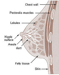
Changes for: thalamic reticular nucleus
- Deleted
Changes for: habenula
- Deleted
- - habenula definition A segment of the epithalamus that has as its parts the lateral and medial habenular nuclei and the habenular commissure. { database cross reference=FMA:62032 }
- - habenula depicted by http://upload.wikimedia.org/wikipedia/commons/thumb/d/d7/Gray715.png/200px-Gray715.png
- Added
- + habenula definition A segment of the epithalamus that has as its parts the habenular nuclei and the habenular commissure[FMA,modified]. { database cross reference=FMA:62032 , database cross reference=UBERON:cjm }
Changes for: pineal body
- Deleted
Changes for: subthalamic nucleus
- Deleted
Changes for: optic tract
- Deleted
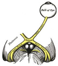
Changes for: habenular commissure
- Deleted
- - habenular commissure editor note This class was created automatically from a combination of ontologies
Changes for: lateral hypothalamic nucleus
- Deleted
- Added
- + lateral hypothalamic nucleus database cross reference http://en.wikipedia.org/wiki/Lateral_hypothalamus
- + lateral hypothalamic nucleus definition The lateral hypothalamus or lateral hypothalamic area is a part of the hypothalamus. It is concerned with hunger. Damage to this area can cause reduced food intake. Stimulating the lateral hypothalamus causes a desire to eat, while stimulating the ventromedial hypothalamus causes a desire to stop eating. { database cross reference=http://en.wikipedia.org/wiki/Lateral_hypothalamus,Wikipedia:Lateral_hypothalamus }
- + lateral hypothalamic nucleus depicted by http://upload.wikimedia.org/wikipedia/commons/8/8c/HypothalamicNuclei.PNG
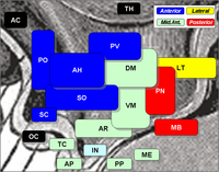
Changes for: dorsomedial nucleus of hypothalamus
- Deleted
Changes for: ventromedial nucleus of hypothalamus
- Deleted
- Added
Changes for: arcuate nucleus of hypothalamus
- Deleted
Changes for: lateral preoptic nucleus
- Deleted
- Added
Changes for: ventral lateral nucleus of thalamus
- Deleted
Changes for: lateral geniculate body
- Deleted
- Added
Changes for: medial geniculate body
- Deleted
- Added
Changes for: preoptic area
- Deleted
Changes for: Verson’s gland
- Deleted
- - Verson’s gland definition A gland found in the larvae of lepidoptera. Each gland is made of three cells: the secretory cell, the reservoir cell and the canal cell.
- Added
- + Verson’s gland definition A gland found in the larvae of lepidoptera. Each gland is made of three cells: the secretory cell, the reservoir cell and the canal cell. { database cross reference=UBERON:cjm }
Changes for: presubiculum
- Deleted
Changes for: white pulp of spleen
- Deleted
Changes for: primary subdivision of skull
- Deleted
- - primary subdivision of skull definition The skull can be divided into two: the neurocranium and the facial skeleton
- Added
- + primary subdivision of skull definition The skull can be divided into two: the neurocranium and the facial skeleton { database cross reference=UBERON:cjm }
Changes for: gular region
- Deleted
- - gular region definition Surface structure that extends from the chin covering pharynx and larynx.
- Added
- + gular region definition Surface structure that extends from the chin covering pharynx and larynx. { database cross reference=UBERON:cjm }
Changes for: pretectal region
- Deleted
- - pretectal region database cross reference BAMS:PT
- - pretectal region definition Region between dorsal thalamus and optic tectum[pub]. It receives binocular sensory input from retinal ganglion cells of the eyes, and is the region responsible for maintaining the pupillary light reflex. [WP,unvetted]. { database cross reference=http://en.wikipedia.org/wiki/Pretectum , database cross reference=https://github.com/obophenotype/uberon/issues/361 , database cross reference=ISBN10:0471888893 }
- Added
- + pretectal region SubClassOf has part some part of some diencephalon
- + pretectal region SubClassOf has part some part of some midbrain
- + pretectal region definition Nuclear complex between dorsal thalamus and optic tectum whose nuclei receive afferents primarily from the retina and the optic tectum and are involved in modulating motor behavior in response to visual input. { database cross reference=https://github.com/obophenotype/uberon/issues/361 , database cross reference=ISBN10:0471888893 }
- + pretectal region function notes It receives binocular sensory input from retinal ganglion cells of the eyes, and is the region responsible for maintaining the pupillary light reflex
- + pretectal region has exact synonym area pretectalis { has synonym type=latin term }
- + pretectal region taxon notes in monotremes, undifferentiated and possesses only a single nucleus[ISBN10:0471888893]
- + pretectal region taxon notes teleost fishes exhibit hypertrophy of the pretectum[ISBN10:0471888893]
- + pretectal region taxon notes within the amniotes, this region is smaller in mammals[ISBN10:0471888893]
Changes for: midbrain tegmentum
- Deleted
Changes for: medial habenular nucleus
- Deleted
- - medial habenular nucleus SubClassOf nucleus of thalamus
- Added
- + medial habenular nucleus SubClassOf habenular nucleus
- + medial habenular nucleus database cross reference http://en.wikipedia.org/wiki/Habenular_nuclei
- + medial habenular nucleus definition The habenular nuclei comprise a small group of nuclei that are part of the epithalamus of the diencephalon, situated at the posterior end of the thalamus, on its upper surface. The habenular nuclei are typically divided into: lateral habenular nucleus medial habenular nucleus The pineal gland is attached to the brain in this region. Nerve impulses from the habenular nuclei are transmitted to the septal nuclei via the stria medullaris, which is found on the medial surface of the thalamus. { database cross reference=http://en.wikipedia.org/wiki/Habenular_nuclei,Wikipedia:Habenular_nuclei }
Changes for: lateral habenular nucleus
- Deleted
- - lateral habenular nucleus SubClassOf nucleus of thalamus
- Added
- + lateral habenular nucleus SubClassOf habenular nucleus
Changes for: red nucleus
- Deleted
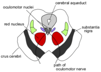
Changes for: superior colliculus
- Deleted
- - superior colliculus comment The term SC is used when discussing mammals, and OT for other vertebrates[WP]. The tectum is a layered structure, with a number of layers that vary by species. The superficial layers are sensory-related, and receive input from the eyes as well as other sensory systems.[1] The optic tectum is one of the fundamental components of the vertebrate brain, existing across the full range of species from hagfish to human.[4] (See the brain article for background.) Some aspects of the structure are very consistent, including a structure composed of a number of layers, with a dense input from the optic nerve to the superficial layers and another strong input conveying somatosensory input to deeper layers. Other aspects are highly variable, such as the total number of layers (from 3 in the African lungfish to 15 in the goldfish[5]), and the number of different types of cells (from 2 in the lungfish to 27 in the house sparrow[5]). In hagfish, lamprey, and shark it is a relatively small structure, but in teleost fish it is greatly expanded, in some cases becoming the largest structure in the brain. (See the adjoining drawing of a codfish brain.) In amphibians, reptiles, and especially birds it is also a very significant component, but in mammals it is dwarfed by the massive expansion of the cerebral cortex.
- - superior colliculus depicted by http://upload.wikimedia.org/wikipedia/commons/thumb/d/da/Cn3nucleus.png/200px-Cn3nucleus.png
- Added
- + superior colliculus comment ). In hagfish, lamprey, and shark it is a relatively small structure, but in teleost fish it is greatly expanded, in some cases becoming the largest structure in the brain. (See the adjoining drawing of a codfish brain.) In amphibians, reptiles, and especially birds it is also a very significant component, but in mammals it is dwarfed by the massive expansion of the cerebral cortex.
- + superior colliculus taxon notes The tectum is a layered structure, with a number of layers that vary by species. The superficial layers are sensory-related, and receive input from the eyes as well as other sensory systems.[1] The optic tectum is one of the fundamental components of the vertebrate brain, existing across the full range of species from hagfish to human.[4] (See the brain article for background.) Some aspects of the structure are very consistent, including a structure composed of a number of layers, with a dense input from the optic nerve to the superficial layers and another strong input conveying somatosensory input to deeper layers. Other aspects are highly variable, such as the total number of layers (from 3 in the African lungfish to 15 in the goldfish[5]), and the number of different types of cells (from 2 in the lungfish to 27 in the house sparrow[5]
- + superior colliculus taxon notes The term SC is used when discussing mammals, and OT for other vertebrates[WP]
Changes for: venule
- Deleted
Changes for: nasal suture
- Deleted
- - nasal suture definition A suture joining a nasal bone to another bone.
- Added
- + nasal suture definition A suture joining a nasal bone to another bone. { database cross reference=UBERON:cjm }
Changes for: semen
- Deleted
- Added
Changes for: least splanchnic nerve
- Deleted
Changes for: elastic cartilage tissue
- Deleted
Changes for: olfactory epithelium
- Deleted
Changes for: sternocostal joint
- Deleted
Changes for: cervical ganglion
- Deleted
- Added
Changes for: middle cervical ganglion
- Deleted
Changes for: fibrocartilage
- Deleted
Changes for: hyaline cartilage tissue
- Deleted
- Added
Changes for: placenta
- Deleted
- Added
Changes for: corneal endothelium
- Deleted
Changes for: superior cervical ganglion
- Deleted
Changes for: superior tibiofibular joint
- Deleted
- Added
Changes for: arteriole
- Deleted
Changes for: crypt of Lieberkuhn
- Deleted
Changes for: blood vessel
- Deleted
Changes for: middle thyroid artery
- Deleted
- Added
- + middle thyroid artery SubClassOf branch of external carotid artery
Changes for: jejunal artery
Changes for: organ system subdivision
- Deleted
- - organ system subdivision definition A subdivision of an anatomical system.
- Added
- + organ system subdivision definition A subdivision of an anatomical system. { database cross reference=UBERON:cjm }
Changes for: lower molar 3
Changes for: egg tooth
Changes for: nucleus of optic tract
- Deleted
- - nucleus of optic tract definition Predominantly gray matter structure consisting of large multipoloar cells lying aong axons of the brachium of the superior colliculus, lyaing adjaent to the dorsal terminal nucleus of the accessory optic system. In several species, cells of this nucleus receive inpu from the contralateral retina (Sefton and Dreher in Paxinos, G. The rat nervous system, 1995, pg. 862). { database cross reference=NIFSTD:birnlex_868 }
- Added
- + nucleus of optic tract definition Predominantly gray matter structure consisting of large multipoloar cells lying aong axons of the brachium of the superior colliculus, lyaing adjaent to the dorsal terminal nucleus of the accessory optic system. In several species, cells of this nucleus receive input from the contralateral retina (Sefton and Dreher in Paxinos, G. The rat nervous system, 1995, pg. 862). { database cross reference=NIFSTD:birnlex_868 }
Changes for: nucleus of Bischoff
- Deleted
- - nucleus of Bischoff definition A midline nucleus of the dorsal motor column found in animals with a prominent tail, including alligator, some birds, kangaroo, rat, shrew, great anteater.
- Added
- + nucleus of Bischoff definition A midline nucleus of the dorsal motor column found in animals with a prominent tail, including alligator, some birds, kangaroo, rat, shrew, great anteater. { database cross reference=UBERON:cjm }
Changes for: subserosal plexus
Changes for: palpebral fissure
- Deleted
Changes for: lacrimal gland
- Deleted
Changes for: tarsal gland
- Deleted
Changes for: lumbosacral nerve plexus
- Deleted
Changes for: spinal nerve plexus
- Deleted
- - somatic nerve plexus EquivalentTo nerve plexus and part of some somatic nervous system
- - somatic nerve plexus SubClassOf part of some somatic nervous system
- - somatic nerve plexus definition A nerve plexus that is part of a somatic nervous system [Automatically generated definition]. { database cross reference=OBOL:automatic }
- - somatic nerve plexus editor note TODO - check
- - somatic nerve plexus label somatic nerve plexus
- Added
- + spinal nerve plexus EquivalentTo nerve plexus and extends fibers into some spinal nerve
- + spinal nerve plexus SubClassOf extends fibers into some spinal nerve
- + spinal nerve plexus definition an intermingling of fiber fascicles from adjacent spinal nerves to form a network. { database cross reference=http://www.medilexicon.com/medicaldictionary.php?t=70024 }
- + spinal nerve plexus has exact synonym somatic nerve plexus { database cross reference=FMA:5903 }
- + spinal nerve plexus label spinal nerve plexus
Changes for: brachial nerve plexus
- Deleted
- Added
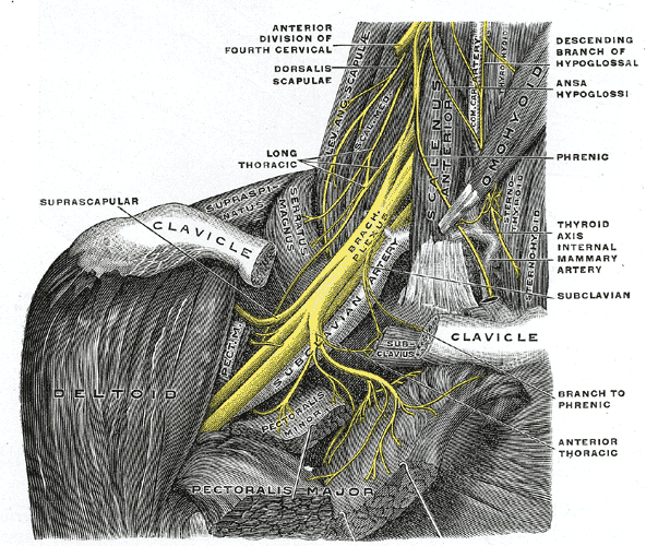
Changes for: conjunctiva
- Deleted
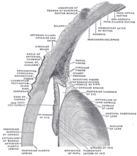
Changes for: sympathetic ganglion
- Deleted
Changes for: paravertebral ganglion
- Deleted
- Added
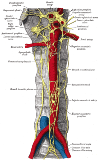
Changes for: parasympathetic ganglion
- Deleted
Changes for: posterior segment of eyeball
- Deleted
Changes for: capsule of lens
- Deleted
Changes for: autopod pad
- Deleted
- Added
Changes for: primitive palate
Changes for: deep cervical artery
- Added
- + deep cervical artery EquivalentTo cervical artery and part of some deep vasculature
- + deep cervical artery SubClassOf part of some deep vasculature
Changes for: diencephalon
- Deleted
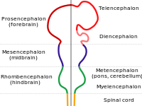
Changes for: medulla oblongata
- Deleted
- Added
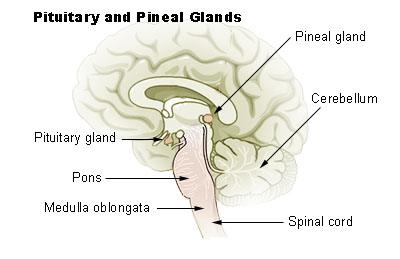
Changes for: metencephalon
- Deleted
Changes for: forebrain
- Deleted
- Added
Changes for: midbrain
- Deleted
Changes for: dorsal plus ventral thalamus
- Deleted
Changes for: epithalamus
- Deleted
Changes for: dentate gyrus of hippocampal formation
- Deleted
Changes for: phrenic nerve
- Deleted
Changes for: olfactory tubercle
- Deleted
Changes for: nucleus accumbens
- Deleted
Changes for: island of Calleja
- Deleted
Changes for: levator claviculae muscle
- Added
- + levator claviculae muscle SubClassOf muscle of shoulder
Changes for: lateral olfactory stria
- Deleted
Changes for: choroid plexus
- Deleted
Changes for: internal capsule of telencephalon
- Deleted
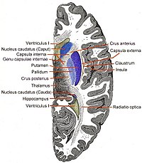
Changes for: parietal lobe
- Deleted
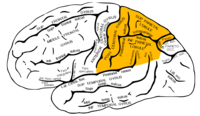
Changes for: temporal lobe
- Deleted
Changes for: putamen
- Deleted
Changes for: caudate nucleus
- Deleted
Changes for: globus pallidus
- Deleted
Changes for: amygdala
- Deleted
Changes for: scala vestibuli
- Deleted
- Added
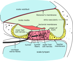
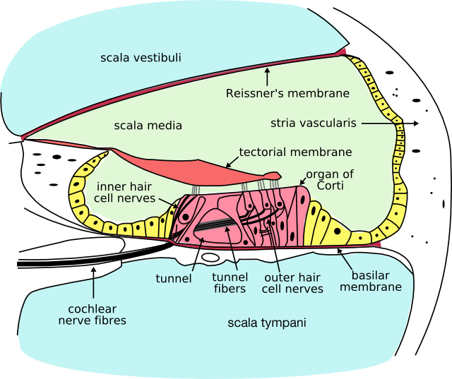
Changes for: vestibular labyrinth
Changes for: endolymphatic duct
- Deleted
Changes for: cartilage of external ear
- Deleted
- - cartilage of external ear has exact synonym auricular region cartilage { database cross reference=OBOL:automatic }
- - cartilage of external ear has exact synonym auricular region of head cartilage { database cross reference=OBOL:automatic }
- - cartilage of external ear has exact synonym cartilage of auricular region of head { database cross reference=OBOL:automatic }
- - cartilage of external ear has exact synonym cartilage of auricular region { database cross reference=OBOL:automatic }
- - cartilage of external ear has exact synonym cartilage of outer ear { database cross reference=OBOL:automatic }
- - cartilage of external ear has exact synonym outer ear cartilage { database cross reference=OBOL:automatic }
Changes for: scala tympani
- Deleted
Changes for: endolymph
- Deleted
Changes for: posterior semicircular duct
- Deleted
- Added
- + posterior semicircular duct definition The semicircular canals are three half-circular, interconnected tubes located inside each ear. The three canals are the horizontal semicircular canal (also known as the lateral semicircular canal), superior semicircular canal (also known as the anterior semicircular canal), and the posterior semicircular canal. The canals are aligned approximately orthogonally to one another. The horizontal canal is aligned roughly horizontally in the head. The superior and anterior canals are aligned roughly at a 45 degree angle to a vertical plane drawn from the nose to the back of the skull. Thus, the horizontal canal detects horizontal head movements (such as when doing a pirouette), while the superior and posterior canals detect vertical head movements. Each canal is filled with a fluid called endolymph and contains a motion sensor with little hairs whose ends are embedded in a gelatinous structure called the cupula. As the skull twists in any direction, the endolymph is thrown into different sections of the canals. The cilia detect when the endolymph rushes past, and a signal is then sent to the brain. The semicircular canals are a component of the Labyrinth. Among species of mammals, the size of the semicircular canals is correlated with their type of locomotion. Specifically, species that are agile and have fast, jerky locomotion have larger canals relative to their body size than those that move more cautiously. { database cross reference=http://en.wikipedia.org/wiki/Semicircular_canal,Wikipedia:Semicircular_canal }
- + posterior semicircular duct depicted by http://upload.wikimedia.org/wikipedia/commons/3/33/Balance_Disorder_Illustration_A.png
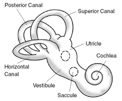
Changes for: lateral semicircular duct
- Deleted
- Added
Changes for: saccule of membranous labyrinth
- Deleted
Changes for: cochlear duct of membranous labyrinth
- Deleted
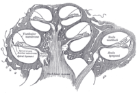
Changes for: rostral organ
- Added
- + rostral organ SubClassOf innervated by some ophthalmic nerve
- + rostral organ SubClassOf surrounded by some adipose tissue
Changes for: semicircular canal
- Deleted
Changes for: anterior semicircular canal
- Deleted
- Added
Changes for: membranous labyrinth
- Deleted
Changes for: auricular cartilage
- Deleted
- - auricular cartilage EquivalentTo cartilage element and part of some pinna
- - auricular cartilage SubClassOf cartilage of external ear
- Added
- + auricular cartilage EquivalentTo zone of organ and part of some pinna and part of some cartilage of external ear
- + auricular cartilage SubClassOf part of some cartilage of external ear
- + auricular cartilage SubClassOf zone of organ
- + auricular cartilage has exact synonym pinna cartilage
Changes for: internal ear
- Deleted
- Added
Changes for: cochlea
- Deleted
- Added
Changes for: lateral semicircular canal
- Deleted
- Added
Changes for: posterior semicircular canal
- Deleted
- Added
- + posterior semicircular canal definition The posterior semicircular canal is a part of the vestibular system and detects rotations of the head in the sagittal plane. { database cross reference=http://en.wikipedia.org/wiki/Posterior_semicircular_canal,Wikipedia:Posterior_semicircular_canal }
Changes for: sublingual gland
- Deleted
Changes for: parotid gland
- Deleted
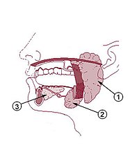
Changes for: upper lip
- Deleted
Changes for: lip
- Deleted
- Added
Changes for: lower lip
- Deleted
Changes for: quadratojugal bone
- Deleted
- Added
Changes for: bony labyrinth
- Deleted
Changes for: nasal cartilage
- Deleted
- Added
Changes for: sebaceous gland
- Deleted
Changes for: sweat gland
- Deleted
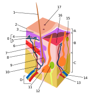
Changes for: nasal cavity mucosa
- Added
Changes for: paranasal sinus
- Deleted
- Added
Changes for: gingiva
- Deleted
Changes for: extrinsic tongue pre-muscle mass
- Deleted
- - extrinsic tongue pre-muscle mass SubClassOf head mesenchyme from mesoderm
- - extrinsic tongue pre-muscle mass SubClassOf mesenchyme of tongue
- - extrinsic tongue pre-muscle mass SubClassOf mixed ectoderm/mesoderm/endoderm-derived structure
- - extrinsic tongue pre-muscle mass SubClassOf part of some skeletal muscle tissue of tongue
- - extrinsic tongue pre-muscle mass SubClassOf presumptive structure
- Added
Changes for: outer limiting layer of retina
- Added
Changes for: photoreceptor layer of retina
- Deleted
Changes for: outer nuclear layer of retina
- Added
Changes for: nerve to stylopharyngeus from glossopharyngeal nerve
- Added
- + nerve to stylopharyngeus from glossopharyngeal nerve database cross reference http://en.wikipedia.org/wiki/Stylopharyngeal_branch_of_glossopharyngeal_nerve
- + nerve to stylopharyngeus from glossopharyngeal nerve definition The stylopharyngeal branch of glossopharyngeal nerve is distributed to the Stylopharyngeus. { database cross reference=http://en.wikipedia.org/wiki/Stylopharyngeal_branch_of_glossopharyngeal_nerve,Wikipedia:Stylopharyngeal_branch_of_glossopharyngeal_nerve }
Changes for: optic disc
- Deleted
Changes for: cranial nerve
- Deleted
- Added
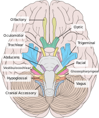
Changes for: fovea centralis
- Deleted
- Added
Changes for: pigmented layer of retina
- Deleted
Changes for: vitreous body
- Deleted
- Added
Changes for: molar 3
- Added
- + molar 3 database cross reference http://en.wikipedia.org/wiki/Wisdom_tooth
- + molar 3 taxon notes A wisdom tooth is in humans any of the usually four third molars, including mandibular third molar and maxillary third molar. Wisdom teeth usually appear between the ages of 17 and 25. Most adults have four wisdom teeth, but it is possible to have morebin which case they are called supernumerary teethbor fewerba condition called hypodontia. Wisdom teeth commonly affect other teeth as they develop, becoming impacted or ‘coming in sideways’. They are often extracted when this occurs. { source=http://en.wikipedia.org/wiki/Wisdom_tooth }
Changes for: aqueous humor of eyeball
- Deleted
- Added
Changes for: vitreous humor
- Deleted
Changes for: inner limiting layer of retina
- Added
Changes for: inner plexiform layer of retina
- Deleted
Changes for: ganglionic layer of retina
- Deleted
Changes for: outer plexiform layer of retina
- Added
Changes for: inner nuclear layer of retina
- Added
Changes for: cervical sympathetic nerve trunk
- Added
- + cervical sympathetic nerve trunk database cross reference http://en.wikipedia.org/wiki/Cervical_ganglia
- + cervical sympathetic nerve trunk definition The cervical ganglia are paravertebral ganglia of the sympathetic nervous system. These emerging postganglionic nerves synapse with preganglionic nerves from the thoracic spinal cord. They consist of three paravertebral ganglia: superior cervical ganglion middle cervical ganglion inferior cervical ganglion. The inferior ganglion may be fused with the first thoracic ganglion to form a single structure, the stellate ganglion. Nerves emerging from cervical sympathetic ganglia contribute to the cardiac plexus, among other things. { database cross reference=http://en.wikipedia.org/wiki/Cervical_ganglia,Wikipedia:Cervical_ganglia }
Changes for: arcuate artery of foot
- Added
- + arcuate artery of foot definition The arcuate artery of the foot (metatarsal artery) arises a little anterior to the lateral tarsal artery; it passes lateralward, over the bases of the metatarsal bones, beneath the tendons of the Extensor digitorum brevis, its direction being influenced by its point of origin; and its anastomoses with the lateral tarsal and lateral plantar arteries. This vessel gives off the second, third, and fourth dorsal metatarsal arteries. It is not present in all individuals. { database cross reference=http://en.wikipedia.org/wiki/Arcuate_artery_of_the_foot,Wikipedia:Arcuate_artery_of_the_foot }
Changes for: anterior pretectal nucleus
- Added
- + anterior pretectal nucleus editor note consider merging with olivary; however, Butler and Hodos treat as distinct within Eutherians
Changes for: mental nerve
- Added
- + mental nerve database cross reference http://en.wikipedia.org/wiki/Mental_nerve
- + mental nerve definition Mental nerve is a general somatic afferent (sensory) nerve which provides sensation to the anterior aspects of the chin and lower lip as well as the buccal gingivae of the mandibular anterior teeth and the premolars. It is a branch of the posterior trunk of the inferior alveolar nerve, which is itself a branch of the mandibular division of the trigeminal nerve (CN V). The nerve emerges at the mental foramen in the mandibula, and divides beneath the Depressor anguli oris muscle into three branches: one descends to the skin of the chin. two ascend to the skin and mucous membrane of the lower lip. These branches communicate freely with the facial nerve. { database cross reference=http://en.wikipedia.org/wiki/Mental_nerve,Wikipedia:Mental_nerve }
Changes for: podotheca
- Deleted
- - podotheca definition A horny sheath covering the lower leg of some birds.
- Added
- + podotheca definition A horny sheath covering the lower leg of some birds. { database cross reference=UBERON:cjm }
Changes for: dental pulp
- Deleted
- Added
Changes for: pinna
- Deleted
- - pinna definition Visible part of the ear that resides outside of the head. The purpose of the pinna is to collect sound. It does so by acting as a funnel, amplifying the sound and directing it to the ear canal. While reflecting from the pinna, sound also goes through a filtering process which adds directional information to the sound (see sound localization, head-related transfer function, pinna notch). The filtering effect of the human pinna preferentially selects sounds in the frequency range of human speech. In various species, the pinna can also signal mood and radiate heat. { database cross reference=http://en.wikipedia.org/wiki/Pinna_(anatomy) }
- Added
- + pinna SubClassOf continuous with some external acoustic meatus
- + pinna definition The part of the ear that projects from the head, connecting to the external acoustic meatus. { database cross reference=http://en.wikipedia.org/wiki/Pinna_(anatomy) , database cross reference=UBERON:cjm }
- + pinna function notes The purpose of the pinna is to collect sound. It does so by acting as a funnel, amplifying the sound and directing it to the ear canal. While reflecting from the pinna, sound also goes through a filtering process which adds directional information to the sound (see sound localization, head-related transfer function, pinna notch).
- + pinna taxon notes In various species, the pinna can also signal mood and radiate heat
Changes for: vagus nerve
- Deleted
Changes for: infundibular organ
- Deleted
- Added
- + infundibular organ definition . { database cross reference=UBERON:cjm }
Changes for: corniculate cartilage
- Deleted
Changes for: arytenoid cartilage
- Deleted
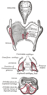
Changes for: secondary nodular lymphoid tissue
- Deleted
- Added
Changes for: brachiocephalic muscle
- Deleted
- - brachiocephalic muscle SubClassOf skeletal muscle organ
- Added
- + brachiocephalic muscle SubClassOf muscle of shoulder
Changes for: sclera
- Deleted
Changes for: corneal epithelium
- Deleted
Changes for: ciliary body
- Deleted
Changes for: pupil
- Deleted
Changes for: optic choroid
- Deleted
Changes for: substantia propria of cornea
- Deleted
- Added
Changes for: iris stroma
- Deleted
- Added
Changes for: cleidobrachialis muscle
- Deleted
- - cleidobrachialis muscle SubClassOf skeletal muscle organ
- Added
- + cleidobrachialis muscle SubClassOf muscle of shoulder
Changes for: cleidocephalicus muscle
- Deleted
- - cleidocephalicus muscle SubClassOf skeletal muscle organ
- Added
- + cleidocephalicus muscle SubClassOf muscle of shoulder
Changes for: maxillary sinus
- Deleted
Changes for: odontogenic papilla
- Deleted
Changes for: nasal concha
- Deleted
Changes for: frontal sinus
- Deleted
Changes for: posterior chamber of eyeball
- Deleted
Changes for: uvea
- Deleted
Changes for: transverse process of atlas
- Added
Changes for: anterior chamber of eyeball
- Deleted
Changes for: nucleus ambiguus
- Deleted
Changes for: male bulbospongiosus muscle
- Deleted
- Added
Changes for: mesencephalic nucleus of trigeminal nerve
- Deleted
Changes for: eyelid
- Deleted
Changes for: lower jaw region
- Deleted
- Added
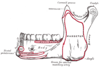
Changes for: lower eyelid
- Deleted
- Added
Changes for: upper eyelid
- Deleted
- Added
Changes for: oculomotor nuclear complex
- Deleted
- Added
Changes for: spinal nucleus of trigeminal nerve
- Deleted
Changes for: secondary palate
- Deleted
Changes for: upper jaw region
- Deleted
- Added
Changes for: nasal cavity
- Deleted
Changes for: eyelash
- Deleted
Changes for: geniculate ganglion
- Deleted
Changes for: nasal septum
- Deleted
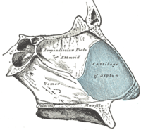
Changes for: submandibular gland
- Deleted
Changes for: thyroid cartilage
- Deleted
Changes for: soft palate
- Deleted
Changes for: pharyngeal tonsil
- Deleted
Changes for: palatine uvula
- Deleted
Changes for: cochlear nucleus
- Deleted
- Added
Changes for: oropharynx
- Deleted
Changes for: nasopharynx
- Deleted
Changes for: taste bud
- Deleted
Changes for: sphenoidal sinus
- Deleted
Changes for: tongue
- Deleted
Changes for: medial vestibular nucleus
- Deleted
- Added
Changes for: tibialis posterior
- Deleted
Changes for: flexor digitorum longus
- Deleted
- Added
Changes for: superior cerebellar vein
- Deleted
- Added
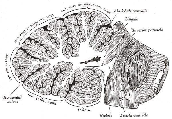
Changes for: anterior auricular vein
- Deleted
Changes for: cerebral vein
- Deleted
Changes for: inferior cerebral vein
- Deleted
Changes for: triceps surae
- Deleted
Changes for: maxillary vein
- Deleted
- Added
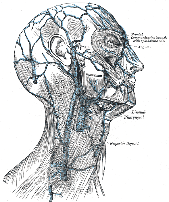
Changes for: deep temporal vein
- Added
- + deep temporal vein EquivalentTo temporal vein and part of some deep vasculature
- + deep temporal vein SubClassOf deep vein
- + deep temporal vein SubClassOf part of some deep vasculature
Changes for: temporal bone
- Deleted
Changes for: sphenoid bone
- Deleted
Changes for: trigeminal ganglion
- Deleted
- - trigeminal ganglion definition A prominent collection of touch-sensory neurons of the trigeminal or fifth cranial nerve, positioned beside the brain between the eye and the ear. { database cross reference=ZFIN:curator }
- - trigeminal ganglion depicted by http://upload.wikimedia.org/wikipedia/commons/thumb/6/69/Gray776.png/200px-Gray776.png
- Added
- + trigeminal ganglion definition The cranial ganglion that is associated with and extends fibers into the trigeminal nerve. { database cross reference=http://www.ncbi.nlm.nih.gov/books/NBK53171 , database cross reference=https://github.com/obophenotype/uberon/issues/693 , database cross reference=UBERON:cjm }
- + trigeminal ganglion editor note consider renaming this ‘trigeminal ganglion complex’. The complex may be fused (amniotes) partially fused (xenopus) or separate (sharks)
- + trigeminal ganglion has exact synonym ganglion of trigeminal complex { database cross reference=UBERON:cjm }
- + trigeminal ganglion has narrow synonym fused trigeminal ganglion { database cross reference=NCBITaxon:32524 , has synonym type=taxonomic disambiguation }
- + trigeminal ganglion taxon notes In Xenopus, the profundal and the trigeminal ganglia are separate distally but fused at their proximal end as they condense around NF stage 24. { source=http://www.ncbi.nlm.nih.gov/pubmed/21452441 , source=XAO:0004093 }
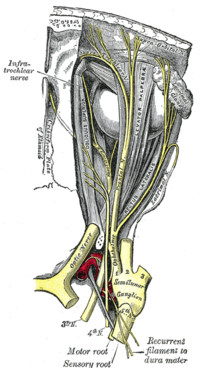
Changes for: occipital bone
- Deleted
Changes for: central retinal vein
- Deleted
Changes for: temporal vein
- Deleted
- Added
- + temporal vein EquivalentTo vein and drains some temporal part of head
- + temporal vein SubClassOf drains some temporal part of head
- + temporal vein definition Any of the veins draining the temporal region. { database cross reference=http://www.thefreedictionary.com/temporal+vein }
- + temporal vein depicted by http://upload.wikimedia.org/wikipedia/commons/8/8c/Gray557.png
- + temporal vein external definition A vein in front of the ear that drains the head and face and joins with the internal maxillary vein to form the temporo-maxillary vein. { source=ncithesaurus:Temporal_Vein }
Changes for: inferior cerebellar vein
- Deleted
- Added
Changes for: malleus bone
- Deleted
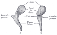
Changes for: incus bone
- Deleted
Changes for: nasal bone
- Deleted
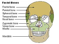
Changes for: palatine bone
- Deleted
Changes for: jugal bone
- Deleted
Changes for: mandible
- Deleted
Changes for: hyoid bone
- Deleted
Changes for: auditory ossicle bone
- Deleted
- Added
Changes for: stapes bone
- Deleted
Changes for: sensory root of facial nerve
- Deleted
- Added
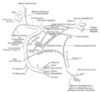
Changes for: exoccipital bone
- Deleted
Changes for: petrous part of temporal bone
- Deleted
- Added
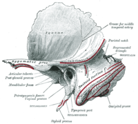
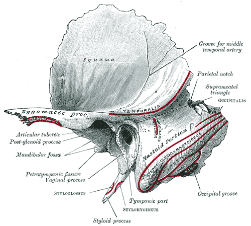
Changes for: external ear
- Deleted
- Added
Changes for: basioccipital bone
- Deleted
Changes for: articulation
- Deleted
- - articulation definition Anatomical cluster that connects two or more adjacent skeletal elements or hardened body parts.
- Added
- + articulation definition Anatomical cluster that connects two or more adjacent skeletal elements or hardened body parts. { database cross reference=UBERON:cjm }
Changes for: crus of penis or clitoris
- Deleted
- - crus of penis or clitoris definition the continuation of each corpus cavernosum of the penis or clitoris, diverging posteriorly to be attached to the pubic arch.
- Added
- + crus of penis or clitoris definition the continuation of each corpus cavernosum of the penis or clitoris, diverging posteriorly to be attached to the pubic arch. { database cross reference=UBERON:cjm }
Changes for: supramarginal gyrus
- Deleted
- - supramarginal gyrus depicted by [http://upload.wikimedia.org/wikipedia/commons/thumb/e/e2/Gehirn%2C_lateral_-Hauptgyri_beschriftet.svg/200px-Gehirn%2C_lateral-Hauptgyri_beschriftet.svg.png](http://upload.wikimedia.org/wikipedia/commons/thumb/e/e2/Gehirn%2C_lateral-Hauptgyri_beschriftet.svg/200px-Gehirn%2C_lateral-_Hauptgyri_beschriftet.svg.png)
- Added
- + supramarginal gyrus depicted by [http://upload.wikimedia.org/wikipedia/commons/e/e2/Gehirn%2C_lateral_-Hauptgyri_beschriftet.svg](http://upload.wikimedia.org/wikipedia/commons/e/e2/Gehirn%2C_lateral-_Hauptgyri_beschriftet.svg)
Changes for: abducens nucleus
- Deleted
Changes for: nucleus raphe obscurus
- Deleted
Changes for: ventral supraoptic decussation
- Deleted
Changes for: vestibular nuclear complex
- Deleted
- Added
Changes for: sphincter of hepatopancreatic ampulla
- Deleted
Changes for: biliary bud
- Deleted
- Added
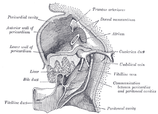
Changes for: duodenal papilla
- Deleted
Changes for: hepatopancreatic ampulla
- Deleted
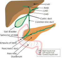
Changes for: ventral tegmental area
- Deleted
Changes for: parieto-occipital sulcus
- Deleted
Changes for: preoccipital notch
Changes for: decussation of medial lemniscus
- Deleted
Changes for: cuneocerebellar tract
- Deleted
- Added
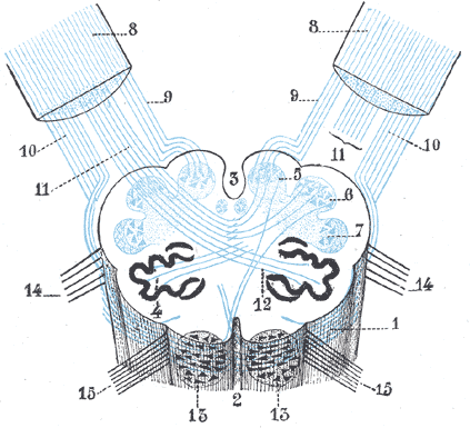
Changes for: ventral anterior nucleus of thalamus
- Deleted
- Added
- + ventral anterior nucleus of thalamus definition The ventral anterior nucleus is a nucleus of the thalamus. { database cross reference=http://en.wikipedia.org/wiki/Ventral_anterior_nucleus,Wikipedia:Ventral_anterior_nucleus }
Changes for: anterior nucleus of hypothalamus
- Deleted
Changes for: motor nucleus of trigeminal nerve
- Deleted
Changes for: cerebral crus
- Deleted
Changes for: superior frontal gyrus
- Deleted
Changes for: superior medullary velum
- Deleted
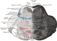
Changes for: anterior horn of lateral ventricle
- Deleted
Changes for: body of lateral ventricle
- Deleted
Changes for: medial tarsal artery
- Added
- + medial tarsal artery definition The medial tarsal arteries are two or three small branches which ramify on the medial border of the foot and join the medial malleolar net-work. { database cross reference=http://en.wikipedia.org/wiki/Medial_tarsal_arteries,Wikipedia:Medial_tarsal_arteries }
Changes for: upper respiratory tract
- Deleted
- Added
Changes for: lower respiratory tract
- Deleted
- Added
Changes for: matrix compartment of caudate nucleus
- Added
- + matrix compartment of caudate nucleus EquivalentTo matrix compartment and part of some caudate nucleus
- + matrix compartment of caudate nucleus SubClassOf part of some caudate nucleus
Changes for: matrix compartment of putamen
- Added
- + matrix compartment of putamen EquivalentTo matrix compartment and part of some putamen
Changes for: popliteal vein
- Deleted
Changes for: popliteal lymph node
- Deleted
- Added
Changes for: medial plantar artery
- Deleted
Changes for: peroneal artery
- Deleted
- Added
Changes for: striosomal part of caudate nucleus
- Added
- + striosomal part of caudate nucleus EquivalentTo striosome and part of some caudate nucleus
- + striosomal part of caudate nucleus SubClassOf part of some caudate nucleus
Changes for: striosomal part of putamen
- Added
- + striosomal part of putamen EquivalentTo striosome and part of some putamen
Changes for: small saphenous vein
- Deleted
Changes for: anterior tibial vein
- Deleted
Changes for: posterior tibial vein
- Deleted
Changes for: genioglossus muscle
- Deleted
- Added
Changes for: inferior pharyngeal constrictor
- Deleted
Changes for: styloglossus
- Deleted
Changes for: hyoglossus muscle
- Deleted
- Added
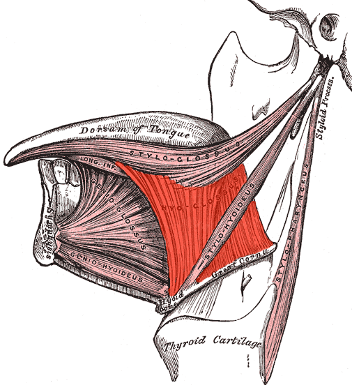
Changes for: lower part of vagina
- Deleted
- - lower part of vagina definition The lower third of the vagina.
- Added
- + lower part of vagina definition The lower third of the vagina. { database cross reference=UBERON:cjm }
Changes for: extrinsic muscle of tongue
- Deleted
- - extrinsic muscle of tongue SubClassOf develops from some extrinsic tongue pre-muscle mass
- - extrinsic muscle of tongue SubClassOf intrinsic muscle of tongue
- - extrinsic muscle of tongue SubClassOf part of some skeletal muscle tissue of tongue
- - extrinsic muscle of tongue SubClassOf structure with developmental contribution from neural crest
- Added
- + extrinsic muscle of tongue SubClassOf immediate transformation of some extrinsic tongue pre-muscle mass
- + extrinsic muscle of tongue SubClassOf tongue muscle
Changes for: palatoglossus muscle
- Deleted
- Added
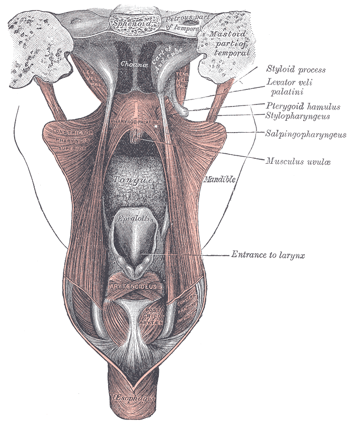
Changes for: facial muscle
- Deleted
- Added
Changes for: intrinsic muscle of tongue
- Deleted
- - intrinsic muscle of tongue SubClassOf part of some skeletal muscle tissue of tongue
- Added
Changes for: orbicularis oculi muscle
- Deleted
Changes for: olfactory nerve
- Deleted
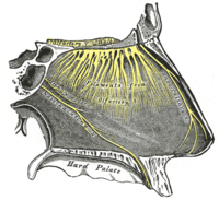
Changes for: digastric muscle group
- Deleted
- - digastric muscle group definition A group of muscles (or in some classifications, muscle bellies) that are located under the jaw and attach to the base of the cranium and typically attaches to the hyoid apparatus via a common tendon.
- - digastric muscle group depicted by http://upload.wikimedia.org/wikipedia/commons/thumb/2/26/Digastricus.png/200px-Digastricus.png
- Added
- + digastric muscle group definition A group of muscles (or in some classifications, muscle bellies) that are located under the jaw and attach to the base of the cranium and typically attaches to the hyoid apparatus via a common tendon. { database cross reference=UBERON:cjm }
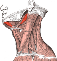
Changes for: cricothyroid muscle
- Deleted
Changes for: geniohyoid muscle
- Deleted
Changes for: longus capitis muscle
- Deleted
Changes for: constrictor muscle of pharynx
- Deleted
- Added
- + constrictor muscle of pharynx database cross reference http://en.wikipedia.org/wiki/Pharyngeal_constrictor
- + constrictor muscle of pharynx definition Pharyngeal constrictor refers to one of the muscles that serves to constrict the pharynx. They include: Superior pharyngeal constrictor muscle Middle pharyngeal constrictor muscle Inferior pharyngeal constrictor muscle { database cross reference=http://en.wikipedia.org/wiki/Pharyngeal_constrictor,Wikipedia:Pharyngeal_constrictor }
- + constrictor muscle of pharynx depicted by http://upload.wikimedia.org/wikipedia/commons/a/ac/Musculusconstrictorpharyngissuperior.png
Changes for: cheek
- Deleted
- Added
Changes for: muscle of larynx
Changes for: masseter muscle
- Deleted
Changes for: temporalis muscle
- Deleted
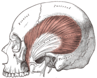
Changes for: stapedius muscle
- Deleted
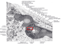
Changes for: venous plexus
- Deleted
Changes for: azygos vein
- Deleted
Changes for: auricular muscle
- Deleted
- - auricular muscle definition A muscle that is part of the pinna and is attached to the auricular cartilage
- - auricular muscle depicted by http://upload.wikimedia.org/wikipedia/commons/thumb/d/d2/Gray906.png/200px-Gray906.png
- Added
- + auricular muscle definition A muscle that is part of the pinna and is attached to the auricular cartilage { database cross reference=UBERON:cjm }
- + auricular muscle depicted by http://upload.wikimedia.org/wikipedia/commons/d/d2/Gray906.png
Changes for: pericardiacophrenic vein
- Deleted
Changes for: thenar muscle
- Deleted
- - thenar muscle external definition The muscles of hand can be remembered using the mnemonic, ‘A OF A OF A’ for, Abductor pollicis brevis, Opponens pollicis, Flexor pollicis brevis, Adductor pollicis (thenar muscles) and Opponens digiti minimi, Flexor digiti minimi, Abductor digiti minimi (hypothenar muscles)[Wikipedia:Thenar_eminence]. { source=http://en.wikipedia.org/wiki/Thenar_eminence }
Changes for: subclavian vein
- Deleted
Changes for: vertebral vein
- Deleted
Changes for: anterior vena cava
- Deleted
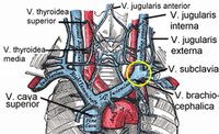
Changes for: internal jugular vein
- Deleted
Changes for: extrinsic auricular muscle
- Deleted
- Added
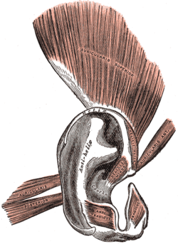
Changes for: left subclavian artery
- Deleted
- Added
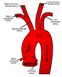
Changes for: depressor labii inferioris
- Deleted
Changes for: buccinator muscle
- Deleted
Changes for: levator labii superioris
- Deleted
Changes for: intercostal nerve
- Deleted
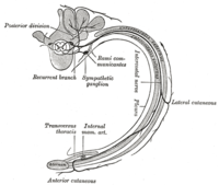
Changes for: mediastinum
- Deleted
Changes for: cervical plexus
- Deleted
Changes for: vestibular nerve
- Deleted
Changes for: musculocutaneous nerve
- Deleted
Changes for: lingual nerve
- Added
- + lingual nerve database cross reference http://en.wikipedia.org/wiki/Lingual_nerve
- + lingual nerve definition The lingual nerve is a branch of the mandibular nerve (CN V3), itself a branch of the trigeminal nerve, which supplies sensory innervation to the tongue. It also carries fibers from the facial nerve, which return taste information from the anterior two thirds of the tongue. { database cross reference=http://en.wikipedia.org/wiki/Lingual_nerve,Wikipedia:Lingual_nerve }
Changes for: anterior cranial fossa
- Deleted
Changes for: levator palpebrae superioris
- Deleted
- Added
Changes for: ciliary muscle
- Deleted
Changes for: stomach non-glandular epithelium
- Deleted
- Added
Changes for: sphincter pupillae
- Deleted
Changes for: tensor tympani
- Deleted
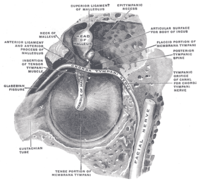
Changes for: extra-ocular muscle
- Deleted
Changes for: medial rectus extraocular muscle
- Deleted
Changes for: lateral rectus extra-ocular muscle
- Deleted
Changes for: dilatator pupillae
- Deleted
Changes for: isthmus of thyroid gland
- Deleted
Changes for: artery of lip
- Deleted
- - labial artery editor note add child terms
- - labial artery label labial artery
- Added
- + artery of lip EquivalentTo artery and supplies some lip
- + artery of lip SubClassOf supplies some lip
- + artery of lip has exact synonym labial artery { database cross reference=MA:0001987 }
- + artery of lip label artery of lip
Changes for: buccal artery
- Deleted
Changes for: transverse facial artery
- Deleted
Changes for: maxillary artery
- Deleted
Changes for: occipital artery
- Deleted
- - occipital artery SubClassOf mixed endoderm/mesoderm-derived structure
- - occipital artery SubClassOf structure with developmental contribution from neural crest
- - occipital artery SubClassOf systemic artery
- - occipital artery depicted by http://upload.wikimedia.org/wikipedia/commons/thumb/7/7b/Gray508.png/200px-Gray508.png
- Added
- + occipital artery SubClassOf branch of external carotid artery
Changes for: superficial temporal artery
- Deleted
- - superficial temporal artery SubClassOf mixed endoderm/mesoderm-derived structure
- - superficial temporal artery SubClassOf structure with developmental contribution from neural crest
- - superficial temporal artery SubClassOf systemic artery
- - superficial temporal artery depicted by http://upload.wikimedia.org/wikipedia/commons/thumb/a/a5/Gray1200.png/200px-Gray1200.png
- Added
Changes for: sublingual artery
- Deleted
Changes for: facial artery
- Deleted
- - facial artery SubClassOf mixed endoderm/mesoderm-derived structure
- - facial artery SubClassOf structure with developmental contribution from neural crest
- - facial artery SubClassOf systemic artery
- - facial artery depicted by http://upload.wikimedia.org/wikipedia/commons/thumb/7/7b/Gray508.png/200px-Gray508.png
- Added
- + facial artery SubClassOf branch of external carotid artery
Changes for: ophthalmic artery
- Deleted
Changes for: lingual artery
- Deleted
- Added
- + lingual artery SubClassOf branch of external carotid artery
Changes for: dorsal nasal artery
- Deleted
Changes for: right coronary artery
- Deleted
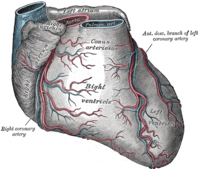
Changes for: anterior cerebral artery
- Deleted
Changes for: middle cerebral artery
- Deleted
Changes for: left coronary artery
- Deleted
Changes for: carotid body
- Deleted
Changes for: posterior communicating artery
- Deleted
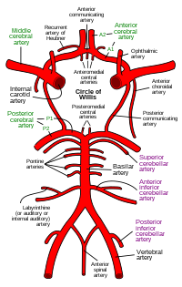
Changes for: central retinal artery
- Deleted
Changes for: coronary artery
- Deleted
- Added
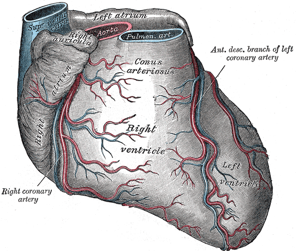
Changes for: posterior cerebral artery
- Deleted
- Added
- + posterior cerebral artery SubClassOf branch of basilar artery
Changes for: superior cerebellar artery
- Deleted
- Added
- + superior cerebellar artery SubClassOf branch of basilar artery
Changes for: basilar artery
- Deleted
- Added
- + basilar artery SubClassOf branch of vertebral artery
Changes for: hepatic portal vein
- Deleted
Changes for: vein
- Deleted
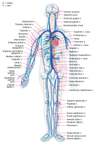
Changes for: artery
Changes for: thoracic duct
- Deleted
Changes for: temporal artery
- Deleted
- Added
Changes for: glossopharyngeal nerve
- Deleted
Changes for: vestibulocochlear nerve
- Deleted
Changes for: trigeminal nerve
- Deleted
Changes for: trochlear nerve
- Deleted
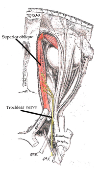
Changes for: facial nerve
- Deleted
Changes for: abducens nerve
- Deleted
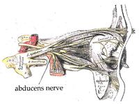
Changes for: celiac artery
- Deleted
Changes for: transverse sinus
- Deleted
- Added
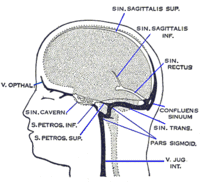
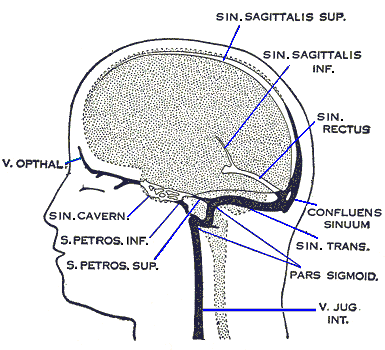
Changes for: superior sagittal sinus
- Deleted
Changes for: oculomotor nerve
- Deleted
Changes for: presumptive midbrain
Changes for: transverse facial vein
- Deleted
- - transverse facial vein SubClassOf facial vein
- - transverse facial vein SubClassOf tributary of some external jugular vein
- - transverse facial vein SubClassOf tributary of some subclavian vein
- - transverse facial vein depicted by http://upload.wikimedia.org/wikipedia/commons/thumb/8/8c/Gray557.png/200px-Gray557.png
- - transverse facial vein editor note This class was created automatically from a combination of ontologies
- Added
- + transverse facial vein EquivalentTo vein and tributary of some retromandibular vein and anastomoses with some facial vein
- + transverse facial vein SubClassOf anastomoses with some facial vein
- + transverse facial vein SubClassOf craniocervical region vein
- + transverse facial vein SubClassOf head blood vessel
- + transverse facial vein SubClassOf neck blood vessel
- + transverse facial vein definition a tributary of the superficial temporal or retromandibular veins, anastomosing with the facial vein. { database cross reference=http://medical-dictionary.thefreedictionary.com/transverse+facial+vein }
- + transverse facial vein depicted by http://upload.wikimedia.org/wikipedia/commons/8/8c/Gray557.png
Changes for: middle temporal vein
- Deleted
- - middle temporal vein SubClassOf tributary of some external jugular vein
- - middle temporal vein SubClassOf tributary of some retromandibular vein
- - middle temporal vein SubClassOf tributary of some subclavian vein
- - middle temporal vein editor note This class was created automatically from a combination of ontologies
- Added
- + middle temporal vein SubClassOf drains some ocular region
- + middle temporal vein SubClassOf tributary of some superficial temporal vein
- + middle temporal vein definition A temporal vein that arises near the lateral angle of the eye; joins the superficial temporal veins to form the retromandibular vein. { database cross reference=http://medical-dictionary.thefreedictionary.com/middle+temporal+vein }
Changes for: superficial temporal vein
- Deleted
- Added
Changes for: retromandibular vein
- Deleted
Changes for: facial vein
- Deleted
Changes for: supraorbital vein
- Deleted
- Added
Changes for: right pulmonary artery
- Deleted
- Added
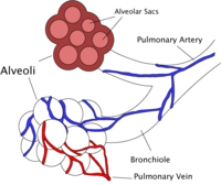
Changes for: left pulmonary artery
- Deleted
- Added
Changes for: cingulate sulcus
- Deleted
Changes for: nucleus of posterior commissure
- Added
- + nucleus of posterior commissure editor note in hagfishes, this is the name of one of the two pretectal nuclei[ISBN10:0471888893]
- + nucleus of posterior commissure editor note in lizards, this is the name of one of the two pretectal nuclei[ISBN10:0471888893]
- + nucleus of posterior commissure has narrow synonym Darkshevich nucleus
- + nucleus of posterior commissure has narrow synonym Darkshevich’s nucleus
- + nucleus of posterior commissure has narrow synonym nucleus of Darkschewitsch
Changes for: rubrospinal tract
- Deleted
- - rubrospinal tract depicted by [http://upload.wikimedia.org/wikipedia/commons/thumb/f/fe/Medulla_spinalis_-tracts-English.svg/200px-Medulla_spinalis-tracts-English.svg.png](http://upload.wikimedia.org/wikipedia/commons/thumb/f/fe/Medulla_spinalis-tracts-English.svg/200px-Medulla_spinalis-tracts-_English.svg.png)
- Added
- + rubrospinal tract depicted by [http://upload.wikimedia.org/wikipedia/commons/f/fe/Medulla_spinalis_-tracts-English.svg](http://upload.wikimedia.org/wikipedia/commons/f/fe/Medulla_spinalis-tracts-_English.svg)
Changes for: circular sulcus of insula
- Added
Changes for: collateral sulcus
- Deleted
Changes for: solitary tract
- Deleted
Changes for: rostral interstitial nucleus of medial longitudinal fasciculus
- Deleted
Changes for: orbital cavity
Changes for: paraaortic lymph node
- Added
- + paraaortic lymph node database cross reference http://en.wikipedia.org/wiki/Paraaortic_lymph_node
- + paraaortic lymph node definition The paraaortic lymph nodes (also known as para-aortic, periaortic, and peri-aortic) are a group of lymph nodes that lie in front of the lumbar vertebral bodies near the aorta. These lymph nodes receive drainage from the lower gastrointestinal tract and the pelvic organs. { database cross reference=http://en.wikipedia.org/wiki/Paraaortic_lymph_node,Wikipedia:Paraaortic_lymph_node }
Changes for: splanchnopleure
- Deleted
- Added
Changes for: urogenital fold
- Deleted
- - urogenital fold comment Less commonly, urogenital fold refers to the fold in the mesonephros which is the precursor of e.g. the suspensory ligament of the ovary. This is the case in older versions of Gray’s anatomy[WP]
- - urogenital fold database cross reference EMAPA:35899
- - urogenital fold database cross reference UMLS:C0231047 { source=ncithesaurus:Urogenital_Ridge }
- - urogenital fold database cross reference http://linkedlifedata.com/resource/umls/id/C0231047
- - urogenital fold database cross reference http://ncicb.nci.nih.gov/xml/owl/EVS/Urogenital_Ridge
- - urogenital fold has exact synonym urethral fold
- Added
- + urogenital fold database cross reference EMAPA:30888
- + urogenital fold has exact synonym urethral fold { database cross reference=EMAPA:30888 }
- + urogenital fold has related synonym urogenital ridge { database cross reference=MP:0011835 , has synonym type=indicates that a synonym is used in an inconsistent or confusing way, typically between species }
- + urogenital fold terminology notes Less commonly, urogenital fold refers to the fold in the mesonephros which is the precursor of e.g. the suspensory ligament of the ovary. This is the case in older versions of Gray’s anatomy[WP]
Changes for: somatopleure
- Deleted
- Added
Changes for: isthmus of cingulate gyrus
- Added
Changes for: medial dorsal nucleus of thalamus
- Deleted
Changes for: intralaminar nuclear group
- Deleted
- Added
Changes for: lateral nuclear group of thalamus
- Deleted
Changes for: perinatal stage
- Deleted
- - perinatal stage definition The period spanning the range immediately before and after birth
- Added
- + perinatal stage definition The period spanning the range immediately before and after birth { database cross reference=UBERON:cjm }
Changes for: sciatic lymph node
- Added
Changes for: eponychium
- Deleted
Changes for: basal forebrain
- Deleted
Changes for: posterior cingulate gyrus
- Deleted
- Added
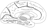
Changes for: diagonal band of Broca
- Deleted
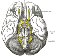
Changes for: lung hilus
Changes for: ileocolic lymph node
- Deleted
- - ileocecocolic lymph node label ileocecocolic lymph node
- Added
- + ileocolic lymph node database cross reference FMA:16611
- + ileocolic lymph node database cross reference http://en.wikipedia.org/wiki/Ileocolic_lymph_nodes
- + ileocolic lymph node database cross reference http://linkedlifedata.com/resource/umls/id/C2697661
- + ileocolic lymph node database cross reference http://ncicb.nci.nih.gov/xml/owl/EVS/Ileocecocolic_Lymph_Node
- + ileocolic lymph node database cross reference http://www.snomedbrowser.com/Codes/Details/29180003
- + ileocolic lymph node definition One of the lymph nodes that form a chain around the ileocolic artery. { database cross reference=http://en.wikipedia.org/wiki/Ileocolic_lymph_nodes }
- + ileocolic lymph node editor note Can be subclassed based on position relative to terminal branches of the vessel. Consider adding subclasses for these: (a) ileal, in relation to the ileal branch of the artery;(b) anterior ileocolic, usually of three glands, in the ileocolic fold, near the wall of the cecum;(c) posterior ileocolic, mostly placed in the angle between the ileum and the colon, but partly lying behind the cecum at its junction with the ascending colon;(d) a single gland, between the layers of the mesenteriole of the vermiform process;(e) right colic, along the medial side of the ascending colon.
- + ileocolic lymph node external definition A lymph node at the ileocecocolic junction. { source=ncithesaurus:Ileocecocolic_Lymph_Node }
- + ileocolic lymph node has exact synonym nodi lymphoidei ileocolici { database cross reference=http://en.wikipedia.org/wiki/Ileocolic_lymph_nodes , has synonym type=latin term }
- + ileocolic lymph node has narrow synonym ileocecocolic lymph node { database cross reference=http://ncicb.nci.nih.gov/xml/owl/EVS/Ileocecocolic_Lymph_Node , database cross reference=MA:0003065 }
- + ileocolic lymph node label ileocolic lymph node
Changes for: accessory XI nerve spinal component
- Added
- + accessory XI nerve spinal component database cross reference http://en.wikipedia.org/wiki/Spinal_root_of_accessory_nerve
- + accessory XI nerve spinal component definition The spinal root of accessory nerve (or part) is firm in texture, and its fibers arise from the motor cells in the lateral part of the anterior column of the gray substance of the medulla spinalis as low as the fifth cervical nerve. Passing through the lateral funiculus of the medulla spinalis, they emerge on its surface and unite to form a single trunk, which ascends between the ligamentum denticulatum and the posterior roots of the spinal nerves; enters the skull through the foramen magnum, and is then directed to the jugular foramen, through which it passes, lying in the same sheath of dura mater as the vagus, but separated from it by a fold of the arachnoid. In the jugular foramen, it receives one or two filaments from the cranial part of the nerve, or else joins it for a short distance and then separates from it again. As its exit from the jugular foramen, it runs backward in front of the internal jugular vein in 66.6 per cent. of cases, and behind in it 33.3 per cent. The nerve then descends obliquely behind the Digastricus and Stylohyoideus to the upper part of the Sternocleidomastoideus; it pierces this muscle, and courses obliquely across the posterior triangle of the neck, to end in the deep surface of the Trapezius. As it traverses the Sternocleidomastoideus it gives several filaments to the muscle, and joins with branches from the second cervical nerve. In the posterior triangle it unites with the second and third cervical nerves, while beneath the Trapezius it forms a plexus with the third and fourth cervical nerves, and from this plexus fibers are distributed to the muscle. { database cross reference=http://en.wikipedia.org/wiki/Spinal_root_of_accessory_nerve,Wikipedia:Spinal_root_of_accessory_nerve }
Changes for: feather barbicel
- Deleted
- - feather barbicel definition A minute hook that projects off of a feather barbule, holding barbules together
- Added
- + feather barbicel definition A minute hook that projects off of a feather barbule, holding barbules together { database cross reference=UBERON:cjm }
Changes for: deep parotid lymph node
- Added
- + deep parotid lymph node database cross reference http://en.wikipedia.org/wiki/Deep_parotid_lymph_nodes
- + deep parotid lymph node definition The deep parotid lymph nodes are lymph nodes found below the parotid gland. The afferents of the subparotid glands drain the nasal part of the pharynx and the posterior parts of the nasal cavities. Their efferents pass to the superior deep cervical glands. { database cross reference=http://en.wikipedia.org/wiki/Deep_parotid_lymph_nodes,Wikipedia:Deep_parotid_lymph_nodes }
Changes for: pedal digitopodium region
- Deleted
- - pedal digitopodium region definition A digitopodium region that is part of a pes[Obol]. This includes the toes and metatarsal region, but excludes the tarsals region.
- Added
- + pedal digitopodium region definition A digitopodium region that is part of a pes[Obol]. This includes the toes and metatarsal region, but excludes the tarsals region. { database cross reference=UBERON:cjm }
Changes for: manual digitopodium region
- Deleted
- - manual digitopodium region definition A digitopodium region that is part of a manus[Obol]. This includes the fingers and metacarpal region, but excludes the carpal region.
- Added
- + manual digitopodium region definition A digitopodium region that is part of a manus[Obol]. This includes the fingers and metacarpal region, but excludes the carpal region. { database cross reference=UBERON:cjm }
Changes for: anterior inferior cerebellar artery
- Deleted
- - anterior inferior cerebellar artery SubClassOf systemic artery
- Added
Changes for: posterior inferior cerebellar artery
- Deleted
- - posterior inferior cerebellar artery SubClassOf systemic artery
- Added
- + posterior inferior cerebellar artery SubClassOf branch of vertebral artery
- + posterior inferior cerebellar artery database cross reference http://en.wikipedia.org/wiki/Posterior_inferior_cerebellar_artery
- + posterior inferior cerebellar artery definition The posterior inferior cerebellar artery (PICA), the largest branch of the vertebral, is one of the three main arterial blood supplies for the cerebellum. { database cross reference=http://en.wikipedia.org/wiki/Posterior_inferior_cerebellar_artery,Wikipedia:Posterior_inferior_cerebellar_artery }
Changes for: posterior nuclear complex of thalamus
- Deleted
- Added
Changes for: posterior nucleus of hypothalamus
- Added
- + posterior nucleus of hypothalamus database cross reference http://en.wikipedia.org/wiki/Posterior_nucleus_of_hypothalamus
- + posterior nucleus of hypothalamus definition The posterior nucleus of the hypothalamus is one of the many nuclei that make up the hypothalamic region of the brain. Its function is thermoregulation (heating) of the body. Damage or destruction of this nucleus causes hypothermia. { database cross reference=http://en.wikipedia.org/wiki/Posterior_nucleus_of_hypothalamus,Wikipedia:Posterior_nucleus_of_hypothalamus }
Changes for: corticospinal tract
- Deleted
- Added
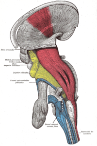
Changes for: metathalamus
- Deleted
Changes for: midline nuclear group
- Deleted
Changes for: middle frontal gyrus
- Deleted
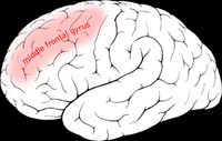
Changes for: precentral gyrus
- Deleted
- Added
Changes for: intergluteal cleft
- Added
- + intergluteal cleft database cross reference http://en.wikipedia.org/wiki/Gluteal_sulcus
- + intergluteal cleft definition The gluteal sulcus (also known as the gluteal fold or horizontal gluteal crease) is an area of the body of humans and great apes, described by a horizontal crease formed by the inferior aspect of the buttocks and the posterior upper leg. It is one of the major defining features of the buttocks. Children with developmental dysplasia of the hips are born with uneven gluteal folds and can be diagnosed with physical examination and sonogram. { database cross reference=http://en.wikipedia.org/wiki/Gluteal_sulcus,Wikipedia:Gluteal_sulcus }
Changes for: decussation of superior cerebellar peduncle
- Deleted
- Added
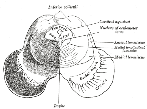
Changes for: brachium of superior colliculus
- Deleted
Changes for: postcentral gyrus
- Deleted
Changes for: pontine reticular formation
- Deleted
- Added
Changes for: temporal pole
- Deleted
- Added
Changes for: olivary pretectal nucleus
- Deleted
- Added
- + olivary pretectal nucleus editor note B&H states this present in urodeles; modify definition
- + olivary pretectal nucleus has exact synonym nucleus praetectalis olivaris { database cross reference=ISBN10:0471888893 , has synonym type=latin term }
- + olivary pretectal nucleus present in taxon http://purl.obolibrary.org/obo/NCBITaxon_8293 { source=ISBN10:0471888893 }
- + olivary pretectal nucleus present in taxon http://purl.obolibrary.org/obo/NCBITaxon_9347 { source=ISBN10:0471888893 }
Changes for: pars endotympanica
- Deleted
- - pars endotympanica definition This is present in the cat and forms the large medial part of the Bulla tympanica. It is not to be regarded as a part of the pars tympanica, which ossifies directly from connective tissue, whereas the Pars endotympanica is preformed in cartilage. The Septum bullae in the cat is formed by the Pars tympanica as well as by the Pars endotympanica(NOMINA ANATOMICA VETERINARIA (2005)).
- Added
- + pars endotympanica definition This is present in the cat and forms the large medial part of the Bulla tympanica. It is not to be regarded as a part of the pars tympanica, which ossifies directly from connective tissue, whereas the Pars endotympanica is preformed in cartilage. The Septum bullae in the cat is formed by the Pars tympanica as well as by the Pars endotympanica(NOMINA ANATOMICA VETERINARIA (2005)). { database cross reference=UBERON:cjm }
Changes for: basal part of pons
- Added
Changes for: superior frontal sulcus
- Deleted
Changes for: mesocolic lymph node
- Deleted
- - colic lymph node EquivalentTo lymph node and located in some colon
- - colic lymph node SubClassOf located in some colon
- - colic lymph node has exact synonym colic node { database cross reference=FMA:16612 }
- - colic lymph node label colic lymph node
- Added
- + mesocolic lymph node EquivalentTo lymph node and located in some hepatic flexure of colon
- + mesocolic lymph node SubClassOf located in some hepatic flexure of colon
- + mesocolic lymph node database cross reference http://en.wikipedia.org/wiki/Mesocolic_lymph_nodes
- + mesocolic lymph node editor note medilexicon distinguishes between the more general mesocolic and the colic lymph nodes
- + mesocolic lymph node external definition Colic lymph node: A mesocolic lymph node that is located along the arteries supplying the colon { source=https://en.wikipedia.org/wiki/Mesocolic_lymph_nodes }
- + mesocolic lymph node external definition colic lymph nodes: those mesocolic lymph nodes located along the arteries supplying the colon. { source=http://www.medilexicon.com }
- + mesocolic lymph node external definition mesocolic lymph nodes: nodes located in the mesocolon; they are of two classes: paracolic lymph nodes, located in immediate proximity to the colon; colic lymph nodes located along the arteries supplying the colon. { source=http://www.medilexicon.com }
- + mesocolic lymph node has broad synonym colic lymph node { database cross reference=FMA:16612 }
- + mesocolic lymph node has broad synonym colic node { database cross reference=FMA:16612 }
- + mesocolic lymph node label mesocolic lymph node
- + mesocolic lymph node present in taxon http://purl.obolibrary.org/obo/NCBITaxon_10090 { source=http://www.ncbi.nlm.nih.gov/pubmed/16624319 }
Changes for: anatomical cavity
Changes for: interstitial nucleus of Cajal
- Deleted
- - interstitial nucleus of Cajal definition The largest and most prominent of the cell groups of the medial longitudinal fasciculus
- Added
- + interstitial nucleus of Cajal definition The largest and most prominent of the cell groups of the medial longitudinal fasciculus { database cross reference=UBERON:cjm }
Changes for: pancreaticoduodenal lymph node
- Added
Changes for: lateral line system
- Deleted
- - lateral line system SubClassOf part of some peripheral nervous system
- - lateral line system definition A line of neuroblasts along the sides of fish and amphibia that detect vibrations and pressure changes. { database cross reference=http://en.wikipedia.org/wiki/Lateral_line_system , database cross reference=ISBN:0471209627 , database cross reference=ISBN:0815318960 }
- Added
- + lateral line system SubClassOf has part some lateral line
- + lateral line system SubClassOf has part some part of some peripheral nervous system
- + lateral line system definition A line of sensory organs and associated structures along the sides of fish and amphibia that detect vibrations and pressure changes. { database cross reference=http://en.wikipedia.org/wiki/Lateral_line_system , database cross reference=ISBN:0471209627 , database cross reference=ISBN:0815318960 , database cross reference=UBERON:cjm }
- + lateral line system editor note we model this as being potentially inclusive of lateral line scales, and thus overlapping with the skeletal system. This is to accomodate TAO
Changes for: scale
- Added
- + scale has exact synonym scale (sensu Metazoa)
- + scale present in taxon http://purl.obolibrary.org/obo/NCBITaxon_9972
Changes for: pharyngeal arch
- Deleted
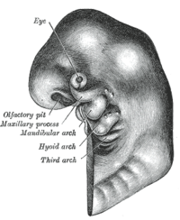
Changes for: male membranous urethra
- Deleted
Changes for: anterior cervical lymph node
- Added
- + anterior cervical lymph node database cross reference http://en.wikipedia.org/wiki/Anterior_cervical_lymph_nodes
- + anterior cervical lymph node definition The anterior cervical lymph nodes are a group of nodes found on the anterior part of the neck. { database cross reference=http://en.wikipedia.org/wiki/Anterior_cervical_lymph_nodes,Wikipedia:Anterior_cervical_lymph_nodes }
Changes for: sacral lymph node
Changes for: brachial lymph node
- Deleted
Changes for: palmar branch of median nerve
- Added
- + palmar branch of median nerve database cross reference http://en.wikipedia.org/wiki/Palmar_branch_of_the_median_nerve
- + palmar branch of median nerve definition The palmar branch of the median nerve is a branch of the median nerve which arises at the lower part of the forearm. { database cross reference=http://en.wikipedia.org/wiki/Palmar_branch_of_the_median_nerve,Wikipedia:Palmar_branch_of_the_median_nerve }
Changes for: submandibular lymph node
- Deleted
- Added
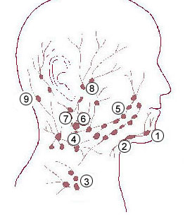
Changes for: tunica intima
- Deleted
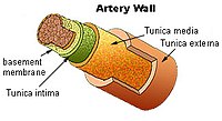
Changes for: tunica media
- Deleted
Changes for: basicranium
- Deleted
Changes for: otolith organ
- Deleted
Changes for: periosteum
- Deleted
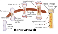
Changes for: corpus luteum
- Deleted
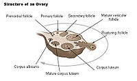
Changes for: anterior fontanel
- Deleted
Changes for: calcaneus
- Deleted
Changes for: distal tarsal bone 1
- Deleted
Changes for: navicular bone of pes
- Deleted
Changes for: distal tarsal bone 3
- Deleted
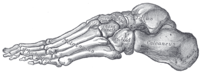
Changes for: distal tarsal bone 2
- Deleted
Changes for: face
- Deleted
Changes for: cuboid bone
- Deleted
Changes for: chest
- Deleted
Changes for: metatarsal bone
- Deleted
- Added
Changes for: fibula
- Deleted
Changes for: tarsal bone
- Deleted
- Added
Changes for: distal carpal bone 3
- Deleted
Changes for: distal carpal bone 4
- Deleted
Changes for: pretectal nucleus
- Deleted
- - pretectal nucleus definition Any of the groups of neurons located between the thalamus and midbrain that receive binocular sensory input from retinal ganglion cells of the eyes and are responsible for maintaining the pupillary light reflex { database cross reference=MP:0009638 }
- Added
- + pretectal nucleus database cross reference BAMS:PTA
- + pretectal nucleus database cross reference FMA:72401
- + pretectal nucleus database cross reference HBA:9083
- + pretectal nucleus database cross reference UMLS:C0175358 { source=NIFSTD:birnlex_864 }
- + pretectal nucleus database cross reference http://braininfo.rprc.washington.edu/centraldirectory.aspx?ID=468
- + pretectal nucleus database cross reference http://braininfo.rprc.washington.edu/centraldirectory.aspx?ID=468 { source=NIFSTD:birnlex_864 }
- + pretectal nucleus database cross reference http://linkedlifedata.com/resource/umls/id/C0175358
- + pretectal nucleus database cross reference http://uri.neuinfo.org/nif/nifstd/birnlex_864
- + pretectal nucleus definition A neural nucleus of the pretectal area (between dorsal thalamus and optic tectum) that receives afferents primarily from the retina and the optic tectum and are involved in modulating motor behavior in response to visual input. { database cross reference=https://github.com/obophenotype/uberon/issues/361 , database cross reference=ISBN10:0471888893 }
- + pretectal nucleus external definition Any of the groups of neurons located between the thalamus and midbrain that receive binocular sensory input from retinal ganglion cells of the eyes and are responsible for maintaining the pupillary light reflex { source=MP:0009638 }
- + pretectal nucleus external ontology notes see notes for pretectal region. Note this is midbrain in FMA, midbrain (mesencephalon) in HBA, midbrain in DHBA { external ontology=FMA }
- + pretectal nucleus has alternative id UBERON:0002994
- + pretectal nucleus has exact synonym nucleus of pretectal area { database cross reference=FMA:72401 }
- + pretectal nucleus has exact synonym pretectal area nucleus { database cross reference=FMA:72401 }
- + pretectal nucleus has related synonym nucleus area pretectalis { database cross reference=NeuroNames:468 , has synonym type=latin term }
- + pretectal nucleus has related synonym nucleus of the pretectal area { database cross reference=NeuroNames:468 }
- + pretectal nucleus present in taxon http://purl.obolibrary.org/obo/NCBITaxon_7761
- + pretectal nucleus present in taxon http://purl.obolibrary.org/obo/NCBITaxon_9255
- + pretectal nucleus taxon notes in hagfishes, there are two: the pretectal nucleus and the nucleus of the posterior commissure[ISBN10:0471888893]
Changes for: carpal bone
- Deleted
Changes for: epiphysis
- Deleted
Changes for: pisiform
- Deleted
Changes for: scaphoid
- Deleted
- Added
Changes for: ulna
- Deleted
Changes for: wrist joint
- Deleted
- Added
Changes for: radial nerve
- Deleted
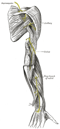
Changes for: axillary nerve
- Deleted
- Added
Changes for: ulnar nerve
- Deleted
- Added
Changes for: pectoral muscle
- Deleted
- Added
Changes for: ascending aorta
- Deleted
Changes for: Pacinian corpuscle
- Deleted
- Added
Changes for: splenic vein
- Deleted
- - splenic vein definition In anatomy, the splenic vein (in the past called the lienal vein) is the blood vessel that drains blood from the spleen. It joins with the superior mesenteric vein, to form the hepatic portal vein and follows a course superior to the pancreas, alongside of the similarly named artery, the splenic artery. It collects branches from the stomach and pancreas and most notably from the large intestine, which is drained by the inferior mesenteric vein and joins with splenic vein shortly before it forms the portal vein. [WP,unvetted]. { database cross reference=http://en.wikipedia.org/wiki/Splenic_vein }
- - splenic vein depicted by http://upload.wikimedia.org/wikipedia/commons/thumb/5/5b/Gray1189.png/200px-Gray1189.png
- Added
- + splenic vein definition A vein arising from the splenic trabecular vein in the hilum of the spleen that drains into the portal vein. { database cross reference=ncithesaurus:Splenic_Vein }
- + splenic vein depicted by http://upload.wikimedia.org/wikipedia/commons/5/5b/Gray1189.png
Changes for: articular capsule
- Deleted
- Added
Changes for: muscle of shoulder
- Deleted
- - muscle of shoulder EquivalentTo skeletal muscle organ and part of some shoulder
- - muscle of shoulder SubClassOf part of some shoulder
- - muscle of shoulder SubClassOf pectoral complex muscle
- Added
- + muscle of shoulder EquivalentTo skeletal muscle organ and attaches to some shoulder bone
- + muscle of shoulder SubClassOf attaches to some shoulder bone
- + muscle of shoulder SubClassOf has part some part of some shoulder
- + muscle of shoulder SubClassOf skeletal muscle organ
Changes for: ankle joint
- Deleted
- Added
Changes for: carotid sinus
- Deleted
Changes for: inguinal canal
- Deleted
- - inguinal canal SubClassOf located in some abdominal wall
- - inguinal canal SubClassOf part of some abdomen
- - inguinal canal depicted by http://upload.wikimedia.org/wikipedia/commons/thumb/b/b4/Gray1227.png/200px-Gray1227.png
- Added
- + inguinal canal SubClassOf part of some abdominal wall
Changes for: circle of Willis
- Deleted
Changes for: glenohumeral joint
- Deleted
Changes for: infraspinatus muscle
- Deleted
- Added
- + infraspinatus muscle SubClassOf pectoral complex muscle
Changes for: teres major muscle
- Deleted
Changes for: vaginal venous plexus
- Deleted
- Added
Changes for: lymphatic vessel
- Deleted
Changes for: sesamoid bone
- Deleted
Changes for: prepyriform area
- Added
- + prepyriform area database cross reference http://en.wikipedia.org/wiki/Prepyriform_area
- + prepyriform area definition Prepyriform area (or prepiriform cortex) is a portion of the rhinencephalon consisting of paleocortex. Some sources state that it is part of the primary olfactory cortex. { database cross reference=http://en.wikipedia.org/wiki/Prepyriform_area,Wikipedia:Prepyriform_area }
Changes for: paracentral sulcus
- Added
- + paracentral sulcus database cross reference http://en.wikipedia.org/wiki/Paracentral_sulcus
- + paracentral sulcus definition The paracentral sulcus is a sulcus of the brain. It forms the paracentral lobule’s anterior border. { database cross reference=http://en.wikipedia.org/wiki/Paracentral_sulcus,Wikipedia:Paracentral_sulcus }
Changes for: shoulder
- Deleted
Changes for: principal sensory nucleus of trigeminal nerve
- Deleted
- Added
Changes for: hip
- Deleted
Changes for: knee
- Deleted
- Added
Changes for: manual digit 1
- Deleted
- Added
Changes for: arm
- Deleted
Changes for: ventral posterior nucleus of thalamus
- Deleted
Changes for: sternoclavicular joint
- Deleted
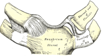
Changes for: manual digit 4
- Deleted
Changes for: manual digit 5
- Deleted
Changes for: manual digit 3
- Deleted
Changes for: manual digit 2
- Deleted
Changes for: pedal digit 5
- Deleted
Changes for: lateral malleolus of fibula
- Added
- + lateral malleolus of fibula database cross reference http://en.wikipedia.org/wiki/Malleolus
- + lateral malleolus of fibula definition Malleolus is the name for the bony prominence on each side of the ankle. { database cross reference=http://en.wikipedia.org/wiki/Malleolus,Wikipedia:Malleolus }
Changes for: hemisphere of embryo
- Deleted
- Added
Changes for: spermaceti organ
- Deleted
- Added
Changes for: radio-ulnar joint
- Deleted
- Added
Changes for: abductor pollicis longus
- Deleted
Changes for: fascia lata
- Deleted
Changes for: synovial bursa
- Deleted
Changes for: brachiocephalic artery
- Deleted
Changes for: extensor carpi radialis longus muscle
- Deleted
Changes for: flexor digitorum profundus
- Deleted
Changes for: extensor carpi ulnaris muscle
- Deleted
- Added
Changes for: pronator teres
- Deleted
Changes for: flexor carpi radialis muscle
- Deleted
Changes for: flexor carpi ulnaris muscle
- Deleted
Changes for: blood vessel layer
- Deleted
- Added
Changes for: posterior tibial artery
- Deleted
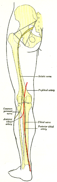
Changes for: anterior tibial artery
- Deleted
Changes for: left common carotid artery plus branches
- Deleted
- Added
Changes for: vertebral artery
- Deleted
Changes for: right subclavian artery
- Deleted
- Added
Changes for: internal carotid artery
- Deleted
Changes for: subclavian artery
- Deleted
Changes for: common carotid artery plus branches
- Deleted
Changes for: right common carotid artery plus branches
- Deleted
- Added
Changes for: brachialis muscle
- Deleted
- Added
- + brachialis muscle SubClassOf muscle of shoulder
Changes for: coracobrachialis muscle
- Deleted
- Added
- + coracobrachialis muscle SubClassOf muscle of shoulder
Changes for: arch of aorta
- Deleted
- Added
Changes for: biceps brachii
- Deleted
- Added
Changes for: triceps brachii
- Deleted
Changes for: tunicate siphon
- Deleted
- - tunicate siphon definition tubular appendage for drawing or ejecting fluids
- Added
- + tunicate siphon definition tubular appendage for drawing or ejecting fluids { database cross reference=UBERON:cjm }
Changes for: intermaxillary process
- Added
Changes for: posterior nasal aperture
- Deleted
Changes for: superior eyelid tarsus
- Added
Changes for: thoracic aorta
- Deleted
Changes for: descending aorta
- Deleted
Changes for: abdominal aorta
- Deleted
Changes for: cochlear nuclear complex
- Deleted
Changes for: dermal skeletal element
- Deleted
- - dermal skeletal element definition dermis-derived entity that is made of skeletal tissue.
- Added
- + dermal skeletal element definition dermis-derived entity that is made of skeletal tissue. { database cross reference=UBERON:cjm }
Changes for: tuber cinereum
- Deleted
Changes for: diaphysis
- Deleted
Changes for: triangular part of inferior frontal gyrus
- Added
Changes for: orbital part of inferior frontal gyrus
- Added
Changes for: cerebral peduncle
- Deleted
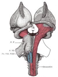
Changes for: foramen magnum
- Deleted
Changes for: tooth socket
- Deleted
Changes for: sella turcica
- Deleted
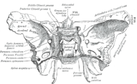
Changes for: abdominal cavity
- Deleted
Changes for: pubic symphysis
- Deleted
Changes for: abdominal wall
- Deleted
- - abdominal wall definition The abdominal wall represents the boundaries of the abdominal cavity. The abdominal wall is split into the posterior (back), lateral (sides) and anterior (front) walls. There is a common set of layers covering and forming all the walls: the deepest being the extraperitoneal fat, the parietal peritoneum, and a layer of fascia which has different names over where it covers (eg transversalis, psoas fascia). Superficial to these, but not present in the posterior wall are the three layers of muscle, the transversus abdominis (transverse abdominal muscle), the internal (obliquus internus) and the external oblique (obliquus externus). { database cross reference=http://en.wikipedia.org/wiki/Abdominal_wall }
- Added
- + abdominal wall definition The tissues that surround the organs that are present within the abdominal cavity. The abdominal wall tissue is composed of layers of fat, parietal peritoneum, fascia, and muscles. { database cross reference=ncithesaurus:Abdominal_Wall }
Changes for: metacarpophalangeal joint
- Deleted
Changes for: limbic lobe
- Deleted
Changes for: retroperitoneal space
- Deleted
- Added
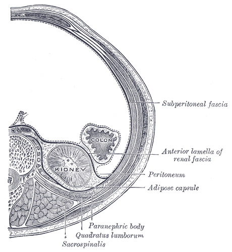
Changes for: acromioclavicular joint
- Deleted
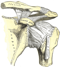
Changes for: fused sacrum
- Deleted
Changes for: ventral nucleus of lateral lemniscus
- Deleted
- Added
Changes for: paraterminal gyrus
- Deleted
Changes for: neuropil
- Deleted
- - neuropil definition A cluster of ➞neurites that is part of a ➞nervous system and forms a network of dendrites and axons where ➞synapses are present and in which neuronal somata do not occur. { database cross reference=http://www.ncbi.nlm.nih.gov/pubmed/21062451 }
- Added
- + neuropil definition A cluster of neurites that is part of a nervous system and forms a network of dendrites and axons where synapses are present and in which neuronal somata do not occur. { database cross reference=http://www.ncbi.nlm.nih.gov/pubmed/21062451 }
Changes for: jugular vein
- Deleted
Changes for: annulus fibrosus disci intervertebralis
- Deleted
Changes for: corpus cavernosum penis
- Deleted
Changes for: septum pellucidum
- Deleted
Changes for: kidney arcuate vein
- Deleted
Changes for: cerebellar vermis
- Deleted
Changes for: interlobular artery
- Deleted
Changes for: deep cervical lymph node
- Deleted
Changes for: medial palpebral ligament
- Deleted
Changes for: piriform cortex
- Deleted
Changes for: vasa recta
- Deleted
Changes for: inferior phrenic artery
- Deleted
- Added
- + inferior phrenic artery definition The inferior phrenic arteries are two small vessels, which supply the diaphragm but present much variety in their origin. They may arise separately from the front of the aorta, immediately above the celiac artery, or by a common trunk, which may spring either from the aorta or from the celiac artery. Sometimes one is derived from the aorta, and the other from one of the renal arteries; they rarely arise as separate vessels from the aorta. They diverge from one another across the crura of the diaphragm, and then run obliquely upward and lateralward upon its under surface. The left phrenic passes behind the esophagus, and runs forward on the left side of the esophageal hiatus. The right phrenic passes behind the inferior vena cava, and along the right side of the foramen which transmits that vein. Near the back part of the central tendon each vessel divides into a medial and a lateral branch. The medial branch curves forward, and anastomoses with its fellow of the opposite side, and with the musculophrenic and pericardiacophrenic arteries. The lateral branch passes toward the side of the thorax, and anastomoses with the lower intercostal arteries, and with the musculophrenic. The lateral branch of the right phrenic gives off a few vessels to the inferior vena cava; and the left one, some branches to the esophagus. Each vessel gives off superior suprarenal branches to the suprarenal gland of its own side. The spleen and the liver also receive a few twigs from the left and right vessels respectively. { database cross reference=http://en.wikipedia.org/wiki/Inferior_phrenic_arteries,Wikipedia:Inferior_phrenic_arteries }
- + inferior phrenic artery depicted by http://upload.wikimedia.org/wikipedia/commons/a/af/Gray531.png
Changes for: bulbus cordis
- Deleted
Changes for: bone fossa
- Deleted
- Added
- + bone fossa has broad synonym fossa
Changes for: superficial cerebral vein
- Added
- + superficial cerebral vein SubClassOf part of some superficial vasculature
- + superficial cerebral vein SubClassOf superficial vein
Changes for: statoconial membrane
- Added
- + statoconial membrane comment See https://sourceforge.net/p/obo/zebrafish-anatomy-zfa-term-requests/132/
Changes for: inferior salivatory nucleus
- Deleted
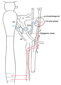
Changes for: hypoglossal nucleus
- Deleted
Changes for: dorsal motor nucleus of vagus nerve
- Deleted
- Added
Changes for: lamina VI of gray matter of spinal cord
- Added
- + lamina VI of gray matter of spinal cord database cross reference http://en.wikipedia.org/wiki/Spinal_lamina_VI
- + lamina VI of gray matter of spinal cord definition Spinal lamina VI is a lamina of the spinal cord. It is also known as the base of the posterior horn. { database cross reference=http://en.wikipedia.org/wiki/Spinal_lamina_VI,Wikipedia:Spinal_lamina_VI }
Changes for: lamina V of gray matter of spinal cord
- Added
- + lamina V of gray matter of spinal cord database cross reference http://en.wikipedia.org/wiki/Spinal_lamina_V
- + lamina V of gray matter of spinal cord definition Spinal lamina V is a lamina of the spinal cord. It is also known as the neck of the posterior horn. { database cross reference=http://en.wikipedia.org/wiki/Spinal_lamina_V,Wikipedia:Spinal_lamina_V }
Changes for: hippocampal sulcus
- Deleted
Changes for: auditory cortex
- Deleted
- Added
Changes for: flexor hallucis longus
- Deleted
Changes for: popliteus muscle
- Deleted
Changes for: sural artery
- Deleted
- Added
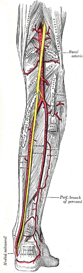
Changes for: deep brachial artery
- Deleted
- Added
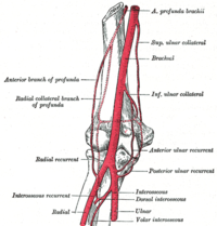
Changes for: brachial artery
- Deleted
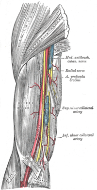
Changes for: subscapular artery
- Deleted
Changes for: lateral thoracic artery
- Deleted
Changes for: thoraco-acromial artery
- Deleted
- Added
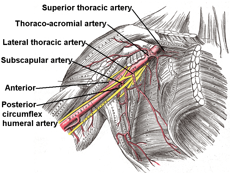
Changes for: axillary artery
- Deleted
Changes for: vastus medialis
- Deleted
Changes for: pectineus muscle
- Deleted
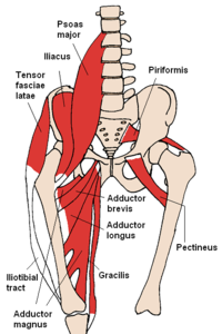
Changes for: semimembranosus muscle
- Deleted
- Added
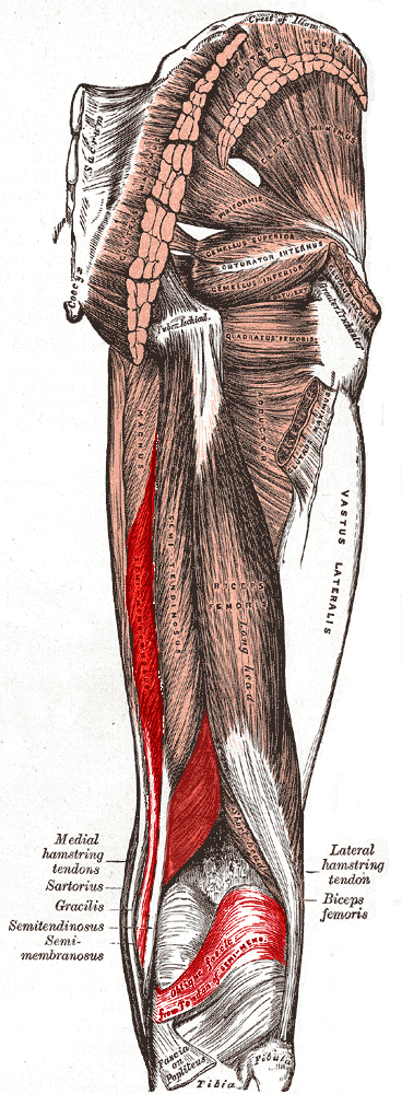
Changes for: gastrocnemius
- Deleted
Changes for: fibularis longus
- Deleted
Changes for: soleus muscle
- Deleted
Changes for: primary motor cortex
- Deleted
Changes for: extensor digitorum longus
- Deleted
Changes for: tibialis anterior
- Deleted
Changes for: perineal artery
- Deleted
Changes for: deep femoral artery
- Deleted
- Added
- + deep femoral artery EquivalentTo artery and part of some deep vasculature and branching part of some femoral artery
- + deep femoral artery SubClassOf part of some deep vasculature
- + deep femoral artery depicted by http://upload.wikimedia.org/wikipedia/commons/8/8b/Gray548.png
Changes for: medial circumflex femoral artery
- Deleted
Changes for: inferior rectal artery
- Deleted
Changes for: coccyx
- Deleted
Changes for: anal region
- Deleted
- Added
Changes for: seminiferous tubule of testis
- Deleted
- Added
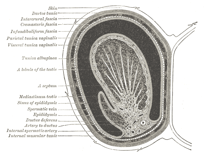
Changes for: lesser sac
- Deleted
- Added
Changes for: mesovarium
- Deleted
Changes for: dorsal artery of penis
- Deleted
- Added
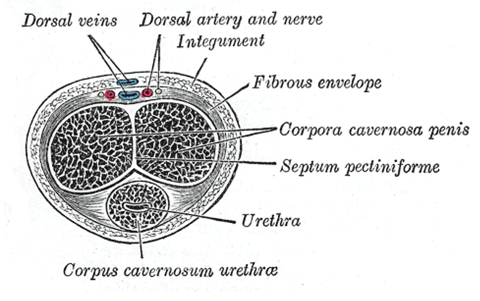
Changes for: psoas minor muscle
- Deleted
Changes for: biceps femoris
- Deleted
Changes for: semitendinosus
- Deleted
Changes for: tensor fasciae latae muscle
- Deleted
- Added
Changes for: quadriceps femoris
- Deleted
Changes for: rectus femoris
- Deleted
Changes for: vastus lateralis
- Deleted
- Added
Changes for: gluteus maximus
- Deleted
Changes for: gluteus medius
- Deleted
Changes for: iliacus muscle
- Deleted
Changes for: femoral vein
- Deleted
Changes for: external anal sphincter
- Deleted
Changes for: obturator externus
- Deleted
Changes for: parietal peritoneum
- Deleted
- Added
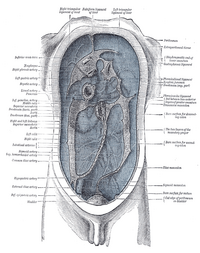
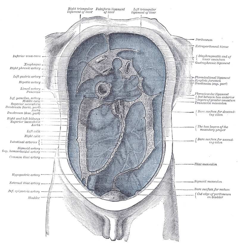
Changes for: deep circumflex iliac vein
- Deleted
- - deep circumflex iliac vein SubClassOf vein
- - deep circumflex iliac vein definition The deep circumflex iliac vein is formed by the union of the venæ comitantes of the deep iliac circumflex artery, and joins the external iliac vein about 2 cm. above the inguinal ligament. It also receives small tributary branches from the thoracoepigastric vein [WP,unvetted]. { database cross reference=http://en.wikipedia.org/wiki/Deep_circumflex_iliac_vein }
- - deep circumflex iliac vein depicted by http://upload.wikimedia.org/wikipedia/commons/thumb/b/bd/Gray585.png/200px-Gray585.png
- Added
- + deep circumflex iliac vein SubClassOf deep vein
- + deep circumflex iliac vein SubClassOf part of some deep vasculature
- + deep circumflex iliac vein definition A vein that corresponds to the deep circumflex iliac artery and empties near or in a common trunk with the inferior epigastric vein into the external iliac vein; deep circumflex iliac vein. { database cross reference=http://medical-dictionary.thefreedictionary.com/circumflex+iliac+vein }
- + deep circumflex iliac vein has exact synonym circumflex iliac vein
- + deep circumflex iliac vein has exact synonym iliac circumflex vein
Changes for: vaginal vein
- Deleted
Changes for: obturator artery
- Deleted
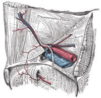
Changes for: superior gluteal artery
- Deleted
Changes for: external iliac vein
- Deleted
Changes for: internal iliac vein
- Deleted
Changes for: umbilical artery
- Deleted
Changes for: iliolumbar artery
- Deleted
Changes for: superior vesical artery
- Deleted
Changes for: internal iliac artery
- Deleted
Changes for: external iliac artery
- Deleted
Changes for: ovarian follicle
- Deleted
- Added
Changes for: cumulus oophorus
- Deleted
Changes for: epididymis
- Deleted
Changes for: spongiose part of urethra
- Deleted
- Added
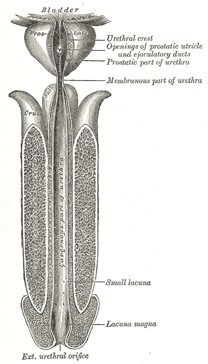
Changes for: ischiocavernosus muscle
- Deleted
- Added
Changes for: pampiniform plexus
- Deleted
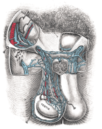
Changes for: male urethra
- Deleted
- Added
Changes for: prostatic urethra
- Deleted
Changes for: female urethra
- Deleted
- Added
Changes for: coccygeus muscle
- Deleted
Changes for: levator ani muscle
- Deleted
Changes for: iliolumbar vein
- Deleted
Changes for: tibial nerve
- Deleted
Changes for: sciatic nerve
- Deleted
Changes for: obturator vein
- Deleted
Changes for: longissimus thoracis muscle
- Deleted
- Added
Changes for: body of uterus
- Deleted
- - body of uterus SubClassOf organ part
- Added
- + body of uterus EquivalentTo zone of organ and continuous with some fallopian tube and continuous with some internal cervical os
- + body of uterus SubClassOf continuous with some fallopian tube
- + body of uterus SubClassOf continuous with some internal cervical os
- + body of uterus SubClassOf zone of organ
Changes for: semispinalis thoracis
- Deleted
Changes for: ulnar artery
- Deleted
Changes for: semispinalis capitis
- Deleted
Changes for: semispinalis cervicis
- Deleted
Changes for: longissimus capitis
- Deleted
- Added
Changes for: longissimus cervicis muscle
- Deleted
- Added
Changes for: spinalis thoracis muscle
- Deleted
- Added
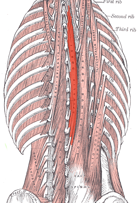
Changes for: radial artery
- Deleted
Changes for: basilic vein
- Deleted
Changes for: median cubital vein
- Deleted
Changes for: brachial vein
- Deleted
- Added
Changes for: zone of stomach
- Deleted
- Added
Changes for: nephrogenic zone
- Deleted
- - nephrogenic zone definition outer portion of the renal cortex. Site of formation of medullary and cortical elements.
- Added
- + nephrogenic zone definition outer portion of the renal cortex. Site of formation of medullary and cortical elements. { database cross reference=UBERON:cjm }
Changes for: Hatschek’s nephridium
- Deleted
- - Hatschek’s nephridium definition An unpaired kidney whose duct opens into the anterior pharynx. It can be seen as a deep ciliated fossa on the dorsal midline of the buccal cavity (mouth). Among other things, it secrets mucus which entraps food particles from the water[WP].
- Added
- + Hatschek’s nephridium definition An unpaired kidney whose duct opens into the anterior pharynx. It can be seen as a deep ciliated fossa on the dorsal midline of the buccal cavity (mouth). Among other things, it secrets mucus which entraps food particles from the water[WP]. { database cross reference=UBERON:cjm }
Changes for: ascidian cerebral ganglion
- Deleted
- - ascidian cerebral ganglion comment the CG appears to be a diencephalic brain. It originates mainly from the posterior sensory vesicle (Takamura 2002; Dufour et al. 2006) and shows endocrine activity. For examples, ascidian genes orthologous to GnRH and OT/VP, which are secreted from the hypothalamus and the NH, respectively, in vertebrates, are expressed in the cortex of the CG, suggesting that the NC is not distinctly compartmentalized as are vertebrate brains (Tsutsui et al. 1998; Kavanaugh et al. 2005; Kawada et al. 2008; Ukena et al. 2008; Kawada et al. 2009). Moreover, PC2 expression in the cortex of the CG supports possible endocrine activity (Sekiguchi et al. 2007). // Editor notes: consider merging into brain, based on http://www.ncbi.nlm.nih.gov/pubmed/21062451 “We suggest using the term ➞brain not only for the ‘dorsal ganglia’ of adult Tunicata and Pterobranchia, but also for the larval ‘cerebral ganglion’ of tunicates.”
- Added
- + ascidian cerebral ganglion comment the CG appears to be a diencephalic brain. It originates mainly from the posterior sensory vesicle (Takamura 2002; Dufour et al. 2006) and shows endocrine activity. For examples, ascidian genes orthologous to GnRH and OT/VP, which are secreted from the hypothalamus and the NH, respectively, in vertebrates, are expressed in the cortex of the CG, suggesting that the NC is not distinctly compartmentalized as are vertebrate brains (Tsutsui et al. 1998; Kavanaugh et al. 2005; Kawada et al. 2008; Ukena et al. 2008; Kawada et al. 2009). Moreover, PC2 expression in the cortex of the CG supports possible endocrine activity (Sekiguchi et al. 2007). // Editor notes: consider merging into brain, based on http://www.ncbi.nlm.nih.gov/pubmed/21062451 “We suggest using the term brain not only for the ‘dorsal ganglia’ of adult Tunicata and Pterobranchia, but also for the larval ‘cerebral ganglion’ of tunicates.”
Changes for: deep inguinal lymph node
- Added
Changes for: superficial inguinal lymph node
- Added
Changes for: carotid sinus nerve
- Added
Changes for: carpal skeleton
- Deleted
- Added
Changes for: lunate sulcus
- Added
- + lunate sulcus database cross reference http://en.wikipedia.org/wiki/Lunate_sulcus
- + lunate sulcus definition The lunate sulcus is a structure in the occipital lobe. It is not present in all individuals. { database cross reference=http://en.wikipedia.org/wiki/Lunate_sulcus,Wikipedia:Lunate_sulcus }
Changes for: occipital pole
- Deleted
- Added
Changes for: lateral occipital sulcus
- Deleted
Changes for: transverse occipital sulcus
- Added
- + transverse occipital sulcus database cross reference http://en.wikipedia.org/wiki/Transverse_occipital_sulcus
- + transverse occipital sulcus definition The transverse occipital sulcus is a structure in the occipital lobe. The transverse occipital sulcus is continuous with the posterior end of the occipital ramus of the intraparietal sulcus, and runs across the upper part of the lobe, a short distance behind the parietoC6ccipital fissure. { database cross reference=http://en.wikipedia.org/wiki/Transverse_occipital_sulcus,Wikipedia:Transverse_occipital_sulcus }
Changes for: parietal operculum
- Deleted
- Added
Changes for: marginal sulcus
- Deleted
Changes for: intraparietal sulcus
- Deleted
Changes for: postcentral sulcus
- Deleted
Changes for: central sulcus
- Deleted
- Added
Changes for: olfactory trigone
- Deleted
Changes for: callosal sulcus
- Deleted
Changes for: longitudinal fissure
- Deleted
- Added
Changes for: trapezoid body
- Deleted
Changes for: tectospinal tract
- Deleted
Changes for: spinothalamic tract of medulla
- Deleted
- - spinothalamic tract of medulla depicted by [http://upload.wikimedia.org/wikipedia/commons/thumb/f/fe/Medulla_spinalis_-tracts-English.svg/200px-Medulla_spinalis-tracts-English.svg.png](http://upload.wikimedia.org/wikipedia/commons/thumb/f/fe/Medulla_spinalis-tracts-English.svg/200px-Medulla_spinalis-tracts-_English.svg.png)
- Added
- + spinothalamic tract of medulla depicted by [http://upload.wikimedia.org/wikipedia/commons/f/fe/Medulla_spinalis_-tracts-English.svg](http://upload.wikimedia.org/wikipedia/commons/f/fe/Medulla_spinalis-tracts-_English.svg)
Changes for: ventral posteromedial nucleus of thalamus
- Deleted
Changes for: ventral posterolateral nucleus
- Deleted
Changes for: lingual gyrus
- Deleted
Changes for: lateral lemniscus
- Deleted
Changes for: cingulate gyrus
- Deleted
- Added
Changes for: caudal pontine reticular nucleus
- Added
- + caudal pontine reticular nucleus definition The caudal pontine reticular nucleus is composed of gigantocellular neurons. In rabbits and cats it is exclusively giant cells, however in humans there are normally sized cells as well. The pontis caudalis is rostral to the gigantocellular nucleus and is located in the caudal pons, as the name would indicate. The pontis caudalis has been known to mediate head movement, in concert with the nucleus gigantocellularis and the superior colliculus. The neurons in the dorsal half of this nuclei fire rhythmically during mastication, and in an anesthetized animal it is possible to induce mastication via electrical stimulation of the PC or adjacent areas of the gigantocellular nucleus. The pontis caudalis is also thought to play a role in the grinding of teeth during sleep. { database cross reference=http://en.wikipedia.org/wiki/Caudal_pontine_reticular_nucleus,Wikipedia:Caudal_pontine_reticular_nucleus }
Changes for: parahippocampal gyrus
- Deleted
Changes for: anterior spinocerebellar tract
- Deleted
- - anterior spinocerebellar tract depicted by [http://upload.wikimedia.org/wikipedia/commons/thumb/f/fe/Medulla_spinalis_-tracts-English.svg/200px-Medulla_spinalis-tracts-English.svg.png](http://upload.wikimedia.org/wikipedia/commons/thumb/f/fe/Medulla_spinalis-tracts-English.svg/200px-Medulla_spinalis-tracts-_English.svg.png)
- Added
- + anterior spinocerebellar tract depicted by [http://upload.wikimedia.org/wikipedia/commons/f/fe/Medulla_spinalis_-tracts-English.svg](http://upload.wikimedia.org/wikipedia/commons/f/fe/Medulla_spinalis-tracts-_English.svg)
Changes for: lateral dorsal nucleus
- Added
- + lateral dorsal nucleus database cross reference http://en.wikipedia.org/wiki/Lateral_dorsal_nucleus_of_thalamus
- + lateral dorsal nucleus definition The lateral dorsal nucleus is a nucleus of the thalamus. It acts in concert with the anterior nuclei of thalamus. It receives significant input from several subdivisions of visual cortex, and has a primary output to parietal cortex on the dorsolateral cortical convexity, giving it access to limbic forebrain nuclei important for emotion and behavior functions. { database cross reference=http://en.wikipedia.org/wiki/Lateral_dorsal_nucleus_of_thalamus,Wikipedia:Lateral_dorsal_nucleus_of_thalamus }
Changes for: lateral posterior nucleus of thalamus
- Added
- + lateral posterior nucleus of thalamus database cross reference http://en.wikipedia.org/wiki/Lateral_posterior_nucleus_of_thalamus
- + lateral posterior nucleus of thalamus definition The lateral posterior nucleus is a nucleus of the thalamus. It acts in concert with the pulvinar. { database cross reference=http://en.wikipedia.org/wiki/Lateral_posterior_nucleus_of_thalamus,Wikipedia:Lateral_posterior_nucleus_of_thalamus }
Changes for: pulvinar nucleus
- Deleted
Changes for: medial superior olivary nucleus
- Deleted
- Added
Changes for: anterior nuclear group
- Deleted
- Added
Changes for: olfactory sulcus
- Deleted
Changes for: middle temporal gyrus
- Deleted
Changes for: posterior hypothalamic region
- Added
Changes for: ventral nuclear group
- Deleted
Changes for: lateral superior olivary nucleus
- Deleted
- Added
Changes for: ventral corticospinal tract
- Deleted
- - ventral corticospinal tract depicted by [http://upload.wikimedia.org/wikipedia/commons/thumb/f/fe/Medulla_spinalis_-tracts-English.svg/200px-Medulla_spinalis-tracts-English.svg.png](http://upload.wikimedia.org/wikipedia/commons/thumb/f/fe/Medulla_spinalis-tracts-English.svg/200px-Medulla_spinalis-tracts-_English.svg.png)
- Added
- + ventral corticospinal tract depicted by [http://upload.wikimedia.org/wikipedia/commons/f/fe/Medulla_spinalis_-tracts-English.svg](http://upload.wikimedia.org/wikipedia/commons/f/fe/Medulla_spinalis-tracts-_English.svg)
Changes for: fusiform gyrus
- Deleted
Changes for: vestibulospinal tract
- Deleted
- - vestibulospinal tract depicted by [http://upload.wikimedia.org/wikipedia/commons/thumb/f/fe/Medulla_spinalis_-tracts-English.svg/200px-Medulla_spinalis-tracts-English.svg.png](http://upload.wikimedia.org/wikipedia/commons/thumb/f/fe/Medulla_spinalis-tracts-English.svg/200px-Medulla_spinalis-tracts-_English.svg.png)
- Added
- + vestibulospinal tract depicted by [http://upload.wikimedia.org/wikipedia/commons/f/fe/Medulla_spinalis_-tracts-English.svg](http://upload.wikimedia.org/wikipedia/commons/f/fe/Medulla_spinalis-tracts-_English.svg)
Changes for: superior temporal gyrus
- Deleted
Changes for: posterior spinocerebellar tract
- Deleted
- - posterior spinocerebellar tract depicted by [http://upload.wikimedia.org/wikipedia/commons/thumb/f/fe/Medulla_spinalis_-tracts-English.svg/200px-Medulla_spinalis-tracts-English.svg.png](http://upload.wikimedia.org/wikipedia/commons/thumb/f/fe/Medulla_spinalis-tracts-English.svg/200px-Medulla_spinalis-tracts-_English.svg.png)
- Added
- + posterior spinocerebellar tract depicted by [http://upload.wikimedia.org/wikipedia/commons/f/fe/Medulla_spinalis_-tracts-English.svg](http://upload.wikimedia.org/wikipedia/commons/f/fe/Medulla_spinalis-tracts-_English.svg)
Changes for: olivocerebellar tract
- Deleted
Changes for: inferior temporal gyrus
- Deleted
Changes for: pyramidal decussation
- Deleted
Changes for: anterior cingulate gyrus
- Deleted
- Added
Changes for: psoas major muscle
- Deleted
Changes for: serosa of uterus
- Deleted
Changes for: myometrium
- Deleted
Changes for: endometrium
- Deleted
Changes for: glans penis
- Deleted
- Added
Changes for: distal convoluted tubule
- Deleted
Changes for: thick ascending limb of loop of Henle
- Deleted
Changes for: junk chamber
- Deleted
- Added
Changes for: nephron
- Deleted
Changes for: renal column
- Deleted
Changes for: proximal convoluted tubule
- Deleted
Changes for: descending limb of loop of Henle
- Deleted
- Added
Changes for: loop of Henle
- Deleted
Changes for: bile canaliculus
- Deleted
Changes for: ischium
- Deleted
Changes for: innominate bone
- Deleted
Changes for: femoral nerve
- Deleted
Changes for: acetabular part of hip bone
- Deleted
Changes for: frontal pole
- Deleted
- Added
Changes for: trabecula of spleen
- Added
Changes for: pancreas
- Deleted
Changes for: phonic lip
- Deleted
- Added
Changes for: dorsal bursa
- Deleted
- Added
Changes for: red pulp of spleen
- Deleted
Changes for: marginal zone of spleen
- Deleted
Changes for: urinary bladder
- Deleted
Changes for: trigone of urinary bladder
- Deleted
Changes for: neck of urinary bladder
- Deleted
Changes for: hilum of spleen
- Deleted
Changes for: nucleus of solitary tract
- Deleted
Changes for: endopiriform nucleus
- Deleted
- Added
- + endopiriform nucleus SubClassOf telencephalic nucleus
Changes for: interlobular bile duct
- Added
Changes for: superficial external pudendal vein
- Added
- + superficial external pudendal vein EquivalentTo external pudendal vein and part of some superficial vasculature
- + superficial external pudendal vein SubClassOf part of some superficial vasculature
- + superficial external pudendal vein SubClassOf superficial vein
Changes for: deep external pudendal vein
- Added
- + deep external pudendal vein EquivalentTo external pudendal vein and part of some deep vasculature
- + deep external pudendal vein SubClassOf deep vein
- + deep external pudendal vein SubClassOf part of some deep vasculature
Changes for: internal anal sphincter
- Deleted
- Added
Changes for: anus
- Deleted
- Added
Changes for: deep circumflex iliac artery
- Added
- + deep circumflex iliac artery EquivalentTo iliac circumflex artery and part of some deep vasculature
- + deep circumflex iliac artery SubClassOf part of some deep vasculature
Changes for: superficial circumflex iliac artery
- Added
- + superficial circumflex iliac artery EquivalentTo iliac circumflex artery and part of some superficial vasculature
- + superficial circumflex iliac artery SubClassOf part of some superficial vasculature
Changes for: paraaortic body
- Deleted
Changes for: renal straight tubule
- Added
Changes for: adrenal cortex
- Deleted
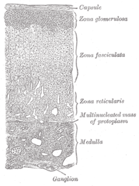
Changes for: adrenal medulla
- Deleted
Changes for: glomerular capsule
- Deleted
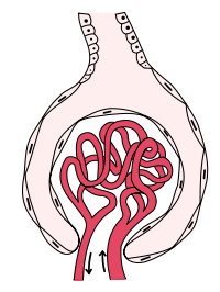
Changes for: renal corpuscle
- Deleted
Changes for: renal papilla
- Deleted
Changes for: minor calyx
- Deleted
Changes for: major calyx
- Deleted
Changes for: renal pelvis
- Deleted
Changes for: cortex of kidney
- Deleted
Changes for: quadratus lumborum
- Deleted
Changes for: transversus abdominis muscle
- Deleted
Changes for: inferior mesenteric vein
- Deleted
Changes for: jejunal vein
- Deleted
Changes for: middle colic vein
- Deleted
Changes for: intestinal villus
- Deleted
Changes for: nauplius eye
- Deleted
- - nauplius eye definition An eye that consists of a cluster of three or four ➞median eyes that form a single structural unit but are separated from one another by pigment layers { database cross reference=http://www.ncbi.nlm.nih.gov/pubmed/21062451 }
- Added
- + nauplius eye definition An eye that consists of a cluster of three or four median eyes that form a single structural unit but are separated from one another by pigment layers { database cross reference=http://www.ncbi.nlm.nih.gov/pubmed/21062451 }
Changes for: primitive superior sagittal sinus
- Deleted
- - primitive superior sagittal sinus comment Editor note: consider merging into developed structure
- Added
- + primitive superior sagittal sinus editor note consider merging into developed structure
Changes for: dentate gyrus subgranular zone
- Deleted
- Added
Changes for: neurogenic placode
- Deleted
- - neurogenic placode EquivalentTo ectodermal placode and has potential to develop into some regional part of nervous system
- - neurogenic placode SubClassOf has potential to develop into some regional part of nervous system
- - neurogenic placode SubClassOf presumptive structure
- Added
- + neurogenic placode SubClassOf develops from some preplacodal ectoderm
- + neurogenic placode SubClassOf has potential to developmentally contribute to some cranial ganglion
- + neurogenic placode SubClassOf has potential to developmentally contribute to some cranial nerve
- + neurogenic placode database cross reference XAO:0004620
Changes for: vomeronasal system
- Deleted
- - vomeronasal system definition A vertebrate nasal chemosensory system that is responsible for detecting intraspecific pheromonal cues as well as environmental odorants
- Added
- + vomeronasal system definition A vertebrate nasal chemosensory system that is responsible for detecting intraspecific pheromonal cues as well as environmental odorants { database cross reference=UBERON:cjm }
Changes for: metapterygial axis
- Deleted
- - metapterygial axis definition proximodistal trajectory through a metapterygium[Wagner]
- Added
- + metapterygial axis definition proximodistal trajectory through a metapterygium[Wagner] { database cross reference=UBERON:cjm }
Changes for: lateral line placode
- Added
- + lateral line placode EquivalentTo ectodermal placode and has potential to develop into some lateral line system
- + lateral line placode SubClassOf has potential to develop into some lateral line system
- + lateral line placode SubClassOf presumptive structure
- + lateral line placode definition An ectodermal placode that will develop into the components of the lateral line system { database cross reference=UBERON:cjm }
- + lateral line placode editor note TODO - compare with protoneuromast
Changes for: ammocoete
- Deleted
- - ammocoete definition lamprey at larval stage
- Added
- + ammocoete definition lamprey at larval stage { database cross reference=UBERON:cjm }
Changes for: horizontal fissure of cerebellum
- Deleted
Changes for: ventral cochlear nucleus
- Deleted
- Added
Changes for: dorsal cochlear nucleus
- Deleted
Changes for: zona arcuata of basilar membrane of cochlea
- Added
Changes for: stratum zonale of thalamus
- Added
- + stratum zonale of thalamus database cross reference http://en.wikipedia.org/wiki/Stratum_zonale_of_thalamus
- + stratum zonale of thalamus definition The thalamus consists chiefly of gray substance, but its upper surface is covered by a layer of white substance, named the stratum zonale. { database cross reference=http://en.wikipedia.org/wiki/Stratum_zonale_of_thalamus,Wikipedia:Stratum_zonale_of_thalamus }
Changes for: accessory cuneate nucleus
- Deleted
Changes for: cerebellar artery
- Deleted
- Added
Changes for: artery of lower lip
- Added
- + artery of lower lip SubClassOf artery of lip
Changes for: artery of upper lip
- Added
- + artery of upper lip SubClassOf artery of lip
Changes for: small intestine Peyer’s patch
- Deleted
- - small intestine Peyer’s patch definition nodular lymphoid structures on the serosal surface of the small intestine.
- Added
- + small intestine Peyer’s patch definition nodular lymphoid structures on the serosal surface of the small intestine. { database cross reference=UBERON:cjm }
Changes for: hyaloid artery
- Deleted
Changes for: vestibular aqueduct
- Deleted
Changes for: perilymphatic space
- Added
Changes for: metatarsus skeleton
- Deleted
Changes for: reticular formation
- Deleted
- Added
Changes for: spiral sulcus
- Deleted
- - spiral sulcus definition A concavity in the floor of the cochlear duct, formed by either the overhanging vestibular lip (inner spiral sulcus) or the spiral prominence and the spiral organ (outer spiral sulcus).
- Added
- + spiral sulcus definition A concavity in the floor of the cochlear duct, formed by either the overhanging vestibular lip (inner spiral sulcus) or the spiral prominence and the spiral organ (outer spiral sulcus). { database cross reference=UBERON:cjm }
Changes for: lamina of spiral limbus
- Deleted
- Added
Changes for: stria vascularis of cochlear duct
- Deleted
- Added
Changes for: vestibular membrane of cochlear duct
- Deleted
- Added
Changes for: otolith
- Deleted
- - otolith comment Currently represented as part of the otolith membrane, but they may also be considered to be embedded in this membrane
- - otolith depicted by http://upload.wikimedia.org/wikipedia/commons/thumb/c/cb/Bigotolith.jpg/200px-Bigotolith.jpg
- Added
- + otolith editor note Currently represented as part of the otolith membrane, but they may also be considered to be embedded in this membrane
- + otolith taxon notes Mammals have many tiny otoconia and fish have three large otoliths[ZFIN:df]
Changes for: midbrain cerebral aqueduct
- Deleted
Changes for: optic recess of third ventricle
- Deleted
Changes for: third ventricle
- Deleted
Changes for: telencephalic ventricle
- Deleted
- Added
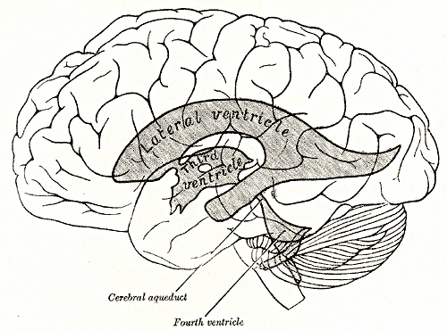
Changes for: olfactory pathway
Changes for: central canal of spinal cord
- Deleted
- - central canal of spinal cord depicted by [http://upload.wikimedia.org/wikipedia/commons/thumb/d/dc/Medulla_spinalis_-Section-English.svg/200px-Medulla_spinalis-Section-English.svg.png](http://upload.wikimedia.org/wikipedia/commons/thumb/d/dc/Medulla_spinalis-Section-English.svg/200px-Medulla_spinalis-Section-_English.svg.png)
- Added
- + central canal of spinal cord database cross reference http://en.wikipedia.org/wiki/Central_canal_of_spinal_cord
- + central canal of spinal cord depicted by [http://upload.wikimedia.org/wikipedia/commons/d/dc/Medulla_spinalis_-Section-English.svg](http://upload.wikimedia.org/wikipedia/commons/d/dc/Medulla_spinalis-Section-_English.svg)
Changes for: costovertebral joint
- Deleted
- Added
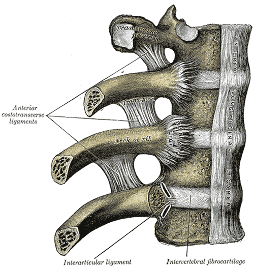
Changes for: scala media
- Deleted
- Added
Changes for: biliary system
- Deleted
- Added
Changes for: dorsal mesentery
- Deleted
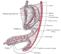
Changes for: brainstem
- Deleted
Changes for: anterior jugular vein
- Deleted
Changes for: costal cartilage
- Deleted
Changes for: external jugular vein
- Deleted
Changes for: true rib
- Deleted
Changes for: pectoralis minor
- Deleted
Changes for: head of rib
- Deleted
Changes for: sternothyroid muscle
- Deleted
Changes for: sternohyoid muscle
- Deleted
Changes for: floating rib
- Deleted
Changes for: omohyoid muscle
- Deleted
Changes for: false rib
- Deleted
- Added
Changes for: clavicle bone
- Deleted
Changes for: uterine horn
- Deleted
Changes for: latissimus dorsi muscle
- Deleted
Changes for: transverse pericardial sinus
- Deleted
- Added
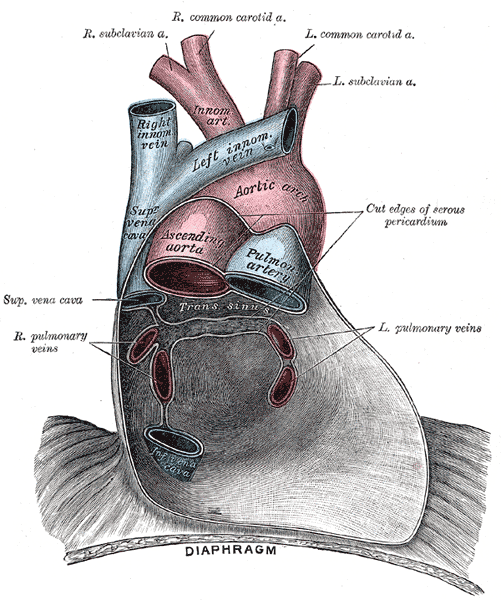
Changes for: left lobe of liver
- Deleted
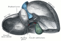
Changes for: right lobe of liver
- Deleted
Changes for: intercostal muscle
- Deleted
Changes for: thyrohyoid muscle
- Deleted
Changes for: nucleus pulposus
- Deleted
Changes for: spinal cord
- Deleted
- - spinal cord database cross reference EMAPA:16666
Changes for: quadrate lobe of liver
- Deleted
Changes for: caudate lobe of liver
- Deleted
Changes for: lobe of thyroid gland
- Deleted
Changes for: scalenus medius
- Deleted
Changes for: longus colli muscle
- Deleted
Changes for: thyroglossal duct
- Deleted
Changes for: vomeronasal organ
- Deleted
Changes for: dorsal horn of spinal cord
- Deleted
- - dorsal horn of spinal cord depicted by [http://upload.wikimedia.org/wikipedia/commons/thumb/d/dc/Medulla_spinalis_-Section-English.svg/200px-Medulla_spinalis-Section-English.svg.png](http://upload.wikimedia.org/wikipedia/commons/thumb/d/dc/Medulla_spinalis-Section-English.svg/200px-Medulla_spinalis-Section-_English.svg.png)
Changes for: serratus ventralis
- Deleted
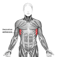
Changes for: dorsal funiculus of spinal cord
- Deleted
- - dorsal funiculus of spinal cord depicted by [http://upload.wikimedia.org/wikipedia/commons/thumb/d/dc/Medulla_spinalis_-Section-English.svg/200px-Medulla_spinalis-Section-English.svg.png](http://upload.wikimedia.org/wikipedia/commons/thumb/d/dc/Medulla_spinalis-Section-English.svg/200px-Medulla_spinalis-Section-_English.svg.png)
- Added
- + dorsal funiculus of spinal cord depicted by [http://upload.wikimedia.org/wikipedia/commons/d/dc/Medulla_spinalis_-Section-English.svg](http://upload.wikimedia.org/wikipedia/commons/d/dc/Medulla_spinalis-Section-_English.svg)
Changes for: scalenus posterior
- Deleted
Changes for: corpora quadrigemina
- Deleted
- - corpora quadrigemina definition In the brain, the corpora quadrigemina (Latin for ‘quadruplet bodies’) are the four colliculi—two inferior, two superior—located on the tectum the dorsal aspect of the midbrain. The corpora quadrigemina are reflex centers involving vision and hearing. [WP,unvetted]. Two pairs of colliculi on the dorsal surface of the midbrain composed of white matter externally and gray matter within, the superior pair containing correlation centers for optic reflexes and the inferior pair containing correlation centers for auditory reflexes[BTO]. { database cross reference=http://en.wikipedia.org/wiki/Corpora_quadrigemina , database cross reference=BTO:0001710 }
- - corpora quadrigemina depicted by http://upload.wikimedia.org/wikipedia/commons/thumb/7/77/Gray709.png/200px-Gray709.png
- Added
- + corpora quadrigemina definition Either of the two pairs of colliculi on the dorsal surface of the midbrain composed of white matter externally and gray matter within, the superior pair containing correlation centers for optic reflexes and the inferior pair containing correlation centers for auditory reflexes[BTO]. { database cross reference=http://en.wikipedia.org/wiki/Corpora_quadrigemina , database cross reference=BTO:0001710 }
Changes for: popliteal artery
- Deleted
Changes for: iliocostalis muscle
- Deleted
Changes for: subscapularis muscle
- Deleted
- Added
- + subscapularis muscle SubClassOf pectoral complex muscle
Changes for: serratus dorsalis inferior muscle
- Deleted
- - serratus dorsalis inferior muscle depicted by http://upload.wikimedia.org/wikipedia/commons/thumb/a/aa/Serratus_posterior.PNG/200px-Serratus_posterior.PNG
- - serratus dorsalis inferior muscle depicted by http://upload.wikimedia.org/wikipedia/commons/thumb/a/aa/Serratus_posterior.PNG/250px-Serratus_posterior.PNG
- Added
Changes for: sternocleidomastoid
- Deleted
- Added
Changes for: vertebral foramen
- Deleted
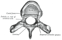
Changes for: olfactory tract
- Deleted
- Added
Changes for: anterior olfactory nucleus
- Deleted
Changes for: cardiac muscle tissue
Changes for: lentiform nucleus
- Deleted
Changes for: olfactory bulb
- Deleted
- Added
Changes for: dorsum
- Deleted
- Added
Changes for: dorsal root of spinal cord
- Deleted
Changes for: celiac ganglion
- Deleted
Changes for: ventral root of spinal cord
- Deleted
Changes for: superior mesenteric vein
- Deleted
Changes for: common iliac vein
- Deleted
Changes for: central nucleus of pallium
- Deleted
- Added
- + central nucleus of pallium SubClassOf telencephalic nucleus
Changes for: renal vein
- Deleted
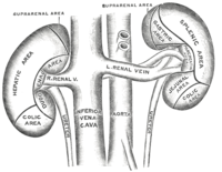
Changes for: epifascicular nucleus
- Deleted
- Added
- + epifascicular nucleus SubClassOf hindbrain nucleus
Changes for: ovarian vein
- Deleted
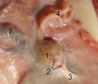
Changes for: suprarenal vein
- Deleted
- Added
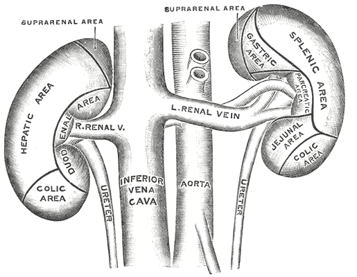
Changes for: median nerve
- Deleted
Changes for: left renal vein
- Deleted
- Added
Changes for: hepatic vein
- Deleted
Changes for: testicular vein
- Deleted
Changes for: manubrium of sternum
- Deleted
Changes for: mammillary body
- Deleted
Changes for: xiphoid process
- Deleted
Changes for: sternebra
- Deleted
- Added
Changes for: body of pancreas
- Deleted
Changes for: tail of pancreas
- Deleted
Changes for: descending colon
- Deleted
Changes for: sigmoid colon
- Deleted
Changes for: ascending colon
- Deleted
Changes for: transverse colon
- Deleted
Changes for: vermiform appendix
- Deleted
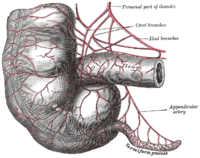
Changes for: cystic duct
- Deleted
- Added
Changes for: caecum
- Deleted
Changes for: subfornical organ
- Deleted
Changes for: symphysis
- Deleted
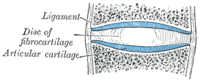
Changes for: fundus of stomach
- Deleted
Changes for: body of stomach
- Deleted
Changes for: cardia of stomach
- Deleted
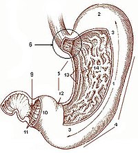
Changes for: lesser curvature of stomach
- Deleted
- Added
Changes for: greater curvature of stomach
- Deleted
- Added
Changes for: pyloric antrum
- Deleted
Changes for: pylorus
- Deleted
Changes for: synchondrosis
- Deleted
Changes for: macula of utricle of membranous labyrinth
- Deleted
Changes for: spiral organ of cochlea
- Deleted
- Added
Changes for: common bile duct
- Deleted
Changes for: common hepatic duct
- Deleted
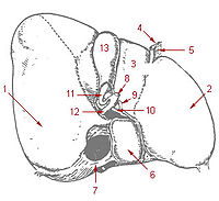
Changes for: fontanelle
- Deleted
Changes for: thoracic cavity
- Deleted
- Added
Changes for: visceral peritoneum
- Deleted
- Added
Changes for: basilar membrane of cochlea
- Deleted
- - basilar membrane of cochlea depicted by http://upload.wikimedia.org/wikipedia/commons/thumb/0/0c/Cochlea-crosssection.png/250px-Cochlea-crosssection.png
- - basilar membrane of cochlea depicted by http://upload.wikimedia.org/wikipedia/commons/thumb/9/9b/Organ_of_corti.svg/200px-Organ_of_corti.svg.png
- Added
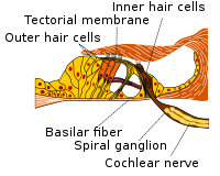
Changes for: right renal artery
- Deleted
- Added
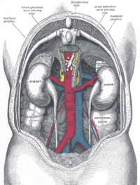
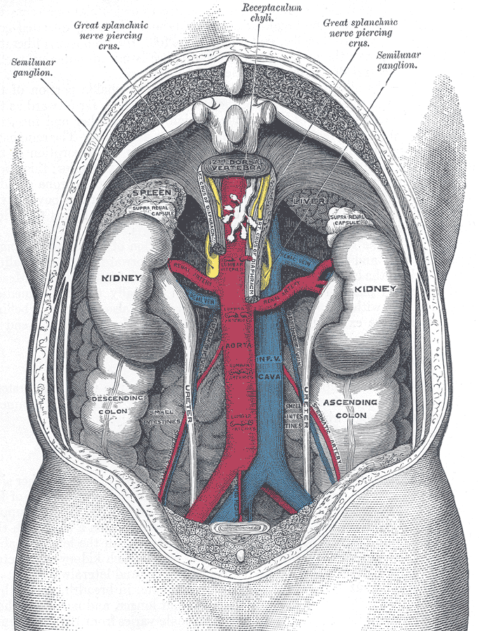
Changes for: testicular artery
- Deleted
Changes for: superior mesenteric artery
- Deleted
- Added
Changes for: renal artery
- Deleted
Changes for: prosomere
- Deleted
- - prosomere definition A neuromere that is part of the presumptive forebrain
- Added
- + prosomere definition A neuromere that is part of the presumptive forebrain { database cross reference=UBERON:cjm }
Changes for: midbrain neuromere
- Deleted
- - midbrain neuromere definition A neuromere that is part of the presumptive midbrain
- Added
- + midbrain neuromere definition A neuromere that is part of the presumptive midbrain { database cross reference=UBERON:cjm }
Changes for: spinal neuromere
- Deleted
- - spinal neuromere definition A neuromere that is part of the presumptive spinal cord
- Added
- + spinal neuromere definition A neuromere that is part of the presumptive spinal cord { database cross reference=UBERON:cjm }
Changes for: lateral palpebral artery
- Added
- + lateral palpebral artery definition The lateral palpebral arteries are small arteries which supply the eyelid. { database cross reference=http://en.wikipedia.org/wiki/Lateral_palpebral_arteries,Wikipedia:Lateral_palpebral_arteries }
Changes for: superior suprarenal artery
- Deleted
Changes for: middle colic artery
- Deleted
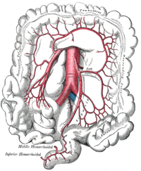
Changes for: splenic artery
- Deleted
Changes for: left gastric artery
- Deleted
Changes for: common iliac artery
- Deleted
Changes for: ovarian artery
- Deleted
Changes for: musculature of pelvic complex
- Added
- + musculature of pelvic complex comment Editor note: currently defined by location rather than attachment
Changes for: musculature of pectoral complex
- Added
- + musculature of pectoral complex comment Editor note: currently defined by location rather than attachment
Changes for: supracoracoideus muscle
- Added
- + supracoracoideus muscle SubClassOf muscle of shoulder
Changes for: teres minor muscle
- Added
- + teres minor muscle SubClassOf pectoral complex muscle
Changes for: pouch scale
- Deleted
- - pouch scale comment Definition reference: Weitzman SH, Menezes NA. 1998. Relationships of the tribes and genera of the Glandulocaudiinae (Ostariophysi: Characiformes: Characidae), with a description of a new genus. In: Malabarba LR, Reis RE, Vari RP, Lucena ZMS, Lucena CAS, editors. Phylogeny and Classification of Neotropical Fishes. Porto Alegre: EDIPUCRS. p 171-192.
- Added
- + pouch scale comment Definition reference: Weitzman SH, Menezes NA. 1998. Relationships of the tribes and genera of the Glandulocaudiinae (Ostariophysi: Characiformes: Characidae), with a description of a new genus. In: Malabarba LR, Reis RE, Vari RP, Lucena ZMS, Lucena CAS, editors. Phylogeny and Classification of Neotropical Fishes. Porto Alegre: EDIPUCRS. p 171-192.
Changes for: dermal scale focus
- Added
Changes for: cotylephore
- Deleted
- - cotylephore comment Definition reference: James Wetzel, John P. Wourms and John Friel 1997. Comparative morphology of cotylephores in Platystacus and Solenostomus: modifications of the integument for egg attachment in skin-brooding fishes. Environmental Biology of Fishes. Volume 50, Number 1 / September, 1997. Pages 13 - 25.
- Added
- + cotylephore comment Definition reference: James Wetzel, John P. Wourms and John Friel 1997. Comparative morphology of cotylephores in Platystacus and Solenostomus: modifications of the integument for egg attachment in skin-brooding fishes. Environmental Biology of Fishes. Volume 50, Number 1 / September, 1997. Pages 13 - 25.
Changes for: nephric duct
Changes for: intermediolateral nucleus
- Deleted
- - intermediolateral nucleus depicted by [http://upload.wikimedia.org/wikipedia/commons/thumb/c/c0/Medulla_spinalis_-Substantia_grisea-English.svg/200px-Medulla_spinalis-Substantia_grisea-English.svg.png](http://upload.wikimedia.org/wikipedia/commons/thumb/c/c0/Medulla_spinalis-Substantia_grisea-English.svg/200px-Medulla_spinalis-Substantia_grisea-_English.svg.png)
Changes for: left main bronchus
- Deleted
- Added
Changes for: right main bronchus
- Deleted
- Added
Changes for: lateral funiculus of spinal cord
- Deleted
- - lateral funiculus of spinal cord depicted by [http://upload.wikimedia.org/wikipedia/commons/thumb/d/dc/Medulla_spinalis_-Section-English.svg/200px-Medulla_spinalis-Section-English.svg.png](http://upload.wikimedia.org/wikipedia/commons/thumb/d/dc/Medulla_spinalis-Section-English.svg/200px-Medulla_spinalis-Section-_English.svg.png)
Changes for: middle lobe of right lung
- Deleted
- Added
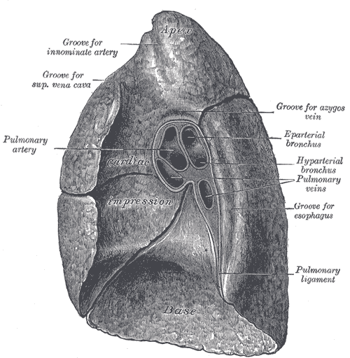
Changes for: respiratory bronchiole
- Deleted
Changes for: terminal bronchiole
- Deleted
Changes for: bronchiole
- Deleted
Changes for: ventral funiculus of spinal cord
- Deleted
- - ventral funiculus of spinal cord depicted by [http://upload.wikimedia.org/wikipedia/commons/thumb/d/dc/Medulla_spinalis_-Section-English.svg/200px-Medulla_spinalis-Section-English.svg.png](http://upload.wikimedia.org/wikipedia/commons/thumb/d/dc/Medulla_spinalis-Section-English.svg/200px-Medulla_spinalis-Section-_English.svg.png)
- Added
- + ventral funiculus of spinal cord depicted by [http://upload.wikimedia.org/wikipedia/commons/d/dc/Medulla_spinalis_-Section-English.svg](http://upload.wikimedia.org/wikipedia/commons/d/dc/Medulla_spinalis-Section-_English.svg)
Changes for: bronchus
- Deleted
Changes for: segmental bronchus
- Deleted
- Added
Changes for: lobar bronchus
- Deleted
Changes for: nucleus raphe pallidus
- Deleted
Changes for: fastigial nucleus
- Deleted
Changes for: nucleus raphe magnus
- Deleted
Changes for: superior cerebellar peduncle
- Deleted
Changes for: middle cerebellar peduncle
- Deleted
Changes for: left lung
- Deleted
- Added
Changes for: right lung
- Deleted
- Added
Changes for: endocardium
- Deleted
Changes for: inferior cerebellar peduncle
- Deleted
Changes for: area postrema
- Deleted
Changes for: gracile nucleus
- Deleted
Changes for: cerebellar nuclear complex
- Deleted
Changes for: anterior lobe of cerebellum
- Deleted
Changes for: vas deferens
- Deleted
Changes for: dentate nucleus
- Deleted
Changes for: tricuspid valve
- Deleted
Changes for: mitral valve
- Deleted
Changes for: respiratory system
- Deleted
Changes for: skin epidermis
- Deleted
- - skin epidermis definition The outer layer of the skin[WP]. cellular, multilayered epithelium derived from the ectoderm[ZFA]. { database cross reference=http://en.wikipedia.org/wiki/Epidermis_(skin) , database cross reference=ISBN10:0073040584 }
- - skin epidermis has exact synonym adult epidermis
- Added
- + skin epidermis definition The outer epithelial layer of the skin that is superficial to the dermis. { database cross reference=ISBN10:0073040584 , database cross reference=UBERON:cjm }
- + skin epidermis has broad synonym epidermis
Changes for: circulatory system
- Deleted
Changes for: subcommissural organ
- Deleted
Changes for: locus ceruleus
- Deleted
- Added
Changes for: superior salivatory nucleus
- Deleted
- Added
Changes for: nervous system
- Deleted
- Added
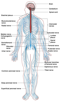
Changes for: pulmonary valve
- Deleted
Changes for: reticulotegmental nucleus
- Added
- + reticulotegmental nucleus database cross reference http://en.wikipedia.org/wiki/Tegmental_pontine_reticular_nucleus
- + reticulotegmental nucleus definition The tegmental pontine reticular nucleus (or pontine reticular nucleus of the tegmentum) is an area within the floor of the midbrain. This area is known to affect the cerebellum with its axonal projections. These efferent connections have been proven to project not only ipsilaterally, but also to decussate and project to the contralateral side of the vermis. It has also been shown that the projections from the tegmenti pontis to the cerebellar lobes are only crossed fibers. The n.r. tegmenti pontis also receives afferent axons from the cerebellum. This nucleus is known for its large amount of multipolar cells and its particularly reticular structure. The n.r. tegmenti pontis is topographically related to pontine nuclei (non-reticular), being just dorsal to them. The nucleus reticularis has been known to mediate eye movements, otherwise known as so-called saccadic movement. This makes sense concerning their connections as it would require a nucleus which receives and projects to the cerebellum to mediate that kind of complex circuitry. Also, behaviorally this makes sense as no one thinks about saccadic movements when scanning a room and the saccadic movements are not directly controlled by the cortex. The nuclei of the cerebellum are the most traditionally studied mostly because it is easy to see which nuclei degrade when the cerebellum is amputated. The neurons of the lateral reticular formation are very important for reflexes and the mediation of posture. It has been shown in cats that electrical stimulation of the reticular formation can make a standing cat lie down. Conversely if the cat is stimulated in an alternate spot it can make a lying cat stand. { database cross reference=http://en.wikipedia.org/wiki/Tegmental_pontine_reticular_nucleus,Wikipedia:Tegmental_pontine_reticular_nucleus }
Changes for: jejunum
- Deleted
Changes for: gall bladder
- Deleted
- Added
Changes for: basis pontis
- Added
- + basis pontis database cross reference http://en.wikipedia.org/wiki/Basilar_part_of_pons
- + basis pontis definition The basis pontis is the anterior portion of the pons. { database cross reference=http://en.wikipedia.org/wiki/Basilar_part_of_pons,Wikipedia:Basilar_part_of_pons }
Changes for: inferior olivary complex
- Deleted
Changes for: thymus lobule
- Added
- + thymus lobule definition A lobule that is part of a thymus. Divided into an outer cortex and inner medulla and separated from each other by connective tissue septa, but with the medullary tissue continuous from lobule to lobule.[ncit,modified]. { database cross reference=http://ncicb.nci.nih.gov/xml/owl/EVS/Thymic_Lobule , database cross reference=UBERON:cjm }
- + thymus lobule structure notes Lymphocytes, reticular cells and macrophages are found in the lobules
Changes for: cerebellar cortex
- Deleted
- Added
Changes for: superior olivary complex
- Deleted
Changes for: saliva-secreting gland
- Deleted
Changes for: pole of lens
- Deleted
- - pole of lens definition A central point or region on the anterior or posterior surface of the lens
- Added
- + pole of lens definition A central point or region on the anterior or posterior surface of the lens { database cross reference=UBERON:cjm }
Changes for: neural glomerulus
- Deleted
- Added
Changes for: neural tube
- Deleted
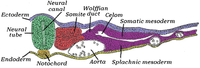
Changes for: yolk sac
- Deleted
Changes for: spleen
- Deleted
Changes for: liver
- Deleted
Changes for: small intestine
- Deleted
Changes for: skin of anterior chest
- Deleted
- - skin of anterior chest definition Zone of skin that is part of the anterior region of the chest
- Added
- + skin of anterior chest definition Zone of skin that is part of the anterior region of the chest { database cross reference=UBERON:cjm }
Changes for: hindlimb
- Deleted
Changes for: rectum
- Deleted
Changes for: tegmentum
Changes for: dento-alveolar joint
- Deleted
Changes for: sheath of Schwann
- Deleted
- Added
Changes for: sensory system
- Deleted
Changes for: megalopa stage
- Deleted
- - megalopa stage definition The crustacean post-larval stage of a crab
- Added
- + megalopa stage definition The crustacean post-larval stage of a crab { database cross reference=UBERON:cjm }
Changes for: calcareous tooth
- Deleted
- Added
Changes for: sacral vertebra
Changes for: incisor tooth
- Deleted
- Added
Changes for: axillary lymph node
- Deleted
Changes for: head of pancreas
- Deleted
Changes for: parotid main excretory duct
- Deleted
Changes for: ventral pancreatic duct
- Deleted
- Added
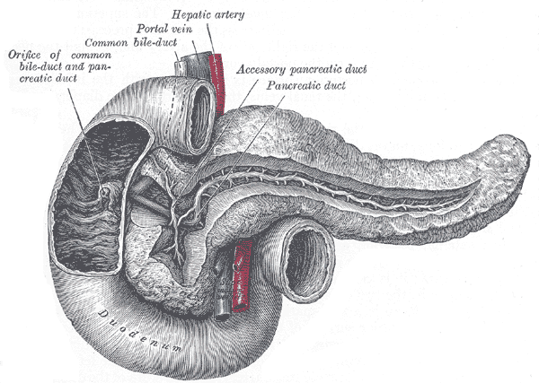
Changes for: vertebral arch joint
- Deleted
Changes for: intervertebral disk
- Deleted
- Added
Changes for: pericardial cavity
- Deleted
Changes for: posterior vena cava
- Deleted
Changes for: superficial cervical artery
- Added
- + superficial cervical artery EquivalentTo cervical artery and part of some superficial vasculature
- + superficial cervical artery SubClassOf part of some superficial vasculature
Changes for: external carotid artery
- Deleted
Changes for: pedicle of vertebra
- Deleted
Changes for: transverse process of vertebra
- Deleted
- Added
Changes for: neural spine
- Deleted
Changes for: bony vertebral centrum
- Deleted
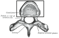
Changes for: blood-testis barrier
- Deleted
- - blood-testis barrier definition A barrier between the blood or haemolymphatic fluid and the testes.
- Added
- + blood-testis barrier definition A barrier between the blood or haemolymphatic fluid and the testes. { database cross reference=UBERON:cjm }
Changes for: feather rachis
- Deleted
- - feather rachis definition A central shaft of feather, with two vanes either side.
- Added
- + feather rachis definition A central shaft of feather, with two vanes either side. { database cross reference=UBERON:cjm }
Changes for: feather vane
- Deleted
- - feather vane definition Collection of feather barbs that branch from rachis
- Added
- + feather vane definition Collection of feather barbs that branch from rachis { database cross reference=UBERON:cjm }
Changes for: plastron
- Added
- + plastron has related synonym ventral armor
Changes for: carapace
- Added
- + carapace has related synonym dorsal armor
Changes for: turtle shell
- Added
- + turtle shell has related synonym armor
Changes for: styliform cartilage
- Deleted
- - styliform cartilage SubClassOf cartilage element
- - styliform cartilage SubClassOf mesoderm-derived structure
- - styliform cartilage SubClassOf part of some forelimb skeleton
- - styliform cartilage definition shaft of skeletal tissue that articulates with the lateral border of the carpus. { database cross reference=ISBN-10:0226313379 }
- Added
- + styliform cartilage EquivalentTo styliform element and composed primarily of some cartilage tissue
- + styliform cartilage SubClassOf composed primarily of some cartilage tissue
- + styliform cartilage SubClassOf forelimb cartilage element
- + styliform cartilage SubClassOf styliform element
- + styliform cartilage definition A styloform element in a cartilaginous state. { database cross reference=https://github.com/obophenotype/uberon/issues/695 }
- + styliform cartilage external definition shaft of skeletal tissue that articulates with the lateral border of the carpus. { source=ISBN-10:0226313379 }
Changes for: efferent lymphatic vessel
- Added
Changes for: subiculum
- Deleted
Changes for: trabecula of lymph node
- Deleted
- Added
Changes for: adenohypophysis
- Deleted
- Added
Changes for: capsule of lymph node
- Deleted
- Added
Changes for: median eminence of neurohypophysis
- Deleted
Changes for: neurohypophysis
- Deleted
- Added
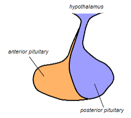
Changes for: lateral globus pallidus
- Deleted
Changes for: medial globus pallidus
- Deleted
Changes for: saphenous nerve
- Deleted
Changes for: internal surface of cranial base
- Deleted
- - internal surface of cranial base definition internal (endosteal) surface of the base of skull
- Added
- + internal surface of cranial base definition internal (endosteal) surface of the base of skull { database cross reference=UBERON:cjm }
Changes for: coronal suture
- Deleted
Changes for: prostate duct
Changes for: glottis
- Deleted
Changes for: trabecular bone tissue
- Deleted
- Added
Changes for: papillary muscle of heart
- Deleted
Changes for: acromion
- Deleted
Changes for: deltopectoral crest
- Deleted
Changes for: cochlear labyrinth
- Deleted
- Added
Changes for: frontal suture
- Deleted
- Added
Changes for: sagittal suture
- Deleted
Changes for: uterine artery
- Deleted
Changes for: abdominal fascia
Changes for: parietal pleura
- Deleted
Changes for: visceral pleura
- Deleted
Changes for: pleural cavity
- Deleted
Changes for: periamygdaloid cortex
- Added
Changes for: parietal serous pericardium
- Deleted
- Added
Changes for: pericardium
- Deleted
Changes for: transversus thoracis
- Deleted
- Added
Changes for: internal intercostal muscle
- Deleted
- Added
Changes for: pericardial sac
- Deleted
- Added
Changes for: cervical vertebra
- Deleted
Changes for: autonomic nervous system
- Deleted
Changes for: clitoris
- Deleted
Changes for: lumbar vertebra
- Deleted
- Added
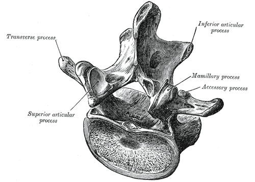
Changes for: basal ganglion
- Deleted
Changes for: hippocampal formation
- Deleted
- - hippocampal formation comment BTO:0000601 is placed here since it includes the DG. GO also includes dentate gyrus development as part of hippocampus development, so we assume when GO says “hippocampus” it means “hippocampal formation”. In ABA HPF = (.. subiculum) + HIP, HIP = DG + AH.
- Added
- + hippocampal formation external ontology notes BTO:0000601 is placed here since it includes the DG. GO also includes dentate gyrus development as part of hippocampus development, so we assume when GO says ‘hippocampus’ it means ‘hippocampal formation’. In ABA HPF = (.. subiculum) + HIP, HIP = DG + AH. { external ontology=BTO }
Changes for: fourth ventricle
- Deleted
Changes for: chest muscle
- Deleted
- - chest muscle SubClassOf part of some musculature of chest
Changes for: visceral serous pericardium
- Deleted
- Added
Changes for: cervical lymph node
- Deleted
- Added
Changes for: distal sesamoid impar ligament
- Deleted
- - distal sesamoid impar ligament definition A short wide fibrous structure that attaches proximally to the navicular (distal sesamoid) and distally to the palmar margin of the flexor surface of the distal phalanx
- Added
- + distal sesamoid impar ligament definition A short wide fibrous structure that attaches proximally to the navicular (distal sesamoid) and distally to the palmar margin of the flexor surface of the distal phalanx { database cross reference=UBERON:cjm }
Changes for: striatum
- Deleted
Changes for: pars tuberalis of adenohypophysis
- Deleted
Changes for: pars intermedia of adenohypophysis
- Deleted
Changes for: lateral hypothalamic area
- Deleted
- Added
Changes for: primary visual cortex
- Deleted
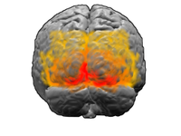
Changes for: patella
- Deleted
Changes for: ulnare
- Deleted
- Added
Changes for: inferior cervical ganglion
- Deleted
Changes for: cervicothoracic ganglion
- Deleted
- Added
Changes for: fungiform papilla
- Deleted
Changes for: ethmoid sinus
- Deleted
- Added
Changes for: endometrial gland
- Deleted
Changes for: decidua
- Deleted
Changes for: common plantar digital arteries
- Deleted
Changes for: dorsal metacarpal artery
- Deleted
- Added
Changes for: vesical vein
- Deleted
Changes for: hamstring muscle
- Deleted
- Added
Changes for: lymphoid system
- Deleted
- Added
Changes for: foliate papilla
- Deleted
Changes for: filiform papilla
- Deleted
Changes for: celiac lymph node
- Deleted
- Added
Changes for: mesenteric lymph node
- Added
Changes for: lesser trochanter
- Deleted
Changes for: round window of inner ear
- Deleted
Changes for: greater trochanter
- Deleted
Changes for: oval window
- Deleted
Changes for: zygomatic arch
- Deleted
Changes for: transverse folds of rectum
- Added
Changes for: ventral body wall
- Added
- + ventral body wall EquivalentTo body wall and part of some ventrum
- + ventral body wall database cross reference FMA:86934
- + ventral body wall has related synonym anterior body wall { database cross reference=FMA:86934 }
Changes for: communicating branch of spinal nerve
- Added
- + communicating branch of spinal nerve database cross reference http://en.wikipedia.org/wiki/Rami_communicans
- + communicating branch of spinal nerve definition Rami communicans (plural rami communicantes) is the term used for a nerve which connects two other nerves. When used without further definition, it almost always refers to a communicating branch between a spinal nerve and the sympathetic trunk. More specifically, it usually refers to one of the following: Gray ramus communicans White ramus communicans { database cross reference=http://en.wikipedia.org/wiki/Rami_communicans,Wikipedia:Rami_communicans }
Changes for: dorsal body wall
- Added
- + dorsal body wall EquivalentTo body wall and part of some dorsum
Changes for: otic ganglion
- Deleted
Changes for: pterygopalatine ganglion
- Deleted
Changes for: sympathetic afferent fiber
- Added
Changes for: fourth ventricle median aperture
- Deleted
Changes for: subarcuate fossa
Changes for: hyoid bone body
- Deleted
Changes for: avian uterine tube isthmus
- Added
Changes for: major sublingual duct
- Added
Changes for: conus arteriosus
- Deleted
Changes for: uterine tube infundibulum
- Deleted
Changes for: medulla oblongata anterior median fissure
- Added
Changes for: tubal tonsil
Changes for: Brodmann (1909) area 20
- Added
- + Brodmann (1909) area 20 database cross reference http://en.wikipedia.org/wiki/Brodmann_area_20
- + Brodmann (1909) area 20 definition Brodmann area 20, or BA20, is part of the temporal cortex in the human brain. The region encompasses most of the ventral temporal cortex, a region believed to play a part in high-level visual processing and recognition memory. This area is also known as inferior temporal area 20, and it refers to a subdivision of the cytoarchitecturally defined temporal region of cerebral cortex. In the human it corresponds approximately to the inferior temporal gyrus. Cytoarchitecturally it is bounded medially by the ectorhinal area 36 (H), laterally by the middle temporal area 21, rostrally by the temporopolar area 38 (H) and caudally by the occipitotemporal area 37 (H) (Brodmann-1909). { database cross reference=http://en.wikipedia.org/wiki/Brodmann_area_20,Wikipedia:Brodmann_area_20 }
Changes for: Brodmann (1909) area 19
- Added
- + Brodmann (1909) area 19 definition Brodmann area 19, or BA19, is part of the occipital lobe cortex in the human brain. Along with area 18, it comprises the extrastriate (or peristriate) cortex. In normally-sighted humans, extrastriate cortex is a visual association area, with feature-extracting, shape recognition, attentional, and multimodal integrating functions. This area is also known as peristriate area 19, and it refers to a subdivision of the cytoarchitecturally defined occipital region of cerebral cortex. In the human it is located in parts of the lingual gyrus, the cuneus, the lateral occipital gyrus (H) and the superior occipital gyrus (H) of the occipital lobe where it is bounded approximately by the parieto-occipital sulcus. Cytoarchitecturally it is bounded on one side by the parastriate area 18 which it surrounds. Rostrally it is bounded by the angular area 39 (H) and the occipitotemporal area 37 (H) (Brodmann-1909). { database cross reference=http://en.wikipedia.org/wiki/Brodmann_area_19,Wikipedia:Brodmann_area_19 }
Changes for: Brodmann (1909) area 25
- Added
- + Brodmann (1909) area 25 database cross reference http://en.wikipedia.org/wiki/Brodmann_area_25
- + Brodmann (1909) area 25 definition Brodmann area 25 (BA25) is an area in the cerebral cortex of the brain and delineated based on its cytoarchitectonic characteristics. It is also called the subgenual area or area subgenualis. It is the 25th ‘Brodmann area’ defined by Korbinian Brodmann (thus its name). BA25 is located in the cingulate region as a narrow band in the caudal portion of the subcallosal area adjacent to the paraterminal gyrus. The posterior parolfactory sulcus separates the paraterminal gyrus from BA25. Rostrally it is bound by the prefrontal area 11 of Brodmann. { database cross reference=http://en.wikipedia.org/wiki/Brodmann_area_25,Wikipedia:Brodmann_area_25 }
Changes for: thymus
- Deleted
Changes for: palatine tonsil
- Deleted
Changes for: metacarpal bone
- Deleted
- Added
Changes for: cricoid cartilage
- Deleted
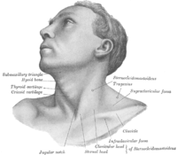
Changes for: anatomical line between outer ears
- Added
- + anatomical line between outer ears EquivalentTo anatomical line and connects some right external ear and connects some left external ear
Changes for: rete testis
- Deleted
Changes for: Brodmann (1909) area 10
- Added
- + Brodmann (1909) area 10 definition Brodmann area 10, or BA10, is part of the frontal cortex in the human brain. BA10 encompasses the most anterior part of the frontal cortex, known as the frontopolar region. This area is believed to play a part in strategic processes involved in memory retrieval and executive function. This area is also called frontopolar area 10, and it refers to a subdivision of the cytoarchitecturally defined frontal region of cerebral cortex. It occupies the most rostral portions of the superior frontal gyrus and the middle frontal gyrus. In humans, on the medial aspect of the hemisphere it is bounded ventrally by the superior rostral sulcus (H). It does not extend as far as the cingulate sulcus. Cytoarchitecturally it is bounded dorsally by the granular frontal area 9, caudally by the middle frontal area 46, and ventrally by the orbital area 47 and by the rostral area 12 or, in an early version of Brodmann’s cortical map (Brodmann-1909), the prefrontal Brodmann area 11-1909. { database cross reference=http://en.wikipedia.org/wiki/Brodmann_area_10,Wikipedia:Brodmann_area_10 }
Changes for: Brodmann (1909) area 14
- Added
- + Brodmann (1909) area 14 definition Brodmann Area 14 is one of Brodmann’s subdivisions of the cerebral cortex in the brain. It was defined by Brodmann in the guenon monkey. No equivalent structure exists in humans. { database cross reference=http://en.wikipedia.org/wiki/Brodmann_area_14,Wikipedia:Brodmann_area_14 }
Changes for: rectus abdominis muscle
- Deleted
- Added
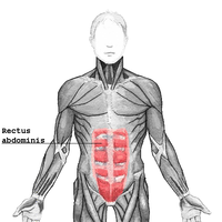
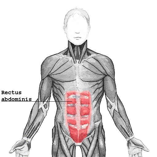
Changes for: supraspinatus muscle
- Deleted
- Added
Changes for: trapezius muscle
- Deleted
Changes for: pectoralis major
- Deleted
- Added
- + pectoralis major SubClassOf muscle of shoulder
Changes for: forelimb zeugopod
- Deleted
- Added
Changes for: pes
- Deleted
Changes for: pancreas primordium
- Deleted
- Added
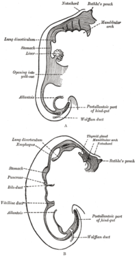
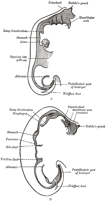
Changes for: pancreatic epithelial bud
- Deleted
- - pancreatic epithelial bud depicted by http://upload.wikimedia.org/wikipedia/commons/thumb/4/49/Suckale08FBS_fig1_pancreas_development.jpeg/300px-Suckale08FBS_fig1_pancreas_development.jpeg
- - pancreatic epithelial bud depicted by http://upload.wikimedia.org/wikipedia/commons/thumb/d/db/Gray982.png/200px-Gray982.png
- Added
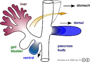
Changes for: dorsal pancreatic bud
- Deleted
- Added
Changes for: ventral pancreatic bud
- Deleted
- Added
Changes for: fibrous pericardium
- Deleted
Changes for: anatomical line between pupils
- Added
- + anatomical line between pupils EquivalentTo anatomical line and connects some left pupil and connects some right pupil
Changes for: pelvis
- Deleted
Changes for: perineum
- Deleted
Changes for: serous pericardium
- Deleted
Changes for: peritoneum
- Deleted
Changes for: sinoatrial node
- Deleted
- Added
Changes for: atrioventricular node
- Deleted
- Added
Changes for: atrioventricular bundle
- Deleted
- Added
Changes for: purkinje fiber
- Deleted
- Added
Changes for: meninx
- Deleted
- Added
Changes for: pia mater
- Deleted
- Added
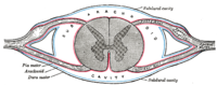
Changes for: pectoral flipper tubercle
- Deleted
- - pectoral flipper tubercle definition A bump or swelling adorining the head and leading edge of a pectoral flipper, associated with the multiple joints and terminal phalanges as found in the multiple phalanges of the humpback whale
- Added
- + pectoral flipper tubercle definition A bump or swelling adorining the head and leading edge of a pectoral flipper, associated with the multiple joints and terminal phalanges as found in the multiple phalanges of the humpback whale { database cross reference=UBERON:cjm }
Changes for: adrenal gland
- Deleted
- Added
Changes for: bulbo-urethral gland
- Deleted
Changes for: prostate gland
- Deleted
Changes for: arachnoid mater
- Deleted
Changes for: accessory nucleus of optic tract
- Added
- + accessory nucleus of optic tract EquivalentTo neural nucleus and part of some accessory optic system
- + accessory nucleus of optic tract SubClassOf part of some accessory optic system
- + accessory nucleus of optic tract taxon notes most vertebrates have at least one AON. Mammals have 4.[ISBN10:0471888893]
Changes for: plica semilunaris of conjunctiva
- Deleted
Changes for: chitinous tooth
- Deleted
- - chitinous tooth definition tooth-like structure composed of chitin.
- Added
- + chitinous tooth definition tooth-like structure composed of chitin. { database cross reference=UBERON:cjm }
Changes for: lesser omentum
- Deleted
Changes for: vomer
- Deleted
Changes for: talus
- Deleted
Changes for: maxilla
- Deleted
Changes for: nasolacrimal duct
- Deleted
Changes for: bile duct
- Deleted
Changes for: posterior choroidal artery
- Deleted
- - posterior choroidal artery SubClassOf head blood vessel
- - posterior choroidal artery SubClassOf systemic artery
- Added
Changes for: Brodmann (1909) area 43
- Added
- + Brodmann (1909) area 43 database cross reference http://en.wikipedia.org/wiki/Brodmann_area_43
- + Brodmann (1909) area 43 definition Brodmann area 43 is a subdivision of the cerebral cortex of the guenon defined on the basis of cytoarchitecture. It was described (but not labeled) on the map of cortical areas in Brodmann-1909, and it was regarded as cytoarchitecturally homologous to area 30 of Mauss in 1908 in the guenon and subcentral area 43 of the human (Brodmann-1909). The Vogts found no distinctive architectonic area of the corresponding location in the guenon (Vogts-1919). { database cross reference=http://en.wikipedia.org/wiki/Brodmann_area_43,Wikipedia:Brodmann_area_43 }
Changes for: deep auricular artery
- Added
- + deep auricular artery EquivalentTo auricular artery and part of some deep vasculature
- + deep auricular artery SubClassOf part of some deep vasculature
Changes for: posterior auricular artery
- Deleted
- Added
Changes for: pterygoid plexus
- Added
- + pterygoid plexus SubClassOf cranial blood vasculature
- + pterygoid plexus SubClassOf part of some head
Changes for: medial longitudinal fasciculus
- Deleted
Changes for: corpus callosum
- Deleted
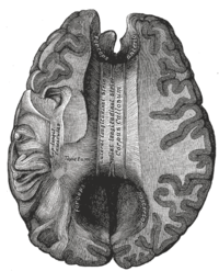
Changes for: submandibular duct
- Deleted
Changes for: pulmonary trunk
- Deleted
Changes for: nucleus H of ventral tegmentum
- Deleted
- Added
- + nucleus H of ventral tegmentum has broad synonym nucleus H
- + nucleus H of ventral tegmentum label nucleus H of ventral tegmentum
Changes for: cerebrocerebellum
Changes for: spinoocerebellum
Changes for: neural crest
- Deleted
- Added
Changes for: thoracic vertebra
- Deleted
- Added
Changes for: epicardium
- Deleted
- - epicardium definition a serous membrane that forms the innermost layer of the pericardium and the outer surface of the heart.
- - epicardium depicted by http://upload.wikimedia.org/wikipedia/commons/thumb/1/16/Gray968.png/200px-Gray968.png
- Added
- + epicardium definition a serous membrane that forms the innermost layer of the pericardium and the outer surface of the heart. { database cross reference=UBERON:cjm }
Changes for: myocardium
- Deleted
- Added
Changes for: midbrain tectum
- Deleted
Changes for: hippocampus fimbria
- Deleted
Changes for: white matter
- Deleted
- Added
Changes for: accessory nerve root
- Added
Changes for: notochord
- Deleted
Changes for: somite
- Deleted
Changes for: costal plate of carapace
- Added
- + costal plate of carapace has related synonym lateral plate of carapace { database cross reference=UBERON:cjm }
Changes for: hypothenar eminence
- Added
- + hypothenar eminence definition Hypothenar refers to a group of three muscles of the palm that control the motion of the little finger. The three muscles are: Abductor digiti minimi Flexor digiti minimi Opponens digiti minimi { database cross reference=http://en.wikipedia.org/wiki/Hypothenar_eminence,Wikipedia:Hypothenar_eminence }
Changes for: thenar eminence
- Added
- + thenar eminence definition A raised fleshy area on the palm of the hand near the base of the thumb. { database cross reference=http://medical-dictionary.thefreedictionary.com/Thenar+muscle }
Changes for: distal limb integumentary appendage
- Added
- + distal limb integumentary appendage has related synonym keratin plate
- + distal limb integumentary appendage has related synonym keratin sheath
Changes for: Mullerian duct
- Deleted
Changes for: skeletal muscle tissue of trunk
- Deleted
- - skeletal muscle tissue of trunk definition A portion of skeletal muscle tissue in the trunk.
- Added
- + skeletal muscle tissue of trunk definition A portion of skeletal muscle tissue in the trunk. { database cross reference=UBERON:cjm }
Changes for: stroma
- Deleted
- - stroma definition Connective, non-functional supportive framework of a biological cell, tissue, or organ. Contrast with parenchyma.
- Added
- + stroma definition Connective, non-functional supportive framework of a biological cell, tissue, or organ. Contrast with parenchyma. { database cross reference=UBERON:cjm }
Changes for: liver primordium
- Deleted
- - liver primordium SubClassOf developing epithelial placode
- Added
- + liver primordium SubClassOf primordium
- + liver primordium SubClassOf simple columnar epithelium
Changes for: mesometrium
Changes for: fallopian tube
- Deleted
Changes for: ctenius
- Added
- + ctenius SubClassOf skeletal element projection
- + ctenius SubClassOf structure with developmental contribution from neural crest
Changes for: parietomastoid suture
- Deleted
- - parietomastoid suture definition unites the mastoid process of the temporal with the region of the mastoid angle of the parietal[WP:Squamosal_suture].
- Added
- + parietomastoid suture definition unites the mastoid process of the temporal with the region of the mastoid angle of the parietal[WP:Squamosal_suture]. { database cross reference=UBERON:cjm }
Changes for: neural arch
- Deleted
Changes for: gonadal vein
- Deleted
Changes for: superficial external pudendal artery
- Added
- + superficial external pudendal artery EquivalentTo external pudendal artery and part of some superficial vasculature
- + superficial external pudendal artery SubClassOf part of some superficial vasculature
Changes for: deep external pudendal artery
- Deleted
- - deep external pudendal artery SubClassOf systemic artery
- Added
- + deep external pudendal artery EquivalentTo external pudendal artery and part of some deep vasculature
- + deep external pudendal artery SubClassOf deep femoral artery
- + deep external pudendal artery SubClassOf part of some deep vasculature
Changes for: roof of mouth
Changes for: outer epithelium
- Deleted
- Added
- + outer epithelium has exact synonym epidermis (sensu Metazoa)
- + outer epithelium has exact synonym epidermis { database cross reference=GO:0008544 }
Changes for: ceratoglossus
- Deleted
- Added
- + ceratoglossus SubClassOf ectoderm-derived structure
Changes for: dermal scale
- Deleted
- - dermal scale SubClassOf ectoderm-derived structure
- - dermal scale SubClassOf mesoderm-derived structure
- Added
- + dermal scale SubClassOf dermal skeletal element
Changes for: epidermal scale
- Added
- + epidermal scale taxon notes Archosaurian (crocodilian and avian) scales and turtle scutes develop from local epithelial thickenings called placodes, though the scales of lizards and snakes (Lepidosauria) develop without placodes as skin folds (Maderson, ‘65; Sawyer, ‘72; Harris et al., 2002; Cherepanov, ‘89, 2006; Alibardi and Minelli, 2014; Moustakas- Verho et al., 2014; we do not consider crocodilian osteoderms, which have been called scutes by some workers, to be homologous to the skin appendages we discuss)[PMID:25877335]
Changes for: future forebrain
Changes for: obsolete magnocellular superficial pretectal nucleus
- Deleted
- - magnocellular superficial pretectal nucleus SubClassOf diencephalic nucleus
- - magnocellular superficial pretectal nucleus SubClassOf part of some superficial pretectum
- - magnocellular superficial pretectal nucleus SubClassOf pretectal nucleus
- - magnocellular superficial pretectal nucleus label magnocellular superficial pretectal nucleus
- Added
- + obsolete magnocellular superficial pretectal nucleus deprecated true
- + obsolete magnocellular superficial pretectal nucleus label obsolete magnocellular superficial pretectal nucleus
Changes for: papillary duct
Changes for: renal interlobular vein
- Deleted
Changes for: inguinal part of abdomen
- Added
Changes for: posterior lobe of cerebellum
- Added
Changes for: pectoral appendage musculature
- Added
- + pectoral appendage musculature comment Editor note: currently defined by location rather than attachment
Changes for: pelvic appendage musculature
- Added
- + pelvic appendage musculature comment Editor note: currently defined by location rather than attachment
Changes for: appendage musculature
- Added
- + appendage musculature comment Editor note: currently defined by location rather than attachment
Changes for: deep artery of clitoris
- Added
- + deep artery of clitoris EquivalentTo artery and part of some deep vasculature and supplies some corpus cavernosum clitoridis
- + deep artery of clitoris SubClassOf part of some deep vasculature
Changes for: dorsal artery of clitoris
- Added
- + dorsal artery of clitoris database cross reference http://en.wikipedia.org/wiki/Dorsal_artery_of_clitoris
- + dorsal artery of clitoris definition The dorsal artery of clitoris is a branch of the internal pudendal artery. { database cross reference=http://en.wikipedia.org/wiki/Dorsal_artery_of_clitoris,Wikipedia:Dorsal_artery_of_clitoris }
Changes for: deep artery of penis
- Added
- + deep artery of penis EquivalentTo artery and part of some deep vasculature and supplies some corpus cavernosum penis
- + deep artery of penis SubClassOf part of some deep vasculature
Changes for: obsolete ventral nucleus
- Deleted
- - obsolete ventral nucleus comment removed axiom: part_of some habenula. Obsoleted because this term refererences a teleost specific region of the habenula, that has a homology relationship with a different portion of the habenula in mammals - that is currently not represented in Uberon. Can reinstate upon further classification of habenular structures.
- - obsolete ventral nucleus database cross reference ZFA:0000302
- Added
- + obsolete ventral nucleus consider ZFA:0000302
Changes for: Brodmann (1909) area 36
- Added
- + Brodmann (1909) area 36 definition Ectorhinal area 36 is a subdivision of the cytoarchitecturally defined temporal region of cerebral cortex. With its medial boundary corresponding approximately to the rhinal sulcus it is located primarily in the fusiform gyrus. Cytoarchitecturally it is bounded laterally and caudally by the inferior temporal area 20, medially by the perirhinal area 35 and rostrally by the temporopolar area 38 (H) (Brodmann-1909). Together with Brodmann area 35, it comprises the perirhinal cortex. { database cross reference=http://en.wikipedia.org/wiki/Brodmann_area_36,Wikipedia:Brodmann_area_36 }
Changes for: Brodmann (1909) area 35
- Added
- + Brodmann (1909) area 35 definition Brodmann area 35, together with Brodmann area 36, is most frequently referred to as perirhinal cortex. { database cross reference=http://en.wikipedia.org/wiki/Brodmann_area_35,Wikipedia:Brodmann_area_35 }
Changes for: palmar digital vein
- Deleted
Changes for: ectodermal placode
- Added
- + ectodermal placode database cross reference AEO:0000218
- + ectodermal placode has related synonym epithelial placode { database cross reference=AEO:0000112 }
Changes for: hemispheric lobule VIII
- Added
- + hemispheric lobule VIII database cross reference http://en.wikipedia.org/wiki/Biventer_lobule
- + hemispheric lobule VIII definition The biventer lobule (or biventral lobule) is triangular in shape; its apex points backward, and is joined by the gray band to the pyramid. The lateral border is separated from the inferior semilunar lobule by the postpyramidal fissure. The base is directed forward, and is on a line with the anterior border of the tonsil, and is separated from the flocculus by the postnodular fissure. { database cross reference=http://en.wikipedia.org/wiki/Biventer_lobule,Wikipedia:Biventer_lobule }
Changes for: posterior column of fornix
- Added
Changes for: mediastinum testis
- Deleted
Changes for: pontine cistern
Changes for: basal cistern
Changes for: primary nerve cord
- Deleted
- - nerve cord SubClassOf tissue
- - nerve cord definition A cluster of ➞neurons that is the most prominent longitudinally extending condensed part of a ➞nervous system. { database cross reference=http://www.ncbi.nlm.nih.gov/pubmed/21062451 }
- - nerve cord label nerve cord
- Added
- + primary nerve cord SubClassOf central nervous system cell part cluster
- + primary nerve cord SubClassOf develops from some ectoderm
- + primary nerve cord comment Usage notes: some species have multiple longitudinal nerve cords; this class refers to the most prominent one, which is dorsal in chordates and ventral in others.
- + primary nerve cord definition A cluster of neurons that is the most prominent longitudinally extending condensed part of a nervous system. { database cross reference=http://www.ncbi.nlm.nih.gov/pubmed/21062451 }
- + primary nerve cord has exact synonym nerve cord { database cross reference=BILA:0000136 }
- + primary nerve cord has exact synonym true nerve cord { database cross reference=UBERON:cjm }
- + primary nerve cord label primary nerve cord
Changes for: primary dorsal nerve cord
- Deleted
- - dorsal nerve cord SubClassOf develops from some ectoderm
- - dorsal nerve cord SubClassOf ectoderm-derived structure
- - dorsal nerve cord definition The dorsal tubular cord of nervous tissue above the notochord of a chordate that comprises or develops into the central nervous system[BTO]. The nerve cord of chordates develops dorsally in the body as a hollow tube above the notochord. In most species it differentiates in embryogeny into the brain anteriorly and spinal cord that runs through the trunk and tail. Together the brain and spinal cord are the central nervous system to which peripheral sensory and motor nerves connect[TOL]. The dorsal nerve cord is one of the embryonic features unique to chordates, along with a notochord, a post-anal tail and pharyngeal slits. The dorsal nerve cord is a hollow cord dorsal to the notochord. It is formed from a part of the ectoderm that rolls, forming the hollow tube, compared to other animal phyla, which have solid, ventral tubes. The dorsal nerve cord is later modified into the brain and spinal cord. Dorsal nerve cord is mainly found in phylum Vertebrata[WP]. { database cross reference=http://en.wikipedia.org/wiki/Dorsal_nerve_cord , database cross reference=http://tolweb.org/Chordata/2499 , database cross reference=BTO:0002329 }
- - dorsal nerve cord label dorsal nerve cord
- Added
- + primary dorsal nerve cord definition A nerve cord in the dorsal mid-line that is the most prominent nerve cord. { database cross reference=http://en.wikipedia.org/wiki/Dorsal_nerve_cord , database cross reference=UBERON:cjm }
- + primary dorsal nerve cord has exact synonym dorsal nerve cord { database cross reference=http://en.wikipedia.org/wiki/Dorsal_nerve_cord , database cross reference=http://tolweb.org/Chordata/2499 , database cross reference=BTO:0002329 }
- + primary dorsal nerve cord has exact synonym true dorsal nerve cord { database cross reference=UBERON:cjm }
- + primary dorsal nerve cord label primary dorsal nerve cord
Changes for: semicircular canal ampulla
- Deleted
Changes for: ganglionic eminence
- Deleted
- - ganglionic eminence comment Development notes: contributes significantly in building up the GABAergic cortical cell population[WP]
- Added
- + ganglionic eminence development notes contributes significantly in building up the GABAergic cortical cell population[WP]
Changes for: feather barbule
- Deleted
- - feather barbule definition Projection from feather bard.
- Added
- + feather barbule definition Projection from feather bard. { database cross reference=UBERON:cjm }
Changes for: golgi tendon organ
Changes for: mucosa of soft palate
- Added
Changes for: vena cava
- Deleted
- Added
Changes for: periorbital region
Changes for: cerebellum vermis lobule I
- Added
Changes for: cerebellum vermis lobule IX
- Added
- + cerebellum vermis lobule IX database cross reference http://en.wikipedia.org/wiki/Uvula_of_cerebellum
- + cerebellum vermis lobule IX definition The uvula (uvular lobe) forms a considerable portion of the inferior vermis; it is separated on either side from the tonsil by the sulcus valleculC&, at the bottom of which it is connected to the tonsil by a ridge of gray matter, indented on its surface by shallow furrows, and hence called the furrowed band. { database cross reference=http://en.wikipedia.org/wiki/Uvula_of_cerebellum,Wikipedia:Uvula_of_cerebellum }
Changes for: frontonasal prominence
- Deleted
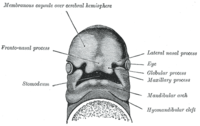
Changes for: lateral nasal prominence
- Deleted
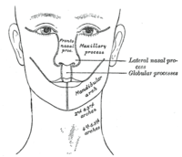
Changes for: medial nasal prominence
- Deleted
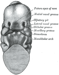
Changes for: quadrigeminal cistern
- Added
Changes for: renal collecting system
- Deleted
Changes for: crus of ear
- Deleted
- - crus of ear SubClassOf auricular cartilage
- Added
- + crus of ear SubClassOf cartilage of external ear
- + crus of ear SubClassOf structure with developmental contribution from neural crest
Changes for: muscle of auditory ossicle
- Added
Changes for: tympanic cavity
- Deleted
Changes for: optic vesicle
- Deleted
- Added
Changes for: skin nerve field
- Deleted
- - skin nerve field depicted by [http://upload.wikimedia.org/wikipedia/commons/thumb/9/97/Dermatomes_and_cutaneous_nerves_-posterior.png/230px-Dermatomes_and_cutaneous_nerves-posterior.png](http://upload.wikimedia.org/wikipedia/commons/thumb/9/97/Dermatomes_and_cutaneous_nerves-posterior.png/230px-Dermatomes_and_cutaneous_nerves-_posterior.png)
- Added
- + skin nerve field depicted by [http://upload.wikimedia.org/wikipedia/commons/9/97/Dermatomes_and_cutaneous_nerves_-posterior.png](http://upload.wikimedia.org/wikipedia/commons/9/97/Dermatomes_and_cutaneous_nerves-_posterior.png)
Changes for: obsolete parvocellular superficial pretectal nucleus
- Deleted
- - parvocellular superficial pretectal nucleus SubClassOf diencephalic nucleus
- - parvocellular superficial pretectal nucleus SubClassOf part of some superficial pretectum
- - parvocellular superficial pretectal nucleus SubClassOf pretectal nucleus
- - parvocellular superficial pretectal nucleus label parvocellular superficial pretectal nucleus
- Added
- + obsolete parvocellular superficial pretectal nucleus deprecated true
- + obsolete parvocellular superficial pretectal nucleus label obsolete parvocellular superficial pretectal nucleus
Changes for: inferior petrosal sinus
- Added
- + inferior petrosal sinus database cross reference http://en.wikipedia.org/wiki/Inferior_petrosal_sinus
- + inferior petrosal sinus definition The inferior petrosal sinus, within the human head, is an area beneath the brain, which allows blood veins to span the area, from the center of the head downward. It drains from the cavernous sinus (beneath the brain) to the sigmoid sinuses above the internal jugular vein. { database cross reference=http://en.wikipedia.org/wiki/Inferior_petrosal_sinus }
Changes for: superior thyroid artery
- Deleted
- Added
- + superior thyroid artery SubClassOf branch of external carotid artery
Changes for: superior epigastric artery
- Added
Changes for: inferior sagittal sinus
- Added
Changes for: superior thyroid vein
- Added
- + superior thyroid vein database cross reference http://en.wikipedia.org/wiki/Superior_thyroid_vein
- + superior thyroid vein definition The superior thyroid vein begins in the substance and on the surface of the thyroid gland, by tributaries corresponding with the branches of the superior thyroid artery, and ends in the upper part of the internal jugular vein. It receives the superior laryngeal and cricothyroid veins. { database cross reference=http://en.wikipedia.org/wiki/Superior_thyroid_vein,Wikipedia:Superior_thyroid_vein }
Changes for: inferior thyroid vein
- Added
- + inferior thyroid vein database cross reference http://en.wikipedia.org/wiki/Inferior_thyroid_veins
- + inferior thyroid vein definition The inferior thyroid veins two, frequently three or four, in number, arise in the venous plexus on the thyroid gland, communicating with the middle and superior thyroid veins. They form a plexus in front of the trachea, behind the Sternothyreoidei. From this plexus, a left vein descends and joins the left innominate trunk, and a right vein passes obliquely downward and to the right across the innominate artery to open into the right innominate vein, just at its junction with the superior vena cava; sometimes the right and left veins open by a common trunk in the latter situation. These veins receive esophageal tracheal, and inferior laryngeal veins, and are provided with valves at their terminations in the innominate veins. { database cross reference=http://en.wikipedia.org/wiki/Inferior_thyroid_veins,Wikipedia:Inferior_thyroid_veins }
Changes for: proximal radio-ulnar joint
- Added
- + proximal radio-ulnar joint database cross reference http://en.wikipedia.org/wiki/Proximal_radioulnar_articulation
- + proximal radio-ulnar joint definition The proximal radioulnar articulation (superior radioulnar joint) is a trochoid or pivot joint between the circumference of the head of the radius and the ring formed by the radial notch of the ulna and the annular ligament. { database cross reference=http://en.wikipedia.org/wiki/Proximal_radioulnar_articulation,Wikipedia:Proximal_radioulnar_articulation }
Changes for: distal radio-ulnar joint
- Added
- + distal radio-ulnar joint database cross reference http://en.wikipedia.org/wiki/Distal_radioulnar_articulation
- + distal radio-ulnar joint definition The Distal Radioulnar Articulation (inferior radioulnar joint) is a pivot-joint formed between the head of the ulna and the ulnar notch on the distal radius. { database cross reference=http://en.wikipedia.org/wiki/Distal_radioulnar_articulation,Wikipedia:Distal_radioulnar_articulation }
Changes for: lateral border of scapula
- Added
- + lateral border of scapula database cross reference http://en.wikipedia.org/wiki/Lateral_border_of_the_scapula
- + lateral border of scapula definition The lateral border (or axillary border, or margin) is the thickest of the three borders of the scapula. It begins above at the lower margin of the glenoid cavity, and inclines obliquely downward and backward to the inferior angle. Immediately below the glenoid cavity is a rough impression, the infraglenoid tuberosity, about 2.5 cm. in length, which gives origin to the long head of the Triceps brachii; in front of this is a longitudinal groove, which extends as far as the lower third of this border, and affords origin to part of the Subscapularis. The inferior third is thin and sharp, and serves for the attachment of a few fibers of the Teres major behind, and of the Subscapularis in front. { database cross reference=http://en.wikipedia.org/wiki/Lateral_border_of_the_scapula,Wikipedia:Lateral_border_of_the_scapula }
Changes for: Brodmann (1909) area 52
- Added
- + Brodmann (1909) area 52 database cross reference http://en.wikipedia.org/wiki/Brodmann_area_52
- + Brodmann (1909) area 52 definition Parainsular area 52 (H) is a subdivision of the cytoarchitecturally defined temporal region of cerebral cortex. It is located in the bank of the lateral sulcus on the dorsal surface of the temporal lobe. Its medial boundary corresponds approximately to the junction between the temporal lobe and the insula. Cytoarchitecturally it is bounded laterally by the anterior transverse temporal area 42 (H) (Brodmann-1909). { database cross reference=http://en.wikipedia.org/wiki/Brodmann_area_52,Wikipedia:Brodmann_area_52 }
Changes for: Brodmann (1909) area 44
- Added
- + Brodmann (1909) area 44 database cross reference http://en.wikipedia.org/wiki/Brodmann_area_44
- + Brodmann (1909) area 44 definition Brodmann area 44, or BA44, is part of the frontal cortex in the human brain. Situated just anterior to premotor cortex and on the lateral surface, inferior to BA9. This area is also known as pars opercularis (of the inferior frontal gyrus), and it refers to a subdivision of the cytoarchitecturally defined frontal region of cerebral cortex. In the human it corresponds approximately to the opercular part of inferior frontal gyrus (H). Thus, it is bounded caudally by the inferior precentral sulcus (H) and rostrally by the anterior ascending limb of lateral sulcus (H). It surrounds the diagonal sulcus (H). In the depth of the lateral sulcus it borders on the insula. Cytoarchitectonically it is bounded caudally and dorsally by the agranular frontal area 6, dorsally by the granular frontal area 9 and rostrally by the triangular area 45 (Brodmann-1909). { database cross reference=http://en.wikipedia.org/wiki/Brodmann_area_44,Wikipedia:Brodmann_area_44 }
Changes for: Brodmann (1909) area 38
- Added
- + Brodmann (1909) area 38 database cross reference http://en.wikipedia.org/wiki/Brodmann_area_38
- + Brodmann (1909) area 38 definition Brodmann area 38, also BA38 or temporopolar area 38 (H), is part of the temporal cortex in the human brain. BA 38 is at the anterior end of the temporal lobe, known as the temporal pole. BA38 is a subdivision of the cytoarchitecturally defined temporal region of cerebral cortex. It is located primarily in the most rostral portions of the superior temporal gyrus and the middle temporal gyrus. Cytoarchitecturally it is bounded caudally by the inferior temporal area 20, the middle temporal area 21, the superior temporal area 22 and the ectorhinal area 36 (Brodmann-1909). Cytoarchitectonic and chemoarchitectonic studies find that it contains at least seven subareas, one of which, TG, is unique to humans. ‘The functional significance of this area TG is not known, but it may bind complex, highly processed perceptual inputs to visceral emotional responses.’ This area is among the earliest affected by Alzheimer’s disease and the earliest involved at the start of temporal lobe seizures. [ { database cross reference=http://en.wikipedia.org/wiki/Brodmann_area_38,Wikipedia:Brodmann_area_38 }
Changes for: ventral accessory optic nucleus
- Deleted
- - ventral accessory optic nucleus SubClassOf pretectal nucleus
- Added
- + ventral accessory optic nucleus SubClassOf central pretectal nucleus
Changes for: trigeminothalamic tract
- Added
Changes for: loop of Henle ascending limb thin segment
- Deleted
Changes for: cardiac vein
Changes for: trigeminal sensory nucleus
- Deleted
- Added
Changes for: orbitofrontal cortex
- Added
Changes for: coronary sinus valve
- Added
Changes for: apex of heart
- Deleted
Changes for: mesentery
- Deleted
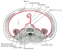
Changes for: interventricular septum
- Deleted
Changes for: skin of body
- Deleted
- Added
Changes for: kidney pyramid
- Deleted
Changes for: suprahyoid muscle
Changes for: left cardiac atrium
- Deleted
- Added
- + left cardiac atrium SubClassOf left cardiac chamber
Changes for: right cardiac atrium
- Deleted
- Added
- + right cardiac atrium SubClassOf right cardiac chamber
Changes for: hypodermis
- Deleted
- Added
Changes for: viscus
- Deleted
- - viscus comment general anatomical term in FMA. Note that we place the MA class here temporarily, although properly systems should be distinguished from organs
- - viscus database cross reference EMAPA:16245
- - viscus definition An organ that is located within the body cavity (or in its extension, in the scrotum); it consists of organ parts that are embryologically derived from endoderm, splanchnic mesoderm or intermediate mesoderm; together with other organs, the viscus constitutes the respiratory, gastrointestinal, urinary, reproductive and immune systems, or is the central organ of the cardiovascular system. Examples: heart, lung, esophagus, kidney, ovary, spleen. { database cross reference=http://en.wikipedia.org/wiki/Viscus , database cross reference=BTO:0001491 }
- Added
- + viscus definition An organ that is located within the body cavity (or in its extension, in the scrotum); it consists of organ parts that are embryologically derived from endoderm, splanchnic mesoderm or intermediate mesoderm; together with other organs, the viscus constitutes the respiratory, gastrointestinal, urinary, reproductive and immune systems, or is the central organ of the cardiovascular system. Examples: heart, lung, esophagus, kidney, ovary, spleen. { database cross reference=http://en.wikipedia.org/wiki/Viscus , database cross reference=https://github.com/obophenotype/mouse-anatomy-ontology/issues/14 , database cross reference=BTO:0001491 }
- + viscus external ontology notes general anatomical term in FMA. Note that we place the MA class here temporarily, although properly systems should be distinguished from organs. { external ontology=FMA }
Changes for: superior pancreaticoduodenal artery
- Deleted
Changes for: lateral thoracic vein
- Deleted
Changes for: atrioventricular canal
- Deleted
Changes for: heart left ventricle
- Deleted
- Added
- + heart left ventricle SubClassOf left cardiac chamber
Changes for: ductus venosus
- Deleted
Changes for: heart right ventricle
- Deleted
- Added
- + heart right ventricle SubClassOf right cardiac chamber
Changes for: sensory canal
- Deleted
- - sensory canal SubClassOf regional part of nervous system
- Added
- + sensory canal SubClassOf multi-tissue structure
Changes for: superficial parotid lymph node
- Added
- + superficial parotid lymph node EquivalentTo parotid lymph node and part of some superficial vasculature
- + superficial parotid lymph node SubClassOf part of some superficial vasculature
- + superficial parotid lymph node database cross reference http://en.wikipedia.org/wiki/Superficial_parotid_lymph_nodes
Changes for: superficial intraparotid lymph node
- Added
- + superficial intraparotid lymph node EquivalentTo intraparotid lymph node and part of some superficial vasculature
- + superficial intraparotid lymph node SubClassOf part of some superficial vasculature
Changes for: future trigeminal ganglion
- Deleted
- - future trigeminal ganglion SubClassOf develops from some trigeminal placode
- Added
- + future trigeminal ganglion SubClassOf transformation of some trigeminal placode complex
- + future trigeminal ganglion comment See: https://github.com/obophenotype/mouse-anatomy-ontology/issues/13 and https://github.com/obophenotype/uberon/issues/693 – consider merging into trigeminal ganglion, as this complicates the model
Changes for: superficial lymph node
- Added
- + superficial lymph node EquivalentTo lymph node and part of some superficial vasculature
- + superficial lymph node SubClassOf part of some superficial vasculature
Changes for: deep lymph node
- Added
- + deep lymph node EquivalentTo lymph node and part of some deep vasculature
- + deep lymph node SubClassOf part of some deep vasculature
Changes for: internal iliac lymph node
- Added
- + internal iliac lymph node database cross reference http://en.wikipedia.org/wiki/Internal_iliac_lymph_nodes
- + internal iliac lymph node definition The internal iliac lymph nodes (or hypogastric) surround the internal iliac artery and its branches (the hypogastric vessels), and receive the lymphatics corresponding to the distribution of the branches of it, i. e. , they receive lymphatics from all the pelvic viscera, from the deeper parts of the perineum, including the membranous and cavernous portions of the urethra, and from the buttock and back of the thigh. It does not receive lymph from the ovary, testis, or superior half of the rectum; the gonads drain to the paraaortic lymph nodes, while the superior half of the rectum drains to the pararectal lymph nodes. { database cross reference=http://en.wikipedia.org/wiki/Internal_iliac_lymph_nodes,Wikipedia:Internal_iliac_lymph_nodes }
Changes for: vasa hyaloidea propria
- Deleted
- - vasa hyaloidea propria definition The vasa hyaloidea propria and tunica vasculosa lentis are small branches of the hyaloid artery which fill the primary vitreous
- Added
- + vasa hyaloidea propria definition The vasa hyaloidea propria and tunica vasculosa lentis are small branches of the hyaloid artery which fill the primary vitreous { database cross reference=UBERON:cjm }
Changes for: facial lymph node
Changes for: stapedial artery
- Added
Changes for: endocardial cushion
- Deleted
- Added
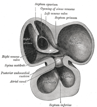
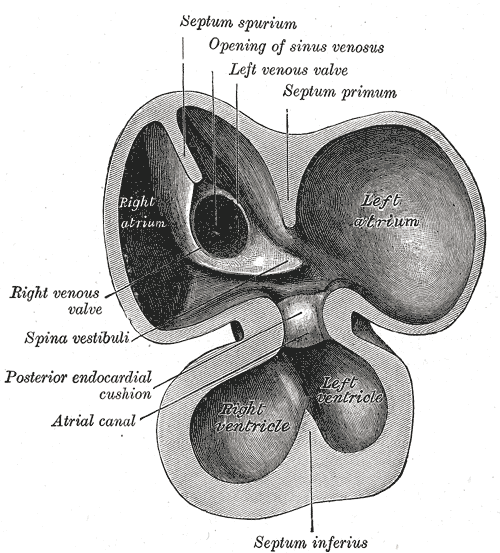
Changes for: sinus venosus
- Deleted
Changes for: common cardinal vein
- Deleted
Changes for: femoral artery
- Deleted
Changes for: umbilical vein
- Deleted
Changes for: urachus
- Deleted
- Added
Changes for: zona glomerulosa of adrenal gland
- Deleted
Changes for: ciliary ganglion
- Deleted
Changes for: inferior suprarenal artery
- Deleted
Changes for: zona fasciculata of adrenal gland
- Deleted
Changes for: zona reticularis of adrenal gland
- Deleted
Changes for: bronchial artery
- Deleted
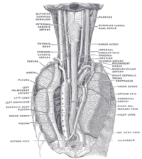
Changes for: cuneate nucleus
- Deleted
Changes for: thyroid gland
- Deleted
- Added
Changes for: vasculature
- Deleted
- Added
Changes for: nipple
- Deleted
Changes for: suprachiasmatic nucleus
- Deleted
Changes for: medial preoptic nucleus
- Deleted
- Added
Changes for: areola
- Deleted
- Added
Changes for: substantia nigra
- Deleted
Changes for: cerebellum
- Deleted
- Added
Changes for: hindbrain
- Deleted
Changes for: stratum corneum of epidermis
- Deleted
- Added
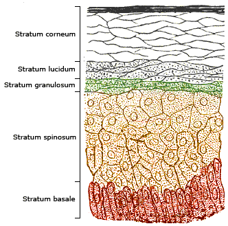
Changes for: occipital lobe
- Deleted
Changes for: internal carotid nerve plexus
- Deleted
- Added
Changes for: gray matter
- Deleted
- Added
Changes for: synovial membrane of synovial joint
- Deleted
- Added
Changes for: accessory XI nerve
- Deleted
- Added
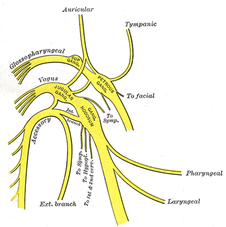
Changes for: portal vein
- Deleted
- Added
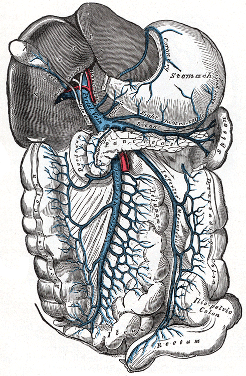
Changes for: pulmonary vein
- Deleted
Changes for: kidney capsule
- Deleted
- Added
Changes for: inferior hypogastric nerve plexus
- Deleted
Changes for: superior hypogastric nerve plexus
- Deleted
Changes for: pulmonary artery
- Deleted
- Added
Changes for: thoracodorsal artery
- Deleted
Changes for: celiac nerve plexus
- Deleted
Changes for: cardiac nerve plexus
- Deleted
Changes for: epithelium of otic placode
- Added
- + epithelium of otic placode SubClassOf ecto-epithelium
Changes for: obsolete skeletal muscle tissue of tongue
- Deleted
- - skeletal muscle tissue of tongue EquivalentTo skeletal muscle tissue and part of some tongue
- - skeletal muscle tissue of tongue SubClassOf contributes to morphology of some musculature
- - skeletal muscle tissue of tongue SubClassOf contributes to morphology of some tongue
- - skeletal muscle tissue of tongue SubClassOf develops from some head somite
- - skeletal muscle tissue of tongue SubClassOf mixed ectoderm/mesoderm/endoderm-derived structure
- - skeletal muscle tissue of tongue SubClassOf part of some tongue muscle
- - skeletal muscle tissue of tongue SubClassOf part of some tongue musculature
- - skeletal muscle tissue of tongue SubClassOf part of some tongue
- - skeletal muscle tissue of tongue SubClassOf skeletal muscle tissue
- - skeletal muscle tissue of tongue database cross reference EHDAA2:0002065
- - skeletal muscle tissue of tongue database cross reference EHDAA:9146
- - skeletal muscle tissue of tongue database cross reference EMAPA:17883
- - skeletal muscle tissue of tongue database cross reference MA:0001596
- - skeletal muscle tissue of tongue database cross reference UMLS:C1710437 { source=ncithesaurus:Tongue_Skeletal_Muscle_Tissue }
- - skeletal muscle tissue of tongue database cross reference VHOG:0001390
- - skeletal muscle tissue of tongue database cross reference http://linkedlifedata.com/resource/umls/id/C1710437
- - skeletal muscle tissue of tongue database cross reference http://ncicb.nci.nih.gov/xml/owl/EVS/Tongue_Skeletal_Muscle_Tissue
- - skeletal muscle tissue of tongue definition A portion of skeletal muscle tissue that is part of a tongue [Automatically generated definition]. { database cross reference=OBOL:automatic }
- - skeletal muscle tissue of tongue editor note consider merging with ‘muscle of tongue’, depending on outcome of issue 331
- - skeletal muscle tissue of tongue has exact synonym skeletal muscle tissue of tongue { database cross reference=OBOL:automatic }
- - skeletal muscle tissue of tongue has exact synonym tongue skeletal muscle tissue { database cross reference=OBOL:automatic }
- - skeletal muscle tissue of tongue has exact synonym tongue skeletal muscle { database cross reference=OBOL:automatic }
- - skeletal muscle tissue of tongue homology notes The lamprey head contains another group of muscles, the epi- and hypo-branchial muscles (EBM and HBM), derivatives of anterior trunk myotomes. (…) The origin and the migration pattern of HBM precursors are very similar to that of the gnathostome MPP, especially to that of the tongue muscle precursors. Other evidence of homology of lamprey HBM to the gnathostome tongue muscle is that HBM is innervated by the nerve termed the hypoglossal nerve based on its morphological position associated with the head/trunk interface. (…) The HBM-specific expression of the LampPax3/7 gene is consistent with the homology of this muscle to the gnathostome tongue muscle, or to the hypobranchial series as a whole (including the infrahyoid and possibly the diaphragm in mammals).[well established][VHOG] { date retrieved=2012-09-17 , external class=VHOG:0001390 , ontology=VHOG , source=http://bgee.unil.ch/ , source=DOI:10.1002/dvdy.20587 Kusakabe R, Kuratani S, Evolution and developmental patterning of the vertebrate skeletal muscles: perspectives from the lamprey. Developmental Dynamics (2005) }
- - skeletal muscle tissue of tongue in subset pheno slim
- - skeletal muscle tissue of tongue label skeletal muscle tissue of tongue
- Added
- + obsolete skeletal muscle tissue of tongue comment Merged as it was equivalent to parent - see https://github.com/obophenotype/uberon/issues/331
- + obsolete skeletal muscle tissue of tongue deprecated true
- + obsolete skeletal muscle tissue of tongue label obsolete skeletal muscle tissue of tongue
- + obsolete skeletal muscle tissue of tongue term replaced by UBERON:0000378
Changes for: median eye
- Deleted
- - median eye definition A median eye is an ➞eye that is connected to a paired or unpaired median anterior ➞neuropil of the ➞syncerebrum by one or several median eye nerves. { database cross reference=http://www.ncbi.nlm.nih.gov/pubmed/21062451 }
- Added
- + median eye definition A median eye is an eye that is connected to a paired or unpaired median anterior neuropil of the syncerebrum by one or several median eye nerves. { database cross reference=http://www.ncbi.nlm.nih.gov/pubmed/21062451 }
Changes for: neural lobe of neurohypophysis
- Deleted
- Added
Changes for: splanchnocranium
- Deleted
- Added
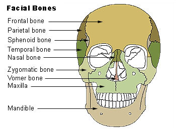
Changes for: pharyngeal gill precursor
- Deleted
- - pharyngeal gill precursor definition A primordium or bud that develops into a gill.
- Added
- + pharyngeal gill precursor definition A primordium or bud that develops into a gill. { database cross reference=UBERON:cjm }
Changes for: distal phalanx
- Deleted
Changes for: primitive streak
- Deleted
- Added
- + primitive streak SubClassOf simple columnar epithelium
Changes for: Descemet’s membrane
- Added
Changes for: pharyngeal arch artery
- Deleted
Changes for: papula
Changes for: pharyngeal arch 1
- Deleted
- - pharyngeal arch 1 definition The first of the series of pharyngeal arches that develop into jaw bones or their homologs, and their associated nerves and arteries
- - pharyngeal arch 1 depicted by http://upload.wikimedia.org/wikipedia/commons/thumb/2/2d/Gray48.png/200px-Gray48.png
- Added
- + pharyngeal arch 1 definition The first of the series of pharyngeal arches that develop into jaw bones or their homologs, and their associated nerves and arteries { database cross reference=UBERON:cjm }
Changes for: tetrapod frontal bone
- Deleted
- Added
Changes for: blastula stage
- Deleted
Changes for: lung bud
- Deleted
- Added
Changes for: olivary body
- Deleted
Changes for: cranial nerve nucleus
- Deleted
Changes for: epineurium
- Deleted
Changes for: perineurium
- Deleted
Changes for: anal canal
- Deleted
Changes for: theca externa
- Deleted
- Added
Changes for: mouth
- Deleted
- Added
Changes for: primitive urogenital sinus
- Deleted
Changes for: haemolymphatic fluid
- Deleted
- - haemolymphatic fluid definition Circulating fluid that is part of the hemolymphoid system. Blood, lymph, interstitial fluid or its analogs.
- Added
- + haemolymphatic fluid definition Circulating fluid that is part of the hemolymphoid system. Blood, lymph, interstitial fluid or its analogs. { database cross reference=UBERON:cjm }
Changes for: pus
- Deleted
- Added
Changes for: duct of epididymis
- Deleted
- - duct of epididymis definition Any duct branching of of the main epididymis duct.
- Added
- + duct of epididymis definition Any duct branching of of the main epididymis duct. { database cross reference=UBERON:cjm }
Changes for: outer spiral sulcus
- Added
Changes for: inner spiral sulcus
- Added
Changes for: skeletal ligament
- Deleted
- Added
Changes for: cranial suspensory ligament
- Deleted
- - cranial suspensory ligament definition Connects the cranial tip of the future gonad to the dorsocranial abdominal cavity.
- Added
- + cranial suspensory ligament definition Connects the cranial tip of the future gonad to the dorsocranial abdominal cavity. { database cross reference=UBERON:cjm }
Changes for: epithelium of mucosa
- Deleted
- - epithelium of mucosa database cross reference EMAPA:16564
- - epithelium of mucosa database cross reference EMAPA:16708
- - epithelium of mucosa database cross reference EMAPA:16835
- - epithelium of mucosa database cross reference EMAPA:17038
- - epithelium of mucosa database cross reference EMAPA:17044
- - epithelium of mucosa database cross reference EMAPA:17181
- - epithelium of mucosa database cross reference EMAPA:17196
- - epithelium of mucosa database cross reference EMAPA:17625
- - epithelium of mucosa database cross reference EMAPA:17629
- - epithelium of mucosa database cross reference EMAPA:17633
- - epithelium of mucosa database cross reference EMAPA:17888
- - epithelium of mucosa database cross reference EMAPA:17894
- - epithelium of mucosa database cross reference EMAPA:17898
- - epithelium of mucosa database cross reference EMAPA:17902
- - epithelium of mucosa database cross reference EMAPA:18259
- - epithelium of mucosa database cross reference EMAPA:18263
- - epithelium of mucosa database cross reference EMAPA:18269
- - epithelium of mucosa database cross reference EMAPA:18668
- - epithelium of mucosa database cross reference EMAPA:18835
- - epithelium of mucosa database cross reference EMAPA:18941
- - epithelium of mucosa database cross reference EMAPA:19078
Changes for: distal epiphysis of radius
- Added
Changes for: distal epiphysis of tibia
- Added
Changes for: rugal fold of stomach
Changes for: third trochanter
- Added
- + third trochanter definition The upper part of the gluteal tuberosity is often elongated into a roughened crest, on which a more or less well-marked, rounded tubercle, the third trochanter, is occasionally developed. { database cross reference=http://en.wikipedia.org/wiki/Third_trochanter,Wikipedia:Third_trochanter }
Changes for: subclavius
- Added
- + subclavius SubClassOf muscle of shoulder
Changes for: proximal epiphysis of fibula
- Added
Changes for: proximal epiphysis of tibia
- Added
Changes for: mesenchyme of pinna
- Added
- + mesenchyme of pinna external ontology notes EMAPA divides this spatially into condensation + rest, and temporally into mesenchyme and cartilage condensation { external ontology=EMAPA }
Changes for: proximal epiphysis of radius
- Added
Changes for: musculature of lower limb
- Added
- + musculature of lower limb comment Editor note: currently defined by location rather than attachment
Changes for: musculature of upper limb
- Added
- + musculature of upper limb comment Editor note: currently defined by location rather than attachment
Changes for: hair root
- Deleted
- Added
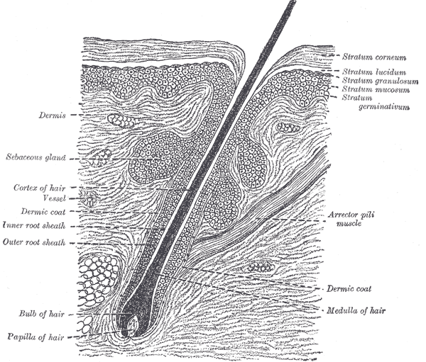
Changes for: musculature of limb
- Added
- + musculature of limb comment Editor note: currently defined by location rather than attachment
Changes for: musculature of manus
- Added
- + musculature of manus comment Editor note: currently defined by location rather than attachment
Changes for: musculature of perineum
- Added
- + musculature of perineum comment Editor note: currently defined by location rather than attachment
Changes for: musculature of pes
- Added
- + musculature of pes comment Editor note: currently defined by location rather than attachment
Changes for: breast
- Deleted
Changes for: subarachnoid space
- Deleted
- Added
Changes for: obsolete musculature of thorax
- Deleted
- - musculature of chest EquivalentTo musculature and part of some chest
- - musculature of chest SubClassOf musculature of thorax
- - musculature of chest SubClassOf part of some chest
- - musculature of chest database cross reference FMA:74779
- - musculature of chest definition Any collection of muscles that is part of a chest [Automatically generated definition]. { database cross reference=OBOL:automatic }
- - musculature of chest has exact synonym chest musculature { database cross reference=FMA:74779 }
- - musculature of chest has exact synonym set of muscles of chest { database cross reference=FMA:74779 }
- - musculature of chest label musculature of chest
- Added
- + obsolete musculature of thorax consider FMA:74779
- + obsolete musculature of thorax consider UBERON:0004464
- + obsolete musculature of thorax deprecated true
- + obsolete musculature of thorax has exact synonym musculature of chest { database cross reference=FMA:74779 }
- + obsolete musculature of thorax has related synonym chest musculature { database cross reference=FMA:74779 }
- + obsolete musculature of thorax has related synonym set of muscles of chest { database cross reference=FMA:74779 }
- + obsolete musculature of thorax label obsolete musculature of thorax
Changes for: cytotrophoblast
- Deleted
Changes for: musculature of trunk
- Added
- + musculature of trunk comment Editor note: currently defined by location rather than attachment
Changes for: musculature of larynx
- Deleted
- - musculature of larynx definition Any collection of muscles that is part of a larynx [Automatically generated definition]. { database cross reference=OBOL:automatic }
- Added
- + musculature of larynx comment Editor note: currently defined by location rather than attachment
- + musculature of larynx database cross reference http://en.wikipedia.org/wiki/Muscles_of_larynx
- + musculature of larynx definition The collection of muscles that are part of the larynx. { database cross reference=UBERON:cjm }
Changes for: obsolete tongue musculature
- Deleted
- - tongue musculature EquivalentTo musculature and part of some tongue
- - tongue musculature SubClassOf ectoderm-derived structure
- - tongue musculature SubClassOf endoderm-derived structure
- - tongue musculature SubClassOf musculature of head
- - tongue musculature SubClassOf part of some tongue
- - tongue musculature database cross reference FMA:71289
- - tongue musculature definition Any collection of muscles that is part of a tongue [Automatically generated definition]. { database cross reference=OBOL:automatic }
- - tongue musculature label tongue musculature
- Added
- + obsolete tongue musculature consider FMA:71289
- + obsolete tongue musculature deprecated true
- + obsolete tongue musculature label obsolete tongue musculature
Changes for: musculature of shoulder
- Deleted
- - musculature of shoulder EquivalentTo musculature and part of some shoulder
- - musculature of shoulder SubClassOf musculature of pectoral complex
- - musculature of shoulder SubClassOf part of some shoulder
- - musculature of shoulder definition Any collection of muscles that is part of a shoulder [Automatically generated definition]. { database cross reference=OBOL:automatic }
- Added
- + musculature of shoulder EquivalentTo musculature and attaches to some shoulder bone
- + musculature of shoulder SubClassOf attaches to some shoulder bone
- + musculature of shoulder SubClassOf has part some part of some shoulder
- + musculature of shoulder SubClassOf musculature
- + musculature of shoulder definition The collection of muscles that form attachments to the shoulder skeleton. { database cross reference=UBERON:cjm }
Changes for: musculature of hip
- Deleted
- - musculature of hip EquivalentTo musculature and part of some hip
- - musculature of hip SubClassOf musculature of pelvic complex
- - musculature of hip SubClassOf part of some hip
- - musculature of hip definition Any collection of muscles that is part of a hip [Automatically generated definition]. { database cross reference=OBOL:automatic }
- Added
- + musculature of hip EquivalentTo musculature and attaches to some bone of hip region
- + musculature of hip SubClassOf attaches to some bone of hip region
- + musculature of hip SubClassOf has part some part of some hip
- + musculature of hip SubClassOf musculature
- + musculature of hip definition The collection of muscles that form attachments to the hip skeleton. { database cross reference=UBERON:cjm }
Changes for: blastula
- Deleted
- Added
Changes for: adductor longus
- Deleted
Changes for: musculature of thorax
- Deleted
- - musculature of thorax external ontology notes BTO and FMA do not mean precisely the same thing here { external ontology=BTO }
- Added
- + musculature of thorax external ontology notes BTO and FMA do not mean precisely the same thing here. BTO thorax muscle is a pectoral muscle or diaphragm { external ontology=BTO }
Changes for: esophageal sphincter
- Deleted
- - esophageal sphincter definition A sphincter that is part of an esophagus.
- Added
- + esophageal sphincter definition A sphincter that is part of an esophagus. { database cross reference=UBERON:cjm }
Changes for: tarsal region
- Deleted
Changes for: metacarpus region
- Deleted
- Added
Changes for: carpal region
- Deleted
- Added
Changes for: entire sense organ system
- Deleted
- - entire sense organ system definition Sum of all sensory systems in an organism.
- Added
- + entire sense organ system definition Sum of all sensory systems in an organism. { database cross reference=UBERON:cjm }
Changes for: tetrapod parietal bone
- Deleted
Changes for: hilum of kidney
Changes for: flocculonodular lobe
- Deleted
Changes for: facial motor nucleus
- Deleted
Changes for: substantia innominata
- Deleted
Changes for: medial lemniscus
- Deleted
Changes for: posterior nucleus of thalamus
- Added
- + posterior nucleus of thalamus EquivalentTo nucleus of brain and part of some posterior nuclear complex of thalamus
Changes for: submedial nucleus of thalamus
- Deleted
- - submedial nucleus of thalamus SubClassOf nucleus of dorsal thalamus
- Added
Changes for: thoracic spinal cord
- Deleted
- Added
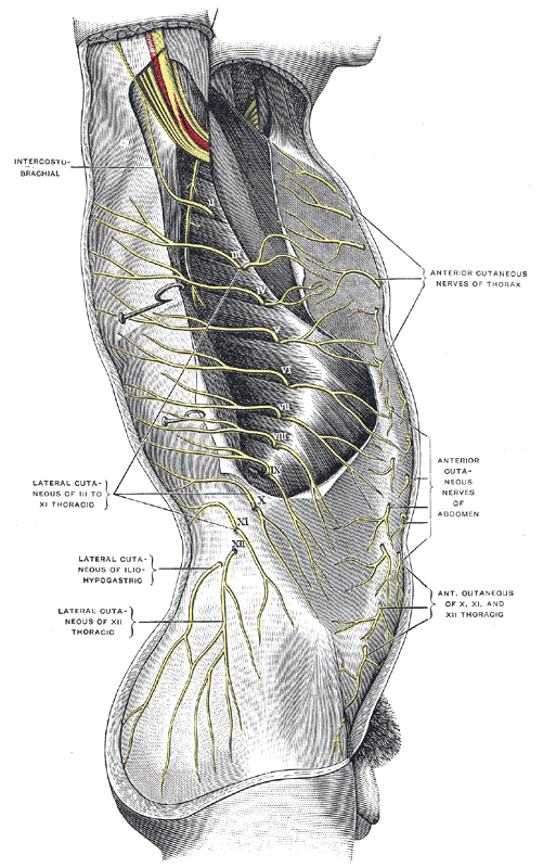
Changes for: central dorsal nucleus of thalamus
- Deleted
- - central dorsal nucleus database cross reference MBA:432
- - central dorsal nucleus has exact synonym nucleus circularis { database cross reference=NIFSTD:birnlex_956 }
- - central dorsal nucleus label central dorsal nucleus
- Added
- + central dorsal nucleus of thalamus has broad synonym circular nucleus { database cross reference=NeuroNames:1853 }
- + central dorsal nucleus of thalamus has exact synonym central dorsal nucleus { database cross reference=http://uri.neuinfo.org/nif/nifstd/birnlex , database cross reference=FMA:62169 }
- + central dorsal nucleus of thalamus has related synonym nucleus circularis { database cross reference=NIFSTD:birnlex_956 , has synonym type=indicates that a synonym is used in an inconsistent or confusing way, typically between species }
- + central dorsal nucleus of thalamus label central dorsal nucleus of thalamus
Changes for: central lobule
- Deleted
Changes for: subcallosal area
- Deleted
Changes for: stria terminalis
- Deleted
Changes for: cingulate cortex
- Deleted
Changes for: brachium of inferior colliculus
- Deleted
Changes for: pontine tegmentum
- Deleted
Changes for: proper plantar digital artery
- Deleted
- Added
Changes for: external capsule of telencephalon
- Deleted
Changes for: cribriform plate
- Deleted
Changes for: sacral region of vertebral column
- Added
Changes for: inferior horn of the lateral ventricle
- Added
Changes for: cuneus cortex
Changes for: precuneus cortex
Changes for: internal arcuate fiber bundle
- Added
- + internal arcuate fiber bundle database cross reference http://en.wikipedia.org/wiki/Internal_arcuate_fibers
- + internal arcuate fiber bundle definition Internal arcuate fibers are the axons of second-order neurons contained within the gracile and cuneate nuclei of the medulla oblongata. These fibers cross from one side of the medulla to the other to form the medial lemniscus. Part of the dorsal column-medial lemniscus system (second neuron), the internal arcuate fibers are important for relaying the sensation of fine touch and proprioception to the thalamus and ultimately to the cerebral cortex. { database cross reference=http://en.wikipedia.org/wiki/Internal_arcuate_fibers,Wikipedia:Internal_arcuate_fibers }
Changes for: perirhinal cortex
- Deleted
- - perirhinal cortex SubClassOf part of some insula
- Added
- + perirhinal cortex external ontology notes in MA classified as part of insular cortex, but this is contested by MM - https://support.crbs.ucsd.edu/browse/NIF-9830 { external ontology=MA }
Changes for: stria medullaris
- Added
- + stria medullaris database cross reference http://en.wikipedia.org/wiki/Stria_medullaris_of_thalamus
- + stria medullaris external ontology notes this is placed under ‘epithalamus related’ in MBA
Changes for: superior parietal cortex
- Added
Changes for: basal nuclear complex
- Deleted
- - basal nuclear complex SubClassOf part of some forebrain
- - basal nuclear complex SubClassOf regional part of brain
- Added
- + basal nuclear complex SubClassOf gray matter of forebrain
- + basal nuclear complex SubClassOf nuclear complex of neuraxis
Changes for: uncinate fasciculus
- Deleted
Changes for: central gray substance of midbrain
- Deleted
Changes for: ear vesicle
- Deleted
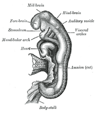
Changes for: olfactory placode
- Deleted
- - olfactory placode SubClassOf developing epithelial placode
- - olfactory placode SubClassOf ecto-epithelium
- - olfactory placode SubClassOf embryonic tissue
Changes for: pharyngeal arch 2
- Deleted
Changes for: axial mesoderm
- Deleted
Changes for: intermediate mesoderm
- Deleted
Changes for: floor plate
- Deleted
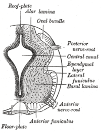
Changes for: paraxial mesoderm
- Deleted
Changes for: epibranchial placode
- Deleted
- - epibranchial placode SubClassOf developing epithelial placode
- - epibranchial placode SubClassOf ecto-epithelium
- - epibranchial placode SubClassOf embryonic tissue
- Added
Changes for: trigeminal placode complex
- Deleted
- - trigeminal placode comment Fig9-5 of ISBN10:0471888893 calls this the profundus placode; XAO says ‘profundal and the trigeminal ganglia are separate distally but fused at their proximal end…’
- - trigeminal placode definition The ectodermal rudiment of the trigeminal ganglion. { database cross reference=VHOG:0000109 }
- - trigeminal placode label trigeminal placode
- Added
- + trigeminal placode complex SubClassOf has potential to developmentally contribute to some somatic sensory system
- + trigeminal placode complex definition A neurogenic placode that arises at the level of the midbrain-hindbrain boundary and develops into a trigeminal ganglion. { database cross reference=http://www.ncbi.nlm.nih.gov/pubmed/22512454 , database cross reference=https://github.com/obophenotype/uberon/issues/693 , database cross reference=UBERON:cjm }
- + trigeminal placode complex has exact synonym trigeminal placode { database cross reference=EHDAA2:0004209 , database cross reference=VHOG:0000109 , database cross reference=XAO:0000225 , database cross reference=ZFA:0000162 }
- + trigeminal placode complex label trigeminal placode complex
- + trigeminal placode complex terminology notes Consider naming this ‘trigeminal placode complex’. Also, Fig9-5 of ISBN10:0471888893 calls this the profundus placode; XAO says ‘profundal and the trigeminal ganglia are separate distally but fused at their proximal end…’
Changes for: mesonephric duct
- Deleted
Changes for: lateral plate mesoderm
- Deleted
Changes for: myotome
- Deleted
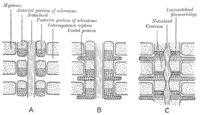
Changes for: sclerotome
- Deleted
Changes for: optic stalk
- Deleted
Changes for: infraorbital artery
- Deleted
Changes for: thoracic vertebra 12
- Added
- + thoracic vertebra 12 definition The twelfth thoracic vertebra counting from the top down. Note that members of this class are not necessarily homologous[ncit,modified]. { database cross reference=http://ncicb.nci.nih.gov/xml/owl/EVS/T12_Vertebra , database cross reference=UBERON:cjm }
Changes for: thoracic vertebra 9
- Added
- + thoracic vertebra 9 definition The ninth thoracic vertebra counting from the top down. Note that members of this class are not necessarily homologous[ncit,modified]. { database cross reference=http://ncicb.nci.nih.gov/xml/owl/EVS/T9_Vertebra , database cross reference=UBERON:cjm }
Changes for: thoracic vertebra 7
- Added
- + thoracic vertebra 7 definition The seventh thoracic vertebra counting from the top down. Note that members of this class are not necessarily homologous[ncit,modified]. { database cross reference=http://ncicb.nci.nih.gov/xml/owl/EVS/T7_Vertebra , database cross reference=UBERON:cjm }
Changes for: thoracic vertebra 11
- Added
- + thoracic vertebra 11 definition The eleventh thoracic vertebra counting from the top down. Note that members of this class are not necessarily homologous[ncit,modified]. { database cross reference=http://ncicb.nci.nih.gov/xml/owl/EVS/T11_Vertebra , database cross reference=UBERON:cjm }
Changes for: thoracic vertebra 10
- Added
- + thoracic vertebra 10 definition The tenth thoracic vertebra counting from the top down. Note that members of this class are not necessarily homologous[ncit,modified]. { database cross reference=http://ncicb.nci.nih.gov/xml/owl/EVS/T10_Vertebra , database cross reference=UBERON:cjm }
Changes for: mesenchyme
- Deleted
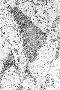
Changes for: thoracic vertebra 6
- Added
- + thoracic vertebra 6 definition The sixth thoracic vertebra counting from the top down. Note that members of this class are not necessarily homologous[ncit,modified]. { database cross reference=http://ncicb.nci.nih.gov/xml/owl/EVS/T6_Vertebra , database cross reference=UBERON:cjm }
Changes for: thoracic vertebra 5
- Added
- + thoracic vertebra 5 definition The fifth thoracic vertebra counting from the top down. Note that members of this class are not necessarily homologous[ncit,modified]. { database cross reference=http://ncicb.nci.nih.gov/xml/owl/EVS/T5_Vertebra , database cross reference=UBERON:cjm }
Changes for: thoracic vertebra 2
- Added
- + thoracic vertebra 2 definition The second thoracic vertebra counting from the top down. Note that members of this class are not necessarily homologous[ncit,modified]. { database cross reference=http://ncicb.nci.nih.gov/xml/owl/EVS/T2_Vertebra , database cross reference=UBERON:cjm }
Changes for: thoracic vertebra 3
- Added
- + thoracic vertebra 3 definition The third thoracic vertebra counting from the top down. Note that members of this class are not necessarily homologous[ncit,modified]. { database cross reference=http://ncicb.nci.nih.gov/xml/owl/EVS/T3_Vertebra , database cross reference=UBERON:cjm }
Changes for: thoracic vertebra 1
- Added
- + thoracic vertebra 1 definition The first thoracic vertebra counting from the top down. Note that members of this class are not necessarily homologous[ncit,modified]. { database cross reference=http://ncicb.nci.nih.gov/xml/owl/EVS/T1_Vertebra , database cross reference=UBERON:cjm }
Changes for: thoracic vertebra 4
- Added
- + thoracic vertebra 4 definition The fourth thoracic vertebra counting from the top down. Note that members of this class are not necessarily homologous[ncit,modified]. { database cross reference=http://ncicb.nci.nih.gov/xml/owl/EVS/T4_Vertebra , database cross reference=UBERON:cjm }
Changes for: lumbar vertebra 4
- Added
- + lumbar vertebra 4 definition The fourth lumbar vertebra counting from the top down. Note that members of this class are not necessarily homologous[ncit,modified]. { database cross reference=http://ncicb.nci.nih.gov/xml/owl/EVS/L4_Vertebra , database cross reference=UBERON:cjm }
Changes for: obsolete developing epithelial placode
- Deleted
- - developing epithelial placode EquivalentTo simple columnar epithelium and has potential to develop into some anatomical structure
- - developing epithelial placode SubClassOf has potential to develop into some anatomical structure
- - developing epithelial placode SubClassOf primordium
- - developing epithelial placode SubClassOf simple columnar epithelium
- - developing epithelial placode database cross reference AEO:0000218
- - developing epithelial placode definition A domain of columnar epithelium in an embryo that represents the early stage of the development of the epithelial component of a new anatomical structure. { database cross reference=AEO:0000218 , database cross reference=JB:DEF }
- - developing epithelial placode has exact synonym epithelial placode { database cross reference=AEO:0000112 }
- - developing epithelial placode label developing epithelial placode
- Added
- + obsolete developing epithelial placode definition OBSOLETE: A domain of columnar epithelium in an embryo that represents the early stage of the development of the epithelial component of a new anatomical structure. { database cross reference=AEO:0000218 , database cross reference=JB:DEF }
- + obsolete developing epithelial placode deprecated true
- + obsolete developing epithelial placode label obsolete developing epithelial placode
- + obsolete developing epithelial placode term replaced by UBERON:0005085
Changes for: lumbar vertebra 5
- Added
- + lumbar vertebra 5 definition The fifth lumbar vertebra counting from the top down. Note that members of this class are not necessarily homologous[ncit,modified]. { database cross reference=http://ncicb.nci.nih.gov/xml/owl/EVS/L5_Vertebra , database cross reference=UBERON:cjm }
Changes for: mammalian cervical vertebra 5
- Added
- + mammalian cervical vertebra 5 definition The fifth of the seven cervical vertebrae[ncit,modified]. { database cross reference=http://ncicb.nci.nih.gov/xml/owl/EVS/C5_Vertebra , database cross reference=UBERON:cjm }
Changes for: mammalian cervical vertebra 6
- Added
- + mammalian cervical vertebra 6 definition The sixth of the seven cervical vertebrae[ncit,modified]. { database cross reference=http://ncicb.nci.nih.gov/xml/owl/EVS/C6_Vertebra , database cross reference=UBERON:cjm }
Changes for: mammalian cervical vertebra 7
- Added
- + mammalian cervical vertebra 7 database cross reference http://en.wikipedia.org/wiki/Vertebra_prominens
- + mammalian cervical vertebra 7 definition Vertebra prominens is the proper name for the seventh cervical vertebra. The most distinctive characteristic of this vertebra is the existence of a long and prominent spinous process which is palpable from the skin surface, hence the name. This spinous process is thick, nearly horizontal in direction, not bifurcated, but terminating in a tubercle to which the lower end of the ligamentum nuchae is attached. The seventh cervical vertebra (C7) has the most prominent spinous process only in about 70% of people; in the remainder, either C6 or T1 (the first thoracic vertebra) will be the most prominent. The transverse processes are of considerable size, their posterior roots are large and prominent, while the anterior are small and faintly marked; the upper surface of each has usually a shallow sulcus for the eighth spinal nerve, and its extremity seldom presents more than a trace of bifurcation. The foramen transversarium may be as large as that in the other cervical vertebrae, but is generally smaller on one or both sides; occasionally it is double, sometimes it is absent. On the left side it occasionally gives passage to the vertebral artery; more frequently the vertebral vein traverses it on both sides; but the usual arrangement is for both artery and vein to pass in front of the transverse process, and not through the foramen. Sometimes the anterior root of the transverse process attains a large size and exists as a separate bone, which is known as a cervical rib. { database cross reference=http://en.wikipedia.org/wiki/Vertebra_prominens,Wikipedia:Vertebra_prominens }
Changes for: lumbar vertebra 1
- Added
- + lumbar vertebra 1 definition The first lumbar vertebra counting from the top down. Note that members of this class are not necessarily homologous[ncit,modified]. { database cross reference=http://ncicb.nci.nih.gov/xml/owl/EVS/L1_Vertebra , database cross reference=UBERON:cjm }
Changes for: lumbar vertebra 2
- Added
- + lumbar vertebra 2 definition The second lumbar vertebra counting from the top down. Note that members of this class are not necessarily homologous[ncit,modified]. { database cross reference=http://ncicb.nci.nih.gov/xml/owl/EVS/L2_Vertebra , database cross reference=UBERON:cjm }
Changes for: lumbar vertebra 3
- Added
- + lumbar vertebra 3 definition The third lumbar vertebra counting from the top down. Note that members of this class are not necessarily homologous[ncit,modified]. { database cross reference=http://ncicb.nci.nih.gov/xml/owl/EVS/L3_Vertebra , database cross reference=UBERON:cjm }
Changes for: rib 12
- Deleted
Changes for: mammalian cervical vertebra 4
- Added
- + mammalian cervical vertebra 4 definition The fourth of the seven cervical vertebrae[ncit,modified]. { database cross reference=http://ncicb.nci.nih.gov/xml/owl/EVS/C4_Vertebra , database cross reference=UBERON:cjm }
Changes for: mammalian cervical vertebra 3
- Added
- + mammalian cervical vertebra 3 definition The third of the seven cervical vertebrae[ncit,modified]. { database cross reference=http://ncicb.nci.nih.gov/xml/owl/EVS/C3_Vertebra , database cross reference=UBERON:cjm }
Changes for: rib 10
- Deleted
Changes for: rib 9
- Added
- + rib 9 definition The ninth rib counting from the top of the rib cage down. Note that members of this class are not necessarily homologous[ncit,modified]. { database cross reference=http://ncicb.nci.nih.gov/xml/owl/EVS/Rib_9 , database cross reference=UBERON:cjm }
Changes for: rib 7
- Added
- + rib 7 definition The seventh rib counting from the top of the rib cage down. Note that members of this class are not necessarily homologous[ncit,modified]. { database cross reference=http://ncicb.nci.nih.gov/xml/owl/EVS/Rib_7 , database cross reference=UBERON:cjm }
Changes for: rib 6
- Added
- + rib 6 definition The sixth rib counting from the top of the rib cage down. Note that members of this class are not necessarily homologous[ncit,modified]. { database cross reference=http://ncicb.nci.nih.gov/xml/owl/EVS/Rib_6 , database cross reference=UBERON:cjm }
Changes for: rib 5
- Added
- + rib 5 definition The fifth rib counting from the top of the rib cage down. Note that members of this class are not necessarily homologous[ncit,modified]. { database cross reference=http://ncicb.nci.nih.gov/xml/owl/EVS/Rib_5 , database cross reference=UBERON:cjm }
Changes for: rib 4
- Added
- + rib 4 definition The fourth rib counting from the top of the rib cage down. Note that members of this class are not necessarily homologous[ncit,modified]. { database cross reference=http://ncicb.nci.nih.gov/xml/owl/EVS/Rib_4 , database cross reference=UBERON:cjm }
Changes for: rib 3
- Added
- + rib 3 definition The third rib counting from the top of the rib cage down. Note that members of this class are not necessarily homologous[ncit,modified]. { database cross reference=http://ncicb.nci.nih.gov/xml/owl/EVS/Rib_3 , database cross reference=UBERON:cjm }
Changes for: rib 1
- Deleted
- - rib 1 definition The first rib of the rib cage
- - rib 1 depicted by http://upload.wikimedia.org/wikipedia/commons/thumb/f/fa/Gray124.png/200px-Gray124.png
- Added
- + rib 1 definition The first rib of the rib cage { database cross reference=UBERON:cjm }
Changes for: rib 2
- Deleted
- - rib 2 definition The second rib of the rib cage
- - rib 2 depicted by http://upload.wikimedia.org/wikipedia/commons/thumb/a/a5/Gray125.png/200px-Gray125.png
- Added
- + rib 2 definition The second rib of the rib cage { database cross reference=UBERON:cjm }
Changes for: habenular nucleus
- Deleted
- - habenular nucleus SubClassOf habenular trigone
- - habenular nucleus definition The gray matter of the habenula, composed of a small-celled medial and a larger-celled lateral habenular nucleus; both nuclei receive fibres from basal forebrain regions (septum, basal nucleus, lateral preoptic nucleus); the lateral habenular nucleus receives an additional projection from the medial segment of the globus pallidus. Both nuclei project by way of the retroflex fasciculus to the interpeduncular nucleus and a medial zone of the midbrain tegmentum. { database cross reference=BTO:0003685 }
- Added
- + habenular nucleus EquivalentTo neural nucleus and part of some habenula
- + habenular nucleus SubClassOf nucleus of thalamus
- + habenular nucleus database cross reference http://en.wikipedia.org/wiki/Habenular_nuclei
- + habenular nucleus definition Either of the two neural nuclei within the habenula. { database cross reference=UBERON:cjm }
Changes for: trachea
- Deleted
- Added
Changes for: costo-cervical trunk
- Deleted
Changes for: naso-frontal vein
- Added
- + naso-frontal vein database cross reference http://en.wikipedia.org/wiki/Nasofrontal_vein
- + naso-frontal vein definition The nasofrontal vein is a vein in the eye which drains to the superior ophthalmic vein. { database cross reference=http://en.wikipedia.org/wiki/Nasofrontal_vein,Wikipedia:Nasofrontal_vein }
Changes for: raphe nuclei
- Deleted
Changes for: middle temporal area
- Deleted
Changes for: body of fornix
- Deleted
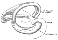
Changes for: corona radiata of neuraxis
- Deleted
Changes for: posterior horn lateral ventricle
- Added
Changes for: gyrus rectus
- Added
- + gyrus rectus database cross reference http://en.wikipedia.org/wiki/Straight_gyrus
- + gyrus rectus definition The portion of the frontal lobe medial to the medial orbital gyrus is named the gyrus rectus (or straight gyrus), and is continuous with the superior frontal gyrus on the medial surface. A specific function for the gyrus rectus has not yet been elucidated. { database cross reference=http://en.wikipedia.org/wiki/Straight_gyrus,Wikipedia:Straight_gyrus }
Changes for: scapula spine
- Deleted
Changes for: mandible condylar process
- Deleted
- Added
Changes for: temporal process of zygomatic bone
- Deleted
Changes for: zygomatic process of temporal bone
- Deleted
Changes for: tertiary ovarian follicle
- Deleted
- Added
Changes for: lamina propria
- Deleted
- Added
Changes for: follicular fluid
- Deleted
Changes for: ganglion
- Deleted
- - ganglion external definition A ganglion is a cluster of ➞neurons. It is part of a ➞nervous system. It may include ➞glial cells. The neurons are arranged in a specific constellation: neuronal somata are concentrated at the surface, thus forming a cell cortex, and ➞neurites are concentrated in the centre of the ganglion to form the ➞neuropil. A ganglion is a distinct unit but several ganglia may be anterio-posteriorly joined by ➞connectives or transversally by ➞commissures { source=http://www.ncbi.nlm.nih.gov/pubmed/21062451 }
- Added
- + ganglion external definition A ganglion is a cluster of neurons. It is part of a nervous system. It may include glial cells. The neurons are arranged in a specific constellation: neuronal somata are concentrated at the surface, thus forming a cell cortex, and neurites are concentrated in the centre of the ganglion to form the neuropil. A ganglion is a distinct unit but several ganglia may be anterio-posteriorly joined by connectives or transversally by commissures { source=http://www.ncbi.nlm.nih.gov/pubmed/21062451 }
Changes for: dorsal root ganglion
- Deleted
Changes for: tendon
- Deleted
Changes for: odontode scale
- Deleted
- - odontode scale SubClassOf dermal skeletal element
- - odontode scale external definition Multi-tissue structure composed of enameloid, dentine surrounding a pulp cavity, and bone of attachment anchoring the element into the dermis.[VSAO] { date retrieved=2012-08-14 , external class=VSAO:0000079 , ontology=VSAO , source=PSPUB:0000170 }
- - odontode scale has exact synonym dermal denticle
- Added
Changes for: preductal region of aortic arch
- Deleted
- - preductal region of aortic arch definition Area of aorta proximal to the ductus arteriosus.
- Added
- + preductal region of aortic arch definition Area of aorta proximal to the ductus arteriosus. { database cross reference=UBERON:cjm }
Changes for: postductal region of aortic arch
- Deleted
- - postductal region of aortic arch definition Area of aorta distal to the ductus arteriosus.
- Added
- + postductal region of aortic arch definition Area of aorta distal to the ductus arteriosus. { database cross reference=UBERON:cjm }
Changes for: juxtaductal region of aortic arch
- Deleted
- - juxtaductal region of aortic arch definition Area of aorta at the area of insertion to the ductus arteriosus.
- Added
- + juxtaductal region of aortic arch definition Area of aorta at the area of insertion to the ductus arteriosus. { database cross reference=UBERON:cjm }
Changes for: parasympathetic nervous system
- Deleted
Changes for: peripheral nervous system
- Deleted
Changes for: sympathetic nervous system
- Deleted
Changes for: zone of skin
- Deleted
- Added
Changes for: obsolete dorsal nucleus
- Deleted
- - obsolete dorsal nucleus comment removed axiom: part_of some habenula. Obsoleted because this term refererences a teleost specific region of the habenula, that has a homology relationship with a different portion of the habenula in mammals - that is currently not represented in Uberon. Can reinstate upon further classification of habenular structures.
- - obsolete dorsal nucleus database cross reference ZFA:0000197
- Added
- + obsolete dorsal nucleus consider ZFA:0000197
Changes for: tracheobronchial lymph node
- Added
Changes for: dorsal accessory optic nucleus
- Deleted
- - dorsal accessory optic nucleus SubClassOf pretectal nucleus
- Added
- + dorsal accessory optic nucleus SubClassOf central pretectal nucleus
Changes for: sense organ
- Deleted
- Added
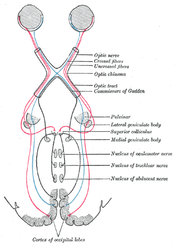
Changes for: forelimb wing
- Deleted
- - forelimb wing definition Forelimb that is used to produce lift for flight through the air.
- Added
- + forelimb wing definition Forelimb that is used to produce lift for flight through the air. { database cross reference=UBERON:cjm }
Changes for: pituitary gland
- Deleted
Changes for: suprapreopercle
- Deleted
- - suprapreopercle comment Synonyms: supraopercle (Roberts 1969) supraopercular bone (Buckup 1998)
- Added
- + suprapreopercle comment Synonyms: supraopercle (Roberts 1969) supraopercular bone (Buckup 1998)
Changes for: bilaminar disc
- Deleted
- Added
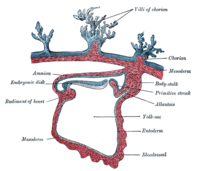
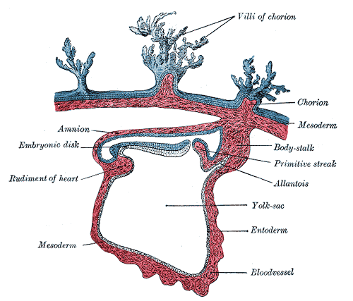
Changes for: blastocele
- Deleted
- Added
Changes for: lateral line ganglion
- Deleted
Changes for: male reproductive system
- Added
Changes for: external ectoderm
- Deleted
- Added
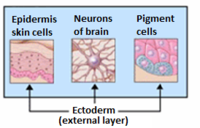
Changes for: ampulla of Lorenzini
- Deleted
- - ampulla of Lorenzini SubClassOf electroreceptor organ
- Added
- + ampulla of Lorenzini SubClassOf lateral line sense organ
Changes for: ampullary organ
- Deleted
- - ampullary organ SubClassOf electroreceptor organ
- - ampullary organ SubClassOf part of some lateral line system
- Added
- + ampullary organ SubClassOf ectoderm-derived structure
- + ampullary organ SubClassOf innervated by some lateral line nerve
- + ampullary organ SubClassOf lateral line sense organ
- + ampullary organ SubClassOf part of some lateral line
- + ampullary organ has exact synonym electrosensory ampullary organ
- + ampullary organ present in taxon http://purl.obolibrary.org/obo/NCBITaxon_7746
- + ampullary organ present in taxon http://purl.obolibrary.org/obo/NCBITaxon_7777
- + ampullary organ present in taxon http://purl.obolibrary.org/obo/NCBITaxon_7900
- + ampullary organ present in taxon http://purl.obolibrary.org/obo/NCBITaxon_7911
- + ampullary organ present in taxon http://purl.obolibrary.org/obo/NCBITaxon_8289
- + ampullary organ present in taxon http://purl.obolibrary.org/obo/NCBITaxon_8296
Changes for: renal glomerulus
- Deleted
Changes for: morula
- Deleted
Changes for: zona pellucida
- Deleted
Changes for: inner cell mass
- Deleted
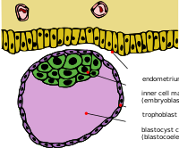
Changes for: trophoblast
- Deleted
Changes for: mesonephros
- Deleted
Changes for: fornix of brain
- Deleted
- Added
Changes for: macula lutea
- Deleted
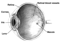
Changes for: ureter
- Deleted
Changes for: urethra
- Deleted
- Added
Changes for: fornix of vagina
- Deleted
- Added
Changes for: organ part
- Deleted
- - organ part definition A multicellular structure that is a part of an organ.
- Added
- + organ part definition A multicellular structure that is a part of an organ. { database cross reference=UBERON:cjm }
Changes for: neuromast
- Deleted
- - neuromast SubClassOf develops from some protoneuromast
- - neuromast SubClassOf sense organ
- - neuromast has exact synonym lateral line organ { database cross reference=ZFA:0000243 }
- Added
- + neuromast SubClassOf develops from some lateral line placode
- + neuromast SubClassOf innervated by some lateral line nerve
- + neuromast SubClassOf lateral line sense organ
- + neuromast SubClassOf transformation of some protoneuromast
- + neuromast has broad synonym lateral line organ { database cross reference=ZFA:0000243 }
Changes for: embryo stage
- Deleted
- - embryo stage definition A life cycle stage that starts with fertilization and ends with the fully formed embryo.
- Added
- + embryo stage definition A life cycle stage that starts with fertilization and ends with the fully formed embryo. { database cross reference=UBERON:cjm }
Changes for: atrioventricular septum
- Added
Changes for: Bachmann’s bundle
- Deleted
- Added
Changes for: coronary vessel
Changes for: crista terminalis
Changes for: outflow part of left ventricle
- Added
Changes for: developing mesenchymal condensation
- Deleted
- - developing mesenchymal condensation database cross reference EMAPA:25351
Changes for: anterior tubercle of transverse process of cervical vertebra
- Added
- + anterior tubercle of transverse process of cervical vertebra database cross reference http://en.wikipedia.org/wiki/Anterior_tubercle_of_cervical_vertebra
- + anterior tubercle of transverse process of cervical vertebra definition The anterior arch forms about one-fifth of the ring: its anterior surface is convex, and presents at its center the anterior tubercle for the attachment of the Longus colli muscles. { database cross reference=http://en.wikipedia.org/wiki/Anterior_tubercle_of_cervical_vertebra,Wikipedia:Anterior_tubercle_of_cervical_vertebra }
Changes for: posterior tubercle of transverse process of cervical vertebra
- Added
- + posterior tubercle of transverse process of cervical vertebra database cross reference http://en.wikipedia.org/wiki/Posterior_tubercle_of_cervical_vertebra
- + posterior tubercle of transverse process of cervical vertebra definition The posterior arch of a cervical vertebra forms about two-fifths of the circumference of the ring: it ends behind in the posterior tubercle, which is the rudiment of a spinous process and gives origin to the Recti capitis posteriores minores. The diminutive size of this process prevents any interference with the movements between the atlas and the skull. { database cross reference=http://en.wikipedia.org/wiki/Posterior_tubercle_of_cervical_vertebra,Wikipedia:Posterior_tubercle_of_cervical_vertebra }
Changes for: cochlear ganglion
- Deleted
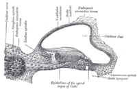
Changes for: vallate papilla
- Deleted
- Added
Changes for: longissimus muscle
- Deleted
Changes for: urinary bladder detrusor smooth muscle
- Deleted
Changes for: tapetum of corpus callosum
- Deleted
Changes for: extensor digitorum brevis
- Deleted
- Added
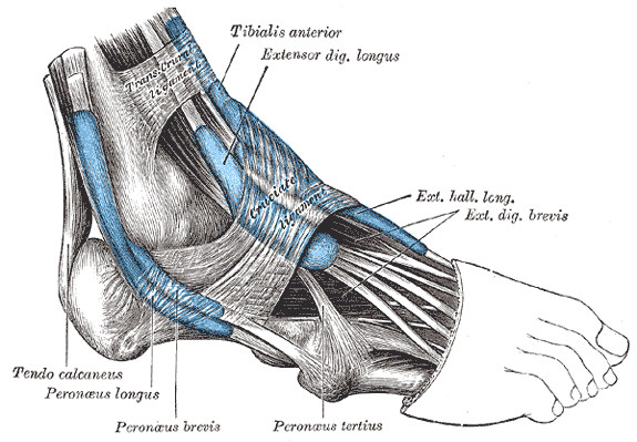
Changes for: maxillary nerve
- Deleted
Changes for: tongue muscle
- Deleted
- - tongue muscle SubClassOf mixed ectoderm/mesoderm/endoderm-derived structure
- - tongue muscle SubClassOf part of some tongue musculature
- Added
- + tongue muscle SubClassOf contributes to morphology of some tongue
- + tongue muscle SubClassOf ectoderm-derived structure
- + tongue muscle SubClassOf part of some mouth
- + tongue muscle database cross reference EHDAA2:0002065
- + tongue muscle database cross reference EHDAA:9146
- + tongue muscle database cross reference EMAPA:17883
- + tongue muscle database cross reference MA:0001596
- + tongue muscle database cross reference UMLS:C1710437 { source=ncithesaurus:Tongue_Skeletal_Muscle_Tissue }
- + tongue muscle database cross reference VHOG:0001390
- + tongue muscle database cross reference http://linkedlifedata.com/resource/umls/id/C1710437
- + tongue muscle database cross reference http://ncicb.nci.nih.gov/xml/owl/EVS/Tongue_Skeletal_Muscle_Tissue
- + tongue muscle has exact synonym skeletal muscle tissue of tongue { database cross reference=OBOL:automatic }
- + tongue muscle has exact synonym tongue skeletal muscle tissue { database cross reference=OBOL:automatic }
- + tongue muscle has exact synonym tongue skeletal muscle { database cross reference=OBOL:automatic }
- + tongue muscle homology notes The lamprey head contains another group of muscles, the epi- and hypo-branchial muscles (EBM and HBM), derivatives of anterior trunk myotomes. (…) The origin and the migration pattern of HBM precursors are very similar to that of the gnathostome MPP, especially to that of the tongue muscle precursors. Other evidence of homology of lamprey HBM to the gnathostome tongue muscle is that HBM is innervated by the nerve termed the hypoglossal nerve based on its morphological position associated with the head/trunk interface. (…) The HBM-specific expression of the LampPax3/7 gene is consistent with the homology of this muscle to the gnathostome tongue muscle, or to the hypobranchial series as a whole (including the infrahyoid and possibly the diaphragm in mammals).[well established][VHOG] { date retrieved=2012-09-17 , external class=VHOG:0001390 , ontology=VHOG , source=http://bgee.unil.ch/ , source=DOI:10.1002/dvdy.20587 Kusakabe R, Kuratani S, Evolution and developmental patterning of the vertebrate skeletal muscles: perspectives from the lamprey. Developmental Dynamics (2005) }
Changes for: mandibular nerve
- Deleted
Changes for: hindlimb stylopod
- Deleted
Changes for: adductor magnus
- Deleted
Changes for: renal medulla
- Deleted
Changes for: adductor brevis
- Deleted
Changes for: corpus striatum
- Deleted
- Added
Changes for: blastocyst
- Deleted
Changes for: throat
- Deleted
Changes for: ophthalmic nerve
- Added
- + ophthalmic nerve SubClassOf develops from some profundal placode
- + ophthalmic nerve has related synonym profundal nerve { database cross reference=UBERON:cjm }
Changes for: limbic system
- Deleted
Changes for: dermal papilla
- Deleted
Changes for: mucous gland
- Deleted
Changes for: visual cortex
- Deleted
- Added
Changes for: scalp
- Deleted
- Added
Changes for: nasal vestibule
- Deleted
Changes for: elasmoid scale
- Deleted
Changes for: limiting layer of elasmoid scale
- Added
Changes for: pelvic girdle skeleton
- Deleted
- Added
Changes for: pectoral girdle skeleton
- Deleted
Changes for: prefrontal cortex
- Deleted
Changes for: simple columnar epithelium
- Deleted
Changes for: simple cuboidal epithelium
- Deleted
Changes for: female reproductive system
- Deleted
- Added
Changes for: testis
- Deleted
- Added
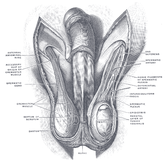
Changes for: reticulospinal tract
- Added
- + reticulospinal tract database cross reference http://en.wikipedia.org/wiki/Reticulospinal_tract
- + reticulospinal tract definition The reticulospinal tract (or anterior reticulospinal tract) is an extrapyramidal motor tract which travels from the reticular formation. { database cross reference=http://en.wikipedia.org/wiki/Reticulospinal_tract,Wikipedia:Reticulospinal_tract }
Changes for: deep dorsal vein of clitoris
- Added
- + deep dorsal vein of clitoris SubClassOf deep vein
- + deep dorsal vein of clitoris SubClassOf part of some deep vasculature
- + deep dorsal vein of clitoris database cross reference http://en.wikipedia.org/wiki/Deep_dorsal_vein_of_clitoris
- + deep dorsal vein of clitoris definition The deep dorsal vein of clitoris is a vein which drains to the vesical plexus. { database cross reference=http://en.wikipedia.org/wiki/Deep_dorsal_vein_of_clitoris,Wikipedia:Deep_dorsal_vein_of_clitoris }
Changes for: pharyngeal tubercle
- Added
- + pharyngeal tubercle database cross reference http://en.wikipedia.org/wiki/Pharyngeal_tubercle
- + pharyngeal tubercle definition On the lower surface of the basilar part of occipital bone, about 1 cm. in front of the foramen magnum, is the pharyngeal tubercle which gives attachment to the fibrous raphC) of the pharynx, also known as the pharyngeal raphe. This is the point of attachment for the superior pharyngeal constrictor muscle. { database cross reference=http://en.wikipedia.org/wiki/Pharyngeal_tubercle,Wikipedia:Pharyngeal_tubercle }
Changes for: spinal vein
Changes for: median sacral vein
- Added
- + median sacral vein database cross reference http://en.wikipedia.org/wiki/Median_sacral_vein
- + median sacral vein definition The median sacral vein (or middle sacral veins) accompany the corresponding artery along the front of the sacrum, and join to form a single vein, which ends in the left common iliac vein; sometimes in the angle of junction of the two iliac veins. { database cross reference=http://en.wikipedia.org/wiki/Median_sacral_vein,Wikipedia:Median_sacral_vein }
Changes for: interthalamic adhesion
- Added
- + interthalamic adhesion database cross reference http://en.wikipedia.org/wiki/Interthalamic_adhesion
- + interthalamic adhesion definition The medial surface of the thalamus constitutes the upper part of the lateral wall of the third ventricle, and is connected to the corresponding surface of the opposite thalamus by a flattened gray band, the Interthalamic adhesion (massa intermedia, middle commissure, gray commissure). In non-human mammals it is a large structure. In humans this mass averages about 1 cm in length in its antero-posterior diameter. It sometimes consists of two parts and occasionally is absent. The interthalamic adhesion is found in 70-80% of humans. It is present more often in females and larger than in males by an average of 53 percent. . When absent in development, no noticeable deficit has been observed. It contains nerve cells and nerve fibers; a few of the latter may cross the middle line, but most of them pass toward the middle line and then curve lateralward on the same side. { database cross reference=http://en.wikipedia.org/wiki/Interthalamic_adhesion,Wikipedia:Interthalamic_adhesion }
Changes for: prefrontal bone
- Deleted
- Added
Changes for: secondary follicle corona
Changes for: rib 8
- Added
- + rib 8 definition The eighth rib counting from the top of the rib cage down. Note that members of this class are not necessarily homologous[ncit,modified]. { database cross reference=http://ncicb.nci.nih.gov/xml/owl/EVS/Rib_8 , database cross reference=UBERON:cjm }
Changes for: meningeal cluster
Changes for: obsolete pretectal periventricular nucleus
- Deleted
- - pretectal periventricular nucleus SubClassOf diencephalic nucleus
- - pretectal periventricular nucleus SubClassOf part of some synencephalon
- - pretectal periventricular nucleus label pretectal periventricular nucleus
- Added
- + obsolete pretectal periventricular nucleus deprecated true
- + obsolete pretectal periventricular nucleus label obsolete pretectal periventricular nucleus
Changes for: deep dorsal vein of penis
- Deleted
- - deep dorsal vein of penis SubClassOf vein
- Added
- + deep dorsal vein of penis SubClassOf deep vein
- + deep dorsal vein of penis SubClassOf part of some deep vasculature
- + deep dorsal vein of penis database cross reference http://en.wikipedia.org/wiki/Deep_dorsal_vein_of_the_penis
- + deep dorsal vein of penis definition The deep dorsal vein of the penis lies beneath the deep fascia of the penis; it receives the blood from the glans penis and corpora cavernosa penis and courses backward in the middle line between the dorsal arteries; near the root of the penis it passes between the two parts of the suspensory ligament and then through an aperture between the arcuate pubic ligament and the transverse ligament of the pelvis, and divides into two branches, which enter the pudendal plexus. The deep vein also communicates below the symphysis pubis with the internal pudendal vein. { database cross reference=http://en.wikipedia.org/wiki/Deep_dorsal_vein_of_the_penis,Wikipedia:Deep_dorsal_vein_of_the_penis }
Changes for: tunica albuginea of testis
- Added
Changes for: deep temporal artery
- Added
- + deep temporal artery EquivalentTo temporal artery and part of some deep vasculature
- + deep temporal artery SubClassOf part of some deep vasculature
Changes for: anterior abdominal wall
- Added
- + anterior abdominal wall database cross reference http://en.wikipedia.org/wiki/Abdominal_wall
- + anterior abdominal wall definition The abdominal wall represents the boundaries of the abdominal cavity. The abdominal wall is split into the posterior (back), lateral (sides) and anterior (front) walls. There is a common set of layers covering and forming all the walls: the deepest being the extraperitoneal fat, the parietal peritoneum, and a layer of fascia which has different names over where it covers (eg transversalis, psoas fascia). Superficial to these, but not present in the posterior wall are the three layers of muscle, the transversus abdominis (transverse abdominal muscle), the internal (obliquus internus) and the external oblique (obliquus externus). { database cross reference=http://en.wikipedia.org/wiki/Abdominal_wall,Wikipedia:Abdominal_wall }
Changes for: obsolete central pretectal nucleus
- Deleted
- - central pretectal nucleus SubClassOf diencephalic nucleus
- - central pretectal nucleus SubClassOf part of some central pretectum
- - central pretectal nucleus SubClassOf pretectal nucleus
- - central pretectal nucleus label central pretectal nucleus
- Added
- + obsolete central pretectal nucleus deprecated true
- + obsolete central pretectal nucleus label obsolete central pretectal nucleus
Changes for: skeleton of manual acropodium
- Added
Changes for: sacral region
Changes for: skeleton of pedal acropodium
- Added
Changes for: thymus lobe
- Added
- + thymus lobe definition Either of the two lateral lobes that constitute the thymus. { database cross reference=UBERON:cjm }
- + thymus lobe has exact synonym lateral lobe of thymus { database cross reference=UBERON:cjm }
- + thymus lobe structure notes each lobe of the thymus can be divided into a central medulla and a peripheral cortex which is surrounded by an outer capsule. Each lobe is composed of numerous lobules held together by delicate areolar tissue; the entire organ being enclosed in an investing capsule of a similar but denser structure. The primary lobules vary in size from that of a pin’s head to that of a small pea, and are made up of a number of small nodules or follicles.[WP]
Changes for: primitive heart tube
- Added
Changes for: superficial palmar arch
- Added
- + superficial palmar arch database cross reference http://en.wikipedia.org/wiki/Superficial_palmar_arch
- + superficial palmar arch definition The superficial palmar arch is formed predominantly by the ulnar artery, with a contribution from the superficial palmar branch of the radial artery. However, in some individuals the contribution from the radial artery might be absent, and instead anastomoses with either the princeps pollicis artery, the radialis indicis artery, or the median artery, the former two of which are branches from the radial artery. Alternative names for this arterial arch are: superficial volar arch, superficial ulnar arch, arcus palmaris superficialis, or arcus volaris superficialis. The arch passes across the palm in a curve with its convexity downward. If one were to fully extend the thumb, the superficial palmar arch would lie approximately at the level of a line drawn from the distal border of the thumb across the palm. The superficial palmar arch is more distal than the deep palmar arch. Three common palmar digital arteries arise from the arch, proceeding down on the second, third, and fourth lumbrical muscles, respectively. They each receive a contribution from a palmar metacarpal artery. Near the level of the metacarpophalangeal joints, each common palmar digital artery divides into two proper palmar digital arteries. { database cross reference=http://en.wikipedia.org/wiki/Superficial_palmar_arch,Wikipedia:Superficial_palmar_arch }
Changes for: statocyst
- Deleted
- Added
Changes for: presumptive hypochord
- Deleted
- - presumptive hypochord comment Taxon notes: Unlike in frogs and axolotl, the hypochord does not appear to be endodermally derived[ZFA]
- Added
- + presumptive hypochord taxon notes Unlike in frogs and axolotl, the hypochord does not appear to be endodermally derived[ZFA]
Changes for: obsolete accessory pretectal nucleus
- Deleted
- - accessory pretectal nucleus SubClassOf diencephalic nucleus
- - accessory pretectal nucleus SubClassOf part of some central pretectum
- - accessory pretectal nucleus SubClassOf pretectal nucleus
- - accessory pretectal nucleus label accessory pretectal nucleus
- Added
- + obsolete accessory pretectal nucleus deprecated true
- + obsolete accessory pretectal nucleus label obsolete accessory pretectal nucleus
Changes for: preputial space
- Deleted
- - preputial space definition space between the prepuce and glans penis or clitoris
- Added
- + preputial space definition space between the prepuce and glans penis or clitoris { database cross reference=UBERON:cjm }
Changes for: renal convoluted tubule
- Added
Changes for: anterior spinal artery
- Added
- + anterior spinal artery SubClassOf branch of vertebral artery
Changes for: longitudinal arch of pes
- Added
Changes for: temporalis fascia
- Added
- + temporalis fascia database cross reference http://en.wikipedia.org/wiki/Temporal_fascia
- + temporalis fascia definition The temporal fascia covers the Temporalis muscle. It is a strong, fibrous investment, covered, laterally, by the Auricularis anterior and superior, by the galea aponeurotica, and by part of the Orbicularis oculi. The superficial temporal vessels and the auriculotemporal nerve cross it from below upward. Above, it is a single layer, attached to the entire extent of the superior temporal line; but below, where it is fixed to the zygomatic arch, it consists of two layers, one of which is inserted into the lateral, and the other into the medial border of the arch. A small quantity of fat, the orbital branch of the superficial temporal artery, and a filament from the zygomatic branch of the maxillary nerve, are contained between these two layers. It affords attachment by its deep surface to the superficial fibers of the Temporalis. { database cross reference=http://en.wikipedia.org/wiki/Temporal_fascia,Wikipedia:Temporal_fascia }
Changes for: central part of mediodorsal nucleus of the thalamus
- Deleted
- - mediodorsal nucleus of the thalamus, central part label mediodorsal nucleus of the thalamus, central part
- Added
- + central part of mediodorsal nucleus of the thalamus has exact synonym mediodorsal nucleus of the thalamus, central part { database cross reference=MBA:617 }
- + central part of mediodorsal nucleus of the thalamus label central part of mediodorsal nucleus of the thalamus
Changes for: lateral part of mediodorsal nucleus of the thalamus
- Deleted
- - mediodorsal nucleus of the thalamus, lateral part label mediodorsal nucleus of the thalamus, lateral part
- Added
- + lateral part of mediodorsal nucleus of the thalamus has exact synonym mediodorsal nucleus of the thalamus, lateral part { database cross reference=MBA:626 }
- + lateral part of mediodorsal nucleus of the thalamus label lateral part of mediodorsal nucleus of the thalamus
Changes for: alimentary part of gastrointestinal system
- Deleted
- - gastrointestinal system external ontology notes that many anatomy ontologies consider this synonymous with digestive system. here we follow MA in dividing digestive system into gastrointestinal and hepatobiliary. hepatobiliary includes the liver and biliary tract. species-specific AO classes are categorized according to whether liver is included. For example, XAO includes liver as part of XAO:0000125 alimentary system, so we assume this class is the more generic class. We have one entity that is part of both gastrointestinal and hepatobiliary systems: hepatopancreatic ampulla { external ontology=MA }
- - gastrointestinal system label gastrointestinal system
- Added
- + alimentary part of gastrointestinal system definition The part of the digestive system that excludes the hepatobiliary system. { database cross reference=UBERON:cjm }
- + alimentary part of gastrointestinal system editor note the decision to split digestive and gastrointestinal in this way may be revisited. As it currently stands, this class more closely aligns what MA calls the alimentary system
- + alimentary part of gastrointestinal system external ontology notes that many anatomy ontologies consider this synonymous with digestive system. here we follow MA in dividing digestive system into gastrointestinal and hepatobiliary. hepatobiliary includes the liver and biliary tract. species-specific AO classes are categorized according to whether liver is included. For example, XAO includes liver as part of XAO:0000125 alimentary system, so we assume this class is the more generic class. We have one entity that is part of both gastrointestinal and hepatobiliary systems: hepatopancreatic ampulla. { external ontology=MA }
- + alimentary part of gastrointestinal system label alimentary part of gastrointestinal system
Changes for: superior vagus X ganglion
- Added
Changes for: peritoneal vaginal process
- Deleted
Changes for: paraflocculus
Changes for: spermatic cord
- Deleted
Changes for: malleus neck
- Deleted
Changes for: striosome
- Deleted
- - striosome has related synonym striosomal compartment { database cross reference=NIFSTD:nlx_anat_20090506 }
- Added
- + striosome has exact synonym striosomal compartment { database cross reference=NIFSTD:nlx_anat_20090506 }
- + striosome has related synonym striosomal part
Changes for: muscle of digastric group
- Deleted
- - muscle of digastric group definition Either of the two muscles that comprise the digastric group (anterior or posterior).
- Added
- + muscle of digastric group definition Either of the two muscles that comprise the digastric group (anterior or posterior). { database cross reference=UBERON:cjm }
Changes for: occipitalis
Changes for: cleidooccipital muscle
- Deleted
- - cleidooccipital muscle SubClassOf skeletal muscle organ
- Added
- + cleidooccipital muscle SubClassOf muscle of shoulder
Changes for: ossicle
Changes for: endostyle
- Deleted
- Added
Changes for: scapula
- Deleted
Changes for: posterior pretectal nucleus
- Deleted
- - posterior pretectal nucleus database cross reference TAO:0000631
- - posterior pretectal nucleus database cross reference ZFA:0000631
- - posterior pretectal nucleus definition Diencephalic nucleus that is part of the superficial pretectum and is rostrally contiguous with the magnocellular superficial pretectal nucleus. Cells of the caudal pretectal nucleus are slightly smaller and less orderly around a central neuropil than those of the magnocellular superficial pretectal nucleus. From Neuroanatomy of the Zebrafish Brain. { database cross reference=ZFA:0000631 }
- - posterior pretectal nucleus has exact synonym caudal pretectal nuclei { database cross reference=ZFA:0000631 }
- - posterior pretectal nucleus has exact synonym posterior pretectal nucleus { database cross reference=ZFA:0000631 }
- - posterior pretectal nucleus in subset dubious grouping
- - posterior pretectal nucleus in subset vertebrate core
Changes for: central vein of liver
- Added
Changes for: tegmen tympani
- Added
Changes for: uterus or analog
- Deleted
- - uterus or analog definition A subdivision of a the reproductive tract in a female organism that is the site of embryo development.
- Added
- + uterus or analog definition A subdivision of a the reproductive tract in a female organism that is the site of embryo development. { database cross reference=UBERON:cjm }
Changes for: chorioallantoic membrane
- Deleted
Changes for: mammary bud
- Deleted
- - mammary bud definition A bulb of epithelial cells that forms from the mammary placode that develops into the mammary gland.
- Added
- + mammary bud definition A bulb of epithelial cells that forms from the mammary placode that develops into the mammary gland. { database cross reference=UBERON:cjm }
Changes for: hypogastric nerve
- Deleted
Changes for: nephrostome
Changes for: preganglionic parasympathetic fiber
- Added
Changes for: postganglionic sympathetic fiber
- Added
Changes for: postganglionic parasympathetic fiber
- Added
Changes for: calcaneofibular ligament
- Deleted
- Added
Changes for: talofibular ligament
- Deleted
- Added
Changes for: myelencephalon
- Deleted
Changes for: ventricular system of central nervous system
- Deleted
Changes for: gonadal ridge
- Deleted
- Added
- + gonadal ridge database cross reference EMAPA:35899
- + gonadal ridge database cross reference UMLS:C0231047 { source=ncithesaurus:Urogenital_Ridge }
- + gonadal ridge database cross reference http://linkedlifedata.com/resource/umls/id/C0231047
- + gonadal ridge database cross reference http://ncicb.nci.nih.gov/xml/owl/EVS/Urogenital_Ridge
- + gonadal ridge has exact synonym genital ridge { database cross reference=http://en.wikipedia.org/wiki/Gonadal_ridge , database cross reference=MP:0011411 }
- + gonadal ridge has related synonym genital cord { database cross reference=MP:0011411 }
- + gonadal ridge label gonadal ridge
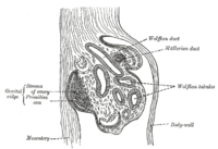
Changes for: prepuce of clitoris
- Deleted
Changes for: accessory XI nerve nucleus
- Added
- + accessory XI nerve nucleus database cross reference http://en.wikipedia.org/wiki/Spinal_accessory_nucleus
- + accessory XI nerve nucleus definition The spinal accessory nucleus lies within the cervical spinal cord (C1-C5) in the ventral horn. The nucleus ambiguus is classically said to provide the ‘cranial component’ of the accessory nerve. However, the very existence of this cranial component has been recently questioned and seen as contributing exclusively to the vagus nerve. The terminology continues to be used in describing both human anatomy, and that of other animals. { database cross reference=http://en.wikipedia.org/wiki/Spinal_accessory_nucleus }
Changes for: efferent nerve
Changes for: striosomal part of body of caudate nucleus
- Deleted
- Added
- + striosomal part of body of caudate nucleus EquivalentTo striosome and part of some body of caudate nucleus
- + striosomal part of body of caudate nucleus SubClassOf striosomal part of caudate nucleus
Changes for: matrix part of head of caudate nucleus
- Deleted
- Added
- + matrix part of head of caudate nucleus EquivalentTo matrix compartment and part of some head of caudate nucleus
- + matrix part of head of caudate nucleus SubClassOf matrix compartment of caudate nucleus
- + matrix part of head of caudate nucleus has exact synonym matrix compartment of head of caudate nucleus
Changes for: matrix part of tail of caudate nucleus
- Deleted
- Added
- + matrix part of tail of caudate nucleus EquivalentTo matrix compartment and part of some tail of caudate nucleus
- + matrix part of tail of caudate nucleus SubClassOf matrix compartment of caudate nucleus
- + matrix part of tail of caudate nucleus has exact synonym matrix compartment of tail of caudate nucleus
Changes for: stomach
- Deleted
Changes for: epithelium of conjunctiva
- Deleted
- - epithelium of conjunctiva definition Layer of nonkeratinized squamous epithelium lining the conjunctiva, overlying scleral tissue
- Added
- + epithelium of conjunctiva definition Layer of nonkeratinized squamous epithelium lining the conjunctiva, overlying scleral tissue { database cross reference=UBERON:cjm }
Changes for: frontal nerve (branch of ophthalmic)
- Deleted
Changes for: anterior commissure
- Deleted
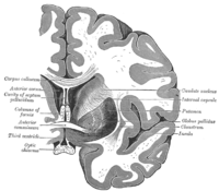
Changes for: posterior commissure
- Deleted
- - posterior commissure EquivalentTo nervous system commissure and connects some nucleus of pretectal area
- - posterior commissure SubClassOf connects some nucleus of pretectal area
- - posterior commissure depicted by http://upload.wikimedia.org/wikipedia/commons/thumb/d/d7/Gray715.png/200px-Gray715.png
- Added
- + posterior commissure EquivalentTo nervous system commissure and connects some pretectal nucleus
- + posterior commissure SubClassOf connects some pretectal nucleus
Changes for: ventral nerve cord
Changes for: stomodeum
- Deleted
Changes for: thoracic ganglion
- Deleted
- Added
Changes for: obsolete pharyngeal nerve
- Deleted
- Added
Changes for: nerve of cervical vertebra
- Deleted
- Added
Changes for: cornea
- Deleted
Changes for: retina
- Deleted
Changes for: stratum lemnisci of superior colliculus
- Added
- + stratum lemnisci of superior colliculus database cross reference http://uri.neuinfo.org/nif/nifstd/nlx_144110
- + stratum lemnisci of superior colliculus definition Composite layer of the superior colliculus lying deep to the stratum opticum, that can be further subdivided into fiber and cell rich layers (Brodal, Neurological Anatomy, 2nd ed., 1981, pg 544) { database cross reference=NIFSTD:nlx_144110 }
- + stratum lemnisci of superior colliculus has broad synonym stratum lemnisci { database cross reference=NIFSTD:nlx_144110 }
Changes for: tectal plate
- Deleted
- - tectal plate SubClassOf part of some embryo
- Added
- + tectal plate SubClassOf part of some presumptive midbrain
Changes for: cerebral cortex
- Deleted
- Added
Changes for: optic chiasma
- Deleted
- Added
Changes for: hippocampal commissure
- Deleted
Changes for: mesoderm
- Deleted
Changes for: pharyngeal branch of vagus nerve
- Deleted
Changes for: endoderm
- Deleted
- Added
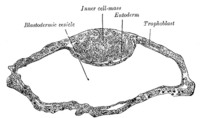
Changes for: penis
- Deleted
- Added
Changes for: pons
- Deleted
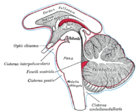
Changes for: axillary vein
- Deleted
Changes for: femur
- Deleted
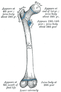
Changes for: pleura
- Deleted
- Added
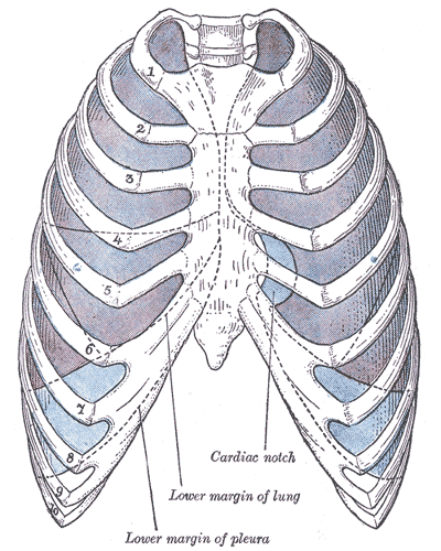
Changes for: tibia
- Deleted
Changes for: neck
- Deleted
- Added
Changes for: humerus
- Deleted
Changes for: sternum
- Deleted
- Added
Changes for: ommatidium
- Deleted
- - ommatidium definition The smallest morphological and functional unit of the ➞compound eye that consists of a usually limited and often constant number of rhabdomeric ➞photoreceptor cells, cornea-secreting epithelial cells, and interommatidial pigment cells, and may additionally contain crystalline cone cells. { database cross reference=http://www.ncbi.nlm.nih.gov/pubmed/21062451 }
- Added
- + ommatidium definition The smallest morphological and functional unit of the compound eye that consists of a usually limited and often constant number of rhabdomeric photoreceptor cells, cornea-secreting epithelial cells, and interommatidial pigment cells, and may additionally contain crystalline cone cells. { database cross reference=http://www.ncbi.nlm.nih.gov/pubmed/21062451 }
Changes for: vagina
- Deleted
- - vagina depicted by [http://upload.wikimedia.org/wikipedia/commons/thumb/a/a4/Vaginal_opening_-english_description.jpg/200px-Vaginal_opening-english_description.jpg](http://upload.wikimedia.org/wikipedia/commons/thumb/a/a4/Vaginal_opening-english_description.jpg/200px-Vaginal_opening-_english_description.jpg)
- Added
- + vagina depicted by [http://upload.wikimedia.org/wikipedia/commons/a/a4/Vaginal_opening_-english_description.jpg](http://upload.wikimedia.org/wikipedia/commons/a/a4/Vaginal_opening-_english_description.jpg)
Changes for: uterus
- Deleted
- Added
Changes for: seminal vesicle
- Deleted
Changes for: ejaculatory duct
- Deleted
Changes for: reproductive system
- Deleted
Changes for: female gonad
- Deleted
- Added
Report for properties
ObjectProperty objects lost from source: 0
ObjectProperty objects new in target: 0
Changed ObjectProperty objects: 0
May 25, 2015
|
Tweet
Comments Section
Feel free to comment on the post but keep it clean and on topic.
comments powered by Disqus