/releases/2014-06-15
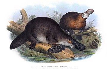
This release includes some improvements to the representation of buccinator, orbicularis oris and digastric muscles, based on the Mammalian Feeding ontology FEED. A number of improvements were made to the skeletal system, continued implementation a standard pattern for endocondral bones with particular attention to the ribs and vertebrae. A number of new classes for a variety of systems have been added, including term requests for ENCODE. This release also marks the debut of the platypus bill.
Note that the diff report for this release is large due to some systematic changes in how textual metadata about classes is stored in the ontology
- skeletal
- AXIAL: rib and vertebrae overhaul. Implemented endochondral pattern consistently.
- CRANIAL: mapped ZFA orbit to orbital cavity. See issue 462
- CRANIAL: renaming jaw joint. Issue issue 463
- LIMB: Abstracted classes with full endochondral triad for proximal and distal mesopodials. Addresses issue 461
- LIMB: Fixed proximal carpals to use consistent pattern
- UPPER: reclassified some classes as being cartilage elements rather than cartilage tissue
- muscles
- FEED: created xrefs to MFMO (Feed ontology)
- FEED: Buccinator improvements. Issue issue 470. FEED:rd and cw
- FEED: Text amd logical def of orbicularis oris. Extended FEED def with surrounds relation. Issue issue 473
- FEED: some changes to digastric based on conv with FEED:rd, added notes about Orangutans
- LIMB: improving defs of calf muscles
- SMOOTH: NTs for smooth muscle tissue and layer of muscle in rectum. Fixes issue issue 465
- EAR: NTs for 3 external auriculars
- other
- admin/general
- PROPERTIES: translation of comments to specific annotation assertions (editor notes, curator notes, taxon notes, etc)
- DEFINITIONS: making WP-derived defs more concise
- METADATA: Fixed consider link. Fixes issue issue 436
- RELATIONS: Fixed RO ID. Fixes issue issue 447
- METADATA: Added DOIs. Issue issue 473
- METADATA: Fixing xref syntax in ext
Ontology Diff Report
Original Ontology
- IRI: http://purl.obolibrary.org/obo/uberon.owl
- VersionIRI: http://purl.obolibrary.org/obo/uberon/releases/2014-05-27/uberon.owl
New Ontology
- IRI: http://purl.obolibrary.org/obo/uberon.owl
- VersionIRI: http://purl.obolibrary.org/obo/uberon/releases/2014-06-15/uberon.owl
Report for classes
Class objects lost from source: 4
- coccygeal vertebra endochondral element
- coccygeal vertebra pre-cartilage condensation
- coccygeal vertebra cartilage element
- coccygeal vertebra
Class objects new in target: 38
New Class : distal mesopodial endochondral element
- distal mesopodial endochondral element definition An endochondral element in the distal portion of the mesopodial skeleton, between the central mesopodial skeleton and the metapodial skeleton { database cross reference=UBERON:cjm }
- distal mesopodial endochondral element id UBERON:0018099
- distal mesopodial endochondral element editor note TODO add logical definition
- distal mesopodial endochondral element SubClassOf autopod endochondral element
- distal mesopodial endochondral element has obo namespace uberon
- distal mesopodial endochondral element SubClassOf part of some mesopodial skeleton
- distal mesopodial endochondral element label distal mesopodial endochondral element
New Class : caudal vertebra endochondral element
- caudal vertebra endochondral element has alternative id UBERON:0015006
- caudal vertebra endochondral element has obo namespace uberon
- caudal vertebra endochondral element has exact synonym tail vertebra element
- caudal vertebra endochondral element SubClassOf vertebra endochondral element
- caudal vertebra endochondral element has exact synonym caudal vertebra element
- caudal vertebra endochondral element definition Any vertebra endochondral element that is part of the caudal region of the vertebral column (tail or coccyx). { database cross reference=UBERON:cjm }
- caudal vertebra endochondral element id UBERON:0018142
- caudal vertebra endochondral element has narrow synonym coccygeal vertebra element
- caudal vertebra endochondral element SubClassOf dorsal region element
- caudal vertebra endochondral element EquivalentTo vertebra endochondral element and part of some caudal region of vertebral column
- caudal vertebra endochondral element SubClassOf part of some caudal region of vertebral column
- caudal vertebra endochondral element label caudal vertebra endochondral element
- caudal vertebra endochondral element has narrow synonym coccyx vertebra element
New Class : anterior perforated substance
- anterior perforated substance has exact synonym substantia perforata anterior { database cross reference=http://en.wikipedia.org/wiki/Anterior_perforated_substance , has synonym type=latin term }
- anterior perforated substance has related synonym eminentia parolfactoria { database cross reference=NeuroNames:282 , has synonym type=latin term }
- anterior perforated substance has exact synonym anterior perforated area { database cross reference=FMA:61891 }
- anterior perforated substance has obo namespace uberon
- anterior perforated substance definition The anterior perforated substance is an irregularly quadrilateral area in front of the optic tract and behind the olfactory trigone, from which it is separated by the fissure prima; medially and in front it is continuous with the subcallosal gyrus; laterally it is bounded by the lateral stria of the olfactory tract and is continued into the uncus. Its gray substance is confluent above with that of the corpus striatum, and is perforated anteriorly by numerous small bloodvessels. { database cross reference=http://en.wikipedia.org/wiki/Anterior_perforated_substance }
- anterior perforated substance has related synonym area olfactoria (Mai) { database cross reference=NeuroNames:282 , has synonym type=latin term }
- anterior perforated substance database cross reference http://braininfo.rprc.washington.edu/centraldirectory.aspx?ID=282 { source=NIF_GrossAnatomy:birnlex_1096 }
- anterior perforated substance id UBERON:0018141
- anterior perforated substance has related synonym olfactory tubercle (Ganser) { database cross reference=NeuroNames:282 }
- anterior perforated substance has related synonym olfactory tubercle { database cross reference=FMA:61891 , has synonym type=indicates that a synonym is used in an inconsistent or confusing way, typically between species }
- anterior perforated substance database cross reference FMA:61891
- anterior perforated substance has related synonym tuber olfactorium { database cross reference=NeuroNames:282 , has synonym type=latin term }
- anterior perforated substance has related synonym olfactory area (Carpenter) { database cross reference=NeuroNames:282 }
- anterior perforated substance has exact synonym olfactory area (mai) { database cross reference=FMA:61891 }
- anterior perforated substance has related synonym rostral perforated substance { database cross reference=NeuroNames:282 }
- anterior perforated substance external definition Subdivision of telencephalon which is a region on the ventro-medial aspect of the telencephalon penetrated by numerous blood vessels. It is bounded by the optic tract posteriorly and the olfactory trigone anteriorly. { source=FMA:61891 }
- anterior perforated substance label anterior perforated substance
- anterior perforated substance database cross reference http://uri.neuinfo.org/nif/nifstd/birnlex_1096
- anterior perforated substance has exact synonym anterior perforated space { database cross reference=FMA:61891 }
- anterior perforated substance SubClassOf part of some telencephalon
- anterior perforated substance database cross reference http://braininfo.rprc.washington.edu/centraldirectory.aspx?ID=282
- anterior perforated substance database cross reference UMLS:C0162436 { source=NIF_GrossAnatomy:birnlex_1096 }
- anterior perforated substance database cross reference BAMS:APS
- anterior perforated substance database cross reference http://linkedlifedata.com/resource/umls/id/C0162436
- anterior perforated substance SubClassOf regional part of telencephalon
- anterior perforated substance database cross reference http://en.wikipedia.org/wiki/Anterior_perforated_substance
- anterior perforated substance database cross reference http://www.snomedbrowser.com/Codes/Details/369108006
New Class : mammary lobe
- mammary lobe database cross reference http://ncicb.nci.nih.gov/xml/owl/EVS/Breast_Lobe
- mammary lobe SubClassOf part of some mammary gland
- mammary lobe SubClassOf ectoderm-derived structure
- mammary lobe has obo namespace uberon
- mammary lobe has narrow synonym lobi glandulae mammariae { database cross reference=BTO:0004718 , database cross reference=NCBITaxon:9606 , has synonym type=preferred term when talking about an instance of this class in Homo sapiens }
- mammary lobe has narrow synonym lobe of breast { database cross reference=BTO:0004718 , database cross reference=NCBITaxon:9606 , has synonym type=preferred term when talking about an instance of this class in Homo sapiens }
- mammary lobe database cross reference http://linkedlifedata.com/resource/umls/id/C0222616
- mammary lobe has related synonym lobus glandulae mammariae { database cross reference=BTO:0004718 , has synonym type=latin term }
- mammary lobe label mammary lobe
- mammary lobe SubClassOf part of some trunk
- mammary lobe has narrow synonym lobe of mammary gland { database cross reference=BTO:0004718 , database cross reference=NCBITaxon:9606 , has synonym type=preferred term when talking about an instance of this class in Homo sapiens }
- mammary lobe SubClassOf has part some lobule of mammary gland
- mammary lobe id UBERON:0018140
- mammary lobe editor note The FMA class ‘lactiferous gland’ may be equivalent to this
- mammary lobe database cross reference BTO:0004718
- mammary lobe definition A cluster of mammary gland lobules. In humans this is a distinct lobe and located in the breast region.
- mammary lobe has narrow synonym breast lobe { database cross reference=BTO:0004718 , database cross reference=NCBITaxon:9606 , has synonym type=preferred term when talking about an instance of this class in Homo sapiens }
- mammary lobe SubClassOf anatomical cluster
New Class : transverse process of lumbar vertebra
- transverse process of lumbar vertebra label transverse process of lumbar vertebra
- transverse process of lumbar vertebra id UBERON:0018146
- transverse process of lumbar vertebra SubClassOf part of some lumbar vertebra
- transverse process of lumbar vertebra definition A transverse process that is part of a lumbar vertebra { database cross reference=UBERON:cjm }
- transverse process of lumbar vertebra database cross reference FMA:65469
- transverse process of lumbar vertebra taxon notes May be homologous with the lumbar ribs of some vertebrates
- transverse process of lumbar vertebra has obo namespace uberon
- transverse process of lumbar vertebra SubClassOf transverse process of vertebra
- transverse process of lumbar vertebra EquivalentTo transverse process of vertebra and part of some lumbar vertebra
- transverse process of lumbar vertebra has exact synonym lumbar transverse process { database cross reference=FMA:16080 }
- transverse process of lumbar vertebra database cross reference http://www.snomedbrowser.com/Codes/Details/280733003
- transverse process of lumbar vertebra database cross reference FMA:16080
New Class : lumbar rib
- lumbar rib has obo namespace uberon
- lumbar rib EquivalentTo rib and connected to some lumbar vertebra
- lumbar rib definition A rib that is attached to a lumbar vertebra. { database cross reference=UBERON:cjm }
- lumbar rib id UBERON:0018145
- lumbar rib label lumbar rib
- lumbar rib SubClassOf rib
- lumbar rib SubClassOf connected to some lumbar vertebra
- lumbar rib taxon notes May be homologous with the transverse processes of lumbar vertebra
New Class : cervical rib
- cervical rib id UBERON:0018144
- cervical rib SubClassOf rib
- cervical rib taxon notes May be homologous with the transverse processes of cervical vertebra
- cervical rib has obo namespace uberon
- cervical rib SubClassOf part of some neck
- cervical rib label cervical rib
- cervical rib EquivalentTo rib and connected to some cervical vertebra
- cervical rib SubClassOf neck bone
- cervical rib definition A rib that is attached to a cervical vertebra. { database cross reference=UBERON:cjm }
- cervical rib SubClassOf connected to some cervical vertebra
New Class : transverse process of cervical vertebra
- transverse process of cervical vertebra database cross reference http://www.snomedbrowser.com/Codes/Details/280730000
- transverse process of cervical vertebra database cross reference FMA:9913
- transverse process of cervical vertebra SubClassOf part of some cervical vertebra
- transverse process of cervical vertebra SubClassOf transverse process of vertebra
- transverse process of cervical vertebra database cross reference FMA:23894
- transverse process of cervical vertebra label transverse process of cervical vertebra
- transverse process of cervical vertebra taxon notes May be homologous with the cervical ribs of some vertebrates
- transverse process of cervical vertebra EquivalentTo transverse process of vertebra and part of some cervical vertebra
- transverse process of cervical vertebra id UBERON:0018143
- transverse process of cervical vertebra definition A transverse process that is part of a cervical vertebra { database cross reference=UBERON:cjm }
- transverse process of cervical vertebra has obo namespace uberon
New Class : ampullary gland secretion
- ampullary gland secretion SubClassOf seminal fluid
- ampullary gland secretion id UBERON:0018148
- ampullary gland secretion label ampullary gland secretion
- ampullary gland secretion has obo namespace uberon
- ampullary gland secretion EquivalentTo bodily secretion and produced by some ampullary gland
- ampullary gland secretion SubClassOf produced by some ampullary gland
New Class : periovarian fat pad
- periovarian fat pad SubClassOf part of some female gonad
- periovarian fat pad label periovarian fat pad
- periovarian fat pad SubClassOf female anatomical structure
- periovarian fat pad definition the encapsulated adipose tissue associated with the ovaries. { database cross reference=UBERON:cjm }
- periovarian fat pad SubClassOf gonadal fat pad
- periovarian fat pad has related synonym ovarian fat pad
- periovarian fat pad has exact synonym periovarian fat depot
- periovarian fat pad database cross reference MA:0003064
- periovarian fat pad id UBERON:0018131
- periovarian fat pad has obo namespace uberon
New Class : monotreme bill
- monotreme bill has related synonym platypus beak
- monotreme bill has related synonym montreme beak
- monotreme bill SubClassOf snout
- monotreme bill has broad synonym beak
- monotreme bill has narrow synonym platypus bill
- monotreme bill has obo namespace uberon
- monotreme bill id UBERON:0018133
- monotreme bill label monotreme bill
- monotreme bill definition An elongated snout found in monotremes.
New Class : tail fat pad
- tail fat pad has obo namespace uberon
- tail fat pad EquivalentTo fat pad and part of some post-anal tail
- tail fat pad id UBERON:0018132
- tail fat pad label tail fat pad
- tail fat pad present in taxon http://purl.obolibrary.org/obo/NCBITaxon_9305
- tail fat pad has exact synonym tail fat depot
- tail fat pad present in taxon http://purl.obolibrary.org/obo/NCBITaxon_9258
- tail fat pad definition Encapsulated adipose tissue that is part of a tail. { database cross reference=UBERON:cjm }
- tail fat pad SubClassOf tail connective tissue
- tail fat pad SubClassOf part of some post-anal tail
- tail fat pad taxon notes The platypus uses its tail for storage of fat reserves (an adaptation also found in animals such as the Tasmanian devil and fat-tailed sheep) { source=WP }
- tail fat pad SubClassOf fat pad
- tail fat pad present in taxon http://purl.obolibrary.org/obo/NCBITaxon_9935
- tail fat pad see also https://en.wikipedia.org/wiki/Tail_fat
New Class : fibrocollagenous connective tissue
- fibrocollagenous connective tissue SubClassOf fibrous connective tissue
- fibrocollagenous connective tissue database cross reference FMA:83521
- fibrocollagenous connective tissue label fibrocollagenous connective tissue
- fibrocollagenous connective tissue id UBERON:0018135
- fibrocollagenous connective tissue has obo namespace uberon
New Class : rugal fold of scrotum
- rugal fold of scrotum label rugal fold of scrotum
- rugal fold of scrotum SubClassOf part of some scrotum skin
- rugal fold of scrotum has obo namespace uberon
- rugal fold of scrotum id UBERON:0018134
- rugal fold of scrotum definition A folded ridge of skin that is part of a scrotum superficial to the dartos muscle
- rugal fold of scrotum has exact synonym folded ridge of skin of scrotum { has synonym type=plural term }
- rugal fold of scrotum SubClassOf scrotum skin
- rugal fold of scrotum has exact synonym scrotal rugae { has synonym type=plural term }
New Class : premaxillary fenestra
- premaxillary fenestra label premaxillary fenestra
- premaxillary fenestra has obo namespace uberon
- premaxillary fenestra EquivalentTo fenestra and part of some premaxilla
- premaxillary fenestra SubClassOf part of some premaxilla
- premaxillary fenestra SubClassOf fenestra
- premaxillary fenestra id UBERON:0018137
New Class : maxillary fenestra
- maxillary fenestra SubClassOf fenestra
- maxillary fenestra has obo namespace uberon
- maxillary fenestra SubClassOf part of some maxilla
- maxillary fenestra definition a fenestra or hole which pierces the maxilla anterior to the antorbital fenestra. { database cross reference=http://palaeos.com/vertebrates/glossary/glossaryM.html }
- maxillary fenestra EquivalentTo fenestra and part of some maxilla
- maxillary fenestra id UBERON:0018136
- maxillary fenestra label maxillary fenestra
New Class : right renal medulla interstitium
- right renal medulla interstitium EquivalentTo renal medulla interstitium and part of some right kidney
- right renal medulla interstitium has obo namespace uberon
- right renal medulla interstitium id UBERON:0018120
- right renal medulla interstitium has exact synonym medullary interstitial tissue of right kidney
- right renal medulla interstitium label right renal medulla interstitium
- right renal medulla interstitium SubClassOf left kidney interstitium
- right renal medulla interstitium SubClassOf part of some right kidney
- right renal medulla interstitium SubClassOf renal medulla interstitium
New Class : right renal cortex interstitium
- right renal cortex interstitium SubClassOf left kidney interstitium
- right renal cortex interstitium SubClassOf part of some right kidney
- right renal cortex interstitium has obo namespace uberon
- right renal cortex interstitium SubClassOf renal cortex interstitium
- right renal cortex interstitium has exact synonym cortical interstitial tissue of right kidney
- right renal cortex interstitium label right renal cortex interstitium
- right renal cortex interstitium id UBERON:0018118
- right renal cortex interstitium EquivalentTo renal cortex interstitium and part of some right kidney
New Class : left renal medulla interstitium
- left renal medulla interstitium has exact synonym medullary interstitial tissue of left kidney
- left renal medulla interstitium label left renal medulla interstitium
- left renal medulla interstitium SubClassOf renal medulla interstitium
- left renal medulla interstitium has obo namespace uberon
- left renal medulla interstitium id UBERON:0018119
- left renal medulla interstitium EquivalentTo renal medulla interstitium and part of some left kidney
- left renal medulla interstitium SubClassOf part of some left kidney
- left renal medulla interstitium SubClassOf right kidney interstitium
New Class : right renal pelvis
- right renal pelvis EquivalentTo renal pelvis and part of some right ureter
- right renal pelvis SubClassOf renal pelvis
- right renal pelvis database cross reference http://www.snomedbrowser.com/Codes/Details/243419002
- right renal pelvis label right renal pelvis
- right renal pelvis has exact synonym pelvis of right kidney { database cross reference=FMA:15578 }
- right renal pelvis has obo namespace uberon
- right renal pelvis SubClassOf part of some right ureter
- right renal pelvis has exact synonym pelvis of right ureter { database cross reference=FMA:15578 }
- right renal pelvis database cross reference FMA:15578
- right renal pelvis id UBERON:0018116
New Class : left renal cortex interstitium
- left renal cortex interstitium EquivalentTo renal cortex interstitium and part of some left kidney
- left renal cortex interstitium has exact synonym cortical interstitial tissue of left kidney
- left renal cortex interstitium SubClassOf right kidney interstitium
- left renal cortex interstitium SubClassOf part of some left kidney
- left renal cortex interstitium id UBERON:0018117
- left renal cortex interstitium SubClassOf renal cortex interstitium
- left renal cortex interstitium has obo namespace uberon
- left renal cortex interstitium label left renal cortex interstitium
New Class : right kidney interstitium
- right kidney interstitium SubClassOf part of some left kidney
- right kidney interstitium label right kidney interstitium
- right kidney interstitium id UBERON:0018114
- right kidney interstitium has obo namespace uberon
- right kidney interstitium EquivalentTo kidney interstitium and part of some left kidney
- right kidney interstitium has exact synonym stroma of left kidney { database cross reference=FMA:74271 }
- right kidney interstitium database cross reference FMA:74271
- right kidney interstitium SubClassOf kidney interstitium
- right kidney interstitium has exact synonym left renal stroma { database cross reference=FMA:74271 }
New Class : left renal pelvis
- left renal pelvis has exact synonym pelvis of left ureter { database cross reference=FMA:15579 }
- left renal pelvis label left renal pelvis
- left renal pelvis EquivalentTo renal pelvis and part of some left ureter
- left renal pelvis SubClassOf part of some left ureter
- left renal pelvis has exact synonym renal pelvis of left kidney { database cross reference=FMA:15579 }
- left renal pelvis database cross reference http://www.snomedbrowser.com/Codes/Details/243436005
- left renal pelvis id UBERON:0018115
- left renal pelvis database cross reference FMA:15579
- left renal pelvis SubClassOf renal pelvis
- left renal pelvis has obo namespace uberon
New Class : rectum smooth muscle tissue
- rectum smooth muscle tissue label rectum smooth muscle tissue
- rectum smooth muscle tissue has exact synonym rectal smooth muscle tissue
- rectum smooth muscle tissue has exact synonym smooth muscle of rectum
- rectum smooth muscle tissue SubClassOf large intestine smooth muscle
- rectum smooth muscle tissue id UBERON:0018112
- rectum smooth muscle tissue EquivalentTo smooth muscle tissue and part of some rectum
- rectum smooth muscle tissue definition Any portion of smooth muscle tissue that is part of the rectum { database cross reference=UBERON:cjm }
- rectum smooth muscle tissue SubClassOf part of some rectum
- rectum smooth muscle tissue SubClassOf anal region smooth muscle
- rectum smooth muscle tissue has obo namespace uberon
- rectum smooth muscle tissue has exact synonym rectum smooth muscle
New Class : left kidney interstitium
- left kidney interstitium id UBERON:0018113
- left kidney interstitium SubClassOf kidney interstitium
- left kidney interstitium database cross reference FMA:74270
- left kidney interstitium has obo namespace uberon
- left kidney interstitium label left kidney interstitium
- left kidney interstitium has exact synonym stroma of right kidney { database cross reference=FMA:74270 }
- left kidney interstitium EquivalentTo kidney interstitium and part of some right kidney
- left kidney interstitium SubClassOf part of some right kidney
- left kidney interstitium has exact synonym right renal stroma { database cross reference=FMA:74270 }
New Class : posterior auricular muscle
- posterior auricular muscle has obo namespace uberon
- posterior auricular muscle has related synonym post auricular muscle { database cross reference=http://en.wikipedia.org/wiki/Posterior_auricular_muscle }
- posterior auricular muscle SubClassOf extrinsic auricular muscle
- posterior auricular muscle database cross reference http://www.snomedbrowser.com/Codes/Details/244761000
- posterior auricular muscle has related synonym post-auricular muscle { database cross reference=http://en.wikipedia.org/wiki/Posterior_auricular_muscle }
- posterior auricular muscle id UBERON:0018110
- posterior auricular muscle database cross reference http://en.wikipedia.org/wiki/Posterior_auricular_muscle
- posterior auricular muscle label posterior auricular muscle
- posterior auricular muscle has exact synonym auricularis posterior { database cross reference=FMA:46857 }
- posterior auricular muscle definition The posterior auricular muscle consists of two or three fleshy fasciculi, which arise from the mastoid portion of the temporal bone by short aponeurotic fibers. They are inserted into the lower part of the cranial surface of the concha. { database cross reference=http://en.wikipedia.org/wiki/Posterior_auricular_muscle_muscle }
- posterior auricular muscle database cross reference FMA:46857
New Class : muscle layer of rectum
- muscle layer of rectum has exact synonym muscular coat of rectum { database cross reference=FMA:15035 }
- muscle layer of rectum has exact synonym tunica muscularis (rectum) { database cross reference=FMA:15035 }
- muscle layer of rectum database cross reference FMA:15035
- muscle layer of rectum has obo namespace uberon
- muscle layer of rectum has exact synonym tunica muscularis recti { database cross reference=FMA:TA }
- muscle layer of rectum has exact synonym muscularis externa of rectum { database cross reference=FMA:15035 }
- muscle layer of rectum label muscle layer of rectum
- muscle layer of rectum has exact synonym rectal muscularis propria { database cross reference=FMA:15035 }
- muscle layer of rectum SubClassOf muscle layer of large intestine
- muscle layer of rectum id UBERON:0018111
- muscle layer of rectum EquivalentTo muscle layer and part of some rectum
- muscle layer of rectum SubClassOf part of some rectum
- muscle layer of rectum definition A muscle layer that is part of the rectum { database cross reference=UBERON:cjm }
New Class : posterior fascicle of palatopharyngeus
- posterior fascicle of palatopharyngeus SubClassOf structure with developmental contribution from neural crest
- posterior fascicle of palatopharyngeus SubClassOf endoderm-derived structure
- posterior fascicle of palatopharyngeus has exact synonym musculus sphincter palatopharyngeus { database cross reference=FMA:TA }
- posterior fascicle of palatopharyngeus has exact synonym velopharyngeal sphincter { database cross reference=http://medical-dictionary.thefreedictionary.com/velopharyngeal+sphincter }
- posterior fascicle of palatopharyngeus id UBERON:0018103
- posterior fascicle of palatopharyngeus SubClassOf part of some palatopharyngeus muscle
- posterior fascicle of palatopharyngeus label posterior fascicle of palatopharyngeus
- posterior fascicle of palatopharyngeus database cross reference FMA:46684
- posterior fascicle of palatopharyngeus has exact synonym fasciculus posterior (musculus palatopharyngeus) { database cross reference=FMA:46684 }
- posterior fascicle of palatopharyngeus has obo namespace uberon
- posterior fascicle of palatopharyngeus SubClassOf organ part
- posterior fascicle of palatopharyngeus has exact synonym palatopharyngeal sphincter { database cross reference=FMA:46684 }
- posterior fascicle of palatopharyngeus definition The thinner portion of the muscle of the palatopharyngeal arch, originating in the region of the midline where its fibers interdigitate with the contralateral partner, then passing posterior to the levator veli palatini muscle to join the longitudinal layer of pharyngeal musculature; acts as a sort of sphincter, reducing the caliber of the isthmus of fauces at the palatopharyngeal arch. { database cross reference=http://medical-dictionary.thefreedictionary.com/velopharyngeal+sphincter }
- posterior fascicle of palatopharyngeus SubClassOf ectoderm-derived structure
- posterior fascicle of palatopharyngeus database cross reference http://www.snomedbrowser.com/Codes/Details/244801005
New Class : parafoveal part of retina
- parafoveal part of retina SubClassOf surrounds some fovea centralis
- parafoveal part of retina label parafoveal part of retina
- parafoveal part of retina definition The intermediate belt surrounding the fovea centralis { database cross reference=UBERON:cjm }
- parafoveal part of retina has related synonym parafoveal belt
- parafoveal part of retina structure notes ganglion cell layer is composed of more than five rows of cells, as well as the highest density of cones
- parafoveal part of retina SubClassOf part of some macula lutea
- parafoveal part of retina SubClassOf ectoderm-derived structure
- parafoveal part of retina has related synonym parafoveal retina
- parafoveal part of retina database cross reference http://www.snomedbrowser.com/Codes/Details/264120002
- parafoveal part of retina id UBERON:0018104
- parafoveal part of retina has obo namespace uberon
- parafoveal part of retina SubClassOf organ part
New Class : perifoveal part of retina
- perifoveal part of retina structure notes the ganglion cell layer contains two to four rows of cells
- perifoveal part of retina has obo namespace uberon
- perifoveal part of retina database cross reference http://www.snomedbrowser.com/Codes/Details/280617008
- perifoveal part of retina definition The outermost region surrounding the fovea centralis { database cross reference=UBERON:cjm }
- perifoveal part of retina has related synonym perifoveal retina
- perifoveal part of retina SubClassOf ectoderm-derived structure
- perifoveal part of retina label perifoveal part of retina
- perifoveal part of retina SubClassOf organ part
- perifoveal part of retina has related synonym perifoveal belt
- perifoveal part of retina id UBERON:0018105
- perifoveal part of retina SubClassOf surrounds some fovea centralis
- perifoveal part of retina SubClassOf part of some macula lutea
New Class : foveola of retina
- foveola of retina definition A region of the fovea centralis that in humans is approximately 0.35 mm in diameter and lies in the center of the fovea and contains only cone cells, and a cone-shaped zone of Müller cells
- foveola of retina has related synonym foveola
- foveola of retina database cross reference http://en.wikipedia.org/wiki/Foveola
- foveola of retina database cross reference http://www.snomedbrowser.com/Codes/Details/399857002
- foveola of retina id UBERON:0018107
- foveola of retina label foveola of retina
- foveola of retina SubClassOf ectoderm-derived structure
- foveola of retina has obo namespace uberon
- foveola of retina SubClassOf part of some fovea centralis
- foveola of retina SubClassOf organ part
New Class : superior auricular muscle
- superior auricular muscle id UBERON:0018108
- superior auricular muscle has obo namespace uberon
- superior auricular muscle SubClassOf extrinsic auricular muscle
- superior auricular muscle database cross reference http://www.snomedbrowser.com/Codes/Details/244759009
- superior auricular muscle database cross reference FMA:46855
- superior auricular muscle database cross reference http://en.wikipedia.org/wiki/Superior_auricular_muscle
- superior auricular muscle label superior auricular muscle
- superior auricular muscle definition The superior auricular muscle, the largest of the three auriculares muscles, is also thin and fan-shaped. Its fibers arise from the galea aponeurotica, and converge to be inserted by a thin, flattened tendon into the upper part of the cranial surface of the auricula. { database cross reference=http://en.wikipedia.org/wiki/Superior_auricular_muscle }
- superior auricular muscle has exact synonym auricularis superior { database cross reference=FMA:46855 }
New Class : anterior auricular muscle
- anterior auricular muscle label anterior auricular muscle
- anterior auricular muscle has obo namespace uberon
- anterior auricular muscle SubClassOf extrinsic auricular muscle
- anterior auricular muscle definition The anterior auricular muscle, the smallest of the three auriculares muscles, is thin and fan-shaped, and its fibers are pale and indistinct. It arises from the lateral edge of the galea aponeurotica, and its fibers converge to be inserted into a projection on the front of the helix. { database cross reference=http://en.wikipedia.org/wiki/Anterior_auricular_muscle_muscle }
- anterior auricular muscle database cross reference FMA:46856
- anterior auricular muscle id UBERON:0018109
- anterior auricular muscle has exact synonym auricularis anterior { database cross reference=FMA:46856 }
- anterior auricular muscle database cross reference http://en.wikipedia.org/wiki/Anterior_auricular_muscle
New Class : distal mesopodial cartilage element
- distal mesopodial cartilage element SubClassOf autopod cartilage
- distal mesopodial cartilage element id UBERON:0018100
- distal mesopodial cartilage element SubClassOf distal mesopodial endochondral element
- distal mesopodial cartilage element label distal mesopodial cartilage element
- distal mesopodial cartilage element SubClassOf develops from some distal mesopodial pre-cartilage condensation
- distal mesopodial cartilage element has obo namespace uberon
- distal mesopodial cartilage element EquivalentTo distal mesopodial endochondral element and composed primarily of some cartilage tissue
- distal mesopodial cartilage element SubClassOf composed primarily of some cartilage tissue
New Class : distal mesopodial pre-cartilage condensation
- distal mesopodial pre-cartilage condensation SubClassOf composed primarily of some pre-cartilage condensation
- distal mesopodial pre-cartilage condensation SubClassOf distal mesopodial endochondral element
- distal mesopodial pre-cartilage condensation label distal mesopodial pre-cartilage condensation
- distal mesopodial pre-cartilage condensation EquivalentTo distal mesopodial endochondral element and composed primarily of some pre-cartilage condensation
- distal mesopodial pre-cartilage condensation id UBERON:0018101
- distal mesopodial pre-cartilage condensation has obo namespace uberon
New Class : distal mesopodial bone
- distal mesopodial bone id UBERON:0018102
- distal mesopodial bone label distal mesopodial bone
- distal mesopodial bone SubClassOf develops from some distal mesopodial cartilage element
- distal mesopodial bone has obo namespace uberon
- distal mesopodial bone SubClassOf mesopodium bone
- distal mesopodial bone EquivalentTo distal mesopodial endochondral element and composed primarily of some bone tissue
- distal mesopodial bone SubClassOf distal mesopodial endochondral element
- distal mesopodial bone SubClassOf composed primarily of some bone tissue
New Class : proximal mesopodial endochondral element
- proximal mesopodial endochondral element label proximal mesopodial endochondral element
- proximal mesopodial endochondral element EquivalentTo endochondral element and part of some mesopodium region and connected to some zeugopodial skeleton
- proximal mesopodial endochondral element id UBERON:0017750
- proximal mesopodial endochondral element SubClassOf autopod endochondral element
- proximal mesopodial endochondral element SubClassOf part of some mesopodial skeleton
- proximal mesopodial endochondral element has obo namespace uberon
- proximal mesopodial endochondral element definition A mesopodial endochondral element that is in the most proximal part of the mesopodial skeleton, connected to the zeugopodial skeleton. { database cross reference=UBERON:cjm }
- proximal mesopodial endochondral element SubClassOf part of some mesopodium region
- proximal mesopodial endochondral element editor note awaiting approval by phenoscape curators
- proximal mesopodial endochondral element SubClassOf connected to some zeugopodial skeleton
New Class : proximal mesopodial cartilage element
- proximal mesopodial cartilage element SubClassOf composed primarily of some cartilage tissue
- proximal mesopodial cartilage element EquivalentTo proximal mesopodial endochondral element and composed primarily of some cartilage tissue
- proximal mesopodial cartilage element has obo namespace uberon
- proximal mesopodial cartilage element label proximal mesopodial cartilage element
- proximal mesopodial cartilage element SubClassOf proximal mesopodial endochondral element
- proximal mesopodial cartilage element SubClassOf autopod cartilage
- proximal mesopodial cartilage element id UBERON:0017751
Changed Class objects: 1427
Changes for: base of crypt of lieberkuhn
- Deleted
- - base of crypt of lieberkuhn taxon notes Clearly defined prolifieration zones found in mammals. Not observed in agnathostomes, larval amphibians. Described in advanced species of fish and adult amphibians. [ISBN:9780521617147]
- Added
- + base of crypt of lieberkuhn taxon notes Clearly defined prolifieration zones found in mammals. Not observed in agnathostomes, larval amphibians. Described in advanced species of fish and adult amphibians. { source=ISBN:9780521617147 }
Changes for: dartos muscle of scrotum
- Deleted
- - dartos muscle of scrotum external ontology notes the FMA class appears to belong here due to its synonyms. Function notes: The tunica dartos acts to regulate the temperature of the testicles, which promotes spermatogenesis. It does this by expanding or contracting to wrinkle the scrotal skin. The wrinkled (rugose) appearance of the scrotum is due to this layer of fascia[WP] { external ontology=FMA }
- Added
- + dartos muscle of scrotum external ontology notes the FMA class appears to belong here due to its synonyms. { external ontology=FMA }
- + dartos muscle of scrotum function notes The tunica dartos acts to regulate the temperature of the testicles, which promotes spermatogenesis. It does this by expanding or contracting to wrinkle the scrotal skin. The wrinkled (rugose) appearance of the scrotum is due to this layer of fascia { source=WP }
Changes for: ilio-marsupialis muscle
- Deleted
- - ilio-marsupialis muscle comment Associated with lumbar musculature. originally postulated to be involved in mile ejection, or for mammary support and for retraction of the teats.
- Added
- + ilio-marsupialis muscle function notes Associated with lumbar musculature. originally postulated to be involved in mile ejection, or for mammary support and for retraction of the teats.
Changes for: body proper
- Added
Changes for: digit 3 digitopodial skeleton
- Deleted
- - digit 3 digitopodial skeleton comment This class represents a series of phalanges plus a metapodial element. In comparative anatomy terminology we would call this a “digit”, but the label “digit” is sometimes used to exclude metapodials and to include soft tissue. This series of elements is hypothesized to be homologous to radials.
- Added
- + digit 3 digitopodial skeleton curator notes this class represents a series of phalanges plus a metapodial element. In comparative anatomy terminology we would call this a ‘digit’, but the label ‘digit’ is sometimes used to exclude metapodials and to include soft tissue. This series of elements is hypothesized to be homologous to radials.
Changes for: digit 4 digitopodial skeleton
- Deleted
- - digit 4 digitopodial skeleton comment This class represents a series of phalanges plus a metapodial element. In comparative anatomy terminology we would call this a “digit”, but the label “digit” is sometimes used to exclude metapodials and to include soft tissue. This series of elements is hypothesized to be homologous to radials.
- Added
- + digit 4 digitopodial skeleton curator notes this class represents a series of phalanges plus a metapodial element. In comparative anatomy terminology we would call this a ‘digit’, but the label ‘digit’ is sometimes used to exclude metapodials and to include soft tissue. This series of elements is hypothesized to be homologous to radials.
Changes for: digit 5 digitopodial skeleton
- Deleted
- - digit 5 digitopodial skeleton comment This class represents a series of phalanges plus a metapodial element. In comparative anatomy terminology we would call this a “digit”, but the label “digit” is sometimes used to exclude metapodials and to include soft tissue. This series of elements is hypothesized to be homologous to radials.
- Added
- + digit 5 digitopodial skeleton curator notes this class represents a series of phalanges plus a metapodial element. In comparative anatomy terminology we would call this a ‘digit’, but the label ‘digit’ is sometimes used to exclude metapodials and to include soft tissue. This series of elements is hypothesized to be homologous to radials.
Changes for: digit 2 digitopodial skeleton
- Deleted
- - digit 2 digitopodial skeleton comment This class represents a series of phalanges plus a metapodial element. In comparative anatomy terminology we would call this a “digit”, but the label “digit” is sometimes used to exclude metapodials and to include soft tissue. This series of elements is hypothesized to be homologous to radials.
- Added
- + digit 2 digitopodial skeleton curator notes this class represents a series of phalanges plus a metapodial element. In comparative anatomy terminology we would call this a ‘digit’, but the label ‘digit’ is sometimes used to exclude metapodials and to include soft tissue. This series of elements is hypothesized to be homologous to radials.
Changes for: digit 1 digitopodial skeleton
- Deleted
- - digit 1 digitopodial skeleton comment This class represents a series of phalanges plus a metapodial element. In comparative anatomy terminology we would call this a “digit”, but the label “digit” is sometimes used to exclude metapodials and to include soft tissue. This series of elements is hypothesized to be homologous to radials.
- Added
- + digit 1 digitopodial skeleton curator notes this class represents a series of phalanges plus a metapodial element. In comparative anatomy terminology we would call this a ‘digit’, but the label ‘digit’ is sometimes used to exclude metapodials and to include soft tissue. This series of elements is hypothesized to be homologous to radials.
Changes for: venom gland
- Deleted
- - venom gland comment Note we include a separate class for snake venom glands
- Added
- + venom gland curator notes we include a separate class for snake venom glands
Changes for: sphincter colli superficialis muscle
- Added
- + sphincter colli superficialis muscle taxon notes M. sphincter colli superficialis is present in Lagomorpha but absent in Rodentia (Meinertz, 1941, 1942; Ryan, 1989; Rinker, 1954). Some errouneous references of this muscle in rodents (e.g., Bezuidenhout and Evans, 2005) are due to decussating fibers of the deep sphincter, which creates a false superficial sphincter (Ryan, 1989)
Changes for: lenticular process of incus
- Deleted
- - lenticular process of incus comment the lenticular process may be homologous to the shaft of the stapes and result from fusion of the former quadrate- stapes articulation. This speculation is consistent with the observation that artiodactyls have a very short lenticular process (Thewissen & Hussain (1993)), with perhaps greater length to the corresponding process of the stapes[http://palaeos.com/vertebrates/bones/ear/incus.html]
- Added
- + lenticular process of incus taxon notes the lenticular process may be homologous to the shaft of the stapes and result from fusion of the former quadrate- stapes articulation. This speculation is consistent with the observation that artiodactyls have a very short lenticular process (Thewissen & Hussain (1993)), with perhaps greater length to the corresponding process of the stapes { source=http://palaeos.com/vertebrates/bones/ear/incus.html }
Changes for: platypus crural gland
- Deleted
- - platypus crural gland definition A kidney-shaped alveolar glands located in the upper thigh connected by a thin-walled duct to a calcaneus spur, or calcar, on each hind lim { database cross reference=Spur and crural gland }
- Added
- + platypus crural gland definition A kidney-shaped alveolar glands located in the upper thigh connected by a thin-walled duct to a calcaneus spur, or calcar, on each hind limb. { database cross reference=Spur and crural gland }
Changes for: supracoracoideus muscle of wing
- Deleted
- - supracoracoideus muscle of wing comment Action: levator of the wing
- Added
- + supracoracoideus muscle of wing actions notes levator of the wing
Changes for: stylohyoid bone
- Deleted
- - stylohyoid bone taxon notes The stylohyoid bone is one of the four bones ( stylohyoid, ceratohyoid, basihyoid, thyrohyoid) of the hyoid apparatus in the horse. Other species have five bones, the fifth being the epihyoid that is not present in the horse. The stylohyoid bone in the horse is significantly larger compared to other bones of the hyoid apparatus and divides the guttural pouch into two chambers, medial and lateral. The hyoid apparatus consists of a series of bony rods, jointed together and forming a means of suspending the tongue and larynx from the skull[MURDOCH]
- Added
- + stylohyoid bone taxon notes The stylohyoid bone is one of the four bones ( stylohyoid, ceratohyoid, basihyoid, thyrohyoid) of the hyoid apparatus in the horse. Other species have five bones, the fifth being the epihyoid that is not present in the horse. The stylohyoid bone in the horse is significantly larger compared to other bones of the hyoid apparatus and divides the guttural pouch into two chambers, medial and lateral. The hyoid apparatus consists of a series of bony rods, jointed together and forming a means of suspending the tongue and larynx from the skull { source=MURDOCH }
Changes for: pleural plate of carapace
- Deleted
- - pleural plate of carapace comment A combination of ribs and fused dermal bone[WP]
- Added
- + pleural plate of carapace structure notes A combination of ribs and fused dermal bone { source=WP }
Changes for: neural plate of carapace
- Deleted
- - neural plate of carapace taxon notes In many species of Pleurodire they are submerged below the pleurals[WP]
- Added
- + neural plate of carapace taxon notes In many species of Pleurodire they are submerged below the pleurals { source=WP }
Changes for: proximal tarsal bone
- Deleted
- - proximal tarsal bone EquivalentTo tarsal bone and connected to some hindlimb zeugopod skeleton
- - proximal tarsal bone SubClassOf connected to some hindlimb zeugopod skeleton
- - proximal tarsal bone SubClassOf part of some tarsal skeleton
- Added
- + proximal tarsal bone EquivalentTo proximal tarsal endochondral element and composed primarily of some bone tissue
- + proximal tarsal bone SubClassOf composed primarily of some bone tissue
- + proximal tarsal bone SubClassOf proximal tarsal endochondral element
Changes for: trunk vertebra
- Deleted
- - trunk vertebra definition A vertebra in the trunk region. For tetrapods, includes lumbar and thoracic vertebrae. Excludes cervical, caudal, sacral (if present). { database cross reference=UBERONREF:0000006 }
- Added
- + trunk vertebra definition A vertebra in the trunk region. For tetrapods, includes lumbar and thoracic vertebrae. Excludes caudal/coccygeal vertebra, which are located posteriorly. In tetrapods this includes thoracic, lumbar and sacral vertebrae, and excludes the cervical vertebrae, which are located anteriorly. { database cross reference=UBERONREF:0000006 }
- + trunk vertebra external ontology notes in TAO this is defined as post-Weberian, this may not precisely align with the definition of being part of the trunk { external ontology=TAO }
- + trunk vertebra has related synonym presacral vertebra { database cross reference=UBERON:cjm }
Changes for: levator palatoquadrati
- Deleted
- - levator palatoquadrati taxon notes In some fishes such as the chimera and in tetrapods the palatoquadrate becomes fused to the braincase and this muscle is absent. { source=Kardong }
- Added
- + levator palatoquadrati taxon notes In some fishes such as the chimera and in tetrapods the palatoquadrate becomes fused to the braincase and this muscle is absent. { source=ISBN10:0073040584 }
Changes for: adductor mandibulae
- Deleted
- - adductor mandibulae taxon notes In teleosts, the complex is composed of several derived muscles that attach to different parts of the highly kinetic skull. { source=Kardong , taxon=NCBITaxon:32443 }
- Added
- + adductor mandibulae taxon notes In teleosts, the complex is composed of several derived muscles that attach to different parts of the highly kinetic skull. { source=ISBN10:0073040584 , taxon=NCBITaxon:32443 }
Changes for: spiracularis muscle
- Deleted
- - spiracularis muscle comment Found in sharks[Kardong]
- Added
Changes for: frontal process of maxilla
- Deleted
- - frontal process of maxilla SubClassOf anatomical projection
- Added
- + frontal process of maxilla SubClassOf mixed ectoderm/mesoderm/endoderm-derived structure
- + frontal process of maxilla SubClassOf neural crest-derived structure
- + frontal process of maxilla SubClassOf part of some maxilla
- + frontal process of maxilla SubClassOf skeletal element projection
- + frontal process of maxilla has related synonym nasal process of maxilla { database cross reference=http://en.wikipedia.org/wiki/Frontal_process_of_maxilla }
Changes for: digestive system element
- Deleted
- - digestive system organ label digestive system organ
- Added
- + digestive system element definition Any of the organs or elements that are part of the digestive system. Examples: tongue, esophagus, spleen, crop, lunge feeding organ, tooth elements. { database cross reference=UBERON:cjm }
- + digestive system element has exact synonym digestive system organ
- + digestive system element label digestive system element
Changes for: ectopterygoid bone
- Deleted
- - ectopterygoid bone comment Provenance notes: taken from TAO but is present in tetrapods
- Added
- + ectopterygoid bone external ontology notes taken from TAO but is present in tetrapods { external ontology=TAO }
Changes for: surangular bone
- Deleted
- - surangular bone taxon notes Nevertheless, it is not completely clear that this[osteichthyans] surangular is homologous with the surangular in tetrapods[paleos]
- Added
- + surangular bone taxon notes Nevertheless, it is not completely clear that this[osteichthyans] surangular is homologous with the surangular in tetrapods { source=paleos }
Changes for: depressor mandibulae muscle
- Deleted
- - depressor mandibulae muscle taxon notes Homolog of levator operculi and epihyoidean - or in mammals, the stapedius (the digastric opens the jaws)[Kardong] The (sphenodon) m. Depressor Mandibulae originates from the posterodorsal edge of the parietal and squamosal, and from a small mid-line portion of connective tissue[http://palaeo-electronica.org/2009_2/179/other.htm]
- Added
- + depressor mandibulae muscle taxon notes Homolog of levator operculi and epihyoidean - or in mammals, the stapedius (the digastric opens the jaws)[Kardong] The (sphenodon) m. Depressor Mandibulae originates from the posterodorsal edge of the parietal and squamosal, and from a small mid-line portion of connective tissue { source=http://palaeo-electronica.org/2009_2/179/other.htm }
Changes for: levator operculi
- Deleted
- - levator operculi comment inserts onto operculum[Kardong]
- Added
- + levator operculi SubClassOf has muscle insertion some opercle
Changes for: interclavicle
- Deleted
- - interclavicle comment See discussion on pectoral girdles - change relationship to overlaps? Kardong: enlarged oval scale. // Present in therapids and monotremes, reduced in size in marsupials and placentals.
- Added
- + interclavicle database cross reference http://en.wikipedia.org/wiki/Interclavicle
- + interclavicle editor note See discussion on pectoral girdles - change relationship to overlaps? Kardong: enlarged oval scale
- + interclavicle has exact synonym interclavicle bone { database cross reference=UBERON:cjm }
- + interclavicle present in taxon http://purl.obolibrary.org/obo/NCBITaxon_9257
- + interclavicle taxon notes Present in therapids and monotremes, reduced in size in marsupials and placentals. In birds, the interclavicle is fused with the clavicles to form the furcula (wishbone); the furcula forms a ‘Y’ shape and the interclavicle is the stem of the ‘Y’.
Changes for: dorsal head of rib
- Deleted
- - dorsal head of rib taxon notes Ribs of primitive tetrapods are bicipital (having two heads)[Kardong]
- Added
- + dorsal head of rib taxon notes Ribs of primitive tetrapods are bicipital (having two heads) { source=ISBN10:0073040584 }
Changes for: diapophysis of rib
- Deleted
- - diapophysis of rib has exact synonym diapophysis { database cross reference=AAO:0000706 }
- Added
- + diapophysis of rib SubClassOf connected to some dorsal head of rib
- + diapophysis of rib editor note taken from AAO, requires attention of amphibian anatomy experts, in particular, relationship to transverse processes
- + diapophysis of rib has broad synonym diapophysis { database cross reference=AAO:0000706 }
- + diapophysis of rib has broad synonym transverse process { database cross reference=AAO:0000706 }
Changes for: ventral head of rib
- Deleted
- - ventral head of rib comment .
- - ventral head of rib taxon notes Ribs of primitive tetrapods are bicipital (having two heads)[Kardong]
- Added
- + ventral head of rib taxon notes Ribs of primitive tetrapods are bicipital (having two heads) { source=ISBN10:0073040584 }
Changes for: manual digit 5 plus metapodial segment
- Deleted
- - manual digit 5 plus metapodial segment comment This class represents a series of phalanges plus a metapodial element plus associated soft tissues. Instances of this class typically do not form a distinct unit.
- Added
- + manual digit 5 plus metapodial segment curator notes this class represents a series of phalanges plus a metapodial element plus associated soft tissues. Instances of this class typically do not form a distinct unit. { source=http://purl.obolibrary.org/obo/uberon/tracker/458 }
Changes for: manual digit 4 plus metapodial segment
- Deleted
- - manual digit 4 plus metapodial segment comment This class represents a series of phalanges plus a metapodial element plus associated soft tissues. Instances of this class typically do not form a distinct unit.
- Added
- + manual digit 4 plus metapodial segment curator notes this class represents a series of phalanges plus a metapodial element plus associated soft tissues. Instances of this class typically do not form a distinct unit. { source=http://purl.obolibrary.org/obo/uberon/tracker/458 }
Changes for: manual digit 3 plus metapodial segment
- Deleted
- - manual digit 3 plus metapodial segment comment This class represents a series of phalanges plus a metapodial element plus associated soft tissues. Instances of this class typically do not form a distinct unit.
- Added
- + manual digit 3 plus metapodial segment curator notes this class represents a series of phalanges plus a metapodial element plus associated soft tissues. Instances of this class typically do not form a distinct unit. { source=http://purl.obolibrary.org/obo/uberon/tracker/458 }
Changes for: manual digit 2 plus metapodial segment
- Deleted
- - manual digit 2 plus metapodial segment comment This class represents a series of phalanges plus a metapodial element plus associated soft tissues. Instances of this class typically do not form a distinct unit.
- Added
- + manual digit 2 plus metapodial segment curator notes this class represents a series of phalanges plus a metapodial element plus associated soft tissues. Instances of this class typically do not form a distinct unit. { source=http://purl.obolibrary.org/obo/uberon/tracker/458 }
Changes for: pedal digit 5 plus metapodial segment
- Deleted
- - pedal digit 5 plus metapodial segment comment This class represents a series of phalanges plus a metapodial element plus associated soft tissues. Instances of this class typically do not form a distinct unit.
- Added
- + pedal digit 5 plus metapodial segment curator notes this class represents a series of phalanges plus a metapodial element plus associated soft tissues. Instances of this class typically do not form a distinct unit. { source=http://purl.obolibrary.org/obo/uberon/tracker/458 }
Changes for: pedal digit 4 plus metapodial segment
- Deleted
- - pedal digit 4 plus metapodial segment comment This class represents a series of phalanges plus a metapodial element plus associated soft tissues. Instances of this class typically do not form a distinct unit.
- Added
- + pedal digit 4 plus metapodial segment curator notes this class represents a series of phalanges plus a metapodial element plus associated soft tissues. Instances of this class typically do not form a distinct unit. { source=http://purl.obolibrary.org/obo/uberon/tracker/458 }
Changes for: pedal digit 1 plus metapodial segment
- Deleted
- - pedal digit 1 plus metapodial segment comment This class represents a series of phalanges plus a metapodial element plus associated soft tissues. Instances of this class typically do not form a distinct unit.
- Added
- + pedal digit 1 plus metapodial segment curator notes this class represents a series of phalanges plus a metapodial element plus associated soft tissues. Instances of this class typically do not form a distinct unit. { source=http://purl.obolibrary.org/obo/uberon/tracker/458 }
Changes for: pedal digit 3 plus metapodial segment
- Deleted
- - pedal digit 3 plus metapodial segment comment This class represents a series of phalanges plus a metapodial element plus associated soft tissues. Instances of this class typically do not form a distinct unit.
- Added
- + pedal digit 3 plus metapodial segment curator notes this class represents a series of phalanges plus a metapodial element plus associated soft tissues. Instances of this class typically do not form a distinct unit. { source=http://purl.obolibrary.org/obo/uberon/tracker/458 }
Changes for: pedal digit 2 plus metapodial segment
- Deleted
- - pedal digit 2 plus metapodial segment comment This class represents a series of phalanges plus a metapodial element plus associated soft tissues. Instances of this class typically do not form a distinct unit.
- Added
- + pedal digit 2 plus metapodial segment curator notes this class represents a series of phalanges plus a metapodial element plus associated soft tissues. Instances of this class typically do not form a distinct unit. { source=http://purl.obolibrary.org/obo/uberon/tracker/458 }
Changes for: anterior prenasal cartilage
- Deleted
- Added
- + anterior prenasal cartilage SubClassOf nasal cartilage
Changes for: pedal digit plus metapodial segment
- Deleted
- - pedal digit plus metapodial segment comment This class represents a series of phalanges plus a metapodial element plus associated soft tissues. Instances of this class typically do not form a distinct unit.
- Added
- + pedal digit plus metapodial segment curator notes this class represents a series of phalanges plus a metapodial element plus associated soft tissues. Instances of this class typically do not form a distinct unit. { source=http://purl.obolibrary.org/obo/uberon/tracker/458 }
Changes for: manual digit 1 plus metapodial segment
- Deleted
- - manual digit 1 plus metapodial segment comment This class represents a series of phalanges plus a metapodial element plus associated soft tissues. Instances of this class typically do not form a distinct unit.
- Added
- + manual digit 1 plus metapodial segment curator notes this class represents a series of phalanges plus a metapodial element plus associated soft tissues. Instances of this class typically do not form a distinct unit. { source=http://purl.obolibrary.org/obo/uberon/tracker/458 }
Changes for: superficial fascia
- Deleted
- - superficial fascia comment We follow FMA, BTO, NCIT and WP in making this a distinct class from hypodermis.
- Added
- + superficial fascia external ontology notes we follow FMA, BTO, NCIT and WP in making this a distinct class from hypodermis. { external ontology=FMA }
Changes for: dense irregular connective tissue
- Added
- + dense irregular connective tissue EquivalentTo irregular connective tissue and composed primarily of some collection of collagen fibrils
- + dense irregular connective tissue SubClassOf composed primarily of some collection of collagen fibrils
- + dense irregular connective tissue SubClassOf dense connective tissue
Changes for: dense connective tissue
- Added
- + dense connective tissue EquivalentTo connective tissue and composed primarily of some collection of collagen fibrils
- + dense connective tissue SubClassOf composed primarily of some collection of collagen fibrils
- + dense connective tissue editor note our OWL definition states that this is differentiated from other connective tissue types by virtue of the fact that the collage fiber component predominates, as opposed to cells and fluid.
Changes for: collection of collagen fibrils
- Deleted
- - collagen fibril label collagen fibril
- Added
- + collection of collagen fibrils label collection of collagen fibrils
Changes for: muscle layer of esophagus
- Added
- + muscle layer of esophagus structure notes In the upper esophagus, part of the externa is skeletal muscle, rather than smooth muscle { source=WP }
Changes for: angular bone
- Deleted
- - angular bone comment The angular fuses with the articular bone in clupeocephalans forming the anguloarticular.
- Added
- + angular bone taxon notes The angular fuses with the articular bone in clupeocephalans forming the anguloarticular.
Changes for: mammary gland
- Deleted
- - mammary gland external ontology notes The FMA class represents an individule lobe
- - mammary gland taxon notes The mammary glands of humans are in the thoracid/breast region. In other mammals they may be located in other locations.
- Added
- + mammary gland external ontology notes The BTO class represents the combination of nipple plus lobe
- + mammary gland external ontology notes The FMA class represents an individule lobe. The nipple is not a part
- + mammary gland external ontology notes The MA class represents a composite structure, including the nipple, fat, connective tissue, smooth muscle as parts
- + mammary gland has narrow synonym milk patch { database cross reference=NCBITaxon:9255 }
- + mammary gland taxon notes The mammary glands of humans are in the thoracid/breast region. In other mammals they may be located elsewhere on the mammary ridges.
Changes for: corpus spongiosum of penis
- Deleted
- - corpus spongiosum of penis function notes The function of the corpus spongiosum in erection is to prevent the urethra from pinching closed, thereby maintaining the urethra as a viable channel for ejaculation. To do this, the corpus spongiosum remains pliable during erection while the corpora cavernosum penis becomes engorged with blood.[WP]
- Added
- + corpus spongiosum of penis function notes The function of the corpus spongiosum in erection is to prevent the urethra from pinching closed, thereby maintaining the urethra as a viable channel for ejaculation. To do this, the corpus spongiosum remains pliable during erection while the corpora cavernosum penis becomes engorged with blood. { source=WP }
Changes for: endothelium of capillary
- Deleted
- - endothelium of capillary comment This class was created automatically from a combination of ontologies
- Added
- + endothelium of capillary editor note This class was created automatically from a combination of ontologies
Changes for: lobule of mammary gland
- Deleted
- - lobule of lactiferous gland SubClassOf organ part
- - lobule of lactiferous gland database cross reference BTO:0004718
- - lobule of lactiferous gland editor note TODO - ensure correct terminology for lobes/lobules. FMA uses lobule for core term, but also has ‘set of mammary gland lobes’. ISBN10:0123813611 states no separate lobes in mouse
- - lobule of lactiferous gland has exact synonym acinus of mammary gland
- - lobule of lactiferous gland has exact synonym lactiferous acinus
- - lobule of lactiferous gland has exact synonym lactiferous gland lobule
- - lobule of lactiferous gland has exact synonym lactiferous lobule
- - lobule of lactiferous gland has exact synonym lobule of mammary gland
- - lobule of lactiferous gland has exact synonym mammary acinus
- - lobule of lactiferous gland has exact synonym mammary gland lobule
- - lobule of lactiferous gland has related synonym breast lobe { database cross reference=BTO:0004718 }
- - lobule of lactiferous gland has related synonym lobe of breast { database cross reference=BTO:0004718 }
- - lobule of lactiferous gland has related synonym lobe of mammary gland { database cross reference=BTO:0004718 }
- - lobule of lactiferous gland has related synonym lobi glandulae mammariae { database cross reference=BTO:0004718 }
- - lobule of lactiferous gland has related synonym lobus glandulae mammariae { database cross reference=BTO:0004718 }
- - lobule of lactiferous gland label lobule of lactiferous gland
- Added
- + lobule of mammary gland SubClassOf lobule
- + lobule of mammary gland editor note TODO - ensure correct terminology for lobes/lobules. In mouse there are no distinct lobes (ISBN10:0123813611), here lobule appears to be synonymous with acinus
- + lobule of mammary gland has exact synonym acinus of mammary gland { database cross reference=FMA:62090 }
- + lobule of mammary gland has exact synonym lactiferous acinus { database cross reference=FMA:62090 }
- + lobule of mammary gland has exact synonym lactiferous gland lobule { database cross reference=FMA:62090 }
- + lobule of mammary gland has exact synonym lactiferous lobule { database cross reference=FMA:62090 }
- + lobule of mammary gland has exact synonym lobule of lactiferous gland { database cross reference=FMA:62090 }
- + lobule of mammary gland has exact synonym lobule of mammary gland { database cross reference=FMA:62090 }
- + lobule of mammary gland has exact synonym mammary acinus { database cross reference=FMA:62090 }
- + lobule of mammary gland has exact synonym mammary gland lobule { database cross reference=MA:0000793 }
- + lobule of mammary gland label lobule of mammary gland
Changes for: endothelium of venule
- Deleted
- - endothelium of venule comment This class was created automatically from a combination of ontologies
- Added
- + endothelium of venule editor note This class was created automatically from a combination of ontologies
Changes for: endothelium of vein
- Deleted
- - endothelium of vein comment This class was created automatically from a combination of ontologies
- Added
- + endothelium of vein editor note This class was created automatically from a combination of ontologies
Changes for: endothelium of arteriole
- Deleted
- - endothelium of arteriole comment This class was created automatically from a combination of ontologies
- Added
- + endothelium of arteriole editor note This class was created automatically from a combination of ontologies
Changes for: endothelium of artery
- Deleted
- - endothelium of artery comment This class was created automatically from a combination of ontologies
- Added
- + endothelium of artery editor note This class was created automatically from a combination of ontologies
Changes for: epithelium of trachea
- Deleted
- - epithelium of trachea comment This class was created automatically from a combination of ontologies
- Added
- + epithelium of trachea editor note This class was created automatically from a combination of ontologies
Changes for: epithelium of small intestine
- Deleted
- - epithelium of small intestine comment This class was created automatically from a combination of ontologies
- Added
- + epithelium of small intestine editor note This class was created automatically from a combination of ontologies
Changes for: subthalamic nucleus
- Deleted
- - subthalamic nucleus comment This class was created automatically from a combination of ontologies
- Added
- + subthalamic nucleus editor note This class was created automatically from a combination of ontologies
Changes for: optic tract
- Deleted
- - optic tract database cross reference BAMS:OT
Changes for: habenular commissure
- Deleted
- - habenular commissure comment This class was created automatically from a combination of ontologies
- Added
- + habenular commissure editor note This class was created automatically from a combination of ontologies
Changes for: tuberomammillary nucleus
- Deleted
- - tuberomammillary nucleus comment This class was created automatically from a combination of ontologies
- Added
- + tuberomammillary nucleus editor note This class was created automatically from a combination of ontologies
Changes for: lateral hypothalamic nucleus
- Deleted
- - lateral hypothalamic nucleus comment This class was created automatically from a combination of ontologies
- Added
- + lateral hypothalamic nucleus editor note This class was created automatically from a combination of ontologies
Changes for: dorsomedial nucleus of hypothalamus
- Deleted
- - dorsomedial nucleus of hypothalamus comment This class was created automatically from a combination of ontologies
- Added
- + dorsomedial nucleus of hypothalamus editor note This class was created automatically from a combination of ontologies
Changes for: arcuate nucleus of hypothalamus
- Deleted
- - arcuate nucleus of hypothalamus comment This class was created automatically from a combination of ontologies
- - arcuate nucleus of hypothalamus has related synonym nucleus arcuatus hypothalami { database cross reference=BTO:0002473 }
- Added
- + arcuate nucleus of hypothalamus database cross reference BTO:0005534
- + arcuate nucleus of hypothalamus editor note This class was created automatically from a combination of ontologies
- + arcuate nucleus of hypothalamus external ontology notes the semantics of the BTO class arcuate nucleus are not clear { external ontology=BTO }
Changes for: retrochiasmatic area
- Deleted
- - retrochiasmatic area comment This class was created automatically from a combination of ontologies
- Added
- + retrochiasmatic area editor note This class was created automatically from a combination of ontologies
Changes for: paraventricular nucleus of hypothalamus
- Deleted
- - paraventricular nucleus of hypothalamus comment This class was created automatically from a combination of ontologies
- Added
- + paraventricular nucleus of hypothalamus editor note This class was created automatically from a combination of ontologies
Changes for: lateral preoptic nucleus
- Deleted
- - lateral preoptic nucleus comment This class was created automatically from a combination of ontologies
- Added
- + lateral preoptic nucleus editor note This class was created automatically from a combination of ontologies
Changes for: lateral mammillary nucleus
- Deleted
- - lateral mammillary nucleus comment This class was created automatically from a combination of ontologies
- Added
- + lateral mammillary nucleus editor note This class was created automatically from a combination of ontologies
Changes for: medial mammillary nucleus
- Deleted
- - medial mammillary nucleus comment This class was created automatically from a combination of ontologies
- Added
- + medial mammillary nucleus editor note This class was created automatically from a combination of ontologies
Changes for: central medial nucleus
- Deleted
- - central medial nucleus comment This class was created automatically from a combination of ontologies
- Added
- + central medial nucleus editor note This class was created automatically from a combination of ontologies
Changes for: ventral lateral nucleus
- Deleted
- - ventral lateral nucleus comment This class was created automatically from a combination of ontologies
- Added
- + ventral lateral nucleus editor note This class was created automatically from a combination of ontologies
Changes for: paraventricular nucleus of thalamus
- Deleted
- - paraventricular nucleus of thalamus comment This class was created automatically from a combination of ontologies
- Added
- + paraventricular nucleus of thalamus editor note This class was created automatically from a combination of ontologies
Changes for: reuniens nucleus
- Deleted
- - reuniens nucleus comment This class was created automatically from a combination of ontologies
- Added
- + reuniens nucleus editor note This class was created automatically from a combination of ontologies
Changes for: parafascicular nucleus
- Deleted
- - parafascicular nucleus comment This class was created automatically from a combination of ontologies
- Added
- + parafascicular nucleus editor note This class was created automatically from a combination of ontologies
Changes for: preoptic area
- Deleted
- - preoptic area comment This class was created automatically from a combination of ontologies
- Added
- + preoptic area editor note This class was created automatically from a combination of ontologies
Changes for: supraoptic nucleus
- Deleted
- - supraoptic nucleus comment This class was created automatically from a combination of ontologies
- Added
- + supraoptic nucleus editor note This class was created automatically from a combination of ontologies
Changes for: presubiculum
- Deleted
- - presubiculum comment Consider merging with BA27
- Added
- + presubiculum editor note consider merging with BA27
Changes for: epithelium of oropharynx
- Deleted
- - epithelium of oropharynx comment Histology/AO notes: Composition varies with species and time. This is classified as nonkeratinizing stratified squamous in FMA, unilaminar in EHDAA2; the majority of the pharyngeal epithelium is unilaminar in zebrafish
- Added
- + epithelium of oropharynx external ontology notes Composition varies with species and time. This is classified as nonkeratinizing stratified squamous in FMA, unilaminar in EHDAA2; the majority of the pharyngeal epithelium is unilaminar in zebrafish { external ontology=FMA }
Changes for: epithelium of respiratory bronchiole
- Deleted
- - epithelium of respiratory bronchiole comment This class was created automatically from a combination of ontologies
- Added
- + epithelium of respiratory bronchiole editor note This class was created automatically from a combination of ontologies
Changes for: submucosa of bronchus
- Deleted
- - submucosa of bronchus comment This class was created automatically from a combination of ontologies
- Added
- + submucosa of bronchus editor note This class was created automatically from a combination of ontologies
Changes for: cartilage of bronchus
- Deleted
- - cartilage of bronchus EquivalentTo cartilage tissue and part of some bronchus
- Added
- + cartilage of bronchus EquivalentTo cartilage element and part of some bronchus
Changes for: white pulp of spleen
- Deleted
- - white pulp of spleen comment This class was created automatically from a combination of ontologies
- Added
- + white pulp of spleen editor note This class was created automatically from a combination of ontologies
Changes for: epithelium of terminal bronchiole
- Deleted
- - epithelium of terminal bronchiole comment This class was created automatically from a combination of ontologies
- Added
- + epithelium of terminal bronchiole editor note This class was created automatically from a combination of ontologies
Changes for: epithelium of nasopharynx
- Deleted
- - epithelium of nasopharynx comment This class was created automatically from a combination of ontologies
- Added
- + epithelium of nasopharynx editor note This class was created automatically from a combination of ontologies
Changes for: jaw depressor muscle
- Deleted
- - jaw depressor muscle taxon notes In amphibians, ‘Muscle that pulls the hyoid apparatus forward and depresses the lower jaw.’ [AAO:0010655]. ‘The protractor hyoideus muscle in teleosts is commonly, albeit mistakenly, referred to as the geniohyoideus muscle, which is involved in the coracomandibularis coupling. According to Edgeworth (1935) and Winterbottom (1974), the protractor hyoideus is composed of a fusion of the intermandibularis posterior and the interhyoideus muscles which resulted in the protractor hyoideus which spans the hyoid and mandible. The intermandibularis spans the mandible while the closely apposed interhyoideus spans the hyoid in other fishes. Furthermore, they concluded that any muscle that is homologous to the geniohyoideus (coracomandibularis coupling) in other lower vertebrates has been lost in teleosts, as well as gars. However, the protractor hyoideus muscle is functionally analogous to the coracomandibularis coupling of other vertebrates and so we use it in our discussion to show the phylogenetically broad roles of these couplings in jaw mechanics.’[doi:10.1006/bijl.2000.0436]
- Added
- + jaw depressor muscle taxon notes In amphibians, ‘Muscle that pulls the hyoid apparatus forward and depresses the lower jaw.’ [AAO:0010655]. ‘The protractor hyoideus muscle in teleosts is commonly, albeit mistakenly, referred to as the geniohyoideus muscle, which is involved in the coracomandibularis coupling. According to Edgeworth (1935) and Winterbottom (1974), the protractor hyoideus is composed of a fusion of the intermandibularis posterior and the interhyoideus muscles which resulted in the protractor hyoideus which spans the hyoid and mandible. The intermandibularis spans the mandible while the closely apposed interhyoideus spans the hyoid in other fishes. Furthermore, they concluded that any muscle that is homologous to the geniohyoideus (coracomandibularis coupling) in other lower vertebrates has been lost in teleosts, as well as gars. However, the protractor hyoideus muscle is functionally analogous to the coracomandibularis coupling of other vertebrates and so we use it in our discussion to show the phylogenetically broad roles of these couplings in jaw mechanics.’ { source=http://dx.doi.org/10.1006/bijl.2000.0436 }
Changes for: medial habenular nucleus
- Deleted
- - medial habenular nucleus comment This class was created automatically from a combination of ontologies
- Added
- + medial habenular nucleus editor note This class was created automatically from a combination of ontologies
Changes for: lateral habenular nucleus
- Deleted
- - lateral habenular nucleus comment This class was created automatically from a combination of ontologies
- Added
- + lateral habenular nucleus editor note This class was created automatically from a combination of ontologies
Changes for: supramammillary nucleus
- Deleted
- - supramammillary nucleus comment This class was created automatically from a combination of ontologies
- Added
- + supramammillary nucleus editor note This class was created automatically from a combination of ontologies
Changes for: venule
- Deleted
- Added
- + venule editor note This class was created automatically from a combination of ontologies
Changes for: parenchyma of pancreas
- Deleted
- - parenchyma of pancreas comment This class was created automatically from a combination of ontologies
- Added
- + parenchyma of pancreas editor note This class was created automatically from a combination of ontologies
Changes for: serosa of esophagus
- Deleted
- - serosa of esophagus comment This class was created automatically from a combination of ontologies
- Added
- + serosa of esophagus editor note This class was created automatically from a combination of ontologies
Changes for: lamina propria of esophagus
- Deleted
- - lamina propria of esophagus comment This class was created automatically from a combination of ontologies
- Added
- + lamina propria of esophagus editor note This class was created automatically from a combination of ontologies
Changes for: serum
- Deleted
- Added
- + serum editor note This class was created automatically from a combination of ontologies
Changes for: bile
- Added
- + bile EquivalentTo bodily secretion and produced by some liver
Changes for: gastric juice
- Deleted
- - gastric juice comment This class was created automatically from a combination of ontologies
- Added
- + gastric juice editor note This class was created automatically from a combination of ontologies
Changes for: semen
- Deleted
- - semen taxon notes Note that in this ontology, semen is defined as the sum of sperm and seminal fluid, where seminal fluid is defined a taxonomically generic way (see comments for UBERON:0006530).
- Added
- + semen taxon notes Note that in this ontology, semen is defined as the sum of sperm and seminal fluid, where seminal fluid is defined ain taxonomically generic way (see comments for UBERON:0006530).
Changes for: reticular lamina of epithelium
- Deleted
- - reticular lamina of epithelium comment This class represents a continuous sheet of reticular lamina which can underlie multiple epithelial cells over large regions. In contrast, the GO class ‘lamina reticularis’ represents a portion of substance on the scale of a single cell.
- Added
- + reticular lamina of epithelium curator notes this class represents a continuous sheet of reticular lamina which can underlie multiple epithelial cells over large regions. In contrast, the GO class ‘lamina reticularis’ represents a portion of substance on the scale of a single cell.
Changes for: substantia nigra pars compacta
- Deleted
- - substantia nigra pars compacta comment This class was created automatically from a combination of ontologies
- Added
- + substantia nigra pars compacta editor note This class was created automatically from a combination of ontologies
Changes for: quadrate-articular joint
- Deleted
- - quadrate-articular joint curator notes see comments for UBERON:0004744 articular/anguloarticular. Taxon notes: over time, the synapsids’ quadrate-articular jaw joint (which the rest of the tetrapods possess) was replaced by a dentary-squamosal joint (which all living mammals possess), while the quadrate and articular migrated, shrank, and became part of the complex of middle ear bones.[http://evolution.berkeley.edu/evolibrary/article/evograms_05]
- Added
- + quadrate-articular joint curator notes over time, the synapsids’ quadrate-articular jaw joint (which the rest of the tetrapods possess) was replaced by a dentary-squamosal joint (which all living mammals possess), while the quadrate and articular migrated, shrank, and became part of the complex of middle ear bones. { source=http://evolution.berkeley.edu/evolibrary/article/evograms_05 }
- + quadrate-articular joint curator notes see comments for UBERON:0004744 articular/anguloarticular.
Changes for: joint connecting upper and lower jaws
- Deleted
- Added
- + joint connecting upper and lower jaws has broad synonym jaw joint
- + joint connecting upper and lower jaws has narrow synonym craniomandibular joint
- + joint connecting upper and lower jaws label joint connecting upper and lower jaws
- + joint connecting upper and lower jaws taxon notes Some synapsids retained two pairs of jaw joints - quadrate-articular and tempero-mandibular, but this unusual paired condition did not last long { source=http://evolution.berkeley.edu }
Changes for: least splanchnic nerve
- Deleted
- - least splanchnic nerve comment This class was created automatically from a combination of ontologies
- Added
- + least splanchnic nerve editor note This class was created automatically from a combination of ontologies
Changes for: olfactory epithelium
- Deleted
- - olfactory epithelium definition Epithelium inside the nasal cavity that is responsible for detecting odors[WP]. { database cross reference=http://en.wikipedia.org/wiki/Olfactory_epithelium }
- - olfactory epithelium has exact synonym main olfactory epithelium { database cross reference=NCBI:NBK55971 }
- - olfactory epithelium has related synonym MOE { database cross reference=NCBI:NBK55971 , has synonym type=abbreviation }
- Added
- + olfactory epithelium definition A sensory epithelium inside the nasal cavity that is responsible for detecting odors[WP]. { database cross reference=http://en.wikipedia.org/wiki/Olfactory_epithelium }
- + olfactory epithelium has exact synonym main olfactory epithelium { database cross reference=http://www.ncbi.nlm.nih.gov/books/NBK55971 }
- + olfactory epithelium has related synonym MOE { database cross reference=http://www.ncbi.nlm.nih.gov/books/NBK55971 , has synonym type=abbreviation }
Changes for: iliopsoas
- Deleted
- Added
- + iliopsoas editor note This class was created automatically from a combination of ontologies
Changes for: cervical ganglion
- Deleted
- - cervical ganglion comment This class was created automatically from a combination of ontologies
- Added
- + cervical ganglion editor note This class was created automatically from a combination of ontologies
Changes for: middle cervical ganglion
- Deleted
- - middle cervical ganglion comment This class was created automatically from a combination of ontologies
- Added
- + middle cervical ganglion editor note This class was created automatically from a combination of ontologies
Changes for: superior cervical ganglion
- Deleted
- - superior cervical ganglion comment This class was created automatically from a combination of ontologies
- Added
- + superior cervical ganglion editor note This class was created automatically from a combination of ontologies
Changes for: arteriole
- Deleted
- Added
- + arteriole editor note This class was created automatically from a combination of ontologies
Changes for: crypt of lieberkuhn of large intestine
- Deleted
- - crypt of lieberkuhn of large intestine comment This class was created automatically from a combination of ontologies
- Added
- + crypt of lieberkuhn of large intestine editor note This class was created automatically from a combination of ontologies
Changes for: crypt of Lieberkuhn
- Deleted
- - crypt of Lieberkuhn taxon notes Most fish lack intestinal glands that extend into the mucosa, Gadidae (Jacobshagen 1937) and Macrouridae (Geisterdoerfer 1973( have glands at the base of surface folds throughout intestine - these have been called crypts, but cell types are the same as for surface epithelium. Crypts have been described in salamander midguts (Reeder 1964) some reptiles (Luppa 1977) and some birds (Ziswiler and Farner 1972). Reptile crypts are less developed than birds and mammals, with epithelium similar to the surface. Avian crypts vary from those with absorprive and goblet cells to those that have cells with basophilic granules[ISBN:9780521617147]
- Added
- + crypt of Lieberkuhn taxon notes Most fish lack intestinal glands that extend into the mucosa, Gadidae (Jacobshagen 1937) and Macrouridae (Geisterdoerfer 1973( have glands at the base of surface folds throughout intestine - these have been called crypts, but cell types are the same as for surface epithelium. Crypts have been described in salamander midguts (Reeder 1964) some reptiles (Luppa 1977) and some birds (Ziswiler and Farner 1972). Reptile crypts are less developed than birds and mammals, with epithelium similar to the surface. Avian crypts vary from those with absorprive and goblet cells to those that have cells with basophilic granules { source=ISBN:9780521617147 }
Changes for: capillary
- Deleted
- Added
- + capillary structure notes capillaries lack tunica media and tunica adventitia; only the endothelial wall of the tunica intima belongs
Changes for: intermetatarsal joint
- Deleted
- - intermetatarsal joint taxon notes In humans: The base of the first metatarsal is not connected with that of the second by any ligaments; in this respect the great toe resembles the thumb[WP]
- Added
- + intermetatarsal joint taxon notes In humans: The base of the first metatarsal is not connected with that of the second by any ligaments; in this respect the great toe resembles the thumb { source=WP }
Changes for: root of vagus nerve
- Deleted
- - root of vagus nerve comment AO notes: FMA distinguishes between roots and rootlets - we mix the two here
- Added
- + root of vagus nerve external ontology notes FMA distinguishes between roots and rootlets - we mix the two here { external ontology=FMA }
Changes for: lumbosacral nerve plexus
- Deleted
- - lumbosacral nerve plexus comment This class was created automatically from a combination of ontologies
- Added
- + lumbosacral nerve plexus editor note This class was created automatically from a combination of ontologies
Changes for: autonomic nerve plexus
- Deleted
- - autonomic nerve plexus comment This class was created automatically from a combination of ontologies
- Added
- + autonomic nerve plexus editor note This class was created automatically from a combination of ontologies
Changes for: brachial nerve plexus
- Deleted
- - brachial nerve plexus comment check superclass - nerve vs collection of fibers vs junction; note that FMA also has ‘brachial autonomic nerve plexus’
- Added
- + brachial nerve plexus editor note check superclass - nerve vs collection of fibers vs junction; note that FMA also has ‘brachial autonomic nerve plexus’
Changes for: paravertebral ganglion
- Deleted
- - paravertebral ganglion comment This class was created automatically from a combination of ontologies
- Added
- + paravertebral ganglion editor note This class was created automatically from a combination of ontologies
Changes for: parasympathetic ganglion
- Deleted
- - parasympathetic ganglion comment This class was created automatically from a combination of ontologies
- Added
- + parasympathetic ganglion editor note This class was created automatically from a combination of ontologies
Changes for: autonomic ganglion
- Deleted
- - autonomic ganglion comment This class was created automatically from a combination of ontologies
- Added
- + autonomic ganglion editor note This class was created automatically from a combination of ontologies
Changes for: sensory ganglion
- Deleted
- - sensory ganglion comment This class was created automatically from a combination of ontologies
- Added
- + sensory ganglion editor note This class was created automatically from a combination of ontologies
Changes for: ethmoid cartilage
- Deleted
- - ethmoid cartilage comment We assume the AAO structure is cartilage
- Added
- + ethmoid cartilage external ontology notes We assume the AAO structure is cartilage { external ontology=AAO }
Changes for: procoracoid bone
- Deleted
- - procoracoid bone comment In primitive synapsis 3 centers develop in the shoulder: the dorsal center gives rise to the scapula and the two ventral centers produce an anterior coracoid (procoracoid) and posterior coracoid (coracoid). Editor notes: VSAO sais that the procoracoid+coracoid are homologous to the teleost coracoid. Consider adding a separate class for posterior coracoid.
- - procoracoid bone homology notes Kardong states that the procoracoid (or the anterior part of the procoracoid) is homologous to the coracoid, but this is controversial { is about=UBERON:0004743 , source=Kardong }
- Added
- + procoracoid bone editor note VSAO sais that the procoracoid+coracoid are homologous to the teleost coracoid. Consider adding a separate class for posterior coracoid.
- + procoracoid bone homology notes Kardong states that the procoracoid (or the anterior part of the procoracoid) is homologous to the coracoid, but this is controversial { is about=UBERON:0004743 , source=ISBN10:0073040584 }
- + procoracoid bone taxon notes In primitive synapsis 3 centers develop in the shoulder: the dorsal center gives rise to the scapula and the two ventral centers produce an anterior coracoid (procoracoid) and posterior coracoid (coracoid)
Changes for: hypothalamus
- Deleted
- - hypothalamus comment .
- - hypothalamus taxon notes all vertebrates contain a hypothalamus[WP]
- Added
- + hypothalamus taxon notes all vertebrates contain a hypothalamus { source=WP }
Changes for: phrenic nerve
- Deleted
- - phrenic nerve comment This class was created automatically from a combination of ontologies
- Added
- + phrenic nerve editor note This class was created automatically from a combination of ontologies
Changes for: olfactory tubercle
- Deleted
- - olfactory tubercle SubClassOf cerebral hemisphere grey matter
- - olfactory tubercle SubClassOf part of some cerebral hemisphere
- - olfactory tubercle SubClassOf segmental subdivision of nervous system
- - olfactory tubercle SubClassOf tubercle
- - olfactory tubercle database cross reference BAMS:APS
- - olfactory tubercle database cross reference FMA:61891
- - olfactory tubercle database cross reference UMLS:C0162436 { source=NIF_GrossAnatomy:birnlex_1096 }
- - olfactory tubercle database cross reference http://braininfo.rprc.washington.edu/centraldirectory.aspx?ID=282
- - olfactory tubercle database cross reference http://braininfo.rprc.washington.edu/centraldirectory.aspx?ID=282 { source=NIF_GrossAnatomy:birnlex_1096 }
- - olfactory tubercle database cross reference http://en.wikipedia.org/wiki/Anterior_perforated_substance
- - olfactory tubercle database cross reference http://linkedlifedata.com/resource/umls/id/C0162436
- - olfactory tubercle database cross reference http://uri.neuinfo.org/nif/nifstd/birnlex_1096
- - olfactory tubercle database cross reference http://www.snomedbrowser.com/Codes/Details/369108006
- - olfactory tubercle external definition Subdivision of telencephalon which is a region on the ventro-medial aspect of the telencephalon penetrated by numerous blood vessels. It is bounded by the optic tract posteriorly and the olfactory trigone anteriorly[FMA:61891, http://en.wikipedia.org/wiki/Anterior_perforated_substance]. { source=FMA:61891 }
- - olfactory tubercle external definition a multi-sensory processing center in the olfactory cortex that plays a role in reward behaviors. The OT is a composite structure that receives direct input from the olfactory bulb and contains the morphological and histochemical characteristics of the ventral pallidum and the striatum of the forebrain (Heimer & Wilson 1975)[Wikipedia:Anterior_perforated_substance]. { source=http://en.wikipedia.org/wiki/Anterior_perforated_substance }
- - olfactory tubercle external ontology notes NIF has separate classes for exact syns in FMA { external ontology=NIF }
- - olfactory tubercle has exact synonym anterior perforated area { database cross reference=FMA:61891 }
- - olfactory tubercle has exact synonym anterior perforated space { database cross reference=FMA:61891 }
- - olfactory tubercle has exact synonym anterior perforated substance { database cross reference=FMA:61891 }
- - olfactory tubercle has exact synonym olfactory area (Mai) { database cross reference=FMA:61891 }
- - olfactory tubercle has exact synonym tuberculum olfactorium { database cross reference=FMA:61891 , database cross reference=FMA:TA , has synonym type=latin term }
- - olfactory tubercle has related synonym area olfactoria (Mai) { database cross reference=NeuroNames:282 , has synonym type=latin term }
- - olfactory tubercle has related synonym eminentia parolfactoria { database cross reference=NeuroNames:282 , has synonym type=latin term }
- - olfactory tubercle has related synonym olfactory area (Carpenter) { database cross reference=NeuroNames:282 }
- - olfactory tubercle has related synonym olfactory tubercle (Ganser) { database cross reference=NeuroNames:282 }
- - olfactory tubercle has related synonym rostral perforated substance { database cross reference=NeuroNames:282 }
- - olfactory tubercle has related synonym substantia perforata anterior { database cross reference=http://en.wikipedia.org/wiki/Anterior_perforated_substance , has synonym type=latin term }
- - olfactory tubercle has related synonym tuber olfactorium { database cross reference=NeuroNames:282 , has synonym type=latin term }
- Added
- + olfactory tubercle SubClassOf telencephalic nucleus
- + olfactory tubercle database cross reference http://en.wikipedia.org/wiki/Olfactory_tubercle
- + olfactory tubercle dubious for taxon http://purl.obolibrary.org/obo/NCBITaxon_9443 { editor note=check EHDAA2 , source=MP }
- + olfactory tubercle external definition The olfactory tubercle is a structure involved in Olfaction. It is present in humans, but much smaller than it is in some other animals. It is a frequent subject of research. { source=http://en.wikipedia.org/wiki/Olfactory_tubercle }
- + olfactory tubercle has exact synonym tuberculum olfactorium { has synonym type=latin term }
- + olfactory tubercle taxon notes Absent in primates { source=MP }
Changes for: nucleus accumbens
- Deleted
- - nucleus accumbens comment This class was created automatically from a combination of ontologies
- Added
- + nucleus accumbens editor note This class was created automatically from a combination of ontologies
Changes for: island of Calleja
- Deleted
- - island of calleja SubClassOf part of some olfactory tubercle
- - island of calleja comment This class was created automatically from a combination of ontologies
- - island of calleja definition The islands of Calleja (IC, ISC, or IClj) are a group of neural granule cells located within the ventral striatum in the brains of most animals. This region of the brain is part of the limbic system, where it aids in the reinforcing effects of reward-like activities. Within most species, the islands are specifically located within the olfactory tubercle; however, in primates these islands are located within the nucleus accumbens, the reward center of the brain, since the olfactory tubercle has practically disappeared in the brains of primates. Both of these structures have been implicated in the processing of incentives as well as addictions to drugs. Projections to and from the islands supplement this knowledge with their involvement in the reward pathways for both cocaine and amphetamines. [WP,unvetted]. { database cross reference=http://en.wikipedia.org/wiki/Islands_of_Calleja }
- - island of calleja has exact synonym calleja island
- - island of calleja has exact synonym islands of calleja
- - island of calleja label island of calleja
- Added
- + island of Calleja SubClassOf contributes to morphology of some nucleus accumbens
- + island of Calleja SubClassOf part of some cerebral hemisphere
- + island of Calleja definition One of the seven small groups of granule cells in the polymorph layer of the olfactory tubercle and one large group, the insula magna, which lies along the border between septum, nucleus accumbens and nucleus of the diagonal band. { database cross reference=http://www.ncbi.nlm.nih.gov/pubmed/80412 , database cross reference=MP:0010010 }
- + island of Calleja has exact synonym Calleja island
- + island of Calleja has exact synonym islands of Calleja
- + island of Calleja label island of Calleja
- + island of Calleja taxon notes The islands of Calleja are located within the olfactory tubercle in most species; however, in primates these islands are located within the nucleus accumbens, since the olfactory tubercle is absent or extremely rudimentary in the brains of primates { source=MP:0010010 }
Changes for: levator claviculae muscle
- Deleted
- - levator claviculae muscle taxon notes The levator claviculae is an infrequently recognized anatomical variant in humans, distinguished from, for example, cervical adenopathy or a thrombosed vein, but a normal muscle in lower mammals and anthropoids. In humans, when present, it often appears unilaterally, most commonly on the left side, or bilaterally.[WP]
- Added
- + levator claviculae muscle taxon notes The levator claviculae is an infrequently recognized anatomical variant in humans, distinguished from, for example, cervical adenopathy or a thrombosed vein, but a normal muscle in lower mammals and anthropoids. In humans, when present, it often appears unilaterally, most commonly on the left side, or bilaterally. { source=WP }
Changes for: lateral olfactory tract
- Deleted
- - lateral olfactory tract editor note We follow neurolex and use ‘tract’ rather than ‘stria’ as the primary label. We make this a subtype of olfactory tract. Currently this groups teleost and mammalian structures by the same name, but this may be a false grouping. For example, in zebrafish, the difference between medial and lateral depends on the region of telencephalon (dorsal or ventral). Taxon notes: In mammals the fibers of the dorsal lateral olfactory tract either pass under the accessory olfactory formation, or they penetrate through it separating the internal granule cells from the output cells.[PMID:7437895]
- Added
- + lateral olfactory tract editor note We follow neurolex and use ‘tract’ rather than ‘stria’ as the primary label. We make this a subtype of olfactory tract. Currently this groups teleost and mammalian structures by the same name, but this may be a false grouping. For example, in zebrafish, the difference between medial and lateral depends on the region of telencephalon (dorsal or ventral).
- + lateral olfactory tract has related synonym lateral olfactory stria { database cross reference=NeuroNames:284 }
- + lateral olfactory tract has related synonym lateral olfactory tract { database cross reference=NeuroNames:284 }
- + lateral olfactory tract has related synonym olfactory tract { database cross reference=NeuroNames:284 }
- + lateral olfactory tract has related synonym stria olfactoria lateralis { database cross reference=NeuroNames:284 , has synonym type=latin term }
- + lateral olfactory tract has related synonym tractus olfactorius lateralis { database cross reference=NeuroNames:284 , has synonym type=latin term }
- + lateral olfactory tract taxon notes In mammals the fibers of the dorsal lateral olfactory tract either pass under the accessory olfactory formation, or they penetrate through it separating the internal granule cells from the output cells. { source=http://www.ncbi.nlm.nih.gov/pubmed/7437895 }
Changes for: trunk of phrenic nerve
- Deleted
- - trunk of phrenic nerve comment This class was created automatically from a combination of ontologies
- Added
- + trunk of phrenic nerve editor note This class was created automatically from a combination of ontologies
Changes for: internal capsule
- Deleted
- - internal capsule comment This class was created automatically from a combination of ontologies
- Added
- + internal capsule editor note This class was created automatically from a combination of ontologies
Changes for: parietal lobe
- Deleted
- - parietal lobe comment This class was created automatically from a combination of ontologies
- Added
- + parietal lobe editor note This class was created automatically from a combination of ontologies
Changes for: putamen
- Deleted
- Added
- + putamen editor note check - rodents. The caudate nucleus and putamen are separated by a clear white matter bundle in most species but not in rodents (MM)
Changes for: septofimbrial nucleus
- Deleted
- - septofimbrial nucleus comment This class was created automatically from a combination of ontologies
- Added
- + septofimbrial nucleus editor note This class was created automatically from a combination of ontologies
Changes for: nucleus of diagonal band
- Deleted
- - nucleus of diagonal band comment This class was created automatically from a combination of ontologies
- Added
- + nucleus of diagonal band editor note This class was created automatically from a combination of ontologies
Changes for: scala vestibuli
- Deleted
- - scala vestibuli comment This class was created automatically from a combination of ontologies
- Added
- + scala vestibuli editor note This class was created automatically from a combination of ontologies
Changes for: mesethmoid bone
- Deleted
- - mesethmoid bone SubClassOf develops from some neurocranial trabecula
- - mesethmoid bone SubClassOf has part some part of some neurocranium
- - mesethmoid bone SubClassOf part of some chondrocranium
- - mesethmoid bone SubClassOf part of some ethmoid region
- - mesethmoid bone comment absent in primitive mammals, ungulates, amphibians, reptiles/birds[Kardong]
- - mesethmoid bone definition Endochondral bone that extends forward from the frontal bones and articulates posterolaterally with the lateral ethmoids and the vomer and parasphenoid ventrally. The mesethmoid is an unpaired median bone. { database cross reference=TAO:0000323 }
- Added
- + mesethmoid bone EquivalentTo mesethmoid element and composed primarily of some bone tissue
- + mesethmoid bone SubClassOf composed primarily of some bone tissue
- + mesethmoid bone SubClassOf has developmental contribution from some ethmoid cartilage
- + mesethmoid bone SubClassOf has developmental contribution from some neurocranial trabecula
- + mesethmoid bone definition An ossified mesethmoid element { database cross reference=UBERON:cjm }
- + mesethmoid bone external definition Endochondral bone that extends forward from the frontal bones and articulates posterolaterally with the lateral ethmoids and the vomer and parasphenoid ventrally. The mesethmoid is an unpaired bone[TAO:0000323]. { source=TAO:0000323 }
Changes for: cartilage of external ear
- Deleted
- - cartilage of external ear EquivalentTo cartilage tissue and part of some external ear
- - cartilage of external ear SubClassOf elastic cartilage tissue
- - cartilage of external ear SubClassOf head connective tissue
- - cartilage of external ear comment This class was created automatically from a combination of ontologies
- Added
- + cartilage of external ear EquivalentTo cartilage element and part of some external ear
- + cartilage of external ear SubClassOf cartilage element
- + cartilage of external ear SubClassOf composed primarily of some elastic cartilage tissue
- + cartilage of external ear editor note This class was created automatically from a combination of ontologies
Changes for: scala tympani
- Deleted
- - scala tympani comment This class was created automatically from a combination of ontologies
- Added
- + scala tympani editor note This class was created automatically from a combination of ontologies
Changes for: cartilaginous external acoustic tube
- Deleted
- - cartilaginous external acoustic tube comment This class was created automatically from a combination of ontologies
- Added
- + cartilaginous external acoustic tube editor note This class was created automatically from a combination of ontologies
Changes for: skin of chest
- Deleted
- - skin of chest comment check FMA:61415
- Added
- + skin of chest editor note check FMA:61415
Changes for: posterior semicircular duct
- Deleted
- - posterior semicircular duct comment This class was created automatically from a combination of ontologies
- - posterior semicircular duct definition The semicircular canals are three half-circular, interconnected tubes located inside each ear. The three canals are the horizontal semicircular canal (also known as the lateral semicircular canal), superior semicircular canal (also known as the anterior semicircular canal), and the posterior semicircular canal. The canals are aligned approximately orthogonally to one another. The horizontal canal is aligned roughly horizontally in the head. The superior and anterior canals are aligned roughly at a 45 degree angle to a vertical plane drawn from the nose to the back of the skull. Thus, the horizontal canal detects horizontal head movements (such as when doing a pirouette), while the superior and posterior canals detect vertical head movements. Each canal is filled with a fluid called endolymph and contains a motion sensor with little hairs whose ends are embedded in a gelatinous structure called the cupula. As the skull twists in any direction, the endolymph is thrown into different sections of the canals. The cilia detect when the endolymph rushes past, and a signal is then sent to the brain. The semicircular canals are a component of the Labyrinth. Among species of mammals, the size of the semicircular canals is correlated with their type of locomotion. Specifically, species that are agile and have fast, jerky locomotion have larger canals relative to their body size than those that move more cautiously. [WP,unvetted]. { database cross reference=http://en.wikipedia.org/wiki/Semicircular_canal }
- Added
- + posterior semicircular duct editor note This class was created automatically from a combination of ontologies
- + posterior semicircular duct function notes detect vertical head movements { source=WP }
Changes for: anterior semicircular duct
- Deleted
- - anterior semicircular duct comment This class was created automatically from a combination of ontologies
- Added
- + anterior semicircular duct editor note This class was created automatically from a combination of ontologies
Changes for: lateral semicircular duct
- Deleted
- - lateral semicircular duct comment This class was created automatically from a combination of ontologies
- - lateral semicircular duct definition The lateral or horizontal canal (external semicircular canal) is the shortest of the three canals. Movement of fluid within this canal corresponds to rotation of the head around a vertical axis (i.e. the neck). [WP,unvetted]. { database cross reference=http://en.wikipedia.org/wiki/Lateral_semicircular_duct }
- Added
- + lateral semicircular duct editor note This class was created automatically from a combination of ontologies
- + lateral semicircular duct has related synonym external semicircular duct
Changes for: semicircular duct
- Deleted
- - semicircular duct definition any of the three loop-shaped membranous inner tubular parts of the semicircular canals that are about one-fourth the diameter of the corresponding outer bony canals, that communicate at each end with the utricle, and that have near one end an expanded ampulla containing an area of sensory epithelium { database cross reference=http://dictionary.reference.com/browse/semicircular+duct }
- Added
- + semicircular duct definition Any of the three loop-shaped membranous inner tubular parts of the semicircular canals that are about one-fourth the diameter of the corresponding outer bony canals, that communicate at each end with the utricle, and that have near one end an expanded ampulla containing an area of sensory epithelium { database cross reference=http://dictionary.reference.com/browse/semicircular+duct }
Changes for: semicircular canal
- Added
- + semicircular canal function notes detects angular acceleration
Changes for: anterior semicircular canal
- Deleted
- - anterior semicircular canal comment This class was created automatically from a combination of ontologies
- - anterior semicircular canal definition One of three fluid-filled toroidal spaces, arranged orthogonally to each other in each ear, that detect angular acceleration. { database cross reference=http://en.wikipedia.org/wiki/Anterior_semicircular_canal , database cross reference=ZFIN:curator }
- Added
- + anterior semicircular canal editor note This class was created automatically from a combination of ontologies
- + anterior semicircular canal function notes detects angular acceleration
Changes for: membranous labyrinth
- Deleted
- - membranous labyrinth comment inner ear has parts membranous and osseuous/bony labyrinth. it’s not clear where the EMAPA term ‘labyrinth’ belong, we place them here for now
- Added
- + membranous labyrinth external ontology notes inner ear has parts membranous and osseuous/bony labyrinth. it’s not clear where the EMAPA term ‘labyrinth’ belong, we place them here for now { external ontology=EMAPA }
Changes for: auricular cartilage
- Deleted
- - auricular cartilage SubClassOf cartilage element
- Added
- + auricular cartilage SubClassOf cartilage of external ear
Changes for: lobule of pinna
- Deleted
- - lobule of pinna comment This class was created automatically from a combination of ontologies
- Added
- + lobule of pinna editor note This class was created automatically from a combination of ontologies
Changes for: internal ear
- Deleted
- - internal ear comment This class was created automatically from a combination of ontologies
- Added
- + internal ear editor note This class was created automatically from a combination of ontologies
Changes for: cochlea
- Deleted
- - cochlea definition The cochlea is the auditory portion of the inner ear. Its core component is the Organ of Corti, the sensory organ of hearing, which is distributed along the partition separating fluid chambers in the coiled tapered tube of the cochlea. The name is from the Latin for snail, which is from the Greek kokhlias ‘snail, screw,’ from kokhlos ‘spiral shell,’ in reference to its coiled shape; the cochlea is coiled in most mammals, monotremes being the exceptions. [WP,unvetted]. { database cross reference=http://en.wikipedia.org/wiki/Cochlea }
- Added
- + cochlea definition The cochlea is the auditory portion of the inner ear. Its core component is the Organ of Corti, the sensory organ of hearing, which is distributed along the partition separating fluid chambers in the coiled tapered tube of the cochlea. [WP,modified]. { database cross reference=http://en.wikipedia.org/wiki/Cochlea }
- + cochlea taxon notes the cochlea is coiled in most mammals, monotremes being the exceptions.
Changes for: lateral semicircular canal
- Deleted
- - lateral semicircular canal comment This class was created automatically from a combination of ontologies
- - lateral semicircular canal definition One of three fluid-filled toroidal spaces, arranged orthogonally to each other in each ear, that detect angular acceleration. { database cross reference=http://en.wikipedia.org/wiki/Lateral_semicircular_canal , database cross reference=ZFIN:curator }
- Added
- + lateral semicircular canal editor note This class was created automatically from a combination of ontologies
- + lateral semicircular canal function notes detects angular acceleration
Changes for: posterior semicircular canal
- Deleted
- - posterior semicircular canal definition One of three fluid-filled toroidal spaces, arranged orthogonally to each other in each ear, that detect angular acceleration. { database cross reference=http://en.wikipedia.org/wiki/Posterior_semicircular_canal , database cross reference=ZFIN:curator }
- Added
- + posterior semicircular canal function notes detects angular acceleration
Changes for: saliva
- Deleted
- - saliva comment Taxon notes: We classify a wide variety of not necessarily homologous fluids here. In humans, the saliva is a turbid and slightly viscous fluid, generally of an alkaline reaction, and is secreted by the parotid, submaxillary, and sublingual glands. In the mouth the saliva is mixed with the secretion from the buccal glands. In man and many animals, saliva is an important digestive fluid on account of the presence of the peculiar enzyme, ptyalin[GO]
- Added
- + saliva taxon notes We classify a wide variety of not necessarily homologous fluids here. In humans, the saliva is a turbid and slightly viscous fluid, generally of an alkaline reaction, and is secreted by the parotid, submaxillary, and sublingual glands. In the mouth the saliva is mixed with the secretion from the buccal glands. In man and many animals, saliva is an important digestive fluid on account of the presence of the peculiar enzyme, ptyalin { source=GO }
Changes for: sublingual duct
- Deleted
- - sublingual duct comment This class was created automatically from a combination of ontologies
- Added
- + sublingual duct editor note This class was created automatically from a combination of ontologies
Changes for: carpometacarpal joint of digit 1
- Deleted
- - carpometacarpal joint of digit 1 taxon notes In humans it connects the trapezium to the first metacarpal bone, plays an irreplaceable role in the normal functioning of the thumb. The most important joint connecting the wrist to the metacarpus, osteoarthritis of the TMC is a severely disabling condition; up to twenty times more common among old women than in average[WP]
- Added
- + carpometacarpal joint of digit 1 taxon notes In humans it connects the trapezium to the first metacarpal bone, plays an irreplaceable role in the normal functioning of the thumb. The most important joint connecting the wrist to the metacarpus, osteoarthritis of the TMC is a severely disabling condition; up to twenty times more common among old women than in average { source=WP }
Changes for: femoral pore
- Deleted
- - femoral pore taxon notes Femoral pores are present in all genera in the families Cordylidae, Crotaphytidae, Hoplocercidae, Iguanidae, Phrynosomatidae, and Xantusiidae. They are absent in all genera in the Anguidae, Chamaeleonidae, Dibamidae, Helodermatidae, Scincidae, Xenosauridae, and Varanidae families.[1] They are present in other lizards and amphisbaenians quite variably, some geckoes, Phelsuma, for example have these pores, others in the same family do not[WP]
- Added
- + femoral pore taxon notes Femoral pores are present in all genera in the families Cordylidae, Crotaphytidae, Hoplocercidae, Iguanidae, Phrynosomatidae, and Xantusiidae. They are absent in all genera in the Anguidae, Chamaeleonidae, Dibamidae, Helodermatidae, Scincidae, Xenosauridae, and Varanidae families.[1] They are present in other lizards and amphisbaenians quite variably, some geckoes, Phelsuma, for example have these pores, others in the same family do not { source=WP }
Changes for: nasal cartilage
- Deleted
- - nasal cartilage EquivalentTo cartilage tissue and part of some olfactory apparatus
- - nasal cartilage comment This class was created automatically from a combination of ontologies
- Added
- + nasal cartilage EquivalentTo cartilage element and part of some olfactory apparatus
- + nasal cartilage SubClassOf cartilage of respiratory system
- + nasal cartilage editor note This class was created automatically from a combination of ontologies
Changes for: orbital septum
- Deleted
- - orbital septum comment This class was created automatically from a combination of ontologies
- Added
- + orbital septum editor note This class was created automatically from a combination of ontologies
Changes for: caudal-sacral region of vertebral column
- Deleted
- - caudal-sacral region of vertebral column comment Definition to be reviewed for generality across tetrapods
- Added
- + caudal-sacral region of vertebral column editor note Definition to be reviewed for generality across tetrapods. Typically we regard the caudal region as immediately following the sacral region, with the caudal (tail) region defined as post-anal.
- + caudal-sacral region of vertebral column has related synonym cloacal vertebrae series { database cross reference=UBERON:cjm }
Changes for: mucosa of larynx
- Deleted
- - mucosa of larynx comment This class was created automatically from a combination of ontologies
- Added
- + mucosa of larynx editor note This class was created automatically from a combination of ontologies
Changes for: major salivary gland
- Deleted
- - major salivary gland comment Taxon notes: There are 3 pairs of major salivary glands in human and mouse[ISBN10:0123813611]
- Added
- + major salivary gland taxon notes There are 3 pairs of major salivary glands in human and mouse { source=ISBN10:0123813611 }
Changes for: photoreceptor layer of retina
- Deleted
- - photoreceptor layer of retina comment we follow MA and ZFA in subdividing this into inner and outer segments
- Added
- + photoreceptor layer of retina external ontology notes we follow MA and ZFA in subdividing this into inner and outer segments { external ontology=MA }
Changes for: outer nuclear layer of retina
- Deleted
- - outer nuclear layer of retina comment This class was created automatically from a combination of ontologies
- Added
- + outer nuclear layer of retina database cross reference BTO:0005600
- + outer nuclear layer of retina editor note This class was created automatically from a combination of ontologies
Changes for: fovea centralis
- Deleted
- - fovea centralis comment As an anatomical term, there are several foveae around the body, including in the head of the femur.(WP)
- - fovea centralis definition a part of the eye, located in the center of the macula region of the retina. [1] [2] The fovea is responsible for sharp central vision (also called foveal vision). The fovea is surrounded by the parafovea belt, and the perifovea outer region:[2] the parafovea is the intermediate belt where the ganglion cell layer is composed of more than five rows of cells; as well as the highest density of cones; the perifovea is the outermost region where the ganglion cell layer contains two to four rows of cells, and is where visual acuity is below the optimum. The perifovea contains an even more diminished density of cones, having 12 per 100 micrometres versus 50 per 100 micrometres in the most central fovea. This, in turn, is surrounded by a larger peripheral area that delivers highly compressed information of low resolution. Approximately 50% of the nerve fibers in the optic nerve carry information from the fovea, while the other 50% carry information from the rest of the retina. The parafovea extends to a distance of 1¼ mm from the central fovea, and the perifovea is found 2¾ mm away from the fovea centralis[WP]. { database cross reference=http://en.wikipedia.org/wiki/Fovea_centralis_in_macula }
- - fovea centralis has exact synonym fovea
- Added
- + fovea centralis definition A depression in the inner retinal surface within the macula lutea, the photoreceptor layer of which is entirely cones and which is specialized for maximum visual acuity { database cross reference=http://en.wikipedia.org/wiki/Fovea , database cross reference=UBERON:cjm }
- + fovea centralis function notes high acuity vision
- + fovea centralis has broad synonym fovea
- + fovea centralis taxon notes The fovea is also a pit in the surface of the retinas of many types of fish, reptiles, and birds. Among mammals, it is found only in simian primates. The retinal fovea takes slightly different forms in different types of animals. For example, in primates, cone photoreceptors line the base of the foveal pit, the cells that elsewhere in the retina form more superficial layers having been displaced away from the foveal region during late fetal and early postnatal life. Other foveae may show only a reduced thickness in the inner cell layers, rather than an almost complete absence
Changes for: spinal nerve
- Deleted
- - spinal nerve comment This class was created automatically from a combination of ontologies
- Added
- + spinal nerve editor note This class was created automatically from a combination of ontologies
Changes for: vitreous chamber of eyeball
- Deleted
- - vitreous chamber of eyeball comment This class was created automatically from a combination of ontologies
- Added
- + vitreous chamber of eyeball editor note This class was created automatically from a combination of ontologies
Changes for: ganglionic layer of retina
- Deleted
- - ganglionic layer of retina comment Check exact synonyms in ZFA / XAO
- Added
- + ganglionic layer of retina editor note check exact synonyms in ZFA / XAO
Changes for: nerve fiber layer of retina
- Deleted
- - nerve fiber layer of retina comment This class was created automatically from a combination of ontologies
- Added
- + nerve fiber layer of retina editor note This class was created automatically from a combination of ontologies
Changes for: outer plexiform layer of retina
- Deleted
- - outer plexiform layer of retina comment This class was created automatically from a combination of ontologies
- Added
- + outer plexiform layer of retina editor note This class was created automatically from a combination of ontologies
Changes for: inner nuclear layer of retina
- Deleted
- - inner nuclear layer of retina comment This class was created automatically from a combination of ontologies
- Added
- + inner nuclear layer of retina editor note This class was created automatically from a combination of ontologies
Changes for: urodeum
- Deleted
- - urodeum taxon notes This is where the urates collect. In the hen the oviduct opens into this compartment.[http://caiquesite.com/glossary.htm]
- Added
- + urodeum taxon notes This is where the urates collect. In the hen the oviduct opens into this compartment. { source=http://caiquesite.com/glossary.htm }
Changes for: oral cirrus
- Deleted
- - oral cirrus comment This class groups a chordate and lophotrochozoan structure; these should probably be split
- Added
- + oral cirrus curator notes this class groups a chordate and lophotrochozoan structure; these should probably be split
Changes for: hepatic cecum
- Deleted
- - hepatic cecum comment Homology notes: may be analagous to liver, Romer says not homologous
- Added
- + hepatic cecum taxon notes may be analagous to liver, Romer says not homologous
Changes for: muscularis mucosae of rectum
- Deleted
- - muscularis mucosae of rectum SubClassOf anal region smooth muscle
- Added
- + muscularis mucosae of rectum SubClassOf rectum smooth muscle tissue
Changes for: mechanoreceptor
- Deleted
- - mechanoreceptor definition a sensory receptor that responds to mechanical pressure or distortion. Normally there are four main types in glabrous skin: Pacinian corpuscles, Meissner’s corpuscles, Merkel’s discs, and Ruffini endings. There are also mechanoreceptors in hairy skin, and the hair cells in the cochlea are the most sensitive mechanoreceptors, transducing air pressure waves into nerve signals sent to the brain. In the periodontal ligament, there are some mechanoreceptors, which allow the jaw to relax when biting down on hard objects; the mesencephalic nucleus is responsible for this reflex. { database cross reference=http://en.wikipedia.org/wiki/Mechanoreceptor }
- Added
- + mechanoreceptor comment Normally there are four main types in glabrous skin: Pacinian corpuscles, Meissner’s corpuscles, Merkel’s discs, and Ruffini endings. There are also mechanoreceptors in hairy skin, and the hair cells in the cochlea are the most sensitive mechanoreceptors, transducing air pressure waves into nerve signals sent to the brain. In the periodontal ligament, there are some mechanoreceptors, which allow the jaw to relax when biting down on hard objects; the mesencephalic nucleus is responsible for this reflex
- + mechanoreceptor definition a sensory receptor that responds to mechanical pressure or distortion. { database cross reference=http://en.wikipedia.org/wiki/Mechanoreceptor }
Changes for: nerve ending
- Deleted
- - nerve ending comment This class and subclasses may be ceded to GO
- Added
- + nerve ending curator notes this class and subclasses may be ceded to GO
Changes for: proctodeum portion of cloaca
- Deleted
- - proctodeum portion of cloaca taxon notes For most birds this is where the phallus is located, but male parrots do not have a phallus. The Bursa of Fabricius is located here.[http://caiquesite.com/glossary.htm]
- Added
- + proctodeum portion of cloaca taxon notes For most birds this is where the phallus is located, but male parrots do not have a phallus. The Bursa of Fabricius is located here. { source=http://caiquesite.com/glossary.htm }
Changes for: lumen of terminal part of digestive tract
- Deleted
- - lumen of terminal part of digestive tract comment includes the cloacal lumen, in species where this is present
- Added
- + lumen of terminal part of digestive tract taxon notes includes the cloacal lumen, in species where this is present
Changes for: epiglottic cartilage
- Deleted
- - epiglottic cartilage SubClassOf elastic cartilage tissue
- - epiglottic cartilage SubClassOf larynx connective tissue
- Added
- + epiglottic cartilage EquivalentTo laryngeal cartilage and part of some epiglottis
- + epiglottic cartilage SubClassOf composed primarily of some elastic cartilage tissue
Changes for: fibrous capsule of thyroid gland
- Deleted
- - fibrous capsule of thyroid gland comment This class was created automatically from a combination of ontologies
- Added
- + fibrous capsule of thyroid gland editor note This class was created automatically from a combination of ontologies
Changes for: lymphoid tissue
- Deleted
- - lymphoid tissue comment This class was created automatically from a combination of ontologies
- - lymphoid tissue dubious for taxon http://purl.obolibrary.org/obo/NCBITaxon_7776 { notes=lampreys lack organized lymphoid tissue , source=NBK27108 }
- Added
- + lymphoid tissue dubious for taxon http://purl.obolibrary.org/obo/NCBITaxon_7776 { notes=lampreys lack organized lymphoid tissue , source=http://www.ncbi.nlm.nih.gov/books/NBK27108 }
- + lymphoid tissue editor note This class was created automatically from a combination of ontologies
Changes for: parenchyma of parathyroid gland
- Deleted
- - parenchyma of parathyroid gland comment This class was created automatically from a combination of ontologies
- Added
- + parenchyma of parathyroid gland editor note This class was created automatically from a combination of ontologies
Changes for: parenchyma of thyroid gland
- Deleted
- - parenchyma of thyroid gland comment This class was created automatically from a combination of ontologies
- Added
- + parenchyma of thyroid gland editor note This class was created automatically from a combination of ontologies
Changes for: capsule of parathyroid gland
- Deleted
- - capsule of parathyroid gland comment This class was created automatically from a combination of ontologies
- Added
- + capsule of parathyroid gland editor note This class was created automatically from a combination of ontologies
Changes for: sclera
- Deleted
- - sclera comment Seko et al demonstrate chondrogenic potential http://dx.doi.org/10.1371/journal.pone.0003709 and identify this as connective tissue. Disease notes: implicated in rheumatoid arthritis. Structure notes: continuous with the dura mater and the cornea; The sclera forms the posterior five-sixths of the connective tissue coat of the globe. Taxon notes: in fish, reptiles and monotremes the connective tissue of the sclera is skeletal
- - sclera development notes Majority derives from NC that surrounds optic cup of neurectoderm; a small temporal portion develops from the mesoderm that contributes to striated extra-ocular muscles and vascular endothelia[Rada&Johnson]. Taxon notes: Multi-tissue structure that composes the opaque fibrous outer layer of the eye[ZFA]
- Added
- + sclera comment Disease notes: implicated in rheumatoid arthritis.
- + sclera development notes Majority derives from NC that surrounds optic cup of neurectoderm; a small temporal portion develops from the mesoderm that contributes to striated extra-ocular muscles and vascular endothelia[Rada&Johnson].
- + sclera development notes Seko et al demonstrate chondrogenic potential and identify this as connective tissue[doi:10.1371/journal.pone.0003709].
- + sclera structure notes continuous with the dura mater and the cornea; The sclera forms the posterior five-sixths of the connective tissue coat of the globe.
- + sclera taxon notes in fish, reptiles and monotremes the connective tissue of the sclera is skeletal
Changes for: pupil
- Deleted
- Added
- + pupil editor note This class was created automatically from a combination of ontologies
Changes for: optic choroid
- Deleted
- - optic choroid comment check choroid vs choroid layer; check ZFA/TAO placement - no homology assertion in VHOG. Note this is entirely in the posterior region of eyeball in ZFA
- Added
- + optic choroid editor note check choroid vs choroid layer; check ZFA/TAO placement - no homology assertion in VHOG. Note this is entirely in the posterior region of eyeball in ZFA
Changes for: substantia propria of cornea
- Deleted
- - substantia propria of cornea SubClassOf head connective tissue
- - substantia propria of cornea comment This class was created automatically from a combination of ontologies
- Added
- + substantia propria of cornea editor note This class was created automatically from a combination of ontologies
Changes for: iris stroma
- Deleted
- - iris stroma SubClassOf head connective tissue
- - iris stroma comment This class was created automatically from a combination of ontologies
- Added
- + iris stroma editor note This class was created automatically from a combination of ontologies
Changes for: maxillary sinus
- Deleted
- - maxillary sinus comment This class was created automatically from a combination of ontologies
- Added
- + maxillary sinus editor note This class was created automatically from a combination of ontologies
Changes for: future foramen cecum
- Deleted
- - future foramen cecum definition A thickened and indented region in the midline in the floor of the rostral part of the pharyngeal region of the foregut, located between the second and third branchial arches that is the first indication of the thyroid primordium. [Kaufman_MH_and_Bard_JBL, The_anatomical_basis_of_mouse_development_(1999)_San_Diego:_Academic_Press, p.132] { database cross reference=VHOG:0000826 }
- Added
- + future foramen cecum definition A thickened and indented region in the midline in the floor of the rostral part of the pharyngeal region of the foregut, located between the second and third branchial arches that is the first indication of the thyroid primordium. [Kaufman_MH_and_Bard_JBL, The_anatomical_basis_of_mouse_development_(1999)_San_Diego:_Academic_Press, p.132] { database cross reference=ISBN10:0124020607 , database cross reference=VHOG:0000826 }
Changes for: frontal sinus
- Deleted
- - frontal sinus taxon notes A unique feature in the horse is that the frontal sinus communicates with the caudal maxillary sinus via the frontomaxillary opening[MURDOCh]
- Added
- + frontal sinus taxon notes A unique feature in the horse is that the frontal sinus communicates with the caudal maxillary sinus via the frontomaxillary opening { source=MURDOCh }
Changes for: nucleus ambiguus
- Deleted
- - nucleus ambiguus comment This class was created automatically from a combination of ontologies
- Added
- + nucleus ambiguus editor note This class was created automatically from a combination of ontologies
Changes for: mesencephalic nucleus of trigeminal nerve
- Deleted
- - mesencephalic nucleus of trigeminal nerve development notes develops from NC in some species[UBERONREF:0000002] Not of NC origin[doi:10.1002/dvdy.1197]. AO notes: part of midbrain tegmentum in NIF and MA. the neurons of the mesencephalic trigeminal nucleus are the only centrally located primary sensory neurons in amniotes; all others are extramedullary[PMID:11747082]
- Added
- + mesencephalic nucleus of trigeminal nerve development notes develops from NC in some species[UBERONREF:0000002] Not of NC origin[doi:10.1002/dvdy.1197]. { source=http://www.ncbi.nlm.nih.gov/pubmed/11747082 }
- + mesencephalic nucleus of trigeminal nerve external ontology notes part of midbrain tegmentum in NIF and MA. the neurons of the mesencephalic trigeminal nucleus are the only centrally located primary sensory neurons in amniotes; all others are extramedullary { external ontology=NIF }
Changes for: facial modiolus
- Deleted
- - facial modiolus definition In facial anatomy, the modiolus is a chiasma of facial muscles held together by fibrous tissue, located lateral and slightly superior to each angle of the mouth. It is important in moving the mouth, facial expression and in dentistry. It derives its motor nerve supply from the facial nerve, and its blood supply from labial branches of the facial artery. It is contributed to by the following muscles: orbicularis oris, buccinator, levator anguli oris, depressor anguli oris, zygomaticus major, risorius quadratus labii superioris, quadratus labii inferioris. { database cross reference=http://en.wikipedia.org/wiki/Modiolus_%28face%29 }
- Added
- + facial modiolus database cross reference MFMO:0000057
- + facial modiolus definition A chiasma of facial muscles held together by fibrous tissue, located lateral and slightly superior to each angle of the mouth. It is important in moving the mouth, facial expression and in dentistry. It derives its motor nerve supply from the facial nerve, and its blood supply from labial branches of the facial artery. It is contributed to by the following muscles: orbicularis oris, buccinator, levator anguli oris, depressor anguli oris, zygomaticus major, risorius quadratus labii superioris, quadratus labii inferioris. { database cross reference=http://en.wikipedia.org/wiki/Modiolus_%28face%29 }
- + facial modiolus has broad synonym modiolus { database cross reference=MFMO:0000057 }
Changes for: eyelid
- Deleted
- - eyelid comment Each eyelid consists of the following layers, starting anteriorly: (1) skin, (2) a layer of subcutaneous connective tissue, (3) a layer of striated muscle fibres of the orbicularis muscle, (4) a layer of submuscular connective tissue, (5) a fibrous layer, including the tarsal plates, (6) a layer of smooth muscle, (7) the palpebral conjunctiva. AO notes: FMA divides this into skin, fascia, conjunctiva. Consider adding ‘eye surface’, like MA
- - eyelid definition An eyelid is a thin fold of skin that covers and protects an eyeball. With the exception of the prepuce and the labia minora, it has the thinnest skin of the whole body. The levator palpebrae superioris muscle retracts the eyelid to ‘open’ the eye. This can be either voluntarily or involuntarily. The human eyelid features a row of eyelashes which serve to heighten the protection of the eye from dust and foreign debris, as well as from perspiration. ‘Palpebral’ (and ‘blepharo’) means relating to the eyelids. Its key function is to regularly spread the tears and other secretion on the eye surface to keep it moist, since the cornea must be continuously moist. They keep the eyes from drying out when asleep. Moreover, the blink reflex protects the eye from foreign bodies. [WP,unvetted]. { database cross reference=http://en.wikipedia.org/wiki/Eyelid }
- Added
- + eyelid definition A fold of skin that covers and protects part of the eyeball. Examples: upper eyelid, lower eyelid, nictitating membrane { database cross reference=UBERON:cjm }
- + eyelid external ontology notes Consider adding ‘eye surface’, like MA { external ontology=MA }
- + eyelid external ontology notes FMA divides this into skin, fascia, conjunctiva { external ontology=FMA }
- + eyelid taxon notes In humans the upper and lower eyelid consists of the following layers, starting anteriorly: (1) skin, (2) a layer of subcutaneous connective tissue, (3) a layer of striated muscle fibres of the orbicularis muscle, (4) a layer of submuscular connective tissue, (5) a fibrous layer, including the tarsal plates, (6) a layer of smooth muscle, (7) the palpebral conjunctiva.
Changes for: oculomotor nuclear complex
- Deleted
- - oculomotor nuclear complex comment This class was created automatically from a combination of ontologies
- Added
- + oculomotor nuclear complex editor note This class was created automatically from a combination of ontologies
Changes for: jaw skeleton
- Deleted
- - jaw skeleton editor note TODO - move ZFA:0001227 (it is the entire jaw skeleton). Editor notes: in FMA, the jaw is an organism subdivision cluster, and includes mucosal tissue such as the gingiva as parts. It appears to be skeletal in MA (and has teeth as parts). It is reasonable to assume that ZFA and XAO consider the upper and lower jaws to be skeletal elements or clusters. EHDAA2 also considers these clusters. TODO - follow EHDAA2 model. These arbitrary differences in terminology and classification have to be reconciled with the genuine well-known biological differences in the skeletal elements across vertebrates
- Added
- + jaw skeleton editor note TODO - move ZFA:0001227 (it is the entire jaw skeleton).
- + jaw skeleton external ontology notes in FMA, the jaw is an organism subdivision cluster, and includes mucosal tissue such as the gingiva as parts. It appears to be skeletal in MA (and has teeth as parts). It is reasonable to assume that ZFA and XAO consider the upper and lower jaws to be skeletal elements or clusters. EHDAA2 also considers these clusters. TODO - follow EHDAA2 model. These arbitrary differences in terminology and classification have to be reconciled with the genuine well-known biological differences in the skeletal elements across vertebrates { external ontology=FMA }
Changes for: nasal cavity
- Deleted
- - nasal cavity comment the cavity includes and starts at the nares and reaches all the way through to the and includes the choanae, the posterior nasal apertures[HP]. Editor note: defne the boundaries of this cavity for a variety of species and check that parthood relations are not too strong.
- Added
- + nasal cavity editor note defne the boundaries of this cavity for a variety of species and check that parthood relations are not too strong.
- + nasal cavity taxon notes in humans, the cavity includes and starts at the nares and reaches all the way through to the and includes the choanae, the posterior nasal apertures { source=HP }
Changes for: nasal septum
- Deleted
- - nasal septum comment We follow AAO in making this part of the nasal skeleton
- Added
- + nasal septum editor note We follow AAO in making this part of the nasal skeleton
Changes for: obsolete viscerocranium
- Deleted
- - obsolete viscerocranium comment This class was made obsolete as it conflated the facial skeleton with the splanchnocranium. The facial skeleton includes: * Inferior nasal concha * Lacrimal bone * Mandible * Maxilla * Nasal bone * Palatine bone * Vomer * Zygomatic bone. The hyoid is sometimes included, and sometimes excluded (FMA includes it) (See also notes for ethmoid bone). The splanchoncranium is the endoskeleton derived from pharyngeal arches.
- Added
- + obsolete viscerocranium curator notes this class was made obsolete as it conflated the facial skeleton with the splanchnocranium. The facial skeleton includes: * Inferior nasal concha * Lacrimal bone * Mandible * Maxilla * Nasal bone * Palatine bone * Vomer * Zygomatic bone. The hyoid is sometimes included, and sometimes excluded (FMA includes it) (See also notes for ethmoid bone). The splanchoncranium is the endoskeleton derived from pharyngeal arches.
Changes for: neurocranium
- Deleted
- - neurocranium development notes The cartilaginous parts of the neurocranium undergo endochondral ossification in most species; ossification has been lost in cartilaginous fishes, but the cartilaginous condition of the skull of lampreys is considered to be primitive (Kardong, 1995)[PMID:11523816]. The neurocranium arises from paraxial mesoderm in the head (first five somites and the unsegmented somitomeres rostral to the first somite) and from ectoderm via the neural crest. In Chondrichthyes and other cartilaginous vertebrates this portion of the cranium does not ossify; it is not replaced via endochondral ossification[WP]. Composition notes: It includes the following bones: Ethmoid bone, Frontal bone, Occipital bone, Parietal bone, Sphenoid bone, Temporal bone. The term cranium can be ambiguous, in that it can refer to the neurocranium, or the neurocranium and the Facial skeleton[WP]
- Added
- + neurocranium development notes The cartilaginous parts of the neurocranium undergo endochondral ossification in most species; ossification has been lost in cartilaginous fishes, but the cartilaginous condition of the skull of lampreys is considered to be primitive (Kardong, 1995)[PMID:11523816]. The neurocranium arises from paraxial mesoderm in the head (first five somites and the unsegmented somitomeres rostral to the first somite) and from ectoderm via the neural crest. In Chondrichthyes and other cartilaginous vertebrates this portion of the cranium does not ossify; it is not replaced via endochondral ossification[WP]. { source=WP }
- + neurocranium structure notes It includes the following bones: Ethmoid bone, Frontal bone, Occipital bone, Parietal bone, Sphenoid bone, Temporal bone. The term cranium can be ambiguous, in that it can refer to the neurocranium, or the neurocranium and the Facial skeleton
Changes for: larynx
- Deleted
- Added
- + larynx editor note This class was created automatically from a combination of ontologies
Changes for: soft palate
- Deleted
- - soft palate comment This class was created automatically from a combination of ontologies
Changes for: pharyngeal tonsil
- Deleted
- - pharyngeal tonsil comment This class was created automatically from a combination of ontologies
- Added
- + pharyngeal tonsil editor note This class was created automatically from a combination of ontologies
Changes for: cochlear nucleus
- Deleted
- - cochlear nucleus comment This class was created automatically from a combination of ontologies
- Added
- + cochlear nucleus editor note This class was created automatically from a combination of ontologies
Changes for: sphenoidal sinus
- Deleted
- - sphenoidal sinus comment This class was created automatically from a combination of ontologies
- Added
- + sphenoidal sinus editor note This class was created automatically from a combination of ontologies
Changes for: tongue
- Deleted
- - tongue editor note in MA the tongue is part of the oral region, which in uberon is treated as the oral opening. consider revising oral opening - oral region equivalence. Taxon notes: Many species of fish have small folds at the base of their mouths that might informally be called tongues, but they lack a muscular structure like the true tongues found in most tetrapods
- Added
- + tongue editor note in MA the tongue is part of the oral region, which in uberon is treated as the oral opening. consider revising oral opening - oral region equivalence.
- + tongue taxon notes Many species of fish have small folds at the base of their mouths that might informally be called tongues, but they lack a muscular structure like the true tongues found in most tetrapods
Changes for: tibialis posterior
- Deleted
- - tibialis posterior comment This class was created automatically from a combination of ontologies
- Added
- + tibialis posterior editor note This class was created automatically from a combination of ontologies
Changes for: flexor digitorum longus
- Deleted
- - flexor digitorum longus comment This class was created automatically from a combination of ontologies
- Added
- + flexor digitorum longus editor note This class was created automatically from a combination of ontologies
Changes for: superior cerebellar vein
- Deleted
- - superior cerebellar vein comment This class was created automatically from a combination of ontologies
- Added
- + superior cerebellar vein editor note This class was created automatically from a combination of ontologies
Changes for: cerebellar vein
- Deleted
- - cerebellar vein comment This class was created automatically from a combination of ontologies
- Added
- + cerebellar vein editor note This class was created automatically from a combination of ontologies
Changes for: anterior auricular vein
- Deleted
- - anterior auricular vein comment This class was created automatically from a combination of ontologies
- Added
- + anterior auricular vein editor note This class was created automatically from a combination of ontologies
Changes for: cerebral vein
- Deleted
- - cerebral vein comment This class was created automatically from a combination of ontologies
- Added
- + cerebral vein editor note This class was created automatically from a combination of ontologies
Changes for: triceps surae
- Deleted
- - triceps surae comment A set of muscles in FMA, a single organ in MA
- Added
- + triceps surae external ontology notes A set of muscles in FMA, a single organ in MA { external ontology=FMA }
Changes for: temporal bone
- Deleted
- - temporal bone comment The temporal bone consists of four parts: * Squama temporalis * Mastoid portion * Petrous portion (Petrosal ridge) * Tympanic part[WP]. AO notes: Note the Temporal bone is not explicitly categorized to a part of the cranium in FMA. Taxon notes: fusion of squamosal + angular (tympanic annulus) [derm.] + petrosal (prootic + opisthotic)[chond.] + styloid process[splanc.] // In many mammals, as in humans, the petrosal, ectotympanic and squamosal bones synostose to form the temporal bone
- Added
- + temporal bone external ontology notes Note the Temporal bone is not explicitly categorized to a part of the cranium in FMA { external ontology=FMA }
- + temporal bone structure notes The temporal bone consists of four parts: * Squama temporalis * Mastoid portion * Petrous portion (Petrosal ridge) * Tympanic part[WP]. { source=[WP] }
- + temporal bone taxon notes In many mammals, as in humans, the petrosal, ectotympanic and squamosal bones synostose to form the temporal bone + styloid process
- + temporal bone taxon notes fusion of squamosal + angular (tympanic annulus) [derm.] + petrosal (prootic + opisthotic) { source=chond. }
Changes for: occipital bone
- Deleted
- - occipital bone taxon notes fusion of basi-, exo- and supra-occipitals (and maybe tabular)[Kardong]. Development notes: formed from the sclerotome of the occipital somites[PMID:11523816]. It is not present in living or fossil agnathans or cartilaginous fishes, but appears to have arisen in parallel in many bony fishes. Incorporation of the occipital ver- tebrae into the skull was associated with the an- nexation of the upper part of the spinal cord into the brain, together with the first 2 spinal nerves as cranial nerves XI and XII[PMID:11523816]
- Added
- + occipital bone taxon notes formed from the sclerotome of the occipital somites. It is not present in living or fossil agnathans or cartilaginous fishes, but appears to have arisen in parallel in many bony fishes. Incorporation of the occipital ver- tebrae into the skull was associated with the an- nexation of the upper part of the spinal cord into the brain, together with the first 2 spinal nerves as cranial nerves XI and XII { source=http://www.ncbi.nlm.nih.gov/pubmed/11523816 }
- + occipital bone taxon notes fusion of basi-, exo- and supra-occipitals (and maybe tabular) { source=Kardong }
Changes for: temporal vein
- Deleted
- - temporal vein comment This class was created automatically from a combination of ontologies
- Added
- + temporal vein editor note This class was created automatically from a combination of ontologies
Changes for: anterior cerebral vein
- Deleted
- - anterior cerebral vein comment This class was created automatically from a combination of ontologies
- Added
- + anterior cerebral vein editor note This class was created automatically from a combination of ontologies
Changes for: inferior cerebellar vein
- Deleted
- - inferior cerebellar vein comment This class was created automatically from a combination of ontologies
- Added
- + inferior cerebellar vein editor note This class was created automatically from a combination of ontologies
Changes for: palatine bone
- Deleted
- - palatine bone comment Fusion of dermopalatine+autopalatine.
- Added
- + palatine bone taxon notes Fusion of dermopalatine+autopalatine.
Changes for: mandible
- Deleted
- - mandible editor note consider merging with dentary - for now we make it a mammal-specific subclass. Terminology notes: ‘mandible’ also refers to either the upper OR lower part of the beak in birds
- Added
- + mandible editor note consider merging with dentary - for now we make it a mammal-specific subclass.
- + mandible terminology notes ‘mandible’ also refers to either the upper OR lower part of the beak in birds
Changes for: hyoid bone
- Deleted
- - hyoid bone taxon notes The hyoid bone is derived from the lower half of the second gill arch in fish, which separates the first gill slit from the spiracle. In many animals, it also incorporates elements of other gill arches, and has a correspondingly greater number of cornua. Amphibians and reptiles may have many cornua, while mammals (including humans) have two pairs, and birds only one. In birds, and some reptiles, the body of the hyoid is greatly extended forward, creating a solid bony support for the tongue. The howler monkey Alouatta has a pneumatized hyoid bone, one of the few cases of postcranial pneumatization of bones outside Saurischia.[WP]
- Added
- + hyoid bone taxon notes The hyoid bone is derived from the lower half of the second gill arch in fish, which separates the first gill slit from the spiracle. In many animals, it also incorporates elements of other gill arches, and has a correspondingly greater number of cornua. Amphibians and reptiles may have many cornua, while mammals (including humans) have two pairs, and birds only one. In birds, and some reptiles, the body of the hyoid is greatly extended forward, creating a solid bony support for the tongue. The howler monkey Alouatta has a pneumatized hyoid bone, one of the few cases of postcranial pneumatization of bones outside Saurischia. { source=WP }
Changes for: auditory ossicle
- Deleted
- - auditory ossicle comment Taxon and editor notes: This should probably be restricted to mammals - the AAO/XAO structures may group non-homologous structures [Wikipedia:Evolution_of_mammalian_auditory_ossicles] see https://github.com/seger/aao/issues/5. Development notes: Studies have shown that ear bones in mammal embryos are attached to the dentary, which is part of the jaw. These are ossified portions of cartilage – called Meckel’s cartilage – that are attached to the jaw. As the embryo develops, the cartilage hardens to form bone. Later in development, the bone structure breaks loose from the jaw and migrates to the inner ear area. The structure is known as the middle ear, and is made up of the incus, stapes, malleus, and tympanic membrane. These correspond to the quadrate, prearticular, articular, and angular structures in the reptile jaw. For this reason, researchers believe the similarity of the results shows that mammals and reptiles have a common ancestry.[WP]
- Added
- + auditory ossicle development notes Studies have shown that ear bones in mammal embryos are attached to the dentary, which is part of the jaw. These are ossified portions of cartilage – called Meckel’s cartilage – that are attached to the jaw. As the embryo develops, the cartilage hardens to form bone. Later in development, the bone structure breaks loose from the jaw and migrates to the inner ear area. The structure is known as the middle ear, and is made up of the incus, stapes, malleus, and tympanic membrane. These correspond to the quadrate, prearticular, articular, and angular structures in the reptile jaw. For this reason, researchers believe the similarity of the results shows that mammals and reptiles have a common ancestry. { source=WP }
- + auditory ossicle editor note This should probably be restricted to mammals - the AAO/XAO structures may group non-homologous structures [Wikipedia:Evolution_of_mammalian_auditory_ossicles] see https://github.com/seger/aao/issues/5.
Changes for: stapes bone
- Deleted
- - stapes bone comment Taxon notes (via VHOG): “This structure [the hyomandibular], on ontogenic grounds alone, can be considered homologous with the amphibian and reptilian columella and the mammalian stapes.” Gerrie J, The phylogeny of the mammalian tympanic cavity and auditory ossicles. The Journal of Laryngology and Otology (1948) 62:339-357? Development notes: As the stapes first develops embryologically from the 6th to 8th week of life, it surrounds the stapedial artery, which supplies the majority of the vasculature of the embryonic head. After that period, the external carotid artery is generated and takes over for the stapedial artery, which subsequently involutes, leaving the stapes with a windowframe-like structure[WP] The stapes develops at the cranial end of the second branchial arch through an independent anlage of the cartilage of this arch. Between the stapedial anlage and the cranial end of the Reichert’s cartilage there is a formation called the interhyale, the internal segment of which gives rise to the tendon of the stapedial muscle. The stapedial anlage is a unique formation with two distinct parts: the superior part that will comprise the base and the inferior part that will be crossed by the stapedial artery during embryonic development and will constitute the limbs and the head of the stapes. According to the results, the otic capsule is not involved in formation of the base of the stapes.[doi:10.1111/j.1469-7580.2005.00441.x]
- Added
- + stapes bone development notes As the stapes first develops embryologically from the 6th to 8th week of life, it surrounds the stapedial artery, which supplies the majority of the vasculature of the embryonic head. After that period, the external carotid artery is generated and takes over for the stapedial artery, which subsequently involutes, leaving the stapes with a windowframe-like structure[WP] The stapes develops at the cranial end of the second branchial arch through an independent anlage of the cartilage of this arch. Between the stapedial anlage and the cranial end of the Reichert’s cartilage there is a formation called the interhyale, the internal segment of which gives rise to the tendon of the stapedial muscle. The stapedial anlage is a unique formation with two distinct parts: the superior part that will comprise the base and the inferior part that will be crossed by the stapedial artery during embryonic development and will constitute the limbs and the head of the stapes. According to the results, the otic capsule is not involved in formation of the base of the stapes. { source=http://dx.doi.org/10.1111/j.1469-7580.2005.00441.x }
- + stapes bone taxon notes ‘This structure [the hyomandibular], on ontogenic grounds alone, can be considered homologous with the amphibian and reptilian columella and the mammalian stapes.’ Gerrie J, The phylogeny of the mammalian tympanic cavity and auditory ossicles. The Journal of Laryngology and Otology (1948) 62:339-357?[VHOG].
Changes for: sensory root of facial nerve
- Deleted
- - sensory root of facial nerve comment This class was created automatically from a combination of ontologies
- Added
- + sensory root of facial nerve editor note This class was created automatically from a combination of ontologies
Changes for: petrous part of temporal bone
- Deleted
- - petrous part of temporal bone comment derivative of prootic and opisthotic
- Added
- + petrous part of temporal bone taxon notes derivative of prootic and opisthotic
Changes for: external ear
- Deleted
- - external ear comment This class was created automatically from a combination of ontologies
- Added
- + external ear editor note This class was created automatically from a combination of ontologies
Changes for: ear
- Deleted
- Added
- + ear editor note This class was created automatically from a combination of ontologies
Changes for: urogenital sinus epithelium
- Deleted
- - urogenital sinus epithelium comment we follow EHDAA2 in dividing the UGS into epithelium and lumen
- Added
- + urogenital sinus epithelium external ontology notes we follow EHDAA2 in dividing the UGS into epithelium and lumen { external ontology=EHDAA2 }
Changes for: postnatal subventricular zone
- Deleted
- - postnatal subventricular zone comment Structure notes: WP divides this into 4 layers.
- Added
- + postnatal subventricular zone structure notes WP divides this into 4 layers.
Changes for: medial tarsal artery
- Deleted
- - medial tarsal artery comment This class was created automatically from a combination of ontologies
- Added
- + medial tarsal artery editor note This class was created automatically from a combination of ontologies
Changes for: kidney arcuate artery
- Deleted
- - kidney arcuate artery comment This class was created automatically from a combination of ontologies
- Added
- + kidney arcuate artery editor note This class was created automatically from a combination of ontologies
Changes for: digestive tract
- Added
- + digestive tract SubClassOf connects some mouth
Changes for: vein of hindlimb zeugopod
- Deleted
- - vein of hindlimb zeugopod terminology notes The term ‘sural veins’ is not sufficient to appropriately designate the complex venous system of the calf. This should be designated as soleal veins (venae solealis), the veins of the soleus muscle and gastrocnemius veins (venae gastrocnemii)[http://www.veinsurg.com/fr/biblio/echodoppler/echodoppler_11.php]
- Added
- + vein of hindlimb zeugopod terminology notes The term ‘sural veins’ is not sufficient to appropriately designate the complex venous system of the calf. This should be designated as soleal veins (venae solealis), the veins of the soleus muscle and gastrocnemius veins (venae gastrocnemii) { source=http://www.veinsurg.com/fr/biblio/echodoppler/echodoppler_11.php }
Changes for: medial marginal vein
- Deleted
- - medial marginal vein comment This class was created automatically from a combination of ontologies
- Added
- + medial marginal vein editor note This class was created automatically from a combination of ontologies
Changes for: oblique cartilage
- Deleted
- - oblique cartilage SubClassOf cartilage of respiratory system
- Added
- + oblique cartilage SubClassOf nasal cartilage
Changes for: popliteal vein
- Deleted
- - popliteal vein comment This class was created automatically from a combination of ontologies
- Added
- + popliteal vein editor note This class was created automatically from a combination of ontologies
Changes for: inguinal lymph node
- Added
- + inguinal lymph node SubClassOf abdominal segment element
Changes for: medial plantar artery
- Deleted
- - medial plantar artery comment This class was created automatically from a combination of ontologies
- Added
- + medial plantar artery editor note This class was created automatically from a combination of ontologies
Changes for: peroneal artery
- Deleted
- - peroneal artery comment This class was created automatically from a combination of ontologies
- Added
- + peroneal artery editor note This class was created automatically from a combination of ontologies
Changes for: dorsal metatarsal vein
- Deleted
- - dorsal metatarsal vein comment This class was created automatically from a combination of ontologies
- Added
- + dorsal metatarsal vein editor note This class was created automatically from a combination of ontologies
Changes for: small saphenous vein
- Deleted
- - small saphenous vein comment This class was created automatically from a combination of ontologies
- Added
- + small saphenous vein editor note This class was created automatically from a combination of ontologies
Changes for: lateral marginal vein
- Deleted
- - lateral marginal vein comment This class was created automatically from a combination of ontologies
- Added
- + lateral marginal vein editor note This class was created automatically from a combination of ontologies
Changes for: anterior tibial vein
- Deleted
- - anterior tibial vein comment This class was created automatically from a combination of ontologies
- Added
- + anterior tibial vein editor note This class was created automatically from a combination of ontologies
Changes for: posterior tibial vein
- Deleted
- - posterior tibial vein comment This class was created automatically from a combination of ontologies
- Added
- + posterior tibial vein editor note This class was created automatically from a combination of ontologies
Changes for: genioglossus muscle
- Deleted
- - genioglossus muscle taxon notes Makes up bulk of tongue in frogs and salamanders; makes up entire tongue in caecilians. [AAO:0010660]
- Added
- + genioglossus muscle taxon notes Makes up bulk of tongue in frogs and salamanders; makes up entire tongue in caecilians. { source=AAO:0010660 }
Changes for: septal organ of Masera
- Deleted
- - septal organ of Masera definition a small island of olfactory neuroepithelium lying bilaterally at the ventral base of the nasal septum near the entrance of the nasopharynx. { database cross reference=NCBI:NBK55971 }
- - septal organ of Masera has related synonym SO of Masera { database cross reference=NCBI:NBK55971 }
- - septal organ of Masera taxon notes This olfactory apparatus has been observed in many mammals, including rat, mouse, hamster, deer mouse, rabbit, opossum, guinea pig, bandicoot, and koala (Rodolfo-Masera 1943; Adams and McFarland 1971; Bojsen-Moller 1975; Katz and Merzel 1977; Breipohl et al. 1983, 1989; Kratzing 1984a, 1984b; Taniguchi et al. 1993), but not in cat (Breipohl et al. 1983) or ferret (Weiler and Farbman 2003) { source=NCBI:NBK55971 }
- Added
- + septal organ of Masera definition a small island of olfactory neuroepithelium lying bilaterally at the ventral base of the nasal septum near the entrance of the nasopharynx. { database cross reference=http://www.ncbi.nlm.nih.gov/books/NBK55971 }
- + septal organ of Masera has related synonym SO of Masera { database cross reference=http://www.ncbi.nlm.nih.gov/books/NBK55971 }
- + septal organ of Masera taxon notes This olfactory apparatus has been observed in many mammals, including rat, mouse, hamster, deer mouse, rabbit, opossum, guinea pig, bandicoot, and koala (Rodolfo-Masera 1943; Adams and McFarland 1971; Bojsen-Moller 1975; Katz and Merzel 1977; Breipohl et al. 1983, 1989; Kratzing 1984a, 1984b; Taniguchi et al. 1993), but not in cat (Breipohl et al. 1983) or ferret (Weiler and Farbman 2003) { source=http://www.ncbi.nlm.nih.gov/books/NBK55971 }
Changes for: extrinsic muscle of tongue
- Deleted
- - extrinsic muscle of tongue comment The extrinsic muscles originate from bone and extend to the tongue. Their main functions are altering the tongue’s position allowing for protrusion, retraction, and side-to-side movement[WP] Innervation: All intrinsic and extrinsic muscles of the tongue are supplied by the hypoglossal nerve (CN XII), except for one of the extrinsic muscles, palatoglossus, which is innervated by the Vagus nerve CN X of the pharyngeal plexus.[WP]
- Added
- + extrinsic muscle of tongue function notes main functions are altering the tongue’s position allowing for protrusion, retraction, and side-to-side movement[WP]
- + extrinsic muscle of tongue structure notes All intrinsic and extrinsic muscles of the tongue are supplied by the hypoglossal nerve (CN XII), except for one of the extrinsic muscles, palatoglossus, which is innervated by the Vagus nerve CN X of the pharyngeal plexus. { source=WP }
Changes for: facial muscle
- Added
- + facial muscle database cross reference MFMO:0000005
Changes for: intrinsic muscle of tongue
- Deleted
- - intrinsic muscle of tongue comment The main function of the intrinsic muscles is to provide shape. They are not involved with changing the position of the tongue and are not attached to bone[WP]
- Added
- + intrinsic muscle of tongue function notes The main function of the intrinsic muscles is to provide shape. They are not involved with changing the position of the tongue and are not attached to bone { source=WP }
Changes for: digastric muscle group
- Deleted
- - digastric muscle definition The digastric muscle (also digastricus) (named digastric as it has two bellies) is a small muscle located under the jaw. It lies below the body of the mandible, and extends, in a curved form, from the mastoid process to the symphysis menti. It belongs to the suprahyoid muscles group. A broad aponeurotic layer is given off from the tendon of the digastricus on either side, to be attached to the body and greater cornu of the hyoid bone; this is termed the suprahyoid aponeurosis. [WP,unvetted]. { database cross reference=http://en.wikipedia.org/wiki/Digastric_muscle }
- - digastric muscle label digastric muscle
- Added
- + digastric muscle group curator notes this class refers to the mereological sum of digastric muscles in an organism (typically two).
- + digastric muscle group definition A group of muscles (or in some classifications, muscle bellies) that are located under the jaw and attach to the base of the cranium and typically attaches to the hyoid apparatus via a common tendon.
- + digastric muscle group external definition The digastric muscle (also digastricus) (named digastric as it has two bellies) is a small muscle located under the jaw. It lies below the body of the mandible, and extends, in a curved form, from the mastoid process to the symphysis menti. It belongs to the suprahyoid muscles group. A broad aponeurotic layer is given off from the tendon of the digastricus on either side, to be attached to body and greater cornu of the hyoid bone; this is termed the suprahyoid aponeurosis. http://en.wikipedia.org/wiki/Digastric_muscle]. { source=http://en.wikipedia.org/wiki/Digastric_muscle }
- + digastric muscle group has related synonym digastric { database cross reference=FMA:46291 }
- + digastric muscle group label digastric muscle group
- + digastric muscle group taxon notes In most mammals there is a common tendon between the ant. and post. muscles, which also makes a connection to the hyoid apparatus, and then the other attachment is to the base of the cranium { source=FEED:rd }
Changes for: subcostal artery
- Deleted
- - subcostal artery comment This class was created automatically from a combination of ontologies
- Added
- + subcostal artery editor note This class was created automatically from a combination of ontologies
Changes for: cricothyroid muscle
- Deleted
- - cricothyroid muscle comment This class was created automatically from a combination of ontologies
- Added
- + cricothyroid muscle editor note This class was created automatically from a combination of ontologies
Changes for: geniohyoid muscle
- Deleted
- - geniohyoid muscle comment Action: draws hyoid cranially
- Added
- + geniohyoid muscle actions notes draws hyoid cranially
Changes for: mylohyoid muscle
- Deleted
- - mylohyoid muscle comment mylohyoideus and digastricus anterior of rats derived from intermandibularis posterior, see e.g. Jarvik [53] http://www.biomedcentral.com/1471-213X/8/24/table/T3
- Added
- + mylohyoid muscle comment Taxon motes: mylohyoideus and digastricus anterior of rats derived from intermandibularis posterior, see e.g. Jarvik [53] http://www.biomedcentral.com/1471-213X/8/24/table/T3
Changes for: longus capitis muscle
- Deleted
- - longus capitis muscle comment Action notes: flexion of neck at atlanto-occipital joint
- Added
- + longus capitis muscle actions notes flexion of neck at atlanto-occipital joint
Changes for: cheek
- Deleted
- - cheek definition Cheeks constitute the area of the face below the eyes and between the nose and the left or right ear. It is fleshy in humans and other mammals, the skin being suspended by the chin and the jaws, and forming the lateral wall of the human mouth, visibly touching the cheekbone below the eye. In vertebrates, markings on the cheek area (malar stripes/spots/… ), particularly immediately beneath the eye, often serve as important distinguishing features between species or individuals. ‘Buccal’ means relating to the cheek. In humans, the region is innervated by the buccal nerve. The inside of the cheek (forming part of the mouth cavity) is lined with a mucous membrane. It is the most common location from which a DNA sample can be taken (during a cheek swab). The cheeks are covered externally by hairy skin, and internally by stratified squamous epithelium. This is mostly smooth, but may have aborally directed papillae . The mucosa is supplied with secretions from the Buccal glands, which are arranged in superior and inferior groups. In carnivores, the superior buccal gland is large and discrete: the Zygomatic gland. During mastication (chewing), the cheeks and tongue between them serve to keep the food between the teeth. [WP,unvetted]. { database cross reference=http://en.wikipedia.org/wiki/Cheek }
- Added
- + cheek definition A fleshy subdivision of one side of the face bounded by an eye, ear and the nose. { database cross reference=http://en.wikipedia.org/wiki/Cheek , database cross reference=UBERON:cjm }
- + cheek taxon notes In many animals the inside of the cheek is lined with mucous membrane covered with stratified squamous epithelium and supplied with secretions from buccal glands.
Changes for: muscle of larynx
- Deleted
- - muscle of larynx comment This class was created automatically from a combination of ontologies
- Added
- + muscle of larynx editor note This class was created automatically from a combination of ontologies
Changes for: intrinsic auricular muscle
- Deleted
- - intrinsic auricular muscle SubClassOf part of some pinna
Changes for: masseter muscle
- Deleted
- - masseter muscle comment The division of the adductor mandibulae in the various lines of tetrapod evolution correlates with divergences in their methods of feeding. (…) As the jaws become stronger and their movements more complex in the line of evolution toward mammals, the adductor complex becomes divided into several distinct muscles (temporalis, masseter, pterygoideus, tensor tympani, tensor veli palati)[VHOG]
- Added
- + masseter muscle taxon notes The division of the adductor mandibulae in the various lines of tetrapod evolution correlates with divergences in their methods of feeding. (…) As the jaws become stronger and their movements more complex in the line of evolution toward mammals, the adductor complex becomes divided into several distinct muscles (temporalis, masseter, pterygoideus, tensor tympani, tensor veli palati) { source=VHOG }
Changes for: bronchial vein
- Deleted
- - bronchial vein comment This class was created automatically from a combination of ontologies
- Added
- + bronchial vein editor note This class was created automatically from a combination of ontologies
Changes for: venous plexus
- Deleted
- - venous plexus comment This class was created automatically from a combination of ontologies
- Added
- + venous plexus editor note This class was created automatically from a combination of ontologies
Changes for: auricular muscle
- Added
- + auricular muscle actions notes In some mammals these muscles can adjust the direction of the pinna. In humans these muscles possess very little action. The auricularis anterior draws the auricula forward and upward; the Auricularis superior slightly raises it; and the Auricularis posterior draws it backward.
- + auricular muscle database cross reference http://en.wikipedia.org/wiki/Auricular_muscles
- + auricular muscle definition A muscle that is part of the pinna and is attached to the auricular cartilage
- + auricular muscle has broad synonym auriclular
- + auricular muscle has exact synonym musculi auriculares { database cross reference=http://en.wikipedia.org/wiki/Auricular_muscles , has synonym type=latin term }
Changes for: pericardiacophrenic vein
- Deleted
- - pericardiacophrenic vein comment This class was created automatically from a combination of ontologies
- Added
- + pericardiacophrenic vein editor note This class was created automatically from a combination of ontologies
Changes for: thymic vein
- Deleted
- - thymic vein comment This class was created automatically from a combination of ontologies
- Added
- + thymic vein editor note This class was created automatically from a combination of ontologies
Changes for: subclavian vein
- Deleted
- - subclavian vein comment This class was created automatically from a combination of ontologies
- Added
- + subclavian vein editor note This class was created automatically from a combination of ontologies
Changes for: vertebral vein
- Deleted
- - vertebral vein comment This class was created automatically from a combination of ontologies
- Added
- + vertebral vein editor note This class was created automatically from a combination of ontologies
Changes for: internal jugular vein
- Deleted
- - internal jugular vein comment This class was created automatically from a combination of ontologies
- Added
- + internal jugular vein editor note This class was created automatically from a combination of ontologies
Changes for: extrinsic auricular muscle
- Deleted
- - extrinsic auricular muscle comment AP Notes: Wikipedia topic refers to the extrinsic auricular muscles
- - extrinsic auricular muscle has broad synonym musculi auriculares { database cross reference=http://en.wikipedia.org/wiki/Auriculares_muscles , has synonym type=latin term }
- Added
- + extrinsic auricular muscle EquivalentTo auricular muscle and has muscle origin some epicranial aponeurosis and has muscle insertion some auricular cartilage
- + extrinsic auricular muscle external ontology notes Wikipedia topic refers to the extrinsic auricular muscles
Changes for: depressor labii inferioris
- Deleted
- - depressor labii inferioris comment This class was created automatically from a combination of ontologies
- Added
- + depressor labii inferioris editor note This class was created automatically from a combination of ontologies
Changes for: buccinator muscle
- Deleted
- - buccinator muscle SubClassOf has muscle origin some temporomandibular joint
- - buccinator muscle definition a thin quadrilateral muscle, occupying the interval between the maxilla and the mandible at the side of the face. { database cross reference=http://en.wikipedia.org/wiki/Buccinator_muscle }
- - buccinator muscle external definition The buccinator is a muscle that is attached to the mandible, skin, pterygomandibular raphe and maxilla, and is innervated by Cranial Nerve VII and participates in oropharyngeal behavior.[FEED] { date retrieved=2012-07-02 , ontology=FEED , url=http://www.feedexp.org }
- - buccinator muscle has exact synonym buccinatorius
- Added
- + buccinator muscle EquivalentTo facial muscle and attaches to some mandible and attaches to some skin of body and attaches to some maxilla and attaches to some pterygomandibular raphe and attaches to some facial modiolus
- + buccinator muscle SubClassOf attaches to some facial modiolus
- + buccinator muscle SubClassOf attaches to some mandible
- + buccinator muscle SubClassOf attaches to some maxilla
- + buccinator muscle SubClassOf attaches to some pterygomandibular raphe
- + buccinator muscle SubClassOf attaches to some skin of body
- + buccinator muscle definition The buccinator is a muscle that is attached to the mandible, skin, pterygomandibular raphe and maxilla, and is innervated by Cranial Nerve VII and participates in oropharyngeal behavior. { database cross reference=http://www.feedexp.org/ , database cross reference=FEED:cw , database cross reference=FEED:rd , database cross reference=MFMO:0000002 }
- + buccinator muscle editor note logical definition provided by FEED
- + buccinator muscle has narrow synonym trumpeters muscle { database cross reference=MFMO:0000002 , database cross reference=NCBITaxon:9606 }
- + buccinator muscle has related synonym buccinatorius { database cross reference=MFMO:0000002 }
Changes for: levator labii superioris
- Deleted
- - levator labii superioris comment This class was created automatically from a combination of ontologies
- Added
- + levator labii superioris editor note This class was created automatically from a combination of ontologies
Changes for: respiratory muscle
- Deleted
- - respiratory muscle comment This class may be merged with ‘respiratory system muscle’; check intercostals
- Added
- + respiratory muscle curator notes this class may be merged with ‘respiratory system muscle’; check intercostals
Changes for: palmar/plantar sweat gland
- Deleted
- - palmar/plantar sweat gland taxon notes in mice these are the only sweat glands present[MP:0000675]
- Added
- + palmar/plantar sweat gland taxon notes in mice these are the only sweat glands present { source=MP:0000675 }
Changes for: proximal mesopodial bone
- Deleted
- - proximal mesopodial bone EquivalentTo mesopodium bone and connected to some zeugopodial skeleton
- - proximal mesopodial bone SubClassOf connected to some zeugopodial skeleton
- - proximal mesopodial bone definition A tarsal bone that connected_to a zeugopod skeleton. { database cross reference=OBOL:automatic }
- Added
- + proximal mesopodial bone EquivalentTo proximal mesopodial endochondral element and composed primarily of some bone tissue
- + proximal mesopodial bone SubClassOf composed primarily of some bone tissue
- + proximal mesopodial bone SubClassOf develops from some proximal mesopodial cartilage element
- + proximal mesopodial bone SubClassOf proximal mesopodial endochondral element
Changes for: ventral part of pharyngeal pouch 2
- Deleted
- - ventral part of pharyngeal pouch 2 development notes in humans, the ventral portion of pouches 1 and 2 are obliterated by the developing tongue[ISBN10:1607950324]
- Added
- + ventral part of pharyngeal pouch 2 development notes in humans, the ventral portion of pouches 1 and 2 are obliterated by the developing tongue { source=ISBN10:1607950324 }
Changes for: dorsal part of pharyngeal pouch 3
- Deleted
- - dorsal part of pharyngeal pouch 3 development notes Derivatives of the dorsal wings include the inferior parathyroid glands[WP] in mammals, the cranial dorsal aspect of the third pouch generates the inferior parathyroids (or parathyroid III)[MP]
- Added
- + dorsal part of pharyngeal pouch 3 development notes Derivatives of the dorsal wings include the inferior parathyroid glands[WP] in mammals, the cranial dorsal aspect of the third pouch generates the inferior parathyroids (or parathyroid III) { source=MP }
Changes for: ventral part of pharyngeal pouch 1
- Deleted
- - ventral part of pharyngeal pouch 1 development notes in humans, the ventral portion of pouches 1 and 2 are obliterated by the developing tongue[ISBN10:1607950324]
- Added
- + ventral part of pharyngeal pouch 1 development notes in humans, the ventral portion of pouches 1 and 2 are obliterated by the developing tongue { source=ISBN10:1607950324 }
Changes for: mediastinum
- Deleted
- - mediastinum definition The mediastinum is a non-delineated group of structures in the thorax, surrounded by loose connective tissue. It is the central compartment of the thoracic cavity. It contains the heart, the great vessels of the heart, esophagus, trachea, phrenic nerve, cardiac nerve, thoracic duct, thymus, and lymph nodes of the central chest. The mediastinum lies between the right and left pleura in and near the median sagittal plane of the chest. It extends from the sternum in front to the vertebral column behind, and contains all the thoracic viscera except the lungs. It may be divided for purposes of description into two parts:an upper portion, above the upper level of the pericardium, which is named the superior mediastinum with its superior limit at the superior thoracic opening and its inferior limit at the plane from the sternal angle to the disc of T4-T5 (Plane of Ludwig at Angle of Louis); and a lower portion, below the upper level of the pericardium. This lower portion is again subdivided into three parts, viz. that in front of the pericardium, the anterior mediastinum; that containing the pericardium and its contents, the middle mediastinum; and that behind the pericardium, the posterior mediastinum. It is surrounded by the chest wall anteriorly, the lungs laterally and the spine posteriorly. It is continuous with the loose connective tissue of the neck, and extends inferiorly onto the diaphragm. { database cross reference=http://en.wikipedia.org/wiki/Mediastinum }
- Added
- + mediastinum definition A central thoracic cavity surrounded by loose connective tissue that contains (where present) the heart, the great vessels of the heart, esophagus, trachea, phrenic nerve, cardiac nerve, thoracic duct, thymus, and lymph nodes of the central chest. { database cross reference=http://en.wikipedia.org/wiki/Mediastinum }
- + mediastinum taxon notes In humans, the mediastinum lies between the right and left pleura in and near the median sagittal plane of the chest. It extends from the sternum in front to the vertebral column behind, and contains all the thoracic viscera except the lungs. It may be divided for purposes of description into two parts:an upper portion, above the upper level of the pericardium, which is named the superior mediastinum with its superior limit at the superior thoracic opening and its inferior limit at the plane from the sternal angle to the disc of T4-T5 (Plane of Ludwig at Angle of Louis); and a lower portion, below the upper level of the pericardium. This lower portion is again subdivided into three parts, viz. that in front of the pericardium, the anterior mediastinum; that containing the pericardium and its contents, the middle mediastinum; and that behind the pericardium, the posterior mediastinum. It is surrounded by the chest wall anteriorly, the lungs laterally and the spine posteriorly. It is continuous with the loose connective tissue of the neck, and extends inferiorly onto the diaphragm { source=WP }
Changes for: dorsal part of pharyngeal pouch 2
- Deleted
- - dorsal part of pharyngeal pouch 2 development notes in humans, persists as the tonsillar fossa - the endodermal lining covers the mesodermal lymphatic tissue to form the palatine tonsil[ISBN10:1607950324]. the dorsal elongation of the second pouch endoderm of all mammals, with the exception of rodents, gives rise to the epithelial lining of palatine tonsils; in rodents, the ventral portion of the second pouch appears to degenerate whereas the remaining part is incorporated into the lateral border of the pharynx; it appears that rodents no longer require tonsils as their function is carried out by the NALT (Nose/Nasal-Associated Lymphoid Tissue) system in the upper respiratory tract[MP]
- Added
- + dorsal part of pharyngeal pouch 2 development notes in humans, persists as the tonsillar fossa - the endodermal lining covers the mesodermal lymphatic tissue to form the palatine tonsil[ISBN10:1607950324]. the dorsal elongation of the second pouch endoderm of all mammals, with the exception of rodents, gives rise to the epithelial lining of palatine tonsils; in rodents, the ventral portion of the second pouch appears to degenerate whereas the remaining part is incorporated into the lateral border of the pharynx; it appears that rodents no longer require tonsils as their function is carried out by the NALT (Nose/Nasal-Associated Lymphoid Tissue) system in the upper respiratory tract { source=MP }
Changes for: dorsal part of pharyngeal pouch 1
- Deleted
- - dorsal part of pharyngeal pouch 1 development notes in humans, dorsal diverticulum deepens as the tubotympanic recess to form the auditory tube[ISBN10:1607950324]
- Added
- + dorsal part of pharyngeal pouch 1 development notes in humans, dorsal diverticulum deepens as the tubotympanic recess to form the auditory tube { source=ISBN10:1607950324 }
Changes for: buccal fat pad
- Added
- + buccal fat pad has exact synonym cheek fat pad
Changes for: ventral part of pharyngeal pouch 3
- Deleted
- - ventral part of pharyngeal pouch 3 development notes The ventral wings fuse to form the cytoreticular cells of the thymus[WP] the caudal ventral part of the pouch gives rise to the thymus epithelium[MP]
- Added
- + ventral part of pharyngeal pouch 3 development notes The ventral wings fuse to form the cytoreticular cells of the thymus[WP] the caudal ventral part of the pouch gives rise to the thymus epithelium { source=MP }
Changes for: basal nucleus of telencephalon
- Deleted
- - basal nucleus of telencephalon comment Location notes: The NBM is inferior to the globus pallidus and within an area known as the substantia innominata
- - basal nucleus of telencephalon external ontology notes MA has basal nucleus as synonym for BG. Function notes: rich in acetylcholine and choline acetyltransferase[WP] { external ontology=MA }
- Added
- + basal nucleus of telencephalon external ontology notes MA has basal nucleus as synonym for BG. { external ontology=MA }
- + basal nucleus of telencephalon function notes rich in acetylcholine and choline acetyltransferase { source=WP }
- + basal nucleus of telencephalon location notes The NBM is inferior to the globus pallidus and within an area known as the substantia innominata
Changes for: spinal nerve root
- Deleted
- - spinal nerve root comment ZFA has part_of to spinal cord but this causes spatial disjointness violations (CNS/PNS)
- Added
- + spinal nerve root external ontology notes ZFA has part_of to spinal cord but this causes spatial disjointness violations (CNS/PNS) { external ontology=ZFA }
Changes for: spiracle
- Deleted
- - spiracle taxon notes The spiracle is still found in all cartilaginous fish except chimaeras, and in bony fishes in the coelacanth, sturgeon, paddlefish and bichirs. It is also seen as an otic notch in the skull of the extinct labyrinthodonts, and is thought to be associated with the ear opening in amniotes and frogs[WP]
- Added
- + spiracle taxon notes The spiracle is still found in all cartilaginous fish except chimaeras, and in bony fishes in the coelacanth, sturgeon, paddlefish and bichirs. It is also seen as an otic notch in the skull of the extinct labyrinthodonts, and is thought to be associated with the ear opening in amniotes and frogs { source=WP }
Changes for: spiral valve of conus arteriosus
- Deleted
- - spiral valve of conus arteriosus comment Evolution notes: The spiral valve is considered to have evolved from the right member of the four embryonic endocardial ridges (Goodrich, 1930 - via Morris 1974)
- Added
- + spiral valve of conus arteriosus taxon notes The spiral valve is considered to have evolved from the right member of the four embryonic endocardial ridges (Goodrich, 1930 - via Morris 1974)
Changes for: muscle of iris
- Deleted
- Added
- + muscle of iris editor note 331
- + muscle of iris taxon notes in aves, stromal mesenchymal cells may migrate to the iris and become skeletal
Changes for: median ovary
- Deleted
- - median ovary taxon notes [In other animals]](In the primitive jawless fish, and some teleosts, there is only one ovary, formed by the fusion of the paired organs in the embryo[Wikipedia:Ovary#In_other_animals])
- Added
- + median ovary taxon notes In the primitive jawless fish, and some teleosts, there is only one ovary, formed by the fusion of the paired organs in the embryo { source=In other animals }
Changes for: extra-ocular muscle
- Deleted
- - extra-ocular muscle external ontology notes BTO and MA consider this part of the eye; we follow them here (inferred from skeletal muscle). Not clear if omission from FMA is deliberate. Editor notes: todo - resolve discrepancies in which muscles are considered extra-ocular { external ontology=BTO }
- Added
- + extra-ocular muscle editor note todo - resolve discrepancies in which muscles are considered extra-ocular
- + extra-ocular muscle external ontology notes BTO and MA consider this part of the eye; we follow them here (inferred from skeletal muscle). Not clear if omission from FMA is deliberate. { external ontology=BTO }
Changes for: oral gland
- Deleted
- - oral gland comment Note that Kardong classifies lacrimal glands here. EHDAA2 includes pituitary primordium, as this develops from oral mucosa
- Added
- + oral gland external ontology notes Note that Kardong classifies lacrimal glands here. EHDAA2 includes pituitary primordium, as this develops from oral mucosa { external ontology=EHDAA2 }
Changes for: isthmus of thyroid gland
- Deleted
- - isthmus of thyroid gland comment This class was created automatically from a combination of ontologies
- - isthmus of thyroid gland definition The thyroid isthmus connects together the lower thirds of the lobes; it measures about 1.25 cm. in breadth, and the same in depth, and usually covers the second and third rings of the trachea. Its situation and size present, however, many variations. In the middle line of the neck it is covered by the skin and fascia, and close to the middle line, on either side, by the Sternothyreoideus. Across its upper border runs an anastomotic branch uniting the two superior thyroid arteries; at its lower border are the inferior thyroid veins. [WP,unvetted]. { database cross reference=http://en.wikipedia.org/wiki/Thyroid_isthmus }
- - isthmus of thyroid gland has exact synonym isthmus glandulae thyroideae
- Added
- + isthmus of thyroid gland definition A narrow median bridge that joins together the lower thirds of the two lateral (right and left) lobes of the thyroid gland and usually covers the second and the third tracheal rings; the thyroid isthmus is variable in presence and size, can change shape and size, and can encompass a cranially extending pyramid lobe (lobus pyramidalis or processus pyramidalis), remnant of the thyroglossal duct { database cross reference=MP:0013163 }
- + isthmus of thyroid gland editor note This class was created automatically from a combination of ontologies
- + isthmus of thyroid gland has exact synonym isthmus glandulae thyroideae { has synonym type=latin term }
Changes for: spermatic artery
- Deleted
- - spermatic artery comment add child terms; consider merging with testicular artery
- Added
- + spermatic artery editor note add child terms; consider merging with testicular artery
Changes for: auricular artery
- Deleted
- - auricular artery comment add child terms
- Added
- + auricular artery editor note add child terms
Changes for: labial artery
- Deleted
- - labial artery comment add child terms
- Added
- + labial artery editor note add child terms
Changes for: alveolar artery
- Deleted
- - alveolar artery comment add child terms
- Added
- + alveolar artery editor note add child terms
Changes for: mental artery
- Deleted
- - mental artery comment This class was created automatically from a combination of ontologies
- Added
- + mental artery editor note This class was created automatically from a combination of ontologies
Changes for: buccal artery
- Deleted
- - buccal artery comment This class was created automatically from a combination of ontologies
- Added
- + buccal artery editor note This class was created automatically from a combination of ontologies
Changes for: transverse facial artery
- Deleted
- - transverse facial artery comment This class was created automatically from a combination of ontologies
- Added
- + transverse facial artery editor note This class was created automatically from a combination of ontologies
Changes for: maxillary artery
- Deleted
- - maxillary artery development notes …1st pair undergoes partial regression, the remnants of which becomes part of the maxillary arteries [http://download.videohelp.com/vitualis/med/Vascular_Devt.html]
- Added
- + maxillary artery development notes …1st pair undergoes partial regression, the remnants of which becomes part of the maxillary arteries { source=http://download.videohelp.com/vitualis/med/Vascular_Devt.html }
Changes for: occipital artery
- Deleted
- - occipital artery comment This class was created automatically from a combination of ontologies
- Added
- + occipital artery editor note This class was created automatically from a combination of ontologies
Changes for: superficial temporal artery
- Deleted
- - superficial temporal artery comment This class was created automatically from a combination of ontologies
- Added
- + superficial temporal artery editor note This class was created automatically from a combination of ontologies
Changes for: copula linguae
- Deleted
- - copula linguae comment add new term for Hypobranchial_eminence?
- Added
- + copula linguae editor note add new term for Hypobranchial_eminence?
Changes for: sublingual artery
- Deleted
- - sublingual artery comment This class was created automatically from a combination of ontologies
- Added
- + sublingual artery editor note This class was created automatically from a combination of ontologies
Changes for: facial artery
- Deleted
- - facial artery comment This class was created automatically from a combination of ontologies
- Added
- + facial artery editor note This class was created automatically from a combination of ontologies
Changes for: acoustico-facial VII-VIII ganglion complex
- Added
- + acoustico-facial VII-VIII ganglion complex database cross reference http://www.snomedbrowser.com/Codes/Details/361514006
- + acoustico-facial VII-VIII ganglion complex has exact synonym acousticofacial ganglion
- + acoustico-facial VII-VIII ganglion complex has exact synonym facio-acoustic ganglion
Changes for: ampullary gland
- Deleted
- - ampullary gland SubClassOf part of some efferent duct
- - ampullary gland comment Ampullary glands are situated in an ampulla-like enlargement of the seminal duct, or embedded in the latter; they are present, eg in the Shrew, Ruminants, and certain Carnivores and Rodents. (Wiedersheim et al., 1907)[MorphoBank]
- Added
- + ampullary gland SubClassOf part of some vas deferens
- + ampullary gland has exact synonym ampulla of the vas
- + ampullary gland has exact synonym ampullae of the vas { has synonym type=plural term }
- + ampullary gland structure notes usually encapsulated by connective tissue and containing smooth muscle
- + ampullary gland taxon notes Ampullary glands are situated in an ampulla-like enlargement of the seminal duct, or embedded in the latter; they are present, eg in the Shrew, Ruminants, and certain Carnivores and Rodents. (Wiedersheim et al., 1907) { source=MorphoBank }
Changes for: lingual artery
- Deleted
- - lingual artery comment This class was created automatically from a combination of ontologies
- Added
- + lingual artery editor note This class was created automatically from a combination of ontologies
Changes for: right coronary artery
- Deleted
- - right coronary artery comment This class was created automatically from a combination of ontologies
- Added
- + right coronary artery editor note This class was created automatically from a combination of ontologies
Changes for: tympanic plate
- Deleted
- - tympanic plate comment Consider merging with tympanic ring. See discussion page on wikipedia page for ectotympanic. editor note: TODO distinguish ecto- and ento- tympanics
- Added
- + tympanic plate editor note TODO distinguish ecto- and ento- tympanics
- + tympanic plate editor note consider merging with tympanic ring. See discussion page on wikipedia page for ectotympanic
Changes for: middle cerebral artery
- Deleted
- - middle cerebral artery comment This class was created automatically from a combination of ontologies
- Added
- + middle cerebral artery editor note This class was created automatically from a combination of ontologies
Changes for: left coronary artery
- Deleted
- - left coronary artery comment This class was created automatically from a combination of ontologies
- Added
- + left coronary artery editor note This class was created automatically from a combination of ontologies
Changes for: carotid body
- Deleted
- - carotid body comment This class was created automatically from a combination of ontologies
- - carotid body definition The carotid body (carotid glomus or glomus caroticum) is a small cluster of chemoreceptors and supporting cells located near the fork of the carotid artery (which runs along both sides of the throat). The carotid body detects changes in the composition of arterial blood flowing through it, mainly the partial pressure of oxygen, but also of carbon dioxide. Furthermore, it is also sensitive to changes in pH and temperature. [WP,unvetted]. { database cross reference=http://en.wikipedia.org/wiki/Carotid_body }
- - carotid body has related synonym glomus caroticum { database cross reference=http://en.wikipedia.org/wiki/Carotid_body , has synonym type=latin term }
- Added
- + carotid body definition A small epithelioid structure consisting of a small cluster of chemoreceptive and supporting cells located near the bifurcation of the common carotid artery that serves as a chemoreceptive organ that senses the pH, carbon dioxide, and oxygen concentrations in the blood and plays a crucial role in their homeostatic control. { database cross reference=MP:0003438 }
- + carotid body has exact synonym carotid glomus { database cross reference=http://en.wikipedia.org/wiki/Carotid_body }
- + carotid body has exact synonym glomus caroticum { database cross reference=http://en.wikipedia.org/wiki/Carotid_body , has synonym type=latin term }
Changes for: central retinal artery
- Deleted
- - central retinal artery comment This class was created automatically from a combination of ontologies
- Added
- + central retinal artery editor note This class was created automatically from a combination of ontologies
Changes for: alveolar wall
- Added
- + alveolar wall external ontology notes part of wall in FMA { external ontology=FMA }
Changes for: obsolete lobar bronchus bronchiole
- Deleted
- - obsolete lobar bronchus bronchiole comment This class was obsoleted as it was generated from an incorrect inference from EMAPA
- Added
- + obsolete lobar bronchus bronchiole curator notes this class was obsoleted as it was generated from an incorrect inference from EMAPA
Changes for: right lung accessory lobe
- Deleted
- - right lung accessory lobe comment In humans, the right lung typically has 3 lobes (superior, middle, inferior). The lobar fissures are often incomplete, making a connection between two apposed lobes. Conversely, more than the expected number of lobes may be produced by new fissures e.g. the azygous lobe of the lung. The additional lobes are termed ‘accessory lobes’ – http://www.gpnotebook.co.uk/simplepage.cfm?ID=1120927803
- - right lung accessory lobe taxon notes In mouse this is sometimes divided into intermediate and diaphragmatic lobes[ISBN10:0123813611]
- Added
- + right lung accessory lobe taxon notes In mouse this is sometimes divided into intermediate and diaphragmatic lobes { source=ISBN10:0123813611 }
- + right lung accessory lobe taxon notes in humans, the right lung typically has 3 lobes (superior, middle, inferior). The lobar fissures are often incomplete, making a connection between two apposed lobes. Conversely, more than the expected number of lobes may be produced by new fissures e.g. the azygous lobe of the lung. The additional lobes are termed ‘accessory lobes’ – http://www.gpnotebook.co.uk/simplepage.cfm?ID=1120927803
Changes for: posterior cerebral artery
- Deleted
- - posterior cerebral artery comment This class was created automatically from a combination of ontologies
- Added
- + posterior cerebral artery editor note This class was created automatically from a combination of ontologies
Changes for: superior cerebellar artery
- Deleted
- - superior cerebellar artery comment This class was created automatically from a combination of ontologies
- Added
- + superior cerebellar artery editor note This class was created automatically from a combination of ontologies
Changes for: basilar artery
- Deleted
- - basilar artery comment This class was created automatically from a combination of ontologies
- - basilar artery definition In human anatomy, the basilar artery is one of the arteries that supplies the brain with oxygen-rich blood. The two vertebral arteries and the basilar artery are sometimes together called the vertebrobasilar system, which supplies blood to the posterior part of circle of Willis and anastomoses with blood supplied to the anterior part of the circle of Willis from the carotid arteries. [WP,unvetted]. { database cross reference=http://en.wikipedia.org/wiki/Basilar_artery }
- Added
- + basilar artery SubClassOf supplies some hindbrain
- + basilar artery definition A major artery that supplues the hindbrain and runs along the ventral keel of the hindbrain. { database cross reference=BGEE:ann }
- + basilar artery external definition Artery that runs along the ventral keel of the hindbrain. It lies between the primordial hindbrain channels(PHBC). It irrigates the hindbrain and is connected, through the hindbrain, to the PHBC by the central arteries. { source=ZFA:0005002 }
- + basilar artery external definition The unpaired artery that is formed by the union of the two vertebral arteries, runs forward within the skull just under the pons, divides into the two posterior cerebral arteries, and supplies the pons, cerebellum, posterior part of the cerebrum, and the inner ear[MP]. { source=MP:0013186 }
Changes for: thoracic duct
- Deleted
- - thoracic duct comment This class was created automatically from a combination of ontologies
- Added
- + thoracic duct editor note This class was created automatically from a combination of ontologies
Changes for: temporal artery
- Deleted
- - temporal artery comment This class was created automatically from a combination of ontologies
- Added
- + temporal artery editor note This class was created automatically from a combination of ontologies
Changes for: glossopharyngeal nerve
- Deleted
- - glossopharyngeal nerve comment This class was created automatically from a combination of ontologies
- Added
- + glossopharyngeal nerve editor note This class was created automatically from a combination of ontologies
Changes for: trigeminal nerve
- Deleted
- - trigeminal nerve taxon notes the ophthalmic usually usually merges with the other two. In some vertebrates, the ophthalmic emerges from the brain separately[Kardong] The trigeminal nerve has 3 branches in mammals - similar branches are present in nonmammalian vertebrates, but in some a separate profundus nerve that corresponds to opthalmic branch in mammls[ISBN10:0471888893]
- Added
- + trigeminal nerve taxon notes the ophthalmic usually usually merges with the other two. In some vertebrates, the ophthalmic emerges from the brain separately[Kardong] The trigeminal nerve has 3 branches in mammals - similar branches are present in nonmammalian vertebrates, but in some a separate profundus nerve that corresponds to opthalmic branch in mammls { source=ISBN10:0471888893 }
Changes for: trochlear nerve
- Deleted
- - trochlear nerve comment This class was created automatically from a combination of ontologies
- Added
- + trochlear nerve editor note This class was created automatically from a combination of ontologies
Changes for: abducens nerve
- Deleted
- - abducens nerve taxon notes Homologous abducens nerves are found in all vertebrates except lampreys and hagfishes.[WP] It controls the movement of a single muscle, the lateral rectus muscle of the eye, in humans. In most other mammals it also innervates the musculus retractor bulbi, which can retract the eye for protection[WP]
- Added
- + abducens nerve taxon notes Homologous abducens nerves are found in all vertebrates except lampreys and hagfishes.[WP] It controls the movement of a single muscle, the lateral rectus muscle of the eye, in humans. In most other mammals it also innervates the musculus retractor bulbi, which can retract the eye for protection { source=WP }
Changes for: oculomotor nerve
- Deleted
- - oculomotor nerve comment This class was created automatically from a combination of ontologies
- Added
- + oculomotor nerve editor note This class was created automatically from a combination of ontologies
Changes for: paries nasi
- Deleted
- - paries nasi SubClassOf cartilage of respiratory system
- Added
- + paries nasi SubClassOf nasal cartilage
Changes for: transverse facial vein
- Deleted
- - transverse facial vein comment This class was created automatically from a combination of ontologies
- Added
- + transverse facial vein editor note This class was created automatically from a combination of ontologies
Changes for: middle temporal vein
- Deleted
- - middle temporal vein comment This class was created automatically from a combination of ontologies
- Added
- + middle temporal vein editor note This class was created automatically from a combination of ontologies
Changes for: superficial temporal vein
- Deleted
- - superficial temporal vein comment This class was created automatically from a combination of ontologies
- Added
- + superficial temporal vein editor note This class was created automatically from a combination of ontologies
Changes for: retromandibular vein
- Deleted
- - retromandibular vein comment This class was created automatically from a combination of ontologies
- Added
- + retromandibular vein editor note This class was created automatically from a combination of ontologies
Changes for: facial vein
- Deleted
- - facial vein comment Comment note MA and NCITA also has ‘anterior facial vein’
- Added
- + facial vein external ontology notes MA and NCITA also has ‘anterior facial vein’ { external ontology=MA }
Changes for: supra-orbital vein
- Deleted
- - supra-orbital vein comment This class was created automatically from a combination of ontologies
- Added
- + supra-orbital vein editor note This class was created automatically from a combination of ontologies
Changes for: paranasal commissure
- Deleted
- - paranasal commissure SubClassOf cartilage of respiratory system
- Added
- + paranasal commissure SubClassOf nasal cartilage
Changes for: cardiovascular system endothelium
- Deleted
- - cardiovascular system endothelium comment consider merging with ‘endothelium’
- Added
- + cardiovascular system endothelium editor note consider merging with ‘endothelium’
Changes for: orbital cavity
- Deleted
- - orbital cavity definition An anatomical cavity that is part of a orbital region. { database cross reference=OBOL:automatic }
- - orbital cavity external ontology notes note that the AAO class is a cavity { external ontology=AAO }
- Added
- + orbital cavity database cross reference ZFA:0005558
- + orbital cavity definition Anatomical cavity bounded by the orbital region of the cranium and forming the location of some or all of the eye. { database cross reference=UBERON:cjm }
- + orbital cavity external ontology notes note that the AAO and ZFA class is a cavity { external ontology=AAO , seeAlso=https://github.com/obophenotype/uberon/issues/462 }
Changes for: somatic layer of lateral plate mesoderm
- Deleted
- - somatic layer of lateral plate mesoderm comment check BILA
- Added
- + somatic layer of lateral plate mesoderm editor note check BILA
Changes for: endometrium epithelium
- Deleted
- - endometrium epithelium comment Taxon/histology notes: In rodents and primates, the uterine epithelium (the endometrium) is eroded away so that maternal blood comes into direct contact with the trophoblast surface (called haemochorial) [PMID:19829370]
- Added
- + endometrium epithelium taxon notes In rodents and primates, the uterine epithelium (the endometrium) is eroded away so that maternal blood comes into direct contact with the trophoblast surface (called haemochorial) { source=http://www.ncbi.nlm.nih.gov/pubmed/19829370 }
Changes for: oronasal membrane
- Deleted
- - oronasal membrane comment split into two?
- Added
- + oronasal membrane editor note split into two?
Changes for: accessory XI nerve cranial component
- Deleted
- - accessory XI nerve cranial component editor note May be obsoleted in future. WP states: Traditional descriptions of the accessory nerve divide it into two parts: a spinal part and a cranial part.[1] But because the cranial component rapidly joins the vagus nerve and serves the same function as other vagal nerve fibers, modern descriptions often consider the cranial component part of the vagus nerve and not part of the accessory nerve proper.[2]
- Added
- + accessory XI nerve cranial component editor note May be obsoleted in future. WP states: Traditional descriptions of the accessory nerve divide it into two parts: a spinal part and a cranial part.[1] But because the cranial component rapidly joins the vagus nerve and serves the same function as other vagal nerve fibers, modern descriptions often consider the cranial component part of the vagus nerve and not part of the accessory nerve proper.
Changes for: fibulare
- Deleted
- - fibulare taxon notes In salamanders this element is aproximatelly rounded, whereas in anurans it is an elongate, cylindrical bone with the proximal and distal heads fused to the heads of the tibiale[AAO]
- Added
- + fibulare taxon notes In salamanders this element is aproximatelly rounded, whereas in anurans it is an elongate, cylindrical bone with the proximal and distal heads fused to the heads of the tibiale { source=AAO }
Changes for: respiratory velum
- Deleted
- - respiratory velum comment Muscular scroll-like flap that moves water through median nostril and over gills // trifid and posteriorly directed lamina arising from roof of pharynx, behind posterior end of naso-pharyngeal duct; Taxon notes: this is restricted to hagfishes; structure with same name found in tunicates
- - respiratory velum database cross reference http://en.wikipedia.org/wiki/http://palaeos.com/vertebrates/glossary/glossaryUV.html
- Added
- + respiratory velum function notes Muscular scroll-like flap that moves water through median nostril and over gills
- + respiratory velum location notes trifid and posteriorly directed lamina arising from roof of pharynx, behind posterior end of naso-pharyngeal duct;
- + respiratory velum taxon notes this is restricted to hagfishes; structure with same name found in tunicates.
Changes for: dental lamina
- Deleted
- - dental lamina comment Homology notes: In the epidermis of embryonic birds, there remains a transient thickening that is comparable to the early formation of the dental lamina in the mouse[PMID:10954731]. AO notes: check MA placement, this structure is the developing structure
- Added
- + dental lamina external ontology notes check MA placement, this structure is the developing structure { external ontology=MA }
- + dental lamina taxon notes In the epidermis of embryonic birds, there remains a transient thickening that is comparable to the early formation of the dental lamina in the mouse { source=http://www.ncbi.nlm.nih.gov/pubmed/10954731 }
Changes for: centrale
- Deleted
- - centrale EquivalentTo centrale endochondral element and composed primarily of some centrale endochondral element
- - centrale SubClassOf composed primarily of some centrale endochondral element
- Added
- + centrale EquivalentTo centrale endochondral element and composed primarily of some bone tissue
- + centrale SubClassOf composed primarily of some bone tissue
- + centrale SubClassOf mesopodium bone
Changes for: olecranon fossa
- Deleted
- - olecranon fossa comment a deep evacuation in the bone, into which the anconeal process of the ulna passes when the elbow joint is extended
- - olecranon fossa definition Above the back part of the trochlea of the humerus is a deep triangular depression, the olecranon fossa, in which the summit of the olecranon is received in extension of the forearm. { database cross reference=http://en.wikipedia.org/wiki/Olecranon_fossa }
- Added
- + olecranon fossa definition a deep evacuation in the bone, into which the anconeal process of the ulna passes when the elbow joint is extended. { database cross reference=MURDOCH:894 }
Changes for: intercuneiform joint
- Deleted
- - intercuneiform joint terminology notes The term ‘cuneocuboid articulation’ is sometimes used to describe the joint between the cuboid and lateral cuneiform, but this term isn’t recognized by Terminologia Anatomica[WP]
- Added
- + intercuneiform joint terminology notes The term ‘cuneocuboid articulation’ is sometimes used to describe the joint between the cuboid and lateral cuneiform, but this term isn’t recognized by Terminologia Anatomica { source=WP }
Changes for: cervix epithelium
- Deleted
- - cervix epithelium comment [Histology]](Histology notes: The epithelium of the cervix is varied. The ectocervix (more distal, by the vagina) is composed of nonkeratinized stratified squamous epithelium. The endocervix (more proximal, within the uterus) is composed of simple columnar epithelium[http://en.wikipedia.org/wiki/Cervix#Histology])
- Added
- + cervix epithelium structure notes The epithelium of the cervix is varied. The ectocervix (more distal, by the vagina) is composed of nonkeratinized stratified squamous epithelium. The endocervix (more proximal, within the uterus) is composed of simple columnar epithelium { source=Histology }
Changes for: renal venous blood vessel
- Deleted
- - renal venous blood vessel comment We follow MA and MP in distinguishing the venous blood vessel from the vein. The kidney cortex vein is not considered a renal vein, nor is the vasa recta
- Added
- + renal venous blood vessel external ontology notes we follow MA and MP in distinguishing the venous blood vessel from the vein. The kidney cortex vein is not considered a renal vein, nor is the vasa recta { external ontology=MA }
Changes for: pancreaticoduodenal lymph node
- Added
- + pancreaticoduodenal lymph node SubClassOf abdominal viscera
Changes for: cranial placode
- Deleted
- - cranial placode comment .
- - cranial placode editor note to avoid confusion, we include neurogenic placode as a subclass. Do not merge. Terminological notes: The term placode or placodes also applies to developing organs such as teeth, mammary glands, hair follicles, feathers and scales. We include a separate parent class for this. Taxon notes: Comparisons of developmental gene expression suggest that the anterior ectoderm in amphioxus may be homologous to the vertebrate olfactory placode, the only vertebrate placode with primary, not secondary, neurons[PMID:11523831]
- Added
- + cranial placode editor note to avoid confusion, we include neurogenic placode as a subclass. Do not merge.
- + cranial placode taxon notes Comparisons of developmental gene expression suggest that the anterior ectoderm in amphioxus may be homologous to the vertebrate olfactory placode, the only vertebrate placode with primary, not secondary, neurons { source=http://www.ncbi.nlm.nih.gov/pubmed/11523831 }
- + cranial placode terminology notes The term placode or placodes also applies to developing organs such as teeth, mammary glands, hair follicles, feathers and scales. We include a separate parent class for this.
Changes for: larva
- Deleted
- Added
- + larva editor note TODO discriminate between stages and organisms
- + larva editor note This class was created automatically from a combination of ontologies
Changes for: hatching gland
- Deleted
- - hatching gland comment This class was created automatically from a combination of ontologies
- Added
- + hatching gland editor note This class was created automatically from a combination of ontologies
Changes for: sacral lymph node
- Added
- + sacral lymph node SubClassOf pelvic region element
Changes for: mediastinal lymph node
- Added
- + mediastinal lymph node SubClassOf thoracic cavity element
Changes for: basicranium
- Deleted
- - basicranium has exact synonym base of skull
- - basicranium has exact synonym basis cranii { database cross reference=FMA:52801 , database cross reference=FMA:TA , has synonym type=latin term }
- - basicranium has exact synonym skull base
- - basicranium has related synonym basis cranii { database cross reference=http://en.wikipedia.org/wiki/Base_of_skull , has synonym type=latin term }
- - basicranium has related synonym cranial base { database cross reference=MA:0002840 }
- Added
- + basicranium has exact synonym base of cranium
- + basicranium has exact synonym base of skull { database cross reference=FMA:52801 }
- + basicranium has exact synonym basis cranii { database cross reference=http://en.wikipedia.org/wiki/Base_of_skull , database cross reference=FMA:52801 , database cross reference=FMA:TA , has synonym type=latin term }
- + basicranium has exact synonym cranial base { database cross reference=FMA:52801 , database cross reference=MA:0002840 }
Changes for: corpus luteum
- Deleted
- - corpus luteum taxon notes maintains the endometrium in mammals[WP]. A corpus luteum is known in hagfish (100), Squalus (101), and salamander (100) but not lamprey (102). There are no data for Branchiostoma and Ciona, and given that they are multiple spawners, there is no a priori reason to conclude that they lack a corpus luteum[PMID:20959416]
- Added
- + corpus luteum taxon notes maintains the endometrium in mammals[WP]. A corpus luteum is known in hagfish (100), Squalus (101), and salamander (100) but not lamprey (102). There are no data for Branchiostoma and Ciona, and given that they are multiple spawners, there is no a priori reason to conclude that they lack a corpus luteum { source=http://www.ncbi.nlm.nih.gov/pubmed/20959416 }
Changes for: trabecula carnea
- Deleted
- - trabecula carnea definition The trabeculae carneae (columnae carneae, or fleshy beams), are rounded or irregular muscular columns which project from the inner surface of the right and left ventricles of the heart.[1] They should not be confused with the pectinate muscles, which are present in the right[1] and left atria only. They are of three kinds: * some are attached along their entire length on one side and merely form prominent ridges,* others are fixed at their extremities but free in the middle, * while a third set, the papillary muscles are continuous by their bases with the wall of the ventricle, while their apices give origin to the chordæ tendineæ which pass to be attached to the segments of both the mitral valve and the tricuspid valve. The purpose of the trabeculae carneae is most likely to prevent suction that would occur with a flat surfaced membrane and thus impair the heart’s ability to pump efficiently. The trabeculae carneae also serve a similar function to papillary muscles in that their contraction pulls on the chordae tendineae, preventing inversion of the mitral (bicuspid) and tricuspid valves, that is, their bulging towards the atrial chambers, which would lead to subsequent leakage of the blood into the atria. So by the action of papillary muscles on the atrioventricular valves, backflow of the blood from the ventricles into the atria is prevented[WP]. { database cross reference=http://en.wikipedia.org/wiki/Trabeculae_carneae }
- Added
- + trabecula carnea SubClassOf composed primarily of some cardiac muscle tissue
- + trabecula carnea SubClassOf part of some cardiac ventricle
- + trabecula carnea SubClassOf structure with developmental contribution from neural crest
- + trabecula carnea definition An irregular muscular column that projects from the inner surface of the ventricles of the heart.. { database cross reference=http://en.wikipedia.org/wiki/Trabeculae_carneae }
- + trabecula carnea function notes The purpose of the trabeculae carneae is most likely to prevent suction that would occur with a flat surfaced membrane and thus impair the heart’s ability to pump efficiently. The trabeculae carneae also serve a similar function to papillary muscles in that their contraction pulls on the chordae tendineae, preventing inversion of the mitral (bicuspid) and tricuspid valves, that is, their bulging towards the atrial chambers, which would lead to subsequent leakage of the blood into the atria. So by the action of papillary muscles on the atrioventricular valves, backflow of the blood from the ventricles into the atria is prevented { source=WP }
Changes for: navicular bone of pes
- Deleted
- - navicular bone of pes taxon notes connections vary depending on species. AAO: Oval tarsal element that is located on the center of the mesopodium. It articulates with all other elements of the tarsus except tarsal 5. In humans: there is a cuboid instead of distal tarsal 4 and 5 - may articulate laterally with the cuboid[WP]
- Added
- + navicular bone of pes taxon notes connections vary depending on species. AAO: Oval tarsal element that is located on the center of the mesopodium. It articulates with all other elements of the tarsus except tarsal 5. In humans: there is a cuboid instead of distal tarsal 4 and 5 - may articulate laterally with the cuboid { source=WP }
Changes for: face
- Deleted
- - face development notes The face develops from outward growth of tissue located rostral to the cranium & pharynx. The lower jaw and most of the upper jaw are formed by growth of the first pharyngeal (branchial) arch. The upper incisor region and the nose and forehead (frontal region) are formed from tissue located rostral to the neural tube (frontonasal prominence)[vanat.cvm.umn.edu/TFFlectPDFs/LectFaceDevelop.pdf]
- Added
- + face development notes The face develops from outward growth of tissue located rostral to the cranium & pharynx. The lower jaw and most of the upper jaw are formed by growth of the first pharyngeal (branchial) arch. The upper incisor region and the nose and forehead (frontal region) are formed from tissue located rostral to the neural tube (frontonasal prominence) { source=vanat.cvm.umn.edu/TFFlectPDFs/LectFaceDevelop.pdf }
Changes for: skin of external ear
- Deleted
- - skin of external ear comment This class was created automatically from a combination of ontologies
- Added
- + skin of external ear editor note This class was created automatically from a combination of ontologies
Changes for: subdivision of head
- Deleted
- - subdivision of head comment This class was created automatically from a combination of ontologies
- Added
- + subdivision of head editor note This class was created automatically from a combination of ontologies
Changes for: chest
- Deleted
- Added
- + chest editor note FMA:24216 present in FMA1, but gone in subsequent versions
Changes for: tarsal bone
- Deleted
- - tarsal bone taxon notes In primitive tetrapods, such as Trematops, the tarsus consists of three rows of bones. There are three proximal tarsals, the tibiale, intermedium, and fibulare, named for their points of articulation with the bones of the lower limb. These are followed by a second row of four bones, referred to as the centralia (singular: centrale), and then a row of five distal tarsals, each articulating with a single metatarsal. In the great majority of tetrapods, including all of those alive today, this simple pattern is modified by the loss and fusion of various of the bones.[3] In reptiles and mammals, there are normally just two proximal tarsals, the calcaneus (equivalent to the amphibian fibulare) and the talus (probably derived from a fusion of multiple bones). In mammals, including humans, the talus forms a hinge joint with the tibia, a feature especially well developed in the artiodactyls. The calcaneus is also modified, forming a heel for the attachment of the Achilles tendon. Neither of these adaptations is found in reptiles, which have a relatively simple structure to both bones.[3] The fifth distal tarsal disappears relatively early in evolution, with the remainder becoming the cuneiform and cuboid bones. Reptiles usually retain two centralia, while mammals typically have only one (the navicular).[3] In birds, the tarsus has disappeared, with the proximal tarsals having fused with the tibia, the centralia having disappeared, and the distal bones having fused with the metatarsals to form a single tarsometatarsus bone, effectively giving the leg a third segment[Wikipedia:Tarsus_(skeleton)]
- Added
- + tarsal bone taxon notes In primitive tetrapods, such as Trematops, the tarsus consists of three rows of bones. There are three proximal tarsals, the tibiale, intermedium, and fibulare, named for their points of articulation with the bones of the lower limb. These are followed by a second row of four bones, referred to as the centralia (singular: centrale), and then a row of five distal tarsals, each articulating with a single metatarsal. In the great majority of tetrapods, including all of those alive today, this simple pattern is modified by the loss and fusion of various of the bones.[3] In reptiles and mammals, there are normally just two proximal tarsals, the calcaneus (equivalent to the amphibian fibulare) and the talus (probably derived from a fusion of multiple bones). In mammals, including humans, the talus forms a hinge joint with the tibia, a feature especially well developed in the artiodactyls. The calcaneus is also modified, forming a heel for the attachment of the Achilles tendon. Neither of these adaptations is found in reptiles, which have a relatively simple structure to both bones.[3] The fifth distal tarsal disappears relatively early in evolution, with the remainder becoming the cuneiform and cuboid bones. Reptiles usually retain two centralia, while mammals typically have only one (the navicular).[3] In birds, the tarsus has disappeared, with the proximal tarsals having fused with the tibia, the centralia having disappeared, and the distal bones having fused with the metatarsals to form a single tarsometatarsus bone, effectively giving the leg a third segment { source=http://en.wikipedia.org/wiki/Tarsus_(skeleton) }
Changes for: distal carpal bone 3
- Deleted
- - distal carpal bone 3 taxon notes medially located and articulates with metacarpals 2-4, distal carpal 5 and the intermedium[VSAO] In the human hand, the capitate bone is the largest of the carpal bones, and occupies the center of the wrist - The capitate articulates with seven bones: the scaphoid and lunate proximally, the second metacarpal, third metacarpal, and fourth metacarpal distally, the lesser multangular on the radial side, and the hamate on the ulnar side[WP]
- Added
- + distal carpal bone 3 taxon notes medially located and articulates with metacarpals 2-4, distal carpal 5 and the intermedium[VSAO] In the human hand, the capitate bone is the largest of the carpal bones, and occupies the center of the wrist - The capitate articulates with seven bones: the scaphoid and lunate proximally, the second metacarpal, third metacarpal, and fourth metacarpal distally, the lesser multangular on the radial side, and the hamate on the ulnar side { source=WP }
Changes for: distal carpal bone 2
- Deleted
- - distal carpal bone 2 taxon notes located in the distal part of the carpus and articulating with the second metacarpal, the distal carpals 1 and 2 and the radiale.[VSAO] in humans - The capitate articulates with 2nd metacarpal distally, scaphoid proximally, trapezium medially, capitate medially[WP] Amphibians: Small element that articulates with the prepollex, metacarpal II(I) and metacarpal III(II). It may be fused to element Y or carpal 3(2)[AAO]
- Added
- + distal carpal bone 2 taxon notes located in the distal part of the carpus and articulating with the second metacarpal, the distal carpals 1 and 2 and the radiale.[VSAO] in humans - The capitate articulates with 2nd metacarpal distally, scaphoid proximally, trapezium medially, capitate medially[WP] Amphibians: Small element that articulates with the prepollex, metacarpal II(I) and metacarpal III(II). It may be fused to element Y or carpal 3(2) { source=AAO }
Changes for: distal carpal bone 4
- Deleted
- - distal carpal bone 4 taxon notes articulates with the lunar, distal carpal 3, the ulnare as well as metacarpals 4 and 5.[VSAO]. In the human hand the lunate proximally the fourth and fifth metacarpals distally the triangular medially the capitate laterally[WP] Amphibians: Small element that articulates anteriorly with metacarpal IV(III). It may be fused to carpal 3(2), 5(4), or other elements[AAO]
- Added
- + distal carpal bone 4 taxon notes articulates with the lunar, distal carpal 3, the ulnare as well as metacarpals 4 and 5.[VSAO]. In the human hand the lunate proximally the fourth and fifth metacarpals distally the triangular medially the capitate laterally[WP] Amphibians: Small element that articulates anteriorly with metacarpal IV(III). It may be fused to carpal 3(2), 5(4), or other elements { source=AAO }
Changes for: distal carpal bone 1
- Deleted
- - distal carpal bone 1 taxon notes located between radiale and first metacarpal[VSAO]. in humans - The capitate articulates with 1st metacarpal distally, scaphoid proximally, trapezoid medially, 2nd metacarpal medially[WP]
- Added
- + distal carpal bone 1 taxon notes located between radiale and first metacarpal[VSAO]. in humans - The capitate articulates with 1st metacarpal distally, scaphoid proximally, trapezoid medially, 2nd metacarpal medially { source=WP }
Changes for: metaphysis
- Deleted
- - metaphysis comment adjacent to or containing epiphyseal plate? Note in FMA the metaphysis is part of the diaphysis, but not in MA.
- Added
- + metaphysis editor note todo - decide - adjacent to or containing epiphyseal plate? AO Notes: FMA the metaphysis is part of the diaphysis, but not in MA.
Changes for: facial lymphatic vessel
- Deleted
- - facial lymphatic vessel comment This class was created automatically from a combination of ontologies
- Added
- + facial lymphatic vessel editor note This class was created automatically from a combination of ontologies
Changes for: subscapular vein
- Deleted
- - subscapular vein comment This class was created automatically from a combination of ontologies
- Added
- + subscapular vein editor note This class was created automatically from a combination of ontologies
Changes for: planum internasale
- Deleted
- - planum internasale SubClassOf cartilage of respiratory system
- Added
- + planum internasale SubClassOf nasal cartilage
Changes for: planum conchale
- Deleted
- - planum conchale SubClassOf cartilage of respiratory system
- Added
- + planum conchale SubClassOf nasal cartilage
Changes for: planum antorbitale
- Deleted
- - planum antorbitale SubClassOf cartilage of respiratory system
- Added
- + planum antorbitale SubClassOf nasal cartilage
Changes for: antorbital fenestra
- Added
- + antorbital fenestra SubClassOf part of some skull
Changes for: lunate
- Deleted
- - lunate taxon notes articulating with the ulnare and the radiale[VSAO}. human articulations: radius proximally capitate and hamate distally scaphoid laterally triangular medially
- Added
- + lunate taxon notes articulating with the ulnare and the radiale[VSAO]. human articulations: radius proximally capitate and hamate distally scaphoid laterally triangular medially
Changes for: pisiform
- Deleted
- Added
- + pisiform editor note check: accessory carpal
Changes for: jugular lymphatic vessel
- Deleted
- - jugular lymphatic vessel comment This class was created automatically from a combination of ontologies
- Added
- + jugular lymphatic vessel editor note This class was created automatically from a combination of ontologies
Changes for: pectoral lymphatic vessel
- Deleted
- - pectoral lymphatic vessel comment This class was created automatically from a combination of ontologies
- Added
- + pectoral lymphatic vessel editor note This class was created automatically from a combination of ontologies
Changes for: radial nerve
- Deleted
- - radial nerve comment This class was created automatically from a combination of ontologies
- Added
- + radial nerve editor note This class was created automatically from a combination of ontologies
Changes for: ulnar nerve
- Deleted
- - ulnar nerve comment This class was created automatically from a combination of ontologies
- Added
- + ulnar nerve editor note This class was created automatically from a combination of ontologies
Changes for: muscle of pes
- Deleted
- - muscle of pes comment This class was created automatically from a combination of ontologies
- Added
- + muscle of pes editor note This class was created automatically from a combination of ontologies
Changes for: neural tissue
- Deleted
- - neural tissue comment AO notes: FMA definition includes vasculature
- Added
- + neural tissue external ontology notes FMA definition includes vasculature { external ontology=FMA }
Changes for: distal carpal bone
- Deleted
- - distal carpal bone taxon notes Primitively, each of the distal bones appears to have articulated with a single metacarpal[WP]
- Added
- + distal carpal bone SubClassOf distal mesopodial bone
- + distal carpal bone taxon notes Primitively, each of the distal bones appears to have articulated with a single metacarpal { source=WP }
Changes for: muscle of shoulder
- Deleted
- - muscle of shoulder comment This class was created automatically from a combination of ontologies
- Added
- + muscle of shoulder editor note This class was created automatically from a combination of ontologies
Changes for: hip joint
- Deleted
- - hip joint comment Alternate def - “The articulation between the femur and the innominate bone.” [BTO:0001502]
- - hip joint definition The hip joint is a synovial joint formed by the articulation of the rounded head of the femur and the cup-like acetabulum of the pelvis. It forms the primary connection between the bones of the lower limb and the axial skeleton of the trunk and pelvis. Both joint surfaces are covered with a strong but lubricated layer called articular hyaline cartilage. The cuplike acetabulum forms at the union of three pelvic bones — the ilium, pubis, and ischium. { database cross reference=Articulation }
- Added
- + hip joint definition A synovial joint that connects the femur to the acetbulum of the innominate bone. { database cross reference=UBERON:cjm }
Changes for: circle of Willis
- Deleted
- - circle of Willis taxon notes In zebrafish, the circle of vessels comprised of the basal communicating artery (BCA) and posterior communicating segments (PCS) superficially resemble but are not homologous to the human circle of Willis[http://zfish.nichd.nih.gov/zfatlas/Intro%20Page/comparative.html]
- Added
- + circle of Willis taxon notes In zebrafish, the circle of vessels comprised of the basal communicating artery (BCA) and posterior communicating segments (PCS) superficially resemble but are not homologous to the human circle of Willis { source=http://zfish.nichd.nih.gov/zfatlas/Intro%20Page/comparative.html }
Changes for: infraspinatus muscle
- Deleted
- - infraspinatus muscle taxon notes The pectoral muscles — the pectoralis major and pectoralis minor — evolved from a primitive muscle sheet that connected the coracoid to the humerus. In late reptilians and early mammals, this muscle structure was displaced dorsally; while most of its components evolved into the pectoralis major, some fibers eventually attached to the scapula and evolved into the supraspinatus, the infraspinatus, and parts of the subscapularis.[WP]
- Added
- + infraspinatus muscle taxon notes The pectoral muscles — the pectoralis major and pectoralis minor — evolved from a primitive muscle sheet that connected the coracoid to the humerus. In late reptilians and early mammals, this muscle structure was displaced dorsally; while most of its components evolved into the pectoralis major, some fibers eventually attached to the scapula and evolved into the supraspinatus, the infraspinatus, and parts of the subscapularis. { source=WP }
Changes for: skin of prepuce of penis
- Deleted
- - skin of prepuce of penis comment This class was created automatically from a combination of ontologies
- Added
- + skin of prepuce of penis editor note This class was created automatically from a combination of ontologies
Changes for: vaginal venous plexus
- Deleted
- - vaginal venous plexus comment This class was created automatically from a combination of ontologies
- Added
- + vaginal venous plexus editor note This class was created automatically from a combination of ontologies
Changes for: bone element
- Deleted
- - bone element comment Alternate definition: Organ with cavitated organ parts, which primarily consists of compact (cortical) and cancellous bone, which surround bone marrow cavities; together with other bones, cartilages and joints, it constitutes the skeletal system. Examples: femur, sternum, maxilla, vertebra, talus[FMA]
Changes for: shoulder
- Deleted
- - shoulder definition In human anatomy, the shoulder joint comprises the part of the body where the humerus attaches to the scapula. The shoulder refers to the group of structures in the region of the joint. It is made up of three bones: the clavicle (collarbone), the scapula (shoulder blade), and the humerus (upper arm bone) as well as associated muscles, ligaments and tendons. The articulations between the bones of the shoulder make up the shoulder joints. There are two kinds of cartilage in the joint. The first type is the white cartilage on the ends of the bones (called articular cartilage) which allows the bones to glide and move on each other. When this type of cartilage starts to wear out (a process called arthritis), the joint becomes painful and stiff. The labrum is a second kind of cartilage in the shoulder which is distinctly different from the articular cartilage. This cartilage is more fibrous or rigid than the cartilage on the ends of the ball and socket. Also, this cartilage is also found only around the socket where it is attached. The shoulder must be flexible for the wide range of motion required in the arms and hands and also strong enough to allow for actions such as lifting, pushing and pulling. The compromise between these two functions results in a large number of shoulder problems not faced by other joints such as the hip. [WP,unvetted]. { database cross reference=http://en.wikipedia.org/wiki/Shoulder }
- Added
- + shoulder SubClassOf has part some shoulder joint
- + shoulder definition A subdivision of the pectoral complex consisting of the structures in the region of the shoulder joint (which connects the humerus, scapula and clavicle). { database cross reference=UBERON:cjm }
Changes for: arm
- Deleted
- - arm comment This class refers to the combination of the stylopod and zeugopod of a forelimb. Sometimes (cf FMA), ‘arm’ denotes the stylopod only; sometimes (cf MA), ‘arm’ denotes the part of the forelimb covering both (i.e. shoulder to hand). Also note distinction between arm and forelimb: hand is part of forelimb, but not part of arm. Editor note: currently declared as overlapping hand, as AOs disagree over whether some wrist parts are in the arm or hand
- Added
- + arm curator notes this class refers to the combination of the stylopod and zeugopod of a forelimb. Sometimes (cf FMA), ‘arm’ denotes the stylopod only; sometimes (cf MA), ‘arm’ denotes the part of the forelimb covering both (i.e. shoulder to hand). Also note distinction between arm and forelimb: hand is part of forelimb, but not part of arm
- + arm editor note currently declared as overlapping hand, as AOs disagree over whether some wrist parts are in the arm or hand
Changes for: renal tubule
- Added
- + renal tubule curator notes this class groups vertebrate nephron tubules with analagous structures such as insect Malpighian tubules
Changes for: animal hemisphere
- Deleted
- - animal hemisphere comment The animal pole consists of small cells that divide rapidly, in contrast with the vegetal pole below it.[WP] Note the XAO class is specific to the zygote.
- Added
- + animal hemisphere comment AO notes: the XAO class is specific to the zygote.
- + animal hemisphere structure notes The animal pole consists of small cells that divide rapidly, in contrast with the vegetal pole below it. { source=WP }
Changes for: vegetal hemisphere
- Deleted
- - vegetal hemisphere comment The vegetal pole contains large yolky cells that divide very slowly, in contrast with the animal pole above it.[WP]
- Added
- + vegetal hemisphere structure notes The vegetal pole contains large yolky cells that divide very slowly, in contrast with the animal pole above it. { source=WP }
Changes for: trachea cartilage
- Deleted
- - trachea cartilage EquivalentTo cartilage tissue and part of some trachea
- - trachea cartilage SubClassOf trachea connective tissue
- Added
- + trachea cartilage EquivalentTo cartilage element and part of some trachea
Changes for: lower respiratory tract cartilage
- Deleted
- - lower respiratory tract cartilage EquivalentTo cartilage tissue and part of some lower respiratory tract
- - lower respiratory tract cartilage SubClassOf cartilage tissue
- - lower respiratory tract cartilage SubClassOf lower respiratory tract connective tissue
- Added
- + lower respiratory tract cartilage EquivalentTo cartilage element and part of some lower respiratory tract
- + lower respiratory tract cartilage SubClassOf cartilage of respiratory system
- + lower respiratory tract cartilage SubClassOf endoderm-derived structure
Changes for: neck cartilage
- Deleted
- - neck cartilage EquivalentTo cartilage tissue and part of some neck
- - neck cartilage SubClassOf cartilage tissue
- - neck cartilage SubClassOf neck connective tissue
- Added
- + neck cartilage EquivalentTo cartilage element and part of some neck
- + neck cartilage SubClassOf cartilage element
Changes for: blubber
- Deleted
- - blubber comment Lipid-rich, collagen fiber–laced blubber comprises the hypodermis and covers the whole body, except for parts of the appendages, strongly attached to the musculature and skeleton by highly organized, fan-shaped networks of tendons and ligaments. It can comprise up to 50% of the body mass of some marine mammals during some points in their lives, and can range from two inches (5 cm) thick in dolphins and smaller whales, to more than 12 inches (30 cm)thick in some bigger whales, such as right and bowhead whales
- Added
- + blubber location notes Lipid-rich, collagen fiber–laced blubber comprises the hypodermis and covers the whole body, except for parts of the appendages, strongly attached to the musculature and skeleton by highly organized, fan-shaped networks of tendons and ligaments. It can comprise up to 50% of the body mass of some marine mammals during some points in their lives, and can range from two inches (5 cm) thick in dolphins and smaller whales, to more than 12 inches (30 cm)thick in some bigger whales, such as right and bowhead whales
Changes for: lymph node medullary cord
- Deleted
- - lymph node medullary cord SubClassOf anatomical structure
- Added
- + lymph node medullary cord SubClassOf organ part
- + lymph node medullary cord has broad synonym medullary cord
Changes for: lymph node medullary sinus
- Added
- + lymph node medullary sinus SubClassOf channel for some lymph
- + lymph node medullary sinus comment Lymph flows into the medullary sinuses from cortical sinuses, and into efferent lymphatic vessels. Medullary sinuses contain histiocytes (immobile macrophages) and reticular cells.[WP]
- + lymph node medullary sinus has broad synonym medullary sinus
- + lymph node medullary sinus has related synonym lymph node medullary sinusoid
Changes for: abductor pollicis longus
- Deleted
- - abductor pollicis longus comment This class was created automatically from a combination of ontologies
- Added
- + abductor pollicis longus editor note This class was created automatically from a combination of ontologies
Changes for: dorsalis pedis artery
- Deleted
- - dorsalis pedis artery comment This class was created automatically from a combination of ontologies
- Added
- + dorsalis pedis artery editor note This class was created automatically from a combination of ontologies
Changes for: posterior tibial artery
- Deleted
- - posterior tibial artery comment This class was created automatically from a combination of ontologies
- Added
- + posterior tibial artery editor note This class was created automatically from a combination of ontologies
Changes for: anterior tibial artery
- Deleted
- - anterior tibial artery comment This class was created automatically from a combination of ontologies
- Added
- + anterior tibial artery editor note This class was created automatically from a combination of ontologies
Changes for: vertebral artery
- Deleted
- - vertebral artery comment This class was created automatically from a combination of ontologies
- Added
- + vertebral artery editor note This class was created automatically from a combination of ontologies
Changes for: lumbrical muscle of manus
- Deleted
- - lumbrical muscle of manus comment This class was created automatically from a combination of ontologies
- Added
- + lumbrical muscle of manus editor note This class was created automatically from a combination of ontologies
Changes for: brachialis muscle
- Deleted
- - brachialis muscle comment This class was created automatically from a combination of ontologies
- Added
- + brachialis muscle editor note This class was created automatically from a combination of ontologies
Changes for: coracobrachialis muscle
- Deleted
- - coracobrachialis muscle comment This class was created automatically from a combination of ontologies
- Added
- + coracobrachialis muscle editor note This class was created automatically from a combination of ontologies
Changes for: cupular organ
- Deleted
- - cupular organ comment Homology: possible homologues of neuromasts in the inner ear (Bone & Ryan, 1978; Wada et al 1998). In the tunicate cupular organ, the sensory cell is a primary neuron sending an axon to the CNS. The sensory cilium is within a deep indentation of the cell and is surrounded by a collar of short microvilli.
- Added
- + cupular organ taxon notes possible homologues of neuromasts in the inner ear (Bone & Ryan, 1978; Wada et al 1998). In the tunicate cupular organ, the sensory cell is a primary neuron sending an axon to the CNS. The sensory cilium is within a deep indentation of the cell and is surrounded by a collar of short microvilli.
Changes for: eyelid tarsus
- Deleted
- - eyelid tarsus taxon notes In most taxa (birds, mammals, lizards), the tarsal plate is described as a dense, fibrous connective tissue, possibly including cartilage, present within one or both of the upper and lower eyelids (Gau- thier et al., 1988; Rieppel, 2000). In humans, the tarsal plate of the upper eyelid is composed of collagens types I, III, and V, as well as glycosaminogly- cans (chondroitin sulphate 4 and 6), aggrecan, and cartilage oligomeric matrix proteins but lacks collagen type II as well as chondrocytes (Milz et al., 2005). Thus, for humans, the upper tarsal plate represents neither a truly fibrous nor a truly cartilagi- nous element but instead one that is composed of a unique transitional tissue (Milz et al., 2005). In many birds, lizards, and Sphenodon (the tuatara), the upper eyelid has lim- ited mobility and a putative tarsal plate is instead found within the lower eyelid (Underwood, 1970; Gau- thier et al., 1988). [PMID:16496288]
- Added
- + eyelid tarsus taxon notes In most taxa (birds, mammals, lizards), the tarsal plate is described as a dense, fibrous connective tissue, possibly including cartilage, present within one or both of the upper and lower eyelids (Gau- thier et al., 1988; Rieppel, 2000). In humans, the tarsal plate of the upper eyelid is composed of collagens types I, III, and V, as well as glycosaminogly- cans (chondroitin sulphate 4 and 6), aggrecan, and cartilage oligomeric matrix proteins but lacks collagen type II as well as chondrocytes (Milz et al., 2005). Thus, for humans, the upper tarsal plate represents neither a truly fibrous nor a truly cartilagi- nous element but instead one that is composed of a unique transitional tissue (Milz et al., 2005). In many birds, lizards, and Sphenodon (the tuatara), the upper eyelid has lim- ited mobility and a putative tarsal plate is instead found within the lower eyelid (Underwood, 1970; Gau- thier et al., 1988). { source=http://www.ncbi.nlm.nih.gov/pubmed/16496288 }
Changes for: posterior nasal aperture
- Deleted
- - posterior nasal aperture comment In addition to tetrapods, the lungfish has internal nostrils too. In tetrapods the exhalant nostril (choana) empties into the buccal cavity.
- - posterior nasal aperture taxon notes Fish don’t have choanae, instead they have two pairs of external nostrils[WP]
- Added
- + posterior nasal aperture taxon notes Fish don’t have choanae, instead they have two pairs of external nostrils { source=WP }
- + posterior nasal aperture taxon notes In addition to tetrapods, the lungfish has internal nostrils too. In tetrapods the exhalant nostril (choana) empties into the buccal cavity.
Changes for: muscle of manus
- Deleted
- - muscle of manus comment This class was created automatically from a combination of ontologies
- Added
- + muscle of manus editor note This class was created automatically from a combination of ontologies
Changes for: skin of pes
- Deleted
- - skin of pes comment This class was created automatically from a combination of ontologies
- Added
- + skin of pes editor note This class was created automatically from a combination of ontologies
Changes for: skin of manus
- Deleted
- - skin of manus comment This class was created automatically from a combination of ontologies
- Added
- + skin of manus editor note This class was created automatically from a combination of ontologies
Changes for: skin of wrist
- Deleted
- - skin of wrist comment This class was created automatically from a combination of ontologies
- Added
- + skin of wrist editor note This class was created automatically from a combination of ontologies
Changes for: skin of elbow
- Deleted
- - skin of elbow comment This class was created automatically from a combination of ontologies
- Added
- + skin of elbow editor note This class was created automatically from a combination of ontologies
Changes for: ventral pancreas
- Deleted
- - ventral pancreas SubClassOf endocrine gland
Changes for: dorsal pancreas
- Deleted
- - dorsal pancreas SubClassOf endocrine gland
Changes for: molar tooth
- Deleted
- - molar tooth comment Adult humans have twelve molars, in four groups of three at the back of the mouth. The third, rearmost molar in each group is called a wisdom tooth. It is the last tooth to appear, breaking through the surface of the gum at about the age of twenty, although this varies from individual to individual. Ethnicity can also have an impact on the age at which this occurs, with statistical variations between groups. The human mouth contains upper (maxillary) and lower (mandibular) molars. They are: maxillary first molar, maxillary second molar, maxillary third molar, mandibular first molar, mandibular second molar, and mandibular third molar.
- Added
- + molar tooth comment Human variation notes: Adult humans have twelve molars, in four groups of three at the back of the mouth. The third, rearmost molar in each group is called a wisdom tooth. It is the last tooth to appear, breaking through the surface of the gum at about the age of twenty, although this varies from individual to individual. Ethnicity can also have an impact on the age at which this occurs, with statistical variations between groups. The human mouth contains upper (maxillary) and lower (mandibular) molars. They are: maxillary first molar, maxillary second molar, maxillary third molar, mandibular first molar, mandibular second molar, and mandibular third molar.
Changes for: skin of knee
- Deleted
- - skin of knee comment This class was created automatically from a combination of ontologies
- Added
- + skin of knee editor note This class was created automatically from a combination of ontologies
Changes for: skin of leg
- Deleted
- - skin of leg comment This class was created automatically from a combination of ontologies
- Added
- + skin of leg editor note This class was created automatically from a combination of ontologies
Changes for: cochlear nuclear complex
- Deleted
- - cochlear nuclear complex comment Location notes: WP says “The CN is located at the dorso-lateral side of the brainstem, spanning the junction of the pons and medulla.”. MA places this in pons, other sources the medulla
- Added
- + cochlear nuclear complex location notes WP says ‘The CN is located at the dorso-lateral side of the brainstem, spanning the junction of the pons and medulla.’. MA places this in pons, other sources the medulla
Changes for: endochondral bone tissue
- Deleted
- - endochondral bone tissue comment The text and logical definition of this term was modified from the definition of ‘endochondral bone’ in ZFA. AO notes: note the distinction between endochondral bone and endochondral bone tissue. Some bones may be a mixture of both tissue types. The MA class most likely refers to a tissue type
- Added
- + endochondral bone tissue external ontology notes the distinction between endochondral bone and endochondral bone tissue. Some bones may be a mixture of both tissue types. The MA class most likely refers to a tissue type { external ontology=MA }
Changes for: intramembranous bone tissue
- Deleted
- - intramembranous bone tissue comment note the distinction between intramembranous bone and intramembranous bone tissue. Some bones may be a mixture of both tissue types. The MA class most likely refers to a tissue type (it is under bone tissue and connective tissue; there are no child classes)
- Added
- + intramembranous bone tissue external ontology notes note the distinction between intramembranous bone and intramembranous bone tissue. Some bones may be a mixture of both tissue types. The MA class most likely refers to a tissue type (it is under bone tissue and connective tissue; there are no child classes) { external ontology=MA }
Changes for: masticatory muscle
- Deleted
- - masticatory muscle curator notes we use this specifically for a jaw muscle that is innervates by the mandibular nerve. Development notes: Masticatory muscles (MM) originate from the somitomeres. These muscles develop late and are not complete even at birth. Tongue muscles develop before masticatory muscles and complete by birth [http://php.med.unsw.edu.au/embryology]
- Added
- + masticatory muscle curator notes we use this specifically for a jaw muscle that is innervates by the mandibular nerve.
- + masticatory muscle development notes originate from the somitomeres. These muscles develop late and are not complete even at birth. Tongue muscles develop before masticatory muscles and complete by birth { source=http://php.med.unsw.edu.au/embryology }
Changes for: pronephric glomerulus
- Deleted
- - pronephric glomerulus comment a glomus differs from a glomerulus in that each vascular glomus services several tubules. ISBN10:0073040584 “Vertebrates, Kardong”. AO notes: GO treats glomus and pronephric glomerulus differently
- Added
- + pronephric glomerulus comment AO notes: GO treats glomus and pronephric glomerulus differently
- + pronephric glomerulus curator notes a glomus differs from a glomerulus in that each vascular glomus services several tubules { source=ISBN10:0073040584 }
Changes for: archenteron
- Deleted
- - archenteron definition The cavity of a gastrula forming a primitive gut[BTO]. The primitive gut that forms during gastrulation in the developing blastula. It develops into the digestive tract of an animal. As primary mesenchyme cells detach from the vegetal pole in the blastula and enter the fluid filled cavity in the center (the blastocoel), the remaining cells at the vegetal pole flatten to form a vegetal plate. This buckles inwards towards the blastocoel in a process called invagination. The cells continue to be rearranged until the shallow dip formed by invagination transforms into a deeper, narrower pouch formed by the gastrula’s endoderm. This narrowing and lengthening of the archenteron is driven by convergent extension. The open end of the archenteron is called the blastopore. The filopodia–thin fibers formed by the mesenchyme cells–found in a late gastrula contract to drag the tip of the archenteron across the blastocoel. The endoderm of the archenteron will fuse with the ectoderm of the blastocoel wall. At this point gastrulation is complete, and the gastrula has a functional digestive tube. The indentation that is actually formed is called the lip of the blastopore in amphibians and fish, and the primitive streak in birds and mammals. Each is controlled by the dorsal blastopore, and primitive node (also known as Hensen’s node), respectively. { database cross reference=http://en.wikipedia.org/wiki/Archenteron }
- Added
- + archenteron definition The cavity of a gastrula forming a primitive gut. { database cross reference=BTO:0001696 }
- + archenteron development notes The primitive gut that forms during gastrulation in the developing blastula. It develops into the digestive tract of an animal. As primary mesenchyme cells detach from the vegetal pole in the blastula and enter the fluid filled cavity in the center (the blastocoel), the remaining cells at the vegetal pole flatten to form a vegetal plate. This buckles inwards towards the blastocoel in a process called invagination. The cells continue to be rearranged until the shallow dip formed by invagination transforms into a deeper, narrower pouch formed by the gastrula’s endoderm. This narrowing and lengthening of the archenteron is driven by convergent extension. The open end of the archenteron is called the blastopore. The filopodia–thin fibers formed by the mesenchyme cells–found in a late gastrula contract to drag the tip of the archenteron across the blastocoel. The endoderm of the archenteron will fuse with the ectoderm of the blastocoel wall. At this point gastrulation is complete, and the gastrula has a functional digestive tube. The indentation that is actually formed is called the lip of the blastopore in amphibians and fish, and the primitive streak in birds and mammals. Each is controlled by the dorsal blastopore, and primitive node (also known as Hensen’s node), respectively { source=WP }
Changes for: coracoid bone
- Deleted
- - coracoid bone editor note check developmental relationships. Taxon notes: In Theria, coracoid bones non-existent or fused with the shoulder blades to form coracoid processes[WP]. Procoracoid+coracoid are homologuous with coracoid of teleostomi. The coracoid is a triangular shaped-bone that usually has an anteriorly directed long process that may joins its counterpart in some fish groups. It may be perforated by the coracoid foramen or it may be notched dorsally and forms the scapulo-coracoid foramen together with a similar notch of the ventral margin of the scapula[VSAO]
- Added
- + coracoid bone editor note check developmental relationships.
- + coracoid bone taxon notes In Theria, coracoid bones non-existent or fused with the shoulder blades to form coracoid processes[WP]. Procoracoid+coracoid are homologuous with coracoid of teleostomi. The coracoid is a triangular shaped-bone that usually has an anteriorly directed long process that may joins its counterpart in some fish groups. It may be perforated by the coracoid foramen or it may be notched dorsally and forms the scapulo-coracoid foramen together with a similar notch of the ventral margin of the scapula { source=VSAO , source=WP }
Changes for: liver stroma
- Deleted
- - liver stroma SubClassOf abdomen connective tissue
Changes for: cleithrum
- Deleted
- - cleithrum taxon notes Found in some early members of Chelonia (e.g. Triassochelys), but missing in all later forms.[VSAO]
- Added
- + cleithrum taxon notes Found in some early members of Chelonia (e.g. Triassochelys), but missing in all later forms. { source=VSAO }
Changes for: fused sacrum
- Deleted
- - sacrum SubClassOf mesoderm-derived structure
- - sacrum SubClassOf part of some postcranial axial skeleton
- - sacrum SubClassOf part of some sacral region
- - sacrum SubClassOf part of some vertebral column
- - sacrum database cross reference AAO:0000552
- - sacrum definition The sacrum is a large, triangular bone at the base of the spine and at the upper and back part of the pelvic cavity, where it is inserted like a wedge between the two hip bones. Its upper part connects with the last lumbar vertebra, and bottom part with the coccyx (tailbone). In children, it consists of usually five unfused vertebrae which begin to fuse between ages 16-18 and are usually completely fused into a single bone by age 26. It is curved upon itself and placed obliquely (that is, tilted forward). It is kyphotic — that is, concave facing forwards. The base projects forward as the sacral promontory internally, and articulates with the last lumbar vertebra to form the prominent sacrovertebral angle. The central part is curved outward towards the posterior, allowing greater room for the pelvic cavity. [WP,unvetted]. { database cross reference=http://en.wikipedia.org/wiki/Sacrum }
- - sacrum external definition Enlarged vertebra with transverse processes (diapophyses), and ocassionally ribs, that are modified and elaborated for support of the pelvic girdle.[AAO] { date retrieved=2012-06-20 , external class=AAO:0000552 , ontology=AAO , source=AAO:Duellman_and_Trueb_1994 }
- - sacrum has exact synonym os sacrum [vertebrae sacrales i - v] { database cross reference=FMA:16202 , database cross reference=FMA:TA , has synonym type=latin term }
- - sacrum has exact synonym sacrum [sacral vertebrae i - v] { database cross reference=FMA:16202 }
- - sacrum has related synonym os sacrum { database cross reference=http://en.wikipedia.org/wiki/Sacrum }
- - sacrum label sacrum
- Added
- + fused sacrum EquivalentTo sacral region of vertebral column and part of some bony pelvis and has fused element some sacral vertebra
- + fused sacrum SubClassOf bone of dorsum
- + fused sacrum SubClassOf sacral region of vertebral column
- + fused sacrum definition A collection of sacral vertebrae in the sacral region that are fused and part of the bony pelvis. { database cross reference=https://orcid.org/0000-0002-6601-2165 }
- + fused sacrum external ontology notes the AAO class ‘sacrum’ is deliberately placed under ‘sacral vertebra’ rather than here { external ontology=AAO }
- + fused sacrum has exact synonym os sacrum [vertebrae sacrales I - V] { database cross reference=FMA:16202 , database cross reference=FMA:TA , has synonym type=latin term }
- + fused sacrum has exact synonym sacrum [sacral vertebrae I - V] { database cross reference=FMA:16202 }
- + fused sacrum has exact synonym sacrum { database cross reference=FMA:16202 , has synonym type=preferred term when talking about an instance of this class in Homo sapiens }
- + fused sacrum label fused sacrum
Changes for: supraoccipital bone
- Deleted
- - supraoccipital bone comment Taxon notes (via VHOG): See Kardong KV, Vertebrates: Comparative Anatomy, Function, Evolution, Fourth Edition (2006) McGraw-Hill, p.237 Table 7.1; usually forming a part of the occipital in the adult, but distinct in the young [PMID:10742104]
- Added
- + supraoccipital bone taxon notes See Kardong KV, Vertebrates: Comparative Anatomy, Function, Evolution, Fourth Edition (2006) McGraw-Hill, p.237 Table 7.1; usually forming a part of the occipital in the adult, but distinct in the young { source=http://www.ncbi.nlm.nih.gov/pubmed/10742104 }
Changes for: cerebellar vermis
- Added
- + cerebellar vermis taxon notes In bony fish, or teleosts, it has been proposed that the cerebellar auricles, which receive a large amount of input from the vestibulolateral line system, constitute the vestibulocerebellum and are homologues of the flocculonodular lobe of higher vertebrates along with the corpus cerebelli, which receives spinocerebellar and tectocerebellar fibers. The labyrinth and the lateral line organs of lampreys have structural and functional similarity. An important difference between the two structures is that the arrangement of the lateral line organs are such that they are sensitive to relative motion of the fluid surrounding the animal, whereas the labyrinths, having very similar sensing mechanisms, are sensitive to endolymph, providing information concerning the animal’s own equilibrium of the body and orientation in space { source=WP }
Changes for: piriform cortex
- Deleted
- - piriform cortex definition In anatomy of animals, the piriform cortex, or pyriform cortex is a region in the brain. The piriform cortex is part of the rhinencephalon situated in the telencephalon. The function of the piriform cortex relates to olfaction, which is the perception of smells. Sometimes called the olfactory cortex, olfactory lobe or paleopallium, piriform cortical regions are present in the brains of amphibians, reptiles and mammals. The piriform cortex is among three areas that emerge in the telencephalon of amphibians, situated caudally to a dorsal area, which is caudal to a hippocampal area. Farther along the phylogenic timeline, the telencephalic bulb of reptiles as viewed in a cross section of the transverse plane extends with the archipallial hippocampus folding toward the midline and down as the dorsal area begins to form a recognizable cortex. As mammalian cerebrums developed, volume of the dorsal cortex increased in slightly greater proportion, as compared proportionally with increased overall brain volume, until it enveloped the hippocampal regions. Recognized as neopallium or neocortex, enlarged dorsal areas envelop the paleopallial piriform cortex in humans and Old World monkeys. Among taxonomic groupings of mammals, the piriform cortex and the olfactory bulb become proportionally smaller in the brains of phylogenically younger species. The piriform cortex occupies a greater proportion of the overall brain and of the telencephalic brains of insectivores than in primates. The piriform cortex continues to occupy a consistent albeit small and declining proportion of the increasingly large telencephalon in the most recent primate species while the volume of the olfactory bulb becomes less in proportion. It is Brodmann area 27. { database cross reference=http://en.wikipedia.org/wiki/Piriform_cortex }
- Added
- + piriform cortex definition A part of the rhinencephalon situated in the telencephalon, the function of which relates to olfaction. { database cross reference=http://en.wikipedia.org/wiki/Piriform_cortex }
- + piriform cortex taxon notes Piriform cortical regions are present in the brains of amphibians, reptiles and mammals. The piriform cortex is among three areas that emerge in the telencephalon of amphibians, situated caudally to a dorsal area, which is caudal to a hippocampal area. Farther along the phylogenic timeline, the telencephalic bulb of reptiles as viewed in a cross section of the transverse plane extends with the archipallial hippocampus folding toward the midline and down as the dorsal area begins to form a recognizable cortex. As mammalian cerebrums developed, volume of the dorsal cortex increased in slightly greater proportion, as compared proportionally with increased overall brain volume, until it enveloped the hippocampal regions. Recognized as neopallium or neocortex, enlarged dorsal areas envelop the paleopallial piriform cortex in humans and Old World monkeys. Among taxonomic groupings of mammals, the piriform cortex and the olfactory bulb become proportionally smaller in the brains of phylogenically younger species. The piriform cortex occupies a greater proportion of the overall brain and of the telencephalic brains of insectivores than in primates. The piriform cortex continues to occupy a consistent albeit small and declining proportion of the increasingly large telencephalon in the most recent primate species while the volume of the olfactory bulb becomes less in proportion { source=WP }
Changes for: cervix glandular epithelium
- Deleted
- - cervix glandular epithelium comment Histology notes: The ‘glandular’ or columnar epithelium of the cervix is located cephalad to the squamocolumnar junction. It covers a variable amount of the ectocervix and lines the endocervical canal. It is comprised of a single layer of mucin-secreting cells. The epithelium is thrown into longitudinal folds and invaginations that make up the so-called endocervical glands (they are not true glands). These infolding crypts and channels make the cytologic and colposcopic detection of neoplasia less reliable and more problematic. The complex architecture of the endocervical glands gives the columnar epithelium a papillary appearance through the colposcope and a grainy appearance upon gross visual inspection. The single cell layer allows the coloration of the underlying vasculature to be seen more easily. Therefore, the columnar epithelium appears more red in comparison with the more opaque squamous epithelium.[http://www.asccp.org/practicemanagement/cervix/histologyofthenormalcervix/tabid/5842/default.aspx]
- Added
- + cervix glandular epithelium structure notes The ‘glandular’ or columnar epithelium of the cervix is located cephalad to the squamocolumnar junction. It covers a variable amount of the ectocervix and lines the endocervical canal. It is comprised of a single layer of mucin-secreting cells. The epithelium is thrown into longitudinal folds and invaginations that make up the so-called endocervical glands (they are not true glands). These infolding crypts and channels make the cytologic and colposcopic detection of neoplasia less reliable and more problematic. The complex architecture of the endocervical glands gives the columnar epithelium a papillary appearance through the colposcope and a grainy appearance upon gross visual inspection. The single cell layer allows the coloration of the underlying vasculature to be seen more easily. Therefore, the columnar epithelium appears more red in comparison with the more opaque squamous epithelium. { source=http://www.asccp.org/practicemanagement/cervix/histologyofthenormalcervix/tabid/5842/default.aspx }
Changes for: digestive syncytial vacuole
- Deleted
- - digestive syncytial vacuole taxon notes in Aceola, Digestion is accomplished by means of a syncytium that forms a vacuole around ingested food. There are no epithelial cells lining the digestive vacuole, although there is sometimes a short pharynx leading from the mouth to the vacuole[WP]
- Added
- + digestive syncytial vacuole taxon notes in Aceola, Digestion is accomplished by means of a syncytium that forms a vacuole around ingested food. There are no epithelial cells lining the digestive vacuole, although there is sometimes a short pharynx leading from the mouth to the vacuole { source=WP }
Changes for: paired limb/fin
- Deleted
- - paired limb/fin comment this class is the union of the classes limb, pectoral fin and pelvic fin.
- Added
- + paired limb/fin SubClassOf has part some musculature
- + paired limb/fin SubClassOf has part some vasculature
- + paired limb/fin SubClassOf has part some zone of skin
- + paired limb/fin curator notes this class is the union of the classes limb, pectoral fin and pelvic fin.
Changes for: secondary olfactory cortex
- Deleted
- - secondary olfactory cortex comment AO notes: FMA considers this a syn of entorhinal cortex. See also brodman definition
- Added
- + secondary olfactory cortex external ontology notes FMA considers this a syn of entorhinal cortex. See also brodman definition { external ontology=FMA }
Changes for: olfactory cortex
- Deleted
- - olfactory cortex comment Editor note - consider relationship: receives_input_from UBERON:0002264 { source=GO }
- Added
- + olfactory cortex editor note consider relationship: receives_input_from UBERON:0002264 olfactory bulb
Changes for: popliteus muscle
- Deleted
- - popliteus muscle comment This class was created automatically from a combination of ontologies
- Added
- + popliteus muscle editor note This class was created automatically from a combination of ontologies
Changes for: deep brachial artery
- Deleted
- - deep brachial artery comment This class was created automatically from a combination of ontologies
- Added
- + deep brachial artery editor note This class was created automatically from a combination of ontologies
Changes for: brachial artery
- Deleted
- - brachial artery comment This class was created automatically from a combination of ontologies
- Added
- + brachial artery editor note This class was created automatically from a combination of ontologies
Changes for: subscapular artery
- Deleted
- - subscapular artery comment This class was created automatically from a combination of ontologies
- Added
- + subscapular artery editor note This class was created automatically from a combination of ontologies
Changes for: lateral thoracic artery
- Deleted
- - lateral thoracic artery comment This class was created automatically from a combination of ontologies
- Added
- + lateral thoracic artery editor note This class was created automatically from a combination of ontologies
Changes for: thoraco-acromial artery
- Deleted
- - thoraco-acromial artery comment This class was created automatically from a combination of ontologies
- Added
- + thoraco-acromial artery editor note This class was created automatically from a combination of ontologies
Changes for: semimembranosus muscle
- Deleted
- - semimembranosus muscle comment This class was created automatically from a combination of ontologies
- Added
- + semimembranosus muscle editor note This class was created automatically from a combination of ontologies
Changes for: gastrocnemius
- Deleted
- - gastrocnemius definition A muscle of the shank. In mammals it has two heads[Kardong]. It runs from its two heads just above the knee to the heel, and is involved in standing, walking, running and jumping. Along with the soleus muscle it forms the calf muscle. [WP]. { database cross reference=http://en.wikipedia.org/wiki/Gastrocnemius_muscle , database cross reference=ISBN10:0073040584 }
- Added
- + gastrocnemius comment Function notes: involved in standing, walking, running and jumping. Taxon notes: In mammals this muscle has two heads[ISBN10:0073040584]
- + gastrocnemius definition The most superficial muscle of the triceps surae group, in the posterior portion of the lower hindleg. { database cross reference=ISBN10:0073040584 , database cross reference=MP:0013188 , database cross reference=UBERON:cjm }
Changes for: fibularis longus
- Deleted
- - fibularis longus comment This class was created automatically from a combination of ontologies
- Added
- + fibularis longus editor note This class was created automatically from a combination of ontologies
Changes for: frontal lobe
- Deleted
- - frontal lobe editor note Many species don’t have lobes but they do have frontal cortex. Lobe isn’t a really well defined term though [MM]
- Added
- + frontal lobe editor note Many species don’t have lobes but they do have frontal cortex. Lobe isn’t a really well defined term though { source=MM }
Changes for: soleus muscle
- Deleted
- - soleus muscle comment This class was created automatically from a combination of ontologies
- - soleus muscle definition a powerful muscle in the back part of the lower leg (the calf). It runs from just below the knee to the heel, and is involved in standing and walking. It is closely connected to the gastrocnemius muscle and some anatomists consider them to be a single muscle, the triceps surae. Its name is derived from the solefish whose shape it resembles. The soleus is located in the superficial posterior compartment of the leg. Not all mammals have a soleus muscle; one familiar species that lacks the soleus is the dog. { database cross reference=http://en.wikipedia.org/wiki/Soleus_muscle }
- Added
- + soleus muscle definition A deep muscle of the triceps surae group, in the superficial posterior compartment of the leg. { database cross reference=http://en.wikipedia.org/wiki/Soleus_muscle , database cross reference=UBERON:cjm }
- + soleus muscle function notes responsible for plantar flexion of the foot { source=MP:0003081 }
Changes for: primary motor cortex
- Deleted
- - primary motor cortex editor note TODO - in MA this is asserted to be part_of BOTH frontal and parietal cortex. in ABA these are disjoint. FMA makes no commitment beyond cerebral cortex. Wikipedia says frontal lobe. Check if species difference or difference in definition. Removed relationship: part_of UBERON:0001872 {source=’MA-modified’} ! parietal lobe
- Added
- + primary motor cortex editor note TODO - in MA this is asserted to be part_of BOTH frontal and parietal cortex. in ABA these are disjoint. FMA makes no commitment beyond cerebral cortex. Wikipedia says frontal lobe. Check if species difference or difference in definition. Removed relationship: part_of UBERON:0001872 parietal lobe
Changes for: tibialis anterior
- Deleted
- - tibialis anterior comment This class was created automatically from a combination of ontologies
- Added
- + tibialis anterior editor note This class was created automatically from a combination of ontologies
Changes for: perineal artery
- Deleted
- - perineal artery comment This class was created automatically from a combination of ontologies
- Added
- + perineal artery editor note This class was created automatically from a combination of ontologies
Changes for: inferior epigastric artery
- Deleted
- - inferior epigastric artery comment This class was created automatically from a combination of ontologies
- Added
- + inferior epigastric artery editor note This class was created automatically from a combination of ontologies
Changes for: deep femoral artery
- Deleted
- - deep femoral artery comment This class was created automatically from a combination of ontologies
- Added
- + deep femoral artery editor note This class was created automatically from a combination of ontologies
Changes for: medial circumflex femoral artery
- Deleted
- - medial circumflex femoral artery comment This class was created automatically from a combination of ontologies
- Added
- + medial circumflex femoral artery editor note This class was created automatically from a combination of ontologies
Changes for: inferior rectal artery
- Deleted
- - inferior rectal artery comment This class was created automatically from a combination of ontologies
- Added
- + inferior rectal artery editor note This class was created automatically from a combination of ontologies
Changes for: coccyx
- Deleted
- - coccyx SubClassOf has fused element some coccygeal vertebra
- - coccyx SubClassOf part of some vertebral column
- - coccyx comment .
- - coccyx definition A collection of caudal vertebrae in the coccygeal region - may be fused[cjm]. { database cross reference=https://orcid.org/0000-0002-6601-2165 }
- - coccyx external ontology notes In MA the subclass hierarchy is cocc V is_a caud V is_a tail bone. Taxon notes: In humans: The coccyx, commonly referred to as the tailbone, is the final segment of the human vertebral column. Comprising three to five separate or fused vertebrae (the coccygeal vertebrae) below the sacrum, it is attached to the sacrum by a fibrocartilaginous joint, the sacrococcygeal symphysis, which permits limited movement between the sacrum and the coccyx. The term coccyx comes originally from the Greek language and means ‘cuckoo’, referring to the curved shape of a cuckoo’s beak when viewed from the side. [WP,unvetted] { external ontology=MA }
- - coccyx has exact synonym coccyx [coccygeal vertebrae i-iv] { database cross reference=FMA:20229 }
- - coccyx has exact synonym coccyx [vertebrae coccygeae i-iv] { database cross reference=FMA:20229 , database cross reference=FMA:TA , has synonym type=latin term }
- Added
- + coccyx EquivalentTo caudal region of vertebral column and part of some bony pelvis and has fused element some caudal vertebra
- + coccyx SubClassOf has fused element some caudal vertebra
- + coccyx definition A collection of caudal vertebrae in the coccygeal region that are fused and part of the bony pelvis. { database cross reference=https://orcid.org/0000-0002-6601-2165 }
- + coccyx has exact synonym coccyx [coccygeal vertebrae I-IV] { database cross reference=FMA:20229 }
- + coccyx has exact synonym coccyx [vertebrae coccygeae I-IV] { database cross reference=FMA:20229 , database cross reference=FMA:TA , has synonym type=latin term }
- + coccyx has exact synonym fused caudal vertebrae { database cross reference=UBERON:cjm }
- + coccyx has exact synonym fused caudal vertebral column { database cross reference=UBERON:cjm }
- + coccyx taxon notes In humans: The coccyx, commonly referred to as the tailbone, is the final segment of the human vertebral column. Comprising three to five separate or fused vertebrae (the coccygeal vertebrae) below the sacrum, it is attached to the sacrum by a fibrocartilaginous joint, the sacrococcygeal symphysis, which permits limited movement between the sacrum and the coccyx. { source=WP,unvetted }
Changes for: lacrimal sac
- Deleted
- - lacrimal sac comment This class was created automatically from a combination of ontologies
- Added
- + lacrimal sac editor note This class was created automatically from a combination of ontologies
Changes for: external acoustic meatus
- Deleted
- - external acoustic meatus external ontology notes subdivision of head in FMA. Editor note: consider revisiting superclass after refinement of CARO/EAO, conduit may not be quite correct. Development notes: tympanic ring, which serves as support for the tympanic membrane, directs invagination of the first pharingeal cleft ectoderm to form the external acoustic meatus (EAM), which provides the outer layer of the membrane[PMID 11237469] { external ontology=FMA }
- Added
- + external acoustic meatus development notes tympanic ring, which serves as support for the tympanic membrane, directs invagination of the first pharingeal cleft ectoderm to form the external acoustic meatus (EAM), which provides the outer layer of the membrane[PMID 11237469]
- + external acoustic meatus editor note consider revisiting superclass after refinement of CARO/EAO, conduit may not be quite correct.
- + external acoustic meatus external ontology notes subdivision of head in FMA. { external ontology=FMA }
Changes for: brown adipose tissue
- Deleted
- - brown adipose tissue external definition Adipose tissue, cells of which contain multiple small lipid droplets.[FMA] { source=FMA:FMA }
- Added
- + brown adipose tissue external definition Adipose tissue, cells of which contain multiple small lipid droplets.[FMA] { source=FMA }
Changes for: white adipose tissue
- Deleted
- - white adipose tissue external definition Adipose tissue, cells of which contain a single large lipid droplet.[FMA[FMA:FMA] { source=FMA:FMA }
- Added
- + white adipose tissue external definition Adipose tissue, cells of which contain a single large lipid droplet. { source=FMA }
Changes for: obsolete fornix
- Deleted
- - obsolete fornix comment This class was created automatically from a combination of ontologies
- Added
- + obsolete fornix editor note This class was created automatically from a combination of ontologies
Changes for: epithelium of vagina
- Deleted
- - epithelium of vagina comment AO notes: FMA: nonkeratinizing stratified squamous epithelium; BTO: Vaginal epithelium is stratified squamous epithelium having a thickness of 15-200 microns; MA has single subclass (squamous)
- Added
- + epithelium of vagina external ontology notes FMA: nonkeratinizing stratified squamous epithelium; BTO: Vaginal epithelium is stratified squamous epithelium having a thickness of 15-200 microns; MA has single subclass (squamous) { external ontology=FMA }
Changes for: semitendinosus
- Deleted
- - semitendinosus comment This class was created automatically from a combination of ontologies
- Added
- + semitendinosus editor note This class was created automatically from a combination of ontologies
Changes for: gluteus maximus
- Deleted
- - gluteus maximus comment This class was created automatically from a combination of ontologies
- Added
- + gluteus maximus editor note This class was created automatically from a combination of ontologies
Changes for: gluteus medius
- Deleted
- - gluteus medius comment This class was created automatically from a combination of ontologies
- Added
- + gluteus medius editor note This class was created automatically from a combination of ontologies
Changes for: nigrostriatal tract
- Added
- + nigrostriatal tract editor note todo - connect this to dopamine related terms
Changes for: iliacus muscle
- Deleted
- - iliacus muscle comment AAO has iliacus major and minor, both of which attach to ilium and femur - consider grouping these somehow
- Added
- + iliacus muscle external ontology notes AAO has iliacus major and minor, both of which attach to ilium and femur - consider grouping these somehow { external ontology=AAO }
Changes for: great saphenous vein
- Deleted
- - great saphenous vein comment This class was created automatically from a combination of ontologies
- Added
- + great saphenous vein editor note This class was created automatically from a combination of ontologies
Changes for: femoral vein
- Deleted
- - femoral vein comment This class was created automatically from a combination of ontologies
- Added
- + femoral vein editor note This class was created automatically from a combination of ontologies
Changes for: perineal vein
- Deleted
- - perineal vein comment This class was created automatically from a combination of ontologies
- Added
- + perineal vein editor note This class was created automatically from a combination of ontologies
Changes for: external anal sphincter
- Deleted
- - external anal sphincter comment This class was created automatically from a combination of ontologies
Changes for: deep circumflex iliac vein
- Deleted
- - deep circumflex iliac vein comment This class was created automatically from a combination of ontologies
- Added
- + deep circumflex iliac vein editor note This class was created automatically from a combination of ontologies
Changes for: inferior vesical vein
- Deleted
- - inferior vesical vein comment This class was created automatically from a combination of ontologies
- Added
- + inferior vesical vein editor note This class was created automatically from a combination of ontologies
Changes for: vaginal vein
- Deleted
- - vaginal vein comment This class was created automatically from a combination of ontologies
- Added
- + vaginal vein editor note This class was created automatically from a combination of ontologies
Changes for: obturator artery
- Deleted
- - obturator artery comment This class was created automatically from a combination of ontologies
- Added
- + obturator artery editor note This class was created automatically from a combination of ontologies
Changes for: superior gluteal artery
- Deleted
- - superior gluteal artery comment This class was created automatically from a combination of ontologies
- Added
- + superior gluteal artery editor note This class was created automatically from a combination of ontologies
Changes for: external iliac vein
- Deleted
- - external iliac vein comment This class was created automatically from a combination of ontologies
- Added
- + external iliac vein editor note This class was created automatically from a combination of ontologies
Changes for: internal iliac vein
- Deleted
- - internal iliac vein comment This class was created automatically from a combination of ontologies
- Added
- + internal iliac vein editor note This class was created automatically from a combination of ontologies
Changes for: inferior vesical artery
- Deleted
- - inferior vesical artery comment This class was created automatically from a combination of ontologies
- Added
- + inferior vesical artery editor note This class was created automatically from a combination of ontologies
Changes for: iliolumbar artery
- Deleted
- - iliolumbar artery comment This class was created automatically from a combination of ontologies
- Added
- + iliolumbar artery editor note This class was created automatically from a combination of ontologies
Changes for: superior vesical artery
- Deleted
- - superior vesical artery comment This class was created automatically from a combination of ontologies
- Added
- + superior vesical artery editor note This class was created automatically from a combination of ontologies
Changes for: internal iliac artery
- Deleted
- - internal iliac artery comment This class was created automatically from a combination of ontologies
- Added
- + internal iliac artery editor note This class was created automatically from a combination of ontologies
Changes for: capsule of ovary
- Deleted
- - capsule of ovary comment This class was created automatically from a combination of ontologies
- Added
- + capsule of ovary editor note This class was created automatically from a combination of ontologies
Changes for: external iliac artery
- Deleted
- - external iliac artery comment This class was created automatically from a combination of ontologies
- Added
- + external iliac artery editor note This class was created automatically from a combination of ontologies
Changes for: ovarian follicle
- Deleted
- - ovarian follicle comment This class was created automatically from a combination of ontologies
- Added
- + ovarian follicle editor note This class was created automatically from a combination of ontologies
Changes for: cumulus oophorus
- Deleted
- - cumulus oophorus comment This class was created automatically from a combination of ontologies
- Added
- + cumulus oophorus editor note This class was created automatically from a combination of ontologies
Changes for: left uterine tube
- Deleted
- - left uterine tube comment This class was created automatically from a combination of ontologies
- Added
- + left uterine tube editor note This class was created automatically from a combination of ontologies
Changes for: right uterine tube
- Deleted
- - right uterine tube comment This class was created automatically from a combination of ontologies
- Added
- + right uterine tube editor note This class was created automatically from a combination of ontologies
Changes for: epididymis
- Deleted
- - epididymis comment Structures notes: Typically divided into three main regions. In reptiles, there is an additional canal between the testis and the head of the epididymis, which receives the various efferent ducts. This is, however, absent in all birds and mammals. The epididymis is covered by a two layered pseudostratified epithelium. The epithelium is separated by a basement membrane from the connective tissue wall which has smooth muscle cells.
- - epididymis taxon notes A similar, but probably non-homologous, structure is found in cartilaginous fishes[WP]
- Added
- + epididymis structure notes Typically divided into three main regions. In reptiles, there is an additional canal between the testis and the head of the epididymis, which receives the various efferent ducts. This is, however, absent in all birds and mammals. The epididymis is covered by a two layered pseudostratified epithelium. The epithelium is separated by a basement membrane from the connective tissue wall which has smooth muscle cells.
- + epididymis taxon notes A similar, but probably non-homologous, structure is found in cartilaginous fishes { source=WP }
Changes for: scrotum
- Deleted
- - scrotum definition In some male mammals the scrotum (also referred to as the cod) is a protuberance of skin and muscle containing the testicles. It is an extension of the abdomen, and is located between the penis and anus. In humans and some other mammals, the base of the scrotum becomes covered with pubic hair at puberty. In common speech, the scrotum is often improperly referred to as the testicles, which actually refer to organs encased inside the scrotum. The scrotum is homologous to the labia majora in females. It becomes tight when sexually aroused. [WP,unvetted]. { database cross reference=http://en.wikipedia.org/wiki/Scrotum }
- Added
- + scrotum definition A protuberance of skin and muscle containing the testicles found in some male mammals. It is an extension of the abdomen, and is located between the penis and anus. In humans and some other mammals, the base of the scrotum becomes covered with pubic hair at puberty [WP,unvetted]. { database cross reference=http://en.wikipedia.org/wiki/Scrotum }
Changes for: spongiose part of urethra
- Deleted
- - spongiose part of urethra comment This class was created automatically from a combination of ontologies
- Added
- + spongiose part of urethra editor note This class was created automatically from a combination of ontologies
Changes for: lumbar vertebra endochondral element
- Deleted
- Added
Changes for: superior prenasal cartilage
- Deleted
- Added
- + superior prenasal cartilage SubClassOf nasal cartilage
Changes for: skin of penis
- Deleted
- - skin of penis comment This class was created automatically from a combination of ontologies
- Added
- + skin of penis editor note This class was created automatically from a combination of ontologies
Changes for: pampiniform plexus
- Deleted
- - pampiniform plexus comment This class was created automatically from a combination of ontologies
- Added
- + pampiniform plexus editor note This class was created automatically from a combination of ontologies
Changes for: thoracic vertebra endochondral element
- Deleted
- - thoracic vertebra endochondral element SubClassOf endochondral element
- - thoracic vertebra endochondral element SubClassOf mesoderm-derived structure
- - thoracic vertebra endochondral element SubClassOf part of some thoracic segment of trunk
- - thoracic vertebra endochondral element definition A thoracic vertebra bone or its cartilage or pre-cartilage precursor. { database cross reference=UBERON:endochondral }
- Added
- + thoracic vertebra endochondral element EquivalentTo vertebra endochondral element and part of some thoracic region of vertebral column
- + thoracic vertebra endochondral element SubClassOf vertebra endochondral element
- + thoracic vertebra endochondral element definition A vertebral endochondral element in the thoracic region of the vertebral column. { database cross reference=UBERON:cjm }
Changes for: cervical vertebra endochondral element
- Deleted
- - cervical vertebra endochondral element SubClassOf endochondral element
- - cervical vertebra endochondral element SubClassOf mesoderm-derived structure
- - cervical vertebra endochondral element SubClassOf part of some neck
- - cervical vertebra endochondral element definition A cervical vertebra bone or its cartilage or pre-cartilage precursor. { database cross reference=UBERON:endochondral }
- Added
- + cervical vertebra endochondral element EquivalentTo vertebra endochondral element and part of some cervical region of vertebral column
- + cervical vertebra endochondral element SubClassOf vertebra endochondral element
- + cervical vertebra endochondral element definition A vertebral endochondral element in the cervical region of the vertebral column. { database cross reference=UBERON:cjm }
Changes for: prostatic urethra
- Deleted
- - prostatic urethra comment This class was created automatically from a combination of ontologies
- Added
- + prostatic urethra editor note This class was created automatically from a combination of ontologies
Changes for: coccygeus muscle
- Deleted
- - coccygeus muscle comment This class was created automatically from a combination of ontologies
- Added
- + coccygeus muscle editor note This class was created automatically from a combination of ontologies
Changes for: lobe of prostate
- Deleted
- - lobe of prostate SubClassOf organ part
- - lobe of prostate comment Anatomically, the human prostate gland is located between the base of the bladder and the rectum, and it completely surrounds the proximal urethra (Fig. 1A). It is a single alobular structure with central (CZ), peripheral (PZ) and transitional (TZ) zones. In contrast, the mouse prostate is not merged into one compact anatomical structure. It comprises four paired lobes situated circumferentially around the urethra, immediately caudal to the urinary bladder—namely, anterior (AP), dorsal (DP), lateral (LP), and ventral (VP) prostate (Fig. 1B). Often, the dorsal and the lateral lobes are thought of in combination and referred to as the dorsolateral (DLP) lobe as they share a ductal system. The mouse AP is considered analogous to the human CZ, which is rarely a site of neoplastic transformation in humans. The mouse DLP is considered most similar to the human PZ, which is the zone in which most carcinomas arise (Xue et al. 1997). These analogies, however, are limited as they are based solely on descriptive data and need to be re-evaluated using molecular techniques before the relationship between specific mouse prostate lobes and the human prostate zones is definitively asserted (Abate-Shen & Shen 2000). The mouse VP does not have a human homologue, and the human TZ does not have a murine homologue [PMID:15163300]
- Added
- + lobe of prostate EquivalentTo anatomical lobe and part of some prostate gland
- + lobe of prostate SubClassOf anatomical lobe
- + lobe of prostate definition A portion of a prostate that forms a lobe.
- + lobe of prostate taxon notes Anatomically, the human prostate gland is located between the base of the bladder and the rectum, and it completely surrounds the proximal urethra (Fig. 1A). It is a single alobular structure with central (CZ), peripheral (PZ) and transitional (TZ) zones. In contrast, the mouse prostate is not merged into one compact anatomical structure. It comprises four paired lobes situated circumferentially around the urethra, immediately caudal to the urinary bladder—namely, anterior (AP), dorsal (DP), lateral (LP), and ventral (VP) prostate (Fig. 1B). Often, the dorsal and the lateral lobes are thought of in combination and referred to as the dorsolateral (DLP) lobe as they share a ductal system. The mouse AP is considered analogous to the human CZ, which is rarely a site of neoplastic transformation in humans. The mouse DLP is considered most similar to the human PZ, which is the zone in which most carcinomas arise (Xue et al. 1997). These analogies, however, are limited as they are based solely on descriptive data and need to be re-evaluated using molecular techniques before the relationship between specific mouse prostate lobes and the human prostate zones is definitively asserted (Abate-Shen & Shen 2000). The mouse VP does not have a human homologue, and the human TZ does not have a murine homologue { source=http://www.ncbi.nlm.nih.gov/pubmed/15163300 }
Changes for: prostate gland anterior lobe
- Deleted
- - prostate gland anterior lobe comment comment: editor note: TODO - check - see comments from http://www.ncbi.nlm.nih.gov/pubmed/15163300
- Added
- + prostate gland anterior lobe editor note TODO - check - see comments from http://www.ncbi.nlm.nih.gov/pubmed/15163300
Changes for: sacral vertebra endochondral element
- Deleted
- - sacral vertebra endochondral element SubClassOf endochondral element
- - sacral vertebra endochondral element SubClassOf mesoderm-derived structure
- - sacral vertebra endochondral element definition A sacral vertebra bone or its cartilage or pre-cartilage precursor. { database cross reference=UBERON:endochondral }
- Added
- + sacral vertebra endochondral element EquivalentTo vertebra endochondral element and part of some sacral region of vertebral column
- + sacral vertebra endochondral element SubClassOf vertebra endochondral element
- + sacral vertebra endochondral element definition A vertebra endochondral element that is part of the sacral region of the vertebral column. { database cross reference=UBERON:cjm }
Changes for: iliolumbar vein
- Deleted
- - iliolumbar vein comment This class was created automatically from a combination of ontologies
- Added
- + iliolumbar vein editor note This class was created automatically from a combination of ontologies
Changes for: tibial nerve
- Deleted
- - tibial nerve comment This class was created automatically from a combination of ontologies
- Added
- + tibial nerve editor note This class was created automatically from a combination of ontologies
Changes for: rib endochondral element
- Added
- + rib endochondral element taxon notes Humans have 24 ribs (12 pairs). The first seven sets of ribs, known as ‘true ribs’, are directly attached to the sternum through the costal cartilage. The following five sets are known as ‘false ribs’, three of these sharing a common cartilaginous connection to the sternum, while the last two (eleventh and twelfth ribs) are termed floating ribs (costae fluitantes) or vertebral ribs. They are attached to the vertebrae only, and not to the sternum or cartilage coming off of the sternum. Some people are missing one of the two pairs of floating ribs, while others have a third pair. Rib removal is the surgical excision of ribs for therapeutic or cosmetic reasons. In fish, there are often two sets of ribs attached to the vertebral column. One set, the dorsal ribs, are found in the dividing septum between the upper and lower parts of the main muscle segments, projecting roughly sideways from the vertebral column. The second set, of ventral ribs arise from the vertebral column just below the dorsal ribs, and enclose the lower body, often joining at the tips. Not all species possess both types of rib, with the dorsal ribs being most commonly absent. Sharks, for example, have no dorsal ribs, and only very short ventral ribs, while lampreys have no ribs at all. In some teleosts, there may be additional rib-like bones within the muscle mass. Tetrapods, however, only ever have a single set of ribs which are probably homologous with the dorsal ribs of fishes. In the early tetrapods, every vertebra bore a pair of ribs, although those on the thoracic vertebrae are typically the longest. The sacral ribs were stout and short, since they formed part of the pelvis, connecting the backbone to the hip bones.[1] In most subsequent forms, many of these early ribs have been lost, and in living amphibians and reptiles, there is great variation in rib structure and number. For example, turtles have only eight pairs of ribs, which are developed into a bony or cartilagenous carapace and plastron, while snakes have numerous ribs running along the full length of their trunk. Frogs typically have no ribs, aside from a sacral pair, which form part of the pelvis.[1]. In birds, ribs are present as distinct bones only on the thoracic region, although small fused ribs are present on the cervical vertebrae. The thoracic ribs of birds possess a wide projection to the rear; this uncinate process is an attachment for the shoulder muscles.[1]. Mammals usually also only have distinct ribs on the thoracic vertebra, although fixed cervical ribs are also present in monotremes. In marsupials and placental mammals, the cervical and lumbar ribs are found only as tiny remnants fused to the vertebrae, where they are referred to as transverse processes. In general, the structure and number of the true ribs in humans is similar to that in other mammals. Unlike reptiles, caudal ribs are never found in mammals.
Changes for: obturator vein
- Deleted
- - obturator vein comment This class was created automatically from a combination of ontologies
- Added
- + obturator vein editor note This class was created automatically from a combination of ontologies
Changes for: dorsolateral prefrontal cortex
- Deleted
- - dorsolateral prefrontal cortex comment check dorsolateral prefrontal neocortex
- Added
- + dorsolateral prefrontal cortex editor note check dorsolateral prefrontal neocortex
Changes for: semispinalis thoracis
- Deleted
- - semispinalis thoracis comment This class was created automatically from a combination of ontologies
- Added
- + semispinalis thoracis editor note This class was created automatically from a combination of ontologies
Changes for: ulnar artery
- Deleted
- - ulnar artery comment This class was created automatically from a combination of ontologies
- Added
- + ulnar artery editor note This class was created automatically from a combination of ontologies
Changes for: semispinalis capitis
- Deleted
- - semispinalis capitis comment This class was created automatically from a combination of ontologies
- Added
- + semispinalis capitis editor note This class was created automatically from a combination of ontologies
Changes for: semispinalis cervicis
- Deleted
- - semispinalis cervicis comment This class was created automatically from a combination of ontologies
- Added
- + semispinalis cervicis editor note This class was created automatically from a combination of ontologies
Changes for: spinalis thoracis muscle
- Deleted
- - spinalis thoracis muscle comment This class was created automatically from a combination of ontologies
- Added
- + spinalis thoracis muscle editor note This class was created automatically from a combination of ontologies
Changes for: radial artery
- Deleted
- - radial artery comment This class was created automatically from a combination of ontologies
- Added
- + radial artery editor note This class was created automatically from a combination of ontologies
Changes for: basilic vein
- Deleted
- - basilic vein SubClassOf arm blood vessel
- - basilic vein SubClassOf part of some arm
- - basilic vein comment This class was created automatically from a combination of ontologies
- - basilic vein definition In human anatomy, the basilic vein is a large superficial vein of the upper limb that helps drain parts of manus and forearm. It originates on the medial (ulnar) side of the dorsal venous network of the manus, and it travels up the base of the forearm and arm. Most of its course is superficial; it generally travels in the subcutaneous fat and other fasciae that lie superficial to the muscles of the upper extremity. Because of this, it is usually visible through the skin. Near the region anterior to the cubital fossa, in the bend of the elbow joint, the basilic vein usually connects with the other large superficial vein of the upper extremity, the cephalic vein, via the median cubital vein. The layout of superficial veins in the forearm is highly variable from person to person, and there are generally a variety of other unnamed superficial veins that the basilic vein communicates with. About halfway up the arm proper (the part between the shoulder and elbow), the basilic vein goes deep, travelling under the muscles. There, around the lower border of the teres major muscle, the anterior and posterior circumflex humeral veins feed into it, just before it joins the brachial veins to form the axillary vein. Along with other superficial veins in the forearm, the basilic vein is a possible site for venipuncture. [WP,unvetted]. { database cross reference=http://en.wikipedia.org/wiki/Basilic_vein }
- Added
- + basilic vein SubClassOf drains some forelimb
- + basilic vein SubClassOf forelimb blood vessel
- + basilic vein SubClassOf part of some forelimb
- + basilic vein definition A superficial vein of the upper limb that drain parts of manus and forearm. { database cross reference=http://en.wikipedia.org/wiki/Basilic_vein }
- + basilic vein editor note This class was created automatically from a combination of ontologies
- + basilic vein taxon notes It originates on the medial (ulnar) side of the dorsal venous network of the manus, and it travels up the base of the forearm and arm. Most of its course is superficial; it generally travels in the subcutaneous fat and other fasciae that lie superficial to the muscles of the upper extremity. Because of this, it is usually visible through the skin. Near the region anterior to the cubital fossa, in the bend of the elbow joint, the basilic vein usually connects with the other large superficial vein of the upper extremity, the cephalic vein, via the median cubital vein. The layout of superficial veins in the forearm is highly variable from person to person, and there are generally a variety of other unnamed superficial veins that the basilic vein communicates with. About halfway up the arm proper (the part between the shoulder and elbow), the basilic vein goes deep, travelling under the muscles. There, around the lower border of the teres major muscle, the anterior and posterior circumflex humeral veins feed into it, just before it joins the brachial veins to form the axillary vein. Along with other superficial veins in the forearm, the basilic vein is a possible site for venipuncture. { source=WP,unvetted }
Changes for: common palmar digital artery
- Deleted
- - common palmar digital artery comment the MA term, even though plural, represents the singular, as it is an isa child of artery
- Added
- + common palmar digital artery external ontology notes the MA term, even though plural, represents the singular, as it is an isa child of artery { external ontology=MA }
Changes for: urogenital sinus mesenchyme
- Deleted
- - urogenital sinus mesenchyme comment Androgen receptor (AR) activation releases instructive signals from UGM that acts on UGS epithelium (UGE) to stimulate cell proliferation, form prostate ductal progenitors (prostatic buds), and regulate cell adhesion dynamics to permit prostatic bud outgrowth
- Added
- + urogenital sinus mesenchyme development notes Androgen receptor (AR) activation releases instructive signals from UGM that acts on UGS epithelium (UGE) to stimulate cell proliferation, form prostate ductal progenitors (prostatic buds), and regulate cell adhesion dynamics to permit prostatic bud outgrowth
Changes for: skin of limb
- Deleted
- - skin of limb comment This class was created automatically from a combination of ontologies
- Added
- + skin of limb editor note This class was created automatically from a combination of ontologies
Changes for: skin of thorax
- Deleted
- - skin of thorax comment This class was created automatically from a combination of ontologies
- Added
- + skin of thorax editor note This class was created automatically from a combination of ontologies
Changes for: skin of neck
- Deleted
- - skin of neck comment This class was created automatically from a combination of ontologies
- Added
- + skin of neck editor note This class was created automatically from a combination of ontologies
Changes for: skin of abdomen
- Deleted
- - skin of abdomen comment This class was created automatically from a combination of ontologies
- Added
- + skin of abdomen editor note This class was created automatically from a combination of ontologies
Changes for: skin of pelvis
- Deleted
- - skin of pelvis comment This class was created automatically from a combination of ontologies
- Added
- + skin of pelvis editor note This class was created automatically from a combination of ontologies
Changes for: median cubital vein
- Deleted
- - median cubital vein SubClassOf arm blood vessel
- - median cubital vein comment This class was created automatically from a combination of ontologies
- Added
- + median cubital vein SubClassOf forelimb blood vessel
- + median cubital vein editor note This class was created automatically from a combination of ontologies
Changes for: brachial vein
- Deleted
- - brachial vein comment This class was created automatically from a combination of ontologies
- Added
- + brachial vein editor note This class was created automatically from a combination of ontologies
Changes for: tarsal skeleton
- Deleted
- - tarsal skeleton taxon notes Not always associated with digits, in sarcopterygians the ulnare is present without true digits being formed, though their homologous radial elements are present[PHENOSCAPE:ad]
- Added
- + tarsal skeleton taxon notes Not always associated with digits, in sarcopterygians the ulnare is present without true digits being formed, though their homologous radial elements are present { source=PHENOSCAPE:ad }
Changes for: proximal carpal cartilage
- Deleted
- - proximal carpal cartilage SubClassOf autopod cartilage
- Added
Changes for: proximal carpal endochondral element
- Deleted
- Added
- + proximal carpal endochondral element EquivalentTo proximal mesopodial endochondral element and part of some forelimb
- + proximal carpal endochondral element SubClassOf part of some forelimb
- + proximal carpal endochondral element SubClassOf proximal mesopodial endochondral element
Changes for: distal carpal endochondral element
- Added
- + distal carpal endochondral element EquivalentTo distal mesopodial endochondral element and part of some forelimb
- + distal carpal endochondral element SubClassOf distal mesopodial endochondral element
- + distal carpal endochondral element SubClassOf part of some forelimb
Changes for: distal carpal cartilage element
- Deleted
- - distal carpal cartilage SubClassOf autopod cartilage
- - distal carpal cartilage label distal carpal cartilage
- Added
- + distal carpal cartilage element SubClassOf distal mesopodial cartilage element
- + distal carpal cartilage element label distal carpal cartilage element
Changes for: distal tarsal endochondral element
- Deleted
- - distal tarsal bone endochondral element label distal tarsal bone endochondral element
- Added
- + distal tarsal endochondral element EquivalentTo distal mesopodial endochondral element and part of some hindlimb
- + distal tarsal endochondral element SubClassOf distal mesopodial endochondral element
- + distal tarsal endochondral element SubClassOf part of some hindlimb
- + distal tarsal endochondral element label distal tarsal endochondral element
Changes for: proximal tarsal cartilage
- Deleted
- - proximal tarsal cartilage SubClassOf autopod cartilage
- Added
Changes for: secondary heart field
- Deleted
- - secondary heart field taxon notes In general, the two studies in chick concluded that the contribution of the SHF was to the outflow tract, whereas the mouse work suggested that the second lineage contributed more broadly to the heart, including the outflow tract and much or all of the right ventricle [11–14]. These different conclusions may represent differences in the experimental approaches used or may represent bona fide differences in the contribution of the second lineage to the hearts of birds compared to mammals [11]. Alternatively, the secondary/anterior heart fields described in the chick may represent a subset of a broader field that makes a more substantial contribution to the heart, as the mouse studies suggested [PMID:17276708]
- Added
- + secondary heart field taxon notes In general, the two studies in chick concluded that the contribution of the SHF was to the outflow tract, whereas the mouse work suggested that the second lineage contributed more broadly to the heart, including the outflow tract and much or all of the right ventricle [11–14]. These different conclusions may represent differences in the experimental approaches used or may represent bona fide differences in the contribution of the second lineage to the hearts of birds compared to mammals [11]. Alternatively, the secondary/anterior heart fields described in the chick may represent a subset of a broader field that makes a more substantial contribution to the heart, as the mouse studies suggested { source=http://www.ncbi.nlm.nih.gov/pubmed/17276708 }
Changes for: proximal tarsal endochondral element
- Deleted
- - proximal tarsal bone endochondral element label proximal tarsal bone endochondral element
- Added
- + proximal tarsal endochondral element EquivalentTo proximal mesopodial endochondral element and part of some hindlimb
- + proximal tarsal endochondral element SubClassOf part of some hindlimb
- + proximal tarsal endochondral element SubClassOf proximal mesopodial endochondral element
- + proximal tarsal endochondral element label proximal tarsal endochondral element
Changes for: interlobar vein
- Deleted
- - interlobar vein comment AO notes: FMA only has the set term
- Added
- + interlobar vein external ontology notes FMA only has the set term { external ontology=FMA }
Changes for: proximal tarsal bone pre-cartilage condensation
- Deleted
- - proximal tarsal bone pre-cartilage condensation SubClassOf part of some tarsal skeleton
Changes for: ciliary stroma
- Deleted
- - ciliary stroma SubClassOf head connective tissue
Changes for: interlobar artery
- Deleted
- - interlobar artery comment AO notes: FMA only has the set term
- Added
- + interlobar artery external ontology notes FMA only has the set term { external ontology=FMA }
Changes for: medullary ray
- Deleted
- - medullary ray comment AO notes: FMA says parenchyma, MP says cortex
- Added
- + medullary ray external ontology notes FMA says parenchyma, MP says cortex { external ontology=FMA }
Changes for: carpal skeleton
- Deleted
- - carpal skeleton taxon notes [Evolutionary variations]](Not always associated with digits, in sarcopterygians the ulnare is present without true digits being formed, though their homologous radial elements are present[PHENOSCAPE:ad]. AO notes: we assume MA:carpus belongs here, as there is a distinct class MA:wrist, with the carpal bone being part of the former. XAO:carpus is part of the forelimb skeleton. FMA set-of class lacks definition but we assume this to be equivalent. Taxon notes: The structure of the carpus varies widely between different groups of tetrapods, even among those that retain the full set of five digits. In primitive fossil amphibians, such as Eryops, the carpus consists of three rows of bones; a proximal row of three carpals, a second row of four bones, and a distal row of five bones. The proximal carpals are referred to as the radiale, intermediale, and ulnare, after their proximal articulations, and are homologous with the scaphoid, lunate, and triquetal bones respectively. The remaining bones are simply numbered, as the first to fourth centralia (singular: centrale), and the first to fifth distal carpals. Primitively, each of the distal bones appears to have articulated with a single metacarpal. However, the vast majority of later vertebrates, including modern amphibians, have undergone varying degrees of loss and fusion of these primitive bones, resulting in a smaller number of carpals. Almost all mammals and reptiles, for example, have lost the fifth distal carpal, and have only a single centrale - and even this is missing in humans. The pisiform bone is somewhat unusual, in that it first appears in primitive reptiles, and is never found in amphibians. Because many tetrapods have less than five digits on the forelimb, even greater degrees of fusion are common, and a huge array of different possible combinations are found. The wing of a modern bird, for example, has only two remaining carpals; the radiale (the scaphoid of mammals) and a bone formed from the fusion of four of the distal carpals. In some macropods, the scaphoid and lunar bones are fused into the scaphollunar bone[14] [Wikipedia:Carpus#Evolutionary_variations])
- Added
- + carpal skeleton external ontology notes we assume MA:carpus belongs here, as there is a distinct class MA:wrist, with the carpal bone being part of the former. XAO:carpus is part of the forelimb skeleton. FMA set-of class lacks definition but we assume this to be equivalent. { external ontology=MA }
- + carpal skeleton taxon notes Not always associated with digits, in sarcopterygians the ulnare is present without true digits being formed, though their homologous radial elements are present[PHENOSCAPE:ad]. { source=Evolutionary variations }
- + carpal skeleton taxon notes The structure of the carpus varies widely between different groups of tetrapods, even among those that retain the full set of five digits. In primitive fossil amphibians, such as Eryops, the carpus consists of three rows of bones; a proximal row of three carpals, a second row of four bones, and a distal row of five bones. The proximal carpals are referred to as the radiale, intermediale, and ulnare, after their proximal articulations, and are homologous with the scaphoid, lunate, and triquetal bones respectively. The remaining bones are simply numbered, as the first to fourth centralia (singular: centrale), and the first to fifth distal carpals. Primitively, each of the distal bones appears to have articulated with a single metacarpal. However, the vast majority of later vertebrates, including modern amphibians, have undergone varying degrees of loss and fusion of these primitive bones, resulting in a smaller number of carpals. Almost all mammals and reptiles, for example, have lost the fifth distal carpal, and have only a single centrale - and even this is missing in humans. The pisiform bone is somewhat unusual, in that it first appears in primitive reptiles, and is never found in amphibians. Because many tetrapods have less than five digits on the forelimb, even greater degrees of fusion are common, and a huge array of different possible combinations are found. The wing of a modern bird, for example, has only two remaining carpals; the radiale (the scaphoid of mammals) and a bone formed from the fusion of four of the distal carpals. In some macropods, the scaphoid and lunar bones are fused into the scaphollunar bone
Changes for: centrale (fore)
- Deleted
- - centrale (fore) SubClassOf carpus endochondral element
- Added
- + centrale (fore) SubClassOf carpal bone
Changes for: centrale 1
- Deleted
- - centrale 1 SubClassOf tarsus endochondral element
- Added
- + centrale 1 SubClassOf tarsal bone
Changes for: centrale 2
- Deleted
- - centrale 2 SubClassOf tarsus endochondral element
- Added
- + centrale 2 SubClassOf tarsal bone
Changes for: vein of abdomen
- Deleted
- - vein of abdomen comment This class groups all veins that are in the abdomen. The term ‘abdominal vein’ may have specific meanings in different contexts. The lateral abdominal veins are present in fishes but usually merged or absent in tetrapods; in amphibians, the L&R abdominal veins merge into the ventral abdominal vein. EDITOR NOTE: TODO mirror representation of abdominal aorta
- Added
- + vein of abdomen curator notes this class groups all veins that are in the abdomen. The term ‘abdominal vein’ may have specific meanings in different contexts. The lateral abdominal veins are present in fishes but usually merged or absent in tetrapods; in amphibians, the L&R abdominal veins merge into the ventral abdominal vein
- + vein of abdomen editor note TODO mirror representation of abdominal aorta
Changes for: interdental plate
- Deleted
- - interdental plate taxon notes In paleobiology, the presence or absence of the interdental plate can determine the place of an animal in the evolutionary scale, and paleontologists use the interdental plate when trying to classify a new specimen. Thecodont reptiles and theropod dinosaur fossils have an interdental plate, whereas acrodont reptiles such as Sphenodontia do not.[3] Its presence in Archaeopteryx, an extinct avian, resulted in the proposal of the dinosaur-bird connection[WP]
- Added
- + interdental plate taxon notes In paleobiology, the presence or absence of the interdental plate can determine the place of an animal in the evolutionary scale, and paleontologists use the interdental plate when trying to classify a new specimen. Thecodont reptiles and theropod dinosaur fossils have an interdental plate, whereas acrodont reptiles such as Sphenodontia do not.[3] Its presence in Archaeopteryx, an extinct avian, resulted in the proposal of the dinosaur-bird connection { source=WP }
Changes for: psoas major muscle
- Deleted
- - psoas major muscle comment This class was created automatically from a combination of ontologies
- Added
- + psoas major muscle editor note This class was created automatically from a combination of ontologies
Changes for: inner medulla of kidney
- Deleted
- - inner medulla of kidney taxon notes unique to mammals[GO:0072053]
- Added
- + inner medulla of kidney taxon notes unique to mammals { source=GO:0072053 }
Changes for: distal convoluted tubule
- Deleted
- - distal convoluted tubule comment This class was created automatically from a combination of ontologies
- Added
- + distal convoluted tubule editor note This class was created automatically from a combination of ontologies
Changes for: junk chamber
- Deleted
- - junk chamber comment May be homologous to melon organ
- Added
- + junk chamber taxon notes May be homologous to melon organ
Changes for: blowhole ligament
- Deleted
- - blowhole ligament comment Likely involved in regulating air movement in perhaps whistle production ISBM10:0120885522
- Added
- + blowhole ligament function notes Likely involved in regulating air movement in perhaps whistle production { source=ISBM10:0120885522 }
Changes for: nephron
- Deleted
- - nephron editor note kidney terms require review for cross-vertebrate compatibility and developmental relationships. Taxon notes: In the avian kidney, three types of nephron are identified: mammalian-type nephrons with long and short loops of Henle, and reptilian type nephrons (Gambaryan, 1992)[GO Kidney]
- Added
- + nephron editor note kidney terms require review for cross-vertebrate compatibility and developmental relationships.
- + nephron taxon notes In the avian kidney, three types of nephron are identified: mammalian-type nephrons with long and short loops of Henle, and reptilian type nephrons (Gambaryan, 1992) { source=GO }
Changes for: hepatic sinusoid
- Deleted
- - hepatic sinusoid comment This class was created automatically from a combination of ontologies
- Added
- + hepatic sinusoid editor note This class was created automatically from a combination of ontologies
Changes for: liver parenchyma
- Deleted
- - liver parenchyma comment This class was created automatically from a combination of ontologies
- Added
- + liver parenchyma editor note This class was created automatically from a combination of ontologies
Changes for: bile canaliculus
- Deleted
- - bile canaliculus comment This class was created automatically from a combination of ontologies
- Added
- + bile canaliculus editor note This class was created automatically from a combination of ontologies
Changes for: entotympanic bone
- Deleted
- - entotympanic bone SubClassOf zone of bone organ
- - entotympanic bone comment endochondral [doi:10.1023/A:1020538313412]
- Added
- + entotympanic bone SubClassOf endochondral bone
- + entotympanic bone SubClassOf head bone
Changes for: ovarian medulla
- Deleted
- - ovarian medulla comment composed of loose connective tissue that contains blood vessels, lymph vessels and nerves[NCIT]
- Added
- + ovarian medulla structure notes composed of loose connective tissue that contains blood vessels, lymph vessels and nerves { source=NCIT }
Changes for: epithelium of large intestine
- Deleted
- - epithelium of large intestine comment This class was created automatically from a combination of ontologies
- Added
- + epithelium of large intestine editor note This class was created automatically from a combination of ontologies
Changes for: ilium
- Deleted
- - ilium external ontology notes this is_a hip bone in MA. Taxon notes: All reptiles have an ilium except snakes, although some snake species have a tiny bone which is considered to be an ilium[WP] { external ontology=MA }
- Added
- + ilium external ontology notes this is_a hip bone in MA. { external ontology=MA }
- + ilium taxon notes All reptiles have an ilium except snakes, although some snake species have a tiny bone which is considered to be an ilium { source=WP }
Changes for: innominate bone
- Deleted
- - innominate bone external ontology notes in FMA this is a paired structure. NCITA has ‘pelvic bone’ but this is the superclass of ilium/ischium/pubis. The MA class ‘pelvis bone’ is actually a superclass of ‘pelvic girdle bone’ and caudal/sacral vertebra. Taxon notes: The hip/pelvic bone/basipterygium first appears in fishes, where it consists of a simple, usually triangular bone, to which the pelvic fin articulates. The hip bones on each side usually connect with each other at the forward end, and are even solidly fused in lungfishes and sharks, but they never attach to the vertebral column[WP] { external ontology=FMA }
- Added
- + innominate bone external ontology notes in FMA this is a paired structure. NCITA has ‘pelvic bone’ but this is the superclass of ilium/ischium/pubis. The MA class ‘pelvis bone’ is actually a superclass of ‘pelvic girdle bone’ and caudal/sacral vertebra. { external ontology=FMA }
- + innominate bone taxon notes The hip/pelvic bone/basipterygium first appears in fishes, where it consists of a simple, usually triangular bone, to which the pelvic fin articulates. The hip bones on each side usually connect with each other at the forward end, and are even solidly fused in lungfishes and sharks, but they never attach to the vertebral column { source=WP }
Changes for: femoral nerve
- Deleted
- - femoral nerve comment This class was created automatically from a combination of ontologies
- Added
- + femoral nerve editor note This class was created automatically from a combination of ontologies
Changes for: acetabular part of hip bone
- Deleted
- - acetabular part of hip bone taxon notes In reptiles and in birds, the acetabula are deep sockets.[WP]
- Added
- + acetabular part of hip bone taxon notes In reptiles and in birds, the acetabula are deep sockets. { source=WP }
Changes for: pancreatic acinus
- Deleted
- - pancreatic acinus comment This class was created automatically from a combination of ontologies
- Added
- + pancreatic acinus editor note This class was created automatically from a combination of ontologies
Changes for: trabecula of spleen
- Deleted
- - trabecula of spleen comment This class was created automatically from a combination of ontologies
- Added
- + trabecula of spleen editor note This class was created automatically from a combination of ontologies
Changes for: pancreas
- Deleted
- - pancreas SubClassOf part of some endocrine system
- - pancreas taxon notes As a secretory organ serving exocrine and endocrine functions, the pancreas is specific to the vertebrates[PMID:16417468] Hagfishes and lampreys are unique in the complete separation of their endocrine pancreas (islet or- gan) and their exocrine pancreas (50). The endocrine and exocrine pancreas are coassociated in crown gnathostomes (50). In Branchiostoma and Ciona, there is no diverticulum as there is in hagfishes, lampreys, and gnathostomes, only dispersed insulin-secreting cells in the walls of the gastrointestinal tract (51, 52) [PMID:20959416]
- Added
- + pancreas SubClassOf has part some endocrine pancreas
- + pancreas SubClassOf located in some coelemic cavity lumen
- + pancreas taxon notes As a secretory organ serving exocrine and endocrine functions, the pancreas is specific to the vertebrates[PMID:16417468] Hagfishes and lampreys are unique in the complete separation of their endocrine pancreas (islet or- gan) and their exocrine pancreas (50). The endocrine and exocrine pancreas are coassociated in crown gnathostomes (50). In Branchiostoma and Ciona, there is no diverticulum as there is in hagfishes, lampreys, and gnathostomes, only dispersed insulin-secreting cells in the walls of the gastrointestinal tract (51, 52) { source=http://www.ncbi.nlm.nih.gov/pubmed/20959416 }
Changes for: lamina propria of urinary bladder
- Deleted
- - lamina propria of urinary bladder comment This class was created automatically from a combination of ontologies
- Added
- + lamina propria of urinary bladder editor note This class was created automatically from a combination of ontologies
Changes for: posterior dorsal bursa
- Deleted
- - posterior dorsal bursa comment right posterior dorsal bursa may be homologous to spermaceti organ
- Added
- + posterior dorsal bursa taxon notes right posterior dorsal bursa may be homologous to spermaceti organ
Changes for: nasal air sac
- Deleted
- - nasal air sac comment May be positioned distally or frontally
- Added
- + nasal air sac location notes May be positioned distally or frontally
Changes for: mucosa of urinary bladder
- Deleted
- - mucosa of urinary bladder comment This class was created automatically from a combination of ontologies
- Added
- + mucosa of urinary bladder editor note This class was created automatically from a combination of ontologies
Changes for: adventitia of ureter
- Deleted
- - adventitia of ureter comment This class was created automatically from a combination of ontologies
Changes for: closed circulatory system
- Deleted
- - closed circulatory system editor note consider merging with cardiovascular system? Taxon notes: The circulatory systems of all vertebrates, as well as of annelids (for example, earthworms) and cephalopods (squid and octopus) are closed, just as in humans. Still, the systems of fish, amphibians, reptiles, and birds show various stages of the evolution of the circulatory system
- Added
- + closed circulatory system editor note consider merging with cardiovascular system?
- + closed circulatory system taxon notes The circulatory systems of all vertebrates, as well as of annelids (for example, earthworms) and cephalopods (squid and octopus) are closed, just as in humans. Still, the systems of fish, amphibians, reptiles, and birds show various stages of the evolution of the circulatory system
Changes for: wall of urinary bladder
- Deleted
- - wall of urinary bladder comment This class was created automatically from a combination of ontologies
- Added
- + wall of urinary bladder editor note This class was created automatically from a combination of ontologies
Changes for: trigone of urinary bladder
- Deleted
- - trigone of urinary bladder development notes Embryologically, the trigone of the bladder is derived from the caudal end of mesonephric ducts, which is of mesodermal origin (the rest of the bladder is endodermal). In the female the mesonephric ducts regresses, causing the trigone to be less prominent, but still present[WP]
- Added
- + trigone of urinary bladder development notes Embryologically, the trigone of the bladder is derived from the caudal end of mesonephric ducts, which is of mesodermal origin (the rest of the bladder is endodermal). In the female the mesonephric ducts regresses, causing the trigone to be less prominent, but still present { source=WP }
Changes for: neck of urinary bladder
- Deleted
- - neck of urinary bladder comment This class was created automatically from a combination of ontologies
- Added
- + neck of urinary bladder editor note This class was created automatically from a combination of ontologies
Changes for: hilum of spleen
- Deleted
- - hilum of spleen comment This class was created automatically from a combination of ontologies
- Added
- + hilum of spleen editor note This class was created automatically from a combination of ontologies
Changes for: muscularis mucosae of intestine
- Deleted
- - muscularis mucosae of intestine comment This class was created automatically from a combination of ontologies
- Added
- + muscularis mucosae of intestine editor note This class was created automatically from a combination of ontologies
Changes for: crypt of Lieberkuhn of small intestine
- Deleted
- - crypt of Lieberkuhn of small intestine comment This class was created automatically from a combination of ontologies
- Added
- + crypt of Lieberkuhn of small intestine editor note This class was created automatically from a combination of ontologies
Changes for: interlobular bile duct
- Deleted
- - interlobular bile duct comment This class was created automatically from a combination of ontologies
- Added
- + interlobular bile duct editor note This class was created automatically from a combination of ontologies
Changes for: internal anal sphincter
- Deleted
- - internal anal sphincter comment This class was created automatically from a combination of ontologies
- Added
- + internal anal sphincter editor note This class was created automatically from a combination of ontologies
Changes for: paraaortic body
- Deleted
- - paraaortic body comment This class was created automatically from a combination of ontologies
- Added
- + paraaortic body editor note This class was created automatically from a combination of ontologies
Changes for: muscularis mucosae of large intestine
- Deleted
- - muscularis mucosae of large intestine comment This class was created automatically from a combination of ontologies
- Added
- + muscularis mucosae of large intestine editor note This class was created automatically from a combination of ontologies
Changes for: right adrenal gland
- Deleted
- - right adrenal gland taxon notes In humans, the right adrenal gland is triangular shaped[WP]
- Added
- + right adrenal gland taxon notes In humans, the right adrenal gland is triangular shaped { source=WP }
Changes for: left adrenal gland
- Deleted
- - left adrenal gland taxon notes In humans, the left adrenal gland is semilunar shaped[WP]
- Added
- + left adrenal gland taxon notes In humans, the left adrenal gland is semilunar shaped { source=WP }
Changes for: right ureter
- Deleted
- - right ureter comment This class was created automatically from a combination of ontologies
- Added
- + right ureter editor note This class was created automatically from a combination of ontologies
Changes for: left ureter
- Deleted
- - left ureter comment This class was created automatically from a combination of ontologies
- Added
- + left ureter editor note This class was created automatically from a combination of ontologies
Changes for: quadratus lumborum
- Deleted
- - quadratus lumborum comment This class was created automatically from a combination of ontologies
- Added
- + quadratus lumborum editor note This class was created automatically from a combination of ontologies
Changes for: inferior mesenteric vein
- Deleted
- - inferior mesenteric vein comment This class was created automatically from a combination of ontologies
- Added
- + inferior mesenteric vein editor note This class was created automatically from a combination of ontologies
Changes for: jejunal vein
- Deleted
- - jejunal vein comment This class was created automatically from a combination of ontologies
- Added
- + jejunal vein editor note This class was created automatically from a combination of ontologies
Changes for: middle colic vein
- Deleted
- - middle colic vein comment This class was created automatically from a combination of ontologies
- Added
- + middle colic vein editor note This class was created automatically from a combination of ontologies
Changes for: ileocolic vein
- Deleted
- - ileocolic vein comment This class was created automatically from a combination of ontologies
- Added
- + ileocolic vein editor note This class was created automatically from a combination of ontologies
Changes for: muscularis mucosae of small intestine
- Deleted
- - muscularis mucosae of small intestine comment This class was created automatically from a combination of ontologies
- Added
- + muscularis mucosae of small intestine editor note This class was created automatically from a combination of ontologies
Changes for: duodenal gland
- Deleted
- - duodenal gland editor note currently defined as equivalent to any submucosal gland in the duodenum. Taxon notes: Said to be absent outside mammlian (Andrew 1959) but Ziswiler and Farner (1972) noted similar glands at the gastroduodenal junction of some birds [ISBN:9780521617147]
- Added
- + duodenal gland editor note currently defined as equivalent to any submucosal gland in the duodenum.
- + duodenal gland taxon notes Said to be absent outside mammlian (Andrew 1959) but Ziswiler and Farner (1972) noted similar glands at the gastroduodenal junction of some birds { source=ISBN:9780521617147 }
Changes for: Peyer’s patch
- Deleted
- - Peyer’s patch comment Pp is a syn for Aggregated lymphoid follicle of small intestine; but MA uses Pp as covering both SI and LI. MA also includes follicle as a part. WP says duodenum and jejunum lack PPs. Taxon notes: Reptiles have accumulations of lymphocytes in their guts that may be primitive Peyer’s patches (Zapata and Solas).
- Added
- + Peyer’s patch external ontology notes Pp is a syn for Aggregated lymphoid follicle of small intestine; but MA uses Pp as covering both SI and LI. MA also includes follicle as a part. WP says duodenum and jejunum lack PPs { external ontology=MA }
- + Peyer’s patch taxon notes Reptiles have accumulations of lymphocytes in their guts that may be primitive Peyer’s patches (Zapata and Solas).
Changes for: serosa of small intestine
- Deleted
- - serosa of small intestine comment Histology notes: duodenum, 1st part serosa, 2nd - 4th adventitia
- Added
- + serosa of small intestine structure notes duodenum, 1st part serosa, 2nd - 4th adventitia
Changes for: inner medulla vasa recta ascending limb
- Deleted
- - inner medulla vasa recta ascending limb comment This class was created in order to align KUPO with CL. The original class was undefined, and the intended meaning is not clear, may be obsoleted in future. The vasa recta is parallel to the loop of Henles, this may refer to the portion of the vasa recta in the inner medulla parallel to the ascending limb
- Added
- + inner medulla vasa recta ascending limb external ontology notes This class was created in order to align KUPO with CL. The original class was undefined, and the intended meaning is not clear, may be obsoleted in future. The vasa recta is parallel to the loop of Henles, this may refer to the portion of the vasa recta in the inner medulla parallel to the ascending limb { external ontology=CL }
Changes for: mucosa of large intestine
- Deleted
- - mucosa of large intestine comment This class was created automatically from a combination of ontologies
- Added
- + mucosa of large intestine editor note This class was created automatically from a combination of ontologies
Changes for: vasa recta ascending limb
- Deleted
- - vasa recta ascending limb comment This class was created in order to align KUPO with CL. The original class was undefined, and the intended meaning is not clear, may be obsoleted in future. The vasa recta is parallel to the loop of Henles, this may refer to the portion of the vasa recta parallel to the ascending limb
- Added
- + vasa recta ascending limb external ontology notes This class was created in order to align KUPO with CL. The original class was undefined, and the intended meaning is not clear, may be obsoleted in future. The vasa recta is parallel to the loop of Henles, this may refer to the portion of the vasa recta parallel to the ascending limb { external ontology=CL }
Changes for: mucosa of small intestine
- Deleted
- - mucosa of small intestine comment This class was created automatically from a combination of ontologies
- Added
- + mucosa of small intestine editor note This class was created automatically from a combination of ontologies
Changes for: outer medulla vasa recta descending limb
- Deleted
- - outer medulla vasa recta descending limb comment This class was created in order to align KUPO with CL. The original class was undefined, and the intended meaning is not clear, may be obsoleted in future. The vasa recta is parallel to the loop of Henles, this may refer to the portion of the vasa recta in the outer medullar parallel to the descending limb
- Added
- + outer medulla vasa recta descending limb external ontology notes This class was created in order to align KUPO with CL. The original class was undefined, and the intended meaning is not clear, may be obsoleted in future. The vasa recta is parallel to the loop of Henles, this may refer to the portion of the vasa recta in the outer medullar parallel to the descending limb { external ontology=CL }
Changes for: submucosa of small intestine
- Deleted
- - submucosa of small intestine comment This class was created automatically from a combination of ontologies
- Added
- + submucosa of small intestine editor note This class was created automatically from a combination of ontologies
Changes for: submucosa of large intestine
- Deleted
- - submucosa of large intestine comment This class was created automatically from a combination of ontologies
- Added
- + submucosa of large intestine editor note This class was created automatically from a combination of ontologies
Changes for: outer medulla vasa recta ascending limb
- Deleted
- - outer medulla vasa recta ascending limb comment This class was created in order to align KUPO with CL. The original class was undefined, and the intended meaning is not clear, may be obsoleted in future. The vasa recta is parallel to the loop of Henles, this may refer to the portion of the vasa recta in the outer medulla parallel to the ascending limb
- Added
- + outer medulla vasa recta ascending limb external ontology notes This class was created in order to align KUPO with CL. The original class was undefined, and the intended meaning is not clear, may be obsoleted in future. The vasa recta is parallel to the loop of Henles, this may refer to the portion of the vasa recta in the outer medulla parallel to the ascending limb { external ontology=CL }
Changes for: inner medulla vasa recta descending limb
- Deleted
- - inner medulla vasa recta descending limb comment This class was created in order to align KUPO with CL. The original class was undefined, and the intended meaning is not clear, may be obsoleted in future. The vasa recta is parallel to the loop of Henles, this may refer to the portion of the vasa recta in the inner medullar parallel to the descending limb
- Added
- + inner medulla vasa recta descending limb external ontology notes This class was created in order to align KUPO with CL. The original class was undefined, and the intended meaning is not clear, may be obsoleted in future. The vasa recta is parallel to the loop of Henles, this may refer to the portion of the vasa recta in the inner medullar parallel to the descending limb { external ontology=CL }
Changes for: muscularis mucosae of stomach
- Deleted
- - muscularis mucosae of stomach comment This class was created automatically from a combination of ontologies
- Added
- + muscularis mucosae of stomach editor note This class was created automatically from a combination of ontologies
Changes for: submucosa of stomach
- Deleted
- - submucosa of stomach comment This class was created automatically from a combination of ontologies
- Added
- + submucosa of stomach editor note This class was created automatically from a combination of ontologies
Changes for: haemal node
- Added
- + haemal node SubClassOf viscus
Changes for: distal tarsal bone pre-cartilage condensation
- Added
Changes for: pessulus
- Deleted
- - pessulus SubClassOf cartilage of respiratory system
- - pessulus SubClassOf endoderm-derived structure
- Added
- + pessulus SubClassOf trachea cartilage
Changes for: distal tarsal cartilage
- Deleted
- - distal tarsal cartilage SubClassOf autopod cartilage
- Added
Changes for: wall of ureter
- Added
- + wall of ureter SubClassOf has part some adventitia
- + wall of ureter SubClassOf has part some muscle layer
Changes for: renal lobe
- Deleted
- - renal lobe editor note todo - add has_part relationships. Taxon notes: human kidneys have multilobar (multipyramidal) architecture while mice and rats have unilobar (unipyramidal) kidneys[MP]
- Added
- + renal lobe editor note todo - add has_part relationships.
- + renal lobe taxon notes human kidneys have multilobar (multipyramidal) architecture while mice and rats have unilobar (unipyramidal) kidneys { source=MP }
Changes for: lobule
- Added
- + lobule editor note todo - provide definition. Clearly distinguish between lobules, lobes and acinar parts of glands (see for example lobule of mammary gland)
Changes for: hypothalamo-hypophyseal system
- Deleted
- - hypothalamo-hypophyseal system taxon notes in lampreys and teleost hormones enter the adenohypophysis by diffusion. Editor notes: we represent this structure strictly as a system of blood vessels (presumably in line with the FMA). It could also be extended to include neurons and fiber tracts, as per the MESH definition. Function notes: One of the most important functions of the hypothalamus is to link the nervous system to the endocrine system via the pituitary gland (hypophysis). The hypothalamus is responsible for certain metabolic processes and other activities of the autonomic nervous system. It synthesizes and secretes certain neurohormones, often called hypothalamic-releasing hormones, and these in turn stimulate or inhibit the secretion of pituitary hormones. The hypothalamus controls body temperature, hunger, thirst, fatigue, sleep, and circadian cycles[WP]
- Added
- + hypothalamo-hypophyseal system editor note we represent this structure strictly as a system of blood vessels (presumably in line with the FMA). It could also be extended to include neurons and fiber tracts, as per the MESH definition
- + hypothalamo-hypophyseal system function notes One of the most important functions of the hypothalamus is to link the nervous system to the endocrine system via the pituitary gland (hypophysis). The hypothalamus is responsible for certain metabolic processes and other activities of the autonomic nervous system. It synthesizes and secretes certain neurohormones, often called hypothalamic-releasing hormones, and these in turn stimulate or inhibit the secretion of pituitary hormones. The hypothalamus controls body temperature, hunger, thirst, fatigue, sleep, and circadian cycles
- + hypothalamo-hypophyseal system taxon notes in lampreys and teleost hormones enter the adenohypophysis by diffusion. . { source=WP }
Changes for: epithelium of pancreatic duct
- Deleted
- - epithelium of pancreatic duct editor note in EHDAA2, the embryonic pancreatic ducts (dorsal, ventral) are classified as eithelial sacs, which would render them subclasses this
- Added
- + epithelium of pancreatic duct editor note in EHDAA2, the embryonic pancreatic ducts (dorsal, ventral) are classified as eithelial sacs, which would render them subclasses of this
Changes for: internodal tract
- Deleted
- - internodal tract comment evidence is sparse [http://www.gpnotebook.co.uk/simplepage.cfm?ID=-483065798] There is some functional evidence for the existence of specialized conducting pathways within the atria (termed internodal tracts), although this is controversial[http://www.cvphysiology.com/Arrhythmias/A003.htm]. Editor note: include 3 subtypes?
- Added
- + internodal tract curator notes evidence is sparse [http://www.gpnotebook.co.uk/simplepage.cfm?ID=-483065798] There is some functional evidence for the existence of specialized conducting pathways within the atria (termed internodal tracts), although this is controversial { source=http://www.cvphysiology.com/Arrhythmias/A003.htm }
- + internodal tract editor note include 3 subtypes?
Changes for: neurogenic placode
- Deleted
- - neurogenic placode comment Includes: trigeminal, otic, lateral line and epibranchial placodes. Taxon notes: While some sensory placodes (otic and olfactory) may have homologues in basal chordates (Wada et al., 1998), the so-called neurogenenic placodes (trigeminal, otic, lateral line and epibranchial placodes) appear to have emerged at a later time (Shimeld and Holland, 2000)[NBK53171]
- Added
- + neurogenic placode curator notes Includes: trigeminal, otic, lateral line and epibranchial placodes.
- + neurogenic placode taxon notes While some sensory placodes (otic and olfactory) may have homologues in basal chordates (Wada et al., 1998), the so-called neurogenenic placodes (trigeminal, otic, lateral line and epibranchial placodes) appear to have emerged at a later time (Shimeld and Holland, 2000) { source=http://www.ncbi.nlm.nih.gov/books/NBK53171 }
Changes for: olfactory bulb plexiform layer
- Deleted
- - olfactory bulb plexiform layer comment check if the NIF class is actually a grouping class
- Added
- + olfactory bulb plexiform layer editor note check if the NIF class is actually a grouping class
Changes for: cartilage of nasal septum
- Deleted
- - cartilage of nasal septum EquivalentTo cartilage tissue and part of some nasal septum
- Added
- + cartilage of nasal septum EquivalentTo cartilage element and part of some nasal septum
- + cartilage of nasal septum SubClassOf cartilage element of chondrocranium
Changes for: patella cartilage element
- Deleted
- - patella cartilage element comment Note some sources have this as intramembranous
- Added
- + patella cartilage element editor note Note some sources have this as intramembranous
Changes for: replacement bone
- Deleted
- - replacement bone comment This class was introduced to be consistent with the ZFA hierarchy. The corresponding TAO term was obsoleted, and never introduced into VSAO. Note that VSAO does have replacement element.
- Added
- + replacement bone external ontology notes This class was introduced to be consistent with the ZFA hierarchy. The corresponding TAO term was obsoleted, and never introduced into VSAO. Note that VSAO does have replacement element. { external ontology=ZFA }
Changes for: geniculate placode
- Deleted
- - geniculate placode definition Rostralmost epibranchial placode. Associated with 1st branchial cleft. { database cross reference=NBK:NBK53175 }
- Added
- + geniculate placode definition Rostralmost epibranchial placode. Associated with 1st branchial cleft. { database cross reference=http://www.ncbi.nlm.nih.gov/books/NBK53175 }
Changes for: petrosal placode
- Deleted
- - petrosal placode definition Epibranchial placode between geniculate and nodose. Associated with 2nd branchial cleft. { database cross reference=NBK:NBK53175 }
- Added
- + petrosal placode definition Epibranchial placode between geniculate and nodose. Associated with 2nd branchial cleft. { database cross reference=http://www.ncbi.nlm.nih.gov/books/NBK53175 }
Changes for: nodosal placode
- Deleted
- - nodosal placode definition Caudalmost epibranchial placode. Associated with 3rd branchial cleft. { database cross reference=NBK:NBK53175 }
- Added
- + nodosal placode definition Caudalmost epibranchial placode. Associated with 3rd branchial cleft. { database cross reference=http://www.ncbi.nlm.nih.gov/books/NBK53175 }
Changes for: adenohypophyseal placode
- Deleted
- - adenohypophyseal placode comment Addtional notes: Fate-mapping studies in amphibian, chick and mouse embryos (Eagleson et al., 1986; 1995; Couly and Le Douarin, 1985; Cobos et al., 2001; Osumi-Yamachita et al., 1994; Kawamura et al., 2002) have shown that the cells contributing to the adenohypophysis develop at the midline of the anterior neural ridge, which delineates the rostral boundary of the neural plate, a region devoid of neural crest. The anterior neural ridge also gives rise to the olfactory placodes and some forebrain tissues including the olfactory bulbs (reviewed in Papalopulu, 1995). Ablation of this region in chick embryos at the 2-4 somite stage confirmed these lineage analyses as it prevented formation of Rathke’s pouch and any further pituitary development (elAmraoui and Dubois, 1993). Upon head folding, the oral ectoderm cells of the adenohypophyseal placode invaginate towards the prospective ventral diencephalon to form Rathke’s pouch, the anlage of the adenohypophysis. Rathke’s pouch starts as an invagination of the oral ectoderm in response to inductive signals from the prospective diencephalon. The region of the diencephalon above the pouch is known as the infundibulum and forms the posterior lobe of the pituitary or neurohypohysis (Figure 3). While in most basal fish and tetrapods the adenohypophyseal anlagen invaginates to form Rathke’s pouch, in teleost fish the adenohypophyseal placode does not invaginate but rather maintains its initial organization forming a solid structure in the head (reviewed in Pogoda and Hammerschmidt; 2009)
- Added
- + adenohypophyseal placode development notes Fate-mapping studies in amphibian, chick and mouse embryos (Eagleson et al., 1986; 1995; Couly and Le Douarin, 1985; Cobos et al., 2001; Osumi-Yamachita et al., 1994; Kawamura et al., 2002) have shown that the cells contributing to the adenohypophysis develop at the midline of the anterior neural ridge, which delineates the rostral boundary of the neural plate, a region devoid of neural crest. The anterior neural ridge also gives rise to the olfactory placodes and some forebrain tissues including the olfactory bulbs (reviewed in Papalopulu, 1995). Ablation of this region in chick embryos at the 2-4 somite stage confirmed these lineage analyses as it prevented formation of Rathke’s pouch and any further pituitary development (elAmraoui and Dubois, 1993). Upon head folding, the oral ectoderm cells of the adenohypophyseal placode invaginate towards the prospective ventral diencephalon to form Rathke’s pouch, the anlage of the adenohypophysis. Rathke’s pouch starts as an invagination of the oral ectoderm in response to inductive signals from the prospective diencephalon. The region of the diencephalon above the pouch is known as the infundibulum and forms the posterior lobe of the pituitary or neurohypohysis (Figure 3). While in most basal fish and tetrapods the adenohypophyseal anlagen invaginates to form Rathke’s pouch, in teleost fish the adenohypophyseal placode does not invaginate but rather maintains its initial organization forming a solid structure in the head (reviewed in Pogoda and Hammerschmidt; 2009)
Changes for: cranial sensory ganglion
- Deleted
- - cranial sensory ganglion editor note check this - merge into cranial ganglion? WP: the geniculate, petrosal and nodose ganglia, appended respectively to cranial nerves VII, IX and X. Other ontology notes: has no subtypes in FMA
- Added
- + cranial sensory ganglion comment ontology notes: has no subtypes in FMA
- + cranial sensory ganglion editor note check this - merge into cranial ganglion? WP: the geniculate, petrosal and nodose ganglia, appended respectively to cranial nerves VII, IX and X. Other
Changes for: branchial basket
- Deleted
- - branchial basket comment May also be present in amphioxus. Editor notes: developmental relationship needs to be changed if the scope is broadened, as NC is vertebrate-specific
- Added
- + branchial basket editor note developmental relationship needs to be changed if the scope is broadened, as NC is vertebrate-specific
- + branchial basket taxon notes May also be present in amphioxus
Changes for: vestibulocochlear ganglion
- Deleted
- - vestibulocochlear ganglion comment The cell bodies of the cochlear nerve lie within the central aspect of the cochlea and are collectively known as the spiral ganglion. This name reflects the fact that the cell bodies, considered as a unit, have a spiral (or perhaps more accurately, a helical) shape, reflecting the shape of the cochlea. The terms “cochlear nerve fiber” and “spiral ganglion cell” are used, to some degree, interchangeably, although the former may be used to more specifically refer to the central axons of the cochlear nerve. These central axons exit the cochlea at its base, where it forms a nerve trunk. In humans, this aspect of the nerve is roughly one inch in length. It projects centrally to the brainstem, where its fibers synapse with the cell bodies of the cochlear nucleus[Wikipedia:Cochlear_nerve]
- Added
- + vestibulocochlear ganglion terminology notes The cell bodies of the cochlear nerve lie within the central aspect of the cochlea and are collectively known as the spiral ganglion. This name reflects the fact that the cell bodies, considered as a unit, have a spiral (or perhaps more accurately, a helical) shape, reflecting the shape of the cochlea. The terms ‘cochlear nerve fiber’ and ‘spiral ganglion cell’ are used, to some degree, interchangeably, although the former may be used to more specifically refer to the central axons of the cochlear nerve. These central axons exit the cochlea at its base, where it forms a nerve trunk. In humans, this aspect of the nerve is roughly one inch in length. It projects centrally to the brainstem, where its fibers synapse with the cell bodies of the cochlear nucleus { source=http://en.wikipedia.org/wiki/Cochlear_nerve }
Changes for: macula lutea proper
- Added
- + macula lutea proper definition The part of the macula lutea that excludes the fovea. { database cross reference=UBERON:cjm }
- + macula lutea proper editor note consider obsoletion
Changes for: mammary gland sebaceous gland
- Deleted
- - mammary gland sebaceous gland comment This class was obsoleted as it was defined inconsistently
- Added
- + mammary gland sebaceous gland curator notes this class was obsoleted as it was defined inconsistently
Changes for: periventricular zone of hypothalamus
- Deleted
- - periventricular zone of hypothalamus comment This class was created automatically from a combination of ontologies
- Added
- + periventricular zone of hypothalamus editor note This class was created automatically from a combination of ontologies
Changes for: hyaloid artery
- Deleted
- - hyaloid artery comment Taxon notes: In humans, Usually fully regressed before birth, its purpose is to supply nutrient to the developing lens in the growing fetus. During the tenth week of development the lens grows independent of a blood supply and the hyaloid artery usually regresses. Its proximal portion remains as the central artery of the retina. Regression of the hyaloid artery leaves a clear central zone through the vitreous called the hyaloid canal or Cloquet’s canal. Occasionally the artery may not fully regress, resulting in the condition persistent hyaloid artery. More commonly, small remnants of the artery may remain. Free remnants can sometimes be seen as ‘floaters’. An anterior remnant of the hyaloid artery can be seen in some people as Mittendorf’s dot, a small pinpoint-like scar on the posterior surface of the lens. A posterior remnant may be seen where the artery left the optic disc, and is known as Bergmeister’s papilla
- Added
- + hyaloid artery taxon notes In humans, Usually fully regressed before birth, its purpose is to supply nutrient to the developing lens in the growing fetus. During the tenth week of development the lens grows independent of a blood supply and the hyaloid artery usually regresses. Its proximal portion remains as the central artery of the retina. Regression of the hyaloid artery leaves a clear central zone through the vitreous called the hyaloid canal or Cloquet’s canal. Occasionally the artery may not fully regress, resulting in the condition persistent hyaloid artery. More commonly, small remnants of the artery may remain. Free remnants can sometimes be seen as ‘floaters’. An anterior remnant of the hyaloid artery can be seen in some people as Mittendorf’s dot, a small pinpoint-like scar on the posterior surface of the lens. A posterior remnant may be seen where the artery left the optic disc, and is known as Bergmeister’s papilla
Changes for: lateral zone of hypothalamus
- Deleted
- - lateral zone of hypothalamus comment This class was created automatically from a combination of ontologies
- Added
- + lateral zone of hypothalamus editor note This class was created automatically from a combination of ontologies
Changes for: medial zone of hypothalamus
- Deleted
- - medial zone of hypothalamus comment This class was created automatically from a combination of ontologies
- Added
- + medial zone of hypothalamus editor note This class was created automatically from a combination of ontologies
Changes for: udder
- Added
- + udder SubClassOf ObjectMinCardinality( has component mammary gland )
- + udder SubClassOf has component some nipple
- + udder SubClassOf has part some nipple
Changes for: vestibular aqueduct
- Deleted
- - vestibular aqueduct comment This class was created automatically from a combination of ontologies
- Added
- + vestibular aqueduct editor note This class was created automatically from a combination of ontologies
Changes for: reticular formation
- Deleted
- - reticular formation comment Usage notes: this class denotes the generic structure, and not a specific one such as medullary or pontine reticular formation.
- Added
- + reticular formation curator notes this class denotes the generic structure, and not a specific one such as medullary or pontine reticular formation.
Changes for: perifornical nucleus
- Deleted
- - perifornical nucleus comment This class was created automatically from a combination of ontologies
- Added
- + perifornical nucleus editor note This class was created automatically from a combination of ontologies
Changes for: lamina of spiral limbus
- Deleted
- - lamina of spiral limbus comment This class was created automatically from a combination of ontologies
- Added
- + lamina of spiral limbus editor note This class was created automatically from a combination of ontologies
Changes for: hyponychium
- Deleted
- - hyponychium taxon notes Extended in mouse compared to human[PMID:23408541]
- Added
- + hyponychium taxon notes Extended in mouse compared to human { source=http://www.ncbi.nlm.nih.gov/pubmed/23408541 }
Changes for: vestibular membrane of cochlear duct
- Deleted
- - vestibular membrane of cochlear duct comment This class was created automatically from a combination of ontologies
- Added
- + vestibular membrane of cochlear duct editor note This class was created automatically from a combination of ontologies
Changes for: Grueneberg ganglion
- Deleted
- - Grueneberg ganglion has related synonym GG { database cross reference=NCBI:NBK55971 , has synonym type=abbreviation }
- Added
Changes for: choroid plexus of third ventricle
- Deleted
- - choroid plexus of third ventricle comment This class was created automatically from a combination of ontologies
- Added
- + choroid plexus of third ventricle editor note This class was created automatically from a combination of ontologies
Changes for: choroid plexus of fourth ventricle
- Deleted
- - choroid plexus of fourth ventricle comment This class was created automatically from a combination of ontologies
- Added
- + choroid plexus of fourth ventricle editor note This class was created automatically from a combination of ontologies
Changes for: biliary system
- Deleted
- - biliary system comment This class was created automatically from a combination of ontologies
- Added
- + biliary system editor note This class was created automatically from a combination of ontologies
Changes for: supraorbital gland
- Deleted
- - supraorbital gland comment Function notes: The gland’s function is similar to that of the kidneys, though it is much more efficient at removing salt, allowing Penguins to survive without access to fresh water. Contrary to popular belief, the gland does not directly convert saltwater to freshwater. The term supraorbital refers to the area just above the eye socket (which is known as the orbit of the eye. ) Living in saltwater environments would naturally pose a large problem for penguins because the ingestion of saltwater would be detrimental to a penguin’s health. Although penguins do not directly drink water, it is taken in when they engulf prey. As a result, saltwater enters their system and must be effectively excreted. The supraorbital gland has thus enabled the penguins’ survival in such environments due to its water-filtering capability. The gland is located just above the eyes and surrounds a capillary in the head. This capillary constantly strains out the salt in the saltwater that a penguin takes in. Since the byproduct of the gland has roughly five times as much salt as would normally be found in the animal’s fluids, the supraorbital gland is highly efficient. The penguin excretes the salt byproduct as a brine through its bill. Often, the fluid drips out, and this gives the appearance of a runny nose. However, the fluid may also be sneezed out. In the absence of saltwater, caused by captivity, the supraorbital gland will lie dormant as it has no other purpose. Having a dormant supraorbital gland does not negatively affect the health of a penguin
- Added
- + supraorbital gland function notes The gland’s function is similar to that of the kidneys, though it is much more efficient at removing salt, allowing Penguins to survive without access to fresh water. Contrary to popular belief, the gland does not directly convert saltwater to freshwater. The term supraorbital refers to the area just above the eye socket (which is known as the orbit of the eye. ) Living in saltwater environments would naturally pose a large problem for penguins because the ingestion of saltwater would be detrimental to a penguin’s health. Although penguins do not directly drink water, it is taken in when they engulf prey. As a result, saltwater enters their system and must be effectively excreted. The supraorbital gland has thus enabled the penguins’ survival in such environments due to its water-filtering capability. The gland is located just above the eyes and surrounds a capillary in the head. This capillary constantly strains out the salt in the saltwater that a penguin takes in. Since the byproduct of the gland has roughly five times as much salt as would normally be found in the animal’s fluids, the supraorbital gland is highly efficient. The penguin excretes the salt byproduct as a brine through its bill. Often, the fluid drips out, and this gives the appearance of a runny nose. However, the fluid may also be sneezed out. In the absence of saltwater, caused by captivity, the supraorbital gland will lie dormant as it has no other purpose. Having a dormant supraorbital gland does not negatively affect the health of a penguin
Changes for: caudofemoralis
- Deleted
- - caudofemoralis comment Action: The Caudofemoralis acts to flex the tail laterally to its respective side when the pelvic limb is bearing weight. When the pelvic limb is lifted off the ground, contraction of the Caudofemoralis causes the limb to abduct and the shank to extend by extending the hip joint (acetabulofemoral or coxofemoral joint)
- Added
- + caudofemoralis actions notes The Caudofemoralis acts to flex the tail laterally to its respective side when the pelvic limb is bearing weight. When the pelvic limb is lifted off the ground, contraction of the Caudofemoralis causes the limb to abduct and the shank to extend by extending the hip joint (acetabulofemoral or coxofemoral joint)
Changes for: anterior jugular vein
- Deleted
- - anterior jugular vein comment This class was created automatically from a combination of ontologies
- Added
- + anterior jugular vein editor note This class was created automatically from a combination of ontologies
Changes for: cartilage of main bronchus
- Deleted
- - cartilage of main bronchus EquivalentTo cartilage tissue and part of some main bronchus
- - cartilage of main bronchus comment This class was created automatically from a combination of ontologies
- Added
- + cartilage of main bronchus EquivalentTo cartilage element and part of some main bronchus
- + cartilage of main bronchus editor note This class was created automatically from a combination of ontologies
Changes for: costal cartilage
- Deleted
- - costal cartilage SubClassOf cartilage tissue
- Added
- + costal cartilage SubClassOf cartilage element
Changes for: external jugular vein
- Deleted
- - external jugular vein comment This class was created automatically from a combination of ontologies
- Added
- + external jugular vein editor note This class was created automatically from a combination of ontologies
Changes for: head of rib
- Deleted
- - head of rib comment This class was created automatically from a combination of ontologies
- Added
- + head of rib editor note This class was created automatically from a combination of ontologies
Changes for: body of rib
- Deleted
- - body of rib comment This class was created automatically from a combination of ontologies
- Added
- + body of rib editor note This class was created automatically from a combination of ontologies
Changes for: entire embryonic mesenchyme
- Deleted
- - entire embryonic mesenchyme comment consider adding new class (EMAPA:16097) for mesenchyme of embryo (some mesenchyme is extraembryonic - e.g. amnion mesoderm)
- Added
- + entire embryonic mesenchyme editor note consider adding new class (EMAPA:16097) for mesenchyme of embryo (some mesenchyme is extraembryonic - e.g. amnion mesoderm)
Changes for: sternothyroid muscle
- Deleted
- - sternothyroid muscle comment Action: draws larynx caudally
- Added
- + sternothyroid muscle actions notes draws larynx caudally
Changes for: sternohyoid muscle
- Deleted
- - sternohyoid muscle comment Action: draws hyoid posteriorly . AO notes: Note that as defined here, this structure attaches to the sternum, which is a tetrapod structure. TAO has class ‘sternohyoid’ as part of mandibular muscle, but no sternum, so excluded here. The TAO/ZFA class has no definition, but the dictionary of icthyology states: “a large muscle originating on the ventral spine of the postcleithrum and inserting on the sides of the urohyal. It functions in rapid opening of the jaw and expanding the buccal cavity” - a generic way of grouping these is via a hyoid - girdle connection
- Added
- + sternohyoid muscle actions notes draws hyoid posteriorly .
- + sternohyoid muscle external ontology notes Note that as defined here, this structure attaches to the sternum, which is a tetrapod structure. TAO has class ‘sternohyoid’ as part of mandibular muscle, but no sternum, so excluded here. The TAO/ZFA class has no definition, but the dictionary of icthyology states: ‘a large muscle originating on the ventral spine of the postcleithrum and inserting on the sides of the urohyal. It functions in rapid opening of the jaw and expanding the buccal cavity’ - a generic way of grouping these is via a hyoid - girdle connection { external ontology=TAO }
Changes for: clavicle
- Deleted
- - clavicle taxon notes The clavicle first appears as part of the skeleton in primitive bony fish, where it is associated with the pectoral fin; they also have a bone called the cleithrum. In such fish, the paired clavicles run behind and below the gills on each side, and are joined by a solid symphysis on the fish’s underside. They are, however, absent in cartilagenous fish and in the vast majority of living bony fish, including all of the teleosts[ISBN 0-03-910284-X] AO Notes: FMA and MA differ in whether they consider this part of the shoulder. Development notes: In most birds and mammals the clavicles are the only dermal elements in the trunk, and is the only membrane bone associated with the pectoral girdle in these taxa. However, there can be secondary cartilage, or subsequent endochondral ossification, or fusion with endochondral elements. In rodents, the lateral ends of the clavicle are endochondral but the main portion is dermal.[ISBN:978-0-12-319060-4]
- Added
- + clavicle development notes In most birds and mammals the clavicles are the only dermal elements in the trunk, and is the only membrane bone associated with the pectoral girdle in these taxa. However, there can be secondary cartilage, or subsequent endochondral ossification, or fusion with endochondral elements. In rodents, the lateral ends of the clavicle are endochondral but the main portion is dermal. { source=ISBN:978-0-12-319060-4 }
- + clavicle external ontology notes FMA and MA differ in whether they consider this part of the shoulder { external ontology=FMA }
- + clavicle taxon notes The clavicle first appears as part of the skeleton in primitive bony fish, where it is associated with the pectoral fin; they also have a bone called the cleithrum. In such fish, the paired clavicles run behind and below the gills on each side, and are joined by a solid symphysis on the fish’s underside. They are, however, absent in cartilagenous fish and in the vast majority of living bony fish, including all of the teleosts[ISBN 0-03-910284-X].
Changes for: cephalic vein
- Deleted
- - cephalic vein comment This class was created automatically from a combination of ontologies
- Added
- + cephalic vein editor note This class was created automatically from a combination of ontologies
Changes for: left lobe of liver
- Deleted
- - left lobe of liver comment This class was created automatically from a combination of ontologies
- Added
- + left lobe of liver editor note This class was created automatically from a combination of ontologies
Changes for: right lobe of liver
- Deleted
- - right lobe of liver comment This class was created automatically from a combination of ontologies
- Added
- + right lobe of liver editor note This class was created automatically from a combination of ontologies
Changes for: dorsal thoracic nucleus
- Deleted
- - dorsal thoracic nucleus comment This class was created automatically from a combination of ontologies
- Added
- + dorsal thoracic nucleus editor note This class was created automatically from a combination of ontologies
Changes for: intercostal muscle
- Deleted
- - intercostal muscle comment This class was created automatically from a combination of ontologies
- Added
- + intercostal muscle editor note This class was created automatically from a combination of ontologies
Changes for: quadrate lobe of liver
- Deleted
- - quadrate lobe of liver comment This class was created automatically from a combination of ontologies
- Added
- + quadrate lobe of liver editor note This class was created automatically from a combination of ontologies
Changes for: median artery
- Deleted
- - median artery comment This class was created automatically from a combination of ontologies
- Added
- + median artery editor note This class was created automatically from a combination of ontologies
Changes for: scalenus medius
- Deleted
- - scalenus medius comment This class was created automatically from a combination of ontologies
- Added
- + scalenus medius editor note This class was created automatically from a combination of ontologies
Changes for: splenius
- Deleted
- Added
- + splenius external ontology notes Muscle of neck in MA { external ontology=MA }
Changes for: thyroglossal duct
- Deleted
- - thyroglossal duct comment AO notes: FMA treats this as vestigial
- Added
- + thyroglossal duct external ontology notes FMA treats this as vestigial { external ontology=FMA }
Changes for: dorsal horn of spinal cord
- Deleted
- - dorsal horn of spinal cord comment This class was created automatically from a combination of ontologies
- Added
- + dorsal horn of spinal cord editor note This class was created automatically from a combination of ontologies
Changes for: serratus ventralis
- Deleted
- - serratus ventralis comment This class was created automatically from a combination of ontologies
- Added
- + serratus ventralis editor note This class was created automatically from a combination of ontologies
Changes for: ventral horn of spinal cord
- Deleted
- - ventral horn of spinal cord comment This class was created automatically from a combination of ontologies
- Added
- + ventral horn of spinal cord editor note This class was created automatically from a combination of ontologies
Changes for: scalenus posterior
- Deleted
- - scalenus posterior comment This class was created automatically from a combination of ontologies
- Added
- + scalenus posterior editor note This class was created automatically from a combination of ontologies
Changes for: popliteal artery
- Deleted
- - popliteal artery comment This class was created automatically from a combination of ontologies
- Added
- + popliteal artery editor note This class was created automatically from a combination of ontologies
Changes for: vertebral foramen
- Deleted
- - vertebral foramen comment This class was created automatically from a combination of ontologies
- Added
- + vertebral foramen editor note This class was created automatically from a combination of ontologies
Changes for: anterior olfactory nucleus
- Deleted
- - anterior olfactory nucleus comment This class was created automatically from a combination of ontologies
- Added
- + anterior olfactory nucleus editor note This class was created automatically from a combination of ontologies
Changes for: cardiac muscle tissue
- Deleted
- - cardiac muscle tissue comment check relationship with myocardium. part_of in MA - but we also have a more specific class ‘cardiac muscle tissue of myocardium’. Check ncit
- Added
- + cardiac muscle tissue editor note check relationship with myocardium. part_of in MA - but we also have a more specific class ‘cardiac muscle tissue of myocardium’. Check ncit
Changes for: parathyroid gland
- Deleted
- - parathyroid gland development notes table 13.1 of Kardong is used to create the taxon-specific developmental relationships here, although some omissions are made for simplicity. Additional notes: Parathyroid glands are found in all adult tetrapods, although they vary in their number, and in their exact position. Mammals typically have four parathyroids, while other groups typically have six. Fish do not possess parathyroid glands, although the ultimobranchial glands, which are found close to the oesophagus, may have a similar function and could even be homologous with the tetrapod parathyroids. Even these glands are absent in the most primitive vertebrates, the jawless fish, but as these species have no bone in their skeletons, only cartilage, it may be that they have less need to regulate calcium metabolism. The conserved homology of genes and calcium-sensing receptors in fish gills with those in the parathryroid glands of birds and mammals is recognized by evolutionary developmental biology as evolution-using genes and gene networks in novel ways to generate new structures with some similar functions and novel functions[WP]. The parathryoid gland is not formed in fish, but is only found in tetrapods. In humans and chick it emerges from pouches 3 and 4, but in mice it is exclusively generated by the third pouch[PMID:16313389]
- Added
- + parathyroid gland comment Additional notes: Parathyroid glands are found in all adult tetrapods, although they vary in their number, and in their exact position. Mammals typically have four parathyroids, while other groups typically have six. Fish do not possess parathyroid glands, although the ultimobranchial glands, which are found close to the oesophagus, may have a similar function and could even be homologous with the tetrapod parathyroids. Even these glands are absent in the most primitive vertebrates, the jawless fish, but as these species have no bone in their skeletons, only cartilage, it may be that they have less need to regulate calcium metabolism. The conserved homology of genes and calcium-sensing receptors in fish gills with those in the parathryroid glands of birds and mammals is recognized by evolutionary developmental biology as evolution-using genes and gene networks in novel ways to generate new structures with some similar functions and novel functions[WP]. The parathryoid gland is not formed in fish, but is only found in tetrapods. In humans and chick it emerges from pouches 3 and 4, but in mice it is exclusively generated by the third pouch
- + parathyroid gland development notes table 13.1 of Kardong is used to create the taxon-specific developmental relationships here, although some omissions are made for simplicity. { source=http://www.ncbi.nlm.nih.gov/pubmed/16313389 }
Changes for: olfactory bulb
- Deleted
- - olfactory bulb comment Note that in uberon ‘main olfactory bulb’ is a separate class, but some ontologies may treat this as partially synonymous. The distinction may only make sense in tetrapods with a vomeronasal organ (olfactory nerves terminate in OB in fishes and in main OB in tetrapods - Butler and Hodos). Development notes: the olfactory bulbs develop as bilateral evaginations from a region of the prosencephalic neural plate intercalated between the septal and the cortical anlagen (Cobos et al. 2001b, Rubenstein et al. 1998). Comparing the structure of the olfactory bulb among vertebrate species, such as the leopard frog and the lab mouse, reveals that they all share the same fundamental layout(WP).
- Added
- + olfactory bulb development notes the olfactory bulbs develop as bilateral evaginations from a region of the prosencephalic neural plate intercalated between the septal and the cortical anlagen (Cobos et al. 2001b, Rubenstein et al. 1998). Comparing the structure of the olfactory bulb among vertebrate species, such as the leopard frog and the lab mouse, reveals that they all share the same fundamental layout(WP).
- + olfactory bulb taxon notes Note that in uberon ‘main olfactory bulb’ is a separate class, but some ontologies may treat this as partially synonymous. The distinction may only make sense in tetrapods with a vomeronasal organ (olfactory nerves terminate in OB in fishes and in main OB in tetrapods - Butler and Hodos)
Changes for: pupillary membrane
- Deleted
- - pupillary membrane comment This class was created automatically from a combination of ontologies
- Added
- + pupillary membrane editor note This class was created automatically from a combination of ontologies
Changes for: laterodorsal tegmental nucleus
- Deleted
- - laterodorsal tegmental nucleus comment This class was created automatically from a combination of ontologies
- Added
- + laterodorsal tegmental nucleus editor note This class was created automatically from a combination of ontologies
Changes for: mesothelium
- Deleted
- - mesothelium comment This class was created automatically from a combination of ontologies
- Added
- + mesothelium editor note This class was created automatically from a combination of ontologies
Changes for: dorsal root of spinal cord
- Deleted
- - dorsal root of spinal cord comment AO notes: FMA xref is ‘general anatomical term’. We xref both to ensure equivalence.
- Added
- + dorsal root of spinal cord external ontology notes FMA xref is ‘general anatomical term’. We xref both to ensure equivalence. { external ontology=FMA }
Changes for: celiac ganglion
- Deleted
- - celiac ganglion comment This class was created automatically from a combination of ontologies
- Added
- + celiac ganglion editor note This class was created automatically from a combination of ontologies
Changes for: ventral root of spinal cord
- Deleted
- - ventral root of spinal cord comment AO notes: FMA:77516 xref is a ‘general anatomical term’. We xref both to ensure equivalence.
- Added
- + ventral root of spinal cord external ontology notes FMA:77516 xref is a ‘general anatomical term’. We xref both to ensure equivalence. { external ontology=FMA }
Changes for: superior mesenteric vein
- Deleted
- - superior mesenteric vein comment This class was created automatically from a combination of ontologies
- Added
- + superior mesenteric vein editor note This class was created automatically from a combination of ontologies
Changes for: common iliac vein
- Deleted
- - common iliac vein comment This class was created automatically from a combination of ontologies
- Added
- + common iliac vein editor note This class was created automatically from a combination of ontologies
Changes for: ovarian vein
- Deleted
- - ovarian vein comment This class was created automatically from a combination of ontologies
- Added
- + ovarian vein editor note This class was created automatically from a combination of ontologies
Changes for: suprarenal vein
- Deleted
- - suprarenal vein comment This class was created automatically from a combination of ontologies
- Added
- + suprarenal vein editor note This class was created automatically from a combination of ontologies
Changes for: trunk of peripheral nerve
- Deleted
- - trunk of peripheral nerve comment This class was created automatically from a combination of ontologies
- Added
- + trunk of peripheral nerve editor note This class was created automatically from a combination of ontologies
Changes for: median nerve
- Deleted
- - median nerve comment This class was created automatically from a combination of ontologies
- Added
- + median nerve editor note This class was created automatically from a combination of ontologies
Changes for: testicular vein
- Deleted
- - testicular vein EquivalentTo vein and connected to some testis
- - testicular vein SubClassOf connected to some testis
- - testicular vein comment comment: editor note: TODO - check testicular vs spermatic vein (MA:0002218)
- - testicular vein definition The testicular vein (or spermatic vein), the male gonadal vein, carries deoxygenated blood from its corresponding testis to the inferior vena cava or one of its tributaries. It is the male equivalent of the ovarian vein, and is the venous counterpart of the testicular artery. It is a paired vein, with one supplying each testis: the right testicular vein generally joins the inferior vena cava; the left testicular vein, unlike the right, often joins the left renal vein instead of the inferior vena cava. The veins emerge from the back of the testis, and receive tributaries from the epididymis; they unite and form a convoluted plexus, called the pampiniform plexus, which constitutes the greater mass of the spermatic cord; the vessels composing this plexus are very numerous, and ascend along the cord, in front of the ductus deferens. Below the subcutaneous inguinal ring, they unite to form three or four veins, which pass along the inguinal canal, and, entering the abdomen through the abdominal inguinal ring, coalesce to form two veins, which ascend on the Psoas major, behind the peritoneum, lying one on either side of the internal spermatic artery. These unite to form a single vein, which opens, on the right side, into the inferior vena cava (at an acute angle), on the left side into the left renal vein (at a right angle). The spermatic veins are provided with valves. The left spermatic vein passes behind the iliac colon and is thus exposed to pressure from the contents of that part of the bowel. [WP,unvetted]. { database cross reference=http://en.wikipedia.org/wiki/Testicular_vein , database cross reference=https://sourceforge.net/tracker/?func=detail&aid=3220553&group_id=76834&atid=1205376 }
- Added
- + testicular vein EquivalentTo vein and drains some testis
- + testicular vein SubClassOf drains some testis
- + testicular vein definition A vein that carries deoxygenated blood from a single male gonad. It is the male equivalent of the ovarian vein, and is the venous counterpart of the testicular artery. It is a paired vein, with one supplying each testis. { database cross reference=http://en.wikipedia.org/wiki/Testicular_vein , database cross reference=https://sourceforge.net/tracker/?func=detail&aid=3220553&group_id=76834&atid=1205376 , database cross reference=UBERON:cjm }
- + testicular vein editor note TODO - check testicular vs spermatic vein (MA:0002218)
- + testicular vein taxon notes In humans, the right testicular vein generally joins the inferior vena cava; the left testicular vein, unlike the right, often joins the left renal vein instead of the inferior vena cava. The veins emerge from the back of the testis, and receive tributaries from the epididymis; they unite and form a convoluted plexus, called the pampiniform plexus, which constitutes the greater mass of the spermatic cord; the vessels composing this plexus are very numerous, and ascend along the cord, in front of the ductus deferens. Below the subcutaneous inguinal ring, they unite to form three or four veins, which pass along the inguinal canal, and, entering the abdomen through the abdominal inguinal ring, coalesce to form two veins, which ascend on the Psoas major, behind the peritoneum, lying one on either side of the internal spermatic artery. These unite to form a single vein, which opens, on the right side, into the inferior vena cava (at an acute angle), on the left side into the left renal vein (at a right angle). The spermatic veins are provided with valves. The left spermatic vein passes behind the iliac colon and is thus exposed to pressure from the contents of that part of the bowel
Changes for: fibrous joint
- Deleted
- - fibrous joint comment This class was created automatically from a combination of ontologies
- Added
- + fibrous joint editor note This class was created automatically from a combination of ontologies
Changes for: xiphoid process
- Deleted
- - xiphoid process comment this class may represent a mixed bony-cartilage element, or it may be the superclass of either purely cartilage or purely ossified elements. Taxon notes: usually ossified in the adult human. By age 15 to 29, the xiphoid usually fuses to the body of the sternum with a fibrous joint. Unlike the synovial articulation of major joints, this is non-movable. Much the way the first seven ribs articulate with the sternum, the cartilage in the celiac plexus joins on the xiphoid process, reinforcing it, and indirectly attaches the costal cartilage to the sternum. [WP,unvetted]
- Added
- + xiphoid process curator notes this class may represent a mixed bony-cartilage element, or it may be the superclass of either purely cartilage or purely ossified elements { source=WP,unvetted }
- + xiphoid process taxon notes usually ossified in the adult human. By age 15 to 29, the xiphoid usually fuses to the body of the sternum with a fibrous joint. Unlike the synovial articulation of major joints, this is non-movable. Much the way the first seven ribs articulate with the sternum, the cartilage in the celiac plexus joins on the xiphoid process, reinforcing it, and indirectly attaches the costal cartilage to the sternum.
Changes for: body of pancreas
- Deleted
- - body of pancreas comment This class was created automatically from a combination of ontologies
- Added
- + body of pancreas editor note This class was created automatically from a combination of ontologies
Changes for: tail of pancreas
- Deleted
- - tail of pancreas comment This class was created automatically from a combination of ontologies
- Added
- + tail of pancreas editor note This class was created automatically from a combination of ontologies
Changes for: paleocortex
- Deleted
- - paleocortex comment Location notes: Paleocortex is present in the parahippocampal gyrus,[1] olfactory bulb, accessory olfactory bulb, olfactory tubercle, piriform cortex, periamygdalar area,[2] anterior olfactory nucleus, anterior perforated substance, and prepyriform area[WP]
- Added
- + paleocortex location notes Paleocortex is present in the parahippocampal gyrus,[1] olfactory bulb, accessory olfactory bulb, olfactory tubercle, piriform cortex, periamygdalar area,[2] anterior olfactory nucleus, anterior perforated substance, and prepyriform area { source=WP }
Changes for: descending colon
- Deleted
- - descending colon comment This class was created automatically from a combination of ontologies
- Added
- + descending colon editor note This class was created automatically from a combination of ontologies
Changes for: submucosa of trachea
- Deleted
- - submucosa of trachea comment This class was created automatically from a combination of ontologies
- Added
- + submucosa of trachea editor note This class was created automatically from a combination of ontologies
Changes for: sigmoid colon
- Deleted
- - sigmoid colon comment This class was created automatically from a combination of ontologies
- Added
- + sigmoid colon editor note This class was created automatically from a combination of ontologies
Changes for: vasculature of trunk
- Deleted
- - vasculature of trunk comment This class was created automatically from a combination of ontologies
- Added
- + vasculature of trunk editor note This class was created automatically from a combination of ontologies
Changes for: transverse colon
- Deleted
- - transverse colon comment This class was created automatically from a combination of ontologies
- Added
- + transverse colon editor note This class was created automatically from a combination of ontologies
Changes for: vasculature of head
- Deleted
- - vasculature of head comment This class was created automatically from a combination of ontologies
- Added
- + vasculature of head editor note This class was created automatically from a combination of ontologies
Changes for: caecum
- Deleted
- - caecum taxon notes A cecum is present in most amniote species, and also in lungfish, but not in any living species of amphibian. In reptiles, it is usually a single median structure, arising from the dorsal side of the large intestine. Birds typically have two paired ceca, as, unlike other mammals, do hyraxes. Most mammalian herbivores have a relatively large cecum, hosting a large number of bacteria, which aid in the enzymatic breakdown of plant materials such as cellulose; in many species, it is considerably wider than the colon. In contrast, obligatory carnivores, whose diets contain little or no plant material, have a reduced cecum, which is often partially or wholly replaced by the vermiform appendix. Many fish have a number of small outpocketings, called pyloric ceca, along their intestine; despite the name they are not homologous with the cecum of amniotes, and their purpose is to increase the overall area of the digestive epithelium.[2] Some invertebrates, such as squid,[3] may also have structures with the same name, but these have no relationship with those of vertebrates.[WP]
- Added
- + caecum taxon notes A cecum is present in most amniote species, and also in lungfish, but not in any living species of amphibian. In reptiles, it is usually a single median structure, arising from the dorsal side of the large intestine. Birds typically have two paired ceca, as, unlike other mammals, do hyraxes. Most mammalian herbivores have a relatively large cecum, hosting a large number of bacteria, which aid in the enzymatic breakdown of plant materials such as cellulose; in many species, it is considerably wider than the colon. In contrast, obligatory carnivores, whose diets contain little or no plant material, have a reduced cecum, which is often partially or wholly replaced by the vermiform appendix. Many fish have a number of small outpocketings, called pyloric ceca, along their intestine; despite the name they are not homologous with the cecum of amniotes, and their purpose is to increase the overall area of the digestive epithelium.[2] Some invertebrates, such as squid,[3] may also have structures with the same name, but these have no relationship with those of vertebrates. { source=WP }
Changes for: lesser curvature of stomach
- Deleted
- - lesser curvature of stomach comment This class was created automatically from a combination of ontologies
- Added
- + lesser curvature of stomach editor note This class was created automatically from a combination of ontologies
Changes for: greater curvature of stomach
- Deleted
- - greater curvature of stomach comment This class was created automatically from a combination of ontologies
- Added
- + greater curvature of stomach editor note This class was created automatically from a combination of ontologies
Changes for: wall of stomach
- Deleted
- - wall of stomach comment This class was created automatically from a combination of ontologies
- Added
- + wall of stomach editor note This class was created automatically from a combination of ontologies
Changes for: synchondrosis
- Deleted
- - synchondrosis comment consider adding link to hyaline cartilage
- Added
- + synchondrosis editor note consider adding link to hyaline cartilage
Changes for: wall of small intestine
- Deleted
- - wall of small intestine comment This class was created automatically from a combination of ontologies
- Added
- + wall of small intestine editor note This class was created automatically from a combination of ontologies
Changes for: macula of utricle of membranous labyrinth
- Deleted
- - macula of utricle of membranous labyrinth comment This class was created automatically from a combination of ontologies
- Added
- + macula of utricle of membranous labyrinth editor note This class was created automatically from a combination of ontologies
Changes for: wall of large intestine
- Deleted
- - wall of large intestine comment This class was created automatically from a combination of ontologies
- Added
- + wall of large intestine editor note This class was created automatically from a combination of ontologies
Changes for: cartilaginous joint
- Deleted
- - cartilaginous joint comment This class was created automatically from a combination of ontologies
- Added
- + cartilaginous joint editor note This class was created automatically from a combination of ontologies
Changes for: macula of saccule of membranous labyrinth
- Deleted
- - macula of saccule of membranous labyrinth comment This class was created automatically from a combination of ontologies
- Added
- + macula of saccule of membranous labyrinth editor note This class was created automatically from a combination of ontologies
Changes for: rib
- Deleted
- - rib comment Humans have 24 ribs (12 pairs). The first seven sets of ribs, known as “true ribs”, are directly attached to the sternum through the costal cartilage. The following five sets are known as “false ribs”, three of these sharing a common cartilaginous connection to the sternum, while the last two (eleventh and twelfth ribs) are termed floating ribs (costae fluitantes) or vertebral ribs. They are attached to the vertebrae only, and not to the sternum or cartilage coming off of the sternum. Some people are missing one of the two pairs of floating ribs, while others have a third pair. Rib removal is the surgical excision of ribs for therapeutic or cosmetic reasons. In fish, there are often two sets of ribs attached to the vertebral column. One set, the dorsal ribs, are found in the dividing septum between the upper and lower parts of the main muscle segments, projecting roughly sideways from the vertebral column. The second set, of ventral ribs arise from the vertebral column just below the dorsal ribs, and enclose the lower body, often joining at the tips. Not all species possess both types of rib, with the dorsal ribs being most commonly absent. Sharks, for example, have no dorsal ribs, and only very short ventral ribs, while lampreys have no ribs at all. In some teleosts, there may be additional rib-like bones within the muscle mass. Tetrapods, however, only ever have a single set of ribs which are probably homologous with the dorsal ribs of fishes. In the early tetrapods, every vertebra bore a pair of ribs, although those on the thoracic vertebrae are typically the longest. The sacral ribs were stout and short, since they formed part of the pelvis, connecting the backbone to the hip bones.[1] In most subsequent forms, many of these early ribs have been lost, and in living amphibians and reptiles, there is great variation in rib structure and number. For example, turtles have only eight pairs of ribs, which are developed into a bony or cartilagenous carapace and plastron, while snakes have numerous ribs running along the full length of their trunk. Frogs typically have no ribs, aside from a sacral pair, which form part of the pelvis.[1]. In birds, ribs are present as distinct bones only on the thoracic region, although small fused ribs are present on the cervical vertebrae. The thoracic ribs of birds possess a wide projection to the rear; this uncinate process is an attachment for the shoulder muscles.[1]. Mammals usually also only have distinct ribs on the thoracic vertebra, although fixed cervical ribs are also present in monotremes. In marsupials and placental mammals, the cervical and lumbar ribs are found only as tiny remnants fused to the vertebrae, where they are referred to as transverse processes. In general, the structure and number of the true ribs in humans is similar to that in other mammals. Unlike reptiles, caudal ribs are never found in mammals.[1]
Changes for: interparietal bone
- Deleted
- - interparietal bone taxon notes rarely present in humans[PMID:3654370]
- Added
- + interparietal bone taxon notes rarely present in humans { source=http://www.ncbi.nlm.nih.gov/pubmed/3654370 }
Changes for: manual minor digit (Aves) digitopodial skeleton
- Deleted
- - manual minor digit (Aves) digitopodial skeleton comment This class represents a series of phalanges plus a metapodial element. In comparative anatomy terminology we would call this a “digit”, but the label “digit” is sometimes used to exclude metapodials and to include soft tissue. This series of elements is hypothesized to be homologous to radials.
- Added
- + manual minor digit (Aves) digitopodial skeleton curator notes this class represents a series of phalanges plus a metapodial element. In comparative anatomy terminology we would call this a ‘digit’, but the label ‘digit’ is sometimes used to exclude metapodials and to include soft tissue. This series of elements is hypothesized to be homologous to radials.
Changes for: manual major digit (Aves) digitopodial skeleton
- Deleted
- - manual major digit (Aves) digitopodial skeleton comment This class represents a series of phalanges plus a metapodial element. In comparative anatomy terminology we would call this a “digit”, but the label “digit” is sometimes used to exclude metapodials and to include soft tissue. This series of elements is hypothesized to be homologous to radials.
- Added
- + manual major digit (Aves) digitopodial skeleton curator notes this class represents a series of phalanges plus a metapodial element. In comparative anatomy terminology we would call this a ‘digit’, but the label ‘digit’ is sometimes used to exclude metapodials and to include soft tissue. This series of elements is hypothesized to be homologous to radials.
Changes for: alular digit digitopodial skeleton
- Deleted
- - alular digit digitopodial skeleton comment This class represents a series of phalanges plus a metapodial element. In comparative anatomy terminology we would call this a “digit”, but the label “digit” is sometimes used to exclude metapodials and to include soft tissue. This series of elements is hypothesized to be homologous to radials.
- Added
- + alular digit digitopodial skeleton curator notes this class represents a series of phalanges plus a metapodial element. In comparative anatomy terminology we would call this a ‘digit’, but the label ‘digit’ is sometimes used to exclude metapodials and to include soft tissue. This series of elements is hypothesized to be homologous to radials.
Changes for: hepatic acinus
- Deleted
- - hepatic acinus comment This class was created automatically from a combination of ontologies
- Added
- + hepatic acinus editor note This class was created automatically from a combination of ontologies
Changes for: portal lobule
- Deleted
- - portal lobule comment This class was created automatically from a combination of ontologies
- Added
- + portal lobule editor note This class was created automatically from a combination of ontologies
Changes for: right hepatic duct
- Deleted
- - right hepatic duct comment This class was created automatically from a combination of ontologies
- Added
- + right hepatic duct editor note This class was created automatically from a combination of ontologies
Changes for: thoracic cavity
- Deleted
- - thoracic cavity comment This class was created automatically from a combination of ontologies
- Added
- + thoracic cavity editor note This class was created automatically from a combination of ontologies
Changes for: visceral peritoneum
- Deleted
- - visceral peritoneum comment This class was created automatically from a combination of ontologies
- Added
- + visceral peritoneum editor note This class was created automatically from a combination of ontologies
Changes for: basilar membrane of cochlea
- Deleted
- - basilar membrane of cochlea comment This class was created automatically from a combination of ontologies
- Added
- + basilar membrane of cochlea editor note This class was created automatically from a combination of ontologies
Changes for: peritoneal cavity
- Deleted
- - peritoneal cavity comment This class was created automatically from a combination of ontologies
- Added
- + peritoneal cavity editor note This class was created automatically from a combination of ontologies
Changes for: testicular artery
- Deleted
- - testicular artery comment This class was created automatically from a combination of ontologies
- Added
- + testicular artery editor note This class was created automatically from a combination of ontologies
Changes for: embryonic urethral groove
- Deleted
- - embryonic urethral groove comment Taxon notes: In humans, the urethral groove is a temporary linear indentation on the underside (ventral side) of the male penis during embryonic development. It typically appears around 8 weeks of gestation and becomes closed into a normal male urethra by the 12th week[Wikipedia:Urethral_groove]
- Added
- + embryonic urethral groove taxon notes In humans, the urethral groove is a temporary linear indentation on the underside (ventral side) of the male penis during embryonic development. It typically appears around 8 weeks of gestation and becomes closed into a normal male urethra by the 12th week { source=http://en.wikipedia.org/wiki/Urethral_groove }
Changes for: superior suprarenal artery
- Deleted
- - superior suprarenal artery comment This class was created automatically from a combination of ontologies
- Added
- + superior suprarenal artery editor note This class was created automatically from a combination of ontologies
Changes for: ileocolic artery
- Deleted
- - ileocolic artery comment This class was created automatically from a combination of ontologies
- Added
- + ileocolic artery editor note This class was created automatically from a combination of ontologies
Changes for: middle colic artery
- Deleted
- - middle colic artery comment This class was created automatically from a combination of ontologies
- Added
- + middle colic artery editor note This class was created automatically from a combination of ontologies
Changes for: inferior pancreaticoduodenal artery
- Deleted
- - inferior pancreaticoduodenal artery comment This class was created automatically from a combination of ontologies
- Added
- + inferior pancreaticoduodenal artery editor note This class was created automatically from a combination of ontologies
Changes for: splenic artery
- Deleted
- - splenic artery comment This class was created automatically from a combination of ontologies
- Added
- + splenic artery editor note This class was created automatically from a combination of ontologies
Changes for: left gastric artery
- Deleted
- - left gastric artery comment This class was created automatically from a combination of ontologies
- Added
- + left gastric artery editor note This class was created automatically from a combination of ontologies
Changes for: cell group
- Deleted
- - cell group comment Consider adding to CARO.
- Added
- + cell group editor note consider adding to CARO.
Changes for: pharyngeal membrane of 1st arch
- Deleted
- - pharyngeal membrane of 1st arch comment check EMAPA - 3 terms appear to match. We follow EMAPA in making each membrane part of the arch with the same number, though in fact it is between these arches
- Added
- + pharyngeal membrane of 1st arch editor note check EMAPA - 3 terms appear to match. We follow EMAPA in making each membrane part of the arch with the same number, though in fact it is between these arches
Changes for: nephric duct
- Added
- + nephric duct EquivalentTo pronephric duct or mesonephric duct
Changes for: vasa recta descending limb
- Deleted
- - vasa recta descending limb comment This class was created in order to align KUPO with CL. The original class was undefined, and the intended meaning is not clear, may be obsoleted in future. The vasa recta is parallel to the loop of Henles, this may refer to the portion of the vasa recta parallel to the descending limb
- Added
- + vasa recta descending limb external ontology notes This class was created in order to align KUPO with CL. The original class was undefined, and the intended meaning is not clear, may be obsoleted in future. The vasa recta is parallel to the loop of Henles, this may refer to the portion of the vasa recta parallel to the descending limb { external ontology=CL }
Changes for: lateral cervical nucleus
- Deleted
- - lateral cervical nucleus comment This class was created automatically from a combination of ontologies
- Added
- + lateral cervical nucleus editor note This class was created automatically from a combination of ontologies
Changes for: lateral funiculus of spinal cord
- Deleted
- - lateral funiculus of spinal cord comment This class was created automatically from a combination of ontologies
- Added
- + lateral funiculus of spinal cord editor note This class was created automatically from a combination of ontologies
Changes for: outer cortex of kidney
- Deleted
- - outer cortex of kidney comment This class was created automatically from a combination of ontologies
- Added
- + outer cortex of kidney editor note This class was created automatically from a combination of ontologies
Changes for: respiratory bronchiole
- Deleted
- - respiratory bronchiole taxon notes mice have few or none of these structures, with the terminal bronchioles transitioning directly to the alveolary ducts[ISBN10:0123813611]
- Added
- + respiratory bronchiole taxon notes mice have few or none of these structures, with the terminal bronchioles transitioning directly to the alveolary ducts { source=ISBN10:0123813611 }
Changes for: bronchiole
- Deleted
- - bronchiole comment Structure notes: lacks submucosa and cartilage plates; they have 3 layers: mucosa, muscular layer and outer layer[ISBN10:0123813611]
- Added
- + bronchiole structure notes lacks submucosa and cartilage plates; they have 3 layers: mucosa, muscular layer and outer layer { source=ISBN10:0123813611 }
Changes for: ventral funiculus of spinal cord
- Deleted
- - ventral funiculus of spinal cord comment This class was created automatically from a combination of ontologies
- Added
- + ventral funiculus of spinal cord editor note This class was created automatically from a combination of ontologies
Changes for: lobar bronchus
- Deleted
- - lobar bronchus comment Structure notes: They have relatively large lumens that are lined by respiratory epithelium. There is a smooth muscle layer below the epithelium arranged as two ribbons of muscle that spiral in opposite directions. This smooth muscle layer contains seromucous glands. Irregularly arranged plates of hyaline cartilage surround the smooth muscle. These plates give structural support to the bronchus and maintain the patency of the lumen.
- Added
- + lobar bronchus structure notes They have relatively large lumens that are lined by respiratory epithelium. There is a smooth muscle layer below the epithelium arranged as two ribbons of muscle that spiral in opposite directions. This smooth muscle layer contains seromucous glands. Irregularly arranged plates of hyaline cartilage surround the smooth muscle. These plates give structural support to the bronchus and maintain the patency of the lumen.
Changes for: primary nodular lymphoid tissue
- Deleted
- - primary nodular lymphoid tissue comment Location notes: The cortex of lymph nodes, the white pulp of spleen, and mucosa associated lymphoid tissue all have these primary follicles
- Added
- + primary nodular lymphoid tissue location notes The cortex of lymph nodes, the white pulp of spleen, and mucosa associated lymphoid tissue all have these primary follicles
Changes for: principal inferior olivary nucleus
- Deleted
- - principal inferior olivary nucleus comment This class was created automatically from a combination of ontologies
- Added
- + principal inferior olivary nucleus editor note This class was created automatically from a combination of ontologies
Changes for: nucleus raphe pallidus
- Deleted
- - nucleus raphe pallidus comment This class was created automatically from a combination of ontologies
- Added
- + nucleus raphe pallidus editor note This class was created automatically from a combination of ontologies
Changes for: medial accessory inferior olivary nucleus
- Deleted
- - medial accessory inferior olivary nucleus comment This class was created automatically from a combination of ontologies
- Added
- + medial accessory inferior olivary nucleus editor note This class was created automatically from a combination of ontologies
Changes for: oropharyngeal choana
- Deleted
- - oropharyngeal choana SubClassOf adjacent to some oropharynx
- - oropharyngeal choana database cross reference AAO:0000025
- Added
- + oropharyngeal choana EquivalentTo internal naris and part of some secondary palate and connects some nasal cavity and connects some oropharynx
- + oropharyngeal choana SubClassOf connects some nasal cavity
- + oropharyngeal choana SubClassOf connects some oropharynx
- + oropharyngeal choana editor note todo - check AAO:0000025 apertura nasalis interna
Changes for: fastigial nucleus
- Deleted
- - fastigial nucleus comment This class was created automatically from a combination of ontologies
- Added
- + fastigial nucleus editor note This class was created automatically from a combination of ontologies
Changes for: nucleus raphe magnus
- Deleted
- - nucleus raphe magnus comment This class was created automatically from a combination of ontologies
- Added
- + nucleus raphe magnus editor note This class was created automatically from a combination of ontologies
Changes for: gigantocellular nucleus
- Deleted
- - gigantocellular nucleus comment This class was created automatically from a combination of ontologies
- Added
- + gigantocellular nucleus editor note This class was created automatically from a combination of ontologies
Changes for: middle cerebellar peduncle
- Deleted
- - middle cerebellar peduncle comment This class was created automatically from a combination of ontologies
- Added
- + middle cerebellar peduncle editor note This class was created automatically from a combination of ontologies
Changes for: pontine nuclear group
- Deleted
- - pontine nuclear group comment This class was created automatically from a combination of ontologies
- Added
- + pontine nuclear group editor note This class was created automatically from a combination of ontologies
Changes for: left lung
- Deleted
- Added
- + left lung editor note This class was created automatically from a combination of ontologies
Changes for: right lung
- Deleted
- - right lung comment This class was created automatically from a combination of ontologies
- Added
- + right lung editor note This class was created automatically from a combination of ontologies
Changes for: endocardium of atrium
- Deleted
- - endocardium of atrium comment This class was created automatically from a combination of ontologies
- Added
- + endocardium of atrium editor note This class was created automatically from a combination of ontologies
Changes for: tectobulbar tract
- Deleted
- - tectobulbar tract comment This class was created automatically from a combination of ontologies
- Added
- + tectobulbar tract editor note This class was created automatically from a combination of ontologies
Changes for: inferior cerebellar peduncle
- Deleted
- - inferior cerebellar peduncle comment This class was created automatically from a combination of ontologies
- Added
- + inferior cerebellar peduncle editor note This class was created automatically from a combination of ontologies
Changes for: area postrema
- Deleted
- - area postrema function notes vomiting center in humans[WP]
- Added
- + area postrema function notes vomiting center in humans { source=WP }
Changes for: gracile nucleus
- Deleted
- - gracile nucleus comment This class was created automatically from a combination of ontologies
- Added
- + gracile nucleus editor note This class was created automatically from a combination of ontologies
Changes for: nucleus prepositus
- Deleted
- - nucleus prepositus comment This class was created automatically from a combination of ontologies
- Added
- + nucleus prepositus editor note This class was created automatically from a combination of ontologies
Changes for: cerebellar nuclear complex
- Deleted
- - cerebellar nuclear complex comment This class was created automatically from a combination of ontologies
- Added
- + cerebellar nuclear complex editor note This class was created automatically from a combination of ontologies
Changes for: anterior lobe of cerebellum
- Deleted
- - anterior lobe of cerebellum comment This class was created automatically from a combination of ontologies
- Added
- + anterior lobe of cerebellum editor note This class was created automatically from a combination of ontologies
Changes for: vas deferens
- Deleted
- - vas deferens taxon notes Most vertebrates have some form of duct to transfer the sperm from the testes to the urethra. In cartilaginous fish and amphibians, sperm is carried through the archinephric duct, which also partially helps to transport urine from the kidneys. In teleosts, there is a distinct sperm duct, separate from the ureters, and often called the vas deferens, although probably not truly homologous with that in humans. In cartilaginous fishes, the part of the archinephric duct closest to the testis is coiled up to form an epididymis. Below this are a number of small glands secreting components of the seminal fluid. The final portion of the duct also receives ducts from the kidneys in most species. In amniotes, however, the archinephric duct has become a true vas deferens, and is used only for conducting sperm, never urine. As in cartilaginous fish, the upper part of the duct forms the epididymis. In many species, the vas deferens ends in a small sac for storing sperm. The only vertebrates to lack any structure resembling a vas deferens are the primitive jawless fishes, which release sperm directly into the body cavity, and then into the surrounding water through a simple opening in the body wall.[WP]
- Added
- + vas deferens taxon notes Most vertebrates have some form of duct to transfer the sperm from the testes to the urethra. In cartilaginous fish and amphibians, sperm is carried through the archinephric duct, which also partially helps to transport urine from the kidneys. In teleosts, there is a distinct sperm duct, separate from the ureters, and often called the vas deferens, although probably not truly homologous with that in humans. In cartilaginous fishes, the part of the archinephric duct closest to the testis is coiled up to form an epididymis. Below this are a number of small glands secreting components of the seminal fluid. The final portion of the duct also receives ducts from the kidneys in most species. In amniotes, however, the archinephric duct has become a true vas deferens, and is used only for conducting sperm, never urine. As in cartilaginous fish, the upper part of the duct forms the epididymis. In many species, the vas deferens ends in a small sac for storing sperm. The only vertebrates to lack any structure resembling a vas deferens are the primitive jawless fishes, which release sperm directly into the body cavity, and then into the surrounding water through a simple opening in the body wall. { source=WP }
Changes for: dentate nucleus
- Deleted
- - dentate nucleus comment This class was created automatically from a combination of ontologies
- Added
- + dentate nucleus editor note This class was created automatically from a combination of ontologies
Changes for: skin epidermis
- Deleted
- - skin epidermis comment Zebrafish epidermis consists only of living cells unlike terrestrial vertebrates in which dead, keratinized cells are present. In terrestrial vertebrates the epidermis often forms an outer keratinized or cornified layer, the stratum corneum. Interaction between the epideris and dermis gives rise to feathers (birds), hair and mammary glands (mammals), teeth and scales (placoid: chondrichthyans; cosmoids, ganoid, cycloid in bony fishes).
- Added
- + skin epidermis taxon notes Zebrafish epidermis consists only of living cells unlike terrestrial vertebrates in which dead, keratinized cells are present. In terrestrial vertebrates the epidermis often forms an outer keratinized or cornified layer, the stratum corneum. Interaction between the epideris and dermis gives rise to feathers (birds), hair and mammary glands (mammals), teeth and scales (placoid: chondrichthyans; cosmoids, ganoid, cycloid in bony fishes).
Changes for: habenulo-interpeduncular tract
- Deleted
- - habenulo-interpeduncular tract taxon notes present in all fishes and tetrapods[ISBN10:0471888893]
- Added
- + habenulo-interpeduncular tract taxon notes present in all fishes and tetrapods { source=ISBN10:0471888893 }
Changes for: digestive system
- Deleted
- - digestive system definition Anatomical system that has as its parts the organs devoted to the ingestion, digestion, and assimilation of food and the discharge of residual wastes. { database cross reference=http://en.wikipedia.org/wiki/Digestive_system, , database cross reference=FB:gg , database cross reference=NLM:alimentary+system }
- Added
- + digestive system definition Anatomical system that has as its parts the organs devoted to the ingestion, digestion, and assimilation of food and the discharge of residual wastes. { database cross reference=http://en.wikipedia.org/wiki/Digestive_system , database cross reference=FB:gg , database cross reference=NLM:alimentary+system }
Changes for: embryonic nasal process
- Deleted
- - embryonic nasal process comment medial or lateral; in EHDAA2 also frontonasal or intermaxillary
- Added
- + embryonic nasal process external ontology notes medial or lateral; in EHDAA2 also frontonasal or intermaxillary { external ontology=EHDAA2 }
Changes for: parvocellular oculomotor nucleus
- Deleted
- - parvocellular oculomotor nucleus comment This class was created automatically from a combination of ontologies
- Added
- + parvocellular oculomotor nucleus editor note This class was created automatically from a combination of ontologies
Changes for: interpeduncular nucleus
- Deleted
- - interpeduncular nucleus comment This class was created automatically from a combination of ontologies
- Added
- + interpeduncular nucleus editor note This class was created automatically from a combination of ontologies
Changes for: pedunculopontine tegmental nucleus
- Deleted
- - pedunculopontine tegmental nucleus comment This class was created automatically from a combination of ontologies
- Added
- + pedunculopontine tegmental nucleus editor note This class was created automatically from a combination of ontologies
Changes for: dorsal tegmental nucleus
- Deleted
- - dorsal tegmental nucleus comment This class was created automatically from a combination of ontologies
- Added
- + dorsal tegmental nucleus editor note This class was created automatically from a combination of ontologies
Changes for: adipose tissue
- Deleted
- - adipose tissue SubClassOf connective tissue
- Added
- + adipose tissue SubClassOf dense irregular connective tissue
Changes for: superior salivatory nucleus
- Deleted
- - superior salivatory nucleus comment This class was created automatically from a combination of ontologies
- Added
- + superior salivatory nucleus editor note This class was created automatically from a combination of ontologies
Changes for: pulmonary valve
- Deleted
- - pulmonary valve comment This class was created automatically from a combination of ontologies
- Added
- + pulmonary valve editor note This class was created automatically from a combination of ontologies
Changes for: reticulotegmental nucleus
- Deleted
- - reticulotegmental nucleus comment This class was created automatically from a combination of ontologies
- Added
- + reticulotegmental nucleus editor note This class was created automatically from a combination of ontologies
Changes for: artery smooth muscle tissue
- Deleted
- - artery smooth muscle tissue comment This class was created automatically from a combination of ontologies
- Added
- + artery smooth muscle tissue editor note This class was created automatically from a combination of ontologies
Changes for: smooth muscle of esophagus
- Deleted
- - smooth muscle of esophagus comment This class was created automatically from a combination of ontologies
- Added
- + smooth muscle of esophagus editor note This class was created automatically from a combination of ontologies
Changes for: right ovary
- Deleted
- - right ovary comment This class was created automatically from a combination of ontologies
- Added
- + right ovary editor note This class was created automatically from a combination of ontologies
Changes for: left ovary
- Deleted
- - left ovary taxon notes [In other animals]](In some elasmobranchs, the left ovary does not mature, with only the right ovary fully developing. In the primitive jawless fish, and some teleosts, there is only one ovary, formed by the fusion of the paired organs in the embryo[Wikipedia:Ovary#In_other_animals])
- Added
- + left ovary taxon notes In some elasmobranchs, the left ovary does not mature, with only the right ovary fully developing. In the primitive jawless fish, and some teleosts, there is only one ovary, formed by the fusion of the paired organs in the embryo { source=In other animals }
Changes for: solitary tract nuclear complex
- Deleted
- - solitary tract nuclear complex comment This class was created automatically from a combination of ontologies
- Added
- + solitary tract nuclear complex editor note This class was created automatically from a combination of ontologies
Changes for: inferior olivary complex
- Deleted
- - inferior olivary complex comment This class was created automatically from a combination of ontologies
- Added
- + inferior olivary complex editor note This class was created automatically from a combination of ontologies
Changes for: medulla of thymus
- Deleted
- - medulla of thymus comment This class was created automatically from a combination of ontologies
- Added
- + medulla of thymus editor note This class was created automatically from a combination of ontologies
Changes for: lobule of thymus
- Deleted
- - lobule of thymus comment This class was created automatically from a combination of ontologies
- Added
- + lobule of thymus editor note This class was created automatically from a combination of ontologies
Changes for: capsule of thymus
- Deleted
- - capsule of thymus comment This class was created automatically from a combination of ontologies
- Added
- + capsule of thymus editor note This class was created automatically from a combination of ontologies
Changes for: saliva-secreting gland
- Deleted
- - saliva-secreting gland comment Taxon notes: The salivary glands in mammals are exocrine glands, glands with ducts, that produce saliva. They also secrete amylase, an enzyme that breaks down starch into maltose. In other organisms such as insects, salivary glands are often used to produce biologically important proteins like silk or glues, and fly salivary glands contain polytene chromosomes that have been useful in genetic research. The salivary glands of some species are modified to produce enzymes; salivary amylase is found in many, but by no means all, bird and mammal species (including humans, as noted above). Furthermore, the venom glands of poisonous snakes, Gila monsters, and some shrews, are modified salivary glands
- - saliva-secreting gland curator notes currently we define saliva and salivary glands very generally in functional terms but it may be more appropriate to split this class. From WP: In most vertebrates, saliva does not contain any enzymes, consisting of mucus and water only, and its primary function is to moisten food while eating. As a result, true salivary glands are rarely found in fish or aquatic tetrapods, although there are often individual mucus-secreting cells. Amphibians have a single salivary gland, the intermaxillary gland, located in the forward part of the palate. Reptiles and birds normally have only very small glands on the lips, palate, and base of the mouth, although there are some birds with large glands, which produce a sticky saliva that helps in nest-building. The distinct parotid, submandibular, and sublingual glands are only developed in mammals.[3]
- Added
- + saliva-secreting gland curator notes currently we define saliva and salivary glands very generally in functional terms but it may be more appropriate to split this class. From WP: In most vertebrates, saliva does not contain any enzymes, consisting of mucus and water only, and its primary function is to moisten food while eating. As a result, true salivary glands are rarely found in fish or aquatic tetrapods, although there are often individual mucus-secreting cells. Amphibians have a single salivary gland, the intermaxillary gland, located in the forward part of the palate. Reptiles and birds normally have only very small glands on the lips, palate, and base of the mouth, although there are some birds with large glands, which produce a sticky saliva that helps in nest-building. The distinct parotid, submandibular, and sublingual glands are only developed in mammals. { source=3 }
- + saliva-secreting gland taxon notes The salivary glands in mammals are exocrine glands, glands with ducts, that produce saliva. They also secrete amylase, an enzyme that breaks down starch into maltose. In other organisms such as insects, salivary glands are often used to produce biologically important proteins like silk or glues, and fly salivary glands contain polytene chromosomes that have been useful in genetic research. The salivary glands of some species are modified to produce enzymes; salivary amylase is found in many, but by no means all, bird and mammal species (including humans, as noted above). Furthermore, the venom glands of poisonous snakes, Gila monsters, and some shrews, are modified salivary glands
Changes for: tarsometatarsus
- Deleted
- - tarsometatarsus taxon notes found in birds[VSAO]
- Added
- + tarsometatarsus taxon notes found in birds { source=VSAO }
Changes for: primordium
- Deleted
- - primordium comment This class was created automatically from a combination of ontologies
- Added
- + primordium editor note This class was created automatically from a combination of ontologies
Changes for: neural tube
- Deleted
- - neural tube development notes The mature structure of the neural tube exists when the tube has been segmented into the forebrain, midbrain, hindbrain and spinal cord regions. In addition neural crest has budded away from the epithelium[GO:0021915]
- Added
- + neural tube development notes The mature structure of the neural tube exists when the tube has been segmented into the forebrain, midbrain, hindbrain and spinal cord regions. In addition neural crest has budded away from the epithelium { source=GO:0021915 }
Changes for: spleen
- Deleted
- - spleen taxon notes Neither hagfish nor lampreys possess what might be considered a discrete and condensed spleen. Hagfish possess dispersed lymphoid tissue within the submucosa of the intestine (96) associated with the portal vein (97), whereas lymphoid tissue is associated with the typhlosole portion of the intestine in lampreys (96) [PMID:20959416]
- Added
- + spleen taxon notes Neither hagfish nor lampreys possess what might be considered a discrete and condensed spleen. Hagfish possess dispersed lymphoid tissue within the submucosa of the intestine (96) associated with the portal vein (97), whereas lymphoid tissue is associated with the typhlosole portion of the intestine in lampreys (96) { source=http://www.ncbi.nlm.nih.gov/pubmed/20959416 }
Changes for: liver
- Deleted
- - liver comment The zebrafish liver differs from the mammalian liver in that the hepatocytes are not clearly organized in cords or lobules and the typical portal triads are not apparent. In addition, the zebrafish liver does not have Kuppfer cells. Furthermore, a clear distinction can be made between the male and female liver in the adult zebrafish. The female hepatocytes are very basophilic (Figure 15c) as a result of the production of vitellogenin (Van der Ven et al. 2003).
- - liver external ontology notes Only ZFA considers this part_of immune system - we weaken this to an overlaps relation, as in general it’s only a subset of cells that have clear immune function. Taxon notes: WP: The liver is found in all vertebrates, and is typically the largest visceral organ. Its form varies considerably in different species, and is largely determined by the shape and arrangement of the surrounding organs. Nonetheless, in most species it is divided into right and left lobes; exceptions to this general rule include snakes, where the shape of the body necessitates a simple cigar-like form. The internal structure of the liver is broadly similar in all vertebrates.[7] An organ sometimes referred to as a liver is found associated with the digestive tract of the primitive chordate Amphioxus. However, this is an enzyme secreting gland, not a metabolic organ, and it is unclear how truly homologous it is to the vertebrate liver. { external ontology=ZFA }
- Added
- + liver comment An organ sometimes referred to as a liver is found associated with the digestive tract of the primitive chordate Amphioxus. However, this is an enzyme secreting gland, not a metabolic organ, and it is unclear how truly homologous it is to the vertebrate liver. The zebrafish liver differs from the mammalian liver in that the hepatocytes are not clearly organized in cords or lobules and the typical portal triads are not apparent. In addition, the zebrafish liver does not have Kuppfer cells. Furthermore, a clear distinction can be made between the male and female liver in the adult zebrafish. The female hepatocytes are very basophilic (Figure 15c) as a result of the production of vitellogenin (Van der Ven et al. 2003).
- + liver external ontology notes Only ZFA considers this part_of immune system - we weaken this to an overlaps relation, as in general it’s only a subset of cells that have clear immune function. { external ontology=ZFA }
- + liver taxon notes The liver is found in all vertebrates, and is typically the largest visceral organ. Its form varies considerably in different species, and is largely determined by the shape and arrangement of the surrounding organs. Nonetheless, in most species it is divided into right and left lobes; exceptions to this general rule include snakes, where the shape of the body necessitates a simple cigar-like form. The internal structure of the liver is broadly similar in all vertebrates. { source=7 }
Changes for: trunk
- Deleted
- - trunk SubClassOf has part some thoracic segment of trunk
Changes for: mushroom body
- Deleted
- - mushroom body taxon notes Also in annelids. ‘Comparison to the vertebrate pallium reveals that the annelid mushroom bodies develop from similar molecular coordinates within a conserved overall molecular brain topology and that their development involves conserved patterning mechanisms and produces conserved neuron types that existed already in the proto- stome-deuterostome ancestors. These data indicate deep homology of pallium and mushroom bodies and date back the origin of higher brain centers to prebilaterian times’ [PMID:20813265]
- Added
- + mushroom body taxon notes Also in annelids. ‘Comparison to the vertebrate pallium reveals that the annelid mushroom bodies develop from similar molecular coordinates within a conserved overall molecular brain topology and that their development involves conserved patterning mechanisms and produces conserved neuron types that existed already in the proto- stome-deuterostome ancestors. These data indicate deep homology of pallium and mushroom bodies and date back the origin of higher brain centers to prebilaterian times’ { source=http://www.ncbi.nlm.nih.gov/pubmed/20813265 }
Changes for: visual system
- Deleted
- - visual system comment This class was created automatically from a combination of ontologies
- Added
- + visual system editor note This class was created automatically from a combination of ontologies
Changes for: nerve
- Deleted
- Added
- + nerve editor note check nerve vs peripheral nerve vs tract. Check isa hierarchy - we include under neural tissue as well as neuron projection bundle for now. FMA also has this under organ segment
Changes for: pedal digit 7 digitopodial skeleton
- Deleted
- - pedal digit 7 digitopodial skeleton comment This class represents a series of phalanges plus a metapodial element. In comparative anatomy terminology we would call this a “digit”, but the label “digit” is sometimes used to exclude metapodials and to include soft tissue. This series of elements is hypothesized to be homologous to radials.
- Added
- + pedal digit 7 digitopodial skeleton curator notes this class represents a series of phalanges plus a metapodial element. In comparative anatomy terminology we would call this a ‘digit’, but the label ‘digit’ is sometimes used to exclude metapodials and to include soft tissue. This series of elements is hypothesized to be homologous to radials.
Changes for: pedal digit 8 digitopodial skeleton
- Deleted
- - pedal digit 8 digitopodial skeleton comment This class represents a series of phalanges plus a metapodial element. In comparative anatomy terminology we would call this a “digit”, but the label “digit” is sometimes used to exclude metapodials and to include soft tissue. This series of elements is hypothesized to be homologous to radials.
- Added
- + pedal digit 8 digitopodial skeleton curator notes this class represents a series of phalanges plus a metapodial element. In comparative anatomy terminology we would call this a ‘digit’, but the label ‘digit’ is sometimes used to exclude metapodials and to include soft tissue. This series of elements is hypothesized to be homologous to radials.
Changes for: endocardium of ventricle
- Deleted
- - endocardium of ventricle comment This class was created automatically from a combination of ontologies
- Added
- + endocardium of ventricle editor note This class was created automatically from a combination of ontologies
Changes for: calcareous tooth
- Added
- + calcareous tooth taxon notes absent in living platypus, present in toothed Miocene platypus Obduron dicksoni { source=http://www.ncbi.nlm.nih.gov/pubmed/10210685 }
Changes for: wall of esophagus
- Deleted
- - wall of esophagus comment This class was created automatically from a combination of ontologies
- Added
- + wall of esophagus editor note This class was created automatically from a combination of ontologies
Changes for: caudal vertebra
- Deleted
- - caudal vertebra EquivalentTo vertebra and part of some caudal region of vertebral column
- - caudal vertebra SubClassOf part of some caudal region of vertebral column
- - caudal vertebra definition bones in the tails or coccyx of mammals. In zebrafish: Vertebra bearing a hemal arch and spine. The most posterior caudal vertebrae support the caudal fin and are referred to as preural vertebrae. { database cross reference=http://en.wikipedia.org/wiki/Caudal_vertebra , database cross reference=ZFIN:curator }
- Added
- + caudal vertebra EquivalentTo caudal vertebra endochondral element and composed primarily of some bone tissue
- + caudal vertebra SubClassOf caudal vertebra endochondral element
- + caudal vertebra SubClassOf composed primarily of some bone tissue
- + caudal vertebra comment Taxon notes In zebrafish, Vertebra bearing a hemal arch and spine. The most posterior caudal vertebrae support the caudal fin and are referred to as preural vertebrae
- + caudal vertebra database cross reference FMA:12527
- + caudal vertebra database cross reference MA:0001420
- + caudal vertebra database cross reference UMLS:C0223616 { source=ncithesaurus:Coccygeal_Vertebra }
- + caudal vertebra database cross reference http://linkedlifedata.com/resource/umls/id/C0223616
- + caudal vertebra database cross reference http://ncicb.nci.nih.gov/xml/owl/EVS/Coccygeal_Vertebra
- + caudal vertebra database cross reference http://www.snomedbrowser.com/Codes/Details/361774004
- + caudal vertebra database cross reference galen:CoccygealVertebra
- + caudal vertebra definition Any vertebral bone that is part of the caudal region of the vertebral column (tail or coccyx). { database cross reference=UBERON:cjm }
- + caudal vertebra external ontology notes we deliberately merge the MA classes as we believe them equivalent { external ontology=MA }
- + caudal vertebra has alternative id UBERON:0006070
- + caudal vertebra has broad synonym caudal vertebra { database cross reference=FMA:12527 , has synonym type=taxonomic disambiguation }
- + caudal vertebra has exact synonym caudal vertebral bone
- + caudal vertebra has exact synonym caudal vertebral bone element
- + caudal vertebra has exact synonym coccygeal segment
- + caudal vertebra has exact synonym coccygeal vertebra { database cross reference=FMA:12527 }
- + caudal vertebra has exact synonym fused tail vertebra { database cross reference=https://orcid.org/0000-0002-6601-2165 }
- + caudal vertebra has related synonym coccygea { database cross reference=MA:0001420 }
Changes for: sacral vertebra
- Deleted
- - sacral vertebra EquivalentTo vertebra and part of some sacral region of vertebral column
- - sacral vertebra SubClassOf part of some sacral region of vertebral column
- - sacral vertebra definition A vertebra that is part of a sacral region of vertebral column. { database cross reference=OBOL:automatic }
- Added
- + sacral vertebra EquivalentTo sacral vertebra endochondral element and composed primarily of some bone tissue
- + sacral vertebra SubClassOf composed primarily of some bone tissue
- + sacral vertebra database cross reference AAO:0000552
- + sacral vertebra definition A vertebra bone that is part of the sacral region of the vertebral column. { database cross reference=UBERON:cjm }
- + sacral vertebra editor note todo - AAO placement to be confirmed by AAO curators (the AAO class may also include modified ribs)
- + sacral vertebra external definition Enlarged vertebra with transverse processes (diapophyses), and ocassionally ribs, that are modified and elaborated for support of the pelvic girdle.[AAO] { date retrieved=2012-06-20 , external class=AAO:0000552 , ontology=AAO , source=AAO:Duellman_and_Trueb_1994 }
Changes for: subcostal vein
- Deleted
- - subcostal vein comment This class was created automatically from a combination of ontologies
- Added
- + subcostal vein editor note This class was created automatically from a combination of ontologies
Changes for: axillary lymph node
- Deleted
- - axillary lymph node comment This class was created automatically from a combination of ontologies
- Added
- + axillary lymph node editor note This class was created automatically from a combination of ontologies
Changes for: flocculus
- Deleted
- - flocculus external ontology notes flocculus is an exact label for two distinct classes in NIF. Function notes: At its base, the flocculus receives input from the middle ears vestibular system and regulates balance. Many floccular projections descend to the spinal cord and connect to the motor nuclei involved in control of eye movement.[WP]. Taxon notes: enlarged in pterosaurs[PMID:14586467] { external ontology=NIF }
- Added
- + flocculus external ontology notes flocculus is an exact label for two distinct classes in NIF. { external ontology=NIF }
- + flocculus function notes At its base, the flocculus receives input from the middle ears vestibular system and regulates balance. Many floccular projections descend to the spinal cord and connect to the motor nuclei involved in control of eye movement. { source=WP }
- + flocculus taxon notes enlarged in pterosaurs { source=http://www.ncbi.nlm.nih.gov/pubmed/14586467 }
Changes for: ventral pancreatic duct
- Deleted
- - ventral pancreatic duct development notes Upon reaching its final destination, the ventral pancreatic bud fuses with the much larger dorsal pancreatic bud. At this point of fusion, the main ducts of the ventral and dorsal pancreatic buds fuse, forming the duct of Wirsung, the main pancreatic duct.[WP]
- Added
- + ventral pancreatic duct development notes Upon reaching its final destination, the ventral pancreatic bud fuses with the much larger dorsal pancreatic bud. At this point of fusion, the main ducts of the ventral and dorsal pancreatic buds fuse, forming the duct of Wirsung, the main pancreatic duct. { source=WP }
- + ventral pancreatic duct editor note in EHDAA2 this is an epithelium, in FMA the wall consists of epithelium and connective tissue
Changes for: transverse process of vertebra
- Added
- + transverse process of vertebra external definition Projections on each side of the vertebra from the point where the neural arch lamina joins the pedicel for the articulation with the head of a single-headed rib or the upper head of a double-headed rib. Also called diapophyses. They provide surfaces for the attachment of muscles. In anurans, the term transverse process is used in reference to the processes associated with the presacral and postsacral vertebrae. { source=AAO:0000698 }
- + transverse process of vertebra has related synonym diapophyses
- + transverse process of vertebra has related synonym diapophysis
Changes for: blood-testis barrier
- Added
- + blood-testis barrier SubClassOf male anatomical structure
- + blood-testis barrier SubClassOf part of some male reproductive system
- + blood-testis barrier SubClassOf reproductive structure
Changes for: cartilago retronarina
- Deleted
- - cartilago retronarina SubClassOf cartilage of respiratory system
- Added
- + cartilago retronarina SubClassOf nasal cartilage
Changes for: cartilago ectochoanalis
- Deleted
- - cartilago ectochoanalis SubClassOf cartilage of respiratory system
- Added
- + cartilago ectochoanalis SubClassOf nasal cartilage
Changes for: cartilago infranarina
- Deleted
- - cartilago infranarina SubClassOf cartilage of respiratory system
- Added
- + cartilago infranarina SubClassOf nasal cartilage
Changes for: spleen marginal sinus
- Deleted
- - spleen marginal sinus editor note Note that there is no consensus in the literature about which compartments constitute the white pulp or the ramifications of the microvasculature. In addition, notable differences exist between humans and rodents. In this ontology, the marginal sinus (which may not exist in human) has been included as part of the white pulp. The perifollicular zone, which is present in human and not rodents, may be the functional equivalent of the marginal sinus in rodents.[MP:0002363]
- Added
- + spleen marginal sinus editor note Note that there is no consensus in the literature about which compartments constitute the white pulp or the ramifications of the microvasculature. In addition, notable differences exist between humans and rodents. In this ontology, the marginal sinus (which may not exist in human) has been included as part of the white pulp. The perifollicular zone, which is present in human and not rodents, may be the functional equivalent of the marginal sinus in rodents. { source=MP:0002363 }
Changes for: pancreas dorsal primordium
- Deleted
- - pancreas dorsal primordium comment Consider merging with dorsal pancreatic bud. Starts at CS12 in human (EHDAA2, embryology.ch ‘bud anlagen’)
- Added
- + pancreas dorsal primordium editor note consider merging with dorsal pancreatic bud. Starts at CS12 in human (EHDAA2, embryology.ch ‘bud anlagen’)
Changes for: alary cartilage
- Deleted
- - alary cartilage SubClassOf cartilage of respiratory system
- Added
- + alary cartilage SubClassOf nasal cartilage
Changes for: ventricular system choroidal fissure
- Deleted
- - ventricular system choroidal fissure external ontology notes Most ontologies appear to have switched to using less confusing primary terms for optic and choroidal fissues. Still need to check FMA - two possible terms? CARO notes: is this a groove or a space or a line? Structure notes: relate to lateral ventricle? { external ontology=FMA }
- Added
- + ventricular system choroidal fissure editor note relate to lateral ventricle?
- + ventricular system choroidal fissure external ontology notes Most ontologies appear to have switched to using less confusing primary terms for optic and choroidal fissues. Still need to check FMA - two possible terms? CARO - is this a groove or a space or a line? { external ontology=FMA }
Changes for: trabecula of lymph node
- Deleted
- - trabecula of lymph node comment This class was created automatically from a combination of ontologies
- Added
- + trabecula of lymph node editor note This class was created automatically from a combination of ontologies
Changes for: adenohypophysis
- Deleted
- - adenohypophysis comment .
- - adenohypophysis taxon notes In contrast to mammalian vertebrates, the adenohypophysis remains in a subepithelial position and there exists no equivalent of Rathke’s pouch in zebrafish[ZFA]
- - adenohypophysis taxon notes While in most basal fish and tetrapods the adenohypophyseal anlagen invaginates to form Rathke’s pouch, in teleost fish the adenohypophyseal placode does not invaginate but rather maintains its initial organization forming a solid structure in the head[NBK53175].
- Added
- + adenohypophysis taxon notes In contrast to mammalian vertebrates, the adenohypophysis remains in a subepithelial position and there exists no equivalent of Rathke’s pouch in zebrafish { source=ZFA }
- + adenohypophysis taxon notes While in most basal fish and tetrapods the adenohypophyseal anlagen invaginates to form Rathke’s pouch, in teleost fish the adenohypophyseal placode does not invaginate but rather maintains its initial organization forming a solid structure in the head[NCBIBook:NBK53175].
Changes for: capsule of lymph node
- Deleted
- - capsule of lymph node comment This class was created automatically from a combination of ontologies
- Added
- + capsule of lymph node editor note This class was created automatically from a combination of ontologies
Changes for: median eminence of neurohypophysis
- Deleted
- - median eminence of neurohypophysis SubClassOf circumventricular organ
- - median eminence of neurohypophysis comment This class was created automatically from a combination of ontologies
- Added
- + median eminence of neurohypophysis SubClassOf gland of diencephalon
- + median eminence of neurohypophysis SubClassOf secretory circumventricular organ
- + median eminence of neurohypophysis editor note This class was created automatically from a combination of ontologies
Changes for: neurohypophysis
- Deleted
- - neurohypophysis comment Editor note - request magnocellular cell from CL. Request oxytocin secretion from GO. Notes: “The hypophysis or pituitary gland is derived, in part from an ectodermal outpocketing of the stomodeum (Rathke’s Pouch) and in part from the floor of the diencephalon” [http://syllabus.med.unc.edu/courseware/embryo_images/]
- Added
- + neurohypophysis editor note request magnocellular cell from CL. Request oxytocin secretion from GO. Notes: ‘The hypophysis or pituitary gland is derived, in part from an ectodermal outpocketing of the stomodeum (Rathke’s Pouch) and in part from the floor of the diencephalon’ { source=http://syllabus.med.unc.edu/courseware/embryo_images/ }
Changes for: sagittal crest
- Deleted
- - sagittal crest comment Compare with: AAO:0000384 parasagittal crest (UBERON:0013402)
- - sagittal crest definition A sagittal crest is a ridge of bone running lengthwise along the midline of the top of the skull of many mammalian and reptilian skulls, among others. The presence of this ridge of bone indicates that there are exceptionally strong jaw muscles. The sagittal crest serves primarily for attachment of the temporalis muscle, which is one of the main chewing muscles. Development of the sagittal crest is thought to be connected to the development of this muscle. A sagittal crest usually develops during the childhood of an animal in conjunction with the growth of the temporalis muscle, as a result of convergence and gradual heightening of the temporal lines. A sagittal crest tends to be present on the skulls of adult animals that rely on powerful biting and clenching of their teeth, usually as a part of their hunting strategy. Skulls of some dinosaur species, including tyrannosaurs, possessed well developed sagittal crests. Among mammals, dogs, cats, lions, and many other carnivores have sagittal crests, as do some leaf eaters, including tapirs and some apes. Sagittal crests are found in robust great apes, and some early hominins. Prominent sagittal crests are found among male gorillas and orangutans, and do occur but only rarely in male chimpanzees such as Bili Apes. The largest sagittal crest ever discovered in the human lineage belongs to the ‘Black Skull’, Paranthropus aethiopicus field number KNM WT 17000, the earliest known robust hominid ancestor and the oldest robust australopithecine discovered to date. The prominence of the crest appears to have been an adaptation for the aethiopicus’s heavy chewing, and the Black Skull’s cheek teeth are correspondingly large. Smaller sagittal crests are also present on the skulls of other Paranthropines, including Paranthropus boisei and Paranthropus robustus. { database cross reference=http://en.wikipedia.org/wiki/Sagittal_crest }
- Added
- + sagittal crest definition A ridge of bone running lengthwise along the midline of the top of the skull of many mammalian and reptilian skulls and serves as an attachment site for the temporailis muscle[WP,modified]. { database cross reference=http://en.wikipedia.org/wiki/Sagittal_crest }
- + sagittal crest external ontology notes Compare with: AAO:0000384 parasagittal crest (UBERON:0013402) { external ontology=AAO }
- + sagittal crest function notes The sagittal crest serves primarily for attachment of the temporalis muscle, which is one of the main chewing muscles
- + sagittal crest taxon notes Skulls of some dinosaur species, including tyrannosaurs, possessed well developed sagittal crests. Among mammals, dogs, cats, lions, and many other carnivores have sagittal crests, as do some leaf eaters, including tapirs and some apes. Sagittal crests are found in robust great apes, and some early hominins. Prominent sagittal crests are found among male gorillas and orangutans, and do occur but only rarely in male chimpanzees such as Bili Apes
Changes for: scleral sesamoid bone
- Deleted
- - scleral sesamoid bone comment Structure notes: During contraction, the scleral sesamoid bone acts to anchor the pyramidal nictitating muscle to the sclera and divert the tendon from pro- jecting over the cornea. It remains to be determined whether this is indeed a true sesamoid (sensu; Vickaryous and Olson, 2006), as the scleral sesa- moid is actually invested within the synovial tendon sheath and not the tendon per se
- Added
- + scleral sesamoid bone structure notes During contraction, the scleral sesamoid bone acts to anchor the pyramidal nictitating muscle to the sclera and divert the tendon from pro- jecting over the cornea. It remains to be determined whether this is indeed a true sesamoid (sensu; Vickaryous and Olson, 2006), as the scleral sesa- moid is actually invested within the synovial tendon sheath and not the tendon per se
Changes for: nictitating membrane lamina
- Deleted
- - nictitating membrane lamina EquivalentTo cartilage tissue and part of some nictitating membrane
- - nictitating membrane lamina SubClassOf cartilage tissue
- - nictitating membrane lamina SubClassOf eyelid connective tissue
- Added
- + nictitating membrane lamina EquivalentTo cartilage element and part of some nictitating membrane
- + nictitating membrane lamina SubClassOf cranial cartilage
- + nictitating membrane lamina SubClassOf skeletal element of eye region
Changes for: parietal pleura
- Deleted
- - parietal pleura comment This class was created automatically from a combination of ontologies
- Added
- + parietal pleura editor note This class was created automatically from a combination of ontologies
Changes for: parietal serous pericardium
- Deleted
- - parietal serous pericardium comment This class was created automatically from a combination of ontologies
- Added
- + parietal serous pericardium editor note This class was created automatically from a combination of ontologies
Changes for: pericardium
- Deleted
- - pericardium comment We follow FMA in dividing pericardial sac in pericardium and pericardial cavity
- Added
- + pericardium external ontology notes we follow FMA in dividing pericardial sac in pericardium and pericardial cavity { external ontology=FMA }
Changes for: transversus thoracis
- Deleted
- - transversus thoracis comment This class was created automatically from a combination of ontologies
- Added
- + transversus thoracis editor note This class was created automatically from a combination of ontologies
Changes for: internal intercostal muscle
- Deleted
- - internal intercostal muscle comment This class was created automatically from a combination of ontologies
- Added
- + internal intercostal muscle editor note This class was created automatically from a combination of ontologies
Changes for: pericardial sac
- Deleted
- - pericardial sac comment We follow FMA in dividing pericardial sac in pericardium and pericardial cavity
- Added
- + pericardial sac external ontology notes we follow FMA in dividing pericardial sac in pericardium and pericardial cavity { external ontology=FMA }
Changes for: cervical vertebra
- Deleted
- - cervical vertebra EquivalentTo vertebra and part of some cervical region of vertebral column
- - cervical vertebra SubClassOf part of some cervical region of vertebral column
- - cervical vertebra taxon notes In some species, some parts of the skull may be composed of vertebra-like elements, e.g. the occipital bone in humans is composed of four vertebra-like segments. In many vertebrate species, cervical vertebrae are variable in number; however, almost all mammals have seven (including those with very short necks relative to body size, such as elephants or whales, and those with very long necks, such as giraffes). The few exceptions include the manatee and the sloths, of which the two-toed sloth has six cervical vertebrae and the three-toed sloth has up to nine cervical vertebrae. In many species, though not in mammals, the cervical vertebrae bear ribs. In many other groups, such as lizards and saurischian dinosaurs, the cervical ribs are large; in birds they are small and completely fused to the vertebrae. The transverse processes of mammals are homologous to the cervical ribs of other amniotes. Thoracic vertebrae in all species are defined as those vertebrae which also carry a pair of ribs, and lie caudal to the cervical vertebrae. n humans, cervical vertebrae are the smallest of the true vertebrae, and can be readily distinguished from those of the thoracic or lumbar regions by the presence of a foramen (hole) in each transverse process, through which passes the vertebral artery.
- Added
- + cervical vertebra EquivalentTo cervical vertebra endochondral element and composed primarily of some bone tissue
- + cervical vertebra SubClassOf composed primarily of some bone tissue
- + cervical vertebra taxon notes In some species, some parts of the skull may be composed of vertebra-like elements, e.g. the occipital bone in humans is composed of four vertebra-like segments. In many vertebrate species, cervical vertebrae are variable in number; however, almost all mammals have seven (including those with very short necks relative to body size, such as elephants or whales, and those with very long necks, such as giraffes). The few exceptions include the manatee and the sloths, of which the two-toed sloth has six cervical vertebrae and the three-toed sloth has up to nine cervical vertebrae. In many species, though not in mammals, the cervical vertebrae bear ribs. In many other groups, such as lizards and saurischian dinosaurs, the cervical ribs are large; in birds they are small and completely fused to the vertebrae. The transverse processes of mammals are homologous to the cervical ribs of other amniotes. Thoracic vertebrae in all species are defined as those vertebrae which also carry a pair of ribs, and lie caudal to the cervical vertebrae. In humans, cervical vertebrae are the smallest of the true vertebrae, and can be readily distinguished from those of the thoracic or lumbar regions by the presence of a foramen (hole) in each transverse process, through which passes the vertebral artery.
Changes for: lumbar vertebra
- Deleted
- - lumbar vertebra EquivalentTo vertebra and part of some lumbar region of vertebral column
- - lumbar vertebra SubClassOf part of some lumbar region of vertebral column
- - lumbar vertebra comment Whereas all rats had six lumbar vertebrae, variable patterns in mice included mostly five vertebrae in DBA/2J, mostly six vertebrae in C57BL/6J, and a mix in B6129PF2/J - http://www.ncbi.nlm.nih.gov/pmc/articles/PMC2700063/
- Added
- + lumbar vertebra EquivalentTo lumbar vertebra endochondral element and composed primarily of some bone tissue
- + lumbar vertebra SubClassOf composed primarily of some bone tissue
- + lumbar vertebra taxon notes Whereas all rats had six lumbar vertebrae, variable patterns in mice included mostly five vertebrae in DBA/2J, mostly six vertebrae in C57BL/6J, and a mix in B6129PF2/J { source=http://www.ncbi.nlm.nih.gov/pmc/articles/PMC2700063 }
Changes for: skin gland
- Deleted
- - skin gland comment check FMA, use plural for now as general class not there
- Added
- + skin gland editor note check FMA, use plural for now as general class not there
Changes for: hippocampal formation
- Deleted
- - hippocampal formation curator notes The term hippocampus is often used synonymously with hippocampal formation which consists of the hippocampus proper or Cornu Ammonis, the dentate gyrus and the subiculum[NIF]
- Added
- + hippocampal formation curator notes The term hippocampus is often used synonymously with hippocampal formation which consists of the hippocampus proper or Cornu Ammonis, the dentate gyrus and the subiculum { source=NIF }
Changes for: oral epithelium
- Deleted
- - oral epithelium editor note consider including separate class for developmental structure (adult human is stratified; in EHDAA2 is unilaminar). Development notes: the avian oral epithelium has the developmental capacity to initiate tooth developmental programs with underlying grafts of non-avian oral ectomesenchyme [PMID:16488870]
- Added
- + oral epithelium development notes the avian oral epithelium has the developmental capacity to initiate tooth developmental programs with underlying grafts of non-avian oral ectomesenchyme { source=http://www.ncbi.nlm.nih.gov/pubmed/16488870 }
- + oral epithelium editor note consider including separate class for developmental structure (adult human is stratified; in EHDAA2 is unilaminar)
Changes for: limb bone
- Deleted
- Added
- + limb bone curator notes the formal definition is very inclusive, and includes sesamoids
Changes for: obsolete pars anterior
- Deleted
- - obsolete pars anterior consider UBERON:0006965
- Added
- + obsolete pars anterior consider UBERON:0006964
Changes for: ventral tegmental nucleus
- Deleted
- - ventral tegmental nucleus comment Location notes: this is part of pons and brainstem in MA, but this leads to an inconsistency with ABA. We err on the side of safety, and exclude the relationship to the pons here.
- Added
- + ventral tegmental nucleus location notes this is part of pons and brainstem in MA, but this leads to an inconsistency with ABA. We err on the side of safety, and exclude the relationship to the pons here.
Changes for: uterine gland
- Deleted
- - uterine gland definition tube like gland lined by ciliated columnar epithelium[WP]. The mucous secreting gland associated with the epithelium lining the uterus. These glands develop and secrete each menstrual cycle and are thought to provide initial blastocyst nutrition prior to implantation[BTO] { database cross reference=http://en.wikipedia.org/wiki/Uterine_glands }
- Added
- + uterine gland definition The mucous secreting gland associated with the epithelium lining the uterus. These glands develop and secrete each menstrual cycle and are thought to provide initial blastocyst nutrition prior to implantation[BTO] { database cross reference=http://en.wikipedia.org/wiki/Uterine_glands }
- + uterine gland has exact synonym endometrial gland { database cross reference=BTO:0003433 }
- + uterine gland has exact synonym endometrium gland
Changes for: dorsal metacarpal artery
- Deleted
- - dorsal metacarpal artery comment check FMA, singular. specific subtypes e.g. FMA:22761 first dorsal metacarpal artery
- Added
- + dorsal metacarpal artery editor note check FMA, singular. specific subtypes e.g. FMA:22761 first dorsal metacarpal artery
Changes for: nerve trunk
- Deleted
- - nerve trunk external definition Segment of neural tree organ which consists of nerve fiber bundles aggregated to form a cable-like structure that branches off to different nerves; together with all nerves of the same tree consproperty_value external_definitiona neural tree. Example: trunk of cranial nerve, trunk of spinal nerve, trunk of peripheral nerve[FMA:5913]. { source=FMA:5913 }
- Added
- + nerve trunk external definition Segment of neural tree organ which consists of nerve fiber bundles aggregated to form a cable-like structure that branches off to different nerves; together with all nerves of the same tree. Example: trunk of cranial nerve, trunk of spinal nerve, trunk of peripheral nerve[FMA:5913]. { source=FMA:5913 }
Changes for: hamstring muscle
- Deleted
- - hamstring muscle comment check FMA, singular.
- Added
- + hamstring muscle editor note check FMA, singular.
Changes for: esophagus mucosa
- Deleted
- - esophagus mucosa has exact synonym esophagus mucosa of organ { database cross reference=OBOL:automatic }
- - esophagus mucosa has exact synonym esophagus organ mucosa { database cross reference=OBOL:automatic }
- - esophagus mucosa has exact synonym gullet mucosa of organ { database cross reference=OBOL:automatic }
- - esophagus mucosa has exact synonym gullet mucosa { database cross reference=OBOL:automatic }
- - esophagus mucosa has exact synonym gullet mucous membrane { database cross reference=OBOL:automatic }
- - esophagus mucosa has exact synonym gullet organ mucosa { database cross reference=OBOL:automatic }
- - esophagus mucosa has exact synonym mucosa of gullet { database cross reference=OBOL:automatic }
- - esophagus mucosa has exact synonym mucosa of organ of esophagus { database cross reference=OBOL:automatic }
- - esophagus mucosa has exact synonym mucosa of organ of gullet { database cross reference=OBOL:automatic }
- - esophagus mucosa has exact synonym mucosa of organ of oesophagus { database cross reference=OBOL:automatic }
- - esophagus mucosa has exact synonym mucous membrane of gullet { database cross reference=OBOL:automatic }
- - esophagus mucosa has exact synonym oesophagus mucosa of organ { database cross reference=OBOL:automatic }
- - esophagus mucosa has exact synonym oesophagus organ mucosa { database cross reference=OBOL:automatic }
- - esophagus mucosa has exact synonym organ mucosa of esophagus { database cross reference=OBOL:automatic }
- - esophagus mucosa has exact synonym organ mucosa of gullet { database cross reference=OBOL:automatic }
- - esophagus mucosa has exact synonym organ mucosa of oesophagus { database cross reference=OBOL:automatic }
- Added
- + esophagus mucosa has exact synonym esophageal mucous membrane
Changes for: abdominal lymph node
- Added
- + abdominal lymph node SubClassOf abdomen element
Changes for: endochondral scleral ossicle
- Deleted
- - endochondral scleral ossicle taxon notes The scleral ossicles [of teleosts] are reported to form indirectly by endochondral (Hall and Miyake, 1992) or by perichondral ossification (Patterson, 1977) from a cartilage element that is also present in the sclera. This is unlike the scleral ossicles of reptiles, which develop directly from ectomesenchyme (i.e., intramembranous ossification) and independently from the scleral cartilage[PMID:17051547]
- Added
- + endochondral scleral ossicle taxon notes The scleral ossicles [of teleosts] are reported to form indirectly by endochondral (Hall and Miyake, 1992) or by perichondral ossification (Patterson, 1977) from a cartilage element that is also present in the sclera. This is unlike the scleral ossicles of reptiles, which develop directly from ectomesenchyme (i.e., intramembranous ossification) and independently from the scleral cartilage { source=http://www.ncbi.nlm.nih.gov/pubmed/17051547 }
Changes for: substantia propria of sclera
- Deleted
- - substantia propria of sclera SubClassOf head connective tissue
Changes for: gas gland
- Added
- + gas gland SubClassOf trunk region element
Changes for: sinus of von Szily
- Deleted
- - sinus of von Szily comment Taxon notes: In humans, present in month 3-7 pc.
- Added
- + sinus of von Szily taxon notes In humans, present in month 3-7 pc.
Changes for: intermaxillary salivary gland
- Deleted
- - intermaxillary salivary gland comment taxon notes: The intermaxillary gland is the first salivary gland to appear in the vertebrate evolution. Its duct system is uniformly lined by ciliated cells and scattered secretory cells. This indicates that in the evolution, of salivary glands, there was a diversification of the duct system, together with progressxve concentration of secretory cells in special áreas of this system[http://dx.doi.org/10.1590/S0101-81751988000400003]. Function notes: Although usually described in the literature as mucous, the secretory cells of the intermaxillary gland have serous characteristics.
- Added
- + intermaxillary salivary gland function notes Although usually described in the literature as mucous, the secretory cells of the intermaxillary gland have serous characteristics.
- + intermaxillary salivary gland taxon notes The intermaxillary gland is the first salivary gland to appear in the vertebrate evolution. Its duct system is uniformly lined by ciliated cells and scattered secretory cells. This indicates that in the evolution, of salivary glands, there was a diversification of the duct system, together with progressxve concentration of secretory cells in special áreas of this system { source=http://dx.doi.org/10.1590/S0101-81751988000400003 }
Changes for: lumbar vertebra cartilage element
- Deleted
- - lumbar vertebra cartilage element EquivalentTo cartilaginous condensation and has potential to develop into some lumbar vertebra
- - lumbar vertebra cartilage element SubClassOf part of some lumbar region of vertebral column
- - lumbar vertebra cartilage element SubClassOf vertebra cartilage element
- Added
- + lumbar vertebra cartilage element EquivalentTo lumbar vertebra endochondral element and composed primarily of some cartilage tissue
- + lumbar vertebra cartilage element SubClassOf composed primarily of some cartilage tissue
- + lumbar vertebra cartilage element SubClassOf postcranial axial cartilage
Changes for: thoracic vertebra pre-cartilage condensation
- Deleted
- - thoracic vertebra pre-cartilage condensation EquivalentTo pre-cartilage condensation and has potential to develop into some thoracic vertebra
- - thoracic vertebra pre-cartilage condensation SubClassOf has potential to develop into some thoracic vertebra
- - thoracic vertebra pre-cartilage condensation SubClassOf trunk mesenchyme
- - thoracic vertebra pre-cartilage condensation SubClassOf vertebra pre-cartilage condensation
- Added
Changes for: thoracic vertebra cartilage element
- Deleted
- - thoracic vertebra cartilage element EquivalentTo cartilaginous condensation and has potential to develop into some thoracic vertebra
- - thoracic vertebra cartilage element SubClassOf vertebra cartilage element
- Added
- + thoracic vertebra cartilage element EquivalentTo thoracic vertebra endochondral element and composed primarily of some cartilage tissue
- + thoracic vertebra cartilage element SubClassOf composed primarily of some cartilage tissue
- + thoracic vertebra cartilage element SubClassOf postcranial axial cartilage
Changes for: cervical vertebra pre-cartilage condensation
- Deleted
- - cervical vertebra pre-cartilage condensation EquivalentTo pre-cartilage condensation and has potential to develop into some cervical vertebra
- - cervical vertebra pre-cartilage condensation SubClassOf has potential to develop into some cervical vertebra
- - cervical vertebra pre-cartilage condensation SubClassOf part of some cervical region of vertebral column
- - cervical vertebra pre-cartilage condensation SubClassOf vertebra pre-cartilage condensation
- Added
Changes for: cervical vertebra cartilage element
- Deleted
- - cervical vertebra cartilage element EquivalentTo cartilaginous condensation and has potential to develop into some cervical vertebra
- - cervical vertebra cartilage element SubClassOf part of some cervical region of vertebral column
- - cervical vertebra cartilage element SubClassOf vertebra cartilage element
- Added
- + cervical vertebra cartilage element EquivalentTo cervical vertebra endochondral element and composed primarily of some cartilage tissue
- + cervical vertebra cartilage element SubClassOf composed primarily of some cartilage tissue
- + cervical vertebra cartilage element SubClassOf neck cartilage
- + cervical vertebra cartilage element SubClassOf postcranial axial cartilage
Changes for: caudal vertebra pre-cartilage condensation
- Deleted
- - caudal vertebra pre-cartilage condensation EquivalentTo pre-cartilage condensation and has potential to develop into some caudal vertebra
- - caudal vertebra pre-cartilage condensation SubClassOf dorsal region organ
- - caudal vertebra pre-cartilage condensation SubClassOf has potential to develop into some caudal vertebra
- - caudal vertebra pre-cartilage condensation SubClassOf vertebra pre-cartilage condensation
- Added
- + caudal vertebra pre-cartilage condensation EquivalentTo caudal vertebra endochondral element and composed primarily of some pre-cartilage condensation
- + caudal vertebra pre-cartilage condensation SubClassOf caudal vertebra endochondral element
- + caudal vertebra pre-cartilage condensation SubClassOf composed primarily of some pre-cartilage condensation
- + caudal vertebra pre-cartilage condensation database cross reference EMAPA:18045
- + caudal vertebra pre-cartilage condensation has alternative id UBERON:0013518
- + caudal vertebra pre-cartilage condensation has exact synonym coccygeal vertebra pre-cartilage condensation { database cross reference=UBERON:cjm }
- + caudal vertebra pre-cartilage condensation has exact synonym tail vertebral pre-cartilage condensation { database cross reference=EMAPA:18045 }
Changes for: caudal vertebra cartilage element
- Deleted
- - caudal vertebra cartilage element EquivalentTo cartilaginous condensation and has potential to develop into some caudal vertebra
- - caudal vertebra cartilage element SubClassOf dorsal region organ
- - caudal vertebra cartilage element SubClassOf vertebra cartilage element
- Added
- + caudal vertebra cartilage element EquivalentTo caudal vertebra endochondral element and composed primarily of some cartilage tissue
- + caudal vertebra cartilage element SubClassOf caudal vertebra endochondral element
- + caudal vertebra cartilage element SubClassOf composed primarily of some cartilage tissue
- + caudal vertebra cartilage element SubClassOf postcranial axial cartilage
- + caudal vertebra cartilage element database cross reference EHDAA2:0000260
- + caudal vertebra cartilage element database cross reference EMAPA:18044
- + caudal vertebra cartilage element has alternative id UBERON:0013517
- + caudal vertebra cartilage element has exact synonym coccygeal vertebra cartilage element { database cross reference=UBERON:cjm }
- + caudal vertebra cartilage element has exact synonym coccygeal vertebral cartilage condensation group { database cross reference=EHDAA2:0000260 }
- + caudal vertebra cartilage element has exact synonym tail vertebral cartilage condensation { database cross reference=EMAPA:18044 }
Changes for: pharyngeal arch mesenchymal region
- Deleted
- - pharyngeal arch mesenchymal region editor note this represents a part of the entire arch mesenchyme (UBERON:0010046), and is therefore a superclass of the individual arch mesenchyme classes. Alternate definition: primordial embryonic connective tissue associated with the branchial arches, consisting of mesenchymal cells supported in interlaminar jelly, that derive mostly from the mesoderm and contribute to facial and cranial nerve-associated structures. [MP:0011262]
- Added
- + pharyngeal arch mesenchymal region editor note this represents a part of the entire arch mesenchyme (UBERON:0010046), and is therefore a superclass of the individual arch mesenchyme classes. Alternate definition: primordial embryonic connective tissue associated with the branchial arches, consisting of mesenchymal cells supported in interlaminar jelly, that derive mostly from the mesoderm and contribute to facial and cranial nerve-associated structures. { source=MP:0011262 }
Changes for: hyoid bone greater horn
- Deleted
- - hyoid bone greater horn taxon notes In humans, the greater cornua are the larger, more lateral projections from the left and right borders of the body of hyoid bone. They are in contrast to the lesser cornua, which also occur in pairs but are comparatively smaller and conical in shape. The greater cornua are derived from the third pharyngeal arches. [http://www.biology-online.org/dictionary/Greater_cornua]
- Added
- + hyoid bone greater horn taxon notes In humans, the greater cornua are the larger, more lateral projections from the left and right borders of the body of hyoid bone. They are in contrast to the lesser cornua, which also occur in pairs but are comparatively smaller and conical in shape. The greater cornua are derived from the third pharyngeal arches. { source=http://www.biology-online.org/dictionary/Greater_cornua }
Changes for: musculus retractor bulbi
- Deleted
- - musculus retractor bulbi taxon notes In most other mammals it also innervates the musculus retractor bulbi, which can retract the eye for protection[wiki/Abducens_nerve]
- Added
- + musculus retractor bulbi taxon notes In most other mammals it also innervates the musculus retractor bulbi, which can retract the eye for protection { source=wiki/Abducens_nerve }
Changes for: conus arteriosus
- Deleted
- - conus arteriosus terminology notes we use the term CA for the anterior chamber if it is composed of cardiac muscle, contractile, and contains conal valves internally[Kardong]
- Added
- + conus arteriosus terminology notes we use the term CA for the anterior chamber if it is composed of cardiac muscle, contractile, and contains conal valves internally { source=ISBN10:0073040584 }
Changes for: scleral cartilage
- Deleted
- - scleral cartilage taxon notes May be calcified in chondrichthyans. Tetrapods without this element (e.g., snakes and mammals) have a collagenous sclera. May not be homologous - see http://www.ncbi.nlm.nih.gov/pubmed/17051547. cup-shaped in birds. Present in monotremes. Editor notes: consider distinguishing cup from predecessor of ossicle. Hall: The scleral cartilaginous cup is a distinctly sepa- rate element composed of hyaline cartilage
- Added
- + scleral cartilage editor note consider distinguishing cup from predecessor of ossicle. Hall: The scleral cartilaginous cup is a distinctly sepa- rate element composed of hyaline cartilage
- + scleral cartilage taxon notes May be calcified in chondrichthyans. Tetrapods without this element (e.g., snakes and mammals) have a collagenous sclera. May not be homologous - see http://www.ncbi.nlm.nih.gov/pubmed/17051547. cup-shaped in birds. Present in monotremes.
Changes for: lumbar vertebra pre-cartilage condensation
- Deleted
- - lumbar vertebra pre-cartilage condensation EquivalentTo pre-cartilage condensation and has potential to develop into some lumbar vertebra
- - lumbar vertebra pre-cartilage condensation SubClassOf has potential to develop into some lumbar vertebra
- - lumbar vertebra pre-cartilage condensation SubClassOf part of some lumbar region of vertebral column
- - lumbar vertebra pre-cartilage condensation SubClassOf trunk mesenchyme
- - lumbar vertebra pre-cartilage condensation SubClassOf vertebra pre-cartilage condensation
- Added
Changes for: retinal tapetum lucidum
- Deleted
- - retinal tapetum lucidum taxon notes as seen in teleosts, crocodiles, marsupials and fruit bats[WP]
- Added
- + retinal tapetum lucidum taxon notes as seen in teleosts, crocodiles, marsupials and fruit bats { source=WP }
Changes for: thymus
- Deleted
- Added
- + thymus editor note check - a subtype of gland? not in GO. NCIT has thymus and thymus gland. EHDAA2 has ductless gland.
Changes for: palatine tonsil
- Deleted
- - palatine tonsil development notes tonsillar fossa develops from dorsal portion of pouch 2. Note that pharyngeal and lingual tonsils do not develop from pharyngeal pouches[ISBN10:1607950324]
- Added
- + palatine tonsil development notes tonsillar fossa develops from dorsal portion of pouch 2. Note that pharyngeal and lingual tonsils do not develop from pharyngeal pouches { source=ISBN10:1607950324 }
Changes for: muscle of neck
- Deleted
- - muscle of neck comment This class was created automatically from a combination of ontologies
- Added
- + muscle of neck editor note This class was created automatically from a combination of ontologies
Changes for: choroidal guanine tapetum
- Deleted
- - choroidal guanine tapetum taxon notes elasmobranchii (skates, rays, and sharks) and chimaeras[WP]
- Added
- + choroidal guanine tapetum taxon notes elasmobranchii (skates, rays, and sharks) and chimaeras { source=WP }
Changes for: perineal muscle
- Deleted
- - perineal muscle comment This class was created automatically from a combination of ontologies
- Added
- + perineal muscle editor note This class was created automatically from a combination of ontologies
Changes for: choroidal tapetum fibrosum
- Deleted
- - choroidal tapetum fibrosum taxon notes cows, sheep, goats and horses[WP]
- Added
- + choroidal tapetum fibrosum taxon notes cows, sheep, goats and horses { source=WP }
Changes for: palatopharyngeus muscle
- Deleted
- - palatopharyngeus muscle comment Structure notes: It is separated from the palatoglossus muscle by an angular interval, in which the palatine tonsil is lodged. It arises from the soft palate, where it is divided into two fasciculi by the levator veli palatini and musculus uvulae
- Added
- + palatopharyngeus muscle structure notes It is separated from the palatoglossus muscle by an angular interval, in which the palatine tonsil is lodged. It arises from the soft palate, where it is divided into two fasciculi by the levator veli palatini and musculus uvulae
Changes for: pes
- Deleted
- - pes editor note consider renaming using less human-centric terminology
- - pes has exact synonym foot { database cross reference=MA:0000044 }
- Added
Changes for: fibrous pericardium
- Deleted
- - fibrous pericardium comment This class was created automatically from a combination of ontologies
- Added
- + fibrous pericardium editor note This class was created automatically from a combination of ontologies
Changes for: serous pericardium
- Deleted
- - serous pericardium comment This class was created automatically from a combination of ontologies
- Added
- + serous pericardium editor note This class was created automatically from a combination of ontologies
Changes for: atrioventricular node
- Deleted
- - atrioventricular node comment This class was created automatically from a combination of ontologies
- Added
- + atrioventricular node editor note This class was created automatically from a combination of ontologies
Changes for: laryngeal apparatus
- Deleted
- - laryngeal apparatus comment Check this. See also UBERONREF:0000008
- Added
- + laryngeal apparatus editor note check this. See also UBERONREF:0000008
Changes for: subdivision of tube
- Added
- + subdivision of tube SubClassOf has part some anatomical space
Changes for: meninx
- Deleted
- - meninx editor note consider separate term for meninges aka collection of meninges. Note in EHDAA2 pia mater etc are part of the meninges. Also consider a new class for primary/primitive meninx. AO notes: Not consider part of the CNS/neuraxis in FMA
- Added
- + meninx editor note consider separate term for meninges aka collection of meninges. Note in EHDAA2 pia mater etc are part of the meninges. Also consider a new class for primary/primitive meninx.
- + meninx external ontology notes Not consider part of the CNS/neuraxis in FMA { external ontology=FMA }
Changes for: bulbo-urethral gland
- Deleted
- - bulbo-urethral gland comment Structure notes: The bulbourethral glands are compound tubulo-alveolar glands. AO notes: see comments for paraurethral gland - ncita placement may not be correct
- Added
- + bulbo-urethral gland external ontology notes see comments for paraurethral gland - ncita placement may not be correct { external ontology=ncit }
- + bulbo-urethral gland structure notes The bulbourethral glands are compound tubulo-alveolar glands.
Changes for: tympanic membrane
- Deleted
- - tympanic membrane comment most AOs consider this part of the middle ear, so we go with this and place it in the middle ear as a border to the external, although it could be considered to overlap the external ear in that it has an EAM layer as part - for this reason we do not consider middle and external ear to be spatially disjoint. Development notes: The tympanic membrane (TM) has three layers, two epithelia provided by the external acoustic meatus (EAM) and the MEC, derived respectively from the first pharyngeal cleft and pouch, and a mesenchymally derived fibrous stratum[PMID:11237469]
- Added
- + tympanic membrane development notes The tympanic membrane (TM) has three layers, two epithelia provided by the external acoustic meatus (EAM) and the MEC, derived respectively from the first pharyngeal cleft and pouch, and a mesenchymally derived fibrous stratum
- + tympanic membrane external ontology notes most AOs consider this part of the middle ear, so we go with this and place it in the middle ear as a border to the external, although it could be considered to overlap the external ear in that it has an EAM layer as part - for this reason we do not consider middle and external ear to be spatially disjoint. { source=http://www.ncbi.nlm.nih.gov/pubmed/11237469 }
Changes for: nictitating membrane
- Deleted
- - nictitating membrane taxon notes Some reptiles, birds, and sharks have full nictitating membranes; in many mammals, a small, vestigial portion of the membrane remains in the corner of the eye. Some mammals, such as camels, polar bears, seals, and aardvarks, have full nictitating membranes[WP]
- Added
- + nictitating membrane taxon notes Some reptiles, birds, and sharks have full nictitating membranes; in many mammals, a small, vestigial portion of the membrane remains in the corner of the eye. Some mammals, such as camels, polar bears, seals, and aardvarks, have full nictitating membranes { source=WP }
Changes for: fused metapodial bones 3 and 4
- Deleted
- - fused metapodial bones 3 and 4 comment This class can be used in annotation when it is unclear whether the element is a fusion of the metacarpal and metatarsal elements.
- Added
- + fused metapodial bones 3 and 4 curator notes this class can be used in annotation when it is unclear whether the element is a fusion of the metacarpal and metatarsal elements.
Changes for: kidney mesenchyme
- Deleted
- - kidney mesenchyme comment check this
- Added
- + kidney mesenchyme editor note check this
Changes for: epithelial tube
- Added
- + epithelial tube curator notes contrast with a multi-tissue tube, which has as parts both epithelium, connective tissue, possibly muscle layers
- + epithelial tube external ontology notes In EHDAA2, the developing spinal cord is classified as an epithelial tube { external ontology=EHDAA2 }
Changes for: vomer
- Deleted
- Added
- + vomer editor note This class was created automatically from a combination of ontologies
Changes for: talus
- Deleted
- - talus taxon notes The talus is apparently derived from the fusion of three separate bones in the feet of primitive amphibians; the tibiale, articulating with tibia, the intermedium, between the bases of the tibia and fibula, and the fourth centrale, lying in the mid-part of the tarsus. These bones are still partially separate in modern amphibians, which therefore do not have a true talus. The talus forms a considerably more flexible joint in mammals than it does in reptiles. This reaches its greatest extent in artiodactyls, where the distal surface of the bone has a smooth keel to allow greater freedom of movement of the foot, and thus increase running speed[WP]
- Added
- + talus taxon notes The talus is apparently derived from the fusion of three separate bones in the feet of primitive amphibians; the tibiale, articulating with tibia, the intermedium, between the bases of the tibia and fibula, and the fourth centrale, lying in the mid-part of the tarsus. These bones are still partially separate in modern amphibians, which therefore do not have a true talus. The talus forms a considerably more flexible joint in mammals than it does in reptiles. This reaches its greatest extent in artiodactyls, where the distal surface of the bone has a smooth keel to allow greater freedom of movement of the foot, and thus increase running speed { source=WP }
Changes for: manus
- Deleted
- - manus has exact synonym hand { database cross reference=MA:0000037 }
- Added
Changes for: lymph
- Deleted
- Added
- + lymph editor note This class was created automatically from a combination of ontologies
Changes for: lateral tuber of ulna
- Deleted
- - lateral tuber of ulna database cross reference PHENOSCAPE: ni
- - lateral tuber of ulna definition Projection on the lateral side of proximal part of the ulna.
- Added
- + lateral tuber of ulna definition Projection on the lateral side of proximal part of the ulna[PHENOSCAPE:ni].
Changes for: layer of dentate gyrus
- Deleted
- - layer of dentate gyrus comment This class was created automatically from a combination of ontologies
- Added
- + layer of dentate gyrus editor note This class was created automatically from a combination of ontologies
Changes for: medial longitudinal fasciculus
- Deleted
- - medial longitudinal fasciculus comment This class was created automatically from a combination of ontologies
- Added
- + medial longitudinal fasciculus editor note This class was created automatically from a combination of ontologies
Changes for: distal interphalangeal joint of digit 2
- Deleted
- - distal interphalangeal joint of digit 2 comment check whether NCITA classes belong with hand or generic autopod
- Added
- + distal interphalangeal joint of digit 2 editor note check whether NCITA classes belong with hand or generic autopod
Changes for: macula densa
- Deleted
- - macula densa comment AO notes: not part of a nephron or distal tubule in MA and FMA
- Added
- + macula densa external ontology notes not part of a nephron or distal tubule in MA and FMA { external ontology=MA }
Changes for: endometrial stroma
- Deleted
- - endometrial stroma comment This class was created automatically from a combination of ontologies
- Added
- + endometrial stroma editor note This class was created automatically from a combination of ontologies
Changes for: lamina propria of bronchus
- Deleted
- - lamina propria of bronchus comment This class was created automatically from a combination of ontologies
- Added
- + lamina propria of bronchus editor note This class was created automatically from a combination of ontologies
Changes for: epithelium of segmental bronchus
- Deleted
- - epithelium of segmental bronchus comment This class was created automatically from a combination of ontologies
- Added
- + epithelium of segmental bronchus editor note This class was created automatically from a combination of ontologies
Changes for: epithelium of main bronchus
- Deleted
- - epithelium of main bronchus comment This class was created automatically from a combination of ontologies
- Added
- + epithelium of main bronchus editor note This class was created automatically from a combination of ontologies
Changes for: abdomen musculature
- Deleted
- - abdomen musculature comment Usage notes: distinction between abdomen muscle and abdomen musculature
- Added
- + abdomen musculature curator notes distinction between abdomen muscle and abdomen musculature
Changes for: descending thoracic aorta
- Deleted
- - descending thoracic aorta comment This class was created automatically from a combination of ontologies
- Added
- + descending thoracic aorta editor note This class was created automatically from a combination of ontologies
Changes for: thoracic vertebra
- Deleted
- - thoracic vertebra EquivalentTo vertebra and part of some thoracic region of vertebral column
- - thoracic vertebra SubClassOf part of some thoracic region of vertebral column
- - thoracic vertebra definition In humans, the 12 thoracic vertebrae compose the middle segment of the vertebral column, between the cervical vertebrae and the lumbar vertebrae. They are intermediate in size between those of the cervical and lumbar regions; they increase in size as one proceeds down the spine, the upper vertebrae being much smaller than those in the lower part of the region. They are distinguished by the presence of facets on the sides of the bodies for articulation with the heads of the ribs, and facets on the transverse processes of all, except the eleventh and twelfth, for articulation with the tubercles of the ribs. [WP,unvetted]. { database cross reference=http://en.wikipedia.org/wiki/Thoracic_vertebrae }
- Added
- + thoracic vertebra EquivalentTo thoracic vertebra endochondral element and composed primarily of some bone tissue
- + thoracic vertebra SubClassOf composed primarily of some bone tissue
- + thoracic vertebra taxon notes Thoracic vertebrae in all species are defined as those vertebrae which also carry a pair of ribs, and lie caudal to the cervical vertebrae. In humans, they are distinguished by the presence of facets on the sides of the bodies for articulation with the heads of the ribs, and facets on the transverse processes of all, except the eleventh and twelfth, for articulation with the tubercles of the ribs
Changes for: myocardium
- Added
- + myocardium SubClassOf composed primarily of some cardiac muscle tissue
- + myocardium SubClassOf has part some conducting system of heart
- + myocardium SubClassOf structure with developmental contribution from neural crest
Changes for: midbrain tectum
- Deleted
- - midbrain tectum comment .
- - midbrain tectum taxon notes In adult humans it is present only in the mesencephalon as the inferior and the superior colliculi[WP]
- Added
- + midbrain tectum taxon notes In adult humans it is present only in the mesencephalon as the inferior and the superior colliculi { source=WP }
Changes for: mesangium
- Deleted
- Added
- + mesangium SubClassOf cell layer
- + mesangium SubClassOf located in some glomerular basement membrane
- + mesangium comment AO notes: WP considers this the same as glomerular mesangium. In MA, glomerular and extraglomerular are distingsuished. FMA class has no definition and is a type of stroma, which is inconsistent with our classification of this here.
Changes for: white matter of spinal cord
- Deleted
- - white matter of spinal cord comment This class was created automatically from a combination of ontologies
- Added
- + white matter of spinal cord editor note This class was created automatically from a combination of ontologies
Changes for: periventricular nucleus
- Deleted
- - periventricular nucleus comment This class was created automatically from a combination of ontologies
- Added
- + periventricular nucleus editor note This class was created automatically from a combination of ontologies
Changes for: epithelium of urethra
- Deleted
- - epithelium of urethra comment This class was created automatically from a combination of ontologies
- Added
- + epithelium of urethra editor note This class was created automatically from a combination of ontologies
Changes for: muscle of back
- Deleted
- - muscle of back comment This class was created automatically from a combination of ontologies
- Added
- + muscle of back editor note This class was created automatically from a combination of ontologies
Changes for: extraglomerular mesangium
- Deleted
- - extraglomerular mesangium SubClassOf abdomen connective tissue
- - extraglomerular mesangium comment editor note - check MA part_ofs - should be spatially disjoint from renal glomerulus?
- Added
- + extraglomerular mesangium editor note check MA part_ofs - should be spatially disjoint from renal glomerulus?
Changes for: glomerular mesangium
- Deleted
- - glomerular mesangium SubClassOf abdomen connective tissue
- - glomerular mesangium comment This class was created automatically from a combination of ontologies
- Added
- + glomerular mesangium editor note This class was created automatically from a combination of ontologies
Changes for: lamina propria of urethra
- Deleted
- - lamina propria of urethra comment This class was created automatically from a combination of ontologies
- Added
- + lamina propria of urethra editor note This class was created automatically from a combination of ontologies
Changes for: trunk of intercostal nerve
- Deleted
- - trunk of intercostal nerve comment This class was created automatically from a combination of ontologies
- Added
- + trunk of intercostal nerve editor note This class was created automatically from a combination of ontologies
Changes for: associated mesenchyme of middle ear
- Deleted
- - associated mesenchyme of middle ear comment Also 1st arch maxillary mesenchyme in EHDAA2
- Added
- + associated mesenchyme of middle ear external ontology notes Also 1st arch maxillary mesenchyme in EHDAA2 { external ontology=EHDAA2 }
Changes for: renal portal system
- Deleted
- - renal portal system taxon notes present in all classes of vertebrates except mammals; the mammalian kidney has a low pressure vascular network that may be its counterpart [ISBN10:0073040584]
- Added
- + renal portal system taxon notes present in all classes of vertebrates except mammals; the mammalian kidney has a low pressure vascular network that may be its counterpart { source=ISBN10:0073040584 }
Changes for: withers
- Deleted
- Added
- + withers taxon notes The withers in horses are formed by the dorsal spinal processes of roughly the 3rd through 11th thoracic vertebrae (most horses have 18 thoracic vertebrae), which are unusually long in this area. The processes at the withers can be more than 12 inches (30 cm) long.
Changes for: carotid duct
- Deleted
- - carotid duct taxon notes In amphibians, closes at metamorphosis[Kardong]
- Added
- + carotid duct taxon notes In amphibians, closes at metamorphosis { source=ISBN10:0073040584 }
Changes for: renal portal vein
- Deleted
- - renal portal vein function notes function not well understood. [ISBN10:0073040584]
- Added
- + renal portal vein function notes function not well understood. { source=ISBN10:0073040584 }
Changes for: stroma
- Deleted
- - stroma SubClassOf connective tissue
- Added
- + stroma SubClassOf composed primarily of some connective tissue
- + stroma SubClassOf organ part
Changes for: CA4 field of hippocampus
- Deleted
- - CA4 field of hippocampus comment consider obsoleting. consider - CA3c, hilus
- Added
- + CA4 field of hippocampus editor note consider obsoleting. consider - CA3c, hilus
Changes for: fallopian tube
- Deleted
- Added
Changes for: inferior prenasal cartilage
- Deleted
- Added
- + inferior prenasal cartilage SubClassOf nasal cartilage
Changes for: hippocampal field
- Deleted
- - hippocampal field comment AO notes: do not include ABA:HIP
- Added
- + hippocampal field external ontology notes do not include ABA:HIP { external ontology=ABA }
Changes for: medial inferior prenasal cartilage
- Deleted
- Added
- + medial inferior prenasal cartilage SubClassOf nasal cartilage
Changes for: sphincter colli muscle
- Deleted
- - sphincter colli muscle comment The m. sphincter in the head region can be divided into 2 layers: M. sphincter colli profundus and superficialis; in studied placentals, the deepest layer has circular fibers, while the superficial layer has oblique and longitudinal fibers. In non-mammalian vertebrates (if differenciated) and in monotremes both the superficialis and the profundus layers are circular (“sphincter”) muscles (Shumacher, 1956; Saban, 1971). M. sphincter colli superficialis is present in Lagomorpha but absent in Rodentia (Meinertz, 1941, 1942; Ryan, 1989; Rinker, 1954). Some errouneous references of this muscle in rodents (e.g., Bezuidenhout and Evans, 2005) are due to decussating fibers of the deep sphincter, which creates a false superficial sphincter (Ryan, 1989).[MorphoBank]
- Added
- + sphincter colli muscle structure notes The m. sphincter in the head region can be divided into 2 layers: M. sphincter colli profundus and superficialis; in studied placentals, the deepest layer has circular fibers, while the superficial layer has oblique and longitudinal fibers. In non-mammalian vertebrates (if differenciated) and in monotremes both the superficialis and the profundus layers are circular (‘sphincter’) muscles (Shumacher, 1956; Saban, 1971). { source=MorphoBank }
Changes for: post-anal tail tip
- Deleted
- - post-anal tail tip SubClassOf tissue
- Added
- + post-anal tail tip EquivalentTo subdivision of organism along main body axis and distalmost part of some post-anal tail
- + post-anal tail tip SubClassOf distalmost part of some post-anal tail
- + post-anal tail tip SubClassOf subdivision of organism along main body axis
- + post-anal tail tip definition The distal end of the tail { database cross reference=UBERON:cjm }
Changes for: sensory circumventricular organ
- Added
- + sensory circumventricular organ EquivalentTo circumventricular organ and part of some sensory system
- + sensory circumventricular organ definition A circumventricular organ that is capable of monitoring the levels of substances to the cerebrospinal fluid. { database cross reference=UBERON:cjm }
Changes for: secretory circumventricular organ
- Added
- + secretory circumventricular organ EquivalentTo circumventricular organ and part of some endocrine system
- + secretory circumventricular organ SubClassOf part of some endocrine system
- + secretory circumventricular organ definition A circumventricular organ that is capable of secreting substances into the cerebrospinal fluid. { database cross reference=UBERON:cjm }
Changes for: dental epithelium
- Deleted
- - dental epithelium comment Development notes: In the developing molar dentition of the mouse, tooth inductive po- tential resides in the dental epithelium until embryonic day (E)12.5 (3). Thereafter, tooth inductive potential shifts to neural crest-derived dental mesenchyme, which acquires the ability to direct tooth formation in nonodontogenic tissues (3, 4)[PMID:10954731]
- Added
- + dental epithelium development notes In the developing molar dentition of the mouse, tooth inductive po- tential resides in the dental epithelium until embryonic day (E)12.5 (3). Thereafter, tooth inductive potential shifts to neural crest-derived dental mesenchyme, which acquires the ability to direct tooth formation in nonodontogenic tissues (3, 4) { source=http://www.ncbi.nlm.nih.gov/pubmed/10954731 }
Changes for: pancreatic duct
- Deleted
- - pancreatic duct SubClassOf continuous with some duodenum
- Added
- + pancreatic duct EquivalentTo duct and part of some pancreas and connected to some duodenum
- + pancreatic duct SubClassOf connected to some duodenum
- + pancreatic duct SubClassOf part of some pancreas
Changes for: rectus capitis posterior minor
- Deleted
- - rectus capitis posterior minor comment Action notes: extends the head at the neck
- Added
- + rectus capitis posterior minor actions notes extends the head at the neck
Changes for: rectus capitis posterior major
- Deleted
- - rectus capitis posterior major comment Action notes: extend and rotate the atlanto-occipital joint
- Added
- + rectus capitis posterior major actions notes extend and rotate the atlanto-occipital joint
Changes for: rectus capitis anterior
- Deleted
- - rectus capitis anterior comment Action notes: Flexion of neck at atlanto-occipital joint
- Added
- + rectus capitis anterior actions notes Flexion of neck at atlanto-occipital joint
Changes for: nidopallium
- Deleted
- - nidopallium definition The nidopallium, meaning nested pallium, is the region of the avian brain that is used mostly for some types of executive functions but also other higher cognitive tasks. The region was renamed to nidopallium in 2002 during the Avian Brain Nomenclature Consortium because the prior name suggested that the region was used for more primitive functions. { database cross reference=http://en.wikipedia.org/wiki/Avian_Brain_Nomenclature_Consortium , database cross reference=http://en.wikipedia.org/wiki/Nidopallium }
- Added
- + nidopallium definition The nidopallium, meaning nested pallium, is the region of the avian brain that is used mostly for some types of executive functions but also other higher cognitive tasks. { database cross reference=http://en.wikipedia.org/wiki/Avian_Brain_Nomenclature_Consortium , database cross reference=http://en.wikipedia.org/wiki/Nidopallium }
- + nidopallium terminology notes The region was renamed from neostriatum to nidopallium in 2002 during the Avian Brain Nomenclature Consortium because the prior name suggested that the region was used for more primitive functions
Changes for: rhamphotheca
- Deleted
- - rhamphotheca development notes This covering arises from the Malpighian cells of the bird’s epidermis,[9] growing from plates at the base of each mandible[WP]
- Added
- + rhamphotheca development notes This covering arises from the Malpighian cells of the bird’s epidermis,[9] growing from plates at the base of each mandible { source=WP }
Changes for: cartilage of pharyngotympanic tube
- Deleted
- - cartilage of pharyngotympanic tube EquivalentTo cartilage tissue and part of some pharyngotympanic tube
- - cartilage of pharyngotympanic tube SubClassOf cartilage tissue
- - cartilage of pharyngotympanic tube SubClassOf head connective tissue
- Added
- + cartilage of pharyngotympanic tube EquivalentTo cartilage element and part of some pharyngotympanic tube
- + cartilage of pharyngotympanic tube SubClassOf cartilage element
Changes for: superficial external pudendal artery
- Deleted
- - superficial external pudendal artery definition The superficial external pudendal artery (superficial external pudic artery) arises from the medial side of the femoral artery, close to the superficial epigastric artery and superficial iliac circumflex artery. { database cross reference=http://en.wikipedia.org/wiki/Superficial_external_pudendal_artery }
- Added
- + superficial external pudendal artery definition An artery that arises from the medial side of the femoral artery, close to the superficial epigastric artery and superficial iliac circumflex artery. { database cross reference=http://en.wikipedia.org/wiki/Superficial_external_pudendal_artery }
- + superficial external pudendal artery has exact synonym superficial external pudic artery
Changes for: rumen
- Deleted
- Added
- + rumen structure notes There is no muscularis mucosae in the rumen
Changes for: stratum compactum
- Deleted
- - stratum compactum comment Histology notes: connective tissue in the skin is usually diffuse and irregular, although in some species collagen bundles are arranged in a distinct ordered layer in the dermis
- Added
- + stratum compactum structure notes connective tissue in the skin is usually diffuse and irregular, although in some species collagen bundles are arranged in a distinct ordered layer in the dermis
Changes for: mammary ridge
- Deleted
- - mammary ridge SubClassOf part of some ectoderm
- - mammary ridge comment split ridge and milk line?
- - mammary ridge definition A bandlike thickening of ectoderm in the embryo extending from just below the axilla to the inguinal region and giving rise to the mammary glands[Stedmans]. One of two parallel lines, formed by thickenings of the epidermis (the mammary ridge) along the ventral surface of mammals of both sexes. They extend from the upper limbs (arms) to the lower limbs (legs) and are developed in the embryo. They give rise to the mammary glands and nipples but are otherwise usually not visible in the adult. The first manifestation of the mammary gland in many mammals is an elevated ridge or milk line, which then fragments into individual buds in specific regions lateral of the dorsal midline. These buds are either located in the thoracic region in primates, in the inguinal area in ungulates or along the entire length of the trunk in rodents and pigs. In humans, milk lines appear in the seventh week of embryonic development. After initial development of the milk lines they go into remission. Nipples develop on the milk lines of mammals. Most humans have two nipples, but in some cases more than two will develop. In most cases, these ‘third nipples’—and sometimes fourth nipple—grow along the milk line[WP]. { database cross reference=http://dictionary.reference.com/browse/mammary%20ridge , database cross reference=http://en.wikipedia.org/wiki/Milk_line }
- Added
- + mammary ridge SubClassOf develops from some ectoderm
- + mammary ridge SubClassOf part of some embryo
- + mammary ridge SubClassOf part of some trunk
- + mammary ridge definition A bandlike thickening of ectoderm in the embryo extending from just below the axilla to the inguinal region and giving rise to the mammary glands. { database cross reference=http://dictionary.reference.com/browse/mammary%20ridge }
- + mammary ridge development notes The first manifestation of the mammary gland in many mammals is an elevated ridge or milk line, which then fragments into individual buds in specific regions lateral of the dorsal midline. After initial development of the milk lines they go into remission.
- + mammary ridge editor note consider splitting ridge (precursor) and milk line
- + mammary ridge taxon notes These buds are either located in the thoracic region in primates, in the inguinal area in ungulates or along the entire length of the trunk in rodents and pigs. In humans, milk lines appear in the seventh week of embryonic development
Changes for: flexor pollicis longus muscle
- Deleted
- - flexor pollicis longus muscle taxon notes Modern humans are unique among hominids in having a flexor pollicis longus (FPL) muscle belly that is separate from that of the flexor digitorum profundus (FDP). While the FPL is not a separate muscle belly in extant great apes, a distinct tendon from the FDP belly might be present. In some individuals this tendon tend to act more like a ligament which restricts extension of the interphalangeal joint of the thumb. In orangutans there is a tendon similar in insertion and function to the FPL in humans, but which has an intrinsic origin on the oblique head of the adductor pollicis[WP]
- Added
- + flexor pollicis longus muscle taxon notes Modern humans are unique among hominids in having a flexor pollicis longus (FPL) muscle belly that is separate from that of the flexor digitorum profundus (FDP). While the FPL is not a separate muscle belly in extant great apes, a distinct tendon from the FDP belly might be present. In some individuals this tendon tend to act more like a ligament which restricts extension of the interphalangeal joint of the thumb. In orangutans there is a tendon similar in insertion and function to the FPL in humans, but which has an intrinsic origin on the oblique head of the adductor pollicis { source=WP }
Changes for: ethmoid bone primordium
- Deleted
- - ethmoid bone primordium comment Consider obsoleting, the ethmoid is endochondral?
- Added
- + ethmoid bone primordium editor note consider obsoleting, the ethmoid is endochondral?
Changes for: scapula pre-cartilage condensation
- Deleted
- - scapula pre-cartilage condensation development notes the scapula has been shown to develop from multiple tissues, such as the dermomyotome of the somites, the mesodermal portion of the soma- topleure (a domain of the lateral plate mesoderm or LPM) and neural crest cell-derived mesenchyme (Huang et al., 2000; Matsuoka et al., 2005; Wang et al., 2005).[PMID:21455939]
- Added
- + scapula pre-cartilage condensation development notes the scapula has been shown to develop from multiple tissues, such as the dermomyotome of the somites, the mesodermal portion of the soma- topleure (a domain of the lateral plate mesoderm or LPM) and neural crest cell-derived mesenchyme (Huang et al., 2000; Matsuoka et al., 2005; Wang et al., 2005). { source=http://www.ncbi.nlm.nih.gov/pubmed/21455939 }
Changes for: pleuropericardial folds
- Deleted
- - pleuropericardial folds development notes As the folds grow towards the midline, carrying the phrenic nerve, the root of the folds migrate ventrally[http://www.indiana.edu/~anat550/cvanim/pericard/pericard.html]
- Added
- + pleuropericardial folds development notes As the folds grow towards the midline, carrying the phrenic nerve, the root of the folds migrate ventrally { source=http://www.indiana.edu/~anat550/cvanim/pericard/pericard.html }
Changes for: pericardio-peritoneal canal
- Deleted
- - pericardio-peritoneal canal definition Each of the canals that connects the pericardial cavity with the peritoneal cavity and becomes a pleural cavity. { database cross reference=The_anatomical_basis_of_mouse_development_(1999)_San_Diego:_Academic_Press , database cross reference=VHOG:0001498 , database cross reference=p.42 , database cross reference=see_Kaufman_MH_and_Bard_JBL }
- Added
- + pericardio-peritoneal canal definition Each of the canals that connects the pericardial cavity with the peritoneal cavity and becomes a pleural cavity. { database cross reference=ISBN10:0124020607 , database cross reference=VHOG:0001498 }
Changes for: thoracic cavity element
- Deleted
- - thoracic cavity organ definition An organ that is in the thoracic cavity. Examples: lung, heart. { database cross reference=http://orcid.org/0000-0002-6601-2165 }
- - thoracic cavity organ label thoracic cavity organ
- Added
- + thoracic cavity element definition An organ or element that is in the thoracic cavity. Examples: lung, heart, longus colli. { database cross reference=http://orcid.org/0000-0002-6601-2165 }
- + thoracic cavity element has exact synonym thoracic cavity organ { database cross reference=MA:0000557 }
- + thoracic cavity element label thoracic cavity element
Changes for: pelvic region element
- Deleted
- - pelvic region organ definition An organ that is part of the pelvic region. Examples: reproductive organs (in some organisms), urinary bladder. { database cross reference=http://orcid.org/0000-0002-6601-2165 }
- - pelvic region organ label pelvic region organ
- Added
- + pelvic region element definition An organ or element that is part of the pelvic region. Examples: reproductive organs (in some organisms), urinary bladder, bones of the pelvis. { database cross reference=http://orcid.org/0000-0002-6601-2165 }
- + pelvic region element has exact synonym pelvic element
- + pelvic region element has exact synonym pelvis region organ
- + pelvic region element label pelvic region element
Changes for: abdomen element
- Deleted
- - abdomen organ definition An organ that is in the abdomen. Examples: spleen, intestine, kidney, abdominal mammary gland. { database cross reference=http://orcid.org/0000-0002-6601-2165 }
- - abdomen organ label abdomen organ
- Added
- + abdomen element definition An organ or element that is in the abdomen. Examples: spleen, intestine, kidney, abdominal mammary gland. { database cross reference=http://orcid.org/0000-0002-6601-2165 }
- + abdomen element has exact synonym abdomen organ { database cross reference=MA:0000522 }
- + abdomen element label abdomen element
Changes for: abdominal segment element
- Deleted
- - abdominal segment organ definition An organ that part of the adbominal segment of the organism. This region can be further subdivided into the abdominal cavity and the pelvic region. { database cross reference=http://orcid.org/0000-0002-6601-2165 }
- - abdominal segment organ label abdominal segment organ
- Added
- + abdominal segment element definition An organ or element that is part of the adbominal segment of the organism. This region can be further subdivided into the abdominal cavity and the pelvic region. { database cross reference=http://orcid.org/0000-0002-6601-2165 }
- + abdominal segment element has exact synonym abdominal segment organ { database cross reference=MA:0000529 }
- + abdominal segment element label abdominal segment element
Changes for: tooth enamel organ
- Deleted
- - tooth enamel organ editor note some sources treat the dental organ as epithelium - we treat it as the mereological sum of dentail epithelium plus dental papilla (consistent with treatment in many AOs), but this may be revised.Development notes: NOT develeops_from NC[UBERONREF:0000002]. Alternate definitioon: A cellular aggregation seen in histologic sections of a developing tooth. It lies above a condensation of ectomesenchymal cells called the odontogenic papilla. Historically, enamel organ has been the term to describe this structure, but it was attempted unsuccessfully in recent years to change the name to dental organ in order to better represent its multiple functions apart from enamel formation[WP]
- Added
- + tooth enamel organ comment revised.Development notes: NOT develeops_from NC[UBERONREF:0000002]. Alternate definitioon: A cellular aggregation seen in histologic sections of a developing tooth. It lies above a condensation of ectomesenchymal cells called the odontogenic papilla. Historically, enamel organ has been the term to describe this structure, but it was attempted unsuccessfully in recent years to change the name to dental organ in order to better represent its multiple functions apart from enamel formation
- + tooth enamel organ editor note some sources treat the dental organ as epithelium - we treat it as the mereological sum of dentail epithelium plus dental papilla (consistent with treatment in many AOs), but this may be { source=WP }
Changes for: trunk region element
- Deleted
- - trunk organ definition An organ that part of the trunk region. The trunk region can be further subdividied into thoracic (including chest and thoracic cavity) and abdominal (including abdomen and pelbis) regions. { database cross reference=http://orcid.org/0000-0002-6601-2165 }
- - trunk organ label trunk organ
- Added
- + trunk region element definition An organ or element that part of the trunk region. The trunk region can be further subdividied into thoracic (including chest and thoracic cavity) and abdominal (including abdomen and pelbis) regions. { database cross reference=http://orcid.org/0000-0002-6601-2165 }
- + trunk region element has exact synonym trunk organ { database cross reference=MA:0000516 }
- + trunk region element label trunk region element
Changes for: dorsal region element
- Deleted
- - dorsal region organ definition An organ that part of the dorsum of the organism. In vertebrates this includes the spinal cord. { database cross reference=http://orcid.org/0000-0002-6601-2165 }
- - dorsal region organ label dorsal region organ
- Added
- + dorsal region element definition An organ or element that part of the dorsum of the organism. Examples: spinal cord, vertebrae, muscles of back. { database cross reference=http://orcid.org/0000-0002-6601-2165 }
- + dorsal region element has exact synonym dorsal region organ
- + dorsal region element label dorsal region element
Changes for: stapes pre-cartilage condensation
- Deleted
- - stapes pre-cartilage condensation comment independent of the mesenchymal condensation that will form the otic capsule
- Added
- + stapes pre-cartilage condensation development notes independent of the mesenchymal condensation that will form the otic capsule
Changes for: pelvic spur
- Deleted
- - pelvic spur comment This class refers to the external portion and its corneal covering rather than the combined pelvis plus femur.
- Added
- + pelvic spur curator notes this class refers to the external portion and its corneal covering rather than the combined pelvis plus femur.
Changes for: neonate stage
- Added
- + neonate stage has narrow synonym pouch stage { database cross reference=NCBITaxon:9263 , has synonym type=taxonomic disambiguation }
- + neonate stage has narrow synonym puggle stage { database cross reference=NCBITaxon:189497 , has synonym type=taxonomic disambiguation }
- + neonate stage has narrow synonym puggle { database cross reference=NCBITaxon:189497 , has synonym type=taxonomic disambiguation }
Changes for: 4 cell stage
- Deleted
- - 4 cell stage SubClassOf starts some 4-8 cell stage
- Added
- + 4 cell stage SubClassOf starts some 4-8 cell stage
Changes for: heart endothelium
- Deleted
- - heart endothelium comment This class includes any endothelia that are part of the heart. This might include the endocardial epithelia, as well as endothelia of vessels that are considered strictly part of the heart (e.g. outflow tract). As a grouping class, this may lack utility, and pending review this class may be obsoleted. For annotation consider a more specific class.
- Added
- + heart endothelium curator notes this class includes any endothelia that are part of the heart. This might include the endocardial epithelia, as well as endothelia of vessels that are considered strictly part of the heart (e.g. outflow tract). As a grouping class, this may lack utility, and pending review this class may be obsoleted. For annotation consider a more specific class.
Changes for: intervertebral disk of cervical vertebra
- Added
- + intervertebral disk of cervical vertebra SubClassOf neck cartilage
Changes for: beak
- Deleted
- - beak definition The avian beak is an external anatomical structure, in the head region, that is adapted for feeding self and young, catching prey, probing, etc. It encompasses, but is not restricted to, the maxilla, mandible, maxillary ramaphotheca, mandibular ramaphotheca, nostril, nasal fossa, nasal bones, egg tooth and rictus[GO]. The beak, bill or rostrum is an external anatomical structure of birds which is used for eating and for grooming, manipulating objects, killing prey, probing for food, courtship and feeding young. The term beak is also used to refer to a similar mouthpart in some Ornithischian dinosaurs, monotremes, cephalopods, cetaceans, pufferfishes, turtles, Anuran tadpoles and sirens[WP]. { database cross reference=http://en.wikipedia.org/wiki/Beak , database cross reference=GO:0071728 }
- Added
- + beak definition The avian beak is an external anatomical structure, in the head region, that is adapted for feeding self and young, catching prey, probing, etc. It encompasses, but is not restricted to, the maxilla, mandible, maxillary ramaphotheca, mandibular ramaphotheca, nostril, nasal fossa, nasal bones, egg tooth and rictus[GO]. The beak, bill or rostrum is an external anatomical structure of birds which is used for eating and for grooming, manipulating objects, killing prey, probing for food, courtship and feeding young. . { database cross reference=http://en.wikipedia.org/wiki/Beak , database cross reference=GO:0071728 }
- + beak terminology notes The term beak is also used to refer to a similar mouthpart in some Ornithischian dinosaurs, monotremes, cephalopods, cetaceans, pufferfishes, turtles, Anuran tadpoles and sirens { source=WP }
Changes for: myelinated nerve fiber
- Deleted
- - myelinated nerve fiber comment consider adding link to UBERON:0002316
- Added
- + myelinated nerve fiber editor note consider adding link to UBERON:0002316 ! white matter of neuraxis
- + myelinated nerve fiber taxon notes Invertebrate myelin
Changes for: proper palmar digital artery
- Deleted
- - proper palmar digital artery comment the MA term, even though plural, represents the singular, as it is an isa child of artery
- Added
- + proper palmar digital artery external ontology notes the MA term, even though plural, represents the singular, as it is an isa child of artery { external ontology=MA }
Changes for: ectodermal placode
- Deleted
- - ectodermal placode comment Homology: With the exception of the adenohypophysis, homologues of the nonneurogenic placodes (e.g. placodes giving rise to the teeth, hair follicles and lens), appear to be lacking in invertebrate chordates. Editor notes: consder adding placodal ectoderm as a distinct class? Also place placodal ectoderm as develops_from ectoderm; need to check taxon-appropriateness // AO notes: in GO, covers optic, otic, olfactory, mammary, hair, tooth, sebaceous.
- Added
- + ectodermal placode editor note consder adding placodal ectoderm as a distinct class? Also place placodal ectoderm as develops_from ectoderm; need to check taxon-appropriateness
- + ectodermal placode external ontology notes in GO, covers optic, otic, olfactory, mammary, hair, tooth, sebaceous. { external ontology=GO }
- + ectodermal placode taxon notes With the exception of the adenohypophysis, homologues of the nonneurogenic placodes (e.g. placodes giving rise to the teeth, hair follicles and lens), appear to be lacking in invertebrate chordates.
Changes for: tooth placode
- Deleted
- - tooth placode comment Location notes: In mammals this may be located in the alveolar ridges, but note that some species such as zebrafish have ceratobranchial teeth. Sharks have dermal denticles that share developmental origins. Currently we don’t include location information due to this diversity, but in future we may include specific subclasses and/or taxon GCIs.
- Added
- + tooth placode location notes In mammals this may be located in the alveolar ridges, but note that some species such as zebrafish have ceratobranchial teeth. Sharks have dermal denticles that share developmental origins. Currently we don’t include location information due to this diversity, but in future we may include specific subclasses and/or taxon GCIs.
Changes for: eggshell
- Deleted
- - eggshell SubClassOf acellular anatomical structure
- Added
- + eggshell SubClassOf acellular membrane
Changes for: dorsal digital artery
- Deleted
- - dorsal digital artery comment Note Wikipedia article refers to manual dorsal digital artery
- Added
- + dorsal digital artery external ontology notes Note Wikipedia article refers to manual dorsal digital artery
Changes for: spongiotrophoblast layer
- Deleted
- - spongiotrophoblast layer taxon notes restriction to Rodentia may be too strong - we apply it for now as we are inheriting the MP definition. Development notes: In the absence of direct lineage analysis, it has been assumed, from his- tological studies and from the continuity of marker gene expression, that the spongiotrophoblast largely derives from the cells of the ectoplacental cone[PMID:19829370]
- Added
- + spongiotrophoblast layer development notes In the absence of direct lineage analysis, it has been assumed, from his- tological studies and from the continuity of marker gene expression, that the spongiotrophoblast largely derives from the cells of the ectoplacental cone { source=http://www.ncbi.nlm.nih.gov/pubmed/19829370 }
- + spongiotrophoblast layer taxon notes restriction to Rodentia may be too strong - we apply it for now as we are inheriting the MP definition
Changes for: periocular mesenchyme
- Deleted
- - periocular mesenchyme external ontology notes Check XAO once it has a definition. Taxon notes: Periocular mesenchyme (PM) is a mesencephalic neural crest derived cell population which as a result of an interaction with the retinal pigment epithelium forms the scleral cartilage of the avian eye[PMID:3709965] { external ontology=XAO }
- Added
- + periocular mesenchyme external ontology notes Check XAO once it has a definition. { external ontology=XAO }
- + periocular mesenchyme taxon notes Periocular mesenchyme (PM) is a mesencephalic neural crest derived cell population which as a result of an interaction with the retinal pigment epithelium forms the scleral cartilage of the avian eye { source=http://www.ncbi.nlm.nih.gov/pubmed/3709965 }
Changes for: labium minora
- Deleted
- - labium minora definition The labia minora or nymphae are two longitudinal cutaneous folds on the human vulva. They are situated between the labia majora, and extend from the clitoris obliquely downward, laterally, and backward on either side of the vulval vestibule, ending between bottom of the vulval vestibule and the labia majora. In the virgin the posterior ends of the labia minora are usually joined across the middle line by a fold of skin, named the frenulum labiorum pudendi or fourchette. Labia minora may vary widely in size from woman to woman. On the front, each labium minus (nympha) divides into two portions: the upper division passes above the clitoris to meet the labium minus of the opposite side—which may not be equal in size—forming a fold which overhangs the glans clitoridis; this fold is named the preputium clitoridis. The lower division passes beneath the glans clitoridis and becomes united to its under surface, forming, with the labium minus of the opposite side—which also may not be equal in size—the frenulum clitoridis. On the opposed surfaces of the labia minora are numerous sebaceous follicles. [WP,unvetted]. { database cross reference=http://en.wikipedia.org/wiki/Labia_minora }
- Added
- + labium minora definition One of the folds of skin which form the inner lips on both sides of the vaginal opening { database cross reference=MP:0003520 }
Changes for: genital labium
- Deleted
- - genital labium definition the folds of skin which form the inner lips (labia minora) on both sides of the vaginal opening { database cross reference=MP:0003520 }
- Added
- + genital labium definition One of the folds of skin which form the inner lips (labia minora) and outer lips (labia majora) on both sides of the vaginal opening. { database cross reference=MP:0003511 }
Changes for: cerebellum vermis lobule VII
- Deleted
- - cerebellum vermis lobule VII comment check MA, MP, FMA, folium?
- Added
- + cerebellum vermis lobule VII external ontology notes check MA, MP, FMA, folium? { external ontology=MA }
Changes for: labium majora
- Deleted
- - labium majora definition The labia majora are two prominent longitudinal cutaneous folds which extend downward and backward from the mons pubis to the perineum and form the lateral boundaries of the cleft of venus, which contains the labia minora, interlabial sulci, clitoral hood, clitoral glans, frenulum clitoridis, the Hart’s Line, and the vulval vestibule, which contains the external openings of the urethra and the vagina. Each labium majus has two surfaces, an outer, pigmented and covered with strong, crisp hairs; and an inner, smooth and beset with large sebaceous follicles. Between the two there is a considerable quantity of areolar tissue, fat, and a tissue resembling the dartos tunic of the scrotum, besides vessels, nerves, and glands. The Labia Majora are thicker in front, where they form by their meeting the anterior commisure of the labia majora. Posteriorly they are not really joined, but appear to become lost in the neighboring integument, ending close to — and nearly parallel with — each other. Together with the connecting skin between them, they form the posterior commisure of the labia majora or posterior boundary of the pudendum. The interval between the posterior commissure of the labia majora and the anus, from 2.5 to 3 cm. in length, constitutes the perineum. The labia majora correspond to the scrotum in the male. Between the labia majora and the inner thighs are the labiocrural folds. Between the labia majora and labia minora are the interlabial sulci. [WP,unvetted]. { database cross reference=http://en.wikipedia.org/wiki/Labia_majora }
- Added
- + labium majora definition One of the folds of skin which form the outer lips on both sides of the vaginal opening { database cross reference=MP:0003515 }
Changes for: cerebellum vermis lobule II
- Deleted
- - cerebellum vermis lobule II comment central vs alar central? II and III? part_of CENT in ABA
- Added
- + cerebellum vermis lobule II external ontology notes central vs alar central? II and III? part_of CENT in ABA { external ontology=ABA }
Changes for: cerebellum vermis lobule III
- Deleted
- - cerebellum vermis lobule III comment II or III?
- Added
- + cerebellum vermis lobule III editor note II or III?
Changes for: cerebellum vermis lobule IX
- Deleted
- - cerebellum vermis lobule IX definition . { database cross reference=MP:0009992 }
- Added
- + cerebellum vermis lobule IX comment The uvula is the second largest lobule, following the culmen. It pertains to the paleocerebellum and is separated from the nodule by the posterolateral fissure.
Changes for: neural tube ventricular layer
- Deleted
- - neural tube ventricular layer comment consider merging with ‘ventricular zone’; note that the MA class probably does not belong here, as this is an embryonic structure
- Added
- + neural tube ventricular layer editor note consider merging with ‘ventricular zone’; note that the MA class probably does not belong here, as this is an embryonic structure
Changes for: medial nasal prominence
- Deleted
- - medial nasal prominence comment Fusion of right and left medial nasal processes forms a primary palate rostrally and the nasal septum caudally. The incisive bone, including upper incisor teeth and the rostral up- per lip, are derived from the primary palate. The nasal septum consists of bone, cartilage, and a patch of soft tissue membrane that separates right & left halves of the nasal cavity.
- Added
- + medial nasal prominence development notes Fusion of right and left medial nasal processes forms a primary palate rostrally and the nasal septum caudally. The incisive bone, including upper incisor teeth and the rostral up- per lip, are derived from the primary palate. The nasal septum consists of bone, cartilage, and a patch of soft tissue membrane that separates right & left halves of the nasal cavity.
Changes for: biliary ductule
- Deleted
- - biliary ductule comment ZFIN: Digestive system duct that collects bile from the canaliculus and transports bile through the liver – (check if the same) // The smallest and the most peripheral branches of the biliary tree consist of a portal part (portal ductule) and an intralobular part (intralobular ductule) //
- Added
- + biliary ductule external ontology notes In ZFA, Digestive system duct that collects bile from the canaliculus and transports bile through the liver – (check if the same) // The smallest and the most peripheral branches of the biliary tree consist of a portal part (portal ductule) and an intralobular part (intralobular ductule) { external ontology=ZFA }
Changes for: subungual region
- Deleted
- - subungual region comment Consider merging with nailbed
- Added
- + subungual region editor note consider merging with nailbed
Changes for: crus of ear
- Deleted
- - crus of ear SubClassOf cartilage of external ear
- - crus of ear SubClassOf structure with developmental contribution from neural crest
- Added
- + crus of ear SubClassOf auricular cartilage
Changes for: clivus of occipital bone
- Deleted
- - clivus of occipital bone comment The clivus is an important landmark for checking for anatomical atlanto-occipital alignment; the clivus, when viewed on a lateral C-spine X-ray, forms a line which, if extended, is known as Wackenheim’s clivus line. Wackenheim’s clivus line should pass through the dens of the axis or be tangential to it. Editor note: check relationships.
- Added
- + clivus of occipital bone comment The clivus is an important landmark for checking for anatomical atlanto-occipital alignment; the clivus, when viewed on a lateral C-spine X-ray, forms a line which, if extended, is known as Wackenheim’s clivus line. Wackenheim’s clivus line should pass through the dens of the axis or be tangential to it.
- + clivus of occipital bone editor note check relationships.
Changes for: anatomical conduit
- Deleted
- - anatomical conduit definition any opening in the body. { database cross reference=http://en.wikipedia.org/wiki/Foramen }
- Added
- + anatomical conduit definition any tube, opening or passage that connects two distinct anatomical spaces.
Changes for: muscle of middle ear
- Deleted
- - muscle of middle ear comment check tympanic cavity vs middle ear. the same for muscles? Consider merging with muscle of auditory ossicle. Extent in MA/NCITA is stapedius and tensor tympani
- Added
- + muscle of middle ear editor note check tympanic cavity vs middle ear. the same for muscles? Consider merging with muscle of auditory ossicle. Extent in MA/NCITA is stapedius and tensor tympani
Changes for: optic vesicle
- Deleted
- - optic vesicle comment Genes: Six3, Pax6, Rx1 are expressed together in the tip of the neural plate [ISBN:9780878932504 “Developmental Biology”]. Development notes: During subsequent develop- ment, the optic vesicle invaginates and becomes a two-layered structure with an inner neural retina and outer retinal pigment epithelium. As soon as the developing optic vesicle makes contact with the overlying ectoderm, it induces the ectoderm to thicken and form the lens placode http://www.ncbi.nlm.nih.gov/pubmed/16496288
- Added
- + optic vesicle comment Genes: Six3, Pax6, Rx1 are expressed together in the tip of the neural plate [ISBN:9780878932504 “Developmental Biology”]. Development notes: During subsequent develop- ment, the optic vesicle invaginates and becomes a two-layered structure with an inner neural retina and outer retinal pigment epithelium. As soon as the developing optic vesicle makes contact with the overlying ectoderm, it induces the ectoderm to thicken and form the lens placode [PMID:16496288]
Changes for: vitelline duct
- Deleted
- - vitelline duct definition In the human embryo, the vitelline duct, also known as the omphalomesenteric duct, is a long narrow tube that joins the yolk-sac to the midgut lumen of the developing fetus. It appears at the end of the fourth week, when the yolk-sac presents the appearance of a small pear-shaped vesicle (the umbilical vesicle). The vesicle can be seen in the afterbirth as a small, somewhat oval-shaped body whose diameter varies from 1 mm to 5 mm. It is situated between the amnion and the chorion and may lie on or at a varying distance from the placenta. Generally, the duct fully obliterates (narrows and disappears) during the seventh week, but a failure of the duct to close is termed a vitelline fistula. This results in discharge of meconium from the umbilicus. About two per cent of fetuses exhibit a type of vitelline fistula characterized by persistence of the proximal part of the vitelline duct as a diverticulum protruding from the small intestine, Meckel’s diverticulum, which is situated about two feet above the ileocecal junction, and may be attached by a fibrous cord to the abdominal wall at the umbilicus. Sometimes a narrowing of the lumen of the ileum is seen opposite the site of attachment of the duct. { database cross reference=http://en.wikipedia.org/wiki/Vitelline_duct }
- Added
- + vitelline duct definition A long narrow tube that joins the yolk-sac to the midgut lumen of the developing fetus. { database cross reference=http://en.wikipedia.org/wiki/Vitelline_duct }
- + vitelline duct development notes In humans, it appears at the end of the fourth week, when the yolk-sac presents the appearance of a small pear-shaped vesicle (the umbilical vesicle). The vesicle can be seen in the afterbirth as a small, somewhat oval-shaped body whose diameter varies from 1 mm to 5 mm. It is situated between the amnion and the chorion and may lie on or at a varying distance from the placenta. Generally, the duct fully obliterates (narrows and disappears) during the seventh week, but a failure of the duct to close is termed a vitelline fistula. This results in discharge of meconium from the umbilicus. About two per cent of fetuses exhibit a type of vitelline fistula characterized by persistence of the proximal part of the vitelline duct as a diverticulum protruding from the small intestine, Meckel’s diverticulum, which is situated about two feet above the ileocecal junction, and may be attached by a fibrous cord to the abdominal wall at the umbilicus. Sometimes a narrowing of the lumen of the ileum is seen opposite the site of attachment of the duct
Changes for: chorionic villus
- Deleted
- - chorionic villus comment Histology notes: The bulk of the villi consist of connective tissues in which blood vessels are found. Most of the cells in the connective tissue core of the villi are fibroblasts. Macrophages known as Hofbauer cells are also present.
- Added
- + chorionic villus structure notes The bulk of the villi consist of connective tissues in which blood vessels are found. Most of the cells in the connective tissue core of the villi are fibroblasts. Macrophages known as Hofbauer cells are also present.
Changes for: venom
- Deleted
- Added
- + venom curator notes Note we include a separate class for snake venom
Changes for: pharyngeal pouch 3
- Deleted
- - pharyngeal pouch 3 development notes The third pouch possesses Dorsal and Ventral wings. Derivatives of the dorsal wings include the inferior parathyroid glands, while the ventral wings fuse to form the cytoreticular cells of the thymus. The main nerve supply to the derivatives of this pouch is Cranial Nerve IX, glossopharyngeal nerve[WP]
- Added
- + pharyngeal pouch 3 development notes The third pouch possesses Dorsal and Ventral wings. Derivatives of the dorsal wings include the inferior parathyroid glands, while the ventral wings fuse to form the cytoreticular cells of the thymus. The main nerve supply to the derivatives of this pouch is Cranial Nerve IX, glossopharyngeal nerve { source=WP }
Changes for: pharyngeal pouch 2
- Deleted
- - pharyngeal pouch 2 comment the dorsal elongation of the second pouch endoderm of all mammals, with the exception of rodents, gives rise to the epithelial lining of palatine tonsils; in rodents, the ventral portion of the second pouch appears to degenerate whereas the remaining part is incorporated into the lateral border of the pharynx; it appears that rodents no longer require tonsils as their function is carried out by the NALT (Nose/Nasal-Associated Lymphoid Tissue) system in the upper respiratory tract.
- - pharyngeal pouch 2 development notes Contributes to the middle ear, epithelial lining of Crypts (spaces) of the palatine tonsils, supplied by the facial nerve[WP]
- Added
- + pharyngeal pouch 2 development notes Contributes to the middle ear, epithelial lining of Crypts (spaces) of the palatine tonsils, supplied by the facial nerve { source=WP }
- + pharyngeal pouch 2 taxon notes the dorsal elongation of the second pouch endoderm of all mammals, with the exception of rodents, gives rise to the epithelial lining of palatine tonsils; in rodents, the ventral portion of the second pouch appears to degenerate whereas the remaining part is incorporated into the lateral border of the pharynx; it appears that rodents no longer require tonsils as their function is carried out by the NALT (Nose/Nasal-Associated Lymphoid Tissue) system in the upper respiratory tract.
Changes for: pharyngeal pouch 1
- Deleted
- - pharyngeal pouch 1 external ontology notes we use adjacency relations rather than part_of, as in EHDAA2. Development notes: The endoderm lines the future auditory tube (Pharyngotympanic Eustachian tube) , middle ear, mastoid antrum, and inner layer of the tympanic membrane. & origin of Mandibular nerve & Maxillary artery[WP] { external ontology=EHDAA2 }
- Added
- + pharyngeal pouch 1 development notes The endoderm lines the future auditory tube (Pharyngotympanic Eustachian tube) , middle ear, mastoid antrum, and inner layer of the tympanic membrane. & origin of Mandibular nerve & Maxillary artery { source=WP }
- + pharyngeal pouch 1 external ontology notes we use adjacency relations rather than part_of, as in EHDAA2. { external ontology=EHDAA2 }
Changes for: pharyngeal pouch 6
- Deleted
- - pharyngeal pouch 6 comment pouches 5 and 6 are hard to distinguish. Development notes: Along with the fourth pouch, contributes to the formation of the musculature and cartilage of the larynx[WP].
- Added
- + pharyngeal pouch 6 development notes Along with the fourth pouch, contributes to the formation of the musculature and cartilage of the larynx
- + pharyngeal pouch 6 taxon notes in humans pouches 5 and 6 are hard to distinguish. { source=WP }
Changes for: pharyngeal pouch 5
- Deleted
- - pharyngeal pouch 5 comment pouches 5 and 6 are hard to distinguish. Rudimentary structure, becomes part of the fourth pouch contributing to thyroid C-cells[WP]
- Added
- + pharyngeal pouch 5 development notes pouches 5 and 6 are hard to distinguish. Rudimentary structure, becomes part of the fourth pouch contributing to thyroid C-cells { source=WP }
Changes for: pharyngeal pouch 4
- Deleted
- - pharyngeal pouch 4 development notes Derivatives include: superior parathyroid glands and ultimobranchial body which forms the parafollicular C-Cells of the thyroid gland. Musculature and cartilage of larynx (along with the sixth pharyngeal pouch)[WP] in most mammals, excluding rodents, the dorsal part of each fourth pouch develops into a superior parathyroid gland (parathyroid IV), which lies on the dorsal surface of the thyroid gland (the parathyroid glands derived from the third pouches descend with the thymus and are carried to a more inferior position than the parathyroid glands that are derived from the fourth pouches); the elongated ventral part of each fourth pouch develops into the ultimopharyngeal body, which fuses with the thyroid gland, giving rise to the calcitonin-producing parafollicular or C-cells of the thyroid gland. In the mouse, but also in the rat and hamster, the dorsal fourth pouch does not generate a parathyroid. Thus, rodents develop only one pair of parathyroid glands.[MP]
- Added
- + pharyngeal pouch 4 development notes Derivatives include: superior parathyroid glands and ultimobranchial body which forms the parafollicular C-Cells of the thyroid gland. Musculature and cartilage of larynx (along with the sixth pharyngeal pouch)[WP] in most mammals, excluding rodents, the dorsal part of each fourth pouch develops into a superior parathyroid gland (parathyroid IV), which lies on the dorsal surface of the thyroid gland (the parathyroid glands derived from the third pouches descend with the thymus and are carried to a more inferior position than the parathyroid glands that are derived from the fourth pouches); the elongated ventral part of each fourth pouch develops into the ultimopharyngeal body, which fuses with the thyroid gland, giving rise to the calcitonin-producing parafollicular or C-cells of the thyroid gland. In the mouse, but also in the rat and hamster, the dorsal fourth pouch does not generate a parathyroid. Thus, rodents develop only one pair of parathyroid glands. { source=MP }
Changes for: posterior spinal artery
- Deleted
- - posterior spinal artery definition The posterior spinal artery (dorsal spinal artery) arises from the vertebral artery, adjacent to the medulla oblongata. { database cross reference=http://en.wikipedia.org/wiki/Posterior_spinal_artery }
- Added
- + posterior spinal artery definition A spinal artery that arises from the vertebral artery, adjacent to the medulla oblongata[WP,modified]. { database cross reference=http://en.wikipedia.org/wiki/Posterior_spinal_artery }
Changes for: medial border of scapula
- Deleted
- - medial border of scapula comment Consider merging with UBERON:1500000 scapular blade, depending on https://github.com/obophenotype/uberon/issues/339. See also http://www.ncbi.nlm.nih.gov/pubmed/20136669
- Added
- + medial border of scapula editor note consider merging with UBERON:1500000 scapular blade, depending on https://github.com/obophenotype/uberon/issues/339. See also http://www.ncbi.nlm.nih.gov/pubmed/20136669
Changes for: vesicular gland
- Added
- + vesicular gland taxon notes bipartite seminal vesicles, with a seminal gland and vesicular gland sharing a common duct are found in lagomorphs { source=Lombardi }
Changes for: xiphoid cartilage
- Added
- + xiphoid cartilage SubClassOf postcranial axial cartilage
- + xiphoid cartilage SubClassOf thoracic segment organ
Changes for: prostate gland stroma
- Deleted
- - prostate gland stroma SubClassOf connective tissue of prostate
- Added
- + prostate gland stroma SubClassOf male anatomical structure
- + prostate gland stroma SubClassOf reproductive structure
Changes for: mammary gland cord
- Deleted
- - mammary gland cord editor note TODO - mammary placode
Changes for: loop of Henle ascending limb thin segment
- Deleted
- - loop of Henle ascending limb thin segment comment check relationship to distal tubule
- Added
- + loop of Henle ascending limb thin segment editor note check relationship to distal tubule
Changes for: superior reticular formation
- Deleted
- - superior reticular formation comment check ZFA structures superior reticular formation medial column & tegmentum
- Added
- + superior reticular formation editor note check ZFA structures superior reticular formation medial column & tegmentum
Changes for: bulbus arteriosus
- Deleted
- - bulbus arteriosus comment Evolution notes: replaces conus arteriosis in cartilaginous fish
- Added
- + bulbus arteriosus taxon notes replaces conus arteriosis in cartilaginous fish
Changes for: small intestine smooth muscle
- Deleted
- - small intestine smooth muscle comment MA class may specifically mean muscle layer, but we treat as the sum of smooth muscle in both muscle layer and muscularis mucosa
- Added
- + small intestine smooth muscle external ontology notes MA class may specifically mean muscle layer, but we treat as the sum of smooth muscle in both muscle layer and muscularis mucosa { external ontology=MA }
Changes for: bone of dorsum
- Deleted
- - bone of dorsum comment This class may be obsoleted. See: https://sourceforge.net/p/obo/mouse-anatomy-requests/79/
- Added
- + bone of dorsum curator notes this class may be obsoleted. See: https://sourceforge.net/p/obo/mouse-anatomy-requests/79/
Changes for: large intestine smooth muscle
- Deleted
- - large intestine smooth muscle comment MA class may specifically mean muscle layer, but we treat as the sum of smooth muscle in both muscle layer and muscularis mucosa
- Added
- + large intestine smooth muscle external ontology notes MA class may specifically mean muscle layer, but we treat as the sum of smooth muscle in both muscle layer and muscularis mucosa { external ontology=MA }
Changes for: intestine smooth muscle
- Deleted
- - intestine smooth muscle comment MA class may specifically mean muscle layer, but we treat as the sum of smooth muscle in both muscle layer and muscularis mucosa
- Added
- + intestine smooth muscle external ontology notes MA class may specifically mean muscle layer, but we treat as the sum of smooth muscle in both muscle layer and muscularis mucosa { external ontology=MA }
Changes for: cardiac septum
- Deleted
- - cardiac septum comment Taxon notes: “Terrestrial vertebrates have divided hearts in which septae separate the oxygenated and deoxygenated blood within the pulmonary and systemic circulations”
- Added
- + cardiac septum taxon notes ‘Terrestrial vertebrates have divided hearts in which septae separate the oxygenated and deoxygenated blood within the pulmonary and systemic circulations’
Changes for: postcranial axial skeleton
- Deleted
- - postcranial axial skeleton database cross reference EMAPA:18043
Changes for: spinal dura mater
- Deleted
- - spinal dura mater comment This class was created automatically from a combination of ontologies
- Added
- + spinal dura mater editor note This class was created automatically from a combination of ontologies
Changes for: interventricular septum
- Deleted
- - interventricular septum development notes involves the recruitment of myocardial cells as well as of non- muscular mesenchymal cells (Kaufman and Bard, 1999)[17064678]
- Added
- + interventricular septum development notes involves the recruitment of myocardial cells as well as of non- muscular mesenchymal cells (Kaufman and Bard, 1999) { source=ISBN10:0124020607 }
Changes for: skin of body
- Added
- + skin of body database cross reference MFMO:0000099
Changes for: cortex of hair
- Deleted
- - cortex of hair comment This class was created automatically from a combination of ontologies
- Added
- + cortex of hair editor note This class was created automatically from a combination of ontologies
Changes for: suprahyoid muscle
- Added
- + suprahyoid muscle EquivalentTo hyoid muscle and anterior to some hyoid bone
- + suprahyoid muscle SubClassOf anterior to some hyoid bone
Changes for: cuticle of hair
- Deleted
- - cuticle of hair comment This class was created automatically from a combination of ontologies
- Added
- + cuticle of hair editor note This class was created automatically from a combination of ontologies
Changes for: viscus
- Deleted
- - viscus EquivalentTo organ and part of some trunk and located in some coelemic cavity lumen
- Added
- + viscus EquivalentTo organ and located in some coelemic cavity lumen
Changes for: superior pancreaticoduodenal artery
- Deleted
- - superior pancreaticoduodenal artery comment This class was created automatically from a combination of ontologies
- Added
- + superior pancreaticoduodenal artery editor note This class was created automatically from a combination of ontologies
Changes for: thoracodorsal vein
- Deleted
- - thoracodorsal vein comment This class was created automatically from a combination of ontologies
- Added
- + thoracodorsal vein editor note This class was created automatically from a combination of ontologies
Changes for: lateral thoracic vein
- Deleted
- - lateral thoracic vein comment This class was created automatically from a combination of ontologies
- Added
- + lateral thoracic vein editor note This class was created automatically from a combination of ontologies
Changes for: ductus venosus
- Deleted
- - ductus venosus definition In the fetus, the ductus venosus shunts a significant majority (80%) of the blood flow of the umbilical vein directly to the inferior vena cava. Thus, it allows oxygenated blood from the placenta to bypass the liver. In conjunction with the other fetal shunts, the foramen ovale and ductus arteriosus, it plays a critical role in preferentially shunting oxygenated blood to the fetal brain. The ductus venosus is open at the time of the birth and is the reason why umbilical vein catheterization works. Ductus venosus naturally closes during the first week of life in most full-term neonates; however, it may take much longer to close in pre-term neonates. Functional closure occurs within minutes of birth. Structural closure in term babies occurs within 3 to 7 days. After it closes, the remnant is known as ligamentum venosum[WP]. { database cross reference=http://en.wikipedia.org/wiki/Ductus_venosus }
- Added
- + ductus venosus definition The vascular channel in the fetus passing through the liver and joining the umbilical vein with the inferior vena cava. { database cross reference=VHOG:0000924 }
- + ductus venosus taxon notes Ductus venosus naturally closes during the first week of life in most full-term neonates; however, it may take much longer to close in pre-term neonates. Functional closure occurs within minutes of birth. Structural closure in term babies occurs within 3 to 7 days. After it closes, the remnant is known as ligamentum venosum { source=WP }
Changes for: cardiac atrium
- Deleted
- - cardiac atrium comment This class was created automatically from a combination of ontologies
- Added
- + cardiac atrium editor note This class was created automatically from a combination of ontologies
Changes for: primitive gut
- Deleted
- - primitive gut development notes The endodermal cells generate only the lining of the digestive tube and its glands; mesodermal mesenchyme cells will surround this tube to provide the muscles for peristalsis[NBK10107]
- Added
- + primitive gut development notes The endodermal cells generate only the lining of the digestive tube and its glands; mesodermal mesenchyme cells will surround this tube to provide the muscles for peristalsis { source=http://www.ncbi.nlm.nih.gov/books/NBK10107 }
Changes for: splenius cervicis
- Deleted
- - splenius cervicis comment Action notes: Bilaterally: Extend the head & neck, Unilaterally: Lateral flexion to the same side, Rotation to the same side.
- Added
- + splenius cervicis actions notes Bilaterally: Extend the head & neck, Unilaterally: Lateral flexion to the same side, Rotation to the same side.
Changes for: infrahyoid muscle
- Added
- + infrahyoid muscle EquivalentTo hyoid muscle and posterior to some hyoid bone
- + infrahyoid muscle SubClassOf posterior to some hyoid bone
Changes for: Zymbal’s gland
- Deleted
- - Zymbal’s gland comment Structure notes: Zymbal’s glands are located beneath squamous epithelium at the anterior and posterior aspect of the ear canal, and as a single external gland that is located anterior and ventral to the base of the external ear canal. The bilateral Zymbal’s glands are adjacent to the auditory canal and are made up of several lobules of modified sebaceous glands - http://ratguide.com/health/neoplasia/zymbals_gland_tumor.php. Editor notes: modified sebacious gland or subtype?
- Added
- + Zymbal’s gland editor note modified sebacious gland or subtype?
- + Zymbal’s gland structure notes Zymbal’s glands are located beneath squamous epithelium at the anterior and posterior aspect of the ear canal, and as a single external gland that is located anterior and ventral to the base of the external ear canal. The bilateral Zymbal’s glands are adjacent to the auditory canal and are made up of several lobules of modified sebaceous glands - http://ratguide.com/health/neoplasia/zymbals_gland_tumor.php.
Changes for: perihilar interstitium
- Deleted
- - perihilar interstitium SubClassOf pelvis connective tissue
Changes for: pelvic lymph node
- Added
- + pelvic lymph node SubClassOf pelvic region element
Changes for: nasal capsule
- Deleted
- - nasal capsule comment Editor’s note: consider splitting this class. Developing cartilage in mammals? Connective tissue sheath in TAO. Structure that has both respiratory and olfactory functions and lies anterior to the braincase, in the foremost section of the cranium[AAO]. Cartilaginous envelope containing the nasal organ[FishBase] Multi-tissue structure composed of connective tissue that surrounds, the internal portions of the olfactory epithelium[ZFA]
- - nasal capsule taxon notes In most mammals, the nasal capsule remains unossified, except in mammals where the ethmoid portion ossifies to form the turbinates[Kardong]
- Added
- + nasal capsule editor note consider splitting this class. Developing cartilage in mammals? { source=ZFA }
- + nasal capsule external ontology notes Connective tissue sheath in TAO. Structure that has both respiratory and olfactory functions and lies anterior to the braincase, in the foremost section of the cranium[AAO]. Cartilaginous envelope containing the nasal organ[FishBase] { external ontology=TAO }
- + nasal capsule taxon notes In most mammals, the nasal capsule remains unossified, except in mammals where the ethmoid portion ossifies to form the turbinates { source=ISBN10:0073040584 }
Changes for: posterior lateral line
- Deleted
- - posterior lateral line comment check XAO/AAO
- Added
- + posterior lateral line editor note check XAO/AAO
Changes for: laryngeal intrinsic muscle
- Deleted
- - laryngeal intrinsic muscle external definition each muscle listed separately.[FEED] { date retrieved=2012-07-02 , ontology=FEED , url=http://www.feedexp.org }
Changes for: bodily fluid
- Deleted
- - bodily fluid comment consider merging with parent class
- Added
- + bodily fluid editor note consider merging with parent class
Changes for: sinus venosus
- Deleted
- - sinus venosus comment Taxon notes: In humans, it exists distinctly only in the embryonic heart, where it is found between the two venae cavae. In the adult, it is incorporated into the wall of the right atrium to form a smooth part called the sinus venarum, also known as the venarum sinus, which is separated from the rest of the atrium by a ridge of fibres called the crista terminalis. The sinus venosus also forms the SA node and the coronary sinus. In the embryo, the thin walls of the sinus venosus are connected below with the right ventricle, and medially with the left atrium, but are free in the rest of their extent. It receives blood from the vitelline vein, umbilical vein and common cardinal vein. It originally starts as a paired structure but shifts towards associating only with the right atrium as the embryonic heart develops. The left portion shrinks in size and eventually forms the coronary sinus and oblique vein of the left atrium, whereas the right part becomes incorporated into the right atrium to form the sinus venarmu
- - sinus venosus taxon notes In zebrafish, the sinus venosus also acts as a pacemaker and is the first to contract[ZFA:0000154]
- Added
- + sinus venosus taxon notes In humans, it exists distinctly only in the embryonic heart, where it is found between the two venae cavae. In the adult, it is incorporated into the wall of the right atrium to form a smooth part called the sinus venarum, also known as the venarum sinus, which is separated from the rest of the atrium by a ridge of fibres called the crista terminalis. The sinus venosus also forms the SA node and the coronary sinus. In the embryo, the thin walls of the sinus venosus are connected below with the right ventricle, and medially with the left atrium, but are free in the rest of their extent. It receives blood from the vitelline vein, umbilical vein and common cardinal vein. It originally starts as a paired structure but shifts towards associating only with the right atrium as the embryonic heart develops. The left portion shrinks in size and eventually forms the coronary sinus and oblique vein of the left atrium, whereas the right part becomes incorporated into the right atrium to form the sinus venarmu
- + sinus venosus taxon notes In zebrafish, the sinus venosus also acts as a pacemaker and is the first to contract { source=ZFA:0000154 }
Changes for: femoral artery
- Deleted
- - femoral artery comment This class was created automatically from a combination of ontologies
- Added
- + femoral artery editor note This class was created automatically from a combination of ontologies
Changes for: posterior cardinal vein
- Deleted
- - posterior cardinal vein comment …join with the corresponding right and left cardinal veins to form the left common cardinal veins, which empty in the sinus venosus. Most of the posterior cardinal veins regress, what remains of them forms the renal segment of the inferior vena cava and the common iliac veins. Later in the development stages, the posterior cardinal veins are replaced by the subcardinal and supracardinal veins. The subcardinal veins form part of the inferior vena cava, renal veins and gonadal veins. The supracardinal veins form part of the inferior vena cava, the intercostal veins, hemiazygos vein and azygos vein[WP]
- Added
- + posterior cardinal vein development notes join with the corresponding right and left cardinal veins to form the left common cardinal veins, which empty in the sinus venosus. Most of the posterior cardinal veins regress, what remains of them forms the renal segment of the inferior vena cava and the common iliac veins. Later in the development stages, the posterior cardinal veins are replaced by the subcardinal and supracardinal veins. The subcardinal veins form part of the inferior vena cava, renal veins and gonadal veins. The supracardinal veins form part of the inferior vena cava, the intercostal veins, hemiazygos vein and azygos vein { source=WP }
Changes for: capsule of adrenal gland
- Deleted
- - capsule of adrenal gland comment This class was created automatically from a combination of ontologies
- Added
- + capsule of adrenal gland editor note This class was created automatically from a combination of ontologies
Changes for: ciliary ganglion
- Deleted
- - ciliary ganglion comment This class was created automatically from a combination of ontologies
- Added
- + ciliary ganglion editor note This class was created automatically from a combination of ontologies
Changes for: submandibular ganglion
- Deleted
- - submandibular ganglion comment This class was created automatically from a combination of ontologies
- Added
- + submandibular ganglion editor note This class was created automatically from a combination of ontologies
Changes for: inferior suprarenal artery
- Deleted
- - inferior suprarenal artery comment This class was created automatically from a combination of ontologies
- Added
- + inferior suprarenal artery editor note This class was created automatically from a combination of ontologies
Changes for: bronchial artery
- Deleted
- - bronchial artery comment This class was created automatically from a combination of ontologies
- Added
- + bronchial artery editor note This class was created automatically from a combination of ontologies
Changes for: terminal bronchus
- Deleted
- - terminal bronchus comment This class was created automatically from a combination of ontologies
- Added
- + terminal bronchus editor note This class was created automatically from a combination of ontologies
Changes for: ventral nucleus of posterior commissure
- Deleted
- - ventral nucleus of posterior commissure comment This class was created automatically from a combination of ontologies
- Added
- + ventral nucleus of posterior commissure editor note This class was created automatically from a combination of ontologies
Changes for: cuneate nucleus
- Deleted
- - cuneate nucleus comment This class was created automatically from a combination of ontologies
- Added
- + cuneate nucleus editor note This class was created automatically from a combination of ontologies
Changes for: pontine raphe nucleus
- Deleted
- - pontine raphe nucleus comment This class was created automatically from a combination of ontologies
- Added
- + pontine raphe nucleus editor note This class was created automatically from a combination of ontologies
Changes for: lung
- Deleted
- - lung comment Taxon notes: respiration organ in all air-breathing animals, including most tetrapods, a few fish and a few snails. In mammals and the more complex life forms, the two lungs are located in the chest on either side of the heart. Their principal function is to transport oxygen from the atmosphere into the bloodstream, and to release carbon dioxide from the bloodstream into the atmosphere. This exchange of gases is accomplished in the mosaic of specialized cells that form millions of tiny, exceptionally thin-walled air sacs called alveoli. // Avian lungs do not have alveoli as mammalian lungs do, they have Faveolar lungs. They contain millions of tiny passages known as para-bronchi, connected at both ends by the dorsobronchi // Snakes and limbless lizards typically possess only the right lung as a major respiratory organ; the left lung is greatly reduced, or even absent. Amphisbaenians, however, have the opposite arrangement, with a major left lung, and a reduced or absent right lung [WP]
- Added
- + lung comment Snakes and limbless lizards typically possess only the right lung as a major respiratory organ; the left lung is greatly reduced, or even absent. Amphisbaenians, however, have the opposite arrangement, with a major left lung, and a reduced or absent right lung [WP]
- + lung taxon notes respiration organ in all air-breathing animals, including most tetrapods, a few fish and a few snails. In mammals and the more complex life forms, the two lungs are located in the chest on either side of the heart. Their principal function is to transport oxygen from the atmosphere into the bloodstream, and to release carbon dioxide from the bloodstream into the atmosphere. This exchange of gases is accomplished in the mosaic of specialized cells that form millions of tiny, exceptionally thin-walled air sacs called alveoli. // Avian lungs do not have alveoli as mammalian lungs do, they have Faveolar lungs. They contain millions of tiny passages known as para-bronchi, connected at both ends by the dorsobronchi
Changes for: vasculature
- Added
- + vasculature SubClassOf has part some haemolymphatic fluid
Changes for: epithelium of bronchus
- Deleted
- - epithelium of bronchus comment This class was created automatically from a combination of ontologies
- Added
- + epithelium of bronchus editor note This class was created automatically from a combination of ontologies
Changes for: suprachiasmatic nucleus
- Deleted
- - suprachiasmatic nucleus comment This class was created automatically from a combination of ontologies
- Added
- + suprachiasmatic nucleus editor note This class was created automatically from a combination of ontologies
Changes for: areola
- Deleted
- Added
- + areola editor note This class was created automatically from a combination of ontologies
Changes for: inferior phrenic vein
- Deleted
- - inferior phrenic vein comment This class was created automatically from a combination of ontologies
- Added
- + inferior phrenic vein editor note This class was created automatically from a combination of ontologies
Changes for: epithelium of gall bladder
- Deleted
- - epithelium of gall bladder comment This class was created automatically from a combination of ontologies
- Added
- + epithelium of gall bladder editor note This class was created automatically from a combination of ontologies
Changes for: stratum corneum of epidermis
- Deleted
- - stratum corneum of epidermis comment Histology notes: some oral epitehlium also has cornified layer / stratum corneum
- Added
- + stratum corneum of epidermis structure notes some oral epitehlium also has cornified layer / stratum corneum
Changes for: internal carotid nerve plexus
- Deleted
- - internal carotid nerve plexus comment This class was created automatically from a combination of ontologies
- Added
- + internal carotid nerve plexus editor note This class was created automatically from a combination of ontologies
Changes for: accessory XI nerve
- Deleted
- - accessory XI nerve comment AO notes: FMA has distinct cranian and spinal parts - WP says ‘Traditional descriptions of the accessory nerve divide it into two parts: a spinal part and a cranial part.[1] However, because the cranial component rapidly joins the vagus nerve, becoming an integral part of said nerve, modern descriptions often consider the cranial component to be part of the vagus nerve and not part of the accessory nerve proper.[2] For this reason, in contemporary discussions of the accessory nerve, the common practice is to dismiss the cranial part altogether, referring to the accessory nerve specifically as the spinal accessory nerve.’
- - accessory XI nerve taxon notes In mice, the spinal branch of this motor nerve innervates the trapezius and the sternocleidomastoid muscles. The cranial branch joins the vagus nerve and innervates the same targets as the vagus nerve[GO]
- Added
- + accessory XI nerve comment For this reason, in contemporary discussions of the accessory nerve, the common practice is to dismiss the cranial part altogether, referring to the accessory nerve specifically as the spinal accessory nerve.’
- + accessory XI nerve external ontology notes FMA has distinct cranian and spinal parts - WP says ‘Traditional descriptions of the accessory nerve divide it into two parts: a spinal part and a cranial part.[1] However, because the cranial component rapidly joins the vagus nerve, becoming an integral part of said nerve, modern descriptions often consider the cranial component to be part of the vagus nerve and not part of the accessory nerve proper.[2] { external ontology=FMA }
- + accessory XI nerve taxon notes In mice, the spinal branch of this motor nerve innervates the trapezius and the sternocleidomastoid muscles. The cranial branch joins the vagus nerve and innervates the same targets as the vagus nerve { source=GO }
Changes for: inferior hypogastric nerve plexus
- Deleted
- - inferior hypogastric nerve plexus comment Check relationship with generic hypogastric nerve/plexus
- Added
- + inferior hypogastric nerve plexus editor note check relationship with generic hypogastric nerve/plexus
Changes for: superior hypogastric nerve plexus
- Deleted
- - superior hypogastric nerve plexus comment Check relationship with generic hypogastric nerve/plexus
- Added
- + superior hypogastric nerve plexus editor note check relationship with generic hypogastric nerve/plexus
Changes for: thoracodorsal artery
- Deleted
- - thoracodorsal artery comment This class was created automatically from a combination of ontologies
- Added
- + thoracodorsal artery editor note This class was created automatically from a combination of ontologies
Changes for: celiac nerve plexus
- Deleted
- - celiac nerve plexus comment This class was created automatically from a combination of ontologies
- Added
- + celiac nerve plexus editor note This class was created automatically from a combination of ontologies
Changes for: medulla of lymph node
- Deleted
- - medulla of lymph node comment This class was created automatically from a combination of ontologies
- - medulla of lymph node database cross reference http://en.wikipedia.org/wiki/Bone_marrow_of_ovary_of_lymph_node
- - medulla of lymph node definition . { database cross reference=http://en.wikipedia.org/wiki/Bone_marrow_of_ovary_of_lymph_node }
- Added
- + medulla of lymph node SubClassOf has part some lymph node medullary cord
- + medulla of lymph node SubClassOf has part some lymph node medullary sinus
- + medulla of lymph node definition The mdeullary portion of the lymph node, which contains large blood vessels, sinuses and medullary cords that contain antibody-secreting plasma cells. { database cross reference=Medulla , database cross reference=UBERON:cjm }
Changes for: cardiac nerve plexus
- Deleted
- - cardiac nerve plexus comment This class was created automatically from a combination of ontologies
- Added
- + cardiac nerve plexus editor note This class was created automatically from a combination of ontologies
Changes for: trunk of sciatic nerve
- Deleted
- - trunk of sciatic nerve comment This class was created automatically from a combination of ontologies
- Added
- + trunk of sciatic nerve editor note This class was created automatically from a combination of ontologies
Changes for: cortex of lymph node
- Added
- + cortex of lymph node SubClassOf adjacent to some capsule of lymph node
Changes for: enteric nervous system
- Deleted
- - enteric nervous system comment This class was created automatically from a combination of ontologies
- Added
- + enteric nervous system editor note This class was created automatically from a combination of ontologies
Changes for: forelimb stylopod muscle
- Deleted
- - forelimb stylopod muscle comment See notes on UBERON:0001460 for possible terminological confusion over term ‘arm’
- Added
- + forelimb stylopod muscle terminology notes See notes on UBERON:0001460 for possible terminological confusion over term ‘arm’
Changes for: rectus capitis lateralis muscle
- Deleted
- - rectus capitis lateralis muscle comment Action notes: Lateral flexion
- Added
- + rectus capitis lateralis muscle actions notes Lateral flexion
Changes for: intermediate gastric gland
- Deleted
- - intermediate gastric gland comment check me
- Added
- + intermediate gastric gland editor note todo, check this class
Changes for: stomach muscularis externa
- Deleted
- - stomach muscularis externa comment Histology notes: the muscularis externa in the stomach differs from that of other GI organs in that it has three layers of smooth muscle instead of two[WP].
- Added
- + stomach muscularis externa structure notes the muscularis externa in the stomach differs from that of other GI organs in that it has three layers of smooth muscle instead of two { source=WP }
Changes for: cardiac gastric gland
- Deleted
- - cardiac gastric gland comment Consistency note: Kardong places this in the glandular epithelium, FMA more generally in the mucosa
- Added
- + cardiac gastric gland external ontology notes Kardong places this in the glandular epithelium, FMA more generally in the mucosa { external ontology=FMA }
Changes for: placenta metrial gland
- Deleted
- - placenta metrial gland definition group of granular epithelial cells in the uterine muscle beneath the placenta that develops during pregnancy in rodents and some other species. { database cross reference=MP:0010777 }
- Added
- + placenta metrial gland definition A group of granular epithelial cells in the uterine muscle beneath the placenta that develops during pregnancy in rodents and some other species. { database cross reference=MP:0010777 }
Changes for: epithelium of mammary gland
- Deleted
- - epithelium of mammary gland definition An epithelium that is part of a lactiferous gland [Automatically generated definition]. { database cross reference=OBOL:automatic }
- - epithelium of mammary gland has exact synonym epithelial tissue of lactiferous gland { database cross reference=OBOL:automatic }
- - epithelium of mammary gland has exact synonym epithelial tissue of lobe of breast { database cross reference=OBOL:automatic }
- - epithelium of mammary gland has exact synonym epithelial tissue of lobe of mammary gland { database cross reference=OBOL:automatic }
- - epithelium of mammary gland has exact synonym epithelial tissue of mammary gland { database cross reference=OBOL:automatic }
- - epithelium of mammary gland has exact synonym epithelium of lobe of breast { database cross reference=OBOL:automatic }
- - epithelium of mammary gland has exact synonym epithelium of lobe of mammary gland { database cross reference=OBOL:automatic }
- - epithelium of mammary gland has exact synonym lactiferous gland epithelial tissue { database cross reference=OBOL:automatic }
- - epithelium of mammary gland has exact synonym lactiferous gland epithelium { database cross reference=OBOL:automatic }
- - epithelium of mammary gland has exact synonym lobe of breast epithelial tissue { database cross reference=OBOL:automatic }
- - epithelium of mammary gland has exact synonym lobe of breast epithelium { database cross reference=OBOL:automatic }
- - epithelium of mammary gland has exact synonym lobe of mammary gland epithelial tissue { database cross reference=OBOL:automatic }
- - epithelium of mammary gland has exact synonym lobe of mammary gland epithelium { database cross reference=OBOL:automatic }
- Added
- + epithelium of mammary gland definition An epithelium that is part of a mammary gland [Automatically generated definition]. { database cross reference=OBOL:automatic }
Changes for: epithelium of otic placode
- Deleted
- - epithelium of otic placode comment editor note - check ZFA, placemeant, appears to refer to both adult structures and primordium
- Added
- + epithelium of otic placode editor note check ZFA, placemeant, appears to refer to both adult structures and primordium
Changes for: skeletal muscle tissue of tongue
- Deleted
- - skeletal muscle tissue of tongue comment Consider merging with ‘muscle of tongue’, depending on outcome of issue 331
- Added
- + skeletal muscle tissue of tongue editor note consider merging with ‘muscle of tongue’, depending on outcome of issue 331
Changes for: rostral migratory stream
- Deleted
- - rostral migratory stream definition Unique telencephalic subventricular zones that extend from the lateral ventricles into the olfactory bulbs. Newly produced GABAergic interneurons migrate along the RMS and settle in the bulb. { database cross reference=http://www.ncbi.nlm.nih.gov/pubmed/12453055, , database cross reference=NIF_GrossAnatomy:birnlex_1702 }
- - rostral migratory stream taxon notes In rodents, the anterior region of the SVZ produces neuroblasts that migrate in chain toward the olfactory bulb along the so-called rostral migratory stream (RMS)[BTO]
- Added
- + rostral migratory stream definition Unique telencephalic subventricular zones that extend from the lateral ventricles into the olfactory bulbs. Newly produced GABAergic interneurons migrate along the RMS and settle in the bulb. { database cross reference=http://www.ncbi.nlm.nih.gov/pubmed/12453055 , database cross reference=NIF_GrossAnatomy:birnlex_1702 }
- + rostral migratory stream taxon notes In rodents, the anterior region of the SVZ produces neuroblasts that migrate in chain toward the olfactory bulb along the so-called rostral migratory stream (RMS) { source=BTO }
Changes for: hypodermis skeletal muscle layer
- Deleted
- - hypodermis skeletal muscle layer comment Consider merging with UBERON:0010934
- Added
- + hypodermis skeletal muscle layer editor note consider merging with UBERON:0010934
Changes for: splanchnocranium
- Deleted
- - splanchnocranium external ontology notes VSAO labels this term ‘pharyngeal arch’ (2012-06-01) - however, it clearly means pharyngeal arch skeleton. Editor/AO notes: many sources define this as the part of the endoskeletal that is from pharyngeal arches - this would make it part-disjoint from dermatocranium. However, there are many bones in AOs that have part-paths to both { external ontology=VSAO }
- Added
- + splanchnocranium external ontology notes VSAO labels this term ‘pharyngeal arch’ (2012-06-01) - however, it clearly means pharyngeal arch skeleton. { external ontology=VSAO }
- + splanchnocranium external ontology notes many sources define this as the part of the endoskeletal that is from pharyngeal arches - this would make it part-disjoint from dermatocranium. However, there are many bones in AOs that have part-paths to both
Changes for: fin
- Deleted
- Added
- + fin taxon notes This class groups together various structures that may have arisen through convergent evolution, including, for example, the dorsal fin of a cetacean
Changes for: phalanx
- Deleted
- - phalanx taxon notes In primates such as humans and monkeys, the thumb and big toe have two phalanges, while the other fingers and toes consist of three[WP]
- Added
- + phalanx taxon notes In primates such as humans and monkeys, the thumb and big toe have two phalanges, while the other fingers and toes consist of three { source=WP }
Changes for: gubernaculum
- Deleted
- - gubernaculum comment WP def is male/female, split term? EMAP is male-specific. vestigial adult in FMA. TODO
- Added
- + gubernaculum external ontology notes WP def is male/female, split term? EMAP is male-specific. vestigial adult in FMA. TODO { external ontology=MA }
Changes for: allantois
- Deleted
- Added
- + allantois comment AO notes: part_of embryo in EHDAA2 - note differences in humans
- + allantois development notes a diverticulum of the hindgut endoderm [ISBN10:0073040584 (Vertebrates, Kardong)]
Changes for: distal phalanx of manual digit 1
- Deleted
- - distal phalanx of manual digit 1 taxon notes The human pollical distal phalanx (PDP) has a pronounced insertion for the flexor pollicis longus (asymmetric towards the radial side), an ungual fossa, and a pair of dissymmetric ungual spines (the ulnar being more prominent). This asymmetry is necessary to ensure that the thumb pulp is always facing the pulps of the other digits, an osteological configuration which provides the maximum contact surface with held objects.[WP]
- Added
- + distal phalanx of manual digit 1 taxon notes The human pollical distal phalanx (PDP) has a pronounced insertion for the flexor pollicis longus (asymmetric towards the radial side), an ungual fossa, and a pair of dissymmetric ungual spines (the ulnar being more prominent). This asymmetry is necessary to ensure that the thumb pulp is always facing the pulps of the other digits, an osteological configuration which provides the maximum contact surface with held objects. { source=WP }
Changes for: cerebellum culmen
- Deleted
- - cerebellum culmen comment ABA splits culmen into IV and V and IV-V
- Added
- + cerebellum culmen external ontology notes ABA splits culmen into IV and V and IV-V { external ontology=ABA }
Changes for: Reichert’s cartilage
- Deleted
- - Reichert’s cartilage SubClassOf cartilage tissue
- - Reichert’s cartilage SubClassOf embryonic tissue
- - Reichert’s cartilage SubClassOf head connective tissue
- - Reichert’s cartilage SubClassOf respiratory system connective tissue
- Added
- + Reichert’s cartilage SubClassOf cartilage of respiratory system
- + Reichert’s cartilage SubClassOf digestive system element
- + Reichert’s cartilage SubClassOf embryonic structure
Changes for: ectoplacental cone
- Deleted
- - ectoplacental cone development notes derivative of the early postimplantation trophoblast, which probably gives rise to the spongiotrophoblast.[PMID:19829370]
- Added
- + ectoplacental cone development notes derivative of the early postimplantation trophoblast, which probably gives rise to the spongiotrophoblast. { source=http://www.ncbi.nlm.nih.gov/pubmed/19829370 }
Changes for: pallium
- Deleted
- - pallium taxon notes In humans the cerebrum has three parts: the archipallium, the paleopallium and the neopallium. The developing telencephalon or forebrain is divided into pallium and subpallium. In amphibians, the cerebrum includes archipallium, paleopallium and some of the basal nuclei. Reptiles first developed a neopallium, which continued to develop in the brains of more recent species to become the neocortex of humans and Old World monkeys. In fish, the archipallium is the largest part of the cerebrum. Some researchers suggest the early archipallium gave rise to the human hippocampus[WP]
- Added
- + pallium taxon notes In humans the cerebrum has three parts: the archipallium, the paleopallium and the neopallium. The developing telencephalon or forebrain is divided into pallium and subpallium. In amphibians, the cerebrum includes archipallium, paleopallium and some of the basal nuclei. Reptiles first developed a neopallium, which continued to develop in the brains of more recent species to become the neocortex of humans and Old World monkeys. In fish, the archipallium is the largest part of the cerebrum. Some researchers suggest the early archipallium gave rise to the human hippocampus { source=WP }
Changes for: cauda epididymis
- Deleted
- - cauda epididymis comment Structure notes: This has a thicker myoepithelium than the head region, as it is involved in absorbing fluid to make the sperm more concentrated.[WP]
- Added
- + cauda epididymis structure notes This has a thicker myoepithelium than the head region, as it is involved in absorbing fluid to make the sperm more concentrated. { source=WP }
Changes for: pharyngeal arch artery
- Deleted
- - pharyngeal arch artery curator notes Do not confuse with arch of aorta. Editor notes: add specific artery derivatives. Development notes: The third, fourth, and sixth arches, along with the seventh intersegmental arteries and the left dorsal aorta, are the primary contributors to the normal aortic arch and its major thoracic branches
- Added
- + pharyngeal arch artery curator notes Do not confuse with arch of aorta.
- + pharyngeal arch artery development notes The third, fourth, and sixth arches, along with the seventh intersegmental arteries and the left dorsal aorta, are the primary contributors to the normal aortic arch and its major thoracic branches
- + pharyngeal arch artery editor note add specific artery derivatives
Changes for: tetrapod frontal bone
- Deleted
- - tetrapod frontal bone taxon notes fused in humans, paired in other animals Development notes: In the mouse the parietal originates from cephalic paraxial mesoderm (Jiang 2000; Morriss-Kay 2001), whereas in the chicken it is traced back to either mesodermal (Noden 1978) or neural crest (Couly 1992, 1993).
- Added
- + tetrapod frontal bone development notes In the mouse the parietal originates from cephalic paraxial mesoderm (Jiang 2000; Morriss-Kay 2001), whereas in the chicken it is traced back to either mesodermal (Noden 1978) or neural crest (Couly 1992, 1993).
- + tetrapod frontal bone taxon notes fused in humans, paired in other animals
Changes for: caput epididymis
- Deleted
- - caput epididymis comment Structure notes: The head of the epididymis receives spermatozoa via the efferent ducts of the mediastinum of the testis. It is characterized histologically by a thin myoepithelium. The concentration of the sperm here is dilute[WP]
- Added
- + caput epididymis structure notes The head of the epididymis receives spermatozoa via the efferent ducts of the mediastinum of the testis. It is characterized histologically by a thin myoepithelium. The concentration of the sperm here is dilute { source=WP }
Changes for: apical ectodermal ridge
- Deleted
- - apical ectodermal ridge comment Function notes: maintaining mesenchyme in plastic proliferating state; maintaining expression of A-P axis genes; interacting with D-V proteins. Genes: AER induced by Fgf10 in mesenchyme. AER secretes Fgf8, which stimulates mitosis in the mesenchyme causing Fgf10 production - positive feedback loop
- - apical ectodermal ridge curator notes note that media, dorsal fins etc have AERs. Taxon/development notes: The AER of tetrapods regresses after specification of skeletal progenitors, the AER of teleosts form an elongating fin fold[PMID:20574421]
- Added
- + apical ectodermal ridge curator notes note that media, dorsal fins etc have AERs.
- + apical ectodermal ridge function notes maintaining mesenchyme in plastic proliferating state; maintaining expression of A-P axis genes; interacting with D-V proteins. Genes: AER induced by Fgf10 in mesenchyme. AER secretes Fgf8, which stimulates mitosis in the mesenchyme causing Fgf10 production - positive feedback loop
- + apical ectodermal ridge taxon notes The AER of tetrapods regresses after specification of skeletal progenitors, the AER of teleosts form an elongating fin fold { source=http://www.ncbi.nlm.nih.gov/pubmed/20574421 }
Changes for: zygote stage
- Deleted
- - zygote stage SubClassOf starts some life cycle
- Added
- + zygote stage SubClassOf starts some life cycle
Changes for: respiratory segment of nasal mucosa
- Deleted
- - respiratory segment of nasal mucosa definition The segment of the nasal mucosa that is lined with ciliated pseudostratified columnar epithelium (respiratory epithelium). The respiratory segment has a very vascularized lamina propria allowing the venous plexuses of the conchal mucosa to engorge with blood, restricting airflow and causing air to be directed to the other side of the nose.[WP,modified] { database cross reference=https://en.wikipedia.org/wiki/Nasal_cavity }
- Added
- + respiratory segment of nasal mucosa EquivalentTo nasal cavity mucosa and part of some respiratory system and has part some nasal cavity respiratory epithelium
- + respiratory segment of nasal mucosa SubClassOf has part some nasal cavity respiratory epithelium
- + respiratory segment of nasal mucosa definition The portion of the nasal mucosa that is part of the respiratory system. { database cross reference=UBERON:cjm }
- + respiratory segment of nasal mucosa structure notes lined with ciliated pseudostratified columnar epithelium (respiratory epithelium). The respiratory segment has a very vascularized lamina propria allowing the venous plexuses of the conchal mucosa to engorge with blood, restricting airflow and causing air to be directed to the other side of the nose. { source=https://en.wikipedia.org/wiki/Nasal_cavity }
Changes for: cell layer
- Deleted
- - cell layer comment consider adding to caro
- Added
- + cell layer editor note consider adding to caro
Changes for: digit 6 plus metapodial segment
- Deleted
- - digit 6 plus metapodial segment comment This class represents a series of phalanges plus a metapodial element plus associated soft tissues. Instances of this class typically do not form a distinct unit.
- Added
- + digit 6 plus metapodial segment curator notes this class represents a series of phalanges plus a metapodial element plus associated soft tissues. Instances of this class typically do not form a distinct unit. { source=http://purl.obolibrary.org/obo/uberon/tracker/458 }
Changes for: digit 7 plus metapodial segment
- Deleted
- - digit 7 plus metapodial segment comment This class represents a series of phalanges plus a metapodial element plus associated soft tissues. Instances of this class typically do not form a distinct unit.
- Added
- + digit 7 plus metapodial segment curator notes this class represents a series of phalanges plus a metapodial element plus associated soft tissues. Instances of this class typically do not form a distinct unit. { source=http://purl.obolibrary.org/obo/uberon/tracker/458 }
Changes for: digit 8 plus metapodial segment
- Deleted
- - digit 8 plus metapodial segment comment This class represents a series of phalanges plus a metapodial element plus associated soft tissues. Instances of this class typically do not form a distinct unit.
- Added
- + digit 8 plus metapodial segment curator notes this class represents a series of phalanges plus a metapodial element plus associated soft tissues. Instances of this class typically do not form a distinct unit. { source=http://purl.obolibrary.org/obo/uberon/tracker/458 }
Changes for: facial nucleus
- Deleted
- - facial nucleus comment AO notes: FMA has 3 subclasses - left, right and motor nucleus of facial nerve, but no defs. It’s not clear how facial nucleus is distinct from facial motor nucleus.
- Added
- + facial nucleus external ontology notes FMA has 3 subclasses - left, right and motor nucleus of facial nerve, but no defs. It’s not clear how facial nucleus is distinct from facial motor nucleus. { external ontology=FMA }
Changes for: epineurium
- Deleted
- - epineurium SubClassOf connective tissue
- Added
- + epineurium SubClassOf dense irregular connective tissue
Changes for: endoneurium
- Deleted
- - endoneurium SubClassOf connective tissue
- Added
- + endoneurium SubClassOf reticular tissue
Changes for: perineurium
- Deleted
- - perineurium SubClassOf connective tissue
- - perineurium comment consider FBbt:00007090 (perineurium): The outer two layers of glial cells that cover the central nervous system and the nerves. The outer layer is the perineurial sheath (sometimes referred to as the perineurial sheath outer layer); the inner layer is the sub-perineural glial sheath (sometimes referred to as the perineurial sheath inner layer)
- Added
- + perineurium SubClassOf dense irregular connective tissue
- + perineurium external ontology notes consider FBbt:00007090 (perineurium): The outer two layers of glial cells that cover the central nervous system and the nerves. The outer layer is the perineurial sheath (sometimes referred to as the perineurial sheath outer layer); the inner layer is the sub-perineural glial sheath (sometimes referred to as the perineurial sheath inner layer) { external ontology=FBbt }
Changes for: primitive urogenital sinus
- Deleted
- - primitive urogenital sinus terminology notes the term ‘urogenital sinus’ may refer to the primitive urogenital sinus present as a transient developmental structure in most mammals or it may refer to a condition in which an unseptated cloaca persists in animals longer than normal[MP]
- Added
- + primitive urogenital sinus terminology notes the term ‘urogenital sinus’ may refer to the primitive urogenital sinus present as a transient developmental structure in most mammals or it may refer to a condition in which an unseptated cloaca persists in animals longer than normal { source=MP }
Changes for: cloaca
- Deleted
- - cloaca development notes hindgut endoderm and proctodeal ectoderm. Taxon notes: Human beings only have an embryonic cloaca, which is split up into separate tracts during the development of the urinary and reproductive organs[WP]
- Added
- + cloaca development notes hindgut endoderm and proctodeal ectoderm. { source=WP }
- + cloaca taxon notes Human beings only have an embryonic cloaca, which is split up into separate tracts during the development of the urinary and reproductive organs
Changes for: oronasal secretion
- Deleted
- - oronasal secretion definition Oronasal secretion is a bodily fluid secreted from the body via the mouth or nose. { database cross reference=ENVO:02000035 }
- Added
- + oronasal secretion definition A bodily fluid secreted from the body via the mouth or nose. { database cross reference=ENVO:02000035 }
Changes for: blood
- Added
- + blood SubClassOf has part some blood plasma
Changes for: forestomach
- Deleted
- - forestomach comment This class is based on the murine structure. Other groups, such as cetaceans, also have a forestomach (which is essentially an enlargement of the esophagus)
- Added
- + forestomach taxon notes This class is based on the murine structure. Other groups, such as cetaceans, also have a forestomach (which is essentially an enlargement of the esophagus)
Changes for: digit 6 digitopodial skeleton
- Deleted
- - digit 6 digitopodial skeleton comment This class represents a series of phalanges plus a metapodial element. In comparative anatomy terminology we would call this a “digit”, but the label “digit” is sometimes used to exclude metapodials and to include soft tissue. This series of elements is hypothesized to be homologous to radials.
- Added
- + digit 6 digitopodial skeleton curator notes this class represents a series of phalanges plus a metapodial element. In comparative anatomy terminology we would call this a ‘digit’, but the label ‘digit’ is sometimes used to exclude metapodials and to include soft tissue. This series of elements is hypothesized to be homologous to radials.
Changes for: digit 8 digitopodial skeleton
- Deleted
- - digit 8 digitopodial skeleton comment This class represents a series of phalanges plus a metapodial element. In comparative anatomy terminology we would call this a “digit”, but the label “digit” is sometimes used to exclude metapodials and to include soft tissue. This series of elements is hypothesized to be homologous to radials.
- Added
- + digit 8 digitopodial skeleton curator notes this class represents a series of phalanges plus a metapodial element. In comparative anatomy terminology we would call this a ‘digit’, but the label ‘digit’ is sometimes used to exclude metapodials and to include soft tissue. This series of elements is hypothesized to be homologous to radials.
Changes for: digit 7 digitopodial skeleton
- Deleted
- - digit 7 digitopodial skeleton comment This class represents a series of phalanges plus a metapodial element. In comparative anatomy terminology we would call this a “digit”, but the label “digit” is sometimes used to exclude metapodials and to include soft tissue. This series of elements is hypothesized to be homologous to radials.
- Added
- + digit 7 digitopodial skeleton curator notes this class represents a series of phalanges plus a metapodial element. In comparative anatomy terminology we would call this a ‘digit’, but the label ‘digit’ is sometimes used to exclude metapodials and to include soft tissue. This series of elements is hypothesized to be homologous to radials.
Changes for: duct of epididymis
- Deleted
- - duct of epididymis EquivalentTo duct and part of some epididymis
- - duct of epididymis definition A duct that is part of a epididymis. { database cross reference=OBOL:automatic }
- Added
- + duct of epididymis EquivalentTo duct and branching part of some epididymis
- + duct of epididymis SubClassOf branching part of some epididymis
- + duct of epididymis definition Any duct branching of of the main epididymis duct.
Changes for: digit 1 plus metapodial segment
- Deleted
- - digit 1 plus metapodial segment comment This class represents a series of phalanges plus a metapodial element plus associated soft tissues. Instances of this class typically do not form a distinct unit.
- Added
- + digit 1 plus metapodial segment curator notes this class represents a series of phalanges plus a metapodial element plus associated soft tissues. Instances of this class typically do not form a distinct unit. { source=http://purl.obolibrary.org/obo/uberon/tracker/458 }
Changes for: digit 2 plus metapodial segment
- Deleted
- - digit 2 plus metapodial segment comment This class represents a series of phalanges plus a metapodial element plus associated soft tissues. Instances of this class typically do not form a distinct unit.
- Added
- + digit 2 plus metapodial segment curator notes this class represents a series of phalanges plus a metapodial element plus associated soft tissues. Instances of this class typically do not form a distinct unit. { source=http://purl.obolibrary.org/obo/uberon/tracker/458 }
Changes for: rugal fold
- Deleted
- - rugal fold SubClassOf anatomical structure
- Added
- + rugal fold SubClassOf organ part
- + rugal fold editor note As currently defined this encompasses only mucuous membranes, and not for example rugal folds of skin such as the scrotal rugae
Changes for: rugal fold of stomach
- Added
- + rugal fold of stomach SubClassOf stomach region
Changes for: cranial fossa
- Deleted
- - cranial fossa comment editor note - todo - review the relationships for this and its subclasses; the relation should be bounded rather than part of, and contains should be used for the brain parts contained by the fossae
- Added
- + cranial fossa editor note todo - review the relationships for this and its subclasses; the relation should be bounded rather than part of, and contains should be used for the brain parts contained by the fossae
Changes for: serosa of duodenum
- Deleted
- - serosa of duodenum comment Histology notes: 1st part serosa, 2nd - 4th adventitia
- Added
- + serosa of duodenum structure notes 1st part serosa, 2nd - 4th adventitia
Changes for: ganglion of peripheral nervous system
- Deleted
- - ganglion of peripheral nervous system comment We follow MA in including separate classes for ganglion and peripheral nervous system, even though for vertebrates gangia are always in the PNS
- Added
- + ganglion of peripheral nervous system external ontology notes we follow MA in including separate classes for ganglion and peripheral nervous system, even though for vertebrates gangia are always in the PNS { external ontology=MA }
Changes for: digit 3 plus metapodial segment
- Deleted
- - digit 3 plus metapodial segment comment This class represents a series of phalanges plus a metapodial element plus associated soft tissues. Instances of this class typically do not form a distinct unit.
- Added
- + digit 3 plus metapodial segment curator notes this class represents a series of phalanges plus a metapodial element plus associated soft tissues. Instances of this class typically do not form a distinct unit. { source=http://purl.obolibrary.org/obo/uberon/tracker/458 }
Changes for: digit 5 plus metapodial segment
- Deleted
- - digit 5 plus metapodial segment comment This class represents a series of phalanges plus a metapodial element plus associated soft tissues. Instances of this class typically do not form a distinct unit.
- Added
- + digit 5 plus metapodial segment curator notes this class represents a series of phalanges plus a metapodial element plus associated soft tissues. Instances of this class typically do not form a distinct unit. { source=http://purl.obolibrary.org/obo/uberon/tracker/458 }
Changes for: digit 4 plus metapodial segment
- Deleted
- - digit 4 plus metapodial segment comment This class represents a series of phalanges plus a metapodial element plus associated soft tissues. Instances of this class typically do not form a distinct unit.
- Added
- + digit 4 plus metapodial segment curator notes this class represents a series of phalanges plus a metapodial element plus associated soft tissues. Instances of this class typically do not form a distinct unit. { source=http://purl.obolibrary.org/obo/uberon/tracker/458 }
Changes for: epaxial musclulature
- Deleted
- - epaxial musclulature comment Taxon notes: In humans, the erector spinae, the transversospinal muscles (including the multifidus, semispinalis and rotatores), the splenius and suboccipital muscles are the only epaxial muscles
- - epaxial musclulature taxon notes The hypaxial muscles are located on the lower side of the body, often below the horizontal septum in many species (primarily fish and amphibians). In amniotes, the positional distinctions have been lost as the body’s muscles have changed through evolution, but in all species, the hypaxial muscles are innervated by the ventral ramus of the spinal nerves, while the epaxial muscles are innervated by the dorsal ramus.[WP]
- Added
- + epaxial musclulature taxon notes In humans, the erector spinae, the transversospinal muscles (including the multifidus, semispinalis and rotatores), the splenius and suboccipital muscles are the only epaxial muscles
- + epaxial musclulature taxon notes The hypaxial muscles are located on the lower side of the body, often below the horizontal septum in many species (primarily fish and amphibians). In amniotes, the positional distinctions have been lost as the body’s muscles have changed through evolution, but in all species, the hypaxial muscles are innervated by the ventral ramus of the spinal nerves, while the epaxial muscles are innervated by the dorsal ramus. { source=WP }
Changes for: hypaxial musclulature
- Deleted
- - hypaxial musclulature taxon notes The hypaxial muscles are located on the lower side of the body, often below the horizontal septum in many species (primarily fish and amphibians). In amniotes, the positional distinctions have been lost as the body’s muscles have changed through evolution, but in all species, the hypaxial muscles are innervated by the ventral ramus of the spinal nerves, while the epaxial muscles are innervated by the dorsal ramus.[WP]
- Added
- + hypaxial musclulature taxon notes The hypaxial muscles are located on the lower side of the body, often below the horizontal septum in many species (primarily fish and amphibians). In amniotes, the positional distinctions have been lost as the body’s muscles have changed through evolution, but in all species, the hypaxial muscles are innervated by the ventral ramus of the spinal nerves, while the epaxial muscles are innervated by the dorsal ramus. { source=WP }
Changes for: uriniferous tubule
- Added
- + uriniferous tubule SubClassOf has part some collecting duct of renal tubule
- + uriniferous tubule SubClassOf has part some nephron
Changes for: breast
- Deleted
- - breast definition The breast is the upper ventral region of an animal’s torso, particularly that of mammals, including human beings. The breasts of a female primate’s body contain the mammary glands, which secrete milk used to feed infants. Both men and women develop breasts from the same embryological tissues. However, at puberty female sex hormones, mainly estrogens, promote breast development, which does not happen with men. As a result women’s breasts become more prominent than men’s. [WP,unvetted]. { database cross reference=http://en.wikipedia.org/wiki/Breast }
- Added
- + breast comment Taxon notes: The breasts of a female primate’s body contain the mammary glands, which secrete milk used to feed infants. Both men and women develop breasts from the same embryological tissues. However, at puberty female sex hormones, mainly estrogens, promote breast development, which does not happen with men. As a result women’s breasts become more prominent than men’s. [WP,unvetted]
- + breast definition The upper ventral region of an animal’s torso. { database cross reference=http://en.wikipedia.org/wiki/Breast }
Changes for: musculature of face
- Deleted
- - musculature of face comment originally created to reflect the distinction drawn in FMA between a facial muscle and the entire musculature of the face. However, naming both does not serve any particular purpose
- Added
- + musculature of face editor note originally created to reflect the distinction drawn in FMA between a facial muscle and the entire musculature of the face. However, naming both does not serve any particular purpose
Changes for: amniotic cavity
- Deleted
- - amniotic cavity SubClassOf has part some amniotic fluid
- Added
- + amniotic cavity SubClassOf contains some amniotic fluid
Changes for: body wall
- Deleted
- Added
- + body wall external ontology notes the BTO class refers to earthworms. { external ontology=BTO }
- + body wall taxon notes defined generically to encompass both vertebrates and invertebrates
Changes for: musculature of body wall
- Deleted
- - musculature of body wall comment Taxon notes: define generically to encompass both vertebrates and invertebrates
- Added
- + musculature of body wall taxon notes define generically to encompass both vertebrates and invertebrates
Changes for: esophageal sphincter
- Deleted
- - esophageal sphincter comment editor note: check for specific subtypes across species. Note mammals have upper and lower, but lower is really part of cardia and thus is not classified under here
- Added
- + esophageal sphincter editor note check for specific subtypes across species. Note mammals have upper and lower, but lower is really part of cardia and thus is not classified under here
Changes for: trunk or cervical vertebra
- Deleted
- - trunk or cervical vertebra has exact synonym presacral vertebra { database cross reference=MA:0002869 }
- Added
- + trunk or cervical vertebra EquivalentTo vertebra and anterior to some caudal vertebra
- + trunk or cervical vertebra SubClassOf anterior to some caudal vertebra
- + trunk or cervical vertebra has related synonym presacral vertebra { database cross reference=MA:0002869 }
Changes for: thoracic lymph node
- Added
- + thoracic lymph node SubClassOf thoracic segment organ
Changes for: nucleus of trapezoid body
- Deleted
- - nucleus of trapezoid body comment consider splitting into nucleus and complex
- Added
- + nucleus of trapezoid body editor note consider splitting into nucleus and complex
Changes for: tetrapod parietal bone
- Deleted
- - tetrapod parietal bone taxon notes The frontal bone in teleost fish is the homolog of the parietal bone in tetrapods[various]. In many non-mammalian tetrapods, they are bordered to the rear by a pair of postparietal bones that may be solely in the roof of the skull, or slope downwards to contribute to the back of the skull, depending on the species. In the living tuatara, and many fossil species, a small opening, the parietal foramen, lies between the two parietal bones. This opening is the location of a third eye in the midline of the skull, which is much smaller than the two main eyes[WP]
- Added
- + tetrapod parietal bone taxon notes The frontal bone in teleost fish is the homolog of the parietal bone in tetrapods[various]. In many non-mammalian tetrapods, they are bordered to the rear by a pair of postparietal bones that may be solely in the roof of the skull, or slope downwards to contribute to the back of the skull, depending on the species. In the living tuatara, and many fossil species, a small opening, the parietal foramen, lies between the two parietal bones. This opening is the location of a third eye in the midline of the skull, which is much smaller than the two main eyes { source=WP }
Changes for: limiting membrane of retina
- Deleted
- - limiting membrane of retina comment MA does treat internal/external limiting membranes as layers - these are classified as laminae. We do not follow this convention. However, we do following MA is grouping these terms together
- Added
- + limiting membrane of retina external ontology notes MA does treat internal/external limiting membranes as layers - these are classified as laminae. We do not follow this convention. However, we do following MA is grouping these terms together { external ontology=MA }
Changes for: median raphe nucleus
- Deleted
- - median raphe nucleus editor note TODO - The term medial raphe nucleus refers to a composite structure that consists of the superior central nucleus and the inferior central nucleus of the pontine reticular formation ( Carpenter-1983 )[http://braininfo.rprc.washington.edu/centraldirectory.aspx?type=h&ID=1391]
- Added
- + median raphe nucleus editor note TODO - The term medial raphe nucleus refers to a composite structure that consists of the superior central nucleus and the inferior central nucleus of the pontine reticular formation ( Carpenter-1983 ) { source=http://braininfo.rprc.washington.edu/centraldirectory.aspx?type=h&ID=1391 }
Changes for: smooth muscle sphincter
- Deleted
- - smooth muscle sphincter SubClassOf smooth muscle tissue
- Added
- + smooth muscle sphincter SubClassOf composed primarily of some smooth muscle tissue
Changes for: proper plantar digital artery
- Deleted
- - proper plantar digital artery comment the MA term, even though plural, represents the singular, as it is an isa child of artery. We use the FMA set term as it is the closest match, but the semantics of set terms are unclear
- Added
- + proper plantar digital artery external ontology notes the MA term, even though plural, represents the singular, as it is an isa child of artery. We use the FMA set term as it is the closest match, but the semantics of set terms are unclear { external ontology=MA }
Changes for: cribriform plate
- Deleted
- - cribriform plate definition The cribriform plate of the ethmoid bone (horizontal lamina) is received into the ethmoidal notch of the frontal bone and roofs in the nasal cavities. Projecting upward from the middle line of this plate is a thick, smooth, triangular process, the crista galli, so called from its resemblance to a cock’s comb. The long thin posterior border of the crista galli serves for the attachment of the falx cerebri. Its anterior border, short and thick, articulates with the frontal bone, and presents two small projecting alæ, which are received into corresponding depressions in the frontal bone and complete the foramen cecum. Its sides are smooth, and sometimes bulging from the presence of a small air sinus in the interior. On either side of the crista galli, the cribriform plate is narrow and deeply grooved; it supports the olfactory bulb and is perforated by foramina for the passage of the olfactory nerves. The foramina in the middle of the groove are small and transmit the nerves to the roof of the nasal cavity; those at the medial and lateral parts of the groove are larger—the former transmit the nerves to the upper part of the nasal septum, the latter those to the superior nasal concha. At the front part of the cribriform plate, on either side of the crista galli, is a small fissure which is occupied by a process of dura mater. Lateral to this fissure is a notch or foramen which transmits the nasociliary nerve; from this notch a groove extends backward to the anterior ethmoidal foramen. [WP,unvetted]. { database cross reference=http://en.wikipedia.org/wiki/Cribriform_plate }
- Added
- + cribriform plate definition A zone of the ethmoid that is received into the ethmoidal notch of the frontal bone and roofs in the nasal cavities. [WP,modified]. { database cross reference=http://en.wikipedia.org/wiki/Cribriform_plate }
- + cribriform plate has exact synonym horizontal lamina of ethmoid bone { database cross reference=http://en.wikipedia.org/wiki/Cribriform_plate }
Changes for: digital vein
- Deleted
- - digital vein comment check part_of - do we need term for digit + metacarpal/tarsal bones?
- Added
- + digital vein editor note check part_of - do we need term for digit + metacarpal/tarsal bones?
Changes for: obsolete set of proper plantar digital arteries
- Deleted
- - obsolete set of proper plantar digital arteries comment this class was made obsolete, as uberon does not include ‘Set of X’ classes. For now, the FMA class for set of plantar digital arteries proper is under UBERON:0004540, but it is unclear if this is correct
- Added
- + obsolete set of proper plantar digital arteries curator notes this class was made obsolete, as uberon does not include ‘Set of X’ classes. For now, the FMA class for set of plantar digital arteries proper is under UBERON:0004540, but it is unclear if this is correct
Changes for: digital artery
- Deleted
- - digital artery comment check part_of - do we need term for digit + metacarpal/tarsal bones?
- Added
- + digital artery editor note check part_of - do we need term for digit + metacarpal/tarsal bones?
Changes for: caudal region of vertebral column
- Added
- + caudal region of vertebral column database cross reference EMAPA:18043
Changes for: sacral region of vertebral column
- Deleted
- - sacral region of vertebral column taxon notes Inadult humans, the sacral skeleton is fused and called the sacrum
- Added
- + sacral region of vertebral column taxon notes In adult humans, the sacral skeleton is fused and called the sacrum
Changes for: caudal region
- Added
- + caudal region SubClassOf posterior to some anus
- + caudal region definition Subdivision of an organism along anterior-posterior axis that is the posteriormost region, extending past the anus (or cloaca, if present), consisting of a tail in some fully formed chordates (or a tail bud in embryos). In animals that have a sacral region, this is the region immediately posterior to this. This region is reduced in humans and corresponds to the coccyx. { database cross reference=UBERON:cjm }
- + caudal region has related synonym post-anal caudal region { database cross reference=UBERON:cjm }
- + caudal region has related synonym post-anal tail region { database cross reference=UBERON:cjm }
Changes for: hemal arch
- Deleted
- - hemal arch taxon notes fish, most reptiles, some birds, and some mammals with long tails.[WP]
- Added
- + hemal arch taxon notes fish, most reptiles, some birds, and some mammals with long tails. { source=WP }
Changes for: bone of tail
- Deleted
- - bone of tail has exact synonym tail bone { database cross reference=MA:0000696 }
- Added
- + bone of tail external ontology notes MA has distinct classes for ‘tail bone’, ‘caudal vertebra’ and ‘coccygeal vertebra’ but these may all be equivalent. This class may be obsoleted in future. { external ontology=MA }
- + bone of tail has related synonym tail bone { database cross reference=MA:0000696 }
- + bone of tail has related synonym tail vertebra { database cross reference=UBERON:cjm }
Changes for: chordal neural plate
- Deleted
- - chordal neural plate comment This class was created automatically from a combination of ontologies
- Added
- + chordal neural plate editor note This class was created automatically from a combination of ontologies
Changes for: periderm
- Added
- + periderm development notes In some mammals, Eyelid Fusion is thought to be driven by a population of cells which are derived from the periderm, the outermost layer of the developing epidermis
Changes for: pre-chordal neural plate
- Deleted
- - pre-chordal neural plate comment This class was created automatically from a combination of ontologies
- Added
- + pre-chordal neural plate editor note This class was created automatically from a combination of ontologies
Changes for: integumentary adnexa
- Deleted
- - integumentary adnexa comment editor note - add subclasses based on resolution of CARO tracker item. Also check: adnexal gland. Note that MP implicitly includes hypodermis.
- Added
- + integumentary adnexa editor note add subclasses based on resolution of CARO tracker item. Also check: adnexal gland. Note that MP implicitly includes hypodermis.
- + integumentary adnexa has narrow synonym body hair or bristle { database cross reference=GO:GO }
- + integumentary adnexa has related synonym skin adnexal structure
Changes for: otic placode
- Deleted
- - otic placode comment With the exception of the pigment cells of the stria vascularis and the secretory epithelium of the cochlea, which are of neural crest origin, all compo- nents of the inner ear derive from the otic placode. In most species the thickening of the ectoderm into a placode occurs in a region adjacent to rhombomere 5 (reviewed in Ohyama et al., 2007), while in amphibians the otic placode is centered onto rhombomere 4 (Ruiz i Altaba and Jessell, 1991).[NBK53175]
- - otic placode definition A cranial placode which, once specified, invaginates to form an otic cup, which eventually separates from the surface ectoderm to form the otic vesicle or otocyst, a rounded structure without appar- ent polarity. As the otic placode invaginates into a cup neuroblasts delaminate from the anterior ventral aspect of the otic epithelium to give rise to neurons of the vestibulocochlear (statoacoustic) ganglion of cranial nerve VIII[NBK] { database cross reference=http://en.wikipedia.org/wiki/Otic_placode , database cross reference=NBK:NBK53175 }
- Added
- + otic placode definition A cranial placode which, once specified, invaginates to form an otic cup, which eventually separates from the surface ectoderm to form the otic vesicle or otocyst, a rounded structure without appar- ent polarity. As the otic placode invaginates into a cup neuroblasts delaminate from the anterior ventral aspect of the otic epithelium to give rise to neurons of the vestibulocochlear (statoacoustic) ganglion of cranial nerve VIII[NBK] { database cross reference=http://en.wikipedia.org/wiki/Otic_placode , database cross reference=http://www.ncbi.nlm.nih.gov/books/NBK53175 }
- + otic placode development notes With the exception of the pigment cells of the stria vascularis and the secretory epithelium of the cochlea, which are of neural crest origin, all compo- nents of the inner ear derive from the otic placode. In most species the thickening of the ectoderm into a placode occurs in a region adjacent to rhombomere 5 (reviewed in Ohyama et al., 2007), while in amphibians the otic placode is centered onto rhombomere 4 (Ruiz i Altaba and Jessell, 1991). { source=http://www.ncbi.nlm.nih.gov/books/NBK53175 }
Changes for: ciliary marginal zone
- Deleted
- - ciliary marginal zone comment This class was created automatically from a combination of ontologies
- Added
- + ciliary marginal zone editor note This class was created automatically from a combination of ontologies
Changes for: epibranchial placode
- Deleted
- - epibranchial placode comment Epibranchial placodes-derived neurons innervate internal organs to transmit information such as heart rate, blood pressure, and visceral distension from the periphery to the central nervous system (Baker and Bronner-Fraser, 2001). From rostral to caudal the epibranchial placodes comprise the geniculate, petrosal, and nodose placodes, each associated in sequence with the first, second and third branchial clefts. Each placode contributes sensory neurons to cranial nerves VII (facial nerve), IX (glossopharyngeal nerve), and X (vagal nerve), respectively[NBK53175]. AO notes: in XAO this develops_from dorsolateral placode, but in NBK53175, this is a separate group
- Added
- + epibranchial placode development notes Epibranchial placodes-derived neurons innervate internal organs to transmit information such as heart rate, blood pressure, and visceral distension from the periphery to the central nervous system (Baker and Bronner-Fraser, 2001). From rostral to caudal the epibranchial placodes comprise the geniculate, petrosal, and nodose placodes, each associated in sequence with the first, second and third branchial clefts. Each placode contributes sensory neurons to cranial nerves VII (facial nerve), IX (glossopharyngeal nerve), and X (vagal nerve), respectively { source=http://www.ncbi.nlm.nih.gov/books/NBK53175 }
- + epibranchial placode external ontology notes in XAO this develops_from dorsolateral placode, but in NBK53175, this is a separate group { external ontology=XAO }
Changes for: lens placode
- Deleted
- - lens placode comment Classical transplantation experiments using amphibian embryos suggested that the optic vesicle is the source of lens-inducing signals sufficient to generate lens tissues in competent ectoderm (reviewed in Grainger et al., 1996). More recent findings suggest a multistep model for lens induction. There is now good evidence that lens specification occurs at the neurula stage, before the optic vesicle contact the surface ectoderm, and that neural crest cell migration in the frontonasal region is required to restrict the position of the lens placode (Bailey et al., 2006)[NBK53175]
- Added
- + lens placode development notes Classical transplantation experiments using amphibian embryos suggested that the optic vesicle is the source of lens-inducing signals sufficient to generate lens tissues in competent ectoderm (reviewed in Grainger et al., 1996). More recent findings suggest a multistep model for lens induction. There is now good evidence that lens specification occurs at the neurula stage, before the optic vesicle contact the surface ectoderm, and that neural crest cell migration in the frontonasal region is required to restrict the position of the lens placode (Bailey et al., 2006) { source=http://www.ncbi.nlm.nih.gov/books/NBK53175 }
Changes for: caudal artery
- Deleted
- - caudal artery comment This class was created automatically from a combination of ontologies
- Added
- + caudal artery editor note This class was created automatically from a combination of ontologies
Changes for: trunk neural crest
- Deleted
- - trunk neural crest comment This class was created automatically from a combination of ontologies
- Added
- + trunk neural crest editor note This class was created automatically from a combination of ontologies
Changes for: caudal vein
- Deleted
- - caudal vein comment This class was created automatically from a combination of ontologies
- Added
- + caudal vein editor note This class was created automatically from a combination of ontologies
Changes for: supraorbital lateral line
- Deleted
- - supraorbital lateral line comment This class was created automatically from a combination of ontologies
- Added
- + supraorbital lateral line editor note This class was created automatically from a combination of ontologies
Changes for: dorsal fin
- Deleted
- - dorsal fin axiom lost from external ontology is_a median fin[TAO]
- Added
- + dorsal fin axiom lost from external ontology is_a median fin { source=TAO }
Changes for: middle lateral line
- Deleted
- - middle lateral line comment This class was created automatically from a combination of ontologies
- Added
- + middle lateral line editor note This class was created automatically from a combination of ontologies
Changes for: dorsal lateral line
- Deleted
- - dorsal lateral line comment This class was created automatically from a combination of ontologies
- Added
- + dorsal lateral line editor note This class was created automatically from a combination of ontologies
Changes for: infraorbital lateral line
- Deleted
- - infraorbital lateral line comment This class was created automatically from a combination of ontologies
- Added
- + infraorbital lateral line editor note This class was created automatically from a combination of ontologies
Changes for: occipital lateral line
- Deleted
- - occipital lateral line comment This class was created automatically from a combination of ontologies
- Added
- + occipital lateral line editor note This class was created automatically from a combination of ontologies
Changes for: thyroid primordium
- Deleted
- - thyroid primordium taxon notes In all vertebrates the thyroid arises from the ventral aspect of the second pouch[PMID:16313389]
- Added
- + thyroid primordium taxon notes In all vertebrates the thyroid arises from the ventral aspect of the second pouch { source=http://www.ncbi.nlm.nih.gov/pubmed/16313389 }
Changes for: pharyngeal arch artery 1
- Deleted
- - pharyngeal arch artery 1 comment ZFA: The paired aortic arch 1 arteries branch off of the ventral aorta, and empty into the right and left lateral dorsal aortas. // Disappears early in development in tetrapods
- Added
- + pharyngeal arch artery 1 external ontology notes ZFA states that The paired aortic arch 1 arteries branch off of the ventral aorta, and empty into the right and left lateral dorsal aortas { external ontology=ZFA }
- + pharyngeal arch artery 1 taxon notes Disappears early in development in tetrapods
Changes for: thoracic vertebra 12
- Deleted
- - thoracic vertebra 12 comment elements are to be defined spatially/topologically
- Added
- + thoracic vertebra 12 curator notes elements are to be defined spatially/topologically
Changes for: Meckel’s cartilage
- Deleted
- - Meckel’s cartilage taxon notes functional lower jaw of Elasmobranchii and Holocephali, the embryonic lower jaw of other gnathostomous vertebrates[http://www.briancoad.com]
- Added
- + Meckel’s cartilage taxon notes functional lower jaw of Elasmobranchii and Holocephali, the embryonic lower jaw of other gnathostomous vertebrates { source=http://www.briancoad.com }
Changes for: thoracic vertebra 9
- Deleted
- - thoracic vertebra 9 comment elements are to be defined spatially/topologically
- Added
- + thoracic vertebra 9 curator notes elements are to be defined spatially/topologically
Changes for: dorsal lateral plate region
- Deleted
- - dorsal lateral plate region comment This class was created automatically from a combination of ontologies
- Added
- + dorsal lateral plate region editor note This class was created automatically from a combination of ontologies
Changes for: thoracic vertebra 7
- Deleted
- - thoracic vertebra 7 comment elements are to be defined spatially/topologically
- Added
- + thoracic vertebra 7 curator notes elements are to be defined spatially/topologically
Changes for: urostyle
- Deleted
- - urostyle taxon notes In zebrafish, it is composed of preural vertebra 1 and ural centra 1 and 2. The number of ural centra that become fused with preural centrum 1 varies among teleost subgroups. Because of varying developmental origins the urostyle may not be homologous across species.[TAO]
- Added
- + urostyle taxon notes In zebrafish, it is composed of preural vertebra 1 and ural centra 1 and 2. The number of ural centra that become fused with preural centrum 1 varies among teleost subgroups. Because of varying developmental origins the urostyle may not be homologous across species. { source=TAO }
Changes for: thoracic vertebra 11
- Deleted
- - thoracic vertebra 11 comment elements are to be defined spatially/topologically
- Added
- + thoracic vertebra 11 curator notes elements are to be defined spatially/topologically
Changes for: thoracic vertebra 10
- Deleted
- - thoracic vertebra 10 comment elements are to be defined spatially/topologically
- Added
- + thoracic vertebra 10 curator notes elements are to be defined spatially/topologically
Changes for: thoracic vertebra 6
- Deleted
- - thoracic vertebra 6 comment elements are to be defined spatially/topologically
- Added
- + thoracic vertebra 6 curator notes elements are to be defined spatially/topologically
Changes for: thoracic vertebra 5
- Deleted
- - thoracic vertebra 5 comment elements are to be defined spatially/topologically
- Added
- + thoracic vertebra 5 curator notes elements are to be defined spatially/topologically
Changes for: thoracic vertebra 2
- Deleted
- - thoracic vertebra 2 comment elements are to be defined spatially/topologically
- Added
- + thoracic vertebra 2 curator notes elements are to be defined spatially/topologically
Changes for: thoracic vertebra 3
- Deleted
- - thoracic vertebra 3 comment elements are to be defined spatially/topologically
- Added
- + thoracic vertebra 3 curator notes elements are to be defined spatially/topologically
Changes for: sacral vertebra 4
- Deleted
- - sacral vertebra 4 comment elements are to be defined spatially/topologically
- Added
- + sacral vertebra 4 curator notes elements are to be defined spatially/topologically
Changes for: thoracic vertebra 1
- Deleted
- - thoracic vertebra 1 comment elements are to be defined spatially/topologically
- Added
- + thoracic vertebra 1 curator notes elements are to be defined spatially/topologically
Changes for: thoracic vertebra 4
- Deleted
- - thoracic vertebra 4 comment elements are to be defined spatially/topologically
- Added
- + thoracic vertebra 4 curator notes elements are to be defined spatially/topologically
Changes for: lumbar vertebra 4
- Deleted
- - lumbar vertebra 4 comment elements are to be defined spatially/topologically
- Added
- + lumbar vertebra 4 curator notes elements are to be defined spatially/topologically
Changes for: developing epithelial placode
- Deleted
- - developing epithelial placode comment Consider merging with ectodermal placode? In EHDAA2 this includes lens, nasal, otic epibranchial and trigeminal placodes
- Added
- + developing epithelial placode editor note consider merging with ectodermal placode? In EHDAA2 this includes lens, nasal, otic epibranchial and trigeminal placodes
Changes for: sacral vertebra 3
- Deleted
- - sacral vertebra 3 comment elements are to be defined spatially/topologically
- Added
- + sacral vertebra 3 curator notes elements are to be defined spatially/topologically
Changes for: sacral vertebra 2
- Deleted
- - sacral vertebra 2 comment elements are to be defined spatially/topologically
- Added
- + sacral vertebra 2 curator notes elements are to be defined spatially/topologically
Changes for: sacral vertebra 1
- Deleted
- - sacral vertebra 1 comment elements are to be defined spatially/topologically
- Added
- + sacral vertebra 1 curator notes elements are to be defined spatially/topologically
Changes for: lumbar vertebra 5
- Deleted
- - lumbar vertebra 5 comment elements are to be defined spatially/topologically
- Added
- + lumbar vertebra 5 curator notes elements are to be defined spatially/topologically
Changes for: mammalian cervical vertebra 5
- Deleted
- - mammalian cervical vertebra 5 comment elements are to be defined spatially/topologically. all mammals have 7 cervical vertebrae
- Added
- + mammalian cervical vertebra 5 curator notes elements are to be defined spatially/topologically. all mammals have 7 cervical vertebrae
Changes for: mammalian cervical vertebra 6
- Deleted
- - mammalian cervical vertebra 6 comment elements are to be defined spatially/topologically. all mammals have 7 cervical vertebrae
- Added
- + mammalian cervical vertebra 6 curator notes elements are to be defined spatially/topologically. all mammals have 7 cervical vertebrae
Changes for: mammalian cervical vertebra 7
- Deleted
- - mammalian cervical vertebra 7 comment elements are to be defined spatially/topologically. all mammals have 7 cervical vertebrae
- Added
- + mammalian cervical vertebra 7 curator notes elements are to be defined spatially/topologically. all mammals have 7 cervical vertebrae
Changes for: lumbar vertebra 1
- Deleted
- - lumbar vertebra 1 comment elements are to be defined spatially/topologically
- Added
- + lumbar vertebra 1 curator notes elements are to be defined spatially/topologically
Changes for: lumbar vertebra 2
- Deleted
- - lumbar vertebra 2 comment elements are to be defined spatially/topologically
- Added
- + lumbar vertebra 2 curator notes elements are to be defined spatially/topologically
Changes for: lumbar vertebra 3
- Deleted
- - lumbar vertebra 3 comment elements are to be defined spatially/topologically
- Added
- + lumbar vertebra 3 curator notes elements are to be defined spatially/topologically
Changes for: rib 12
- Deleted
- Added
- + rib 12 curator notes elements are to be defined spatially/topologically
Changes for: rib 11
- Deleted
- Added
- + rib 11 curator notes elements are to be defined spatially/topologically
Changes for: mammalian cervical vertebra 4
- Deleted
- - mammalian cervical vertebra 4 comment elements are to be defined spatially/topologically. all mammals have 7 cervical vertebrae
- Added
- + mammalian cervical vertebra 4 curator notes elements are to be defined spatially/topologically. all mammals have 7 cervical vertebrae
Changes for: mammalian cervical vertebra 3
- Deleted
- - mammalian cervical vertebra 3 comment elements are to be defined spatially/topologically. all mammals have 7 cervical vertebrae
- Added
- + mammalian cervical vertebra 3 curator notes elements are to be defined spatially/topologically. all mammals have 7 cervical vertebrae
Changes for: rib 10
- Deleted
- Added
- + rib 10 curator notes elements are to be defined spatially/topologically
Changes for: rib 9
- Deleted
- Added
- + rib 9 curator notes elements are to be defined spatially/topologically
Changes for: rib 7
- Deleted
- Added
- + rib 7 curator notes elements are to be defined spatially/topologically
Changes for: rib 6
- Deleted
- Added
- + rib 6 curator notes elements are to be defined spatially/topologically
Changes for: rib 5
- Deleted
- Added
- + rib 5 curator notes elements are to be defined spatially/topologically
Changes for: rib 4
- Deleted
- Added
- + rib 4 curator notes elements are to be defined spatially/topologically
Changes for: rib 3
- Deleted
- Added
- + rib 3 curator notes elements are to be defined spatially/topologically
Changes for: rib 1
- Deleted
- Added
- + rib 1 curator notes elements are to be defined spatially/topologically
Changes for: rib 2
- Deleted
- Added
- + rib 2 curator notes elements are to be defined spatially/topologically
Changes for: arthropod tibia
- Deleted
- - arthropod tibia editor note this term should be ceded to the arthropod anatomy ontology. Homology notes: homology of leg segments between groups are difficult to prove and are the source of much argument. Some authors posit up to eleven segments per leg for the most recent common ancestor of extant arthropods [1], but modern arthropods have eight or fewer. It has been argued [2][3]
- Added
- + arthropod tibia editor note this term should be ceded to the arthropod anatomy ontology. { source=3 }
- + arthropod tibia taxon notes homology of leg segments between groups are difficult to prove and are the source of much argument. Some authors posit up to eleven segments per leg for the most recent common ancestor of extant arthropods, but modern arthropods have eight or fewer.
Changes for: skull
- Deleted
- - skull comment A skull that is missing a mandible is only a cranium; this is the source of a very commonly made error in terminology. Those animals having skulls are called craniates[WP]. Editor/AO notes: in many ontologies, the structure called the cranium is inclusive of the mandible/lower jaw skeleton
- Added
- + skull external ontology notes in many ontologies, the structure called the cranium is inclusive of the mandible/lower jaw skeleton
- + skull terminology notes A skull that is missing a mandible is only a cranium; this is the source of a very commonly made error in terminology. Those animals having skulls are called craniates { source=WP }
Changes for: chorion
- Deleted
- - chorion comment .
- - chorion development notes Extra-embryonic ectoderm[PMID:19829370]. Taxon notes: The chorion of placentals is bilaminar as in reptiles and birds, but forms from the trophoblast and includes the ajdacent mesodermal layer [ISBN10:0073040584 ‘Vertebrates, Kardong’]
- Added
- + chorion development notes Extra-embryonic ectoderm { source=http://www.ncbi.nlm.nih.gov/pubmed/19829370 }
- + chorion taxon notes The chorion of placentals is bilaminar as in reptiles and birds, but forms from the trophoblast and includes the ajdacent mesodermal layer [ISBN10:0073040584 ‘Vertebrates, Kardong’]
Changes for: pharyngeal arch artery 6
- Deleted
- - pharyngeal arch artery 6 development notes the distal left sixth arch becomes the ductus arteriosus, the proximal sixth arches bilaterally contribute to the proximal branch pulmonary arteries[http://emedicine.medscape.com/article/899609-overview]
- Added
- + pharyngeal arch artery 6 development notes the distal left sixth arch becomes the ductus arteriosus, the proximal sixth arches bilaterally contribute to the proximal branch pulmonary arteries { source=http://emedicine.medscape.com/article/899609-overview }
Changes for: cranium
- Deleted
- - cranium comment The term cranium can be ambiguous, in that it can refer to the neurocranium, or the neurocranium and the Facial skeleton[WP]. AO notes: the inclusion of the FMA class here is probably not quite correct, as this class includes as parts sutures, whereas this is not part of the bones of the cranium
- Added
- + cranium external ontology notes the inclusion of the FMA class here is probably not quite correct, as this class includes as parts sutures, whereas this is not part of the bones of the cranium { external ontology=FMA }
- + cranium terminology notes The term cranium can be ambiguous, in that it can refer to the neurocranium, or the neurocranium and the Facial skeleton { source=WP }
Changes for: trachea
- Deleted
- - trachea taxon notes in mouse 15-18 C-rings, 15-20 in human. [ISBN10:0123813611]
- Added
- + trachea taxon notes in mouse 15-18 C-rings, 15-20 in human. { source=ISBN10:0123813611 }
Changes for: nucleus of midbrain tegmentum
- Deleted
- - nucleus of midbrain tegmentum comment This class groups together any nuclei in the tegmentum region, possibly including disparate structures across species
- Added
- + nucleus of midbrain tegmentum curator notes this class groups together any nuclei in the tegmentum region, possibly including disparate structures across species
Changes for: bulbourethral gland secretion
- Deleted
- - bulbourethral gland secretion definition A clear, viscous secretion that helps to lubricate the urethra for spermatozoa to pass through, neutralizing traces of acidic urine in the urethra,[2] and helps flush out any residual urine or foreign matter. It is possible for this fluid to pick up sperm, remaining in the urethral bulb from previous ejaculations, and carry them out prior to the next ejaculation[WP]. { database cross reference=Function }
- Added
- + bulbourethral gland secretion definition A clear, viscous secretion that helps to lubricate the urethra for spermatozoa to pass through, neutralizing traces of acidic urine in the urethra, and helps flush out any residual urine or foreign matter. It is possible for this fluid to pick up sperm, remaining in the urethral bulb from previous ejaculations, and carry them out prior to the next ejaculation[WP]. { database cross reference=Function }
Changes for: vestibular system
- Deleted
- - vestibular system comment AO notes. FMA and NIF have no part information for this class. This is a GAT in FMA
- Added
- + vestibular system external ontology notes FMA and NIF have no part information for this class. This is a GAT in FMA { external ontology=FMA }
Changes for: ependyma
- Deleted
- Added
- + ependyma external ontology notes FMA breaks ependyma into ependyma proper (a subdivsion of epithelium) and epithelium of choroid plexus { external ontology=FMA }
Changes for: follicular antrum
- Added
- + follicular antrum SubClassOf contains some follicular fluid
Changes for: mucus body coating
- Deleted
- - mucus body coating function notes The epidermal mucus of fish regulates swimming speed by controlling the hydrodynamic resistance of the skin surface. The mucus is also presumed to serve as a defence against pathogenic organisms and is intimately associated with osmoregulation. [BTO:0005082]
- Added
- + mucus body coating function notes The epidermal mucus of fish regulates swimming speed by controlling the hydrodynamic resistance of the skin surface. The mucus is also presumed to serve as a defence against pathogenic organisms and is intimately associated with osmoregulation. { source=BTO:0005082 }
Changes for: endocrine pancreas
- Deleted
- - endocrine pancreas SubClassOf viscus
- - endocrine pancreas definition The endocrine pancreas is made up of islet cells that produce insulin, glucagon and somatostatin[GO]. { database cross reference=http://en.wikipedia.org/wiki/Islets_of_Langerhans , database cross reference=GOC:GO }
- - endocrine pancreas editor note endocrine and exocrine pancrease are not co-associated in hagfishes or lampreys [PMID:20959416]
- Added
- + endocrine pancreas SubClassOf composed primarily of some islet of Langerhans
- + endocrine pancreas SubClassOf compound organ component
- + endocrine pancreas SubClassOf part of some endocrine system
- + endocrine pancreas definition The part of the pancreas that is part of the endocrine system and is made up of islet cells, which produce insulin, glucagon and somatostatin. { database cross reference=GO:GO , database cross reference=UBERON:cjm }
- + endocrine pancreas taxon notes endocrine and exocrine pancrease are not co-associated in hagfishes or lampreys { source=http://www.ncbi.nlm.nih.gov/pubmed/20959416 }
Changes for: exocrine pancreas
- Deleted
- - exocrine pancreas SubClassOf viscus
- - exocrine pancreas definition The exocrine pancreas produces and store zymogens of digestive enzymes, such as chymotrypsinogen and trypsinogen in the acinar cells [GO]. The exocrine pancreas is composed of acinar epithelial cells and ductal epithelium that manufacture the proteolytic enzymes and bicarbonate required for digestion. { database cross reference=http://en.wikipedia.org/wiki/Exocrine_component_of_pancreas , database cross reference=GOC:GO }
- Added
- + exocrine pancreas SubClassOf has part some pancreatic acinus
- + exocrine pancreas SubClassOf has part some pancreatic duct
- + exocrine pancreas SubClassOf multi-tissue structure
- + exocrine pancreas definition The part of the pancrease that is part of the exocrine system and which produces and store zymogens of digestive enzymes, such as chymotrypsinogen and trypsinogen in the acinar cells [GO]. { database cross reference=GOC:GO , database cross reference=UBERON:cjm }
- + exocrine pancreas has related synonym exocrine component of pancreas
Changes for: camera-type eye
- Added
- + camera-type eye SubClassOf has part some lens of camera-type eye
- + camera-type eye SubClassOf has part some retina
Changes for: scrotum skin
Changes for: frenulum of upper lip
- Added
- + frenulum of upper lip has exact synonym f. labii superioris { has synonym type=latin term }
Changes for: feather
- Deleted
- - feather comment Structure notes: The β-keratins in feathers, beaks and claws — and the claws, scales and shells of reptiles — are composed of protein strands hydrogen-bonded into β-pleated sheets, which are then further twisted and crosslinked by disulfide bridges into structures even tougher than the α-keratins of mammalian hair, horns and hoof.
- Added
- + feather structure notes The β-keratins in feathers, beaks and claws — and the claws, scales and shells of reptiles — are composed of protein strands hydrogen-bonded into β-pleated sheets, which are then further twisted and crosslinked by disulfide bridges into structures even tougher than the α-keratins of mammalian hair, horns and hoof.
Changes for: cutaneous appendage
- Deleted
- - cutaneous appendage comment Examples: hair, nail (FMA); feather, claw, hoof, horn, wattle, spur, beak, scale(?), antler, bristle. Editors note: Mammary glands develop by similar mechanisms, and there is an argument for including them here (e.g. http://www.ncbi.nlm.nih.gov/pubmed/20484386), but these structures do not fit the current definition (lactiferous glands are part of the integumentary system in FMA). Note the FMA class is a subdivision of epidermis, which may be too restrictive for our purposes here. EDITOR NOTE: TODO revise after definition of appendage is finalized in CARO2; in addition, verify subtypes w.r.t definition - e.g. beaks may overlap with skin of body rather than being part of it - rhamphotheca may be more appropriate
- - cutaneous appendage definition Anatomical projection that protrudes from the skin. { database cross reference=https://orcid.org/0000-0002-6601-2165 }
- Added
- + cutaneous appendage definition Anatomical projection that protrudes from the skin. Examples: hair, nail, feather, claw, hoof, horn, wattle, spur, beak, antler, bristle and some scales. { database cross reference=https://orcid.org/0000-0002-6601-2165 }
- + cutaneous appendage editor note Mammary glands develop by similar mechanisms, and there is an argument for including them here (e.g. http://www.ncbi.nlm.nih.gov/pubmed/20484386), but these structures do not fit the current definition (lactiferous glands are part of the integumentary system in FMA). Note the FMA class is a subdivision of epidermis, which may be too restrictive for our purposes here.
Changes for: tube
- Added
- + tube SubClassOf has part some anatomical space
- + tube curator notes not every anatomical conduit is a tube - for example, a bone foramen is an opening in the bone, and there is no distinct separate tube structure. Tubes may transport large mixed objects (for example, a bolus of food in the digestive tube) or they may transport the secretions of a single gland (for example, gland ducts)
Changes for: wing
- Deleted
- Added
- + wing taxon notes This class is not monophyletic. See also: GO:0007629
Changes for: lymph node
- Deleted
- - lymph node SubClassOf organ part
- - lymph node definition Oval or bean shaped bodies (1 - 30 mm in diameter) located along the lymphatic system. Lymph nodes are garrisons of B, T, and other immune cells. Lymph nodes are found all through the body, and act as filters or traps for foreign particles. They contain white blood cells that use oxygen to process. Thus they are important in the proper functioning of the immune system. The lymph node is surrounded by a fibrous capsule, and inside the lymph node the fibrous capsule extends to form trabeculae. The substance of the lymph node is divided into the outer cortex and the inner medulla surrounded by the former all around except for at the hilum, where the medulla comes in direct contact with the surface. Thin reticular fibers, elastin and reticular fibers form a supporting meshwork called reticular network (RN) inside the node, within which the white blood cells (WBCs), most prominently, lymphocytes are tightly packed as follicles in the cortex. Elsewhere, there are only occasional WBCs. The RN provides not just the structural support, but also provide surface for adhesion of the dendritic cells, macrophages and lymphocytes. It allows for exchange of material with blood through the high endothelial venules and provides the growth and regulatory factors necessary for activation and maturation of immune cells[WP]. { database cross reference=http://en.wikipedia.org/wiki/Lymph_node , database cross reference=GAID:947 }
- Added
- + lymph node SubClassOf has part some capsule of lymph node
- + lymph node SubClassOf has part some cortex of lymph node
- + lymph node SubClassOf has part some medulla of lymph node
- + lymph node SubClassOf immune organ
- + lymph node definition Any of the rounded masses of lymphoid tissue that are surrounded by a capsule of connective tissue, are distributed along the lymphatic vessels, and contain numerous lymphocytes which filter the flow of lymph. { database cross reference=BTO:0000784 }
Changes for: islet of Langerhans
- Deleted
- - islet of Langerhans taxon notes A primitive exocrine pancreas can be found in holocephalan cartilaginous fish; a pancreatic duct directly ending in the gut lumen is connected to a glandular structure made of exocrine cells and associated with cell islets, which comprises three different hormone-producing cell types: insulin, somatostatin and glucagon (Yui and Fujita, 1986)[PMID:16417468]
- Added
- + islet of Langerhans taxon notes A primitive exocrine pancreas can be found in holocephalan cartilaginous fish; a pancreatic duct directly ending in the gut lumen is connected to a glandular structure made of exocrine cells and associated with cell islets, which comprises three different hormone-producing cell types: insulin, somatostatin and glucagon (Yui and Fujita, 1986) { source=http://www.ncbi.nlm.nih.gov/pubmed/16417468 }
Changes for: left lung lobe
- Deleted
- - left lung lobe taxon notes 1 in mice, divided into 2 in human by a single interlobar fissure, the oblqiue fissure [ISBN-10:0123813611]
- Added
- + left lung lobe taxon notes 1 in mice, divided into 2 in human by a single interlobar fissure, the oblqiue fissure { source=ISBN-10:0123813611 }
Changes for: auditory bulla
- Deleted
- - auditory bulla comment Comment: may be formed from entotympanic and ectotympanic
- - auditory bulla taxon notes In extant primates, the structure is found in tarsiers, lemurs, and lorises[WP]. When present in marsupials, it usually forms the alisphenoid; in placentals variously constructed of ectotympanic, entotympanic, petrosal, or a combination of these and others[Rose]
- Added
- + auditory bulla development notes may be formed from entotympanic and ectotympanic
- + auditory bulla taxon notes In extant primates, the structure is found in tarsiers, lemurs, and lorises[WP]. When present in marsupials, it usually forms the alisphenoid; in placentals variously constructed of ectotympanic, entotympanic, petrosal, or a combination of these and others { source=Rose }
Changes for: primary somatosensory cortex
- Deleted
- - primary somatosensory cortex comment ABA disjointness - Removed relationship: part_of UBERON:0001870 { source=MA }
- Added
- + primary somatosensory cortex external ontology notes ABA disjointness - Removed relationship: part_of UBERON:0001870 frontal cortex { external ontology=ABA }
Changes for: chondral bone
- Deleted
- - chondral bone comment This class was introduced to be consistent with the ZFA hierarchy. The term was never introduced into VSAO.
- Added
- + chondral bone external ontology notes This class was introduced to be consistent with the ZFA hierarchy. The term was never introduced into VSAO. { external ontology=ZFA }
Changes for: renal glomerulus
- Deleted
- - renal glomerulus comment ZFA - The multi-tissue structure where the glomerular basement membrane supported by mesonephric podocytes filters blood from the glomerular capillaries. TODO - split glomerulus and glomerular tuft? DONE. GUDMAP: ‘Together, the Bowman’s capsule and the glomerulus comprise the definitive renal corpuscle.’ - here the glomerulus is part of the capsule?
- Added
- + renal glomerulus external ontology notes ZFA - The multi-tissue structure where the glomerular basement membrane supported by mesonephric podocytes filters blood from the glomerular capillaries. GUDMAP: ‘Together, the Bowman’s capsule and the glomerulus comprise the definitive renal corpuscle.’ - here the glomerulus is part of the capsule? { external ontology=ZFA }
Changes for: subdivision of skeletal system
- Added
- + subdivision of skeletal system SubClassOf composed primarily of some skeletal element
Changes for: morula
- Deleted
- Added
- + morula external ontology notes EHDAA2 has embryo starting later { external ontology=EHDAA2 }
Changes for: trophoblast
- Deleted
- - trophoblast development notes The postimplantation derivatives of the trophectoderm, which make up most of the fetal part of the placenta[PMID:19829370]
- Added
- + trophoblast development notes The postimplantation derivatives of the trophectoderm, which make up most of the fetal part of the placenta { source=http://www.ncbi.nlm.nih.gov/pubmed/19829370 }
Changes for: metanephros
- Deleted
- - metanephros definition In mammals, the metanephros is the excretory organ of the fetus, which develops into the mature kidney and is formed from the rear portion of the nephrogenic cord. The metanephros is an endocrine and metabolic organ that filters the blood and excretes the end products of body metabolism in the form of urine[GO]. During the fifth week of gestation, the mesonephric duct develops an outpouching, the ureteric bud, near its attachment to the cloaca. This bud, also called the metanephrogenic diverticulum, grows posteriorly and towards the head of the embryo. The elongated stalk of the ureteric bud, the metanephric duct, later forms the ureter. As the cranial end of the bud extends into the intermediate mesoderm, it undergoes a series of branchings to form the collecting duct system of the kidney. It also forms the major and minor calyces and the renal pelvis. { database cross reference=Metanephros , database cross reference=GO:0001656 }
- Added
- + metanephros definition In mammals, the metanephros is the excretory organ of the fetus, which develops into the mature kidney and is formed from the rear portion of the nephrogenic cord. The metanephros is an endocrine and metabolic organ that filters the blood and excretes the end products of body metabolism in the form of urine[GO] { database cross reference=Metanephros , database cross reference=GO:0001656 }
- + metanephros development notes During the fifth week of gestation, the mesonephric duct develops an outpouching, the ureteric bud, near its attachment to the cloaca. This bud, also called the metanephrogenic diverticulum, grows posteriorly and towards the head of the embryo. The elongated stalk of the ureteric bud, the metanephric duct, later forms the ureter. As the cranial end of the bud extends into the intermediate mesoderm, it undergoes a series of branchings to form the collecting duct system of the kidney. It also forms the major and minor calyces and the renal pelvis
Changes for: macula lutea
- Deleted
- - macula lutea definition Oval yellow spot near the center of the retina of the human eye. It has a diameter of around 5 mm and is often histologically defined as having two or more layers of ganglion cells. Near its center is the fovea, a small pit that contains the largest concentration of cone cells in the eye and is responsible for central vision, and also contains the parafovea and perifovea[WP]. It is devoid of retinal blood vessels, except in its periphery, and receives nourishment from the choriocapillaris of the choroid. (From Cline et al., Dictionary of Visual Science, 4th ed) { database cross reference=http://en.wikipedia.org/wiki/Macula_of_retina , database cross reference=MESH:A09.371.729.522 }
- Added
- + macula lutea definition An oval area in the retina, usually located temporal to the posterior pole of the eye and slightly below the level of the optic disk. It is characterized by the presence of a yellow pigment diffusely permeating the inner layers, contains the fovea centralis in its center, and provides the best phototopic visual acuity. It is devoid of retinal blood vessels, except in its periphery, and receives nourishment from the choriocapillaris of the choroid. (From Cline et al., Dictionary of Visual Science, 4th ed) { database cross reference=MESH:A09.371.729.522 }
Changes for: duct
- Deleted
- Added
- + duct SubClassOf channel for some bodily secretion
- + duct external ontology notes Most ducts, but not all, are exocrine gland ducts. Some ontologies classify structures such as the oviduct here.
Changes for: ureter
- Deleted
- - ureter definition Muscular duct that propels urine from the kidneys to the urinary bladder, or related organs. In humans, organ with organ cavity which connects the renal sinus to the urinary bladder. Examples: the right and the left ureters. { database cross reference=http://en.wikipedia.org/wiki/Ureter , database cross reference=FMA:9704 , database cross reference=GO:0090189 }
- Added
- + ureter definition Muscular duct that propels urine from the kidneys to the urinary bladder, or related organs. { database cross reference=http://en.wikipedia.org/wiki/Ureter , database cross reference=GO:0090189 }
- + ureter structure notes in humans, consists of adventitial, muscular and mucoa layers
Changes for: urethra
- Deleted
- Added
- + urethra taxon notes In human males, the urethra travels through the penis, and carries semen as well as urine. In females, the urethra is shorter and emerges above the vaginal opening.
Changes for: larval stage
- Deleted
- - larval stage comment It is a misunderstanding that the larval form always reflects the group’s evolutionary history. It could be the case, but often the larval stage has evolved secondarily, as in insects. In these cases the larval form might differ more from the group’s common origin than the adult form[WP]. Uberon includes clade-specific subclasses.
- Added
- + larval stage curator notes Uberon includes clade-specific subclasses, e.g. nematod larva
- + larval stage taxon notes It is a misunderstanding that the larval form always reflects the group’s evolutionary history. It could be the case, but often the larval stage has evolved secondarily, as in insects. In these cases the larval form might differ more from the group’s common origin than the adult form { source=WP }
Changes for: organ
- Deleted
- - organ has related synonym element
- Added
- + organ has related synonym element { database cross reference=UBERON:cjm }
Changes for: individual digit of digitopodial skeleton
- Deleted
- - individual digit of digitopodial skeleton comment This class represents a series of phalanges plus a metapodial element. In comparative anatomy terminology we would call this a “digit”, but the label “digit” is sometimes used to exclude metapodials and to include soft tissue. This series of elements is hypothesized to be homologous to radials.
- Added
- + individual digit of digitopodial skeleton curator notes this class represents a series of phalanges plus a metapodial element. In comparative anatomy terminology we would call this a ‘digit’, but the label ‘digit’ is sometimes used to exclude metapodials and to include soft tissue. This series of elements is hypothesized to be homologous to radials.
Changes for: pedal digit digitopodial skeleton
- Deleted
- - pedal digit digitopodial skeleton comment This class represents a series of phalanges plus a metapodial element. In comparative anatomy terminology we would call this a “digit”, but the label “digit” is sometimes used to exclude metapodials and to include soft tissue. This series of elements is hypothesized to be homologous to radials.
- Added
- + pedal digit digitopodial skeleton curator notes this class represents a series of phalanges plus a metapodial element. In comparative anatomy terminology we would call this a ‘digit’, but the label ‘digit’ is sometimes used to exclude metapodials and to include soft tissue. This series of elements is hypothesized to be homologous to radials.
Changes for: manual digit 1 digitopodial skeleton
- Deleted
- - manual digit 1 digitopodial skeleton comment This class represents a series of phalanges plus a metapodial element. In comparative anatomy terminology we would call this a “digit”, but the label “digit” is sometimes used to exclude metapodials and to include soft tissue. This series of elements is hypothesized to be homologous to radials.
- Added
- + manual digit 1 digitopodial skeleton curator notes this class represents a series of phalanges plus a metapodial element. In comparative anatomy terminology we would call this a ‘digit’, but the label ‘digit’ is sometimes used to exclude metapodials and to include soft tissue. This series of elements is hypothesized to be homologous to radials.
Changes for: obsolete ectepicondylar flange
- Deleted
- - obsolete ectepicondylar flange definition Flange on the dorsal part of the humerus, associated with extensor musculature of the forelimb (manus. carpus). { database cross reference=PHENOSCAPE: ni }
- Added
- + obsolete ectepicondylar flange definition Flange on the dorsal part of the humerus, associated with extensor musculature of the forelimb (manus. carpus). { database cross reference=PHENOSCAPE:ni }
Changes for: obsolete astragalus-calcaneum unit
- Deleted
- - obsolete astragalus-calcaneum unit definition Anatomical cluster that is an osteological unit consisting of the astragalus and calcaneum. { database cross reference=PHENOSCAPE: ni }
- Added
- + obsolete astragalus-calcaneum unit definition Anatomical cluster that is an osteological unit consisting of the astragalus and calcaneum. { database cross reference=PHENOSCAPE:ni }
Changes for: manual digit 5 digitopodial skeleton
- Deleted
- - manual digit 5 digitopodial skeleton comment This class represents a series of phalanges plus a metapodial element. In comparative anatomy terminology we would call this a “digit”, but the label “digit” is sometimes used to exclude metapodials and to include soft tissue. This series of elements is hypothesized to be homologous to radials.
- Added
- + manual digit 5 digitopodial skeleton curator notes this class represents a series of phalanges plus a metapodial element. In comparative anatomy terminology we would call this a ‘digit’, but the label ‘digit’ is sometimes used to exclude metapodials and to include soft tissue. This series of elements is hypothesized to be homologous to radials.
Changes for: manual digit 4 digitopodial skeleton
- Deleted
- - manual digit 4 digitopodial skeleton comment This class represents a series of phalanges plus a metapodial element. In comparative anatomy terminology we would call this a “digit”, but the label “digit” is sometimes used to exclude metapodials and to include soft tissue. This series of elements is hypothesized to be homologous to radials.
- Added
- + manual digit 4 digitopodial skeleton curator notes this class represents a series of phalanges plus a metapodial element. In comparative anatomy terminology we would call this a ‘digit’, but the label ‘digit’ is sometimes used to exclude metapodials and to include soft tissue. This series of elements is hypothesized to be homologous to radials.
Changes for: manual digit 3 digitopodial skeleton
- Deleted
- - manual digit 3 digitopodial skeleton comment This class represents a series of phalanges plus a metapodial element. In comparative anatomy terminology we would call this a “digit”, but the label “digit” is sometimes used to exclude metapodials and to include soft tissue. This series of elements is hypothesized to be homologous to radials.
- Added
- + manual digit 3 digitopodial skeleton curator notes this class represents a series of phalanges plus a metapodial element. In comparative anatomy terminology we would call this a ‘digit’, but the label ‘digit’ is sometimes used to exclude metapodials and to include soft tissue. This series of elements is hypothesized to be homologous to radials.
Changes for: manual digit 2 digitopodial skeleton
- Deleted
- - manual digit 2 digitopodial skeleton comment This class represents a series of phalanges plus a metapodial element. In comparative anatomy terminology we would call this a “digit”, but the label “digit” is sometimes used to exclude metapodials and to include soft tissue. This series of elements is hypothesized to be homologous to radials.
- Added
- + manual digit 2 digitopodial skeleton curator notes this class represents a series of phalanges plus a metapodial element. In comparative anatomy terminology we would call this a ‘digit’, but the label ‘digit’ is sometimes used to exclude metapodials and to include soft tissue. This series of elements is hypothesized to be homologous to radials.
Changes for: suspensory ligament of duodenum
- Deleted
- - suspensory ligament of duodenum comment an especially important landmark to note when looking at the bowel for the presence of malrotation of the gut, a syndrome often suspected in young children when they have episodes of recurrent vomiting. Visualizing a normal location of the ligament of Treitz in radiological images is critical in ruling out malrotation of the gut in a child; it is abnormally located when malrotation is present. An abnormally low and fixed position of the ligament of Treitz is a known cause of superior mesenteric artery syndrome. However, identifying it on a CT scan is difficult. Hematemesis (blood in the vomit) or melena (black tarry stools) usually indicate a gastrointestinal bleed from a location proximal to the ligament. Hematochezia (bright red blood or clots in the stools) usually indicates a gastrointestinal bleed from a location distal to the ligament.
Changes for: pedal digit 4 digitopodial skeleton
- Deleted
- - pedal digit 4 digitopodial skeleton comment This class represents a series of phalanges plus a metapodial element. In comparative anatomy terminology we would call this a “digit”, but the label “digit” is sometimes used to exclude metapodials and to include soft tissue. This series of elements is hypothesized to be homologous to radials.
- Added
- + pedal digit 4 digitopodial skeleton curator notes this class represents a series of phalanges plus a metapodial element. In comparative anatomy terminology we would call this a ‘digit’, but the label ‘digit’ is sometimes used to exclude metapodials and to include soft tissue. This series of elements is hypothesized to be homologous to radials.
Changes for: pedal digit 3 digitopodial skeleton
- Deleted
- - pedal digit 3 digitopodial skeleton comment This class represents a series of phalanges plus a metapodial element. In comparative anatomy terminology we would call this a “digit”, but the label “digit” is sometimes used to exclude metapodials and to include soft tissue. This series of elements is hypothesized to be homologous to radials.
- Added
- + pedal digit 3 digitopodial skeleton curator notes this class represents a series of phalanges plus a metapodial element. In comparative anatomy terminology we would call this a ‘digit’, but the label ‘digit’ is sometimes used to exclude metapodials and to include soft tissue. This series of elements is hypothesized to be homologous to radials.
Changes for: pedal digit 2 digitopodial skeleton
- Deleted
- - pedal digit 2 digitopodial skeleton comment This class represents a series of phalanges plus a metapodial element. In comparative anatomy terminology we would call this a “digit”, but the label “digit” is sometimes used to exclude metapodials and to include soft tissue. This series of elements is hypothesized to be homologous to radials.
- Added
- + pedal digit 2 digitopodial skeleton curator notes this class represents a series of phalanges plus a metapodial element. In comparative anatomy terminology we would call this a ‘digit’, but the label ‘digit’ is sometimes used to exclude metapodials and to include soft tissue. This series of elements is hypothesized to be homologous to radials.
Changes for: pedal digit 1 digitopodial skeleton
- Deleted
- - pedal digit 1 digitopodial skeleton comment This class represents a series of phalanges plus a metapodial element. In comparative anatomy terminology we would call this a “digit”, but the label “digit” is sometimes used to exclude metapodials and to include soft tissue. This series of elements is hypothesized to be homologous to radials.
- Added
- + pedal digit 1 digitopodial skeleton curator notes this class represents a series of phalanges plus a metapodial element. In comparative anatomy terminology we would call this a ‘digit’, but the label ‘digit’ is sometimes used to exclude metapodials and to include soft tissue. This series of elements is hypothesized to be homologous to radials.
Changes for: pedal digit 5 digitopodial skeleton
- Deleted
- - pedal digit 5 digitopodial skeleton comment This class represents a series of phalanges plus a metapodial element. In comparative anatomy terminology we would call this a “digit”, but the label “digit” is sometimes used to exclude metapodials and to include soft tissue. This series of elements is hypothesized to be homologous to radials.
- Added
- + pedal digit 5 digitopodial skeleton curator notes this class represents a series of phalanges plus a metapodial element. In comparative anatomy terminology we would call this a ‘digit’, but the label ‘digit’ is sometimes used to exclude metapodials and to include soft tissue. This series of elements is hypothesized to be homologous to radials.
Changes for: manual digit 7 plus metapodial segment
- Deleted
- - manual digit 7 plus metapodial segment comment This class represents a series of phalanges plus a metapodial element plus associated soft tissues. Instances of this class typically do not form a distinct unit.
- Added
- + manual digit 7 plus metapodial segment curator notes this class represents a series of phalanges plus a metapodial element plus associated soft tissues. Instances of this class typically do not form a distinct unit. { source=http://purl.obolibrary.org/obo/uberon/tracker/458 }
Changes for: manual digit 6 plus metapodial segment
- Deleted
- - manual digit 6 plus metapodial segment comment This class represents a series of phalanges plus a metapodial element plus associated soft tissues. Instances of this class typically do not form a distinct unit.
- Added
- + manual digit 6 plus metapodial segment curator notes this class represents a series of phalanges plus a metapodial element plus associated soft tissues. Instances of this class typically do not form a distinct unit. { source=http://purl.obolibrary.org/obo/uberon/tracker/458 }
Changes for: pedal digit 6 plus metapodial segment
- Deleted
- - pedal digit 6 plus metapodial segment comment This class represents a series of phalanges plus a metapodial element plus associated soft tissues. Instances of this class typically do not form a distinct unit.
- Added
- + pedal digit 6 plus metapodial segment curator notes this class represents a series of phalanges plus a metapodial element plus associated soft tissues. Instances of this class typically do not form a distinct unit. { source=http://purl.obolibrary.org/obo/uberon/tracker/458 }
Changes for: manual digit 8 plus metapodial segment
- Deleted
- - manual digit 8 plus metapodial segment comment This class represents a series of phalanges plus a metapodial element plus associated soft tissues. Instances of this class typically do not form a distinct unit.
- Added
- + manual digit 8 plus metapodial segment curator notes this class represents a series of phalanges plus a metapodial element plus associated soft tissues. Instances of this class typically do not form a distinct unit. { source=http://purl.obolibrary.org/obo/uberon/tracker/458 }
Changes for: inferior nasal concha
- Added
- + inferior nasal concha SubClassOf endochondral bone
Changes for: zone of bone organ
- Added
- + zone of bone organ SubClassOf composed primarily of some bone tissue
Changes for: slow muscle tissue
- Deleted
- - slow muscle tissue comment The fast and slow skeletal muscles are different in terms of twitching speed (Fitts 1994), and they are believed to be derived from distinct myogenic precursors (Stockdale 1992).
- - slow muscle tissue external ontology notes check if CALOHA term is for fiber or tissue. Editor notes: check if relationship to cell types should be more specific type I cells. { external ontology=CALOHA }
- Added
- + slow muscle tissue development notes The fast and slow skeletal muscles are different in terms of twitching speed (Fitts 1994), and they are believed to be derived from distinct myogenic precursors (Stockdale 1992)
- + slow muscle tissue editor note check if relationship to cell types should be more specific type I cells
- + slow muscle tissue external ontology notes check if CALOHA term is for fiber or tissue. . { external ontology=CALOHA }
Changes for: squamous epithelium
- Deleted
- - squamous epithelium comment consider FBbt:00007028 (squamous epithelium) A type of epithelium that is made up of flattened cells which are arranged with their long axes in the plane of the epithelium
- Added
- + squamous epithelium external ontology notes FBbt:00007028 (squamous epithelium) A type of epithelium that is made up of flattened cells which are arranged with their long axes in the plane of the epithelium { external ontology=FBbt }
Changes for: pharyngeal cleft
- Deleted
- - pharyngeal cleft comment Note we use overlaps as the relationship to the arches
- Added
- + pharyngeal cleft editor note Note we use overlaps as the relationship to the arches
Changes for: esophagus squamous epithelium
- Deleted
- - esophagus squamous epithelium comment MA introduces basal and squamous subtypes of esophagus epithelium; consider adding the FMA class ‘epithelium of esophagus’ here, because it is classified as squamous. BTO also has a singleton subclass
- Added
- + esophagus squamous epithelium external ontology notes MA introduces basal and squamous subtypes of esophagus epithelium; consider adding the FMA class ‘epithelium of esophagus’ here, because it is classified as squamous. BTO also has a singleton subclass { external ontology=MA }
Changes for: locomotive weight-bearing appendage
- Deleted
- - locomotive weight-bearing appendage comment This class was originally introduced to support GO:0035110, but this has been obsoleted, because leg is a mechano-functional grouping
- Added
- + locomotive weight-bearing appendage curator notes this class was originally introduced to support GO:0035110, but this has been obsoleted, because leg is a mechano-functional grouping
Changes for: mammary myoepithelium
- Deleted
- - mammary myoepithelium definition A myoepithelium that is part of a mammary gland. { database cross reference=OBOL:automatic }
- Added
- + mammary myoepithelium definition The part of the mammary epithelium that consists of myo-epithelieal cells. { database cross reference=UBERON:cjm }
Changes for: caudal segment of spinal cord
- Added
Changes for: splenius capitis
- Deleted
- - splenius capitis comment Action notes: Extend, rotate, and laterally flex the head
- Added
- + splenius capitis actions notes Extend, rotate, and laterally flex the head
Changes for: cervical flexure
- Deleted
- - cervical flexure taxon notes In humans develops by the 5th week of gestation[WP]
- Added
- + cervical flexure taxon notes In humans develops by the 5th week of gestation { source=WP }
Changes for: apocrine sweat gland
- Deleted
- - apocrine sweat gland comment A note of confusion is the difference between apocrine secretion and apocrine sweat gland. Apocrine secretion refers to the apical loss of a cell’s membrane to create cell blebs, thus creating a secretory product. Apocrine sweat gland refers to a structure, not a secretion process. Apocrine sweat glands actually use a merocrine type secretion, or that mediated by exocytosis of secretory granules. When “apocrine gland” is used without the word sweat, it is often referring to the gland which uses merocrine secretion, and not the secretion method. The important difference in apocrine sweat gland structure is its larger lumen, compared to the more narrow lumen of the eccrine sweat gland.
- Added
- + apocrine sweat gland curator notes A note of confusion is the difference between apocrine secretion and apocrine sweat gland. Apocrine secretion refers to the apical loss of a cell’s membrane to create cell blebs, thus creating a secretory product. Apocrine sweat gland refers to a structure, not a secretion process. Apocrine sweat glands actually use a merocrine type secretion, or that mediated by exocytosis of secretory granules. When ‘apocrine gland’ is used without the word sweat, it is often referring to the gland which uses merocrine secretion, and not the secretion method. The important difference in apocrine sweat gland structure is its larger lumen, compared to the more narrow lumen of the eccrine sweat gland.
Changes for: epiglottis
- Deleted
- - epiglottis definition a flap of elastic cartilage tissue covered with a mucous membrane, attached to the entrance of the larynx. It projects obliquely upwards behind the tongue and the hyoid bone, pointing dorsally. { database cross reference=http://en.wikipedia.org/wiki/Epiglottis }
- - epiglottis editor note refine definition to better distinguish cartilage from the whole structure
- Added
- + epiglottis SubClassOf has component some epiglottic cartilage
- + epiglottis SubClassOf has component some mucosa of epiglottis
- + epiglottis definition A subdivision of the pharynx that consists of a flap of elastic cartilage tissue plus a mucous membrane covering, attached to the entrance of the larynx[WP,modified]. { database cross reference=http://en.wikipedia.org/wiki/Epiglottis }
Changes for: urothelium
- Deleted
- - urothelium comment consider merging with transitional epithelium. MA uses the label ‘urothelium’ exclusively. FMA uses ‘transitional epithelium’ and has ‘urothelium’ as a sibling. FMA also lists ‘urothelium’ as the only example of ‘transitional epithelium’. MP treats them differently
- Added
- + urothelium editor note consider merging with transitional epithelium. MA uses the label ‘urothelium’ exclusively. FMA uses ‘transitional epithelium’ and has ‘urothelium’ as a sibling. FMA also lists ‘urothelium’ as the only example of ‘transitional epithelium’. MP treats them differently
Changes for: reticuloendothelial system
- Deleted
- - reticuloendothelial system SubClassOf anatomical system
- - reticuloendothelial system definition A part of the immune system that consists of the phagocytic cells located in reticular connective tissue. The cells are primarily monocytes and macrophages, and they accumulate in lymph nodes and the spleen. The Kupffer cells of the liver and tissue histiocytes are also part of the RES. A lymphoma of the reticuloendothelial system is called reticuloendotheliosis[WP]. { database cross reference=http://en.wikipedia.org/wiki/Reticuloendothelial_system }
- Added
- + reticuloendothelial system SubClassOf anatomical group
- + reticuloendothelial system definition A part of the immune system that consists of the phagocytic cells located in reticular connective tissue. The cells are primarily monocytes and macrophages, and they accumulate in lymph nodes and the spleen. The Kupffer cells of the liver and tissue histiocytes are also part of the RES[WP]. { database cross reference=http://en.wikipedia.org/wiki/Reticuloendothelial_system }
Changes for: renal medulla
- Deleted
- - renal medulla definition The renal medulla is the innermost part of the kidney. The renal medulla is split up into a number of sections, known as the renal pyramids. Blood enters into the kidney via the renal artery, which then splits up to form the arcuate arterioles. The arcuate arterioles each in turn branch into interlobular arterioles, which finally reach the glomeruli. At the glomerulus the blood reaches a highly disfavourable pressure gradient and a large exchange surface area, which forces the serum portion of the blood out of the vessel into the renal tubules. Flow continues through the renal tubules, including the proximal tubule, the Loop of Henle, and finally leaves the kidney by means of the collecting duct, leading to the renal ureter. The renal medulla (latin renes medulla = kidney middle) contains the structures of the nephrons responsible for maintaining the salt and water balance of the blood. The renal medulla is hypertonic to the filtrate in the nephron and aids in the reabsorption of water. [WP,unvetted]. { database cross reference=http://en.wikipedia.org/wiki/Renal_medulla }
- Added
- + renal medulla SubClassOf has part some kidney pyramid
- + renal medulla comment The renal medulla is split up into a number of sections, known as the renal pyramids. Blood enters into the kidney via the renal artery, which then splits up to form the arcuate arterioles. The arcuate arterioles each in turn branch into interlobular arterioles, which finally reach the glomeruli. At the glomerulus the blood reaches a highly disfavourable pressure gradient and a large exchange surface area, which forces the serum portion of the blood out of the vessel into the renal tubules. Flow continues through the renal tubules, including the proximal tubule, the Loop of Henle, and finally leaves the kidney by means of the collecting duct, leading to the renal ureter. The renal medulla contains the structures of the nephrons responsible for maintaining the salt and water balance of the blood. The renal medulla is hypertonic to the filtrate in the nephron and aids in the reabsorption of water
- + renal medulla definition The renal medulla is the innermost part of the kidney. [WP,unvetted]. { database cross reference=http://en.wikipedia.org/wiki/Renal_medulla }
Changes for: parenchyma
- Deleted
- - parenchyma definition functional part of an organ in the body. This is in contrast to the stroma, which refers to the structural tissue of organs, being exactly, connective tissues. Early in development the mammalian embryo has three distinct layers: ectoderm (external layer), endoderm (internal layer) and in between those two layers the middle layer or mesoderm. The parenchyma of most organs is of ectodermal (brain, skin) or endodermal origin (lungs, gastrointestinal tract, liver, pancreas). The parenchyma of a few organs (spleen, kidneys, heart) is of mesodermal origin. The stroma of all organs is of mesodermal origin[WP]. { database cross reference=http://en.wikipedia.org/wiki/Parenchyma }
- Added
- + parenchyma comment AO notes: the FMA definition is more restrictive, and limits this to solid organs. This would seem to cause problems for the lung parenchyma, except FMA classifies Lung as solid rather than cavitated
- + parenchyma definition functional part of an organ in the body. This is in contrast to the stroma, which refers to the structural tissue of organs, being exactly, connective tissues. { database cross reference=http://en.wikipedia.org/wiki/Parenchyma }
- + parenchyma development notes Early in development the mammalian embryo has three distinct layers: ectoderm (external layer), endoderm (internal layer) and in between those two layers the middle layer or mesoderm. The parenchyma of most organs is of ectodermal (brain, skin) or endodermal origin (lungs, gastrointestinal tract, liver, pancreas). The parenchyma of a few organs (spleen, kidneys, heart) is of mesodermal origin. The stroma of all organs is of mesodermal origin { source=WP }
- + parenchyma has relational adjective parenchymal
Changes for: nuchal ligament
- Deleted
- - nuchal ligament definition a fibrous membrane, which, in the neck, represents the supraspinal ligaments of the upper vertebræ. As it is required for running, not all animals have a nuchal ligament { database cross reference=http://en.wikipedia.org/wiki/Nuchal_ligament }
- Added
- + nuchal ligament definition a fibrous membrane, which, in the neck, represents the supraspinal ligaments of the upper vertebræ. { database cross reference=http://en.wikipedia.org/wiki/Nuchal_ligament }
- + nuchal ligament function notes As it is required for running, not all animals have a nuchal ligament
Changes for: mucosa
- Deleted
- - mucosa database cross reference EHDAA2:0003234
- Added
- + mucosa SubClassOf has part some connective tissue
- + mucosa SubClassOf has part some epithelium
- + mucosa database cross reference EHDAA2_RETIRED:0003234
Changes for: ophthalmic nerve
- Deleted
- - ophthalmic nerve comment Taxon notes. See: http://palaeos.com/vertebrates/glossary/glossaryPo.html
- Added
- + ophthalmic nerve comment Terminology notes. This nerve is known by a ridiculous number of names[http://palaeos.com/vertebrates/glossary/glossaryPo.html]
- + ophthalmic nerve has exact synonym cranial nerve V, branch V1 { database cross reference=UBERON:cjm }
- + ophthalmic nerve has exact synonym first branch of fifth cranial nerve { database cross reference=UBERON:cjm }
- + ophthalmic nerve has related synonym profundus { database cross reference=http://palaeos.com/vertebrates/glossary/glossaryPo.html }
Changes for: intestinal gland
- Deleted
- - intestinal gland comment Note that some sources treat ‘intestinal gland’ as synonymous with crypts
- Added
- + intestinal gland external ontology notes Note that some sources treat ‘intestinal gland’ as synonymous with crypts
Changes for: yellow bone marrow
- Deleted
- - yellow bone marrow SubClassOf adipose tissue
Changes for: scapholunate
- Deleted
- - scapholunate taxon notes present in mouse[based on MA], carnivora[Websters] and macropods[WP]
- Added
- + scapholunate taxon notes present in mouse[based on MA], carnivora[Websters] and macropods { source=WP }
Changes for: pedal digit 6 digitopodial skeleton
- Deleted
- - pedal digit 6 digitopodial skeleton comment This class represents a series of phalanges plus a metapodial element. In comparative anatomy terminology we would call this a “digit”, but the label “digit” is sometimes used to exclude metapodials and to include soft tissue. This series of elements is hypothesized to be homologous to radials.
- Added
- + pedal digit 6 digitopodial skeleton curator notes this class represents a series of phalanges plus a metapodial element. In comparative anatomy terminology we would call this a ‘digit’, but the label ‘digit’ is sometimes used to exclude metapodials and to include soft tissue. This series of elements is hypothesized to be homologous to radials.
Changes for: manual digit 6 digitopodial skeleton
- Deleted
- - manual digit 6 digitopodial skeleton comment This class represents a series of phalanges plus a metapodial element. In comparative anatomy terminology we would call this a “digit”, but the label “digit” is sometimes used to exclude metapodials and to include soft tissue. This series of elements is hypothesized to be homologous to radials.
- Added
- + manual digit 6 digitopodial skeleton curator notes this class represents a series of phalanges plus a metapodial element. In comparative anatomy terminology we would call this a ‘digit’, but the label ‘digit’ is sometimes used to exclude metapodials and to include soft tissue. This series of elements is hypothesized to be homologous to radials.
Changes for: manual digit 8 digitopodial skeleton
- Deleted
- - manual digit 8 digitopodial skeleton comment This class represents a series of phalanges plus a metapodial element. In comparative anatomy terminology we would call this a “digit”, but the label “digit” is sometimes used to exclude metapodials and to include soft tissue. This series of elements is hypothesized to be homologous to radials.
- Added
- + manual digit 8 digitopodial skeleton curator notes this class represents a series of phalanges plus a metapodial element. In comparative anatomy terminology we would call this a ‘digit’, but the label ‘digit’ is sometimes used to exclude metapodials and to include soft tissue. This series of elements is hypothesized to be homologous to radials.
Changes for: acellular membrane
- Added
- + acellular membrane EquivalentTo acellular anatomical structure and bounding layer of some anatomical structure
- + acellular membrane SubClassOf bounding layer of some anatomical structure
Changes for: manual digit 7 digitopodial skeleton
- Deleted
- - manual digit 7 digitopodial skeleton comment This class represents a series of phalanges plus a metapodial element. In comparative anatomy terminology we would call this a “digit”, but the label “digit” is sometimes used to exclude metapodials and to include soft tissue. This series of elements is hypothesized to be homologous to radials.
- Added
- + manual digit 7 digitopodial skeleton curator notes this class represents a series of phalanges plus a metapodial element. In comparative anatomy terminology we would call this a ‘digit’, but the label ‘digit’ is sometimes used to exclude metapodials and to include soft tissue. This series of elements is hypothesized to be homologous to radials.
Changes for: basement membrane of epithelium
- Deleted
- - basement membrane of epithelium comment This class represents a continuous sheet of basement membrane which can underlie multiple epithelial cells over large regions. In contrast, the GO class ‘basal membrane’ represents a portion of substance on the scale of a single cell.
- Added
- + basement membrane of epithelium curator notes this class represents a continuous sheet of basement membrane which can underlie multiple epithelial cells over large regions. In contrast, the GO class ‘basal membrane’ represents a portion of substance on the scale of a single cell.
Changes for: decidual cell
- Deleted
- - decidual cell definition Before the fertilized ovum reaches the uterus, the mucous membrane of the body of the uterus undergoes important changes and is then known as the decidua. The thickness and vascularity of the mucous membrane are greatly increased; its glands are elongated and open on its free surface by funnel-shaped orifices, while their deeper portions are tortuous and dilated into irregular spaces. The interglandular tissue is also increased in quantity, and is crowded with large round, oval, or polygonal cells, termed decidual cells. Their enlargement is due to glycogen and lipid accumulation in the cytoplasm allowing these cells to provide a rich source of nutrition for the developing embryo. [WP,unvetted]. { database cross reference=http://en.wikipedia.org/wiki/Decidual_cells }
Changes for: trabecula
- Deleted
- - trabecula definition A small, often microscopic, tissue element in the form of a small beam, strut or rod, generally having a mechanical function, and usually but not necessarily composed of dense collagenous tissue. On histological section, a trabecula can look like a septum, but in three dimensions they are topologically distinct, with trabeculae being roughly rod or pillar-shaped and septa being sheet-like.. Trabeculae are usually composed of dense fibrous tissue, i.e. mainly of collagen, and in most cases provide mechanical strengthening or stiffening to a soft solid organ, such as the spleen. They can be composed of other materials, such as bone or muscle[WP]. { database cross reference=http://en.wikipedia.org/wiki/Trabecula , database cross reference=GO:0060343 }
- Added
- + trabecula SubClassOf has part some connective tissue
- + trabecula definition A small, often microscopic, tissue element in the form of a small beam, strut or rod, generally having a mechanical function, and usually but not necessarily composed of dense collagenous tissue. On histological section, a trabecula can look like a septum, but in three dimensions they are topologically distinct, with trabeculae being roughly rod or pillar-shaped and septa being sheet-like. Trabeculae are usually composed of dense fibrous tissue, i.e. mainly of collagen, and in most cases provide mechanical strengthening or stiffening to a soft solid organ, such as the spleen. They can be composed of other materials, such as bone or muscle[WP]. { database cross reference=http://en.wikipedia.org/wiki/Trabecula , database cross reference=GO:0060343 }
Changes for: lymphoid follicle
- Deleted
- - lymphoid follicle comment introducing part_of to ‘cortex of lymph node’ as in issue 7 introduces equivalencies; the part_of has instead been added to ‘lymph node follicle’
- Added
- + lymphoid follicle editor note introducing part_of to ‘cortex of lymph node’ as in issue 7 introduces equivalencies; the part_of has instead been added to ‘lymph node follicle’
Changes for: subdural space
- Deleted
- - subdural space comment Consider obsoleting - see http://www.ncbi.nlm.nih.gov/pubmed/8421539
- Added
- + subdural space editor note consider obsoleting - see http://www.ncbi.nlm.nih.gov/pubmed/8421539
Changes for: dermal papilla
- Deleted
- - dermal papilla comment Consider adding grouping class for TAO:0001995
- Added
- + dermal papilla editor note consider adding grouping class for TAO:0001995 ! papilla
Changes for: mucous gland
- Deleted
- - mucous gland taxon notes In frogs and salamanders, this is the smaller of the two types of gland, the other being the granular (poison) gland. In these species the mucous gland is a cluster of cells that release secretion into a common duct[Kardong] { has scope=NCBITaxon:8292 , source=Kardong }
- Added
- + mucous gland taxon notes In frogs and salamanders, this is the smaller of the two types of gland, the other being the granular (poison) gland. In these species the mucous gland is a cluster of cells that release secretion into a common duct[Kardong] { has scope=NCBITaxon:8292 , source=ISBN10:0073040584 }
Changes for: glomerular tuft
- Deleted
- - glomerular tuft definition the capillary loops of the kidney that normally function as a filtration unit[MP]. …Soon afterwards, endothelial cells invade to make a capillary knot-like outgrowth, the glomerular tuft, which goes on to form the glomerulus[GudMap]. { database cross reference=http://en.wikipedia.org/wiki/Glomerulus , database cross reference=DMK Nephron , database cross reference=MP:0005325 }
- Added
- + glomerular tuft definition the capillary loops of the kidney that normally function as a filtration unit[MP]. { database cross reference=http://en.wikipedia.org/wiki/Glomerulus , database cross reference=DMK Nephron , database cross reference=MP:0005325 }
- + glomerular tuft development notes …Soon afterwards, endothelial cells invade to make a capillary knot-like outgrowth, the glomerular tuft, which goes on to form the glomerulus { source=GudMap }
Changes for: scalp
- Added
- + scalp SubClassOf has part some connective tissue
- + scalp SubClassOf has part some periosteum
- + scalp SubClassOf has part some zone of skin
Changes for: olfactory system
- Deleted
- - olfactory system taxon notes In mammals, the main olfactory system detects odorants that are inhaled through the nose, where they contact the main olfactory epithelium, which contains various olfactory receptors. These olfactory receptors are membrane proteins of bipolar olfactory receptor neurons in the olfactory epithelium. Rather than binding specific ligands like most receptors, olfactory receptors display affinity for a range of odor molecules. Olfactory neurons transduce receptor activation into electrical signals in neurons. The signals travel along the olfactory nerve, which belongs to the peripheral nervous system. This nerve terminates in the olfactory bulb, which belongs to the central nervous system. The complex set of olfactory receptors on different olfactory neurons can distinguish a new odor from the background environmental odors and determine the concentration of the odor[WP]. Editor note: consider splitting into main and accessory. See also: vomeronasal organ
- Added
- + olfactory system editor note consider splitting into main and accessory. See also: vomeronasal organ
- + olfactory system taxon notes In mammals, the main olfactory system detects odorants that are inhaled through the nose, where they contact the main olfactory epithelium, which contains various olfactory receptors. These olfactory receptors are membrane proteins of bipolar olfactory receptor neurons in the olfactory epithelium. Rather than binding specific ligands like most receptors, olfactory receptors display affinity for a range of odor molecules. Olfactory neurons transduce receptor activation into electrical signals in neurons. The signals travel along the olfactory nerve, which belongs to the peripheral nervous system. This nerve terminates in the olfactory bulb, which belongs to the central nervous system. The complex set of olfactory receptors on different olfactory neurons can distinguish a new odor from the background environmental odors and determine the concentration of the odor[WP].
Changes for: post-anal tail
- Added
- + post-anal tail EquivalentTo tail and part of some caudal region
- + post-anal tail SubClassOf part of some caudal region
Changes for: unilaminar epithelium
- Deleted
- - unilaminar epithelium comment AO/editor notes: consider adding disjointness axiom between unilaminar and multilaminar - but note that this will render EHDAA2:0003244 (chorionic trophoblast) unsatisfiable
- Added
- + unilaminar epithelium editor note consider adding disjointness axiom between unilaminar and multilaminar - but note that this will render EHDAA2:0003244 (chorionic trophoblast) unsatisfiable
Changes for: osseus semicircular canal
- Deleted
- - osseus semicircular canal comment consider adding FMA:60186
- Added
- + osseus semicircular canal editor note consider adding FMA:60186 ! Semicircular canal here, as it is part of the bony labyrinth
Changes for: pelvic girdle bone/zone
- Deleted
- - pelvic girdle bone/zone comment typically the ilium, ischium and pubis. These can alternately be considered individual bones, or zones of a single fused bone. Editor note: check sacrum, coccyx (MA does NOT include these)
- Added
- + pelvic girdle bone/zone editor note check sacrum, coccyx (MA does NOT include these)
- + pelvic girdle bone/zone taxon notes typically the ilium, ischium and pubis. These can alternately be considered individual bones, or zones of a single fused bone
Changes for: minor vestibular gland
- Deleted
- - minor vestibular gland comment Structure notes: check and part_of relationships to mammalian vulva / urethra
- Added
- + minor vestibular gland structure notes check and part_of relationships to mammalian vulva / urethra
Changes for: furcula
- Deleted
- - furcula comment Comment: Found in both avian and non-avian theropods including, but not limited to: Coelophysoids, Allosauroids, Spinosauridae, Tyrannosauroids, Oviraptorosauria, Troodontidae and Dromaeosauridae. Editor notes: currently assigned to be part of the pectoral girdle, but this may be incorrect if PG is a paired structure.
- Added
- + furcula editor note currently assigned to be part of the pectoral girdle, but this may be incorrect if PG is a paired structure.
- + furcula taxon notes Found in both avian and non-avian theropods including, but not limited to: Coelophysoids, Allosauroids, Spinosauridae, Tyrannosauroids, Oviraptorosauria, Troodontidae and Dromaeosauridae
Changes for: regular connective tissue
- Added
- + regular connective tissue SubClassOf has part some collection of collagen fibrils
Changes for: dense regular connective tissue
- Added
- + dense regular connective tissue EquivalentTo regular connective tissue and composed primarily of some collection of collagen fibrils
- + dense regular connective tissue SubClassOf composed primarily of some collection of collagen fibrils
Changes for: basal lamina of epithelium
- Deleted
- - basal lamina of epithelium comment This class represents a continuous sheet of basal lamina which can underlie multiple epithelial cells over large regions. In contrast, the GO class ‘basal lamina’ represents a portion of substance on the scale of a single cell.
- Added
- + basal lamina of epithelium curator notes this class represents a continuous sheet of basal lamina which can underlie multiple epithelial cells over large regions. In contrast, the GO class ‘basal lamina’ represents a portion of substance on the scale of a single cell.
Changes for: right dorsal aorta
- Deleted
- - right dorsal aorta function notes Normally regresses in humans[http://emedicine.medscape.com/article/899609-overview]
- Added
- + right dorsal aorta function notes Normally regresses in humans { source=http://emedicine.medscape.com/article/899609-overview }
Changes for: secondary palatal shelf
- Deleted
- - secondary palatal shelf comment Consider splitting into embryonic/fully formed
- Added
- + secondary palatal shelf editor note consider splitting into embryonic/fully formed
Changes for: head somite
- Deleted
- - head somite comment Taxon notes: in humans this includes somites 1-4
- - head somite development notes They provide the epaxial and hypaxial muscles of the neck, the pharyngeal and laryngeal muscles that develop in the caudal branchial arches and the musculature of the tongue. Despite their localisation in the head, myogenic precursors from occipital somites essentially follow the trunk programmes (E. H. Walters and S. D., unpublished). during evolution, have been secondarily incorporated into the head (Gans and Northcutt, 1983)[PMID:11830559]
- Added
- + head somite development notes They provide the epaxial and hypaxial muscles of the neck, the pharyngeal and laryngeal muscles that develop in the caudal branchial arches and the musculature of the tongue. Despite their localisation in the head, myogenic precursors from occipital somites essentially follow the trunk programmes (E. H. Walters and S. D., unpublished). during evolution, have been secondarily incorporated into the head (Gans and Northcutt, 1983) { source=http://www.ncbi.nlm.nih.gov/pubmed/11830559 }
- + head somite taxon notes in humans this includes somites 1-4
Changes for: distal tarsal bone
- Added
- + distal tarsal bone SubClassOf distal mesopodial bone
Changes for: foramen cecum of tongue
- Deleted
- - foramen cecum of tongue comment consider merging with FC of thyroid
- Added
- + foramen cecum of tongue editor note consider merging with FC of thyroid
Changes for: alular digit plus metapodial segment
- Deleted
- - alular digit plus metapodial segment comment This class represents a series of phalanges plus a metapodial element plus associated soft tissues. Instances of this class typically do not form a distinct unit.
- Added
- + alular digit plus metapodial segment curator notes this class represents a series of phalanges plus a metapodial element plus associated soft tissues. Instances of this class typically do not form a distinct unit. { source=http://purl.obolibrary.org/obo/uberon/tracker/458 }
Changes for: manual minor digit (Aves) plus metapodial segment
- Deleted
- - manual minor digit (Aves) plus metapodial segment comment This class represents a series of phalanges plus a metapodial element plus associated soft tissues. Instances of this class typically do not form a distinct unit.
- Added
- + manual minor digit (Aves) plus metapodial segment curator notes this class represents a series of phalanges plus a metapodial element plus associated soft tissues. Instances of this class typically do not form a distinct unit. { source=http://purl.obolibrary.org/obo/uberon/tracker/458 }
Changes for: manual major digit (Aves) plus metapodial segment
- Deleted
- - manual major digit (Aves) plus metapodial segment comment This class represents a series of phalanges plus a metapodial element plus associated soft tissues. Instances of this class typically do not form a distinct unit.
- Added
- + manual major digit (Aves) plus metapodial segment curator notes this class represents a series of phalanges plus a metapodial element plus associated soft tissues. Instances of this class typically do not form a distinct unit. { source=http://purl.obolibrary.org/obo/uberon/tracker/458 }
Changes for: carina of trachea
- Deleted
- - carina of trachea definition In anatomy, the carina is a cartilaginous ridge within the trachea that runs anteroposteriorly between the two primary bronchi at the site of the tracheal bifurcation at the lower end of the trachea (usually at the level of the 4th to 5th thoracic vertebrae). The mucous membrane of the carina is the most sensitive area of the trachea and larynx for triggering a cough reflex. Widening and distortion of the carina is a serious sign because it usually indicates carcinoma of the lymph nodes around the region where the trachea divides. Tracheobronchial injury, an injury to the airways, occurs within 2.5 cm of the carina 40b80% of the time. { database cross reference=http://en.wikipedia.org/wiki/Carina_of_trachea }
- Added
- + carina of trachea definition A cartilaginous ridge within the trachea that runs anteroposteriorly between the two primary bronchi at the site of the tracheal bifurcation at the lower end of the trachea. { database cross reference=http://en.wikipedia.org/wiki/Carina_of_trachea }
Changes for: prefrontal bone
- Deleted
- - prefrontal bone taxon notes It first evolved in the sarcopterygian clade Rhipidistia, which includes lungfish and the Tetrapodomorpha. The prefrontal is found in most modern and extinct lungfish, amphibians and reptiles. It is very small, fused to the frontals or lost in many groups of coelurosaurian theropod dinosaurs and is completely absent in their modern descendants, the birds. The prefrontal was also lost in early mammaliaforms and so is not present in modern mammals either[WP]
- Added
- + prefrontal bone taxon notes It first evolved in the sarcopterygian clade Rhipidistia, which includes lungfish and the Tetrapodomorpha. The prefrontal is found in most modern and extinct lungfish, amphibians and reptiles. It is very small, fused to the frontals or lost in many groups of coelurosaurian theropod dinosaurs and is completely absent in their modern descendants, the birds. The prefrontal was also lost in early mammaliaforms and so is not present in modern mammals either { source=WP }
Changes for: rib 8
- Deleted
- Added
- + rib 8 curator notes elements are to be defined spatially/topologically
Changes for: iliac blade
- Deleted
- - iliac blade comment .
- - iliac blade taxon notes Often dorsally expanded in amniotes[ad]
- Added
- + iliac blade taxon notes Often dorsally expanded in amniotes { source=ad }
Changes for: sacral vertebra cartilage element
- Deleted
- - sacral vertebra cartilage element SubClassOf part of some sacral region of vertebral column
- - sacral vertebra cartilage element SubClassOf vertebra cartilage element
Changes for: sacral vertebra pre-cartilage condensation
- Deleted
- - sacral vertebra pre-cartilage condensation SubClassOf developing mesenchymal structure
- - sacral vertebra pre-cartilage condensation SubClassOf part of some sacral region of vertebral column
- - sacral vertebra pre-cartilage condensation SubClassOf trunk mesenchyme
- - sacral vertebra pre-cartilage condensation SubClassOf vertebra pre-cartilage condensation
Changes for: bone of pectoral complex
- Deleted
- - bone of pectoral complex external ontology notes note that the MA class includes girdle parts so it belongs here; Editor notes: considering adding pectoral and pelvic complex skeleton classes { external ontology=MA }
- Added
- + bone of pectoral complex editor note considering adding pectoral and pelvic complex skeleton classes
- + bone of pectoral complex external ontology notes note that the MA class includes girdle parts so it belongs here; { external ontology=MA }
Changes for: distal carpal bone 5
- Deleted
- - distal carpal bone 5 taxon notes lost in most reptiles and mammals[VSAO]. Small element that articulates anteriorly with metacarpal IV(V). It may be fused to carpal 4(3), 5(4), the ulnare, radiale, or other elements[AAO]
- Added
- + distal carpal bone 5 taxon notes lost in most reptiles and mammals[VSAO]. Small element that articulates anteriorly with metacarpal IV(V). It may be fused to carpal 4(3), 5(4), the ulnare, radiale, or other elements { source=AAO }
Changes for: mandibular symphysis
- Deleted
- - mandibular symphysis taxon notes in snakes, the mandibular symphysis is composed of soft tissues, allowing independent movement of each mandible[Kardong]
- Added
- + mandibular symphysis taxon notes in snakes, the mandibular symphysis is composed of soft tissues, allowing independent movement of each mandible { source=ISBN10:0073040584 }
Changes for: tectum synoticum
- Deleted
- - tectum synoticum comment AAO def - “Roof between frontoparietal foramen and otic capsules, confluent with taenia tecti marginales.” [AAO:0010129]. In TAO/ZFA this gives rise to supraoccipital and sphenotic // posterior tectum synoticum in humans - a skull roof element of the chondrocranium - temporarily exists in the territory of the interparietal during fetal life, and may be resposible for the sutura mendosa [PMC1259625]
- Added
- + tectum synoticum comment Taxon notes: posterior tectum synoticum in humans - a skull roof element of the chondrocranium - temporarily exists in the territory of the interparietal during fetal life, and may be resposible for the sutura mendosa [PMC1259625]
- + tectum synoticum external ontology notes In AAO the def is ‘Roof between frontoparietal foramen and otic capsules, confluent with taenia tecti marginales.’, In TAO/ZFA this gives rise to supraoccipital and sphenotic. { external ontology=AAO }
Changes for: muscle layer
- Added
- + muscle layer editor note currently this class conflates a generic layer of muscle and a specific layer found lining the digestive tract (the muscularis externa), typically composed of circular and longitudinal layers (although this varies)
Changes for: appendix testis
- Deleted
- - appendix testis editor note the label hydatid of morgagni is ambiguous w.r.t male vs female. The ncita structure ‘hydatic of morgani[sic]
- Added
- + appendix testis editor note the label hydatid of morgagni is ambiguous w.r.t male vs female. The ncita structure ‘hydatic of morgani { source=sic }
Changes for: tunica vaginalis testis
- Deleted
- - tunica vaginalis testis comment We follow FMA is classifying as membranous layer - but this does not account for relationship to mesolthelium (subclass or has part?). The FMA also has ‘sac of tunica vaginalis’, which is a serous sac. // tunica vaginalis testis has two layers: visceral and parietal; the visceral layer lies on the anterolateral surface of the testis and epididymis; the parietal layer lines the inner surface of the scrotal sac
- Added
- + tunica vaginalis testis comment Structure notes: tunica vaginalis testis has two layers: visceral and parietal; the visceral layer lies on the anterolateral surface of the testis and epididymis; the parietal layer lines the inner surface of the scrotal sac
- + tunica vaginalis testis external ontology notes we follow FMA is classifying as membranous layer - but this does not account for relationship to mesolthelium (subclass or has part?). The FMA also has ‘sac of tunica vaginalis’, which is a serous sac.[FMA] { external ontology=FMA }
Changes for: glenoid fossa
- Deleted
- - glenoid fossa taxon notes The glenoid fossa can be associated with the metapterygium in sarcopterygians, or either the meso- or propterygium in basal gnathostomes such as Chondrichthyans[ad]
- Added
- + glenoid fossa taxon notes The glenoid fossa can be associated with the metapterygium in sarcopterygians, or either the meso- or propterygium in basal gnathostomes such as Chondrichthyans { source=ad }
Changes for: gonad primordium
- Deleted
- - gonad primordium SubClassOf reproductive structure
- - gonad primordium SubClassOf tissue
- Added
- + gonad primordium has exact synonym future gonad { database cross reference=UBERON:cjm }
- + gonad primordium has exact synonym primitive gonad { database cross reference=UBERON:cjm }
- + gonad primordium has related synonym immature gonad { database cross reference=UBERON:cjm }
Changes for: thymus primordium
- Deleted
- - thymus primordium editor note we follow Kardong table 13.1 in having some developmental contribution of pouch 4 in mammals, but this isn’t reflected in EHDAA2. Development notes: Consider adding distinct term for mesenchyme (see EHDAA2), to indicate NC constribution. Taxon notes: variability of developmental origin: In fish, thymic primordia are generated by all the pouches except the first. However, in avians the thymus arises from pouches 3 and 4, whereas in humans it is only generated by the third pouch[PMID:16313389] The thymus arises from the second pouch in frogs, 2-6 in cartilaginous fish, 2-3 in reptiles, 3 or 4 in bony fish, birds and mammals. The final number is variable - 5 paired organs in sharks, 4 in caecilians, 3 in urodeles, 1 in many teleost, anurans and many mammals[ISBN-10:0781714125]
- Added
- + thymus primordium editor note we follow Kardong table 13.1 in having some developmental contribution of pouch 4 in mammals, but this isn’t reflected in EHDAA2. Consider adding distinct term for mesenchyme (see EHDAA2), to indicate NC constribution. { source=ISBN-10:0781714125 }
- + thymus primordium taxon notes variability of developmental origin: In fish, thymic primordia are generated by all the pouches except the first. However, in avians the thymus arises from pouches 3 and 4, whereas in humans it is only generated by the third pouch[PMID:16313389] The thymus arises from the second pouch in frogs, 2-6 in cartilaginous fish, 2-3 in reptiles, 3 or 4 in bony fish, birds and mammals. The final number is variable - 5 paired organs in sharks, 4 in caecilians, 3 in urodeles, 1 in many teleost, anurans and many mammals { source=ISBN-10:0781714125 }
Changes for: digit plus metapodial segment
- Deleted
- - digit plus metapodial segment comment This class represents a series of phalanges plus a metapodial element plus associated soft tissues. Instances of this class typically do not form a distinct unit.
- Added
- + digit plus metapodial segment curator notes this class represents a series of phalanges plus a metapodial element plus associated soft tissues. Instances of this class typically do not form a distinct unit. { source=http://purl.obolibrary.org/obo/uberon/tracker/458 }
Changes for: platysma
- Deleted
- - platysma SubClassOf develops from some head mesenchyme from mesoderm
- Added
- + platysma SubClassOf develops from some pharyngeal arch 2
Changes for: sacral region
- Added
- + sacral region definition A subdivision of an organism along the anterior-posterior axis in the pelvic region immediately posterior to the lumbar region and anterior to the caudal region. Sometimes an intermediate region is defined between the sacral and caudal. { database cross reference=UBERON:cjm }
Changes for: tentorial sinus
- Deleted
- - tentorial sinus comment AO notes: retired in NCIT
- Added
- + tentorial sinus external ontology notes retired in NCIT { external ontology=NCIT }
Changes for: non-neural ectoderm
- Deleted
- - non-neural ectoderm comment Gene notes: One of the first genes to be expressed in nonneural ectoderm in amphioxus is BMP2/4 (Panopoulou et al. 1998). BMP2/4 homologues appear to have a very ancient role in distinguishing neural from nonneural ectoderm; in Drosophila as well as in amphioxus and vertebrates, BMP2/4 homologues are expressed in nonneural ectoderm and function in distinguishing neural from nonneural ectoderm (Francois & Bier, 1995 ; Sasai et al. 1995 ; Wilson & Hemmati-Brivanlou, 1995; Panopoulou et al. 1998). A change in level of BMP2/4 from very high in nonneural ectoderm to low in neural ectoderm appears to be a key factor in development of neural crest (Baker & Bronner-Fraser,1997a,1997b;Erickson&Reedy,1998;Marchantet al.1998 ;Selleck et al. 1998)
- - non-neural ectoderm development notes After gastrulation, neural crest cells are specified at the border of the neural plate and the non-neural ectoderm. Editor note: todo - epidermis? todo - track down EHDAA2 class in new release
- Added
- + non-neural ectoderm comment Gene notes: One of the first genes to be expressed in nonneural ectoderm in amphioxus is BMP2/4 (Panopoulou et al. 1998). BMP2/4 homologues appear to have a very ancient role in distinguishing neural from nonneural ectoderm; in Drosophila as well as in amphioxus and vertebrates, BMP2/4 homologues are expressed in nonneural ectoderm and function in distinguishing neural from nonneural ectoderm (Francois & Bier, 1995 ; Sasai et al. 1995 ; Wilson & Hemmati-Brivanlou, 1995; Panopoulou et al. 1998). A change in level of BMP2/4 from very high in nonneural ectoderm to low in neural ectoderm appears to be a key factor in development of neural crest (Baker & Bronner-Fraser,1997a,1997b;Erickson&Reedy,1998;Marchantet al.1998 ;Selleck et al. 1998). Editor note: todo - epidermis? todo - track down EHDAA2 class in new release
- + non-neural ectoderm development notes After gastrulation, neural crest cells are specified at the border of the neural plate and the non-neural ectoderm.
Changes for: hyaloid vessel
- Deleted
- - hyaloid vessel comment Editor notes: we are currently conflating vessel and vasculature - we may wish to split this class - see also vascular plexus term. The ZFA class represents the vasculature
- Added
- + hyaloid vessel editor note we are currently conflating vessel and vasculature - we may wish to split this class - see also vascular plexus term. The ZFA class represents the vasculature
Changes for: hyoid muscle
- Added
- + hyoid muscle taxon notes attachment sites may vary by species; for example, in orangutans, the posterior digastric does not attach to the hyoid, but we may still consider this a (supra)hyoid muscle.
Changes for: neural tube lateral wall
- Deleted
- - neural tube lateral wall SubClassOf anatomical wall
Changes for: pedal digit 7 plus metapodial segment
- Deleted
- - pedal digit 7 plus metapodial segment comment This class represents a series of phalanges plus a metapodial element plus associated soft tissues. Instances of this class typically do not form a distinct unit.
- Added
- + pedal digit 7 plus metapodial segment curator notes this class represents a series of phalanges plus a metapodial element plus associated soft tissues. Instances of this class typically do not form a distinct unit. { source=http://purl.obolibrary.org/obo/uberon/tracker/458 }
Changes for: pedal digit 8 plus metapodial segment
- Deleted
- - pedal digit 8 plus metapodial segment comment This class represents a series of phalanges plus a metapodial element plus associated soft tissues. Instances of this class typically do not form a distinct unit.
- Added
- + pedal digit 8 plus metapodial segment curator notes this class represents a series of phalanges plus a metapodial element plus associated soft tissues. Instances of this class typically do not form a distinct unit. { source=http://purl.obolibrary.org/obo/uberon/tracker/458 }
Changes for: pectinate line
- Deleted
- - pectinate line comment AO notes: FMA also has FMA:29321 but it’s not clear how this differs
- Added
- + pectinate line external ontology notes FMA also has FMA:29321 but it’s not clear how this differs { external ontology=FMA }
Changes for: quadrate bone
- Deleted
- - quadrate bone taxon notes In snakes, the quadrate bone has become elongated and very mobile, and contributes greatly to their ability to swallow very large prey items. In mammals the articular and quadrate bones have migrated to the middle ear and are known as the malleus and incus. AO notes: Endochondral bone that commonly articulates with the ectopterygoid anteriorly, the metapterygoid dorsally, and the symplectic postero-medially. The quadrate is formed from the posterior part of the palatoquadrate cartilage, or pars quadrata. The quadrate is a paired bone.[TAO:0000621]
- Added
- + quadrate bone external ontology notes Endochondral bone that commonly articulates with the ectopterygoid anteriorly, the metapterygoid dorsally, and the symplectic postero-medially. The quadrate is formed from the posterior part of the palatoquadrate cartilage, or pars quadrata. The quadrate is a paired bone.
- + quadrate bone taxon notes In snakes, the quadrate bone has become elongated and very mobile, and contributes greatly to their ability to swallow very large prey items. In mammals the articular and quadrate bones have migrated to the middle ear and are known as the malleus and incus. { source=TAO:0000621 }
Changes for: right lung lobe
- Deleted
- - right lung lobe taxon notes 4 in mice, divided into 3 in human by two interlobal fissures [ISBN-10:0123813611]
- Added
- + right lung lobe taxon notes 4 in mice, divided into 3 in human by two interlobal fissures { source=ISBN-10:0123813611 }
Changes for: oculomotor muscle
- Deleted
- - oculomotor muscle comment we follow MA in making this a subclass of extra-ocular muscle but the distinction is not clear
- Added
- + oculomotor muscle external ontology notes we follow MA in making this a subclass of extra-ocular muscle but the distinction is not clear { external ontology=MA }
Changes for: seminal fluid
- Deleted
- - seminal fluid comment Usage notes: this ontology contains a number of subtypes of seminal fluid, defined according to glands (which are more taxonomically restricted)
- - seminal fluid taxon notes This term covers a variety of species. Example: In D. melanogaster, seminal fluid proteins affect female receptivity, ovulation, oogenesis, sperm storage, sperm competition and mating plug formation [doi:10.1046/j.1365-2540.2001.00961.x]
- Added
- + seminal fluid EquivalentTo bodily secretion and part of some semen and produced by some male reproductive gland
- + seminal fluid SubClassOf produced by some male reproductive gland
- + seminal fluid curator notes this ontology contains a number of subtypes of seminal fluid, defined according to glands (which are more taxonomically restricted)
- + seminal fluid function notes seminal fluid may have a role in sperm capacitation, sperm motility
- + seminal fluid taxon notes This term covers a variety of species. Example: In D. melanogaster, seminal fluid proteins affect female receptivity, ovulation, oogenesis, sperm storage, sperm competition and mating plug formation { source=http://dx.doi.org/10.1046/j.1365-2540.2001.00961.x }
Changes for: ansa cervicalis
- Deleted
- - ansa cervicalis definition The ansa cervicalis (or ansa hypoglossi in older literature) is a loop of nerves that are part of the cervical plexus. It lies superficial to the internal jugular vein in the carotid sheath. Branches from the ansa cervicalis innervate three of the four infrahyoid muscles, including the sternohyoid muscle, the sternothyroid muscle, and the omohyoid muscle. { database cross reference=http://en.wikipedia.org/wiki/Ansa_cervicalis }
- Added
- + ansa cervicalis definition A loop of nerves that are part of the cervical plexus that lies superficial to the internal jugular vein in the carotid sheath. Branches from the ansa cervicalis innervate three of the four infrahyoid muscles, including the sternohyoid muscle, the sternothyroid muscle, and the omohyoid muscle. { database cross reference=http://en.wikipedia.org/wiki/Ansa_cervicalis }
Changes for: vagal neural crest
- Deleted
- - vagal neural crest taxon notes In chicken fate mapping studies it was found to originate from neural crest residing between Somite 1-7 (S1-7); In mouse it is considered to be derived from rhombencephalic (post otic) neural crest cells and trunk neural crest cells (anterior to S5)[filemaps.com]
- Added
- + vagal neural crest taxon notes In chicken fate mapping studies it was found to originate from neural crest residing between Somite 1-7 (S1-7); In mouse it is considered to be derived from rhombencephalic (post otic) neural crest cells and trunk neural crest cells (anterior to S5) { source=filemaps.com }
Changes for: dorsal pancreatic duct
- Deleted
- - dorsal pancreatic duct definition Most people have just one pancreatic duct. However, some have an additional accessory pancreatic duct also called the Duct of Santorini, which connects straight to the duodenum at the minor duodenal papilla. Both these ducts connect to the second part (the vertical one) of the duodenum. However, the Duct of Santorini, which bypasses the Ampulla of Vater, is non-functional whereas the Duct of Wirsung is functional and carries the digestive enzymes released by the pancreas. It is named for Giovanni Domenico Santorini. { database cross reference=http://en.wikipedia.org/wiki/Accessory_pancreatic_duct }
- Added
- + dorsal pancreatic duct definition A pancreatic duct that develops from the dorsal pancreatic bud. { database cross reference=http://en.wikipedia.org/wiki/Accessory_pancreatic_duct }
- + dorsal pancreatic duct taxon notes Most humans have just one pancreatic duct. However, some have an additional accessory pancreatic duct also called the Duct of Santorini, which connects straight to the duodenum at the minor duodenal papilla. Both these ducts connect to the second part (the vertical one) of the duodenum. However, the Duct of Santorini, which bypasses the Ampulla of Vater, is non-functional whereas the Duct of Wirsung is functional and carries the digestive enzymes released by the pancreas. It is named for Giovanni Domenico Santorini { source=WP }
Changes for: lens vesicle
- Deleted
- - lens vesicle development notes the lens of zebrafish (and other teleosts) forms from delamination of cells from the lens placode; thus, there is no formation of a hollow lens vesicle, in contrast to the situation in mammals and birds (Glass and Dahm, 2004) [PMID:16496288] The lens of the zebrafish forms by delamination of lens placodal cells and not through invagination. This results in a solid spherical mass as opposed to a hollow lens vesicle. Detachment of the solid lens vesicle of zebrafish at 24b26 hpf is accomplished in part by apoptosis, similar to mammals[ZFA]
- Added
- + lens vesicle development notes the lens of zebrafish (and other teleosts) forms from delamination of cells from the lens placode; thus, there is no formation of a hollow lens vesicle, in contrast to the situation in mammals and birds (Glass and Dahm, 2004) [PMID:16496288] The lens of the zebrafish forms by delamination of lens placodal cells and not through invagination. This results in a solid spherical mass as opposed to a hollow lens vesicle. Detachment of the solid lens vesicle of zebrafish at 24b26 hpf is accomplished in part by apoptosis, similar to mammals { source=ZFA }
Changes for: foramen rotundum
- Deleted
- - foramen rotundum comment AAO to be checked
- Added
- + foramen rotundum external ontology notes todo - check placement of AAO class { external ontology=AAO }
Changes for: filum terminale
- Deleted
- - filum terminale definition The filum terminale (‘terminal thread’), is a delicate strand of fibrous tissue, about 20 cm. in length, proceeding downward from the apex of the conus medullaris. It gives longitudinal support to the spinal cord and consists of two parts: The upper part, or filum terminale internum, is about 15 cm. long and reaches as far as the lower border of the second sacral vertebra. It is continuous above with the pia mater and contained within a tubular sheath of the dura mater. In addition, it is surrounded by the nerves forming the cauda equina, from which it can be easily recognized by its bluish-white color. The lower part, or filum terminale externum, closely adheres to the dura mater. It extends downward from the apex of the tubular sheath and is attached to the back of the first segment of the coccyx in a structure sometimes referred to as the coccygeal ligament. The most inferior of the spinal nerves, the coccygeal nerve leaves the spinal cord at the level of the conus medullaris, superior to the filum terminale. However, adhering to the outer surface of the filum terminale are a few strands of nerve fibers which probably represent rudimentary second and third coccygeal nerves. Furthermore, the central canal of the spinal cord extends 5 to 6 cm. beyond the conus medullaris, downward into the filum terminale. { database cross reference=http://en.wikipedia.org/wiki/Filum_terminale }
- Added
- + filum terminale definition A strand of fibrous tissue in the spinal cord consisting of the filum terminale internum and the filum terminale externum. { database cross reference=http://en.wikipedia.org/wiki/Filum_terminale }
- + filum terminale editor note definition to be verified, wikipedia def conflicts with VHOG?
Changes for: inferior glossopharyngeal IX ganglion
- Deleted
- - inferior glossopharyngeal IX ganglion comment We follow FMA and VHOG in making the relation subclass
- Added
- + inferior glossopharyngeal IX ganglion external ontology notes We follow FMA and VHOG in making the relation subclass { external ontology=FMA }
Changes for: copula pyramidis
- Deleted
- - copula pyramidis comment This is a structure present in rodents and is not present in humans. The cerebellum hemisphere structure and lobulation is dramatically different between these species.
- Added
- + copula pyramidis taxon notes This is a structure present in rodents and is not present in humans. The cerebellum hemisphere structure and lobulation is dramatically different between these species.
Changes for: Rathke’s pouch
- Deleted
- - Rathke’s pouch comment EHDAA2 distinguishes between the adenohypophyseal pouch and it’s precursor Rathke’s pouch epithelium. Taxon notes: median funnel-shaped invagination of ectoderm, the nasohypophyseal pouch. It will contribute to the formation of the pituitary gland of the adult lamprey and is homologous to Rathke’s pouch and perhaps to Hatschek ‘s pit (amphioxus).[http://webs.lander.edu/rsfox/invertebrates/ammocoetes.html]
- Added
- + Rathke’s pouch external ontology notes EHDAA2 distinguishes between the adenohypophyseal pouch and it’s precursor Rathke’s pouch epithelium. { external ontology=EHDAA2 }
- + Rathke’s pouch taxon notes median funnel-shaped invagination of ectoderm, the nasohypophyseal pouch. It will contribute to the formation of the pituitary gland of the adult lamprey and is homologous to Rathke’s pouch and perhaps to Hatschek ‘s pit (amphioxus). { source=http://webs.lander.edu/rsfox/invertebrates/ammocoetes.html }
Changes for: olfactory segment of nasal mucosa
- Deleted
- - olfactory segment of nasal mucosa EquivalentTo nasal cavity mucosa and part of some olfactory system
- - olfactory segment of nasal mucosa SubClassOf has part some nasal cavity epithelium
- Added
- + olfactory segment of nasal mucosa EquivalentTo nasal cavity mucosa and part of some olfactory system and has part some olfactory epithelium
- + olfactory segment of nasal mucosa SubClassOf has part some olfactory epithelium
Changes for: nasal cavity respiratory epithelium
- Added
- + nasal cavity respiratory epithelium definition An epithelium that is part of the respiratory segment of the nasal mucosa [Automatically generated definition]. { database cross reference=OBOL:automatic }
Changes for: caudate-putamen
- Deleted
- - caudate-putamen comment CHECK - rodents. The caudate nucleus and putamen are separated by a clear white matter bundle in most species but not in rodents (MM). Editor notes: we do not currently place rodent-specific taxon constraints here as this would cause taxon violations. There is an argument for merging this into dorsal striatum and treating the distinction as phenotypic
- Added
- + caudate-putamen editor note check - rodents. The caudate nucleus and putamen are separated by a clear white matter bundle in most species but not in rodents (MM)
- + caudate-putamen editor note we do not currently place rodent-specific taxon constraints here as this would cause taxon violations. There is an argument for merging this into dorsal striatum and treating the distinction as phenotypic
Changes for: occipitofrontalis muscle
- Deleted
- - occipitofrontalis muscle comment AO notes: A zone of muscle in FMA
- Added
- + occipitofrontalis muscle external ontology notes A zone of muscle in FMA { external ontology=FMA }
Changes for: anterior digastric muscle
- Deleted
- - anterior belly of digastric label anterior belly of digastric
- Added
- + anterior digastric muscle SubClassOf develops from some pharyngeal arch 1
- + anterior digastric muscle has exact synonym anterior belly of digastric { database cross reference=FMA:46302 }
- + anterior digastric muscle label anterior digastric muscle
- + anterior digastric muscle taxon notes absent in orangutans { source=http://dx.doi.org/10.1007/BF02547558 }
Changes for: posterior digastric muscle
- Deleted
- - posterior belly of digastric SubClassOf has muscle origin some mastoid process of temporal bone
- - posterior belly of digastric SubClassOf innervated by some facial nerve
- - posterior belly of digastric label posterior belly of digastric
- Added
- + posterior digastric muscle SubClassOf develops from some pharyngeal arch 2
- + posterior digastric muscle SubClassOf facial muscle
- + posterior digastric muscle SubClassOf has muscle origin some temporal bone
- + posterior digastric muscle SubClassOf innervated by some digastric branch of facial nerve
- + posterior digastric muscle has exact synonym posterior belly of digastric { database cross reference=FMA:46303 }
- + posterior digastric muscle label posterior digastric muscle
- + posterior digastric muscle taxon notes in orangutans, this muscle attaches to the mandible rather than the hyoid { source=http://dx.doi.org/10.1007/BF02547558 }
Changes for: muscle of digastric group
- Deleted
- - belly of digastric EquivalentTo muscle belly and part of some digastric muscle
- - belly of digastric SubClassOf muscle belly
- - belly of digastric definition A muscle belly that is part of a digastric muscle. { database cross reference=OBOL:automatic }
- - belly of digastric has exact synonym digastric belly { database cross reference=FMA:46297 }
- - belly of digastric label belly of digastric
- Added
- + muscle of digastric group EquivalentTo muscle organ and part of some digastric muscle group
- + muscle of digastric group SubClassOf muscle of neck
- + muscle of digastric group definition Either of the two muscles that comprise the digastric group (anterior or posterior).
- + muscle of digastric group external ontology notes FMA represents this as a muscle belly, but this classification does not work well across mammals (pers. comm., FEED:rd), so we classify this is a distinct muscle organ here. { external ontology=FMA }
- + muscle of digastric group has narrow synonym belly of digastric { database cross reference=FMA:46297 }
- + muscle of digastric group has narrow synonym digastric belly { database cross reference=FMA:46297 }
- + muscle of digastric group has related synonym digastric muscle { database cross reference=http://en.wikipedia.org/wiki/Digastric_muscle }
- + muscle of digastric group has related synonym digastric { database cross reference=FMA:46291 }
- + muscle of digastric group has related synonym digastricus { database cross reference=http://en.wikipedia.org/wiki/Digastric_muscle }
- + muscle of digastric group has related synonym musculus digastricus { database cross reference=http://en.wikipedia.org/wiki/Digastric_muscle , has synonym type=latin term }
- + muscle of digastric group label muscle of digastric group
Changes for: orbicularis oris muscle
- Deleted
- - orbicularis oris muscle definition In human anatomy, the orbicularis oris muscle is the sphincter muscle around the mouth. This muscle closes the mouth and puckers the lips when it contracts. { database cross reference=http://en.wikipedia.org/wiki/Orbicularis_oris_muscle }
- Added
- + orbicularis oris muscle EquivalentTo facial muscle and surrounds some oral opening and attaches to some mandible and attaches to some skin of body and attaches to some maxilla and attaches to some facial modiolus
- + orbicularis oris muscle SubClassOf attaches to some mandible
- + orbicularis oris muscle SubClassOf attaches to some maxilla
- + orbicularis oris muscle SubClassOf attaches to some skin of body
- + orbicularis oris muscle SubClassOf surrounds some oral opening
- + orbicularis oris muscle database cross reference MFMO:0000058
- + orbicularis oris muscle definition The orbicularis oris is a superficial facial muscle with fibers that encircle the opening of the oral cavity that attaches to the maxilla, mandible, skin and modiolus, and that participates in oral/pharyngeal behaviors, and is innervated by the facial nerve (CN VII). { database cross reference=http://dx.doi.org/10.1002/ar.21355 , database cross reference=http://www.feedexp.org/wiki/Mammalian_Muscle_Ontology_Workshop , database cross reference=FEED:rd , database cross reference=MFMO:0000058 , database cross reference=http://dx.doi.org/ 10.1111/j.1469-7580.2009.01111.x }
Changes for: interhyoideus
- Deleted
- - interhyoideus curator notes may be obsoleted. Muscle between hyoid arches in fish; Taxon notes: the [mammalian] interhyoideus is homologous with the simple interhyoideus of early stages of amphibia[Edgeworth, 1935]
- Added
- + interhyoideus curator notes may be obsoleted. Muscle between hyoid arches in fish;
- + interhyoideus taxon notes the [mammalian] interhyoideus is homologous with the simple interhyoideus of early stages of amphibia[Edgeworth, 1935]
Changes for: thyropharyngeus muscle
- Added
- + thyropharyngeus muscle SubClassOf muscle belly
Changes for: opisthotic
- Deleted
- - opisthotic taxon notes teleost fishes have a bone which is called the opisthotic. However, it is not homologous to the tetrapod opisthotic and the teleost otic capsule is all of a piece[palaeos]
- Added
- + opisthotic taxon notes teleost fishes have a bone which is called the opisthotic. However, it is not homologous to the tetrapod opisthotic and the teleost otic capsule is all of a piece { source=palaeos }
Changes for: cricopharyngeus muscle
- Added
- + cricopharyngeus muscle SubClassOf muscle belly
Changes for: manual digit plus metapodial segment
- Deleted
- - manual digit plus metapodial segment comment This class represents a series of phalanges plus a metapodial element plus associated soft tissues. Instances of this class typically do not form a distinct unit.
- Added
- + manual digit plus metapodial segment curator notes this class represents a series of phalanges plus a metapodial element plus associated soft tissues. Instances of this class typically do not form a distinct unit. { source=http://purl.obolibrary.org/obo/uberon/tracker/458 }
Changes for: manual digit digitopodial skeleton
- Deleted
- - manual digit digitopodial skeleton comment This class represents a series of phalanges plus a metapodial element. In comparative anatomy terminology we would call this a “digit”, but the label “digit” is sometimes used to exclude metapodials and to include soft tissue. This series of elements is hypothesized to be homologous to radials.
- Added
- + manual digit digitopodial skeleton curator notes this class represents a series of phalanges plus a metapodial element. In comparative anatomy terminology we would call this a ‘digit’, but the label ‘digit’ is sometimes used to exclude metapodials and to include soft tissue. This series of elements is hypothesized to be homologous to radials.
Changes for: paraglossale
- Deleted
- - paraglossale definition in the avian hyoid apparatus, the arrowhead-shaped bone which forms the base of the tongue { database cross reference=P }
- Added
- + paraglossale definition An arrowhead-shaped bone in the avian hyoid apparatus which forms the base of the tongue { database cross reference=P }
Changes for: parafibula
Changes for: swim bladder
- Added
- + swim bladder SubClassOf viscus
Changes for: swim bladder bud
- Added
- + swim bladder bud SubClassOf develops from some gut epithelium
Changes for: scapula
- Deleted
- - scapula taxon notes present in all tetrapods with even vestiges of anterior limbs, e.g., turtles & birds & mammals. Development notes: chick scapula is of dual origin and segmentally organized[ISBN:978-0-12-319060-4]
- Added
- + scapula development notes chick scapula is of dual origin and segmentally organized
- + scapula taxon notes present in all tetrapods with even vestiges of anterior limbs, e.g., turtles & birds & mammals. { source=ISBN:978-0-12-319060-4 }
Changes for: cusp of tooth
- Deleted
- - cusp of tooth comment This class may be obsoleted and split - the TAO class is generic, the mammalian class refers specifically to a molariform tooth
- Added
- + cusp of tooth curator notes this class may be obsoleted and split - the TAO class is generic, the mammalian class refers specifically to a molariform tooth
Changes for: tegmen tympani
- Deleted
- - tegmen tympani editor note TODO - check AAO; grouping is made on basis of related synonym. Crista parotica: seems to be the same as the crista prootica. However, since this lateral ridge of the otic capsule is frequently formed by the opisthotic, rather than the prootic, this may be a better name.; Crista prootica: a ledge or ridge which elaborates the dorsal surface of the prootic or, more generally, the roof of the otic capsule, laterally. The crista may extend posteriorly & laterally, continuous with the paroccipital process, and/or anteriorly or anteroventrally to protect cranial nerve foramina and form part of the fenestra ovalis/vestibuli. In basal tetrapodomorphs and tetrapods, the crista prootica forms the floor of the posttemporal fenestra. See The Prootic. In many tetrapods, the crista prootica is not ossified, or incompletely ossified. However, in frogs, the crista is the main strut joining the skull roofing bones (frontoparietals) with the squamosals. The crista prootica has a similarly expanded role in mammals where it forms an important part of the epitympanic recess and anchors the tympanohyal.[http://www.palaeos.com]
- Added
- + tegmen tympani editor note TODO - check AAO; grouping is made on basis of related synonym. Crista parotica: seems to be the same as the crista prootica. However, since this lateral ridge of the otic capsule is frequently formed by the opisthotic, rather than the prootic, this may be a better name.; Crista prootica: a ledge or ridge which elaborates the dorsal surface of the prootic or, more generally, the roof of the otic capsule, laterally. The crista may extend posteriorly & laterally, continuous with the paroccipital process, and/or anteriorly or anteroventrally to protect cranial nerve foramina and form part of the fenestra ovalis/vestibuli. In basal tetrapodomorphs and tetrapods, the crista prootica forms the floor of the posttemporal fenestra. See The Prootic. In many tetrapods, the crista prootica is not ossified, or incompletely ossified. However, in frogs, the crista is the main strut joining the skull roofing bones (frontoparietals) with the squamosals. The crista prootica has a similarly expanded role in mammals where it forms an important part of the epitympanic recess and anchors the tympanohyal. { source=http://www.palaeos.com }
Changes for: arthropod oviduct
- Deleted
- - arthropod oviduct SubClassOf continuous with some female gonad
- - arthropod oviduct SubClassOf continuous with some uterus or analog
- - arthropod oviduct SubClassOf epithelial tube
- - arthropod oviduct SubClassOf female anatomical structure
- - arthropod oviduct SubClassOf meso-epithelium
- - arthropod oviduct SubClassOf part of some female reproductive system
- - arthropod oviduct SubClassOf reproductive structure
- - arthropod oviduct database cross reference FBbt:00004911
- - arthropod oviduct database cross reference TADS:0000415
- - arthropod oviduct editor note this term should be ceded to the arthropod anatomy ontology. Note that this is not even analagous to the class ‘oviduct’ in uberon (as the oviduct is the entire tube, from gonad to outside)
- Added
- + arthropod oviduct consider FBbt:00004911
- + arthropod oviduct consider TADS:0000415
- + arthropod oviduct deprecated true
- + arthropod oviduct editor note to be ceded to ATHRRO
Changes for: body of sternum
- Deleted
- - body of sternum comment AAO - Proximal portion of the sternum, usually elaborated into a stylum that may be ossified. [AAO:0000939]
- Added
- + body of sternum external ontology notes AAO defines as ‘Proximal portion of the sternum, usually elaborated into a stylum that may be ossified’ [AAO:0000939] { external ontology=AAO }
Changes for: olecranon
- Deleted
- Added
- + olecranon editor note check AAO
Changes for: mammary bud
- Deleted
- - mammary bud definition Mammary gland buds form by an outpocketing of the mammary placodes and grow to invade the mammary fat, when they form the mammary cord[GO]. produced along the length of the embryonic mammary ridge these buds represent the future locations of the mammary glands. Invagination of ectoderm at each bud leads to the development of epithelial diverticula, later maturing as lactiferous ducts[TFD]. { database cross reference=http://medical-dictionary.thefreedictionary.com/mammary+bud , database cross reference=GO:0060648 }
- Added
- + mammary bud comment Development notes: produced along the length of the embryonic mammary ridge these buds represent the future locations of the mammary glands. Invagination of ectoderm at each bud leads to the development of epithelial diverticula, later maturing as lactiferous ducts[http://medical-dictionary.thefreedictionary.com/mammary+bud]
- + mammary bud definition An outpocketing of the mammary placode that develops into the mammary gland.
- + mammary bud has exact synonym lactiferous gland bud
Changes for: mesonephric glomerulus
- Deleted
- - mesonephric glomerulus comment AO notes: XAO/AAO class belongs here, as it refers to mesonephric structure
- Added
- + mesonephric glomerulus external ontology notes XAO/AAO class belongs here, as it refers to mesonephric structure { external ontology=XAO }
Changes for: male preputial gland
- Deleted
- - male preputial gland comment AO notes: FMA:19653 was previously placed here, but ISBN10:0123813611 states that no true equivalent in humans. See also https://sourceforge.net/tracker/index.php?func=detail&aid=3588536&group_id=76834&atid=1109502
- Added
- + male preputial gland external ontology notes FMA:19653 was previously placed here, but ISBN10:0123813611 states that no true equivalent in humans. See also https://sourceforge.net/tracker/index.php?func=detail&aid=3588536&group_id=76834&atid=1109502 { external ontology=FMA }
Changes for: hypogastric nerve
- Deleted
- - hypogastric nerve comment check plexus vs nerve. WP indicates it is a ‘transition’
- Added
- + hypogastric nerve editor note check plexus vs nerve. WP indicates it is a ‘transition’
Changes for: vibrissa unit
- Deleted
- - vibrissa unit comment MA has distinct classes for vibrissa and vibrissa hair - the former includes the follicle and is thus not the hair proper. We model this as the ‘vibrissa unit’
- Added
- + vibrissa unit external ontology notes MA has distinct classes for vibrissa and vibrissa hair - the former includes the follicle and is thus not the hair proper. We model this as the ‘vibrissa unit’ { external ontology=MA }
Changes for: yolk sac cavity
- Deleted
- - yolk sac cavity taxon notes In mammals, the yolk sac cavity contains fluid rather than platelets[Kardong]
- Added
- + yolk sac cavity taxon notes In mammals, the yolk sac cavity contains fluid rather than platelets { source=ISBN10:0073040584 }
Changes for: head mesenchyme
- Added
- + head mesenchyme has related synonym cranial mesenchyme { database cross reference=MGI:anna }
Changes for: glenoid labrum of scapula
- Deleted
- - glenoid labrum of scapula comment Consider classifying as ‘annulus fibrosus’
- Added
- + glenoid labrum of scapula editor note consider classifying as ‘annulus fibrosus’
Changes for: acetabular labrum
- Deleted
- - acetabular labrum comment Structure notes: some sources treat this as fibrocartilage
- Added
- + acetabular labrum structure notes some sources treat this as fibrocartilage
Changes for: mesethmoid element
- Deleted
- - mesethmoid element SubClassOf skeletal element
- - mesethmoid element definition region of the nasal capsule that forms septum between nasal capsules and usually remains unossified in mammals { database cross reference=ISBN10:0073040584 }
- Added
- + mesethmoid element SubClassOf endochondral element
- + mesethmoid element SubClassOf has part some part of some neurocranium
- + mesethmoid element SubClassOf part of some chondrocranium
- + mesethmoid element definition An endochondral element of the nasal capsule that forms septum between nasal capsules and usually remains unossified in mammals { database cross reference=ISBN10:0073040584 }
- + mesethmoid element dubious for taxon http://purl.obolibrary.org/obo/NCBITaxon_8292 { editor note=check AAO }
- + mesethmoid element taxon notes absent in primitive mammals, ungulates, amphibians, reptiles/birds { source=ISBN10:0073040584 }
Changes for: dorsal skin of finger
- Deleted
- - dorsal skin of finger comment this class is explicitly the union of the two FMA classes
- Added
- + dorsal skin of finger external ontology notes this class is explicitly the union of the two FMA classes { external ontology=FMA }
Changes for: renal cortex interstitium
- Deleted
- - renal cortex interstitium definition A portion of interstitial tissue that is part of a cortex of kidney [Automatically generated definition]. { database cross reference=OBOL:automatic }
- Added
- + renal cortex interstitium database cross reference http://en.wikipedia.org/wiki/Medullary_interstitium
- + renal cortex interstitium database cross reference http://www.snomedbrowser.com/Codes/Details/243707006
- + renal cortex interstitium definition A compartment situated between basement membranes of epithelia and vessels that contains two contiguous cellular networks in mutual contact, one formed by interstitial fibroblasts, the other by dendritic cells. { database cross reference=http://www.ncbi.nlm.nih.gov/pubmed/18575881 }
- + renal cortex interstitium has exact synonym renal cortical interstitial tissue
- + renal cortex interstitium has narrow synonym medullary interstitium { database cross reference=http://en.wikipedia.org/wiki/Medullary_interstitium }
Changes for: dorsal skin of toe
- Deleted
- - dorsal skin of toe comment this class is explicitly the union of the two FMA classes
- Added
- + dorsal skin of toe external ontology notes this class is explicitly the union of the two FMA classes { external ontology=FMA }
Changes for: gastralium
- Deleted
- - gastralium comment These bones may have been derived from the ventral scales found in animals like rhipidistians, labyrinthodonts, and Acanthostega, and may be related to ventral elements of turtle plastrons[WP]
- Added
- + gastralium taxon notes These bones may have been derived from the ventral scales found in animals like rhipidistians, labyrinthodonts, and Acanthostega, and may be related to ventral elements of turtle plastrons { source=WP }
Changes for: piston cartilage
- Deleted
- - piston cartilage SubClassOf cartilage tissue
- - piston cartilage comment Composition notes: composed of lamprin, an elastin-like protein, unique to lampreys
- Added
- + piston cartilage SubClassOf cartilage element
- + piston cartilage structure notes composed of lamprin, an elastin-like protein, unique to lampreys
Changes for: tela choroidea
- Deleted
- - tela choroidea comment Vascularized tela choroidea is choroid plexus. AO notes: Note that the structure in ZFA may be different.
- Added
- + tela choroidea external ontology notes Note that the structure in ZFA may be different { external ontology=ZFA }
- + tela choroidea structure notes Vascularized tela choroidea is choroid plexus.
Changes for: calcar
- Deleted
- - calcar SubClassOf cartilage tissue
- Added
- + calcar SubClassOf cartilage element
Changes for: propatagium
- Deleted
- - propatagium taxon notes In birds: The triangular fold of skin on leading edge of the wing[http://caiquesite.com/glossary.htm]
- Added
- + propatagium taxon notes In birds: The triangular fold of skin on leading edge of the wing { source=http://caiquesite.com/glossary.htm }
Changes for: capitulum of humerus
- Deleted
- - capitulum of humerus editor note TODO - check AAO. Taxon notes: In birds, where forelimb anatomy has adaptation for flight, its functional if not ontogenetic equivalent is the dorsal condyle of the humerus[WP]
- Added
- + capitulum of humerus editor note TODO - check AAO. { source=WP }
- + capitulum of humerus taxon notes In birds, where forelimb anatomy has adaptation for flight, its functional if not ontogenetic equivalent is the dorsal condyle of the humerus
Changes for: clavicle cartilage element
- Deleted
- - clavicle cartilage element taxon notes In most birds and mammals the clavicles are the only dermal elements in the trunk, and is the only membrane bone associated with the pectoral girdle in these taxa. However, there can be secondary cartilage, or subsequent endochondral ossification, or fusion with endochondral elements. In rodents, the lateral ends of the clavicle are endochondral but the main portion is dermal.[ISBN:978-0-12-319060-4]
- Added
- + clavicle cartilage element taxon notes In most birds and mammals the clavicles are the only dermal elements in the trunk, and is the only membrane bone associated with the pectoral girdle in these taxa. However, there can be secondary cartilage, or subsequent endochondral ossification, or fusion with endochondral elements. In rodents, the lateral ends of the clavicle are endochondral but the main portion is dermal. { source=ISBN:978-0-12-319060-4 }
Changes for: clavicle pre-cartilage condensation
- Deleted
- - clavicle pre-cartilage condensation taxon notes In most birds and mammals the clavicles are the only dermal elements in the trunk, and is the only membrane bone associated with the pectoral girdle in these taxa. However, there can be secondary cartilage, or subsequent endochondral ossification, or fusion with endochondral elements. In rodents, the lateral ends of the clavicle are endochondral but the main portion is dermal.[ISBN:978-0-12-319060-4]
- Added
- + clavicle pre-cartilage condensation taxon notes In most birds and mammals the clavicles are the only dermal elements in the trunk, and is the only membrane bone associated with the pectoral girdle in these taxa. However, there can be secondary cartilage, or subsequent endochondral ossification, or fusion with endochondral elements. In rodents, the lateral ends of the clavicle are endochondral but the main portion is dermal. { source=ISBN:978-0-12-319060-4 }
Changes for: stomach
- Deleted
- - stomach taxon notes [In other animals]](We restrict this to the vertebrate specific structure - see the grouping class ‘food storage organ’ for analogous structures in other species. Teleosts: Zebrafish is functionally stomach-less, but may retain ontogenic footprint. Although the precise shape and size of the stomach varies widely among different vertebrates, the relative positions of the oesophageal and duodenal openings remain relatively constant. As a result, the organ always curves somewhat to the left before curving back to meet the pyloric sphincter. However, lampreys, hagfishes, chimaeras, lungfishes, and some teleost fish have no stomach at all, with the oesophagus opening directly into the intestine. The gastric lining is usually divided into two regions, an anterior portion lined by fundic glands, and a posterior with pyloric glands. Cardiac glands are unique to mammals, and even then are absent in a number of species. The distributions of these glands vary between species, and do not always correspond with the same regions as in man. Furthermore, in many non-human mammals, a portion of the stomach anterior to the cardiac glands is lined with epithelium essentially identical to that of the oesophagus. Ruminants, in particular, have a complex stomach, the first three chambers of which are all lined with oesophageal mucosa [WP:Stomach#In_other_animals])
- Added
- + stomach taxon notes We restrict this to the vertebrate specific structure - see the grouping class ‘food storage organ’ for analogous structures in other species. Teleosts: Zebrafish is functionally stomach-less, but may retain ontogenic footprint. Although the precise shape and size of the stomach varies widely among different vertebrates, the relative positions of the oesophageal and duodenal openings remain relatively constant. As a result, the organ always curves somewhat to the left before curving back to meet the pyloric sphincter. However, lampreys, hagfishes, chimaeras, lungfishes, and some teleost fish have no stomach at all, with the oesophagus opening directly into the intestine. The gastric lining is usually divided into two regions, an anterior portion lined by fundic glands, and a posterior with pyloric glands. Cardiac glands are unique to mammals, and even then are absent in a number of species. The distributions of these glands vary between species, and do not always correspond with the same regions as in man. Furthermore, in many non-human mammals, a portion of the stomach anterior to the cardiac glands is lined with epithelium essentially identical to that of the oesophagus. Ruminants, in particular, have a complex stomach, the first three chambers of which are all lined with oesophageal mucosa { source=In other animals }
Changes for: aorta
- Deleted
- - aorta comment .
- - aorta editor note This class is currently a mixed bag, encompassing (1) the entirety of the mammalian aorta together with (2) the developmental and phylogenetic homologs of its segments: the ventral aorta and dorsal aortae. Taxon notes: All amniotes have a broadly similar arrangement to that of humans, albeit with a number of individual variations. In fish, however, there are two separate vessels referred to as aortas. The ventral aorta carries de-oxygenated blood from the heart to the gills; part of this vessel forms the ascending aorta in tetrapods (the remainder forms the pulmonary artery). A second, dorsal aorta carries oxygenated blood from the gills to the rest of the body, and is homologous with the descending aorta of tetrapods. The two aortas are connected by a number of vessels, one passing through each of the gills. Amphibians also retain the fifth connecting vessel, so that the aorta has two parallel arches[WP]
- Added
- + aorta editor note This class is currently a mixed bag, encompassing (1) the entirety of the mammalian aorta together with (2) the developmental and phylogenetic homologs of its segments: the ventral aorta and dorsal aortae. { source=WP }
- + aorta taxon notes All amniotes have a broadly similar arrangement to that of humans, albeit with a number of individual variations. In fish, however, there are two separate vessels referred to as aortas. The ventral aorta carries de-oxygenated blood from the heart to the gills; part of this vessel forms the ascending aorta in tetrapods (the remainder forms the pulmonary artery). A second, dorsal aorta carries oxygenated blood from the gills to the rest of the body, and is homologous with the descending aorta of tetrapods. The two aortas are connected by a number of vessels, one passing through each of the gills. Amphibians also retain the fifth connecting vessel, so that the aorta has two parallel arches
Changes for: cranial nerve II
- Deleted
- - cranial nerve II editor note A sensory tract that develops as an outpocketing of the brain - we classify as a cranial nerve as is traditional, but not strictly a nerve. AO notes: (relaion to eye): MA, XAO, AAO and BTO consider this part of the eye. This is in contrast to GO, FMA, EHDAA2 (FMA has a class ‘intra-ocular part of optic nerve’ which represents the region of overlap). Relation to brain: part of diencephalon in EHDAA2, ZFA. In NIF, has the optic nerve root as part, which is a feature part of the diencphalon. Structure notes: We are consistent here with the FMA in considering CN-II continuous with the retina
- Added
- + cranial nerve II editor note A sensory tract that develops as an outpocketing of the brain - we classify as a cranial nerve as is traditional, but not strictly a nerve.
- + cranial nerve II external ontology notes (relaion to eye): MA, XAO, AAO and BTO consider this part of the eye. This is in contrast to GO, FMA, EHDAA2 (FMA has a class ‘intra-ocular part of optic nerve’ which represents the region of overlap). Relation to brain: part of diencephalon in EHDAA2, ZFA. In NIF, has the optic nerve root as part, which is a feature part of the diencphalon { external ontology=MA }
- + cranial nerve II structure notes We are consistent here with the FMA in considering CN-II continuous with the retina
Changes for: lateral lingual swelling
- Deleted
- - lateral lingual swelling comment Structure notes: paired
- Added
- + lateral lingual swelling structure notes paired
Changes for: imaginal disc
- Deleted
- - imaginal disc comment This class was created automatically from a combination of ontologies
- Added
- + imaginal disc editor note This class was created automatically from a combination of ontologies
Changes for: thoracic ganglion
- Deleted
- - thoracic ganglion comment This class was created automatically from a combination of ontologies
- Added
- + thoracic ganglion editor note This class was created automatically from a combination of ontologies
Changes for: head sensillum
- Deleted
- - head sensillum comment This class was created automatically from a combination of ontologies
- Added
- + head sensillum editor note This class was created automatically from a combination of ontologies
Changes for: rotator muscle of the vertebral column
- Deleted
- - rotator muscle of the vertebral column comment This class was created automatically from a combination of ontologies
- Added
- + rotator muscle of the vertebral column editor note This class was created automatically from a combination of ontologies
Changes for: medulla of organ
- Deleted
- - medulla of organ comment This class is used in a very general sense
- Added
- + medulla of organ curator notes this class is used in a very general sense
Changes for: brain
- Deleted
- Added
- + brain editor note requires review for applicability to invertebrate structures, e.g. synganglion
Changes for: hippocampal commissure
- Deleted
- - hippocampal commissure comment This class was created automatically from a combination of ontologies
- Added
- + hippocampal commissure editor note This class was created automatically from a combination of ontologies
Changes for: alisphenoid bone
- Deleted
- - alisphenoid bone taxon notes The alisphenoid in mammals is part cartilage bone, part membrane bone[PMC1231738] In many mammals, e.g. the dog, the greater wing of the sphenoid bone stays through life a separate bone called the alisphenoid[WP]. Development notes: The mammalian alisphenoid forms the greater wing of the sphenoid bone, which contributes a small part of the skull vault just caudal to the orbit, and also underlies the basal part of the frontal and parietal bones in the fetus (Iseki et al. 1997)[PMID:11523816]
- Added
- + alisphenoid bone development notes The mammalian alisphenoid forms the greater wing of the sphenoid bone, which contributes a small part of the skull vault just caudal to the orbit, and also underlies the basal part of the frontal and parietal bones in the fetus (Iseki et al. 1997)
- + alisphenoid bone taxon notes The alisphenoid in mammals is part cartilage bone, part membrane bone[PMC1231738] In many mammals, e.g. the dog, the greater wing of the sphenoid bone stays through life a separate bone called the alisphenoid[WP]. { source=http://www.ncbi.nlm.nih.gov/pubmed/11523816 }
Changes for: mesoderm
- Deleted
- - mesoderm taxon notes sponges do not seem to have a mesoderm and accordingly Amphimedon lacks transcription factors involved in mesoderm development (Fkh, Gsc, Twist, Snail)[http://www.nature.com/nature/journal/v466/n7307/full/nature09201.html]. Mesoderm may not be homologous across verteberates[UBERONREF:0000002]
- Added
- + mesoderm taxon notes sponges do not seem to have a mesoderm and accordingly Amphimedon lacks transcription factors involved in mesoderm development (Fkh, Gsc, Twist, Snail)[http://www.nature.com/nature/journal/v466/n7307/full/nature09201.html]. Mesoderm may not be homologous across verteberates { source=UBERONREF:0000002 }
Changes for: pharyngeal branch of vagus nerve
- Deleted
- - pharyngeal branch of vagus nerve comment This class was created automatically from a combination of ontologies
- Added
- + pharyngeal branch of vagus nerve editor note This class was created automatically from a combination of ontologies
Changes for: embryonic segment
- Deleted
- - embryonic segment comment This class was created automatically from a combination of ontologies
- Added
- + embryonic segment editor note This class was created automatically from a combination of ontologies
Changes for: penis
- Deleted
- - penis taxon notes Most male birds (e.g., roosters and turkeys) have a cloaca (also present on the female), but not a penis. Among bird species with a penis are paleognathes (tinamous and ratites), Anatidae (ducks, geese and swans), and a very few other species (such as flamingoes). A bird penis is different in structure from mammal penises, being an erectile expansion of the cloacal wall and being erected by lymph, not blood. It is usually partially feathered and in some species features spines and brush-like filaments, and in flaccid state curls up inside the cloaca[WP]
- Added
- + penis taxon notes Most male birds (e.g., roosters and turkeys) have a cloaca (also present on the female), but not a penis. Among bird species with a penis are paleognathes (tinamous and ratites), Anatidae (ducks, geese and swans), and a very few other species (such as flamingoes). A bird penis is different in structure from mammal penises, being an erectile expansion of the cloacal wall and being erected by lymph, not blood. It is usually partially feathered and in some species features spines and brush-like filaments, and in flaccid state curls up inside the cloaca { source=WP }
Changes for: axillary vein
- Deleted
- - axillary vein taxon notes In human anatomy, the axillary vein is a large blood vessel that conveys blood from the lateral aspect of the thorax, axilla (armpit) and upper limb toward the heart. There is one axillary vein on each side of the body. Its origin is at the lower margin of the teres major muscle and a continuation of the brachial vein. Its tributaries include the basilic vein and cephalic vein, which are both superficial veins. It terminates at the lateral margin of the first rib, at which it becomes the subclavian vein. It is accompanied along its course by a similarly named artery, the axillary artery[WP]
- Added
- + axillary vein taxon notes In human anatomy, the axillary vein is a large blood vessel that conveys blood from the lateral aspect of the thorax, axilla (armpit) and upper limb toward the heart. There is one axillary vein on each side of the body. Its origin is at the lower margin of the teres major muscle and a continuation of the brachial vein. Its tributaries include the basilic vein and cephalic vein, which are both superficial veins. It terminates at the lateral margin of the first rib, at which it becomes the subclavian vein. It is accompanied along its course by a similarly named artery, the axillary artery { source=WP }
Changes for: leg
- Deleted
- - leg comment The term leg can mean: [1] an appendage on which an animal walks [2] the entire hindlimb of a tetrapod [3] the segment of a human leg between knee and ankle (cf FMA) [4] the region of a hindlimb include the stylopod and zeugopod, but excluding the autopod. We define this class as [4], and thus ‘leg’ is compltely analagous to ‘arm’. For [1], see the class ‘locomotive weight-bearing appendage’. For [2] we use ‘hindlimb’. For [3] we use the less open to misinterpretation ‘hindlimb zeugopod’. Editor note: currently declared as overlapping foot, as AOs disagree over whether some ankle parts are in the leg or foot
- Added
- + leg comment we use the less open to misinterpretation ‘hindlimb zeugopod’. Editor note: currently declared as overlapping foot, as AOs disagree over whether some ankle parts are in the leg or foot
- + leg terminology notes The term leg can mean: [1] an appendage on which an animal walks [2] the entire hindlimb of a tetrapod [3] the segment of a human leg between knee and ankle (cf FMA) [4] the region of a hindlimb include the stylopod and zeugopod, but excluding the autopod. We define this class as [4], and thus ‘leg’ is compltely analagous to ‘arm’. For [1], see the class ‘locomotive weight-bearing appendage’. For [2] we use ‘hindlimb’. For { source=3 }
Changes for: pleura
- Deleted
- - pleura external definition The serous membrane that covers the lungs and lines the pleural cavity. [Kaufman_MH_and_Bard_JBL, The_anatomical_basis_of_mouse_development_(1999)_San_Diego:_Academic_Press, p.264][VHOG] { date retrieved=2012-09-17 , external class=VHOG:0000394 , ontology=VHOG , source=http://bgee.unil.ch/ }
- - pleura taxon notes In mouse, is_a cavity lining. In FMA, is_a (viscous) serous membrane (which includes mesothelium plus connective tissue). Note the MA structure should probably be associated with the mesothelium of pleura in FMA. JB/EHDAA2 argues the term ‘pleura’ is best used for the mesothelial lining (thus excluding connective tissue). See http://purl.obolibrary.org/obo/uberon/tracker/86
- Added
- + pleura external definition The serous membrane that covers the lungs and lines the pleural cavity. [Kaufman_MH_and_Bard_JBL, The_anatomical_basis_of_mouse_development_(1999)_San_Diego:_Academic_Press, p.264][VHOG] { date retrieved=2012-09-17 , external class=VHOG:0000394 , ontology=VHOG , source=http://bgee.unil.ch/ , source=ISBN10:0124020607 }
- + pleura external ontology notes In MA, is_a cavity lining. In FMA, is_a (viscous) serous membrane (which includes mesothelium plus connective tissue). Note the MA structure should probably be associated with the mesothelium of pleura in FMA. JB/EHDAA2 argues the term ‘pleura’ is best used for the mesothelial lining (thus excluding connective tissue). See http://purl.obolibrary.org/obo/uberon/tracker/86
Changes for: sternum
- Deleted
- - sternum development notes While its developmental origins are similar to the ribs (from the myospetum and septum of the trunk region), it is often associated with the pectoral skeleton, due to both function and physical connections (i.e. the coracoid of archosaurs). It may be a single element or a series of smaller structure (sternebrae). Based on Kardong 2012. [ad]. Editor note: in EMAPA and EHDAA2 this is part of the skeleton of pectoral girdle; the classic def of pectoral girdle (see WP) excludes the rib cage and sternum. Taxon notes: It probably first evolved in early tetrapods as an extension of the pectoral girdle; it is not found in fish. In amphibians and reptiles it is typically a shield-shaped structure, often composed entirely of cartilage. It is absent in both turtles and snakes. In birds it is a relatively large bone and typically bears an enormous projecting keel to which the flight muscles are attached. Only in mammals does the sternum take on the elongated, segmented form seen in humans. In some mammals, such as opossums, the individual segments never fuse and remain separated by cartilagenous plates throughout life[WP]
- Added
- + sternum development notes While its developmental origins are similar to the ribs (from the myospetum and septum of the trunk region), it is often associated with the pectoral skeleton, due to both function and physical connections (i.e. the coracoid of archosaurs). It may be a single element or a series of smaller structure (sternebrae). Based on Kardong 2012. [PHENOSCAPE:ad]. { source=WP }
- + sternum editor note in EMAPA and EHDAA2 this is part of the skeleton of pectoral girdle; the classic def of pectoral girdle (see WP) excludes the rib cage and sternum
- + sternum taxon notes It probably first evolved in early tetrapods as an extension of the pectoral girdle; it is not found in fish. In amphibians and reptiles it is typically a shield-shaped structure, often composed entirely of cartilage. It is absent in both turtles and snakes. In birds it is a relatively large bone and typically bears an enormous projecting keel to which the flight muscles are attached. Only in mammals does the sternum take on the elongated, segmented form seen in humans. In some mammals, such as opossums, the individual segments never fuse and remain separated by cartilagenous plates throughout life
Changes for: eye
- Deleted
- - eye comment Structure notes: Note that whilst this is classified as an organ, it is in fact more of a unit composed of different structures: in Drosophila, it includes the interommatidial bristle as a part; we consider here the vertebrate eye to include the eyeball/eye proper as a part, with the eye having as parts (when present): eyelids, conjuctiva,
- Added
- + eye structure notes Note that whilst this is classified as an organ, it is in fact more of a unit composed of different structures: in Drosophila, it includes the interommatidial bristle as a part; we consider here the vertebrate eye to include the eyeball/eye proper as a part, with the eye having as parts (when present): eyelids, conjuctiva,
Changes for: kidney interstitium
- Deleted
- - kidney interstitium SubClassOf abdomen connective tissue
- Added
- + kidney interstitium database cross reference http://www.snomedbrowser.com/Codes/Details/362210003
- + kidney interstitium has exact synonym interstitial tissue of kidney
- + kidney interstitium has exact synonym renal interstitial tissue
Changes for: renal medulla interstitium
- Deleted
- - renal medulla interstitium definition tissue surrounding the loop of Henle in the renal medulla.. { database cross reference=http://en.wikipedia.org/wiki/Bone_marrow_of_ovaryry_interstitium }
- Added
- + renal medulla interstitium database cross reference http://www.snomedbrowser.com/Codes/Details/245693005
- + renal medulla interstitium definition tissue surrounding the loop of Henle in the renal medulla. { database cross reference=http://en.wikipedia.org/wiki/Bone_marrow_of_ovaryry_interstitium }
- + renal medulla interstitium has exact synonym renal medullary interstitial tissue
Changes for: seminal vesicle
- Deleted
- - seminal vesicle function notes The seminal vesicles secrete a significant proportion of the fluid that ultimately becomes semen. Lipofuscin granules from dead epithelial cells give the secretion its yellowish color. About 50-70%[2] of the seminal fluid in humans originates from the seminal vesicles, but is not expelled in the first ejaculate fractions which are dominated by spermatozoa and zinc-rich prostatic fluid.
- Added
- + seminal vesicle function notes The seminal vesicles secrete a significant proportion of the fluid that ultimately becomes semen. Lipofuscin granules from dead epithelial cells give the secretion its yellowish color. About 50-70% of the seminal fluid in humans originates from the seminal vesicles, but is not expelled in the first ejaculate fractions which are dominated by spermatozoa and zinc-rich prostatic fluid.
Changes for: thoracic mammary gland
- Deleted
- - thoracic mammary gland comment Most primates have one pair of mammary glands, which are thoracic.
- Added
- + thoracic mammary gland taxon notes Most primates have one pair of mammary glands, which are thoracic.
Changes for: reproductive system
- Added
- + reproductive system SubClassOf has part some gonad
Changes for: gonad
- Deleted
- - gonad SubClassOf located in some coelemic cavity lumen
Changes for: female gonad
- Deleted
- - female gonad comment .
- - female gonad taxon notes Ovaries of some kind are found in the female reproductive system of many animals that employ sexual reproduction, including invertebrates. However, they develop in a very different way in most invertebrates than they do in vertebrates, and are not truly homologous. Many of the features found in human ovaries are common to all vertebrates, including the presence of follicular cells, tunica albuginea, and so on. However, many species produce a far greater number of eggs during their lifetime than do humans, so that, in fish and amphibians, there may be hundreds, or even millions of fertile eggs present in the ovary at any given time. In these species, fresh eggs may be developing from the germinal epithelium throughout life. Corpora lutea are found only in mammals, and in some elasmobranch fish; in other species, the remnants of the follicle are quickly resorbed by the ovary. In birds, reptiles, and monotremes, the egg is relatively large, filling the follicle, and distorting the shape of the ovary at maturity. Amphibians and reptiles have no ovarian medulla; the central part of the ovary is a hollow, lymph-filled space. The ovary of teleosts is also often hollow, but in this case, the eggs are shed into the cavity, which opens into the oviduct. Although most normal female vertebrates have two ovaries, this is not the case in all species. In birds and platypuses, the right ovary never matures, so that only the left is functional. In some elasmobranchs, the reverse is true, with only the right ovary fully developing. In the primitive jawless fish, and some teleosts, there is only one ovary, formed by the fusion of the paired organs in the embryo[WP]
- Added
- + female gonad taxon notes Ovaries of some kind are found in the female reproductive system of many animals that employ sexual reproduction, including invertebrates. However, they develop in a very different way in most invertebrates than they do in vertebrates, and are not truly homologous. Many of the features found in human ovaries are common to all vertebrates, including the presence of follicular cells, tunica albuginea, and so on. However, many species produce a far greater number of eggs during their lifetime than do humans, so that, in fish and amphibians, there may be hundreds, or even millions of fertile eggs present in the ovary at any given time. In these species, fresh eggs may be developing from the germinal epithelium throughout life. Corpora lutea are found only in mammals, and in some elasmobranch fish; in other species, the remnants of the follicle are quickly resorbed by the ovary. In birds, reptiles, and monotremes, the egg is relatively large, filling the follicle, and distorting the shape of the ovary at maturity. Amphibians and reptiles have no ovarian medulla; the central part of the ovary is a hollow, lymph-filled space. The ovary of teleosts is also often hollow, but in this case, the eggs are shed into the cavity, which opens into the oviduct. Although most normal female vertebrates have two ovaries, this is not the case in all species. In birds and platypuses, the right ovary never matures, so that only the left is functional. In some elasmobranchs, the reverse is true, with only the right ovary fully developing. In the primitive jawless fish, and some teleosts, there is only one ovary, formed by the fusion of the paired organs in the embryo { source=WP }
Changes for: spermathecum
- Deleted
- - spermathecum comment This class was created automatically from a combination of ontologies
- Added
- + spermathecum editor note This class was created automatically from a combination of ontologies
Report for properties
ObjectProperty objects lost from source: 1
ObjectProperty objects new in target: 3
New ObjectProperty : has material contribution from
- has material contribution from id has_material_contribution_from
- has material contribution from database cross reference RO:0002507
- has material contribution from shorthand has_material_contribution_from
- has material contribution from has obo namespace uberon
- has material contribution from label has material contribution from
- TransitiveObjectProperty( has material contribution from )
New ObjectProperty : provenance_notes
- provenance notes deprecated true
- provenance notes label provenance_notes
- provenance notes shorthand provenance_notes
- provenance notes database cross reference UBPROP:0000004
- provenance notes has obo namespace uberon
- provenance notes id provenance_notes
New ObjectProperty : starts
- starts id starts
- starts shorthand starts
- starts has obo namespace uberon
- starts database cross reference RO:0002223
- starts definition Relation between occurrents, shares a start boundary with. { database cross reference=https://orcid.org/0000-0002-6601-2165 , database cross reference=Allen:starts }
- starts label starts
- SubObjectPropertyOf( starts part of )
Changed ObjectProperty objects: 5
Changes for: simultaneous_with
- Deleted
- - SubObjectPropertyOf( simultaneous with starts )
- Added
- + SubObjectPropertyOf( simultaneous with starts )
Changes for: has developmental contribution from
- Added
- + SubObjectPropertyOf( has developmental contribution from has material contribution from )
Changes for: has_part
- Added
- + SubObjectPropertyOf( has part has material contribution from )
Changes for: develops_from
- Added
- + SubObjectPropertyOf( develops from has material contribution from )
Changes for: branching_part_of
- Deleted
- - branching part of comment Source: FMA. Editor note: we need to check if FMA branch_of implies part_of. the relation we intend to use here should - for example, see vestibulocochlear nerve
- Added
- + branching part of editor note we need to check if FMA branch_of implies part_of. the relation we intend to use here should - for example, see vestibulocochlear nerve
June 15, 2014
|
Tweet
Comments Section
Feel free to comment on the post but keep it clean and on topic.
comments powered by Disqus