2015-07-24 release
Improvements to branchy structures (nerves and vasculature), overhaul of image URLs

Ontology Diff Report
- branching structures
- Addition axiomatizing of nerves: new innervates and extends_fibers_into relationships
- Improvements to veins and arteries
- Added explicit classification in superficial and deep veins
- vesical arteries
- NTR: paraumbilical vein. Fixes 1134
- images
- Taxon constraints and evolution
- Misc.
- obsoleting NMJ (replaced_by the equivalent GO class)
- placed reproductive structure in grouping_class anti-slim. Issue 1133
- fixed synonym case. 720 - Changed label of pelvis to ‘pelvic region of trunk’, this better communicates the fact this is not the bony parts
- NTR: ocular region. Added mereological sum axioms. Fixes 1132
- axiomatizing breast quadrants
- NTs: pharyngeal spaces. MGI:cs. Fixes 1131
- relabeled cervical segments. Added prelim defs. Issue 725
Original Ontology
- IRI: http://purl.obolibrary.org/obo/uberon.owl
- VersionIRI: http://purl.obolibrary.org/obo/uberon/releases/2015-06-22/uberon.owl
New Ontology
- IRI: http://purl.obolibrary.org/obo/uberon.owl
- VersionIRI: http://purl.obolibrary.org/obo/uberon/releases/2015-07-24/uberon.owl
Report for classes
Class objects lost from source: 1
Class objects new in target: 19
New Class : nerve of penis
- nerve of penis definition Any nerve that innervates the penis. { database cross reference=UBERON:cjm }
- nerve of penis has obo namespace uberon
- nerve of penis label nerve of penis
- nerve of penis SubClassOf nerve
- nerve of penis has exact synonym penile nerve
- nerve of penis id UBERON:0035649
- nerve of penis database cross reference EMAPA:35057
- nerve of penis EquivalentTo nerve and innervates some penis
- nerve of penis SubClassOf innervates some penis
- nerve of penis has exact synonym penis nerve
New Class : anterior superior alveolar nerve
- anterior superior alveolar nerve definition The anterior superior alveolar branch (anterior superior dental branch), of considerable size, is given off from the maxillary nerve just before its exit from the infraorbital foramen; it descends in a canal in the anterior wall of the maxillary sinus, and divides into branches which supply the incisor and canine teeth. It communicates with the middle superior alveolar branch, and gives off a nasal branch, which passes through a minute canal in the lateral wall of the inferior meatus, and supplies the mucous membrane of the anterior part of the inferior meatus and the floor of the nasal cavity, communicating with the nasal branches from the sphenopalatine ganglion. { database cross reference=http://en.wikipedia.org/wiki/Anterior_superior_alveolar_nerve }
- anterior superior alveolar nerve has exact synonym anterior superior dental nerve { database cross reference=FMA:52935 }
- anterior superior alveolar nerve database cross reference FMA:52935
- anterior superior alveolar nerve has related synonym anterior superior alveolar branch { database cross reference=http://en.wikipedia.org/wiki/Anterior_superior_alveolar_nerve }
- anterior superior alveolar nerve label anterior superior alveolar nerve
- anterior superior alveolar nerve database cross reference http://www.snomedbrowser.com/Codes/Details/280241004
- anterior superior alveolar nerve has obo namespace uberon
- anterior superior alveolar nerve id UBERON:0035646
- anterior superior alveolar nerve database cross reference http://en.wikipedia.org/wiki/Anterior_superior_alveolar_nerve
- anterior superior alveolar nerve SubClassOf superior alveolar nerve
New Class : posterior auricular nerve
- posterior auricular nerve database cross reference FMA:53278
- posterior auricular nerve database cross reference http://en.wikipedia.org/wiki/Posterior_auricular_nerve
- posterior auricular nerve has related synonym posterior auricular branch of the facial { database cross reference=http://en.wikipedia.org/wiki/Posterior_auricular_nerve }
- posterior auricular nerve SubClassOf branching part of some cervical plexus
- posterior auricular nerve definition A branch of the facial nerve that innervates the posterior auricular and intrinsic muscles of the auricle and, through its occipital branch, innervates the occipital belly of the occipitofrontal muscle. { database cross reference=http://dictionary.reference.com/browse/posterior+auricular+nerve }
- posterior auricular nerve database cross reference http://www.snomedbrowser.com/Codes/Details/280277006
- posterior auricular nerve SubClassOf innervates some posterior auricular muscle
- posterior auricular nerve SubClassOf innervates some pinna
- posterior auricular nerve SubClassOf innervates some occipitofrontalis muscle
- posterior auricular nerve has obo namespace uberon
- posterior auricular nerve SubClassOf nerve innervating pinna
- posterior auricular nerve label posterior auricular nerve
- posterior auricular nerve has exact synonym auricularis posterior branch of facial nerve { database cross reference=FMA:53278 }
- posterior auricular nerve id UBERON:0035647
New Class : nerve innervating pinna
- nerve innervating pinna database cross reference MA:0001232
- nerve innervating pinna definition Any nerve that innervates the pinna. { database cross reference=UBERON:cjm }
- nerve innervating pinna EquivalentTo nerve and innervates some pinna
- nerve innervating pinna has exact synonym auricular nerve { database cross reference=MA:0001232 }
- nerve innervating pinna id UBERON:0035648
- nerve innervating pinna label nerve innervating pinna
- nerve innervating pinna SubClassOf nerve of head region
- nerve innervating pinna SubClassOf branching part of some facial nerve
- nerve innervating pinna SubClassOf innervates some pinna
- nerve innervating pinna has obo namespace uberon
New Class : laryngeal nerve
- laryngeal nerve SubClassOf nerve
- laryngeal nerve label laryngeal nerve
- laryngeal nerve id UBERON:0035642
- laryngeal nerve SubClassOf innervates some larynx
- laryngeal nerve EquivalentTo nerve and innervates some larynx
- laryngeal nerve definition Any nerve that innervates the larynx. { database cross reference=UBERON:cjm }
- laryngeal nerve database cross reference MESH:A08.800.050.050.925.450
- laryngeal nerve has obo namespace uberon
- laryngeal nerve database cross reference http://www.snomedbrowser.com/Codes/Details/307013006
New Class : middle vesical artery
- middle vesical artery definition The middle vesical artery, usually a branch of the superior vesical artery, is distributed to the fundus of the bladder and the seminal vesicles. { database cross reference=http://en.wikipedia.org/wiki/Middle_vesical_artery }
- middle vesical artery SubClassOf inferior vesical artery
- middle vesical artery has obo namespace uberon
- middle vesical artery label middle vesical artery
- middle vesical artery SubClassOf supplies some seminal vesicle
- middle vesical artery database cross reference http://en.wikipedia.org/wiki/Middle_vesical_artery
- middle vesical artery SubClassOf supplies some fundus of urinary bladder
- middle vesical artery id UBERON:0035640
- middle vesical artery comment This artery is not usually described in modern anatomy textbooks. Instead, it is described that the superior vesical artery may exist as multiple vessels that arise from a common origin
New Class : paraumbilical vein
- paraumbilical vein SubClassOf part of some abdomen
- paraumbilical vein database cross reference http://en.wikipedia.org/wiki/Paraumbilical_vein
- paraumbilical vein has related synonym vein of Sappey { database cross reference=FMA:71591 }
- paraumbilical vein has exact synonym venae paraumbilicales { database cross reference=FMA:TA , has synonym type=latin term }
- paraumbilical vein has related synonym para-umbilical veins { database cross reference=FMA:71591 , has synonym type=plural term }
- paraumbilical vein label paraumbilical vein
- paraumbilical vein has related synonym para-umbilical vein { database cross reference=http://en.wikipedia.org/wiki/Paraumbilical_vein }
- paraumbilical vein has related synonym parumbilical veins { database cross reference=http://en.wikipedia.org/wiki/Paraumbilical_vein , has synonym type=plural term }
- paraumbilical vein has related synonym veins of Sappey { database cross reference=FMA:71591 , has synonym type=plural term }
- paraumbilical vein SubClassOf vein of abdomen
- paraumbilical vein has related synonym paraumbilical vasculature
- paraumbilical vein has related synonym parumbilical vein { database cross reference=http://en.wikipedia.org/wiki/Paraumbilical_vein }
- paraumbilical vein id UBERON:0035655
- paraumbilical vein has related synonym para-umbilical veins set { database cross reference=FMA:71591 , has synonym type=plural term }
- paraumbilical vein database cross reference http://www.snomedbrowser.com/Codes/Details/286094009
- paraumbilical vein database cross reference FMA:71591
- paraumbilical vein has obo namespace uberon
- paraumbilical vein definition The small superficial veins that arise from the cutaneous veins, and run from the umbilicus along the round ligament of the liver; these terminate as accessory portal veins in the liver. { database cross reference=http://www.ncbi.nlm.nih.gov/pubmed/7400038 , database cross reference=https://github.com/obophenotype/uberon/issues/1134 , database cross reference=ISBN:0683400088 , database cross reference=MGI:cs }
New Class : fibular nerve
- fibular nerve database cross reference MESH:A08.800.800.720.450.760.640
- fibular nerve EquivalentTo nerve and innervates some peroneus
- fibular nerve SubClassOf innervates some peroneus
- fibular nerve definition Any nerve that innervates the fibularis. Includes the common fibular nerve, and its branches { database cross reference=UBERON:cjm }
- fibular nerve label fibular nerve
- fibular nerve has obo namespace uberon
- fibular nerve SubClassOf nerve
- fibular nerve id UBERON:0035652
- fibular nerve in subset grouping class
- fibular nerve has exact synonym peroneal nerve
New Class : glans
- glans label glans
- glans has obo namespace uberon
- glans database cross reference http://en.wikipedia.org/wiki/Glans
- glans SubClassOf develops from some future glans
- glans id UBERON:0035651
- glans SubClassOf reproductive structure
- glans SubClassOf organ part
- glans EquivalentTo glans penis or glans clitoris
- glans SubClassOf part of some reproductive structure
New Class : nerve of clitoris
- nerve of clitoris id UBERON:0035650
- nerve of clitoris EquivalentTo nerve and innervates some clitoris
- nerve of clitoris has exact synonym clitoral nerve
- nerve of clitoris definition Any nerve that innervates the clitoris. { database cross reference=UBERON:cjm }
- nerve of clitoris SubClassOf nerve
- nerve of clitoris SubClassOf innervates some clitoris
- nerve of clitoris has obo namespace uberon
- nerve of clitoris label nerve of clitoris
- nerve of clitoris database cross reference EMAPA:35044
- nerve of clitoris has exact synonym clitoris nerve
New Class : lower quadrant of breast
- lower quadrant of breast SubClassOf quadrant of breast
- lower quadrant of breast EquivalentTo quadrant of breast and in posterior side of some breast
- lower quadrant of breast has obo namespace uberon
- lower quadrant of breast SubClassOf immediately posterior to some line connecting laterally paired nipples
- lower quadrant of breast SubClassOf in posterior side of some breast
- lower quadrant of breast label lower quadrant of breast
- lower quadrant of breast id UBERON:0035636
New Class : inner quadrant of breast
- inner quadrant of breast has obo namespace uberon
- inner quadrant of breast label inner quadrant of breast
- inner quadrant of breast id UBERON:0035637
- inner quadrant of breast EquivalentTo quadrant of breast and in innermost side of some breast
- inner quadrant of breast SubClassOf in innermost side of some breast
- inner quadrant of breast SubClassOf quadrant of breast
- inner quadrant of breast SubClassOf lateral structure
New Class : quadrant of breast
- quadrant of breast database cross reference FMA:61373
- quadrant of breast has exact synonym breast quadrant { database cross reference=FMA:61373 }
- quadrant of breast has obo namespace uberon
- quadrant of breast id UBERON:0035634
- quadrant of breast label quadrant of breast
- quadrant of breast definition The part of the breast that is bordered by the intermammary line and a perpendicular line bisecting the nipple. { database cross reference=UBERON:cjm }
- quadrant of breast SubClassOf adjacent to some line connecting laterally paired nipples
- quadrant of breast SubClassOf part of some breast
- quadrant of breast SubClassOf organism subdivision
New Class : upper quadrant of breast
- upper quadrant of breast id UBERON:0035635
- upper quadrant of breast SubClassOf in anterior side of some breast
- upper quadrant of breast EquivalentTo quadrant of breast and in anterior side of some breast
- upper quadrant of breast SubClassOf quadrant of breast
- upper quadrant of breast label upper quadrant of breast
- upper quadrant of breast has obo namespace uberon
- upper quadrant of breast SubClassOf immediately anterior to some line connecting laterally paired nipples
New Class : outer quadrant of breast
- outer quadrant of breast EquivalentTo quadrant of breast and in outermost side of some breast
- outer quadrant of breast SubClassOf in outermost side of some breast
- outer quadrant of breast id UBERON:0035638
- outer quadrant of breast SubClassOf lateral structure
- outer quadrant of breast SubClassOf quadrant of breast
- outer quadrant of breast label outer quadrant of breast
- outer quadrant of breast has obo namespace uberon
New Class : ocular adnexa
- ocular adnexa has related synonym structurae oculi accessoriae { database cross reference=http://en.wikipedia.org/wiki/Accessory_visual_structures , has synonym type=latin term }
- ocular adnexa has related synonym adnexal parts of ocular region
- ocular adnexa has related synonym eye adnexa
- ocular adnexa label ocular adnexa
- ocular adnexa has obo namespace uberon
- ocular adnexa definition The parts of the ocular region that are outside of the the eyeball, including the lacrimal apparatus, the extraocular muscles and the eyelids, eyelashes, eyebrows and the conjunctiva { database cross reference=http://medical-dictionary.thefreedictionary.com/ocular+adnexa , database cross reference=https://github.com/obophenotype/uberon/issues/1132 , database cross reference=HPO:pr , database cross reference=UBERON:cjm }
- ocular adnexa database cross reference FMA:76554
- ocular adnexa database cross reference http://en.wikipedia.org/wiki/Accessory_visual_structures
- ocular adnexa has related synonym appendage of eye { database cross reference=http://medical-dictionary.thefreedictionary.com/ocular+adnexa }
- ocular adnexa database cross reference http://linkedlifedata.com/resource/umls/id/C0229243
- ocular adnexa database cross reference http://ncicb.nci.nih.gov/xml/owl/EVS/Eye_Appendage
- ocular adnexa has related synonym appendages of the eye { database cross reference=http://medical-dictionary.thefreedictionary.com/ocular+adnexa , has synonym type=plural term }
- ocular adnexa has related synonym accessory visual structures { database cross reference=http://en.wikipedia.org/wiki/Accessory_visual_structures }
- ocular adnexa id UBERON:0035639
- ocular adnexa has related synonym set of accessory visual structures { database cross reference=FMA:76554 }
- ocular adnexa has exact synonym adnexa oculi { database cross reference=http://medical-dictionary.thefreedictionary.com/ocular+adnexa , has synonym type=latin term }
- ocular adnexa has related synonym accessory parts of ocular region
- ocular adnexa has related synonym accessory visual structures { database cross reference=FMA:76554 }
- ocular adnexa SubClassOf anatomical cluster
- ocular adnexa SubClassOf part of some ocular region
- ocular adnexa has related synonym accessory visual structures set { database cross reference=FMA:76554 }
- ocular adnexa database cross reference http://www.snomedbrowser.com/Codes/Details/120612006
New Class : retropharyngeal space
- retropharyngeal space has obo namespace uberon
- retropharyngeal space has exact synonym Henke’s space { database cross reference=ISBN:0683400088 , database cross reference=MGI:cs }
- retropharyngeal space id UBERON:0035619
- retropharyngeal space database cross reference FMA:84965
- retropharyngeal space label retropharyngeal space
- retropharyngeal space has exact synonym postpharyngeal space { database cross reference=ISBN:0683400088 , database cross reference=MGI:cs }
- retropharyngeal space SubClassOf peripharyngeal space
- retropharyngeal space definition The portion of the peripharyngeal space that is located posterior to the pharynx. { database cross reference=ISBN:0683400088 , database cross reference=MGI:cs }
New Class : parapharyngeal space
- parapharyngeal space has exact synonym parapharyngeal space { database cross reference=FMA:55081 }
- parapharyngeal space database cross reference http://en.wikipedia.org/wiki/Parapharyngeal_space
- parapharyngeal space label parapharyngeal space
- parapharyngeal space has related synonym lateral pharyngeal space { database cross reference=CALOHA:TS-2357 }
- parapharyngeal space database cross reference CALOHA:TS-2357
- parapharyngeal space id UBERON:0035618
- parapharyngeal space has exact synonym lateral pharyngeal space { database cross reference=FMA:55081 }
- parapharyngeal space SubClassOf peripharyngeal space
- parapharyngeal space has exact synonym spatium parapharyngeum { database cross reference=FMA:TA }
- parapharyngeal space database cross reference FMA:55081
- parapharyngeal space database cross reference http://www.snomedbrowser.com/Codes/Details/180439004
- parapharyngeal space definition The portion of the peripharyngeal space limited by the lateral wall of the pharynx, the cervical vertebrae and medial pterygoid muscle. { database cross reference=ISBN:0683400088 , database cross reference=MGI:cs }
- parapharyngeal space has obo namespace uberon
- parapharyngeal space database cross reference http://www.snomedbrowser.com/Codes/Details/280190005
New Class : peripharyngeal space
- peripharyngeal space database cross reference http://www.snomedbrowser.com/Codes/Details/314725004
- peripharyngeal space SubClassOf anatomical space
- peripharyngeal space has obo namespace uberon
- peripharyngeal space label peripharyngeal space
- peripharyngeal space id UBERON:0035617
- peripharyngeal space database cross reference FMA:84964
- peripharyngeal space SubClassOf surrounds some chordate pharynx
- peripharyngeal space definition The space filled with loose areolar tissue surrounding the pharynx; it is divided into two major divisions, the parapharyngeal space and the retropharyngeal space. { database cross reference=ISBN:0683400088 , database cross reference=MGI:cs }
- peripharyngeal space database cross reference http://en.wikipedia.org/wiki/Peripharyngeal_space
- peripharyngeal space SubClassOf part of some craniocervical region
Changed Class objects: 165
Changes for: venom gland
- Deleted
- - venom gland present in taxon http://purl.obolibrary.org/obo/NCBITaxon_6656
- - venom gland present in taxon http://purl.obolibrary.org/obo/NCBITaxon_7898
- - venom gland present in taxon http://purl.obolibrary.org/obo/NCBITaxon_8314
- - venom gland present in taxon http://purl.obolibrary.org/obo/NCBITaxon_9257
Changes for: foramen of skull
- Deleted
- - foramen of skull has exact synonym cranial conduit { database cross reference=FMA:53165 }
- - foramen of skull has exact synonym cranial foramen
- - foramen of skull has related synonym foramen of skull { database cross reference=http://en.wikipedia.org/wiki/Foramina_of_the_skull }
- Added
- + foramen of skull has exact synonym foramen of skull { database cross reference=http://en.wikipedia.org/wiki/Foramina_of_the_skull }
- + foramen of skull has narrow synonym cranial conduit { database cross reference=FMA:53165 }
- + foramen of skull has narrow synonym cranial foramen
Changes for: lateral pterygoid nerve
- Deleted
- - lateral pterygoid nerve definition External Pterygoid Nerve (or lateral pterygoid nerve): The nerve to the Pterygoideus externus frequently arises in conjunction with the buccinator nerve, but it may be given off separately from the anterior division of the mandibular nerve. It enters the deep surface of the muscle. { database cross reference=http://en.wikipedia.org/wiki/Lateral_pterygoid_nerve }
- Added
- + lateral pterygoid nerve EquivalentTo nerve and innervates some lateral pterygoid muscle
- + lateral pterygoid nerve SubClassOf innervates some lateral pterygoid muscle
- + lateral pterygoid nerve definition A nerve that innervates the lateral pterygoid muscle. { database cross reference=UBERON:cjm }
- + lateral pterygoid nerve external definition The nerve to the Pterygoideus externus frequently arises in conjunction with the buccinator nerve, but it may be given off separately from the anterior division of the mandibular nerve. It enters the deep surface of the muscle. { source=http://en.wikipedia.org/wiki/Lateral_pterygoid_nerve }
Changes for: buccal nerve
- Added
- + buccal nerve SubClassOf has part some part of some cheek
Changes for: paired limb/fin skeleton
Changes for: early premaxilla
- Deleted
- - early premaxilla SubClassOf part of some maxillary process mesenchyme
- Added
- + early premaxilla has exact synonym future premaxilla
Changes for: right recurrent laryngeal nerve
- Deleted
- - vagus X nerve right recurrent laryngeal branch label vagus X nerve right recurrent laryngeal branch
- Added
- + right recurrent laryngeal nerve EquivalentTo recurrent laryngeal nerve and in right side of some multicellular organism
- + right recurrent laryngeal nerve SubClassOf in right side of some multicellular organism
- + right recurrent laryngeal nerve has exact synonym vagus X nerve right recurrent laryngeal branch { database cross reference=MA:0001108 }
- + right recurrent laryngeal nerve label right recurrent laryngeal nerve
Changes for: left recurrent laryngeal nerve
- Deleted
- - vagus X nerve left recurrent laryngeal branch comment The left RLN is longer than the right, because it crosses under the arch of the aorta at the ligamentum arteriosum.[WP]
- - vagus X nerve left recurrent laryngeal branch label vagus X nerve left recurrent laryngeal branch
- Added
- + left recurrent laryngeal nerve EquivalentTo recurrent laryngeal nerve and in left side of some multicellular organism
- + left recurrent laryngeal nerve SubClassOf in left side of some multicellular organism
- + left recurrent laryngeal nerve depicted by https://upload.wikimedia.org/wikipedia/commons/6/64/Recurrent_laryngeal_nerve.svg
- + left recurrent laryngeal nerve has exact synonym vagus X nerve left recurrent laryngeal branch { database cross reference=MA:0001107 }
- + left recurrent laryngeal nerve label left recurrent laryngeal nerve
- + left recurrent laryngeal nerve taxon notes The left nerve is longer than the right, because it crosses under the arch of the aorta at the ligamentum arteriosum.[WP]
Changes for: long ciliary nerve
- Deleted
- - long ciliary nerve definition The long ciliary nerves, two or three in number, are given off from the nasociliary, as it crosses the optic nerve. They accompany the short ciliary nerves from the ciliary ganglion, pierce the posterior part of the sclera, and running forward between it and the choroid, are distributed to the iris and cornea. The long ciliary nerves provide sensory innervation to the eyeball. In addition, they contain sympathetic fibers from the superior cervical ganglion to the dilator pupillC& muscle. The sympathetic fibers to the dilator pupillae muscle mainly travel in the nasociliary nerve but there are also sympathetic fibers in the short ciliary nerves that pass through the ciliary ganglion without forming synapses. { database cross reference=http://en.wikipedia.org/wiki/Long_ciliary_nerves }
- Added
- + long ciliary nerve EquivalentTo nerve and innervates some cornea and innervates some ciliary muscle and innervates some iris and branching part of some nasociliary nerve
- + long ciliary nerve SubClassOf innervates some ciliary muscle
- + long ciliary nerve SubClassOf innervates some cornea
- + long ciliary nerve SubClassOf innervates some iris
- + long ciliary nerve SubClassOf iris nerve
- + long ciliary nerve definition A branch of the nasociliary nerve that innervates the ciliary muscles, the iris, and the cornea. { database cross reference=http://medical-dictionary.thefreedictionary.com/long+ciliary+nerve , database cross reference=UBERON:cjm }
Changes for: nasociliary nerve
- Deleted
- - nasociliary nerve definition The nasociliary nerve is a branch of the ophthalmic nerve. It is intermediate in size between the two other main branches of the ophthalmic nerve, the frontal nerve and the lacrimal nerve, and is more deeply placed. { database cross reference=http://en.wikipedia.org/wiki/Nasociliary_nerve }
- Added
- + nasociliary nerve definition A branch of the ophthalmic nerve that serves to supply the mucous membrane of the nose, the tip of the nose, and the conjunctiva. { database cross reference=http://medical-dictionary.thefreedictionary.com/nasociliary+nerve }
Changes for: short ciliary nerve
- Deleted
- - short ciliary nerve definition The branches of the ciliary ganglion are the short ciliary nerves. These are delicate filaments, from six to ten in number, which arise from the forepart of the ganglion in two bundles connected with its superior and inferior angles; the lower bundle is the larger. They run forward with the ciliary arteries in a wavy course, one set above and the other below the optic nerve, and are accompanied by the long ciliary nerves from the nasociliary. They pierce the sclera at the back part of the bulb of the eye, pass forward in delicate grooves on the inner surface of the sclera, and are distributed to the Ciliaris muscle, iris, and cornea. The short ciliary nerve contains parasympathetic and sympathetic nerve fibers. The parasympathetics arise from the Edinger-Westphal nucleus and synapse in the ciliary ganglion via the oculomotor nerve, the postganglionic parasympathetics leave the ciliary ganglion in the short ciliary nerve and supply the ciliary body and iris. Sympathetics are provided by the superior cervical ganglion and they reach the ganglion either as branches of the nasociliary nerve or directly from the extension of the plexus on the ophthalmic artery (sympathetic branch to ciliary ganglion). Damage to the short ciliary nerve may result in loss of the pupillary light reflex, or mydriasis. { database cross reference=http://en.wikipedia.org/wiki/Short_ciliary_nerves }
- Added
- + short ciliary nerve EquivalentTo nerve and innervates some ciliary muscle and innervates some iris and innervates some tunica fibrosa of eyeball and branching part of some ciliary ganglion
- + short ciliary nerve SubClassOf innervates some ciliary muscle
- + short ciliary nerve SubClassOf innervates some iris
- + short ciliary nerve SubClassOf innervates some tunica fibrosa of eyeball
- + short ciliary nerve SubClassOf iris nerve
- + short ciliary nerve definition A branch of the ciliary ganglion that innervates the ciliary muscles, the iris, and the tunics of the eyeball. { database cross reference=http://medical-dictionary.thefreedictionary.com/long+ciliary+nerve , database cross reference=UBERON:cjm }
- + short ciliary nerve has related synonym nervi ciliares brevis { has synonym type=latin term }
- + short ciliary nerve structure notes Contains preganglionic parasympathetic axons.
Changes for: lacrimal nerve
- Deleted
- Added
- + lacrimal nerve EquivalentTo nerve and innervates some lacrimal gland
- + lacrimal nerve SubClassOf innervates some lacrimal gland
- + lacrimal nerve has exact synonym nervus lacrimalis { database cross reference=http://en.wikipedia.org/wiki/Lacrimal_nerve , has synonym type=latin term }
Changes for: inferior ophthalmic vein
- Deleted
- Added
Changes for: superior ophthalmic vein
- Deleted
- Added
Changes for: ophthalmic vein
- Deleted
- Added
Changes for: infra-orbital canal of maxilla
- Deleted
- - infra-orbital canal of maxilla definition One of the canals of the orbital surface of the maxilla, the infraorbital canal, opens just below the margin of the orbit. { database cross reference=http://en.wikipedia.org/wiki/Infraorbital_canal }
- Added
- + infra-orbital canal of maxilla EquivalentTo bone foramen and part of some maxilla and continuous with some infra-orbital foramen of maxilla and continuous with some infra-orbital groove of maxilla and conduit for some infraorbital artery and conduit for some infra-orbital nerve
- + infra-orbital canal of maxilla SubClassOf continuous with some infra-orbital foramen of maxilla
- + infra-orbital canal of maxilla SubClassOf continuous with some infra-orbital groove of maxilla
- + infra-orbital canal of maxilla definition A canal through the maxilla that is connects the infra-orbital groove and foramen and is the conduit for the passage of the infraorbital nerve and artery. { database cross reference=UBERON:cjm }
Changes for: nerve to quadratus femoris
- Added
- + nerve to quadratus femoris EquivalentTo nerve and innervates some quadratus femoris
- + nerve to quadratus femoris SubClassOf innervates some quadratus femoris
Changes for: sacral nerve plexus
- Deleted
- - sacral nerve plexus definition A nerve plexus which provides motor and sensory nerves for the posterior thigh, most of the lower leg, the entire foot, and part of the pelvis.. { database cross reference=http://en.wikipedia.org/wiki/Sacral_plexus }
- Added
- + sacral nerve plexus definition A nerve plexus which provides motor and sensory nerves for the posterior thigh, most of the lower leg, the entire foot, and part of the pelvis. { database cross reference=http://en.wikipedia.org/wiki/Sacral_plexus }
Changes for: masseteric nerve
- Deleted
- - masseteric nerve definition The masseteric nerve passes laterally, above the Pterygoideus externus, in front of the temporomandibular articulation, and behind the tendon of the Temporalis; it crosses the mandibular notch with the masseteric artery, to the deep surface of the Masseter, in which it ramifies nearly as far as its anterior border. It gives a filament to the temporomandibular joint. { database cross reference=http://en.wikipedia.org/wiki/Masseteric_nerve }
- Added
- + masseteric nerve EquivalentTo nerve and innervates some masseter muscle
- + masseteric nerve SubClassOf innervates some masseter muscle
- + masseteric nerve definition A muscular branch of the mandibular nerve passing to the medial surface of the masseter muscle, which it innervates. { database cross reference=http://medical-dictionary.thefreedictionary.com/masseteric+nerve }
Changes for: mylohyoid nerve
- Added
- + mylohyoid nerve EquivalentTo nerve and innervates some mylohyoid muscle
- + mylohyoid nerve SubClassOf innervates some mylohyoid muscle
Changes for: superior alveolar nerve
- Added
- + superior alveolar nerve EquivalentTo nerve and innervates some tooth socket
- + superior alveolar nerve SubClassOf innervates some tooth socket
Changes for: pharyngeal nerve plexus
- Added
- + pharyngeal nerve plexus SubClassOf innervates some pharynx
Changes for: deep temporal nerve
- Added
- + deep temporal nerve SubClassOf innervates some temporalis muscle
Changes for: superior laryngeal nerve
- Added
- + superior laryngeal nerve SubClassOf innervates some larynx
- + superior laryngeal nerve SubClassOf laryngeal nerve
Changes for: digastric branch of facial nerve
- Added
- + digastric branch of facial nerve EquivalentTo nerve and innervates some posterior digastric muscle and branching part of some facial nerve
- + digastric branch of facial nerve SubClassOf innervates some posterior digastric muscle
Changes for: infra-orbital groove of maxilla
- Deleted
- - infra-orbital groove definition Near the middle of the posterior part of the orbital surface of the maxilla is the infraorbital groove (or sulcus), for the passage of the infraorbital vessels and nerve. The groove begins at the middle of the posterior border, where it is continuous with that near the upper edge of the infratemporal surface, and, passing forward, ends in a canal, which subdivides into two branches. { database cross reference=http://en.wikipedia.org/wiki/Infraorbital_groove }
- - infra-orbital groove has related synonym infra-orbital groove { database cross reference=http://en.wikipedia.org/wiki/Infraorbital_groove }
- - infra-orbital groove label infra-orbital groove
- Added
- + infra-orbital groove of maxilla EquivalentTo surface groove and part of some maxilla and continuous with some infra-orbital canal of maxilla and conduit for some infraorbital artery and conduit for some infra-orbital nerve
- + infra-orbital groove of maxilla SubClassOf continuous with some infra-orbital canal of maxilla
- + infra-orbital groove of maxilla definition A groove on the interior of the maxilla that is continuous with the infra-orbital canal and is the conduit for the passage of the infraorbital nerve and artery. { database cross reference=UBERON:cjm }
- + infra-orbital groove of maxilla has exact synonym infra-orbital groove { database cross reference=http://en.wikipedia.org/wiki/Infraorbital_groove }
- + infra-orbital groove of maxilla has exact synonym infraorbital groove { database cross reference=http://en.wikipedia.org/wiki/Infraorbital_groove }
- + infra-orbital groove of maxilla label infra-orbital groove of maxilla
Changes for: infra-orbital nerve
- Deleted
- - infra-orbital nerve definition A branch of the maxillary nerve that enters the infraorbital canal. This nerve innervates the lower eyelid, upper lip, and part of the vestibule and exits the infraorbital foramen of the maxilla. { database cross reference=http://en.wikipedia.org/wiki/Infraorbital_nerve }
- Added
- + infra-orbital nerve EquivalentTo nerve and innervates some nasal vestibule and innervates some lower eyelid and innervates some upper lip and innervates some gingiva of upper jaw and branching part of some maxillary nerve
- + infra-orbital nerve SubClassOf innervates some gingiva of upper jaw
- + infra-orbital nerve SubClassOf innervates some nasal vestibule
- + infra-orbital nerve definition A branch of the maxillary nerve that enters the infraorbital canal and innervates the lower eyelid, upper lip, the nasal vestibule, the upper incisors, canines, premolars, upper gums, lower eyelid and conjunctiva, and part of the nose. { database cross reference=http://en.wikipedia.org/wiki/Infraorbital_nerve , database cross reference=http://medical-dictionary.thefreedictionary.com/infra-orbital+nerve }
Changes for: mental nerve
- Added
- + mental nerve EquivalentTo nerve and innervates some chin
- + mental nerve SubClassOf innervates some chin
- + mental nerve SubClassOf sensory nerve
Changes for: infra-orbital foramen of maxilla
- Deleted
- - infra-orbital foramen definition The infraorbital foramen is located above the second cheek tooth in the maxilla. Immediately rostral to the foramen, the bone is often cribriform, covering a honeycomb area between medial and lateral plates of the maxilla. The infraorbital nerve exits the skull via this foramen. { database cross reference=MURDOCH:900 }
- - infra-orbital foramen has related synonym infra-orbital foramen { database cross reference=http://en.wikipedia.org/wiki/Infraorbital_foramen }
- - infra-orbital foramen label infra-orbital foramen
- Added
- + infra-orbital foramen of maxilla EquivalentTo bone foramen and part of some maxilla and continuous with some infra-orbital canal of maxilla and conduit for some infraorbital artery and conduit for some infra-orbital nerve
- + infra-orbital foramen of maxilla SubClassOf continuous with some infra-orbital canal of maxilla
- + infra-orbital foramen of maxilla definition A foramen on the exterior of the maxilla that is continuous with the infra-orbital canal and is the conduit for the passage of the infraorbital nerve and artery. { database cross reference=UBERON:cjm }
- + infra-orbital foramen of maxilla has exact synonym infra-orbital foramen { database cross reference=FMA:57718 }
- + infra-orbital foramen of maxilla has exact synonym infraorbital foramen { database cross reference=MFMO:0000054 }
- + infra-orbital foramen of maxilla label infra-orbital foramen of maxilla
Changes for: pudendal nerve
- Deleted
- - pudendal nerve SubClassOf in lateral side of some anal part of perineum
- Added
- + pudendal nerve EquivalentTo nerve and innervates some external genitalia and innervates some anal sphincter and innervates some external urethral sphincter
- + pudendal nerve SubClassOf part of some somatic nervous system
Changes for: perineal nerve
- Added
- + perineal nerve EquivalentTo nerve and innervates some perineum
- + perineal nerve SubClassOf innervates some perineum
Changes for: inferior cerebral vein
- Deleted
Changes for: central retinal vein
- Deleted
Changes for: vidian nerve
- Deleted
- - vidian nerve definition A nerve that is formed by the junction of the great petrosal nerve and the deep petrosal nerve in the cartilaginous substance which fills the foramen lacerum. It passes forward, through the pterygoid canal, with the corresponding artery, and is joined by a small ascending sphenoidal branch from the otic ganglion. Finally, it enters the pterygopalatine fossa, and joins the posterior angle of the pterygopalatine ganglion. It contains preganglionic parasympathetic fibers from the greater petrosal nerve, a branch of the facial nerve (CN VII), and postganglionic sympathetic fibers from the deep petrosal nerve. { database cross reference=http://en.wikipedia.org/wiki/Nerve_of_pterygoid_canal }
- Added
- + vidian nerve SubClassOf innervates some roof of mouth
- + vidian nerve definition A nerve that is formed by the junction of the great petrosal nerve and the deep petrosal nerve and continues on to innervate the palate, nose and lacrimal gland. { database cross reference=http://en.wikipedia.org/wiki/Nerve_of_pterygoid_canal , database cross reference=http://medical-dictionary.thefreedictionary.com/vidian+nerve }
Changes for: medial marginal vein
- Deleted
- - medial marginal vein SubClassOf vein
- Added
- + medial marginal vein SubClassOf superficial vein
Changes for: lateral marginal vein
- Deleted
- - lateral marginal vein SubClassOf vein
- Added
- + lateral marginal vein SubClassOf superficial vein
Changes for: vertebral endplate
- Added
- + vertebral endplate SubClassOf composed primarily of some trabecular bone tissue
Changes for: musculocutaneous nerve
- Deleted
- - musculocutaneous nerve definition The musculocutaneous nerve arises from the lateral cord of the brachial plexus, opposite the lower border of the Pectoralis minor, its fibers being derived from C5, C6, C7. [WP,unvetted]. { database cross reference=http://en.wikipedia.org/wiki/Musculocutaneous_nerve }
- Added
- + musculocutaneous nerve SubClassOf branching part of some brachial nerve plexus
- + musculocutaneous nerve SubClassOf innervates some forelimb
- + musculocutaneous nerve definition a large branch of the brachial plexus supplying various parts of the upper arm (as flexor muscles) and forearm (as the skin). { database cross reference=http://www.merriam-webster.com/medical/musculocutaneous%20nerve }
Changes for: lingual nerve
- Added
- + lingual nerve EquivalentTo nerve and innervates some tongue
- + lingual nerve SubClassOf innervates some tongue
Changes for: anterior cerebral artery
- Deleted
- Added
Changes for: middle cerebral artery
- Deleted
- Added
Changes for: posterior cerebral artery
- Deleted
- Added
Changes for: nasion
- Deleted
- - nasion definition The nasion (nDb2zD C$nb2) is the intersection of the frontal and two nasal bones of the human skull. Its manifestation on the visible surface of the face is a distinctly depressed area directly between the eyes, just superior to the bridge of the nose. { database cross reference=http://en.wikipedia.org/wiki/Nasion }
- Added
- + nasion definition The intersection of the frontal and two nasal bones of the human skull. Its manifestation on the visible surface of the face is a distinctly depressed area directly between the eyes, just superior to the bridge of the nose. { database cross reference=http://en.wikipedia.org/wiki/Nasion }
Changes for: intergluteal cleft
- Deleted
- - intergluteal cleft definition The gluteal sulcus (also known as the gluteal fold or horizontal gluteal crease) is an area of the body of humans and great apes, described by a horizontal crease formed by the inferior aspect of the buttocks and the posterior upper leg. It is one of the major defining features of the buttocks. Children with developmental dysplasia of the hips are born with uneven gluteal folds and can be diagnosed with physical examination and sonogram. { database cross reference=http://en.wikipedia.org/wiki/Gluteal_sulcus }
- Added
- + intergluteal cleft comment Children with developmental dysplasia of the hips are born with uneven gluteal folds and can be diagnosed with physical examination and sonogram
- + intergluteal cleft definition An area of the body of humans and great apes, described by a horizontal crease formed by the inferior aspect of the buttocks and the posterior upper leg. It is one of the major defining features of the buttocks. { database cross reference=http://en.wikipedia.org/wiki/Gluteal_sulcus }
- + intergluteal cleft has exact synonym gluteal fold { database cross reference=http://en.wikipedia.org/wiki/Gluteal_sulcus }
- + intergluteal cleft has exact synonym gluteal sulcus { database cross reference=http://en.wikipedia.org/wiki/Gluteal_sulcus }
- + intergluteal cleft has exact synonym horizontal gluteal crease { database cross reference=http://en.wikipedia.org/wiki/Gluteal_sulcus }
Changes for: medullary cavity of long bone
- Deleted
- - medullary cavity of long bone SubClassOf anatomical cavity
- Added
- + medullary cavity of long bone EquivalentTo bone marrow cavity and part of some long bone
- + medullary cavity of long bone SubClassOf bone marrow cavity
Changes for: esophageal hiatus
- Added
- + esophageal hiatus EquivalentTo orifice and part of some diaphragm and conduit for some esophagus
- + esophageal hiatus SubClassOf conduit for some esophagus
Changes for: fibularis quartus
- Added
- + fibularis quartus SubClassOf has muscle insertion some calcaneus
- + fibularis quartus SubClassOf has muscle origin some fibularis brevis
Changes for: palmar branch of median nerve
- Deleted
- - palmar branch of median nerve definition The palmar branch of the median nerve is a branch of the median nerve which arises at the lower part of the forearm. { database cross reference=http://en.wikipedia.org/wiki/Palmar_branch_of_the_median_nerve }
- - palmar branch of median nerve has exact synonym ramus palmaris (nervus medianus) { database cross reference=FMA:44836 }
- Added
- + palmar branch of median nerve SubClassOf innervates some palmar part of manus
- + palmar branch of median nerve SubClassOf innervates some thenar eminence
- + palmar branch of median nerve SubClassOf part of some forelimb
- + palmar branch of median nerve definition The palmar branch of the median nerve is a branch of the median nerve which arises at the lower part of the forearm and innervates skin of proximal central palm and thenar eminence. { database cross reference=http://en.wikipedia.org/wiki/Palmar_branch_of_the_median_nerve , database cross reference=http://medical-dictionary.thefreedictionary.com/palmar+branch+of+median+nerve }
- + palmar branch of median nerve has exact synonym ramus palmaris (nervus medianus) { database cross reference=FMA:44836 , has synonym type=latin term }
- + palmar branch of median nerve structure notes does not pass through the carpal tunnel
Changes for: radial nerve
- Deleted
- - radial nerve SubClassOf supplies some musculature of forelimb zeugopod
- - radial nerve SubClassOf supplies some skin of forearm
- - radial nerve SubClassOf supplies some triceps brachii
- Added
- + radial nerve SubClassOf innervates some musculature of forelimb zeugopod
- + radial nerve SubClassOf innervates some skin of forearm
- + radial nerve SubClassOf innervates some triceps brachii
Changes for: axillary nerve
- Added
- + axillary nerve SubClassOf innervates some axilla
Changes for: ulnar nerve
- Deleted
- - ulnar nerve definition In human anatomy, the ulnar nerve is a nerve which runs near the ulna bone. The ulnar nerve is the largest unprotected nerve in the human body (meaning, unprotected by muscle or bone), and the only unprotected nerve that does not serve a purely sensory function (those nerves specifically meant to perceive changes in the environment, such as nerves in the skin). This nerve is directly connected to the little finger, and the adjacent half of the ring finger, supplying the palmar side of these fingers, including both front and back of the tips, perhaps as far back as the fingernail beds. [WP,unvetted]. { database cross reference=http://en.wikipedia.org/wiki/Ulnar_nerve }
- Added
- + ulnar nerve definition A nerve which runs near the ulna bone. { database cross reference=http://en.wikipedia.org/wiki/Ulnar_nerve }
Changes for: recurrent laryngeal nerve
- Deleted
- Added
- + recurrent laryngeal nerve SubClassOf innervates some larynx
- + recurrent laryngeal nerve SubClassOf laryngeal nerve
Changes for: splanchnic nerve
- Added
- + splanchnic nerve EquivalentTo nerve and innervates some abdomen
- + splanchnic nerve SubClassOf innervates some abdomen
- + splanchnic nerve structure notes All carry sympathetic fibers except for the pelvic splanchnic nerves, which carry parasympathetic fibers
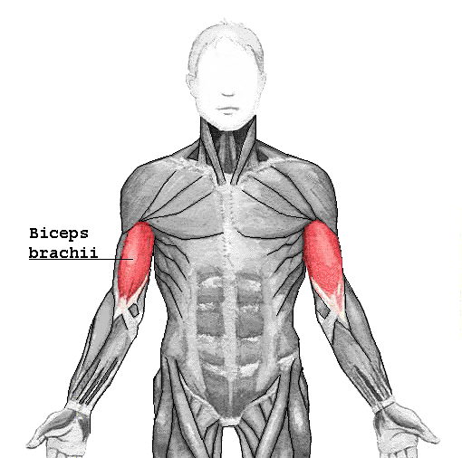
Changes for: biceps brachii
- Deleted
- Added
Changes for: pectoral appendage
Changes for: interlobular artery
- Deleted
- Added
Changes for: paired limb/fin
Changes for: pelvic appendage
Changes for: pit
- Added
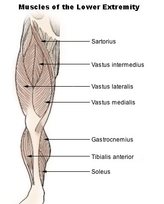
Changes for: vastus medialis
- Deleted
- Added
Changes for: femoral vein
- Deleted
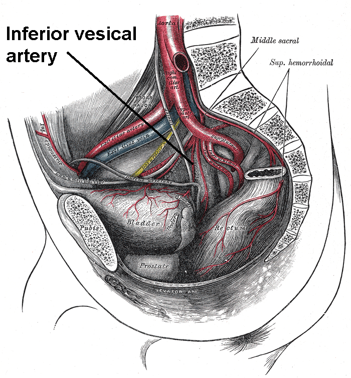
Changes for: inferior vesical artery
- Deleted
- - inferior vesical artery SubClassOf deferent duct artery
- - inferior vesical artery SubClassOf in lateral side of some fundus of urinary bladder
- - inferior vesical artery SubClassOf lateral structure
- - inferior vesical artery SubClassOf supplies some prostate gland
- - inferior vesical artery SubClassOf supplies some seminal vesicle
- - inferior vesical artery SubClassOf supplies some ureter
- - inferior vesical artery SubClassOf supplies some vas deferens
- - inferior vesical artery SubClassOf ureteric segment of renal artery
- - inferior vesical artery definition The inferior vesical artery is an artery in the pelvis that supplies the lower part of the bladder. [WP,unvetted]. { database cross reference=http://en.wikipedia.org/wiki/Inferior_vesical_artery }
- - inferior vesical artery depicted by http://upload.wikimedia.org/wikipedia/commons/1/13/Inferiorvesical.png
- - inferior vesical artery has exact synonym arteria vesicali inferior { has synonym type=latin term }
- Added
- + inferior vesical artery EquivalentTo vesical artery and supplies some fundus of urinary bladder
- + inferior vesical artery SubClassOf branching part of some internal iliac artery
- + inferior vesical artery SubClassOf systemic artery
- + inferior vesical artery definition An artery that supplies blood to the lower urinary bladder. { database cross reference=http://orcid.org/0000-0002-6601-2165 }
- + inferior vesical artery depicted by https://upload.wikimedia.org/wikipedia/commons/1/13/Inferiorvesical.png
- + inferior vesical artery has exact synonym arteria vesicali inferior { database cross reference=http://en.wikipedia.org/wiki/Inferior_vesical_artery , has synonym type=latin term }
- + inferior vesical artery has exact synonym arteria vesicalis inferior { database cross reference=http://en.wikipedia.org/wiki/Inferior_vesical_artery , has synonym type=latin term }
Changes for: superior vesical artery
- Deleted
- - superior vesical artery SubClassOf in lateral side of some urinary bladder
- - superior vesical artery SubClassOf lateral structure
- - superior vesical artery SubClassOf trunk blood vessel
- - superior vesical artery definition The superior vesical artery supplies numerous branches to the upper part of the bladder. From one of these a slender vessel, the artery to the ductus deferens, takes origin and accompanies the duct in its course to the testis, where it anastomoses with the internal spermatic artery. Other branches supply the ureter. The first part of the superior vesical artery represents the terminal section of the previous portion of the umbilical artery. [WP,unvetted]. { database cross reference=http://en.wikipedia.org/wiki/Superior_vesical_artery }
- - superior vesical artery depicted by http://upload.wikimedia.org/wikipedia/commons/2/20/Internal_iliac_branches.PNG
- - superior vesical artery has exact synonym arteria vesicalis superior
- Added
- + superior vesical artery EquivalentTo vesical artery and supplies some ureter
- + superior vesical artery SubClassOf supplies some ureter
- + superior vesical artery SubClassOf supplies some urinary bladder
- + superior vesical artery SubClassOf ureteric segment of renal artery
- + superior vesical artery definition An artery that supplies blood to the upper urinary bladder. { database cross reference=http://orcid.org/0000-0002-6601-2165 }
- + superior vesical artery depicted by https://upload.wikimedia.org/wikipedia/commons/0/02/Internal_iliac_branches.PNG
- + superior vesical artery development notes The first part of the superior vesical artery represents the terminal section of the previous portion of the umbilical artery (fetal hypogastric artery)[WP]
- + superior vesical artery has exact synonym arteria vesicali superior { database cross reference=http://en.wikipedia.org/wiki/Superior_vesical_artery , has synonym type=latin term }
- + superior vesical artery has exact synonym arteria vesicalis superior { database cross reference=http://en.wikipedia.org/wiki/Superior_vesical_artery , has synonym type=latin term }
Changes for: common fibular nerve
- Deleted
- - common fibular nerve definition A nerve arising at the terminal division of the sciatic nerve at the popliteal fossa and extending to the neck of the fibula, ultimately innervating the Peroneus muscle[MP]. The common peroneal nerve (common fibular nerve; external popliteal nerve; peroneal nerve), about one-half the size of the tibial nerve, is derived from the dorsal branches of the fourth and fifth lumbar and the first and second sacral nerves. It descends obliquely along the lateral side of the popliteal fossa to the head of the fibula, close to the medial margin of the biceps femoris muscle. It lies between the tendon of the biceps femoris and lateral head of the gastrocnemius muscle, winds around the neck of the fibula, between the peronæus longus and the bone, and divides beneath the muscle into the superficial peroneal nerve (superficial fibular nerve) and deep peroneal nerve (deep fibular nerve). [WP,unvetted]. { database cross reference=http://en.wikipedia.org/wiki/Common_fibular_nerve , database cross reference=MP:0011211 }
- - common fibular nerve has exact synonym peroneal nerve
- Added
- + common fibular nerve SubClassOf branching part of some sciatic nerve
- + common fibular nerve SubClassOf fibular nerve
- + common fibular nerve SubClassOf innervates some peroneus
- + common fibular nerve definition A nerve arising at the terminal division of the sciatic nerve at the popliteal fossa and extending to the neck of the fibula, ultimately innervating the Peroneus muscle[MP]. { database cross reference=MP:0011211 }
- + common fibular nerve structure notes derives into deep and superficial branches
Changes for: sciatic nerve
- Added
- + sciatic nerve SubClassOf innervates some skin of leg
Changes for: basilic vein
- Deleted
- - basilic vein SubClassOf vein
- Added
- + basilic vein SubClassOf superficial vein
Changes for: skin of limb
- Deleted
- - skin of limb editor note This class was created automatically from a combination of ontologies
Changes for: skin of thorax
- Deleted
- - skin of thorax editor note This class was created automatically from a combination of ontologies
Changes for: skin of neck
- Deleted
- - skin of neck editor note This class was created automatically from a combination of ontologies
Changes for: skin of abdomen
- Deleted
- - skin of abdomen editor note This class was created automatically from a combination of ontologies
Changes for: skin of pelvis
- Deleted
- - skin of pelvis SubClassOf part of some skin of trunk
- - skin of pelvis editor note This class was created automatically from a combination of ontologies
Changes for: median basilic vein
- Deleted
- - median cubital vein SubClassOf forelimb blood vessel
- - median cubital vein SubClassOf vein
- - median cubital vein definition In human anatomy, the median cubital vein (or median basilic vein) a superficial vein of the upper limb. It connects the basilic and cephalic vein and is often used for venipuncture (taking blood). It lies in the cubital fossa superficial to the bicipital aponeurosis. There exists a fair amount of variation of the median cubital vein. More commonly the vein forms an H-pattern with the cephalic and basilic veins making up the sides. Other forms include an M-pattern, where the vein branches to the cephalic and basilic veins. [WP,unvetted]. { database cross reference=http://en.wikipedia.org/wiki/Median_cubital_vein }
- - median cubital vein editor note This class was created automatically from a combination of ontologies
- - median cubital vein label median cubital vein
- Added
- + median basilic vein SubClassOf basilic vein
- + median basilic vein SubClassOf part of some forelimb
- + median basilic vein database cross reference http://linkedlifedata.com/resource/umls/id/C0226807
- + median basilic vein database cross reference http://ncicb.nci.nih.gov/xml/owl/EVS/Median_Basilic_Vein
- + median basilic vein database cross reference http://www.snomedbrowser.com/Codes/Details/368496000
- + median basilic vein definition A vein between the biceps and pronator radii teres muscles that unites with the common ulnar vein to form the basilic vein within the forearm. { database cross reference=ncithesaurus:Median_Basilic_Vein }
- + median basilic vein editor note Merged median cubital and basilic veins on basis of wikipedia synonym - needs verificied
- + median basilic vein has exact synonym median cubital vein { database cross reference=http://en.wikipedia.org/wiki/Median_cubital_vein , database cross reference=MA:0002176 }
- + median basilic vein has related synonym medial antecubital vein { database cross reference=http://en.wikipedia.org/wiki/Median_cubital_vein }
- + median basilic vein has related synonym median cubital { database cross reference=http://en.wikipedia.org/wiki/Median_cubital_vein }
- + median basilic vein label median basilic vein
Changes for: carotid sinus nerve
- Deleted
- - carotid sinus nerve SubClassOf part of some glossopharyngeal nerve
- - carotid sinus nerve definition Carotid branch of glossopharyngeal nerve. { database cross reference=BTO:0004978 }
- - carotid sinus nerve has exact synonym ramus sinus carotici { database cross reference=FMA:53488 }
- Added
- + carotid sinus nerve SubClassOf branching part of some glossopharyngeal nerve
- + carotid sinus nerve SubClassOf extends fibers into some nucleus of solitary tract
- + carotid sinus nerve SubClassOf has part some baroreceptor
- + carotid sinus nerve definition A sensory branch of the glossopharyngeal nerve (CN IX) carrying signals from the baroceptors (blood pressure receptors) in the bifurcation of the carotid artery to the nucleus of the solitary tract (nucleus solitarius). { database cross reference=http://medical-dictionary.thefreedictionary.com/carotid+sinus+nerve }
- + carotid sinus nerve has exact synonym Hering sinus nerve { database cross reference=http://medical-dictionary.thefreedictionary.com/carotid+sinus+nerve }
- + carotid sinus nerve has exact synonym ramus sinus carotici nervi glossopharyngei { has synonym type=latin term }
- + carotid sinus nerve has exact synonym ramus sinus carotici { database cross reference=FMA:53488 , has synonym type=latin term }
- + carotid sinus nerve has exact synonym sinus nerve of Hering { database cross reference=http://medical-dictionary.thefreedictionary.com/carotid+sinus+nerve }
Changes for: anal column
- Deleted
- Added
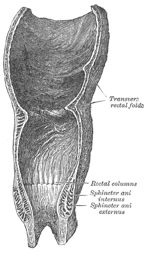
Changes for: glans penis
- Deleted
- - glans penis SubClassOf organ part
- - glans penis SubClassOf reproductive structure
- Added
- + glans penis EquivalentTo glans and part of some penis
- + glans penis SubClassOf glans
Changes for: thick ascending limb of loop of Henle
- Deleted
- Added
Changes for: nephron
- Deleted
- Added
Changes for: proximal convoluted tubule
- Deleted
- Added
Changes for: descending limb of loop of Henle
- Deleted
- Added
Changes for: loop of Henle
- Deleted
- Added
Changes for: pancreas
- Deleted
- Added
Changes for: left outer canthus
- Deleted
- - left outer canthus SubClassOf lateral structure
Changes for: right outer canthus
- Deleted
- - right outer canthus SubClassOf lateral structure
Changes for: helicine artery
- Added
- + helicine artery EquivalentTo helicine branch of uterine artery or helicine artery of penis
Changes for: iris nerve
- Added
- + iris nerve definition Any nerve that innervates the iris. { database cross reference=UBERON:cjm }
- + iris nerve editor note added for consistency with mouse ontologies. The term ‘ciliary nerve’ is usually used to denote either of the two nerves (with different origins) that innervate the iris, as well as ciliary muscles and cornea
- + iris nerve has narrow synonym ciliary nerve
Changes for: marsupium
- Deleted
- - marsupium SubClassOf reproductive structure
- Added
- + marsupium SubClassOf external soft tissue zone
Changes for: paired limb/fin segment
Changes for: future glans
- Added
- + future glans EquivalentTo future glans penis or future glans clitoris
- + future glans SubClassOf has potential to develop into some glans
Changes for: otic notch
- Deleted
- - otic notch definition Otic notches are invagination in the posterior margin of the skull roof, one behind each orbit. Such notches are found in labyrinthodonts and some of their immediate ancestors, but not their reptilian descendants. The presence or absence of the otic notches is one of the traits used to separate the amniotes from the amphibian grade tetrapods. The notches have been interpreted as part of an auditory structure, and are often shown holding a tympanum similar to those seen in modern anurans. Analysis of the columella (the stapes in amphibians and reptiles) of labyrinthodonts however indicate it did not function in transmitting low energy vibrations, thus rendering them effectively deaf to airborne sound. The otic notch instead functioned as a spiracle, at least in the early forms. { database cross reference=http://en.wikipedia.org/wiki/Otic_notch }
- Added
- + otic notch definition Otic notches are invagination in the posterior margin of the skull roof, one behind each orbit. Such notches are found in labyrinthodonts and some of their immediate ancestors, but not their reptilian descendants. The presence or absence of the otic notches is one of the traits used to separate the amniotes from the amphibian grade tetrapods. { database cross reference=http://en.wikipedia.org/wiki/Otic_notch }
- + otic notch function notes The notches have been interpreted as part of an auditory structure, and are often shown holding a tympanum similar to those seen in modern anurans. Analysis of the columella (the stapes in amphibians and reptiles) of labyrinthodonts however indicate it did not function in transmitting low energy vibrations, thus rendering them effectively deaf to airborne sound. The otic notch instead functioned as a spiracle, at least in the early forms
Changes for: costal cartilage
- Deleted
- - costal cartilage comment Editor note: we currently model this as part of the rib, is this correct?
- Added
- + costal cartilage editor note we currently model this as part of the rib, is this correct?
Changes for: cephalic vein
- Deleted
- - cephalic vein SubClassOf vein
- Added
- + cephalic vein SubClassOf superficial vein
Changes for: gall bladder
- Deleted
- Added
Changes for: inferior orbital fissure
- Added
- + inferior orbital fissure EquivalentTo orbital fissure and connects some maxilla and connects some alisphenoid bone
- + inferior orbital fissure SubClassOf connects some alisphenoid bone
- + inferior orbital fissure SubClassOf connects some maxilla
Changes for: obsolete neuromuscular junction
- Deleted
- - neuromuscular junction SubClassOf cell part
- - neuromuscular junction SubClassOf part of some nervous system
- - neuromuscular junction database cross reference FBbt:00005142
- - neuromuscular junction database cross reference FMA:61803
- - neuromuscular junction database cross reference GAID:809
- - neuromuscular junction database cross reference GO:0031594
- - neuromuscular junction database cross reference MESH:D009469
- - neuromuscular junction database cross reference NIF_Subcellular:sao1124888485
- - neuromuscular junction definition . { database cross reference=http://en.wikipedia.org/wiki/Neuromuscular_junction }
- - neuromuscular junction depicted by http://upload.wikimedia.org/wikipedia/commons/2/23/NMJ.jpg
- - neuromuscular junction editor note TODO - use GO
- - neuromuscular junction has exact synonym myoneural junction
- - neuromuscular junction has related synonym synapsis neuromuscularis; junctio neuromuscularis { database cross reference=http://en.wikipedia.org/wiki/Neuromuscular_junction , has synonym type=latin term }
- - neuromuscular junction in subset pheno slim
- - neuromuscular junction label neuromuscular junction
- Added
- + obsolete neuromuscular junction consider FBbt:00005142
- + obsolete neuromuscular junction consider FMA:61803
- + obsolete neuromuscular junction consider GAID:809
- + obsolete neuromuscular junction consider MESH:D009469
- + obsolete neuromuscular junction consider NIF_Subcellular:sao1124888485
- + obsolete neuromuscular junction deprecated true
- + obsolete neuromuscular junction has related synonym junctio neuromuscularis { database cross reference=http://en.wikipedia.org/wiki/Neuromuscular_junction , has synonym type=latin term }
- + obsolete neuromuscular junction has related synonym synapsis neuromuscularis { database cross reference=http://en.wikipedia.org/wiki/Neuromuscular_junction , has synonym type=latin term }
- + obsolete neuromuscular junction label obsolete neuromuscular junction
- + obsolete neuromuscular junction term replaced by GO:0031594
Changes for: obsolete motor nerve
- Added
- + obsolete motor nerve term replaced by UBERON:0006798
Changes for: sensory nerve
- Deleted
- - sensory nerve definition A group of neurons that provides a nervous response from a tissue[AEO]. { database cross reference=http://en.wikipedia.org/wiki/Sensory_nerve , database cross reference=AEO:0000201 }
- - sensory nerve external definition General anatomical term applied to nerves in which somatic afferent nerve fibers predominate[FMA]. nerves that receive sensory stimuli made up of nerve fibers, called sensory fibers (mechanoreceptor fibers sense body movement and pressure placed against the body, and nociceptor fibers sense tissue injury)[WP][Wikipedia:Sensory_nerve]. { source=http://en.wikipedia.org/wiki/Sensory_nerve }
- - sensory nerve has exact synonym nervus sensorius
- - sensory nerve has related synonym nervus sensorius { database cross reference=http://en.wikipedia.org/wiki/Sensory_nerve , has synonym type=latin term }
- Added
- + sensory nerve definition A nerve that transmits from sensory receptors on the surface of the body to the central nervous system. { database cross reference=http://en.wikipedia.org/wiki/Sensory_nerve , database cross reference=UBERON:cjm }
- + sensory nerve has exact synonym nervus sensorius { database cross reference=http://en.wikipedia.org/wiki/Sensory_nerve , has synonym type=latin term }
Changes for: pericardial cavity
- Deleted
Changes for: pouch sphincter
- Deleted
- - pouch sphincter SubClassOf reproductive structure
Changes for: dorsal nerve of clitoris
- Deleted
- - dorsal nerve of clitoris definition The dorsal nerve of the clitoris is a nerve in females that branches off the pudendal nerve to innervate the clitoris. { database cross reference=http://en.wikipedia.org/wiki/Dorsal_nerve_of_clitoris }
- Added
- + dorsal nerve of clitoris EquivalentTo nerve and innervates some clitoris and innervates some glans clitoris and branching part of some pudendal nerve
- + dorsal nerve of clitoris SubClassOf innervates some glans clitoris
- + dorsal nerve of clitoris SubClassOf nerve of clitoris
- + dorsal nerve of clitoris definition The deep terminal division of the pudendal nerve that runs along the dorsum of the clitoral shaft and innervates the the glans clitoris. { database cross reference=http://en.wikipedia.org/wiki/Dorsal_nerve_of_clitoris , database cross reference=http://medical-dictionary.thefreedictionary.com/dorsal%20nerve%20of%20clitoris }
Changes for: dorsal nerve of penis
- Deleted
- - dorsal nerve of penis definition The dorsal nerve of the penis is the deepest division of the pudendal nerve; it accompanies the internal pudendal artery along the ramus of the ischium; it then runs forward along the margin of the inferior ramus of the pubis, between the superior and inferior layers of the fascia of the urogenital diaphragm. Piercing the inferior layer it gives a branch to the corpus cavernosum penis, and passes forward, in company with the dorsal artery of the penis, between the layers of the suspensory ligament, on to the dorsum of the penis, and ends on the glans penis. { database cross reference=http://en.wikipedia.org/wiki/Dorsal_nerve_of_the_penis }
- Added
- + dorsal nerve of penis EquivalentTo nerve and innervates some penis and innervates some glans penis and innervates some prepuce of penis and innervates some corpus cavernosum penis and branching part of some pudendal nerve
- + dorsal nerve of penis SubClassOf innervates some corpus cavernosum penis
- + dorsal nerve of penis SubClassOf innervates some glans penis
- + dorsal nerve of penis SubClassOf innervates some prepuce of penis
- + dorsal nerve of penis SubClassOf nerve of penis
- + dorsal nerve of penis definition The deep terminal division of the pudendal nerve that runs along the dorsum of the penis and innervates the the corpus cavernosum penis, the prepuce and the glans penis. { database cross reference=http://en.wikipedia.org/wiki/Dorsal_nerve_of_the_penis , database cross reference=http://medical-dictionary.thefreedictionary.com/dorsal%20nerve%20of%20penis }
Changes for: saphenous nerve
- Deleted
- - saphenous nerve definition The saphenous nerve (long or internal saphenous nerve) is the largest cutaneous branch of the femoral nerve. [WP,unvetted]. { database cross reference=http://en.wikipedia.org/wiki/Saphenous_nerve }
- Added
- + saphenous nerve definition The largest cutaneous branch of the femoral nerve. [WP,unvetted]. { database cross reference=http://en.wikipedia.org/wiki/Saphenous_nerve }
Changes for: parietal pleura
- Deleted
Changes for: visceral pleura
- Deleted
Changes for: visceral serous pericardium
- Deleted
Changes for: lymphoid system
- Deleted
Changes for: eyeball of camera-type eye
- Deleted
- - eyeball of camera-type eye definition The camera-type eye apart from its appendages/adnexa. { database cross reference=http://en.wikipedia.org/wiki/Globe_(human_eye) }
- Added
- + eyeball of camera-type eye definition The core globe-shaped component of the camera-type eye. { database cross reference=UBERON:cjm }
Changes for: fibrous pericardium
- Deleted
Changes for: pelvic region of trunk
- Deleted
- - pelvis definition lower segment of the trunk, inferioposterior to the abdomen proper, in the transition area between the trunk and the lower limbs. { database cross reference=http://en.wikipedia.org/wiki/Pelvis , database cross reference=http://orcid.org/0000-0002-6601-2165 }
- - pelvis depicted by http://upload.wikimedia.org/wikipedia/commons/f/fe/Gray242.png
- - pelvis label pelvis
- Added
- + pelvic region of trunk definition The lower segment of the trunk, inferioposterior to the abdomen proper, in the transition area between the trunk and the lower limbs. { database cross reference=http://en.wikipedia.org/wiki/Pelvis , database cross reference=http://orcid.org/0000-0002-6601-2165 , database cross reference=https://github.com/obophenotype/uberon/issues/720 }
- + pelvic region of trunk has exact synonym pelvis { database cross reference=FMA:9578 , database cross reference=MA:0000030 }
- + pelvic region of trunk label pelvic region of trunk
Changes for: serous pericardium
- Deleted
Changes for: superficial epigastric vein
- Added
- + superficial epigastric vein SubClassOf superficial vein
Changes for: right gastric vein
- Deleted
- - right gastric vein EquivalentTo gastric vein and in right side of some multicellular organism
- - right gastric vein SubClassOf in right side of some multicellular organism
- - right gastric vein SubClassOf lateral structure
- Added
- + right gastric vein EquivalentTo gastric vein and in right side of some trunk
- + right gastric vein SubClassOf in right side of some trunk
Changes for: left gastric vein
- Deleted
- - left gastric vein EquivalentTo gastric vein and in left side of some multicellular organism
- - left gastric vein SubClassOf in left side of some multicellular organism
- - left gastric vein SubClassOf lateral structure
- Added
- + left gastric vein EquivalentTo gastric vein and in left side of some trunk
- + left gastric vein SubClassOf in left side of some trunk
Changes for: epicardium
Changes for: bona-fide anatomical boundary
Changes for: eyebrow
- Deleted
- - eyebrow SubClassOf part of some ocular region
- Added
- + eyebrow SubClassOf part of some ocular adnexa
Changes for: saphenous vein
- Deleted
- - saphenous vein SubClassOf vein
- Added
- + saphenous vein SubClassOf superficial vein
Changes for: paired limb/fin cartilage
Changes for: deferent duct artery
- Deleted
- - deferent duct artery SubClassOf artery
- - deferent duct artery SubClassOf trunk blood vessel
- Added
- + deferent duct artery SubClassOf systemic artery
Changes for: reproductive structure
Changes for: orbital fissure
- Added
- + orbital fissure EquivalentTo foramen of skull and part of some orbit of skull
- + orbital fissure SubClassOf part of some orbit of skull
Changes for: renal interlobular vein
- Deleted
- Added
Changes for: descending thin limb
- Deleted
- Added
Changes for: renal connecting tubule
- Deleted
- Added
Changes for: renal collecting system
- Deleted
- Added
Changes for: venom
- Added
- + venom present in taxon http://purl.obolibrary.org/obo/NCBITaxon_264759 { source=Wikipedia }
- + venom present in taxon http://purl.obolibrary.org/obo/NCBITaxon_6656
- + venom present in taxon http://purl.obolibrary.org/obo/NCBITaxon_7898
- + venom present in taxon http://purl.obolibrary.org/obo/NCBITaxon_79805 { source=Wikipedia }
- + venom present in taxon http://purl.obolibrary.org/obo/NCBITaxon_8314
- + venom present in taxon http://purl.obolibrary.org/obo/NCBITaxon_9257
Changes for: C2 segment of cervical spinal cord
- Deleted
- - second cervical spinal cord segment label second cervical spinal cord segment
- Added
- + C2 segment of cervical spinal cord definition The segment of the spinal cord that corresponds to the second cervical vertebra in most mammals. { database cross reference=https://github.com/obophenotype/uberon/issues/725 }
- + C2 segment of cervical spinal cord label C2 segment of cervical spinal cord
Changes for: C3 segment of cervical spinal cord
- Deleted
- - third cervical spinal cord segment label third cervical spinal cord segment
- Added
- + C3 segment of cervical spinal cord definition The segment of the spinal cord that corresponds to the third cervical vertebra in most mammals. { database cross reference=https://github.com/obophenotype/uberon/issues/725 }
- + C3 segment of cervical spinal cord label C3 segment of cervical spinal cord
Changes for: C8 segment of cervical spinal cord
- Deleted
- - eighth cervical spinal cord segment label eighth cervical spinal cord segment
- Added
- + C8 segment of cervical spinal cord label C8 segment of cervical spinal cord
Changes for: C4 segment of cervical spinal cord
- Deleted
- - fourth cervical spinal cord segment label fourth cervical spinal cord segment
- Added
- + C4 segment of cervical spinal cord definition The segment of the spinal cord that corresponds to the fourth cervical vertebra in most mammals. { database cross reference=https://github.com/obophenotype/uberon/issues/725 }
- + C4 segment of cervical spinal cord label C4 segment of cervical spinal cord
Changes for: C5 segment of cervical spinal cord
- Deleted
- - fifth cervical spinal cord segment label fifth cervical spinal cord segment
- Added
- + C5 segment of cervical spinal cord definition The segment of the spinal cord that corresponds to the fifth cervical vertebra in most mammals. { database cross reference=https://github.com/obophenotype/uberon/issues/725 }
- + C5 segment of cervical spinal cord label C5 segment of cervical spinal cord
Changes for: C6 segment of cervical spinal cord
- Deleted
- - sixth cervical spinal cord segment label sixth cervical spinal cord segment
- Added
- + C6 segment of cervical spinal cord definition The segment of the spinal cord that corresponds to the sixth cervical vertebra in most mammals. { database cross reference=https://github.com/obophenotype/uberon/issues/725 }
- + C6 segment of cervical spinal cord label C6 segment of cervical spinal cord
Changes for: C7 segment of cervical spinal cord
- Deleted
- - seventh cervical spinal cord segment label seventh cervical spinal cord segment
- Added
- + C7 segment of cervical spinal cord definition The segment of the spinal cord that corresponds to the seventh cervical vertebra in most mammals. { database cross reference=https://github.com/obophenotype/uberon/issues/725 }
- + C7 segment of cervical spinal cord label C7 segment of cervical spinal cord
Changes for: C1 segment of cervical spinal cord
- Deleted
- - first cervical spinal cord segment label first cervical spinal cord segment
- Added
- + C1 segment of cervical spinal cord definition The segment of the spinal cord that corresponds to the first cervical vertebra in most mammals. { database cross reference=https://github.com/obophenotype/uberon/issues/725 }
- + C1 segment of cervical spinal cord has exact synonym C1 cervical spinal cord
- + C1 segment of cervical spinal cord label C1 segment of cervical spinal cord
Changes for: loop of Henle ascending limb thin segment
- Deleted
- Added
Changes for: strand of vibrissa hair
- Deleted
Changes for: accessory XI nerve
- Deleted
- - accessory XI nerve SubClassOf nerve
- Added
- + accessory XI nerve SubClassOf laryngeal nerve
Changes for: paired limb/fin bud
Changes for: bone of free limb or fin
Changes for: anal canal
- Deleted
- Added
Changes for: intertarsal-type crurotarsal joint
- Deleted
- - intertarsal-type crurotarsal joint comment The ankle joint of pseudosuchians (including crocodilians) and phytosaurs, passing between the astragalus and calcaneum, is also called crurotarsal joint in the literature.[5][6] In the skeletons of the phytosaurs and most of the pseudosuchians this joint bends around a peg on the astragalus which fits into a socket in the calcaneum (the “crocodile normal” tarsus); only in the skeletons of the ornithosuchid pseudosuchians a peg on the calcaneum fits into a socket in the astragalus (the “crocodile reversed” tarsus).[3] Strictly speaking this ankle isn’t a crurotarsal joint in the previously discussed sense, as it’s situated between the two proximal tarsal bones. However, while calcaneum isn’t fixed to the fibula, the astragalus is fixed to the tibia by a suture and thus in practice it functions as an extension of the crus.[7]
- Added
- + intertarsal-type crurotarsal joint taxon notes The ankle joint of pseudosuchians (including crocodilians) and phytosaurs, passing between the astragalus and calcaneum, is also called crurotarsal joint in the literature.[5][6] In the skeletons of the phytosaurs and most of the pseudosuchians this joint bends around a peg on the astragalus which fits into a socket in the calcaneum (the ‘crocodile normal’ tarsus); only in the skeletons of the ornithosuchid pseudosuchians a peg on the calcaneum fits into a socket in the astragalus (the ‘crocodile reversed’ tarsus).[3] Strictly speaking this ankle isn’t a crurotarsal joint in the previously discussed sense, as it’s situated between the two proximal tarsal bones. However, while calcaneum isn’t fixed to the fibula, the astragalus is fixed to the tibia by a suture and thus in practice it functions as an extension of the crus.[7]
Changes for: nasopalatine nerve
- Deleted
- - nasopalatine nerve definition One branch of the posterior superior nasal branches (trigeminal, maxillary branch), longer and larger than the others, is named the nasopalatine nerve (sometimes called the long sphenopalatine nerve). It enters the nasal cavity through the sphenopalatine foramen, passes across the roof of the nasal cavity below the orifice of the sphenoidal sinus to reach the septum, and then runs obliquely downward and forward between the periosteum and mucous membrane of the lower part of the septum. It descends to the roof of the mouth through the incisive canal and communicates with the corresponding nerve of the opposite side and with the greater palatine nerve. It supplies the palatal structures around the upper central and lateral incisors and the canines (the upper front six teeth). It also furnishes a few filaments to the mucous membrane of the nasal septum. The medial superior posterior nasal branches of maxillary nerve usually branches from the nasopalatine nerve. { database cross reference=http://en.wikipedia.org/wiki/Nasopalatine_nerve }
- Added
- + nasopalatine nerve EquivalentTo nerve and innervates some mucosa of hard palate and branching part of some maxillary nerve
- + nasopalatine nerve SubClassOf innervates some mucosa of hard palate
- + nasopalatine nerve definition A branch of that maxillary nerve that innervates the mucous membrane of the hard palate. { database cross reference=http://medical-dictionary.thefreedictionary.com/nasopalatine+nerve , database cross reference=UBERON:cjm }
Changes for: great auricular nerve
- Deleted
- - great auricular nerve SubClassOf nerve
- - great auricular nerve database cross reference MA:0001232
- - great auricular nerve definition The great auricular nerve originates from the cervical plexus, composed of branches of spinal nerves C2 and C3. It provides sensory innervation for the skin over parotid gland and mastoid process, and both surfaces of the outer ear. { database cross reference=http://en.wikipedia.org/wiki/Great_auricular_nerve }
- - great auricular nerve editor note TODO - check MA grouping; in MA this is part_of pinna - should this be a different relationship?
- - great auricular nerve has exact synonym auricular nerve { database cross reference=MA:0001232 }
- - great auricular nerve has related synonym nervus auricularis magnus { database cross reference=http://en.wikipedia.org/wiki/Great_auricular_nerve , has synonym type=latin term }
- Added
- + great auricular nerve SubClassOf innervates some pinna
- + great auricular nerve SubClassOf nerve innervating pinna
- + great auricular nerve definition A nerve arising from the second and third cervical nerves from the cervical plexus and innervating the skin over the parotid gland, the skin of part of the ear, the adjacent portion of the scalp, cheek, and angle of the jaw. { database cross reference=http://en.wikipedia.org/wiki/Great_auricular_nerve , database cross reference=http://medical-dictionary.thefreedictionary.com/great+auricular+nerve }
- + great auricular nerve has exact synonym nervus auricularis magnus { database cross reference=http://en.wikipedia.org/wiki/Great_auricular_nerve , has synonym type=latin term }
Changes for: gastric vein
- Deleted
- - gastric vein definition Either of two veins (left gastric and right gastric) that drain blood away from the stomach and lesser curvature. { database cross reference=ncithesaurus:Gastric_Vein }
- Added
- + gastric vein SubClassOf in lateral side of some trunk
- + gastric vein SubClassOf lateral structure
- + gastric vein SubClassOf trunk blood vessel
- + gastric vein definition A laterally paired vein that drains blood away from the lesser curvature of the stomach. { database cross reference=http://ncicb.nci.nih.gov/xml/owl/EVS/Gastric_Vein , database cross reference=UBERON:cjm }
Changes for: renal efferent arteriole
- Deleted
- Added
Changes for: renal afferent arteriole
- Deleted
- Added
Changes for: dorsal root ganglion
- Added
- + dorsal root ganglion database cross reference http://uri.neuinfo.org/nif/nifstd/birnlex_2598
- + dorsal root ganglion has alternative id UBERON:0026602
- + dorsal root ganglion has exact synonym spinal ganglion part of peripheral nervous system { database cross reference=NIFSTD:birnlex_2598 }
Changes for: hypoblast (generic)
- Deleted
Changes for: simple eye with multiple lenses
- Deleted
- - simple eye with multiple lenses SubClassOf has part some lens of camera-type eye
- Added
- + simple eye with multiple lenses SubClassOf has component some transparent eye structure
Changes for: superficial fibular nerve
- Deleted
- - superficial fibular nerve SubClassOf innervates some hindlimb
- - superficial fibular nerve definition A branch of the common peroneal nerve. It innervates the surface of the calf and foot. { database cross reference=ncithesaurus:Superficial_Peroneal_Nerve }
- Added
- + superficial fibular nerve EquivalentTo nerve and innervates some fibularis longus and innervates some pes and innervates some fibularis brevis and branching part of some common fibular nerve
- + superficial fibular nerve SubClassOf fibular nerve
- + superficial fibular nerve SubClassOf innervates some fibularis brevis
- + superficial fibular nerve SubClassOf innervates some fibularis longus
- + superficial fibular nerve SubClassOf innervates some pes
- + superficial fibular nerve definition A branch of the common fibular nerve that innervates the fibularis longus and brevis muscles, and via branches innervates parts of the dorsal part of the pes. { database cross reference=http://medical-dictionary.thefreedictionary.com/superficial%20fibular%20nerve , database cross reference=http://ncicb.nci.nih.gov/xml/owl/EVS/Superficial_Peroneal_Nerve , database cross reference=UBERON:cjm }
Changes for: lower inner quadrant of breast
- Deleted
- - lower inner quadrant of breast SubClassOf organism subdivision
- - lower inner quadrant of breast SubClassOf part of some breast
- Added
- + lower inner quadrant of breast EquivalentTo quadrant of breast and in posterior side of some breast and in innermost side of some breast
- + lower inner quadrant of breast SubClassOf in innermost side of some breast
- + lower inner quadrant of breast SubClassOf in posterior side of some breast
- + lower inner quadrant of breast SubClassOf inner quadrant of breast
- + lower inner quadrant of breast SubClassOf lower quadrant of breast
Changes for: upper inner quadrant of breast
- Deleted
- - upper inner quadrant of breast SubClassOf organism subdivision
- - upper inner quadrant of breast SubClassOf part of some breast
- Added
- + upper inner quadrant of breast EquivalentTo quadrant of breast and in anterior side of some breast and in innermost side of some breast
- + upper inner quadrant of breast SubClassOf in anterior side of some breast
- + upper inner quadrant of breast SubClassOf in innermost side of some breast
- + upper inner quadrant of breast SubClassOf inner quadrant of breast
- + upper inner quadrant of breast SubClassOf upper quadrant of breast
Changes for: insect leg
- Added
- + insect leg editor note this class is present primarily to support GO
Changes for: chemosensory system
Changes for: paired limb/fin field
Changes for: inner canthus
- Added
- + inner canthus EquivalentTo canthus and in innermost side of some camera-type eye
- + inner canthus SubClassOf in innermost side of some camera-type eye
- + inner canthus SubClassOf lateral structure
Changes for: glans clitoris
- Deleted
- - glans clitoris SubClassOf reproductive structure
- Added
- + glans clitoris EquivalentTo glans and part of some clitoris
- + glans clitoris SubClassOf glans
Changes for: obturator nerve
- Added
- + obturator nerve SubClassOf innervates some hindlimb stylopod
Changes for: superior orbital fissure
- Added
- + superior orbital fissure EquivalentTo orbital fissure and connects some orbitosphenoid and connects some alisphenoid bone
- + superior orbital fissure SubClassOf connects some alisphenoid bone
- + superior orbital fissure SubClassOf connects some orbitosphenoid
Changes for: transverse palatine suture
- Deleted
- - transverse palatine suture SubClassOf suture of hard palate
- Added
- + transverse palatine suture SubClassOf endoderm-derived structure
- + transverse palatine suture SubClassOf neural crest-derived structure
- + transverse palatine suture SubClassOf part of some hard palate
Changes for: adrenal/interrenal gland
- Deleted
- Added
Changes for: deep fibular nerve
- Deleted
- - deep fibular nerve SubClassOf common fibular nerve
- Added
- + deep fibular nerve EquivalentTo nerve and innervates some tibialis anterior and innervates some fibularis tertius and branching part of some common fibular nerve
- + deep fibular nerve SubClassOf branching part of some common fibular nerve
- + deep fibular nerve SubClassOf fibular nerve
- + deep fibular nerve SubClassOf innervates some fibularis tertius
- + deep fibular nerve SubClassOf innervates some tibialis anterior
- + deep fibular nerve SubClassOf leg nerve
Changes for: efferent nerve
- Deleted
- - efferent nerve definition A group of neurons that provides a nervous signal to a tissue. { database cross reference=AEO:JB }
- Added
- + efferent nerve database cross reference FMA:5867
- + efferent nerve definition A nerve that transmits nerve impulses from the central nervous system to effectors such as muscles or glands. { database cross reference=UBERON:cjm }
- + efferent nerve fma set term http://purl.obolibrary.org/obo/FMA_76571
- + efferent nerve has exact synonym nervus efferente { has synonym type=latin term }
- + efferent nerve has related synonym motor nerve
- + efferent nerve has related synonym nervus motorius { database cross reference=FMA:5867 , has synonym type=latin term }
- + efferent nerve has related synonym neurofibrae efferentes { has synonym type=latin term }
Changes for: outer canthus
- Added
- + outer canthus EquivalentTo canthus and in outermost side of some camera-type eye
- + outer canthus SubClassOf in outermost side of some camera-type eye
- + outer canthus SubClassOf lateral structure
Changes for: upper outer quadrant of breast
- Deleted
- - upper outer quadrant of breast SubClassOf organism subdivision
- - upper outer quadrant of breast SubClassOf part of some breast
- Added
- + upper outer quadrant of breast EquivalentTo quadrant of breast and in anterior side of some breast and in outermost side of some breast
- + upper outer quadrant of breast SubClassOf in anterior side of some breast
- + upper outer quadrant of breast SubClassOf in outermost side of some breast
- + upper outer quadrant of breast SubClassOf outer quadrant of breast
- + upper outer quadrant of breast SubClassOf upper quadrant of breast
Changes for: lower outer quadrant of breast
- Deleted
- - lower outer quadrant of breast SubClassOf organism subdivision
- - lower outer quadrant of breast SubClassOf part of some breast
- Added
- + lower outer quadrant of breast EquivalentTo quadrant of breast and in posterior side of some breast and in outermost side of some breast
- + lower outer quadrant of breast SubClassOf in outermost side of some breast
- + lower outer quadrant of breast SubClassOf in posterior side of some breast
- + lower outer quadrant of breast SubClassOf lower quadrant of breast
- + lower outer quadrant of breast SubClassOf outer quadrant of breast
Report for properties
ObjectProperty objects lost from source: 0
ObjectProperty objects new in target: 4
New ObjectProperty : in_innermost_side_of
- in innermost side of label in_innermost_side_of
- in innermost side of has obo namespace uberon
- SubObjectPropertyOf( in innermost side of in lateral side of )
- in innermost side of id in_innermost_side_of
New ObjectProperty : immediately_anterior_to
- immediately anterior to shorthand immediately_anterior_to
- immediately anterior to database cross reference BSPO:0015009
- immediately anterior to id immediately_anterior_to
- SubObjectPropertyOf( immediately anterior to anterior to )
- immediately anterior to has obo namespace uberon
- immediately anterior to label immediately_anterior_to
- SubObjectPropertyOf( immediately anterior to adjacent to )
New ObjectProperty : immediately_posterior_to
- immediately posterior to id immediately_posterior_to
- SubObjectPropertyOf( immediately posterior to adjacent to )
- immediately posterior to has obo namespace uberon
- immediately posterior to label immediately_posterior_to
- immediately posterior to database cross reference BSPO:0015012
- immediately posterior to shorthand immediately_posterior_to
- SubObjectPropertyOf( immediately posterior to posterior to )
New ObjectProperty : in_outermost_side_of
- SubObjectPropertyOf( in outermost side of in lateral side of )
- in outermost side of id in_outermost_side_of
- in outermost side of has obo namespace uberon
- in outermost side of label in_outermost_side_of
Changed ObjectProperty objects: 0
July 24, 2015
|
Tweet
Comments Section
Feel free to comment on the post but keep it clean and on topic.
comments powered by Disqus