2015-01-01 release

Summary
This release encompasses multiple imrpovements in the representation of the nervous system. Many wikipedia-sourced definitions have been replaced with definitions from neurolex (632). We have defined and implemented a design pattern for cranial and spinal nerves, ganglia and nuclei (634). We made improvements to the mapping to Allen ontologies. Additionally, we now use the numeric identifiers for all Allen classes, rather than acronyms. Please note that the “ABA” prefix has been superseded by “MBA” (mouse brain atlas), to be consistent with the others (HBA, PBA, DMBA, DHBA). New terms were added from the HarvardOxford Cortical atlas based on work by Russ Poldrack and Trish Whetzel (630).
This release also ensures that every class from NIF_GrossAnatomy (but not its successor, neurolex) is represented in Uberon. Note that not all classes will be in the bridge file. Many of the region-part-of-X classes are present in Uberon as obsolete classes.
Changes
- neuro
- cranial nerve & ganglia improvements. Fixes issue 634
- spinal nerves: nerve roots now neutral w.r.t. CNS/PNS
- NTs for taste buds
- Fixes to developing neural tube / CNS
- NTs: forceps major & minor. Issue issue 630
- Syn for basolateral amygdala. Issue issue 630
- Replaced multiple wikipedia definitions with ones from neurolex
- Realigned with nlx and allen atlases
- added remaining nifstd classes. Note many may be obsoleted in future. See issue 632
- Obsoleted multiple regional-part-of-X terms. issue 632
- replaced all NIF_GrossAnatomy prefixes with NIFSTD. See issue issue 632
- NT: palatal taste bud. Refined taste bud axioms. Merged some new nifstd predominant part terms
- merging Vestibulocochlear nerve, root + nerve fiber bundle. Issue issue 300
- NTs for rostral ACC and olfactory EC and primary OC
- merging nifstd superior olive & SOC. Issue issue 300
- Refactored SOC, fixes issue 631
- entorhinal complex layers, ref doi:10.1155/2008/381243
- NTs, cingula. Issue issue 630
- NTs, corona radiata parts. Issue issue 630
- new MBA alignment (was ABA). Issue issue 609.
- NTs from HarvardOxford-Cortial atlas, via Russ Poldrack, Trish Whetzel. Rows 11-18. Issue issue 630
- Mapped Allen Brain xrefs from acronyms to IDs. Issue issue 609
- Fixed multiple incorrect BAMS xrefs
- skeletal
- added meckelian bone and meckelian foramen [adececchi]
- added hypophysial region [adececchi]
- embryonic/reproductive
- removing taxon constraint on extra-embryonic membrane, as yolk sacs are found in anamniotes. Fixes issue 635
- oviduct fixes
- vitelline vasculature improvement
- other
- Using nlx and mp for definition sources replacing select WP defs issue 633
- mandibular gland - improved defs sourced from MP, tax constr sourced from Bgee, more precise developmental lineage
- Improvements to:
- ligaments
- vertebral arches
- reproductive system development. NTs: gubernacular bulb parts
- heart and veins. NTs: inflow tracts
Ontology Diff Report
Original Ontology
- IRI: http://purl.obolibrary.org/obo/uberon.owl
- VersionIRI: http://purl.obolibrary.org/obo/uberon/releases/2014-12-09/uberon.owl
New Ontology
- IRI: http://purl.obolibrary.org/obo/uberon.owl
- VersionIRI: http://purl.obolibrary.org/obo/uberon/releases/2015-01-01/uberon.owl
Report for classes
Class objects lost from source: 3
- otic cup – merged into otic pit
- trunk of peripheral nerve – merged into nerve trunk
- paravertebral ganglia – merged into paravertebral ganglion
Class objects new in target: 546
New Class : obsolete regional part of trapezoid nuclear complex
- obsolete regional part of trapezoid nuclear complex label obsolete regional part of trapezoid nuclear complex
- obsolete regional part of trapezoid nuclear complex deprecated true
- obsolete regional part of trapezoid nuclear complex has obo namespace uberon
- obsolete regional part of trapezoid nuclear complex has exact synonym regional part of trapezoid nuclear complex { database cross reference=NIFSTD:birnlex_2574 }
- obsolete regional part of trapezoid nuclear complex has exact synonym regional part of trapezoid nuclear complex { database cross reference=NIFSTD:birnlex_2574 , has synonym type=preferred term when talking about an instance of this class in Homo sapiens }
- obsolete regional part of trapezoid nuclear complex consider http://uri.neuinfo.org/nif/nifstd/birnlex_2574
- obsolete regional part of trapezoid nuclear complex id UBERON:0029861
New Class : obsolete predominantly gray regional part of pretectal region
- obsolete predominantly gray regional part of pretectal region id UBERON:0022480
- obsolete predominantly gray regional part of pretectal region has exact synonym predominantly gray regional part of pretectal region { database cross reference=NIFSTD:birnlex_1003 }
- obsolete predominantly gray regional part of pretectal region has obo namespace uberon
- obsolete predominantly gray regional part of pretectal region deprecated true
- obsolete predominantly gray regional part of pretectal region label obsolete predominantly gray regional part of pretectal region
- obsolete predominantly gray regional part of pretectal region consider http://uri.neuinfo.org/nif/nifstd/birnlex_1003
- obsolete predominantly gray regional part of pretectal region has exact synonym predominantly gray regional part of pretectal region { database cross reference=NIFSTD:birnlex_1003 , has synonym type=preferred term when talking about an instance of this class in Homo sapiens }
New Class : obsolete superficial feature part of hypophysis
- obsolete superficial feature part of hypophysis consider http://uri.neuinfo.org/nif/nifstd/birnlex_1593
- obsolete superficial feature part of hypophysis SubClassOf part of some pituitary gland
- obsolete superficial feature part of hypophysis label obsolete superficial feature part of hypophysis
- obsolete superficial feature part of hypophysis has exact synonym superficial feature part of hypophysis { database cross reference=NIFSTD:birnlex_1593 }
- obsolete superficial feature part of hypophysis has exact synonym superficial feature part of hypophysis { database cross reference=NIFSTD:birnlex_1593 , has synonym type=preferred term when talking about an instance of this class in Homo sapiens }
- obsolete superficial feature part of hypophysis id UBERON:0024708
- obsolete superficial feature part of hypophysis deprecated true
- obsolete superficial feature part of hypophysis has obo namespace uberon
New Class : obsolete regional part of cingulate gyrus
- obsolete regional part of cingulate gyrus has obo namespace uberon
- obsolete regional part of cingulate gyrus SubClassOf part of some limbic lobe
- obsolete regional part of cingulate gyrus has exact synonym regional part of cingulate gyrus { database cross reference=NIFSTD:birnlex_1599 , has synonym type=preferred term when talking about an instance of this class in Homo sapiens }
- obsolete regional part of cingulate gyrus deprecated true
- obsolete regional part of cingulate gyrus id UBERON:0024714
- obsolete regional part of cingulate gyrus consider http://uri.neuinfo.org/nif/nifstd/birnlex_1599
- obsolete regional part of cingulate gyrus has exact synonym regional part of cingulate gyrus { database cross reference=NIFSTD:birnlex_1599 }
- obsolete regional part of cingulate gyrus label obsolete regional part of cingulate gyrus
New Class : obsolete regional part of outer ear
- obsolete regional part of outer ear id UBERON:0024721
- obsolete regional part of outer ear deprecated true
- obsolete regional part of outer ear has obo namespace uberon
- obsolete regional part of outer ear SubClassOf part of some ear
- obsolete regional part of outer ear label obsolete regional part of outer ear
- obsolete regional part of outer ear consider http://uri.neuinfo.org/nif/nifstd/birnlex_1605
- obsolete regional part of outer ear has exact synonym regional part of outer ear { database cross reference=NIFSTD:birnlex_1605 }
- obsolete regional part of outer ear has exact synonym regional part of outer ear { database cross reference=NIFSTD:birnlex_1605 , has synonym type=preferred term when talking about an instance of this class in Homo sapiens }
New Class : obsolete regional part of basilar membrane
- obsolete regional part of basilar membrane has obo namespace uberon
- obsolete regional part of basilar membrane has exact synonym regional part of basilar membrane { database cross reference=NIFSTD:birnlex_2527 , has synonym type=preferred term when talking about an instance of this class in Homo sapiens }
- obsolete regional part of basilar membrane SubClassOf part of some spiral organ of cochlea
- obsolete regional part of basilar membrane id UBERON:0029822
- obsolete regional part of basilar membrane has exact synonym regional part of basilar membrane { database cross reference=NIFSTD:birnlex_2527 }
- obsolete regional part of basilar membrane deprecated true
- obsolete regional part of basilar membrane consider http://uri.neuinfo.org/nif/nifstd/birnlex_2527
- obsolete regional part of basilar membrane label obsolete regional part of basilar membrane
New Class : obsolete predominantly gray regional part of temporal lobe
- obsolete predominantly gray regional part of temporal lobe deprecated true
- obsolete predominantly gray regional part of temporal lobe label obsolete predominantly gray regional part of temporal lobe
- obsolete predominantly gray regional part of temporal lobe has exact synonym predominantly gray regional part of temporal lobe { database cross reference=NIFSTD:birnlex_1633 }
- obsolete predominantly gray regional part of temporal lobe has obo namespace uberon
- obsolete predominantly gray regional part of temporal lobe id UBERON:0024749
- obsolete predominantly gray regional part of temporal lobe consider http://uri.neuinfo.org/nif/nifstd/birnlex_1633
- obsolete predominantly gray regional part of temporal lobe has exact synonym predominantly gray regional part of temporal lobe { database cross reference=NIFSTD:birnlex_1633 , has synonym type=preferred term when talking about an instance of this class in Homo sapiens }
- obsolete predominantly gray regional part of temporal lobe consider UBERON:0016538
New Class : regional part of lumbar spinal cord white matter
- regional part of lumbar spinal cord white matter has exact synonym regional part of lumbar spinal cord white matter { database cross reference=NIFSTD:birnlex_1647 }
- regional part of lumbar spinal cord white matter EquivalentTo multi-tissue structure and part of some lumbar spinal cord white matter
- regional part of lumbar spinal cord white matter has obo namespace uberon
- regional part of lumbar spinal cord white matter has exact synonym regional part of lumbar spinal cord white matter { database cross reference=NIFSTD:birnlex_1647 , has synonym type=preferred term when talking about an instance of this class in Homo sapiens }
- regional part of lumbar spinal cord white matter database cross reference http://uri.neuinfo.org/nif/nifstd/birnlex_1647
- regional part of lumbar spinal cord white matter SubClassOf regional part of lumbar spinal cord
- regional part of lumbar spinal cord white matter SubClassOf part of some lumbar spinal cord white matter
- regional part of lumbar spinal cord white matter id UBERON:0024762
- regional part of lumbar spinal cord white matter label regional part of lumbar spinal cord white matter
- regional part of lumbar spinal cord white matter SubClassOf part of some lumbar spinal cord
New Class : obsolete predominantly gray regional part of pontine reticular formation
- obsolete predominantly gray regional part of pontine reticular formation has exact synonym predominantly gray regional part of pontine reticular formation { database cross reference=NIFSTD:birnlex_1071 , has synonym type=preferred term when talking about an instance of this class in Homo sapiens }
- obsolete predominantly gray regional part of pontine reticular formation has exact synonym predominantly gray regional part of pontine reticular formation { database cross reference=NIFSTD:birnlex_1071 }
- obsolete predominantly gray regional part of pontine reticular formation SubClassOf part of some pontine reticular formation
- obsolete predominantly gray regional part of pontine reticular formation has obo namespace uberon
- obsolete predominantly gray regional part of pontine reticular formation deprecated true
- obsolete predominantly gray regional part of pontine reticular formation label obsolete predominantly gray regional part of pontine reticular formation
- obsolete predominantly gray regional part of pontine reticular formation consider http://uri.neuinfo.org/nif/nifstd/birnlex_1071
- obsolete predominantly gray regional part of pontine reticular formation id UBERON:0027453
New Class : paravermic Lobule III
- paravermic Lobule III has exact synonym paravermic lobule iii { database cross reference=NIFSTD:nlx_anat_20081216 , has synonym type=preferred term when talking about an instance of this class in Homo sapiens }
- paravermic Lobule III label paravermic Lobule III
- paravermic Lobule III id UBERON:0028919
- paravermic Lobule III has exact synonym paravermic lobule iii { database cross reference=NIFSTD:nlx_anat_20081216 }
- paravermic Lobule III has obo namespace uberon
- paravermic Lobule III SubClassOf regional Parts of the Paravermal Lobules
- paravermic Lobule III database cross reference http://uri.neuinfo.org/nif/nifstd/nlx_anat_20081216
New Class : paravermic Lobule II
- paravermic Lobule II id UBERON:0028918
- paravermic Lobule II SubClassOf regional Parts of the Paravermal Lobules
- paravermic Lobule II has exact synonym paravermic lobule ii { database cross reference=NIFSTD:nlx_anat_20081215 , has synonym type=preferred term when talking about an instance of this class in Homo sapiens }
- paravermic Lobule II has exact synonym paravermic lobule ii { database cross reference=NIFSTD:nlx_anat_20081215 }
- paravermic Lobule II database cross reference http://uri.neuinfo.org/nif/nifstd/nlx_anat_20081215
- paravermic Lobule II has obo namespace uberon
- paravermic Lobule II label paravermic Lobule II
New Class : paravermic Lobule VII
- paravermic Lobule VII label paravermic Lobule VII
- paravermic Lobule VII has obo namespace uberon
- paravermic Lobule VII id UBERON:0028924
- paravermic Lobule VII has exact synonym paravermic lobule vii { database cross reference=NIFSTD:nlx_anat_20081221 , has synonym type=preferred term when talking about an instance of this class in Homo sapiens }
- paravermic Lobule VII database cross reference http://uri.neuinfo.org/nif/nifstd/nlx_anat_20081221
- paravermic Lobule VII has exact synonym paravermic lobule vii { database cross reference=NIFSTD:nlx_anat_20081221 }
- paravermic Lobule VII SubClassOf regional Parts of the Paravermal Lobules
New Class : paravermic Lobule VIII
- paravermic Lobule VIII has obo namespace uberon
- paravermic Lobule VIII has exact synonym paravermic lobule viii { database cross reference=NIFSTD:nlx_anat_20081222 , has synonym type=preferred term when talking about an instance of this class in Homo sapiens }
- paravermic Lobule VIII label paravermic Lobule VIII
- paravermic Lobule VIII has exact synonym paravermic lobule viii { database cross reference=NIFSTD:nlx_anat_20081222 }
- paravermic Lobule VIII database cross reference http://uri.neuinfo.org/nif/nifstd/nlx_anat_20081222
- paravermic Lobule VIII SubClassOf regional Parts of the Paravermal Lobules
- paravermic Lobule VIII id UBERON:0028925
New Class : paravermic Lobule IX
- paravermic Lobule IX id UBERON:0028921
- paravermic Lobule IX database cross reference http://uri.neuinfo.org/nif/nifstd/nlx_anat_20081218
- paravermic Lobule IX has exact synonym paravermic lobule ix { database cross reference=NIFSTD:nlx_anat_20081218 }
- paravermic Lobule IX has exact synonym paravermic lobule ix { database cross reference=NIFSTD:nlx_anat_20081218 , has synonym type=preferred term when talking about an instance of this class in Homo sapiens }
- paravermic Lobule IX has obo namespace uberon
- paravermic Lobule IX has related synonym ventralis { database cross reference=NIFSTD:nlx_anat_20081218 }
- paravermic Lobule IX label paravermic Lobule IX
- paravermic Lobule IX SubClassOf regional Parts of the Paravermal Lobules
New Class : paravermic Lobule IV
- paravermic Lobule IV label paravermic Lobule IV
- paravermic Lobule IV database cross reference http://uri.neuinfo.org/nif/nifstd/nlx_anat_20081217
- paravermic Lobule IV SubClassOf regional Parts of the Paravermal Lobules
- paravermic Lobule IV id UBERON:0028920
- paravermic Lobule IV has obo namespace uberon
- paravermic Lobule IV has exact synonym paravermic lobule iv { database cross reference=NIFSTD:nlx_anat_20081217 , has synonym type=preferred term when talking about an instance of this class in Homo sapiens }
- paravermic Lobule IV has exact synonym paravermic lobule iv { database cross reference=NIFSTD:nlx_anat_20081217 }
New Class : paravermic Lobule VI
- paravermic Lobule VI has obo namespace uberon
- paravermic Lobule VI label paravermic Lobule VI
- paravermic Lobule VI has exact synonym paravermic lobule vi { database cross reference=NIFSTD:nlx_anat_20081220 }
- paravermic Lobule VI SubClassOf regional Parts of the Paravermal Lobules
- paravermic Lobule VI database cross reference http://uri.neuinfo.org/nif/nifstd/nlx_anat_20081220
- paravermic Lobule VI id UBERON:0028923
- paravermic Lobule VI has exact synonym paravermic lobule vi { database cross reference=NIFSTD:nlx_anat_20081220 , has synonym type=preferred term when talking about an instance of this class in Homo sapiens }
New Class : paravermic Lobule V
- paravermic Lobule V has exact synonym paravermic lobule v { database cross reference=NIFSTD:nlx_anat_20081219 }
- paravermic Lobule V SubClassOf regional Parts of the Paravermal Lobules
- paravermic Lobule V label paravermic Lobule V
- paravermic Lobule V id UBERON:0028922
- paravermic Lobule V has exact synonym paravermic lobule v { database cross reference=NIFSTD:nlx_anat_20081219 , has synonym type=preferred term when talking about an instance of this class in Homo sapiens }
- paravermic Lobule V has obo namespace uberon
- paravermic Lobule V database cross reference http://uri.neuinfo.org/nif/nifstd/nlx_anat_20081219
New Class : obsolete predominantly gray regional part of dentate nucleus
- obsolete predominantly gray regional part of dentate nucleus has obo namespace uberon
- obsolete predominantly gray regional part of dentate nucleus label obsolete predominantly gray regional part of dentate nucleus
- obsolete predominantly gray regional part of dentate nucleus has exact synonym predominantly gray regional part of dentate nucleus { database cross reference=NIFSTD:birnlex_1034 , has synonym type=preferred term when talking about an instance of this class in Homo sapiens }
- obsolete predominantly gray regional part of dentate nucleus SubClassOf part of some dentate nucleus
- obsolete predominantly gray regional part of dentate nucleus consider http://uri.neuinfo.org/nif/nifstd/birnlex_1034
- obsolete predominantly gray regional part of dentate nucleus id UBERON:0027417
- obsolete predominantly gray regional part of dentate nucleus has exact synonym predominantly gray regional part of dentate nucleus { database cross reference=NIFSTD:birnlex_1034 }
- obsolete predominantly gray regional part of dentate nucleus deprecated true
New Class : medial precentral sulcus
- medial precentral sulcus database cross reference http://uri.neuinfo.org/nif/nifstd/birnlex_4008
- medial precentral sulcus SubClassOf sulcus of brain
- medial precentral sulcus has exact synonym medial precentral sulcus { database cross reference=NIFSTD:birnlex_4008 , has synonym type=preferred term when talking about an instance of this class in Homo sapiens }
- medial precentral sulcus SubClassOf part of some frontal lobe
- medial precentral sulcus label medial precentral sulcus
- medial precentral sulcus id UBERON:0026721
- medial precentral sulcus definition The term medial precentral sulcus (H) refers to a superficial feature of the frontal lobe that extends from the dorsal surface onto the medial surface of the superior frontal gyrus (Ono-90). (NN) { database cross reference=NIFSTD:birnlex_4008 }
- medial precentral sulcus has exact synonym medial precentral sulcus { database cross reference=NIFSTD:birnlex_4008 }
- medial precentral sulcus has obo namespace uberon
- medial precentral sulcus database cross reference http://braininfo.rprc.washington.edu/centraldirectory.aspx?ID=1610
New Class : obsolete regional part of posterior superior frontal sulcus
- obsolete regional part of posterior superior frontal sulcus id UBERON:0026720
- obsolete regional part of posterior superior frontal sulcus has obo namespace uberon
- obsolete regional part of posterior superior frontal sulcus consider http://uri.neuinfo.org/nif/nifstd/birnlex_4007
- obsolete regional part of posterior superior frontal sulcus has exact synonym regional part of posterior superior frontal sulcus { database cross reference=NIFSTD:birnlex_4007 , has synonym type=preferred term when talking about an instance of this class in Homo sapiens }
- obsolete regional part of posterior superior frontal sulcus label obsolete regional part of posterior superior frontal sulcus
- obsolete regional part of posterior superior frontal sulcus deprecated true
- obsolete regional part of posterior superior frontal sulcus has exact synonym regional part of posterior superior frontal sulcus { database cross reference=NIFSTD:birnlex_4007 }
New Class : inferior Parietal Sulcus
- inferior Parietal Sulcus definition The term superior parietal sulcus (H) refers to a superficial feature of the parietal lobe that extends from the dorsal surface of the superior parietal lobule across the margin of the parietal lobe into the precuneus (Ono-90). (NN) { database cross reference=NIFSTD:birnlex_4010 }
- inferior Parietal Sulcus has exact synonym inferior parietal sulcus { database cross reference=NIFSTD:birnlex_4010 , has synonym type=preferred term when talking about an instance of this class in Homo sapiens }
- inferior Parietal Sulcus database cross reference http://uri.neuinfo.org/nif/nifstd/birnlex_4010
- inferior Parietal Sulcus has exact synonym inferior parietal sulcus { database cross reference=NIFSTD:birnlex_4010 }
- inferior Parietal Sulcus id UBERON:0026723
- inferior Parietal Sulcus has obo namespace uberon
- inferior Parietal Sulcus label inferior Parietal Sulcus
- inferior Parietal Sulcus SubClassOf superficial feature part of parietal cortex
New Class : transverse parietal sulcus
- transverse parietal sulcus has exact synonym transverse parietal sulcus { database cross reference=NIFSTD:birnlex_4009 , has synonym type=preferred term when talking about an instance of this class in Homo sapiens }
- transverse parietal sulcus has exact synonym transverse parietal sulcus { database cross reference=NIFSTD:birnlex_4009 }
- transverse parietal sulcus database cross reference http://braininfo.rprc.washington.edu/centraldirectory.aspx?ID=2295
- transverse parietal sulcus label transverse parietal sulcus
- transverse parietal sulcus id UBERON:0026722
- transverse parietal sulcus database cross reference http://uri.neuinfo.org/nif/nifstd/birnlex_4009
- transverse parietal sulcus definition The term transverse parietal sulcus (H) refers to a sulcus in the human PARIETAL LOBE that branches dorsally from the intraparietal sulcus (Savel’ev-96). (NN) { database cross reference=NIFSTD:birnlex_4009 }
- transverse parietal sulcus has obo namespace uberon
- transverse parietal sulcus SubClassOf superficial feature part of parietal cortex
New Class : superior parietal sulcus
- superior parietal sulcus SubClassOf superficial feature part of parietal cortex
- superior parietal sulcus database cross reference http://uri.neuinfo.org/nif/nifstd/birnlex_4011
- superior parietal sulcus definition The term superior parietal sulcus (H) refers to a superficial feature of the parietal lobe that extends from the dorsal surface of the superior parietal lobule across the margin of the parietal lobe into the precuneus (Ono-90). (NN) { database cross reference=NIFSTD:birnlex_4011 }
- superior parietal sulcus has exact synonym superior parietal sulcus { database cross reference=NIFSTD:birnlex_4011 , has synonym type=preferred term when talking about an instance of this class in Homo sapiens }
- superior parietal sulcus has exact synonym superior parietal sulcus { database cross reference=NIFSTD:birnlex_4011 }
- superior parietal sulcus id UBERON:0026724
- superior parietal sulcus database cross reference http://braininfo.rprc.washington.edu/centraldirectory.aspx?ID=1611
- superior parietal sulcus label superior parietal sulcus
- superior parietal sulcus has obo namespace uberon
New Class : angular sulcus
- angular sulcus database cross reference http://braininfo.rprc.washington.edu/centraldirectory.aspx?ID=2386
- angular sulcus SubClassOf located in some angular gyrus
- angular sulcus has exact synonym angular sulcus { database cross reference=NIFSTD:birnlex_4012 }
- angular sulcus has exact synonym angular sulcus { database cross reference=NIFSTD:birnlex_4012 , has synonym type=preferred term when talking about an instance of this class in Homo sapiens }
- angular sulcus SubClassOf superficial feature part of parietal cortex
- angular sulcus label angular sulcus
- angular sulcus id UBERON:0026725
- angular sulcus SubClassOf part of some superior temporal sulcus
- angular sulcus database cross reference HBA:9384
- angular sulcus database cross reference http://uri.neuinfo.org/nif/nifstd/birnlex_4012
- angular sulcus definition The term angular sulcus (H) refers to a superficial feature of the parietal lobe. It is the portion of the superior temporal sulcus that extends into the angular gyrus (Ono-90). (NN) { database cross reference=NIFSTD:birnlex_4012 }
- angular sulcus has obo namespace uberon
New Class : thalamic Fiber Tracts
- thalamic Fiber Tracts id UBERON:0025261
- thalamic Fiber Tracts has exact synonym thalamic fiber tracts { database cross reference=NIFSTD:birnlex_749 , has synonym type=preferred term when talking about an instance of this class in Homo sapiens }
- thalamic Fiber Tracts database cross reference http://uri.neuinfo.org/nif/nifstd/birnlex_749
- thalamic Fiber Tracts SubClassOf regional part of brain
- thalamic Fiber Tracts has obo namespace uberon
- thalamic Fiber Tracts SubClassOf part of some dorsal plus ventral thalamus
- thalamic Fiber Tracts label thalamic Fiber Tracts
- thalamic Fiber Tracts has exact synonym thalamic fiber tracts { database cross reference=NIFSTD:birnlex_749 }
New Class : obsolete regional part of hippocampal formation
- obsolete regional part of hippocampal formation id UBERON:0025268
- obsolete regional part of hippocampal formation label obsolete regional part of hippocampal formation
- obsolete regional part of hippocampal formation SubClassOf part of some limbic lobe
- obsolete regional part of hippocampal formation consider http://uri.neuinfo.org/nif/nifstd/birnlex_757
- obsolete regional part of hippocampal formation has exact synonym regional part of hippocampal formation { database cross reference=NIFSTD:birnlex_757 , has synonym type=preferred term when talking about an instance of this class in Homo sapiens }
- obsolete regional part of hippocampal formation deprecated true
- obsolete regional part of hippocampal formation has exact synonym regional part of hippocampal formation { database cross reference=NIFSTD:birnlex_757 }
- obsolete regional part of hippocampal formation has obo namespace uberon
New Class : intermediate frontal sulcus
- intermediate frontal sulcus label intermediate frontal sulcus
- intermediate frontal sulcus database cross reference http://uri.neuinfo.org/nif/nifstd/birnlex_4006
- intermediate frontal sulcus has related synonym sulcus frontalis intermedius { database cross reference=NIFSTD:birnlex_4006 }
- intermediate frontal sulcus SubClassOf sulcus of brain
- intermediate frontal sulcus database cross reference http://linkedlifedata.com/resource/umls/id/C0228199
- intermediate frontal sulcus SubClassOf part of some frontal lobe
- intermediate frontal sulcus has exact synonym intermediate frontal sulcus { database cross reference=NIFSTD:birnlex_4006 , has synonym type=preferred term when talking about an instance of this class in Homo sapiens }
- intermediate frontal sulcus has obo namespace uberon
- intermediate frontal sulcus id UBERON:0026719
- intermediate frontal sulcus has exact synonym intermediate frontal sulcus { database cross reference=NIFSTD:birnlex_4006 }
New Class : obsolete predominantly gray regional part of hypothalamus
- obsolete predominantly gray regional part of hypothalamus has obo namespace uberon
- obsolete predominantly gray regional part of hypothalamus id UBERON:0023127
- obsolete predominantly gray regional part of hypothalamus label obsolete predominantly gray regional part of hypothalamus
- obsolete predominantly gray regional part of hypothalamus consider http://uri.neuinfo.org/nif/nifstd/birnlex_1656
- obsolete predominantly gray regional part of hypothalamus deprecated true
- obsolete predominantly gray regional part of hypothalamus has exact synonym predominantly gray regional part of hypothalamus { database cross reference=NIFSTD:birnlex_1656 , has synonym type=preferred term when talking about an instance of this class in Homo sapiens }
- obsolete predominantly gray regional part of hypothalamus has exact synonym predominantly gray regional part of hypothalamus { database cross reference=NIFSTD:birnlex_1656 }
New Class : obsolete regional part of substantia nigra
- obsolete regional part of substantia nigra consider http://uri.neuinfo.org/nif/nifstd/birnlex_775
- obsolete regional part of substantia nigra SubClassOf part of some cerebral peduncle
- obsolete regional part of substantia nigra id UBERON:0025284
- obsolete regional part of substantia nigra label obsolete regional part of substantia nigra
- obsolete regional part of substantia nigra deprecated true
- obsolete regional part of substantia nigra has exact synonym regional part of substantia nigra { database cross reference=NIFSTD:birnlex_775 , has synonym type=preferred term when talking about an instance of this class in Homo sapiens }
- obsolete regional part of substantia nigra has obo namespace uberon
- obsolete regional part of substantia nigra has exact synonym regional part of substantia nigra { database cross reference=NIFSTD:birnlex_775 }
New Class : Hadjikhani et al. (1998) visuotopic partition scheme region
- Hadjikhani et al. (1998) visuotopic partition scheme region label Hadjikhani et al. (1998) visuotopic partition scheme region
- Hadjikhani et al. (1998) visuotopic partition scheme region SubClassOf part of some occipital lobe
- Hadjikhani et al. (1998) visuotopic partition scheme region id UBERON:0026765
- Hadjikhani et al. (1998) visuotopic partition scheme region has exact synonym hadjikhani et al. (1998) visuotopic partition scheme region { database cross reference=NIFSTD:birnlex_4052 , has synonym type=preferred term when talking about an instance of this class in Homo sapiens }
- Hadjikhani et al. (1998) visuotopic partition scheme region has related synonym hadjikhani visuotopic parcellation scheme { database cross reference=NIFSTD:birnlex_4052 }
- Hadjikhani et al. (1998) visuotopic partition scheme region has obo namespace uberon
- Hadjikhani et al. (1998) visuotopic partition scheme region database cross reference http://uri.neuinfo.org/nif/nifstd/birnlex_4052
- Hadjikhani et al. (1998) visuotopic partition scheme region SubClassOf structure with developmental contribution from neural crest
- Hadjikhani et al. (1998) visuotopic partition scheme region has related synonym hadjikhani visuotopic partition scheme { database cross reference=NIFSTD:birnlex_4052 }
- Hadjikhani et al. (1998) visuotopic partition scheme region has exact synonym hadjikhani et al. (1998) visuotopic partition scheme region { database cross reference=NIFSTD:birnlex_4052 }
- Hadjikhani et al. (1998) visuotopic partition scheme region has related synonym hadjikhani visuotopic areas { database cross reference=NIFSTD:birnlex_4052 }
- Hadjikhani et al. (1998) visuotopic partition scheme region SubClassOf regional part of brain
New Class : regional part of cervical spinal cord white matter
- regional part of cervical spinal cord white matter has exact synonym regional part of cervical spinal cord white matter { database cross reference=NIFSTD:birnlex_723 }
- regional part of cervical spinal cord white matter SubClassOf regional part of cervical spinal cord
- regional part of cervical spinal cord white matter has obo namespace uberon
- regional part of cervical spinal cord white matter SubClassOf part of some cervical spinal cord
- regional part of cervical spinal cord white matter EquivalentTo multi-tissue structure and part of some cervical spinal cord white matter
- regional part of cervical spinal cord white matter label regional part of cervical spinal cord white matter
- regional part of cervical spinal cord white matter id UBERON:0025236
- regional part of cervical spinal cord white matter has exact synonym regional part of cervical spinal cord white matter { database cross reference=NIFSTD:birnlex_723 , has synonym type=preferred term when talking about an instance of this class in Homo sapiens }
- regional part of cervical spinal cord white matter SubClassOf part of some cervical spinal cord white matter
- regional part of cervical spinal cord white matter database cross reference http://uri.neuinfo.org/nif/nifstd/birnlex_723
New Class : inferior Sagittal Sulcus
- inferior Sagittal Sulcus label inferior Sagittal Sulcus
- inferior Sagittal Sulcus database cross reference http://uri.neuinfo.org/nif/nifstd/birnlex_4047
- inferior Sagittal Sulcus has exact synonym inferior sagittal sulcus { database cross reference=NIFSTD:birnlex_4047 }
- inferior Sagittal Sulcus has exact synonym inferior sagittal sulcus { database cross reference=NIFSTD:birnlex_4047 , has synonym type=preferred term when talking about an instance of this class in Homo sapiens }
- inferior Sagittal Sulcus SubClassOf part of some longitudinal fissure
- inferior Sagittal Sulcus SubClassOf regional part of brain
- inferior Sagittal Sulcus has obo namespace uberon
- inferior Sagittal Sulcus id UBERON:0026760
New Class : superior Sagittal Sulcus
- superior Sagittal Sulcus id UBERON:0026761
- superior Sagittal Sulcus label superior Sagittal Sulcus
- superior Sagittal Sulcus has exact synonym superior sagittal sulcus { database cross reference=NIFSTD:birnlex_4048 }
- superior Sagittal Sulcus SubClassOf regional part of brain
- superior Sagittal Sulcus database cross reference http://uri.neuinfo.org/nif/nifstd/birnlex_4048
- superior Sagittal Sulcus has exact synonym superior sagittal sulcus { database cross reference=NIFSTD:birnlex_4048 , has synonym type=preferred term when talking about an instance of this class in Homo sapiens }
- superior Sagittal Sulcus has obo namespace uberon
- superior Sagittal Sulcus SubClassOf part of some longitudinal fissure
New Class : obsolete regional part of calcarine sulcus
- obsolete regional part of calcarine sulcus has obo namespace uberon
- obsolete regional part of calcarine sulcus id UBERON:0026762
- obsolete regional part of calcarine sulcus has exact synonym regional part of calcarine sulcus { database cross reference=NIFSTD:birnlex_4049 }
- obsolete regional part of calcarine sulcus label obsolete regional part of calcarine sulcus
- obsolete regional part of calcarine sulcus deprecated true
- obsolete regional part of calcarine sulcus consider http://uri.neuinfo.org/nif/nifstd/birnlex_4049
- obsolete regional part of calcarine sulcus has exact synonym regional part of calcarine sulcus { database cross reference=NIFSTD:birnlex_4049 , has synonym type=preferred term when talking about an instance of this class in Homo sapiens }
New Class : obsolete regional part of anterior commissure
- obsolete regional part of anterior commissure has exact synonym regional part of anterior commissure { database cross reference=NIFSTD:birnlex_742 , has synonym type=preferred term when talking about an instance of this class in Homo sapiens }
- obsolete regional part of anterior commissure deprecated true
- obsolete regional part of anterior commissure id UBERON:0025254
- obsolete regional part of anterior commissure consider http://uri.neuinfo.org/nif/nifstd/birnlex_742
- obsolete regional part of anterior commissure has exact synonym regional part of anterior commissure { database cross reference=NIFSTD:birnlex_742 }
- obsolete regional part of anterior commissure has obo namespace uberon
- obsolete regional part of anterior commissure label obsolete regional part of anterior commissure
- obsolete regional part of anterior commissure SubClassOf part of some cerebral hemisphere white matter
New Class : obsolete predominantly white regional part of fornix
- obsolete predominantly white regional part of fornix has obo namespace uberon
- obsolete predominantly white regional part of fornix has exact synonym predominantly white regional part of fornix { database cross reference=NIFSTD:birnlex_738 , has synonym type=preferred term when talking about an instance of this class in Homo sapiens }
- obsolete predominantly white regional part of fornix consider http://uri.neuinfo.org/nif/nifstd/birnlex_738
- obsolete predominantly white regional part of fornix has exact synonym predominantly white regional part of fornix { database cross reference=NIFSTD:birnlex_738 }
- obsolete predominantly white regional part of fornix label obsolete predominantly white regional part of fornix
- obsolete predominantly white regional part of fornix deprecated true
- obsolete predominantly white regional part of fornix id UBERON:0025250
New Class : Ongur, Price, and Ferry (2003) prefrontal cortical partition scheme region
- Ongur, Price, and Ferry (2003) prefrontal cortical partition scheme region database cross reference http://uri.neuinfo.org/nif/nifstd/birnlex_4064
- Ongur, Price, and Ferry (2003) prefrontal cortical partition scheme region id UBERON:0026777
- Ongur, Price, and Ferry (2003) prefrontal cortical partition scheme region has exact synonym ongur, price, and ferry (2003) prefrontal cortical partition scheme region { database cross reference=NIFSTD:birnlex_4064 , has synonym type=preferred term when talking about an instance of this class in Homo sapiens }
- Ongur, Price, and Ferry (2003) prefrontal cortical partition scheme region SubClassOf regional part of brain
- Ongur, Price, and Ferry (2003) prefrontal cortical partition scheme region has related synonym ongur, price, and ferry (2003) prefrontal cortical areas { database cross reference=NIFSTD:birnlex_4064 }
- Ongur, Price, and Ferry (2003) prefrontal cortical partition scheme region label Ongur, Price, and Ferry (2003) prefrontal cortical partition scheme region
- Ongur, Price, and Ferry (2003) prefrontal cortical partition scheme region has obo namespace uberon
- Ongur, Price, and Ferry (2003) prefrontal cortical partition scheme region SubClassOf part of some frontal lobe
- Ongur, Price, and Ferry (2003) prefrontal cortical partition scheme region SubClassOf structure with developmental contribution from neural crest
- Ongur, Price, and Ferry (2003) prefrontal cortical partition scheme region has exact synonym ongur, price, and ferry (2003) prefrontal cortical partition scheme region { database cross reference=NIFSTD:birnlex_4064 }
- Ongur, Price, and Ferry (2003) prefrontal cortical partition scheme region has related synonym ongur, price, and ferry (2003) prefrontal cortical partition scheme { database cross reference=NIFSTD:birnlex_4064 }
- Ongur, Price, and Ferry (2003) prefrontal cortical partition scheme region has related synonym ongur, price, and ferry (2003) prefrontal cortical parcellation scheme { database cross reference=NIFSTD:birnlex_4064 }
New Class : Tootell and Hadjikhani (2001) LOC/LOP complex
- Tootell and Hadjikhani (2001) LOC/LOP complex SubClassOf structure with developmental contribution from neural crest
- Tootell and Hadjikhani (2001) LOC/LOP complex SubClassOf part of some occipital lobe
- Tootell and Hadjikhani (2001) LOC/LOP complex has obo namespace uberon
- Tootell and Hadjikhani (2001) LOC/LOP complex database cross reference http://uri.neuinfo.org/nif/nifstd/birnlex_4062
- Tootell and Hadjikhani (2001) LOC/LOP complex has exact synonym tootell and hadjikhani (2001) loc/lop complex { database cross reference=NIFSTD:birnlex_4062 , has synonym type=preferred term when talking about an instance of this class in Homo sapiens }
- Tootell and Hadjikhani (2001) LOC/LOP complex definition Cortical parcel in occipital cortex of human according to Tootell and Hadjikhani 2001 that is activated preferentially by more peripheral stimuli compared to the adjacent lateral occipital central parcel { database cross reference=NIFSTD:birnlex_4062 }
- Tootell and Hadjikhani (2001) LOC/LOP complex has exact synonym tootell and hadjikhani (2001) loc/lop complex { database cross reference=NIFSTD:birnlex_4062 }
- Tootell and Hadjikhani (2001) LOC/LOP complex id UBERON:0026775
- Tootell and Hadjikhani (2001) LOC/LOP complex SubClassOf regional part of brain
- Tootell and Hadjikhani (2001) LOC/LOP complex label Tootell and Hadjikhani (2001) LOC/LOP complex
New Class : Press, Brewer, Dougherty, Wade and Wandell (2001) Visuotopic area V7
- Press, Brewer, Dougherty, Wade and Wandell (2001) Visuotopic area V7 has obo namespace uberon
- Press, Brewer, Dougherty, Wade and Wandell (2001) Visuotopic area V7 has exact synonym press, brewer, dougherty, wade and wandell (2001) visuotopic area v7 { database cross reference=NIFSTD:birnlex_4063 }
- Press, Brewer, Dougherty, Wade and Wandell (2001) Visuotopic area V7 id UBERON:0026776
- Press, Brewer, Dougherty, Wade and Wandell (2001) Visuotopic area V7 SubClassOf part of some occipital lobe
- Press, Brewer, Dougherty, Wade and Wandell (2001) Visuotopic area V7 database cross reference http://uri.neuinfo.org/nif/nifstd/birnlex_4063
- Press, Brewer, Dougherty, Wade and Wandell (2001) Visuotopic area V7 SubClassOf structure with developmental contribution from neural crest
- Press, Brewer, Dougherty, Wade and Wandell (2001) Visuotopic area V7 SubClassOf regional part of brain
- Press, Brewer, Dougherty, Wade and Wandell (2001) Visuotopic area V7 has exact synonym press, brewer, dougherty, wade and wandell (2001) visuotopic area v7 { database cross reference=NIFSTD:birnlex_4063 , has synonym type=preferred term when talking about an instance of this class in Homo sapiens }
- Press, Brewer, Dougherty, Wade and Wandell (2001) Visuotopic area V7 label Press, Brewer, Dougherty, Wade and Wandell (2001) Visuotopic area V7
New Class : obsolete predominantly gray regional part of intermediate hypothalamic region
- obsolete predominantly gray regional part of intermediate hypothalamic region has obo namespace uberon
- obsolete predominantly gray regional part of intermediate hypothalamic region id UBERON:0023102
- obsolete predominantly gray regional part of intermediate hypothalamic region has exact synonym predominantly gray regional part of intermediate hypothalamic region { database cross reference=NIFSTD:birnlex_1630 }
- obsolete predominantly gray regional part of intermediate hypothalamic region has exact synonym predominantly gray regional part of intermediate hypothalamic region { database cross reference=NIFSTD:birnlex_1630 , has synonym type=preferred term when talking about an instance of this class in Homo sapiens }
- obsolete predominantly gray regional part of intermediate hypothalamic region deprecated true
- obsolete predominantly gray regional part of intermediate hypothalamic region consider http://uri.neuinfo.org/nif/nifstd/birnlex_1630
- obsolete predominantly gray regional part of intermediate hypothalamic region label obsolete predominantly gray regional part of intermediate hypothalamic region
New Class : obsolete regional part of organ cavity
- obsolete regional part of organ cavity id UBERON:0022515
- obsolete regional part of organ cavity deprecated true
- obsolete regional part of organ cavity has obo namespace uberon
- obsolete regional part of organ cavity consider http://uri.neuinfo.org/nif/nifstd/birnlex_1039
- obsolete regional part of organ cavity label obsolete regional part of organ cavity
New Class : obsolete superficial feature part of temporal lobe
- obsolete superficial feature part of temporal lobe id UBERON:0024653
- obsolete superficial feature part of temporal lobe consider http://uri.neuinfo.org/nif/nifstd/birnlex_1538
- obsolete superficial feature part of temporal lobe SubClassOf part of some temporal lobe
- obsolete superficial feature part of temporal lobe has exact synonym superficial feature part of temporal lobe { database cross reference=NIFSTD:birnlex_1538 , has synonym type=preferred term when talking about an instance of this class in Homo sapiens }
- obsolete superficial feature part of temporal lobe has exact synonym superficial feature part of temporal lobe { database cross reference=NIFSTD:birnlex_1538 }
- obsolete superficial feature part of temporal lobe label obsolete superficial feature part of temporal lobe
- obsolete superficial feature part of temporal lobe has obo namespace uberon
- obsolete superficial feature part of temporal lobe deprecated true
New Class : obsolete regional part of solitary nucleus
- obsolete regional part of solitary nucleus SubClassOf part of some medulla oblongata
- obsolete regional part of solitary nucleus consider http://uri.neuinfo.org/nif/nifstd/birnlex_1068
- obsolete regional part of solitary nucleus deprecated true
- obsolete regional part of solitary nucleus id UBERON:0022543
- obsolete regional part of solitary nucleus label obsolete regional part of solitary nucleus
- obsolete regional part of solitary nucleus has exact synonym regional part of solitary nucleus { database cross reference=NIFSTD:birnlex_1068 , has synonym type=preferred term when talking about an instance of this class in Homo sapiens }
- obsolete regional part of solitary nucleus has obo namespace uberon
- obsolete regional part of solitary nucleus has exact synonym regional part of solitary nucleus { database cross reference=NIFSTD:birnlex_1068 }
New Class : sacral spinal cord ventral horn
- sacral spinal cord ventral horn has exact synonym sacral spinal cord ventral horn { database cross reference=NIFSTD:birnlex_1457 }
- sacral spinal cord ventral horn SubClassOf part of some sacral spinal cord
- sacral spinal cord ventral horn has exact synonym sacral spinal cord ventral horn { database cross reference=NIFSTD:birnlex_1457 , has synonym type=preferred term when talking about an instance of this class in Homo sapiens }
- sacral spinal cord ventral horn EquivalentTo ventral horn of spinal cord and part of some sacral spinal cord
- sacral spinal cord ventral horn has related synonym sacral spinal cord anterior horn { database cross reference=NIFSTD:birnlex_1457 }
- sacral spinal cord ventral horn has obo namespace uberon
- sacral spinal cord ventral horn id UBERON:0032748
- sacral spinal cord ventral horn label sacral spinal cord ventral horn
- sacral spinal cord ventral horn SubClassOf part of some sacral spinal cord gray matter
- sacral spinal cord ventral horn database cross reference http://uri.neuinfo.org/nif/nifstd/birnlex_1457
- sacral spinal cord ventral horn SubClassOf ventral horn of spinal cord
New Class : pericalcarine cortex
- pericalcarine cortex has exact synonym pericalcarine cortex { database cross reference=NIFSTD:birnlex_1059 }
- pericalcarine cortex has exact synonym pericalcarine cortex { database cross reference=NIFSTD:birnlex_1059 , has synonym type=preferred term when talking about an instance of this class in Homo sapiens }
- pericalcarine cortex SubClassOf regional part of brain
- pericalcarine cortex SubClassOf structure with developmental contribution from neural crest
- pericalcarine cortex SubClassOf part of some parietal lobe
- pericalcarine cortex has obo namespace uberon
- pericalcarine cortex definition Component of the parietal lobe. The rostral boundary of the pericalcarine cortex was the first appearance of the calcarine sulcus whereas the caudal boundary was the most posterior coronal slice where the calcarine sulcus was visualized. The medial and lateral boundaries were the medial portion of the temporal and occipital cortices and the inferomedial end of the calcarine sulcus respectively (Christine Fennema-Notestine). { database cross reference=NIFSTD:birnlex_1059 }
- pericalcarine cortex id UBERON:0022534
- pericalcarine cortex database cross reference http://uri.neuinfo.org/nif/nifstd/birnlex_1059
- pericalcarine cortex label pericalcarine cortex
New Class : regional part of a lobe of the cerebellum
- regional part of a lobe of the cerebellum id UBERON:0022569
- regional part of a lobe of the cerebellum SubClassOf part of some cerebellum lobe
- regional part of a lobe of the cerebellum has obo namespace uberon
- regional part of a lobe of the cerebellum has exact synonym regional part of a lobe of the cerebellum { database cross reference=NIFSTD:birnlex_1094 , has synonym type=preferred term when talking about an instance of this class in Homo sapiens }
- regional part of a lobe of the cerebellum SubClassOf regional part of cerebellar cortex
- regional part of a lobe of the cerebellum has exact synonym regional part of a lobe of the cerebellum { database cross reference=NIFSTD:birnlex_1094 }
- regional part of a lobe of the cerebellum label regional part of a lobe of the cerebellum
- regional part of a lobe of the cerebellum database cross reference http://uri.neuinfo.org/nif/nifstd/birnlex_1094
- regional part of a lobe of the cerebellum EquivalentTo multi-tissue structure and part of some cerebellum lobe
New Class : obsolete regional part of septum
- obsolete regional part of septum id UBERON:0023163
- obsolete regional part of septum consider http://uri.neuinfo.org/nif/nifstd/birnlex_1693
- obsolete regional part of septum SubClassOf part of some telencephalon
- obsolete regional part of septum has exact synonym regional part of septum { database cross reference=NIFSTD:birnlex_1693 , has synonym type=preferred term when talking about an instance of this class in Homo sapiens }
- obsolete regional part of septum has exact synonym regional part of septum { database cross reference=NIFSTD:birnlex_1693 }
- obsolete regional part of septum deprecated true
- obsolete regional part of septum label obsolete regional part of septum
- obsolete regional part of septum has obo namespace uberon
New Class : obsolete regional part of body system
- obsolete regional part of body system deprecated true
- obsolete regional part of body system id UBERON:0022575
- obsolete regional part of body system has obo namespace uberon
- obsolete regional part of body system label obsolete regional part of body system
- obsolete regional part of body system consider http://uri.neuinfo.org/nif/nifstd/birnlex_11
New Class : posterior superior frontal sulcus
- posterior superior frontal sulcus has exact synonym posterior superior frontal sulcus { database cross reference=NIFSTD:birnlex_1130 }
- posterior superior frontal sulcus SubClassOf regional part of brain
- posterior superior frontal sulcus SubClassOf part of some superior frontal sulcus
- posterior superior frontal sulcus id UBERON:0027513
- posterior superior frontal sulcus has exact synonym posterior superior frontal sulcus { database cross reference=NIFSTD:birnlex_1130 , has synonym type=preferred term when talking about an instance of this class in Homo sapiens }
- posterior superior frontal sulcus label posterior superior frontal sulcus
- posterior superior frontal sulcus has obo namespace uberon
- posterior superior frontal sulcus database cross reference http://uri.neuinfo.org/nif/nifstd/birnlex_1130
New Class : anterior parahippocampal gyrus
- anterior parahippocampal gyrus has obo namespace uberon
- anterior parahippocampal gyrus source atlas http://neuro.imm.dtu.dk/wiki/Harvard-Oxford_Atlas
- anterior parahippocampal gyrus id UBERON:0022383
- anterior parahippocampal gyrus SubClassOf part of some parahippocampal gyrus
- anterior parahippocampal gyrus has exact synonym parahippocampal gyrus, anterior division { database cross reference=http://orcid.org/0000-0001-6755-0259 }
- anterior parahippocampal gyrus SubClassOf regional part of brain
- anterior parahippocampal gyrus label anterior parahippocampal gyrus
- anterior parahippocampal gyrus database cross reference DHBA:12163
- anterior parahippocampal gyrus SubClassOf structure with developmental contribution from neural crest
- anterior parahippocampal gyrus has related synonym APH { database cross reference=DHBA:12163 , has synonym type=abbreviation }
New Class : occipital fusiform gyrus
- occipital fusiform gyrus database cross reference DHBA:12152
- occipital fusiform gyrus database cross reference http://neuro.imm.dtu.dk/wiki/Occipital_fusiform_gyrus
- occipital fusiform gyrus SubClassOf structure with developmental contribution from neural crest
- occipital fusiform gyrus has exact synonym occipitotemporal (fusiform) gyrus, occipital part { database cross reference=DHBA:12152 }
- occipital fusiform gyrus id UBERON:0022364
- occipital fusiform gyrus has related synonym FuGo { database cross reference=DHBA:12152 , has synonym type=abbreviation }
- occipital fusiform gyrus has exact synonym occipital fusiform gyrus (OF) { database cross reference=FMA:71033 }
- occipital fusiform gyrus SubClassOf regional part of brain
- occipital fusiform gyrus label occipital fusiform gyrus
- occipital fusiform gyrus database cross reference FMA:71033
- occipital fusiform gyrus SubClassOf part of some fusiform gyrus
- occipital fusiform gyrus has obo namespace uberon
- occipital fusiform gyrus source atlas http://neuro.imm.dtu.dk/wiki/Harvard-Oxford_Atlas
New Class : inferior lateral occipital cortex
- inferior lateral occipital cortex SubClassOf part of some lateral occipital cortex
- inferior lateral occipital cortex source atlas http://neuro.imm.dtu.dk/wiki/Harvard-Oxford_Atlas
- inferior lateral occipital cortex has exact synonym lateral occipital cortex, inferior division (OLI) { database cross reference=FMA:71034 }
- inferior lateral occipital cortex has obo namespace uberon
- inferior lateral occipital cortex id UBERON:0022367
- inferior lateral occipital cortex SubClassOf structure with developmental contribution from neural crest
- inferior lateral occipital cortex label inferior lateral occipital cortex
- inferior lateral occipital cortex database cross reference FMA:71034
- inferior lateral occipital cortex has exact synonym lateral occipital cortex, inferior division { database cross reference=https://github.com/obophenotype/uberon/issues/630 }
- inferior lateral occipital cortex SubClassOf regional part of brain
New Class : superior lateral occipital cortex
- superior lateral occipital cortex id UBERON:0022368
- superior lateral occipital cortex database cross reference FMA:71035
- superior lateral occipital cortex has obo namespace uberon
- superior lateral occipital cortex label superior lateral occipital cortex
- superior lateral occipital cortex SubClassOf structure with developmental contribution from neural crest
- superior lateral occipital cortex source atlas http://neuro.imm.dtu.dk/wiki/Harvard-Oxford_Atlas
- superior lateral occipital cortex SubClassOf part of some lateral occipital cortex
- superior lateral occipital cortex SubClassOf regional part of brain
- superior lateral occipital cortex has exact synonym lateral occipital cortex, superior division { database cross reference=https://github.com/obophenotype/uberon/issues/630 }
New Class : obsolete predominantly gray part of basal amygdaloid nucleus
- obsolete predominantly gray part of basal amygdaloid nucleus has exact synonym predominantly gray part of basal amygdaloid nucleus { database cross reference=NIFSTD:birnlex_2694 , has synonym type=preferred term when talking about an instance of this class in Homo sapiens }
- obsolete predominantly gray part of basal amygdaloid nucleus consider http://uri.neuinfo.org/nif/nifstd/birnlex_2694
- obsolete predominantly gray part of basal amygdaloid nucleus has exact synonym predominantly gray part of basal amygdaloid nucleus { database cross reference=NIFSTD:birnlex_2694 }
- obsolete predominantly gray part of basal amygdaloid nucleus deprecated true
- obsolete predominantly gray part of basal amygdaloid nucleus SubClassOf part of some basal amygdaloid nucleus
- obsolete predominantly gray part of basal amygdaloid nucleus id UBERON:0029960
- obsolete predominantly gray part of basal amygdaloid nucleus has obo namespace uberon
- obsolete predominantly gray part of basal amygdaloid nucleus label obsolete predominantly gray part of basal amygdaloid nucleus
New Class : posterior temporal fusiform gyrus
- posterior temporal fusiform gyrus has exact synonym occipitotemporal (fusiform) gyrus, posterior division { database cross reference=http://orcid.org/0000-0001-6755-0259 }
- posterior temporal fusiform gyrus has obo namespace uberon
- posterior temporal fusiform gyrus SubClassOf part of some fusiform gyrus
- posterior temporal fusiform gyrus SubClassOf structure with developmental contribution from neural crest
- posterior temporal fusiform gyrus label posterior temporal fusiform gyrus
- posterior temporal fusiform gyrus source atlas http://neuro.imm.dtu.dk/wiki/Harvard-Oxford_Atlas
- posterior temporal fusiform gyrus id UBERON:0022397
- posterior temporal fusiform gyrus SubClassOf regional part of brain
New Class : paracingulate gyrus
- paracingulate gyrus id UBERON:0022398
- paracingulate gyrus label paracingulate gyrus
- paracingulate gyrus has related synonym PaCG { database cross reference=DHBA:146034872 , has synonym type=abbreviation }
- paracingulate gyrus SubClassOf regional part of brain
- paracingulate gyrus has exact synonym paracingulate gyrus (PAC) { database cross reference=FMA:71037 }
- paracingulate gyrus SubClassOf part of some frontal lobe
- paracingulate gyrus has obo namespace uberon
- paracingulate gyrus database cross reference FMA:71037
- paracingulate gyrus database cross reference DHBA:146034872
- paracingulate gyrus SubClassOf structure with developmental contribution from neural crest
- paracingulate gyrus SubClassOf part of some cerebral cortex
New Class : temporal fusiform gyrus
- temporal fusiform gyrus label temporal fusiform gyrus
- temporal fusiform gyrus SubClassOf structure with developmental contribution from neural crest
- temporal fusiform gyrus source atlas http://neuro.imm.dtu.dk/wiki/Harvard-Oxford_Atlas
- temporal fusiform gyrus id UBERON:0022395
- temporal fusiform gyrus has exact synonym occipitotemporal (fusiform) gyrus, temporal part { database cross reference=DHBA:12143 }
- temporal fusiform gyrus database cross reference DHBA:12143
- temporal fusiform gyrus SubClassOf part of some fusiform gyrus
- temporal fusiform gyrus has related synonym FuGt { database cross reference=DHBA:12143 , has synonym type=abbreviation }
- temporal fusiform gyrus has obo namespace uberon
- temporal fusiform gyrus SubClassOf regional part of brain
New Class : anterior temporal fusiform gyrus
- anterior temporal fusiform gyrus id UBERON:0022396
- anterior temporal fusiform gyrus source atlas http://neuro.imm.dtu.dk/wiki/Harvard-Oxford_Atlas
- anterior temporal fusiform gyrus SubClassOf structure with developmental contribution from neural crest
- anterior temporal fusiform gyrus label anterior temporal fusiform gyrus
- anterior temporal fusiform gyrus SubClassOf regional part of brain
- anterior temporal fusiform gyrus has obo namespace uberon
- anterior temporal fusiform gyrus has exact synonym occipitotemporal (fusiform) gyrus, anterior division { database cross reference=http://orcid.org/0000-0001-6755-0259 }
- anterior temporal fusiform gyrus SubClassOf part of some fusiform gyrus
New Class : posterior parahippocampal white matter
- posterior parahippocampal white matter has related synonym posterior parahippocampal white matter { database cross reference=NeuroNames:827 }
- posterior parahippocampal white matter EquivalentTo white matter and part of some posterior parahippocampal gyrus
- posterior parahippocampal white matter SubClassOf white matter of temporal lobe
- posterior parahippocampal white matter SubClassOf part of some posterior parahippocampal gyrus
- posterior parahippocampal white matter has obo namespace uberon
- posterior parahippocampal white matter database cross reference http://braininfo.rprc.washington.edu/centraldirectory.aspx?ID=827
- posterior parahippocampal white matter id UBERON:0022394
- posterior parahippocampal white matter has related synonym substantia medullaris parahippocampalis posterior { database cross reference=NeuroNames:827 , has synonym type=latin term }
- posterior parahippocampal white matter label posterior parahippocampal white matter
- posterior parahippocampal white matter SubClassOf white matter of limbic lobe
New Class : regional part of sacral spinal cord white matter
- regional part of sacral spinal cord white matter has obo namespace uberon
- regional part of sacral spinal cord white matter id UBERON:0024621
- regional part of sacral spinal cord white matter SubClassOf regional part of sacral spinal cord
- regional part of sacral spinal cord white matter has exact synonym regional part of sacral spinal cord white matter { database cross reference=NIFSTD:birnlex_1506 , has synonym type=preferred term when talking about an instance of this class in Homo sapiens }
- regional part of sacral spinal cord white matter label regional part of sacral spinal cord white matter
- regional part of sacral spinal cord white matter has exact synonym regional part of sacral spinal cord white matter { database cross reference=NIFSTD:birnlex_1506 }
- regional part of sacral spinal cord white matter EquivalentTo multi-tissue structure and part of some sacral spinal cord white matter
- regional part of sacral spinal cord white matter database cross reference http://uri.neuinfo.org/nif/nifstd/birnlex_1506
- regional part of sacral spinal cord white matter SubClassOf part of some sacral spinal cord white matter
- regional part of sacral spinal cord white matter SubClassOf part of some sacral spinal cord
New Class : obsolete predominantly gray regional part of basal nuclear complex
- obsolete predominantly gray regional part of basal nuclear complex has exact synonym predominantly gray regional part of basal nuclear complex { database cross reference=NIFSTD:birnlex_1507 }
- obsolete predominantly gray regional part of basal nuclear complex id UBERON:0024622
- obsolete predominantly gray regional part of basal nuclear complex has exact synonym predominantly gray regional part of basal nuclear complex { database cross reference=NIFSTD:birnlex_1507 , has synonym type=preferred term when talking about an instance of this class in Homo sapiens }
- obsolete predominantly gray regional part of basal nuclear complex has obo namespace uberon
- obsolete predominantly gray regional part of basal nuclear complex deprecated true
- obsolete predominantly gray regional part of basal nuclear complex consider http://uri.neuinfo.org/nif/nifstd/birnlex_1507
- obsolete predominantly gray regional part of basal nuclear complex label obsolete predominantly gray regional part of basal nuclear complex
New Class : obsolete regional part of lateral amygdaloid nucleus
- obsolete regional part of lateral amygdaloid nucleus label obsolete regional part of lateral amygdaloid nucleus
- obsolete regional part of lateral amygdaloid nucleus id UBERON:0029954
- obsolete regional part of lateral amygdaloid nucleus deprecated true
- obsolete regional part of lateral amygdaloid nucleus has exact synonym regional part of lateral amygdaloid nucleus { database cross reference=NIFSTD:birnlex_2688 }
- obsolete regional part of lateral amygdaloid nucleus has obo namespace uberon
- obsolete regional part of lateral amygdaloid nucleus consider http://uri.neuinfo.org/nif/nifstd/birnlex_2688
- obsolete regional part of lateral amygdaloid nucleus has exact synonym regional part of lateral amygdaloid nucleus { database cross reference=NIFSTD:birnlex_2688 , has synonym type=preferred term when talking about an instance of this class in Homo sapiens }
New Class : obsolete regional part of caudate nucleus
- obsolete regional part of caudate nucleus has exact synonym regional part of caudate nucleus { database cross reference=NIFSTD:birnlex_1205 }
- obsolete regional part of caudate nucleus SubClassOf part of some striatum
- obsolete regional part of caudate nucleus label obsolete regional part of caudate nucleus
- obsolete regional part of caudate nucleus deprecated true
- obsolete regional part of caudate nucleus consider http://uri.neuinfo.org/nif/nifstd/birnlex_1205
- obsolete regional part of caudate nucleus has obo namespace uberon
- obsolete regional part of caudate nucleus id UBERON:0027584
- obsolete regional part of caudate nucleus has exact synonym regional part of caudate nucleus { database cross reference=NIFSTD:birnlex_1205 , has synonym type=preferred term when talking about an instance of this class in Homo sapiens }
New Class : obsolete predominantly gray regional part of dentate gyrus
- obsolete predominantly gray regional part of dentate gyrus consider http://uri.neuinfo.org/nif/nifstd/birnlex_1183
- obsolete predominantly gray regional part of dentate gyrus label obsolete predominantly gray regional part of dentate gyrus
- obsolete predominantly gray regional part of dentate gyrus id UBERON:0027563
- obsolete predominantly gray regional part of dentate gyrus has exact synonym predominantly gray regional part of dentate gyrus { database cross reference=NIFSTD:birnlex_1183 }
- obsolete predominantly gray regional part of dentate gyrus SubClassOf part of some dentate gyrus of hippocampal formation
- obsolete predominantly gray regional part of dentate gyrus deprecated true
- obsolete predominantly gray regional part of dentate gyrus has obo namespace uberon
- obsolete predominantly gray regional part of dentate gyrus has exact synonym predominantly gray regional part of dentate gyrus { database cross reference=NIFSTD:birnlex_1183 , has synonym type=preferred term when talking about an instance of this class in Homo sapiens }
New Class : obsolete predominantly gray regional part of substantia nigra
- obsolete predominantly gray regional part of substantia nigra has exact synonym predominantly gray regional part of substantia nigra { database cross reference=NIFSTD:birnlex_782 }
- obsolete predominantly gray regional part of substantia nigra deprecated true
- obsolete predominantly gray regional part of substantia nigra id UBERON:0026921
- obsolete predominantly gray regional part of substantia nigra has exact synonym predominantly gray regional part of substantia nigra { database cross reference=NIFSTD:birnlex_782 , has synonym type=preferred term when talking about an instance of this class in Homo sapiens }
- obsolete predominantly gray regional part of substantia nigra has obo namespace uberon
- obsolete predominantly gray regional part of substantia nigra label obsolete predominantly gray regional part of substantia nigra
- obsolete predominantly gray regional part of substantia nigra consider http://uri.neuinfo.org/nif/nifstd/birnlex_782
- obsolete predominantly gray regional part of substantia nigra SubClassOf part of some substantia nigra
New Class : obsolete predominantly white regional part of septum pellucidum
- obsolete predominantly white regional part of septum pellucidum has obo namespace uberon
- obsolete predominantly white regional part of septum pellucidum has exact synonym predominantly white regional part of septum pellucidum { database cross reference=NIFSTD:birnlex_1549 }
- obsolete predominantly white regional part of septum pellucidum label obsolete predominantly white regional part of septum pellucidum
- obsolete predominantly white regional part of septum pellucidum consider http://uri.neuinfo.org/nif/nifstd/birnlex_1549
- obsolete predominantly white regional part of septum pellucidum deprecated true
- obsolete predominantly white regional part of septum pellucidum has exact synonym predominantly white regional part of septum pellucidum { database cross reference=NIFSTD:birnlex_1549 , has synonym type=preferred term when talking about an instance of this class in Homo sapiens }
- obsolete predominantly white regional part of septum pellucidum id UBERON:0023020
New Class : obsolete superficial feature part of posterior hypothalamic region
- obsolete superficial feature part of posterior hypothalamic region label obsolete superficial feature part of posterior hypothalamic region
- obsolete superficial feature part of posterior hypothalamic region has exact synonym superficial feature part of posterior hypothalamic region { database cross reference=NIFSTD:birnlex_1542 }
- obsolete superficial feature part of posterior hypothalamic region has exact synonym superficial feature part of posterior hypothalamic region { database cross reference=NIFSTD:birnlex_1542 , has synonym type=preferred term when talking about an instance of this class in Homo sapiens }
- obsolete superficial feature part of posterior hypothalamic region id UBERON:0023013
- obsolete superficial feature part of posterior hypothalamic region has obo namespace uberon
- obsolete superficial feature part of posterior hypothalamic region SubClassOf part of some posterior hypothalamic region
- obsolete superficial feature part of posterior hypothalamic region deprecated true
- obsolete superficial feature part of posterior hypothalamic region consider http://uri.neuinfo.org/nif/nifstd/birnlex_1542
New Class : obsolete predominantly gray regional part of Preoptic area
- obsolete predominantly gray regional part of Preoptic area has obo namespace uberon
- obsolete predominantly gray regional part of Preoptic area label obsolete predominantly gray regional part of Preoptic area
- obsolete predominantly gray regional part of Preoptic area SubClassOf part of some preoptic area
- obsolete predominantly gray regional part of Preoptic area has exact synonym predominantly gray regional part of preoptic area { database cross reference=NIFSTD:birnlex_700 , has synonym type=preferred term when talking about an instance of this class in Homo sapiens }
- obsolete predominantly gray regional part of Preoptic area has exact synonym predominantly gray regional part of preoptic area { database cross reference=NIFSTD:birnlex_700 }
- obsolete predominantly gray regional part of Preoptic area consider http://uri.neuinfo.org/nif/nifstd/birnlex_700
- obsolete predominantly gray regional part of Preoptic area id UBERON:0026845
- obsolete predominantly gray regional part of Preoptic area deprecated true
New Class : obsolete predominantly white regional part of posterior hypothalamic region
- obsolete predominantly white regional part of posterior hypothalamic region has exact synonym predominantly white regional part of posterior hypothalamic region { database cross reference=NIFSTD:birnlex_1536 , has synonym type=preferred term when talking about an instance of this class in Homo sapiens }
- obsolete predominantly white regional part of posterior hypothalamic region label obsolete predominantly white regional part of posterior hypothalamic region
- obsolete predominantly white regional part of posterior hypothalamic region id UBERON:0023007
- obsolete predominantly white regional part of posterior hypothalamic region has exact synonym predominantly white regional part of posterior hypothalamic region { database cross reference=NIFSTD:birnlex_1536 }
- obsolete predominantly white regional part of posterior hypothalamic region deprecated true
- obsolete predominantly white regional part of posterior hypothalamic region has obo namespace uberon
- obsolete predominantly white regional part of posterior hypothalamic region consider http://uri.neuinfo.org/nif/nifstd/birnlex_1536
New Class : temporal cortex cingulum
- temporal cortex cingulum label temporal cortex cingulum
- temporal cortex cingulum database cross reference DHBA:15540
- temporal cortex cingulum has exact synonym cingulum bundle in temporal gyrus
- temporal cortex cingulum SubClassOf part of some temporal lobe
- temporal cortex cingulum has obo namespace uberon
- temporal cortex cingulum id UBERON:0022429
- temporal cortex cingulum has related synonym cb-tx { database cross reference=DHBA:15540 , has synonym type=abbreviation }
- temporal cortex cingulum EquivalentTo cingulum of brain and part of some temporal lobe
- temporal cortex cingulum has exact synonym cingulum bundle in temporal cortex { database cross reference=DHBA:15540 }
- temporal cortex cingulum SubClassOf cingulum of brain
- temporal cortex cingulum has exact synonym cingulum (temporal gyrus)
- temporal cortex cingulum SubClassOf structure with developmental contribution from neural crest
New Class : posterior corona radiata
- posterior corona radiata SubClassOf regional part of brain
- posterior corona radiata has obo namespace uberon
- posterior corona radiata SubClassOf structure with developmental contribution from neural crest
- posterior corona radiata label posterior corona radiata
- posterior corona radiata has related synonym cor-p { database cross reference=DHBA:15543 , has synonym type=abbreviation }
- posterior corona radiata id UBERON:0022427
- posterior corona radiata source atlas http://neuro.debian.net/pkgs/fsl-jhu-dti-whitematter-atlas.html
- posterior corona radiata has exact synonym posterior portion of corona radiata { database cross reference=DHBA:15543 }
- posterior corona radiata SubClassOf part of some corona radiata of neuraxis
- posterior corona radiata database cross reference DHBA:15543
New Class : cingulate cortex cingulum
- cingulate cortex cingulum has related synonym cb-cx { database cross reference=DHBA:15539 , has synonym type=abbreviation }
- cingulate cortex cingulum EquivalentTo cingulum of brain and part of some cingulate cortex
- cingulate cortex cingulum has obo namespace uberon
- cingulate cortex cingulum label cingulate cortex cingulum
- cingulate cortex cingulum has exact synonym cingulum bundle in cingulate cortex { database cross reference=DHBA:15539 }
- cingulate cortex cingulum SubClassOf cingulum of brain
- cingulate cortex cingulum has exact synonym cingulum bundle in cingulate gyrus
- cingulate cortex cingulum database cross reference DHBA:15539
- cingulate cortex cingulum SubClassOf structure with developmental contribution from neural crest
- cingulate cortex cingulum has exact synonym cingulum (cingulate gyrus)
- cingulate cortex cingulum id UBERON:0022428
- cingulate cortex cingulum SubClassOf part of some cingulate cortex
New Class : anterior corona radiata
- anterior corona radiata has obo namespace uberon
- anterior corona radiata database cross reference DHBA:15541
- anterior corona radiata source atlas http://neuro.debian.net/pkgs/fsl-jhu-dti-whitematter-atlas.html
- anterior corona radiata SubClassOf part of some corona radiata of neuraxis
- anterior corona radiata has related synonym cor-a { database cross reference=DHBA:15541 , has synonym type=abbreviation }
- anterior corona radiata id UBERON:0022425
- anterior corona radiata label anterior corona radiata
- anterior corona radiata has exact synonym anterior portion of corona radiata { database cross reference=DHBA:15541 }
- anterior corona radiata SubClassOf structure with developmental contribution from neural crest
- anterior corona radiata SubClassOf regional part of brain
New Class : superior corona radiata
- superior corona radiata has exact synonym superior portion of corona radiata { database cross reference=DHBA:15542 }
- superior corona radiata SubClassOf part of some corona radiata of neuraxis
- superior corona radiata has related synonym cor-s { database cross reference=DHBA:15542 , has synonym type=abbreviation }
- superior corona radiata SubClassOf regional part of brain
- superior corona radiata SubClassOf structure with developmental contribution from neural crest
- superior corona radiata label superior corona radiata
- superior corona radiata has obo namespace uberon
- superior corona radiata source atlas http://neuro.debian.net/pkgs/fsl-jhu-dti-whitematter-atlas.html
- superior corona radiata id UBERON:0022426
- superior corona radiata database cross reference DHBA:15542
New Class : sagulum nucleus
- sagulum nucleus has related synonym Sag { database cross reference=HBA:9486 , has synonym type=abbreviation }
- sagulum nucleus has related synonym nucleus sagulum { database cross reference=NeuroNames:1308 , has synonym type=latin term }
- sagulum nucleus SubClassOf part of some midbrain tectum
- sagulum nucleus database cross reference HBA:9486
- sagulum nucleus database cross reference FMA:77492
- sagulum nucleus SubClassOf nucleus of midbrain tectum
- sagulum nucleus id UBERON:0022423
- sagulum nucleus database cross reference http://braininfo.rprc.washington.edu/centraldirectory.aspx?ID=1308
- sagulum nucleus has related synonym sagulum { database cross reference=NeuroNames:1308 , has synonym type=latin term }
- sagulum nucleus label sagulum nucleus
- sagulum nucleus database cross reference DHBA:12313
- sagulum nucleus has obo namespace uberon
- sagulum nucleus has related synonym sagular nucleus
New Class : supragenual nucleus
- supragenual nucleus database cross reference DMBA:17190
- supragenual nucleus id UBERON:0022424
- supragenual nucleus SubClassOf part of some pontine tegmentum
- supragenual nucleus has obo namespace uberon
- supragenual nucleus SubClassOf regional part of brain
- supragenual nucleus label supragenual nucleus
- supragenual nucleus SubClassOf structure with developmental contribution from neural crest
- supragenual nucleus has related synonym SGe { database cross reference=DHBA:12662 , has synonym type=abbreviation }
- supragenual nucleus database cross reference DHBA:12662
- supragenual nucleus database cross reference http://braininfo.rprc.washington.edu/centraldirectory.aspx?ID=1981
- supragenual nucleus has related synonym supragenual nucleus { database cross reference=NeuroNames:1981 }
New Class : obsolete predominantly gray regional part of septal nuclear complex
- obsolete predominantly gray regional part of septal nuclear complex has obo namespace uberon
- obsolete predominantly gray regional part of septal nuclear complex deprecated true
- obsolete predominantly gray regional part of septal nuclear complex consider http://uri.neuinfo.org/nif/nifstd/birnlex_832
- obsolete predominantly gray regional part of septal nuclear complex id UBERON:0025338
- obsolete predominantly gray regional part of septal nuclear complex has exact synonym predominantly gray regional part of septal nuclear complex { database cross reference=NIFSTD:birnlex_832 , has synonym type=preferred term when talking about an instance of this class in Homo sapiens }
- obsolete predominantly gray regional part of septal nuclear complex has exact synonym predominantly gray regional part of septal nuclear complex { database cross reference=NIFSTD:birnlex_832 }
- obsolete predominantly gray regional part of septal nuclear complex label obsolete predominantly gray regional part of septal nuclear complex
New Class : obsolete predominantly gray regional part of limbic lobe
- obsolete predominantly gray regional part of limbic lobe consider http://uri.neuinfo.org/nif/nifstd/birnlex_831
- obsolete predominantly gray regional part of limbic lobe id UBERON:0025337
- obsolete predominantly gray regional part of limbic lobe label obsolete predominantly gray regional part of limbic lobe
- obsolete predominantly gray regional part of limbic lobe consider UBERON:0016542
- obsolete predominantly gray regional part of limbic lobe has exact synonym predominantly gray regional part of limbic lobe { database cross reference=NIFSTD:birnlex_831 }
- obsolete predominantly gray regional part of limbic lobe has obo namespace uberon
- obsolete predominantly gray regional part of limbic lobe deprecated true
New Class : pontocerebellar tract
- pontocerebellar tract has obo namespace uberon
- pontocerebellar tract database cross reference DHBA:12769
- pontocerebellar tract has exact synonym pontocerebellar fibers { database cross reference=NeuroNames:1345 }
- pontocerebellar tract has exact synonym pontine crossing tract { database cross reference=UBERON:cjm }
- pontocerebellar tract has exact synonym tractus pontocerebellaris { database cross reference=NeuroNames:1345 , has synonym type=latin term }
- pontocerebellar tract has related synonym pncb { database cross reference=DHBA:12769 , has synonym type=abbreviation }
- pontocerebellar tract SubClassOf tract of brain
- pontocerebellar tract SubClassOf part of some white matter of cerebellum
- pontocerebellar tract database cross reference http://braininfo.rprc.washington.edu/centraldirectory.aspx?ID=1345
- pontocerebellar tract database cross reference HBA:265505470
- pontocerebellar tract database cross reference http://www.snomedbrowser.com/Codes/Details/360457005
- pontocerebellar tract SubClassOf structure with developmental contribution from neural crest
- pontocerebellar tract SubClassOf part of some middle cerebellar peduncle
- pontocerebellar tract label pontocerebellar tract
- pontocerebellar tract id UBERON:0022421
New Class : temporal part of superior longitudinal fasciculus
- temporal part of superior longitudinal fasciculus has obo namespace uberon
- temporal part of superior longitudinal fasciculus label temporal part of superior longitudinal fasciculus
- temporal part of superior longitudinal fasciculus SubClassOf part of some superior longitudinal fasciculus
- temporal part of superior longitudinal fasciculus SubClassOf cerebral nerve fasciculus
- temporal part of superior longitudinal fasciculus has exact synonym superior longitudinal fasciculus, temporal division
- temporal part of superior longitudinal fasciculus id UBERON:0022420
New Class : obsolete predominantly gray regional part of habenula
- obsolete predominantly gray regional part of habenula id UBERON:0024559
- obsolete predominantly gray regional part of habenula deprecated true
- obsolete predominantly gray regional part of habenula has obo namespace uberon
- obsolete predominantly gray regional part of habenula consider http://uri.neuinfo.org/nif/nifstd/birnlex_1443
- obsolete predominantly gray regional part of habenula has exact synonym predominantly gray regional part of habenula { database cross reference=NIFSTD:birnlex_1443 }
- obsolete predominantly gray regional part of habenula label obsolete predominantly gray regional part of habenula
- obsolete predominantly gray regional part of habenula has exact synonym predominantly gray regional part of habenula { database cross reference=NIFSTD:birnlex_1443 , has synonym type=preferred term when talking about an instance of this class in Homo sapiens }
New Class : obsolete regional part of inferior frontal gyrus
- obsolete regional part of inferior frontal gyrus deprecated true
- obsolete regional part of inferior frontal gyrus SubClassOf part of some frontal lobe
- obsolete regional part of inferior frontal gyrus has exact synonym regional part of inferior frontal gyrus { database cross reference=NIFSTD:birnlex_821 , has synonym type=preferred term when talking about an instance of this class in Homo sapiens }
- obsolete regional part of inferior frontal gyrus label obsolete regional part of inferior frontal gyrus
- obsolete regional part of inferior frontal gyrus has obo namespace uberon
- obsolete regional part of inferior frontal gyrus consider http://uri.neuinfo.org/nif/nifstd/birnlex_821
- obsolete regional part of inferior frontal gyrus has exact synonym regional part of inferior frontal gyrus { database cross reference=NIFSTD:birnlex_821 }
- obsolete regional part of inferior frontal gyrus id UBERON:0025327
New Class : obsolete predominantly white regional part of red nucleus
- obsolete predominantly white regional part of red nucleus deprecated true
- obsolete predominantly white regional part of red nucleus has exact synonym predominantly white regional part of red nucleus { database cross reference=NIFSTD:birnlex_1434 , has synonym type=preferred term when talking about an instance of this class in Homo sapiens }
- obsolete predominantly white regional part of red nucleus has exact synonym predominantly white regional part of red nucleus { database cross reference=NIFSTD:birnlex_1434 }
- obsolete predominantly white regional part of red nucleus id UBERON:0024550
- obsolete predominantly white regional part of red nucleus consider http://uri.neuinfo.org/nif/nifstd/birnlex_1434
- obsolete predominantly white regional part of red nucleus label obsolete predominantly white regional part of red nucleus
- obsolete predominantly white regional part of red nucleus has obo namespace uberon
New Class : posterodorsal nucleus of medial geniculate body
- posterodorsal nucleus of medial geniculate body database cross reference HBA:4447
- posterodorsal nucleus of medial geniculate body SubClassOf nucleus of dorsal thalamus
- posterodorsal nucleus of medial geniculate body database cross reference http://braininfo.rprc.washington.edu/centraldirectory.aspx?ID=851
- posterodorsal nucleus of medial geniculate body id UBERON:0023094
- posterodorsal nucleus of medial geniculate body SubClassOf part of some medial geniculate body
- posterodorsal nucleus of medial geniculate body has obo namespace uberon
- posterodorsal nucleus of medial geniculate body has exact synonym posterodorsal nucleus of medial geniculate body { database cross reference=NIFSTD:birnlex_1622 , has synonym type=preferred term when talking about an instance of this class in Homo sapiens }
- posterodorsal nucleus of medial geniculate body has related synonym nucleus corporis geniculati medialis, pars posterodorsalis { database cross reference=NIFSTD:birnlex_1622 }
- posterodorsal nucleus of medial geniculate body has related synonym posterodorsal nucleus of medial geniculate complex { database cross reference=NIFSTD:birnlex_1622 }
- posterodorsal nucleus of medial geniculate body has exact synonym posterodorsal nucleus of medial geniculate body { database cross reference=NIFSTD:birnlex_1622 }
- posterodorsal nucleus of medial geniculate body label posterodorsal nucleus of medial geniculate body
- posterodorsal nucleus of medial geniculate body database cross reference http://uri.neuinfo.org/nif/nifstd/birnlex_1622
New Class : obsolete predominantly white regional part of epithalamus
- obsolete predominantly white regional part of epithalamus has exact synonym predominantly white regional part of epithalamus { database cross reference=NIFSTD:birnlex_1620 , has synonym type=preferred term when talking about an instance of this class in Homo sapiens }
- obsolete predominantly white regional part of epithalamus has obo namespace uberon
- obsolete predominantly white regional part of epithalamus deprecated true
- obsolete predominantly white regional part of epithalamus consider http://uri.neuinfo.org/nif/nifstd/birnlex_1620
- obsolete predominantly white regional part of epithalamus label obsolete predominantly white regional part of epithalamus
- obsolete predominantly white regional part of epithalamus id UBERON:0023092
- obsolete predominantly white regional part of epithalamus has exact synonym predominantly white regional part of epithalamus { database cross reference=NIFSTD:birnlex_1620 }
New Class : obsolete regional part of temporal lobe
- obsolete regional part of temporal lobe deprecated true
- obsolete regional part of temporal lobe has exact synonym regional part of temporal lobe { database cross reference=NIFSTD:birnlex_1625 , has synonym type=preferred term when talking about an instance of this class in Homo sapiens }
- obsolete regional part of temporal lobe has exact synonym regional part of temporal lobe { database cross reference=NIFSTD:birnlex_1625 }
- obsolete regional part of temporal lobe id UBERON:0023097
- obsolete regional part of temporal lobe has obo namespace uberon
- obsolete regional part of temporal lobe label obsolete regional part of temporal lobe
- obsolete regional part of temporal lobe consider http://uri.neuinfo.org/nif/nifstd/birnlex_1625
New Class : obsolete predominantly gray regional part of red nucleus
- obsolete predominantly gray regional part of red nucleus deprecated true
- obsolete predominantly gray regional part of red nucleus id UBERON:0024541
- obsolete predominantly gray regional part of red nucleus label obsolete predominantly gray regional part of red nucleus
- obsolete predominantly gray regional part of red nucleus consider http://uri.neuinfo.org/nif/nifstd/birnlex_1425
- obsolete predominantly gray regional part of red nucleus has obo namespace uberon
- obsolete predominantly gray regional part of red nucleus has exact synonym predominantly gray regional part of red nucleus { database cross reference=NIFSTD:birnlex_1425 , has synonym type=preferred term when talking about an instance of this class in Homo sapiens }
- obsolete predominantly gray regional part of red nucleus has exact synonym predominantly gray regional part of red nucleus { database cross reference=NIFSTD:birnlex_1425 }
New Class : primary olfactory cortex
- primary olfactory cortex SubClassOf structure with developmental contribution from neural crest
- primary olfactory cortex has related synonym primary olfactory areas { database cross reference=NIFSTD:birnlex_2706 }
- primary olfactory cortex definition The term primary olfactory areas in the primate, refers to a group of structures defined by connectivity, namely, structures that receive axons from the olfactory bulb. They include the anterior olfactory nucleus, the anterior perforated substance, the rostral part of the medial amygdaloid nucleus (anterior cortical nucleus of amygdala), and structures in the anterior part of the parahippocampal gyrus, namely, the prepyriform area, most of the periamygdaloid area, and the rostral part of the entorhinal area (Price-90). Note that some authors have regarded the olfactory bulb itself as the primary olfactory area and the areas to which it projects as secondary (Anthoney-94). In the mouse (Dong-2004) and the rat (Swanson-2004), olfactory areas include the olfactory bulb, accessory olfactory bulb, anterior olfactory nucleus, tenia tecta, prepyriform area, postpiriform transition area, piriform amygdaloid area, nucleus of lateral olfactory tract, and cortical amygdaloid area. { database cross reference=NIFSTD:birnlex_2706 }
- primary olfactory cortex has obo namespace uberon
- primary olfactory cortex SubClassOf part of some olfactory cortex
- primary olfactory cortex SubClassOf regional part of brain
- primary olfactory cortex database cross reference http://uri.neuinfo.org/nif/nifstd/birnlex_2706
- primary olfactory cortex label primary olfactory cortex
- primary olfactory cortex id UBERON:0022469
New Class : regional part of lumbar spinal cord
- regional part of lumbar spinal cord SubClassOf lumbar spinal cord
- regional part of lumbar spinal cord database cross reference http://uri.neuinfo.org/nif/nifstd/birnlex_1602
- regional part of lumbar spinal cord has exact synonym regional part of lumbar spinal cord { database cross reference=NIFSTD:birnlex_1602 }
- regional part of lumbar spinal cord SubClassOf part of some lumbar spinal cord
- regional part of lumbar spinal cord EquivalentTo multi-tissue structure and part of some lumbar spinal cord
- regional part of lumbar spinal cord id UBERON:0023074
- regional part of lumbar spinal cord has obo namespace uberon
- regional part of lumbar spinal cord has exact synonym regional part of lumbar spinal cord { database cross reference=NIFSTD:birnlex_1602 , has synonym type=preferred term when talking about an instance of this class in Homo sapiens }
- regional part of lumbar spinal cord label regional part of lumbar spinal cord
New Class : olfactory entorhinal cortex
- olfactory entorhinal cortex label olfactory entorhinal cortex
- olfactory entorhinal cortex id UBERON:0022453
- olfactory entorhinal cortex database cross reference http://braininfo.rprc.washington.edu/centraldirectory.aspx?ID=2274
- olfactory entorhinal cortex SubClassOf part of some entorhinal cortex
- olfactory entorhinal cortex SubClassOf part of some olfactory cortex
- olfactory entorhinal cortex definition A part of the entorhinal area defined on the basis of connectivity. It is the rostral portion, which receives a projection from the olfactory bulb (Price-90)-Adapted from Brain Info { database cross reference=NIFSTD:birnlex_2705 }
- olfactory entorhinal cortex database cross reference http://uri.neuinfo.org/nif/nifstd/birnlex_2705
- olfactory entorhinal cortex SubClassOf structure with developmental contribution from neural crest
- olfactory entorhinal cortex SubClassOf regional part of brain
- olfactory entorhinal cortex has related synonym EOl { database cross reference=NIFSTD:birnlex_2705 , has synonym type=abbreviation }
- olfactory entorhinal cortex has obo namespace uberon
New Class : primary superior olive
- primary superior olive has obo namespace uberon
- primary superior olive SubClassOf gray matter of hindbrain
- primary superior olive SubClassOf nuclear complex of neuraxis
- primary superior olive definition Part of the superior olivary complex of nuclei in the caudal pontine tegmentum, situated within the trapezoid body in humans. (Brodal, Neurological Anatomy, 3rd edition, 1981, pg 617). In many species, it comprises a medial superior olivary nucleus and a lateral superior olivary nucleus. { database cross reference=NIFSTD:birnlex_900 }
- primary superior olive database cross reference BAMS:SOl
- primary superior olive id UBERON:0022434
- primary superior olive terminology notes many sources refer to this simply as the ‘superior olive’ or the ‘superior olivary nucleus’. However, this is confusing as superior olive is sometimes used to denote the full SOC, which includes periolvary nuclei[https://github.com/obophenotype/uberon/issues/631]
- primary superior olive database cross reference http://braininfo.rprc.washington.edu/centraldirectory.aspx?ID=570
- primary superior olive has broad synonym superior olive { database cross reference=NIFSTD:birnlex_900 , has synonym type=indicates that a synonym is used in an inconsistent or confusing way, typically between species }
- primary superior olive SubClassOf part of some superior olivary complex
- primary superior olive external ontology notes Primary nuclei
- primary superior olive database cross reference DHBA:12468
- primary superior olive label primary superior olive
- primary superior olive database cross reference http://uri.neuinfo.org/nif/nifstd/birnlex_900
- primary superior olive database cross reference http://linkedlifedata.com/resource/umls/id/C0228444
- primary superior olive database cross reference HBA:9182
New Class : dorsal periolivary nucleus
- dorsal periolivary nucleus SubClassOf part of some superior olivary complex
- dorsal periolivary nucleus SubClassOf part of some periolivary nucleus
- dorsal periolivary nucleus database cross reference DMBA:17260
- dorsal periolivary nucleus definition . { database cross reference=Dorsal periolivary nucleus .28DPO.29 }
- dorsal periolivary nucleus database cross reference DHBA:15561
- dorsal periolivary nucleus has related synonym DPeO { database cross reference=DMBA:17260 , has synonym type=abbreviation }
- dorsal periolivary nucleus label dorsal periolivary nucleus
- dorsal periolivary nucleus has broad synonym nuclei periolivares { database cross reference=FMA:TA , has synonym type=latin term }
- dorsal periolivary nucleus database cross reference FMA:72475
- dorsal periolivary nucleus SubClassOf nucleus of superior olivary complex
- dorsal periolivary nucleus has broad synonym peri-olivary nuclei { database cross reference=FMA:72475 }
- dorsal periolivary nucleus has related synonym DPO { has synonym type=abbreviation }
- dorsal periolivary nucleus has obo namespace uberon
- dorsal periolivary nucleus id UBERON:0022437
New Class : rostral anterior cingulate cortex
- rostral anterior cingulate cortex definition Component of the cingulate cortex. The rostral boundary was the first appearance of the cingulate sulcus (inferior to the superior frontal sulcus) whereas the caudal boundary was the first appearance of the genu of the corpus callosum. The medial boundary was the medial aspect of the cortex. The supero-lateral boundary was the superior frontal gyrus whereas the infero-lateral boundary was defined as the medial division of the orbitofrontal gyrus (Christine Fennema-Notestine). { database cross reference=NIFSTD:birnlex_975 }
- rostral anterior cingulate cortex has exact synonym rostral anterior cingulate cortex { database cross reference=NIFSTD:birnlex_975 , has synonym type=preferred term when talking about an instance of this class in Homo sapiens }
- rostral anterior cingulate cortex id UBERON:0022438
- rostral anterior cingulate cortex has obo namespace uberon
- rostral anterior cingulate cortex label rostral anterior cingulate cortex
- rostral anterior cingulate cortex database cross reference http://uri.neuinfo.org/nif/nifstd/birnlex_975
- rostral anterior cingulate cortex SubClassOf regional part of brain
- rostral anterior cingulate cortex SubClassOf part of some anterior cingulate cortex
- rostral anterior cingulate cortex SubClassOf structure with developmental contribution from neural crest
New Class : obsolete regional part of hypophysis
- obsolete regional part of hypophysis deprecated true
- obsolete regional part of hypophysis has obo namespace uberon
- obsolete regional part of hypophysis label obsolete regional part of hypophysis
- obsolete regional part of hypophysis has exact synonym regional part of hypophysis { database cross reference=NIFSTD:birnlex_1576 }
- obsolete regional part of hypophysis SubClassOf part of some intermediate hypothalamic region
- obsolete regional part of hypophysis consider http://uri.neuinfo.org/nif/nifstd/birnlex_1576
- obsolete regional part of hypophysis has exact synonym regional part of hypophysis { database cross reference=NIFSTD:birnlex_1576 , has synonym type=preferred term when talking about an instance of this class in Homo sapiens }
- obsolete regional part of hypophysis id UBERON:0023047
New Class : hippocampus cortex cingulum
- hippocampus cortex cingulum has exact synonym cingulum (hippocampus)
- hippocampus cortex cingulum has exact synonym cingulum bundle in hippocampus
- hippocampus cortex cingulum EquivalentTo cingulum of brain and part of some Ammon’s horn
- hippocampus cortex cingulum has exact synonym cingulum (Ammon’s horn)
- hippocampus cortex cingulum id UBERON:0022430
- hippocampus cortex cingulum has obo namespace uberon
- hippocampus cortex cingulum SubClassOf part of some Ammon’s horn
- hippocampus cortex cingulum SubClassOf cingulum of brain
- hippocampus cortex cingulum SubClassOf structure with developmental contribution from neural crest
- hippocampus cortex cingulum label hippocampus cortex cingulum
New Class : obsolete simian fossa (macaque)
- obsolete simian fossa (macaque) consider http://uri.neuinfo.org/nif/nifstd/birnlex_1616
- obsolete simian fossa (macaque) has exact synonym simian fossa { database cross reference=NIFSTD:birnlex_1616 , has synonym type=preferred term when talking about an instance of this class in Homo sapiens }
- obsolete simian fossa (macaque) has obo namespace uberon
- obsolete simian fossa (macaque) has related synonym fossa simiarum { database cross reference=NIFSTD:birnlex_1616 }
- obsolete simian fossa (macaque) label obsolete simian fossa (macaque)
- obsolete simian fossa (macaque) consider http://braininfo.rprc.washington.edu/centraldirectory.aspx?ID=46
- obsolete simian fossa (macaque) has related synonym simian fossa { database cross reference=NeuroNames:46 }
- obsolete simian fossa (macaque) id UBERON:0022431
- obsolete simian fossa (macaque) consider UMLS:C0694579
- obsolete simian fossa (macaque) deprecated true
- obsolete simian fossa (macaque) has broad synonym FS { database cross reference=NIFSTD:birnlex_1616 , has synonym type=abbreviation }
- obsolete simian fossa (macaque) has related synonym fossa simiarum { database cross reference=NeuroNames:46 , has synonym type=latin term }
New Class : obsolete regional part of cerebral white matter
- obsolete regional part of cerebral white matter label obsolete regional part of cerebral white matter
- obsolete regional part of cerebral white matter id UBERON:0023044
- obsolete regional part of cerebral white matter consider http://uri.neuinfo.org/nif/nifstd/birnlex_1573
- obsolete regional part of cerebral white matter has exact synonym regional part of cerebral white matter { database cross reference=NIFSTD:birnlex_1573 , has synonym type=preferred term when talking about an instance of this class in Homo sapiens }
- obsolete regional part of cerebral white matter has exact synonym regional part of cerebral white matter { database cross reference=NIFSTD:birnlex_1573 }
- obsolete regional part of cerebral white matter deprecated true
- obsolete regional part of cerebral white matter has obo namespace uberon
- obsolete regional part of cerebral white matter SubClassOf part of some brain
New Class : obsolete predominantly gray regional part of occipital lobe
- obsolete predominantly gray regional part of occipital lobe has exact synonym predominantly gray regional part of occipital lobe { database cross reference=NIFSTD:birnlex_1386 }
- obsolete predominantly gray regional part of occipital lobe has obo namespace uberon
- obsolete predominantly gray regional part of occipital lobe deprecated true
- obsolete predominantly gray regional part of occipital lobe label obsolete predominantly gray regional part of occipital lobe
- obsolete predominantly gray regional part of occipital lobe consider http://uri.neuinfo.org/nif/nifstd/birnlex_1386
- obsolete predominantly gray regional part of occipital lobe consider UBERON:0016540
- obsolete predominantly gray regional part of occipital lobe id UBERON:0024501
New Class : obsolete regional part of neuraxis cavity
- obsolete regional part of neuraxis cavity id UBERON:0024518
- obsolete regional part of neuraxis cavity has exact synonym regional part of neuraxis cavity { database cross reference=NIFSTD:birnlex_1402 }
- obsolete regional part of neuraxis cavity has obo namespace uberon
- obsolete regional part of neuraxis cavity SubClassOf part of some organ cavity
- obsolete regional part of neuraxis cavity deprecated true
- obsolete regional part of neuraxis cavity label obsolete regional part of neuraxis cavity
- obsolete regional part of neuraxis cavity has exact synonym regional part of neuraxis cavity { database cross reference=NIFSTD:birnlex_1402 , has synonym type=preferred term when talking about an instance of this class in Homo sapiens }
- obsolete regional part of neuraxis cavity consider http://uri.neuinfo.org/nif/nifstd/birnlex_1402
New Class : obsolete predominantly gray regional part of medial geniculate body
- obsolete predominantly gray regional part of medial geniculate body has exact synonym predominantly gray regional part of medial geniculate body { database cross reference=NIFSTD:birnlex_1592 }
- obsolete predominantly gray regional part of medial geniculate body has exact synonym predominantly gray regional part of medial geniculate body { database cross reference=NIFSTD:birnlex_1592 , has synonym type=preferred term when talking about an instance of this class in Homo sapiens }
- obsolete predominantly gray regional part of medial geniculate body consider http://uri.neuinfo.org/nif/nifstd/birnlex_1592
- obsolete predominantly gray regional part of medial geniculate body has obo namespace uberon
- obsolete predominantly gray regional part of medial geniculate body deprecated true
- obsolete predominantly gray regional part of medial geniculate body id UBERON:0029603
- obsolete predominantly gray regional part of medial geniculate body label obsolete predominantly gray regional part of medial geniculate body
New Class : obsolete predominantly gray regional part of oculomotor nuclear complex
- obsolete predominantly gray regional part of oculomotor nuclear complex deprecated true
- obsolete predominantly gray regional part of oculomotor nuclear complex has exact synonym predominantly gray regional part of oculomotor nuclear complex { database cross reference=NIFSTD:birnlex_1412 }
- obsolete predominantly gray regional part of oculomotor nuclear complex label obsolete predominantly gray regional part of oculomotor nuclear complex
- obsolete predominantly gray regional part of oculomotor nuclear complex has obo namespace uberon
- obsolete predominantly gray regional part of oculomotor nuclear complex consider http://uri.neuinfo.org/nif/nifstd/birnlex_1412
- obsolete predominantly gray regional part of oculomotor nuclear complex has exact synonym predominantly gray regional part of oculomotor nuclear complex { database cross reference=NIFSTD:birnlex_1412 , has synonym type=preferred term when talking about an instance of this class in Homo sapiens }
- obsolete predominantly gray regional part of oculomotor nuclear complex id UBERON:0024528
New Class : obsolete predominantly gray regional part of midbrain reticular formation
- obsolete predominantly gray regional part of midbrain reticular formation id UBERON:0024524
- obsolete predominantly gray regional part of midbrain reticular formation has exact synonym predominantly gray regional part of midbrain reticular formation { database cross reference=NIFSTD:birnlex_1408 }
- obsolete predominantly gray regional part of midbrain reticular formation deprecated true
- obsolete predominantly gray regional part of midbrain reticular formation label obsolete predominantly gray regional part of midbrain reticular formation
- obsolete predominantly gray regional part of midbrain reticular formation has exact synonym predominantly gray regional part of midbrain reticular formation { database cross reference=NIFSTD:birnlex_1408 , has synonym type=preferred term when talking about an instance of this class in Homo sapiens }
- obsolete predominantly gray regional part of midbrain reticular formation consider http://uri.neuinfo.org/nif/nifstd/birnlex_1408
- obsolete predominantly gray regional part of midbrain reticular formation has obo namespace uberon
New Class : obsolete predominantly gray regional part of ventral nuclear group
- obsolete predominantly gray regional part of ventral nuclear group consider http://uri.neuinfo.org/nif/nifstd/birnlex_1225
- obsolete predominantly gray regional part of ventral nuclear group deprecated true
- obsolete predominantly gray regional part of ventral nuclear group label obsolete predominantly gray regional part of ventral nuclear group
- obsolete predominantly gray regional part of ventral nuclear group has obo namespace uberon
- obsolete predominantly gray regional part of ventral nuclear group has exact synonym predominantly gray regional part of ventral nuclear group { database cross reference=NIFSTD:birnlex_1225 }
- obsolete predominantly gray regional part of ventral nuclear group has exact synonym predominantly gray regional part of ventral nuclear group { database cross reference=NIFSTD:birnlex_1225 , has synonym type=preferred term when talking about an instance of this class in Homo sapiens }
- obsolete predominantly gray regional part of ventral nuclear group id UBERON:0027604
New Class : cervical spinal cord gray commissure
- cervical spinal cord gray commissure SubClassOf dorsal gray commissure of spinal cord
- cervical spinal cord gray commissure database cross reference http://uri.neuinfo.org/nif/nifstd/birnlex_1614
- cervical spinal cord gray commissure has exact synonym cervical spinal cord gray commissure { database cross reference=NIFSTD:birnlex_1614 }
- cervical spinal cord gray commissure SubClassOf part of some cervical spinal cord
- cervical spinal cord gray commissure has obo namespace uberon
- cervical spinal cord gray commissure SubClassOf part of some cervical spinal cord gray matter
- cervical spinal cord gray commissure EquivalentTo dorsal gray commissure of spinal cord and part of some cervical spinal cord
- cervical spinal cord gray commissure has exact synonym cervical spinal cord gray commissure { database cross reference=NIFSTD:birnlex_1614 , has synonym type=preferred term when talking about an instance of this class in Homo sapiens }
- cervical spinal cord gray commissure label cervical spinal cord gray commissure
- cervical spinal cord gray commissure id UBERON:0029626
- cervical spinal cord gray commissure SubClassOf cervical spinal cord gray matter
New Class : obsolete predominantly white regional part of globus pallidus
- obsolete predominantly white regional part of globus pallidus deprecated true
- obsolete predominantly white regional part of globus pallidus SubClassOf part of some globus pallidus
- obsolete predominantly white regional part of globus pallidus has obo namespace uberon
- obsolete predominantly white regional part of globus pallidus has exact synonym predominantly white regional part of globus pallidus { database cross reference=NIFSTD:birnlex_1617 , has synonym type=preferred term when talking about an instance of this class in Homo sapiens }
- obsolete predominantly white regional part of globus pallidus has exact synonym predominantly white regional part of globus pallidus { database cross reference=NIFSTD:birnlex_1617 }
- obsolete predominantly white regional part of globus pallidus label obsolete predominantly white regional part of globus pallidus
- obsolete predominantly white regional part of globus pallidus consider http://uri.neuinfo.org/nif/nifstd/birnlex_1617
- obsolete predominantly white regional part of globus pallidus id UBERON:0029629
New Class : obsolete regional part of globus pallidus
- obsolete regional part of globus pallidus has exact synonym regional part of globus pallidus { database cross reference=NIFSTD:birnlex_1224 , has synonym type=preferred term when talking about an instance of this class in Homo sapiens }
- obsolete regional part of globus pallidus deprecated true
- obsolete regional part of globus pallidus consider http://uri.neuinfo.org/nif/nifstd/birnlex_1224
- obsolete regional part of globus pallidus has exact synonym regional part of globus pallidus { database cross reference=NIFSTD:birnlex_1224 }
- obsolete regional part of globus pallidus SubClassOf part of some basal nuclear complex
- obsolete regional part of globus pallidus label obsolete regional part of globus pallidus
- obsolete regional part of globus pallidus has obo namespace uberon
- obsolete regional part of globus pallidus id UBERON:0027603
New Class : obsolete predominantly gray regional part of neostriatum
- obsolete predominantly gray regional part of neostriatum label obsolete predominantly gray regional part of neostriatum
- obsolete predominantly gray regional part of neostriatum SubClassOf part of some striatum
- obsolete predominantly gray regional part of neostriatum has exact synonym predominantly gray regional part of neostriatum { database cross reference=NIFSTD:birnlex_1221 , has synonym type=preferred term when talking about an instance of this class in Homo sapiens }
- obsolete predominantly gray regional part of neostriatum deprecated true
- obsolete predominantly gray regional part of neostriatum consider http://uri.neuinfo.org/nif/nifstd/birnlex_1221
- obsolete predominantly gray regional part of neostriatum has exact synonym predominantly gray regional part of neostriatum { database cross reference=NIFSTD:birnlex_1221 }
- obsolete predominantly gray regional part of neostriatum has obo namespace uberon
- obsolete predominantly gray regional part of neostriatum id UBERON:0027600
New Class : lumbar spinal cord gray matter
- lumbar spinal cord gray matter id UBERON:0029636
- lumbar spinal cord gray matter has exact synonym lumbar spinal cord gray matter { database cross reference=NIFSTD:birnlex_1624 }
- lumbar spinal cord gray matter label lumbar spinal cord gray matter
- lumbar spinal cord gray matter has exact synonym lumbar spinal cord gray matter { database cross reference=NIFSTD:birnlex_1624 , has synonym type=preferred term when talking about an instance of this class in Homo sapiens }
- lumbar spinal cord gray matter database cross reference http://uri.neuinfo.org/nif/nifstd/birnlex_1624
- lumbar spinal cord gray matter SubClassOf part of some lumbar spinal cord
- lumbar spinal cord gray matter SubClassOf regional part of lumbar spinal cord
- lumbar spinal cord gray matter has obo namespace uberon
New Class : obsolete predominantly gray regional part of lateral nuclear group
- obsolete predominantly gray regional part of lateral nuclear group has exact synonym predominantly gray regional part of lateral nuclear group { database cross reference=NIFSTD:birnlex_997 , has synonym type=preferred term when talking about an instance of this class in Homo sapiens }
- obsolete predominantly gray regional part of lateral nuclear group has obo namespace uberon
- obsolete predominantly gray regional part of lateral nuclear group consider http://uri.neuinfo.org/nif/nifstd/birnlex_997
- obsolete predominantly gray regional part of lateral nuclear group label obsolete predominantly gray regional part of lateral nuclear group
- obsolete predominantly gray regional part of lateral nuclear group has exact synonym predominantly gray regional part of lateral nuclear group { database cross reference=NIFSTD:birnlex_997 }
- obsolete predominantly gray regional part of lateral nuclear group deprecated true
- obsolete predominantly gray regional part of lateral nuclear group id UBERON:0028744
New Class : obsolete predominantly gray regional part of medial dorsal nucleus
- obsolete predominantly gray regional part of medial dorsal nucleus deprecated true
- obsolete predominantly gray regional part of medial dorsal nucleus id UBERON:0027631
- obsolete predominantly gray regional part of medial dorsal nucleus consider http://uri.neuinfo.org/nif/nifstd/birnlex_1252
- obsolete predominantly gray regional part of medial dorsal nucleus has obo namespace uberon
- obsolete predominantly gray regional part of medial dorsal nucleus has exact synonym predominantly gray regional part of medial dorsal nucleus { database cross reference=NIFSTD:birnlex_1252 , has synonym type=preferred term when talking about an instance of this class in Homo sapiens }
- obsolete predominantly gray regional part of medial dorsal nucleus label obsolete predominantly gray regional part of medial dorsal nucleus
- obsolete predominantly gray regional part of medial dorsal nucleus has exact synonym predominantly gray regional part of medial dorsal nucleus { database cross reference=NIFSTD:birnlex_1252 }
- obsolete predominantly gray regional part of medial dorsal nucleus SubClassOf part of some medial dorsal nucleus of thalamus
New Class : obsolete regional part of lateral ventricle
- obsolete regional part of lateral ventricle label obsolete regional part of lateral ventricle
- obsolete regional part of lateral ventricle id UBERON:0027649
- obsolete regional part of lateral ventricle has exact synonym regional part of lateral ventricle { database cross reference=NIFSTD:birnlex_1270 }
- obsolete regional part of lateral ventricle has obo namespace uberon
- obsolete regional part of lateral ventricle consider http://uri.neuinfo.org/nif/nifstd/birnlex_1270
- obsolete regional part of lateral ventricle has exact synonym regional part of lateral ventricle { database cross reference=NIFSTD:birnlex_1270 , has synonym type=preferred term when talking about an instance of this class in Homo sapiens }
- obsolete regional part of lateral ventricle SubClassOf part of some brain ventricle
- obsolete regional part of lateral ventricle deprecated true
New Class : obsolete predominantly gray regional part of median eminence
- obsolete predominantly gray regional part of median eminence has exact synonym predominantly gray regional part of median eminence { database cross reference=NIFSTD:birnlex_1272 , has synonym type=preferred term when talking about an instance of this class in Homo sapiens }
- obsolete predominantly gray regional part of median eminence label obsolete predominantly gray regional part of median eminence
- obsolete predominantly gray regional part of median eminence consider http://uri.neuinfo.org/nif/nifstd/birnlex_1272
- obsolete predominantly gray regional part of median eminence deprecated true
- obsolete predominantly gray regional part of median eminence has obo namespace uberon
- obsolete predominantly gray regional part of median eminence id UBERON:0027651
- obsolete predominantly gray regional part of median eminence SubClassOf part of some median eminence of neurohypophysis
- obsolete predominantly gray regional part of median eminence has exact synonym predominantly gray regional part of median eminence { database cross reference=NIFSTD:birnlex_1272 }
New Class : obsolete predominantly gray regional part of metathalamus
- obsolete predominantly gray regional part of metathalamus has obo namespace uberon
- obsolete predominantly gray regional part of metathalamus has exact synonym predominantly gray regional part of metathalamus { database cross reference=NIFSTD:birnlex_1286 , has synonym type=preferred term when talking about an instance of this class in Homo sapiens }
- obsolete predominantly gray regional part of metathalamus id UBERON:0027665
- obsolete predominantly gray regional part of metathalamus deprecated true
- obsolete predominantly gray regional part of metathalamus has exact synonym predominantly gray regional part of metathalamus { database cross reference=NIFSTD:birnlex_1286 }
- obsolete predominantly gray regional part of metathalamus consider http://uri.neuinfo.org/nif/nifstd/birnlex_1286
- obsolete predominantly gray regional part of metathalamus label obsolete predominantly gray regional part of metathalamus
New Class : caudal anterior cingulate cortex
- caudal anterior cingulate cortex has obo namespace uberon
- caudal anterior cingulate cortex database cross reference http://uri.neuinfo.org/nif/nifstd/birnlex_967
- caudal anterior cingulate cortex label caudal anterior cingulate cortex
- caudal anterior cingulate cortex has exact synonym caudal anterior cingulate cortex { database cross reference=NIFSTD:birnlex_967 }
- caudal anterior cingulate cortex has exact synonym caudal anterior cingulate cortex { database cross reference=NIFSTD:birnlex_967 , has synonym type=preferred term when talking about an instance of this class in Homo sapiens }
- caudal anterior cingulate cortex definition Component of the cingulate cortex. The rostral boundary was the first appearance of the genu of the corpus callosum whereas the caudal boundary was established as the first appearance of the mammillary bodies. The medial boundary was the medial aspect of the cortex. The supero-lateral boundary was the superior frontal gyrus whereas the infero-lateral boundary was the corpus callosum (Christine Fennema-Notestine). { database cross reference=NIFSTD:birnlex_967 }
- caudal anterior cingulate cortex SubClassOf part of some anterior cingulate cortex
- caudal anterior cingulate cortex SubClassOf regional part of brain
- caudal anterior cingulate cortex id UBERON:0028715
- caudal anterior cingulate cortex SubClassOf structure with developmental contribution from neural crest
New Class : obsolete regional part of substantia nigra pars reticulata
- obsolete regional part of substantia nigra pars reticulata has exact synonym regional part of substantia nigra pars reticulata { database cross reference=NIFSTD:birnlex_978 }
- obsolete regional part of substantia nigra pars reticulata has exact synonym regional part of substantia nigra pars reticulata { database cross reference=NIFSTD:birnlex_978 , has synonym type=preferred term when talking about an instance of this class in Homo sapiens }
- obsolete regional part of substantia nigra pars reticulata label obsolete regional part of substantia nigra pars reticulata
- obsolete regional part of substantia nigra pars reticulata deprecated true
- obsolete regional part of substantia nigra pars reticulata consider http://uri.neuinfo.org/nif/nifstd/birnlex_978
- obsolete regional part of substantia nigra pars reticulata id UBERON:0028725
- obsolete regional part of substantia nigra pars reticulata has obo namespace uberon
New Class : regional part of spinal cord gray commissure
- regional part of spinal cord gray commissure database cross reference http://uri.neuinfo.org/nif/nifstd/birnlex_2675
- regional part of spinal cord gray commissure label regional part of spinal cord gray commissure
- regional part of spinal cord gray commissure SubClassOf regional part of spinal cord
- regional part of spinal cord gray commissure has exact synonym regional part of spinal cord gray commissure { database cross reference=NIFSTD:birnlex_2675 }
- regional part of spinal cord gray commissure EquivalentTo multi-tissue structure and part of some spinal cord gray commissure
- regional part of spinal cord gray commissure has obo namespace uberon
- regional part of spinal cord gray commissure id UBERON:0023401
- regional part of spinal cord gray commissure SubClassOf part of some spinal cord gray commissure
- regional part of spinal cord gray commissure SubClassOf part of some spinal cord
- regional part of spinal cord gray commissure has exact synonym regional part of spinal cord gray commissure { database cross reference=NIFSTD:birnlex_2675 , has synonym type=preferred term when talking about an instance of this class in Homo sapiens }
New Class : obsolete predominantly white dorsal regional part of metathalamus
- obsolete predominantly white dorsal regional part of metathalamus has exact synonym predominantly white dorsal regional part of metathalamus { database cross reference=NIFSTD:birnlex_1308 }
- obsolete predominantly white dorsal regional part of metathalamus has exact synonym predominantly white dorsal regional part of metathalamus { database cross reference=NIFSTD:birnlex_1308 , has synonym type=preferred term when talking about an instance of this class in Homo sapiens }
- obsolete predominantly white dorsal regional part of metathalamus consider http://uri.neuinfo.org/nif/nifstd/birnlex_1308
- obsolete predominantly white dorsal regional part of metathalamus deprecated true
- obsolete predominantly white dorsal regional part of metathalamus label obsolete predominantly white dorsal regional part of metathalamus
- obsolete predominantly white dorsal regional part of metathalamus id UBERON:0027687
- obsolete predominantly white dorsal regional part of metathalamus has obo namespace uberon
New Class : regional part of amygdala
- regional part of amygdala SubClassOf structure with developmental contribution from neural crest
- regional part of amygdala SubClassOf regional part of brain
- regional part of amygdala SubClassOf part of some amygdala
- regional part of amygdala has obo namespace uberon
- regional part of amygdala id UBERON:0023404
- regional part of amygdala EquivalentTo multi-tissue structure and part of some amygdala
- regional part of amygdala database cross reference http://uri.neuinfo.org/nif/nifstd/birnlex_2678
- regional part of amygdala SubClassOf part of some brain
- regional part of amygdala label regional part of amygdala
- regional part of amygdala has exact synonym regional part of amygdala { database cross reference=NIFSTD:birnlex_2678 , has synonym type=preferred term when talking about an instance of this class in Homo sapiens }
- regional part of amygdala has exact synonym regional part of amygdala { database cross reference=NIFSTD:birnlex_2678 }
New Class : lateral amygdaloid nucleus, ventrolateral part
- lateral amygdaloid nucleus, ventrolateral part has exact synonym lateral amygdaloid nucleus, ventrolateral part { database cross reference=NIFSTD:birnlex_2690 , has synonym type=preferred term when talking about an instance of this class in Homo sapiens }
- lateral amygdaloid nucleus, ventrolateral part SubClassOf part of some brain
- lateral amygdaloid nucleus, ventrolateral part database cross reference http://uri.neuinfo.org/nif/nifstd/birnlex_2690
- lateral amygdaloid nucleus, ventrolateral part has obo namespace uberon
- lateral amygdaloid nucleus, ventrolateral part has exact synonym lateral amygdaloid nucleus, ventrolateral part { database cross reference=NIFSTD:birnlex_2690 }
- lateral amygdaloid nucleus, ventrolateral part SubClassOf regional part of brain
- lateral amygdaloid nucleus, ventrolateral part id UBERON:0023416
- lateral amygdaloid nucleus, ventrolateral part label lateral amygdaloid nucleus, ventrolateral part
New Class : lateral amygdaloid nucleus, dorsolateral part
- lateral amygdaloid nucleus, dorsolateral part has obo namespace uberon
- lateral amygdaloid nucleus, dorsolateral part SubClassOf regional part of brain
- lateral amygdaloid nucleus, dorsolateral part label lateral amygdaloid nucleus, dorsolateral part
- lateral amygdaloid nucleus, dorsolateral part database cross reference http://uri.neuinfo.org/nif/nifstd/birnlex_2689
- lateral amygdaloid nucleus, dorsolateral part has exact synonym lateral amygdaloid nucleus, dorsolateral part { database cross reference=NIFSTD:birnlex_2689 , has synonym type=preferred term when talking about an instance of this class in Homo sapiens }
- lateral amygdaloid nucleus, dorsolateral part has exact synonym lateral amygdaloid nucleus, dorsolateral part { database cross reference=NIFSTD:birnlex_2689 }
- lateral amygdaloid nucleus, dorsolateral part SubClassOf part of some brain
- lateral amygdaloid nucleus, dorsolateral part id UBERON:0023415
New Class : lateral amygdaloid nucleus, ventromedial part
- lateral amygdaloid nucleus, ventromedial part has exact synonym lateral amygdaloid nucleus, ventromedial part { database cross reference=NIFSTD:birnlex_2691 , has synonym type=preferred term when talking about an instance of this class in Homo sapiens }
- lateral amygdaloid nucleus, ventromedial part id UBERON:0023417
- lateral amygdaloid nucleus, ventromedial part SubClassOf regional part of brain
- lateral amygdaloid nucleus, ventromedial part has exact synonym lateral amygdaloid nucleus, ventromedial part { database cross reference=NIFSTD:birnlex_2691 }
- lateral amygdaloid nucleus, ventromedial part has obo namespace uberon
- lateral amygdaloid nucleus, ventromedial part SubClassOf part of some brain
- lateral amygdaloid nucleus, ventromedial part label lateral amygdaloid nucleus, ventromedial part
- lateral amygdaloid nucleus, ventromedial part database cross reference http://uri.neuinfo.org/nif/nifstd/birnlex_2691
New Class : regional part of thoracic spinal cord gray matter
- regional part of thoracic spinal cord gray matter has exact synonym regional part of thoracic spinal cord gray matter { database cross reference=NIFSTD:birnlex_989 }
- regional part of thoracic spinal cord gray matter database cross reference http://uri.neuinfo.org/nif/nifstd/birnlex_989
- regional part of thoracic spinal cord gray matter EquivalentTo multi-tissue structure and part of some thoracic spinal cord gray matter
- regional part of thoracic spinal cord gray matter id UBERON:0025473
- regional part of thoracic spinal cord gray matter SubClassOf regional part of thoracic spinal cord
- regional part of thoracic spinal cord gray matter has exact synonym regional part of thoracic spinal cord gray matter { database cross reference=NIFSTD:birnlex_989 , has synonym type=preferred term when talking about an instance of this class in Homo sapiens }
- regional part of thoracic spinal cord gray matter SubClassOf part of some thoracic spinal cord
- regional part of thoracic spinal cord gray matter label regional part of thoracic spinal cord gray matter
- regional part of thoracic spinal cord gray matter SubClassOf part of some thoracic spinal cord gray matter
- regional part of thoracic spinal cord gray matter has obo namespace uberon
New Class : dorsomedial subnucleus of solitary tract
- dorsomedial subnucleus of solitary tract database cross reference http://braininfo.rprc.washington.edu/centraldirectory.aspx?ID=744
- dorsomedial subnucleus of solitary tract has exact synonym dorsomedial subnucleus of solitary tract { database cross reference=NIFSTD:birnlex_1799 }
- dorsomedial subnucleus of solitary tract SubClassOf gray matter of hindbrain
- dorsomedial subnucleus of solitary tract has obo namespace uberon
- dorsomedial subnucleus of solitary tract database cross reference http://uri.neuinfo.org/nif/nifstd/birnlex_1799
- dorsomedial subnucleus of solitary tract has related synonym nucleus of the solitary tract, dorsomedial part { database cross reference=NIFSTD:birnlex_1799 }
- dorsomedial subnucleus of solitary tract SubClassOf part of some nucleus of solitary tract
- dorsomedial subnucleus of solitary tract label dorsomedial subnucleus of solitary tract
- dorsomedial subnucleus of solitary tract id UBERON:0026541
- dorsomedial subnucleus of solitary tract database cross reference http://linkedlifedata.com/resource/umls/id/C0175520
- dorsomedial subnucleus of solitary tract has exact synonym dorsomedial subnucleus of solitary tract { database cross reference=NIFSTD:birnlex_1799 , has synonym type=preferred term when talking about an instance of this class in Homo sapiens }
New Class : principal neuronal circuit
- principal neuronal circuit database cross reference http://uri.neuinfo.org/nif/nifstd/birnlex_2514
- principal neuronal circuit id UBERON:0026546
- principal neuronal circuit SubClassOf circuit part of central nervous system
- principal neuronal circuit has exact synonym principal neuronal circuit { database cross reference=NIFSTD:birnlex_2514 , has synonym type=preferred term when talking about an instance of this class in Homo sapiens }
- principal neuronal circuit definition A neuronal circuit bearing a principal projection neuron whose axon extends beyond the region where its soma is located to synapse on the dendrites of cells in other regions of the nervous system (BB). { database cross reference=NIFSTD:birnlex_2514 }
- principal neuronal circuit has obo namespace uberon
- principal neuronal circuit has exact synonym principal neuronal circuit { database cross reference=NIFSTD:birnlex_2514 }
- principal neuronal circuit label principal neuronal circuit
New Class : intrinsic neuronal circuit
- intrinsic neuronal circuit has exact synonym intrinsic neuronal circuit { database cross reference=NIFSTD:birnlex_2515 }
- intrinsic neuronal circuit id UBERON:0026547
- intrinsic neuronal circuit database cross reference http://uri.neuinfo.org/nif/nifstd/birnlex_2515
- intrinsic neuronal circuit SubClassOf circuit part of central nervous system
- intrinsic neuronal circuit has obo namespace uberon
- intrinsic neuronal circuit label intrinsic neuronal circuit
- intrinsic neuronal circuit definition A neuronal circuit where all the participating neurons are bounded within a defined and limited region of the nervous system.(BB). { database cross reference=NIFSTD:birnlex_2515 }
- intrinsic neuronal circuit has exact synonym intrinsic neuronal circuit { database cross reference=NIFSTD:birnlex_2515 , has synonym type=preferred term when talking about an instance of this class in Homo sapiens }
New Class : obsolete regional part of cochlea
- obsolete regional part of cochlea SubClassOf part of some internal ear
- obsolete regional part of cochlea has exact synonym regional part of cochlea { database cross reference=NIFSTD:birnlex_1794 , has synonym type=preferred term when talking about an instance of this class in Homo sapiens }
- obsolete regional part of cochlea consider UMLS:C0931696
- obsolete regional part of cochlea id UBERON:0026536
- obsolete regional part of cochlea has exact synonym regional part of cochlea { database cross reference=NIFSTD:birnlex_1794 }
- obsolete regional part of cochlea label obsolete regional part of cochlea
- obsolete regional part of cochlea consider http://uri.neuinfo.org/nif/nifstd/birnlex_1794
- obsolete regional part of cochlea has obo namespace uberon
- obsolete regional part of cochlea deprecated true
New Class : medullary anterior horn
- medullary anterior horn database cross reference FMA:72610
- medullary anterior horn has exact synonym medullary anterior horn { database cross reference=NIFSTD:birnlex_2649 }
- medullary anterior horn has obo namespace uberon
- medullary anterior horn database cross reference http://linkedlifedata.com/resource/umls/id/C0175539
- medullary anterior horn has exact synonym medullary anterior horn { database cross reference=NIFSTD:birnlex_2649 , has synonym type=preferred term when talking about an instance of this class in Homo sapiens }
- medullary anterior horn id UBERON:0023378
- medullary anterior horn database cross reference http://braininfo.rprc.washington.edu/centraldirectory.aspx?ID=777
- medullary anterior horn database cross reference http://uri.neuinfo.org/nif/nifstd/birnlex_2649
- medullary anterior horn SubClassOf gray matter of hindbrain
- medullary anterior horn SubClassOf part of some medulla oblongata
- medullary anterior horn label medullary anterior horn
New Class : obsolete predominantly gray part of medulla oblongata
- obsolete predominantly gray part of medulla oblongata has exact synonym predominantly gray part of medulla oblongata { database cross reference=NIFSTD:birnlex_2633 }
- obsolete predominantly gray part of medulla oblongata consider http://uri.neuinfo.org/nif/nifstd/birnlex_2633
- obsolete predominantly gray part of medulla oblongata id UBERON:0023362
- obsolete predominantly gray part of medulla oblongata deprecated true
- obsolete predominantly gray part of medulla oblongata label obsolete predominantly gray part of medulla oblongata
- obsolete predominantly gray part of medulla oblongata has obo namespace uberon
- obsolete predominantly gray part of medulla oblongata has exact synonym predominantly gray part of medulla oblongata { database cross reference=NIFSTD:birnlex_2633 , has synonym type=preferred term when talking about an instance of this class in Homo sapiens }
New Class : regional part of spinal cord dorsal horn
- regional part of spinal cord dorsal horn id UBERON:0023399
- regional part of spinal cord dorsal horn EquivalentTo multi-tissue structure and part of some dorsal horn of spinal cord
- regional part of spinal cord dorsal horn SubClassOf part of some spinal cord
- regional part of spinal cord dorsal horn label regional part of spinal cord dorsal horn
- regional part of spinal cord dorsal horn database cross reference http://uri.neuinfo.org/nif/nifstd/birnlex_2671
- regional part of spinal cord dorsal horn has obo namespace uberon
- regional part of spinal cord dorsal horn has exact synonym regional part of spinal cord dorsal horn { database cross reference=NIFSTD:birnlex_2671 }
- regional part of spinal cord dorsal horn has exact synonym regional part of spinal cord dorsal horn { database cross reference=NIFSTD:birnlex_2671 , has synonym type=preferred term when talking about an instance of this class in Homo sapiens }
- regional part of spinal cord dorsal horn SubClassOf part of some dorsal horn of spinal cord
- regional part of spinal cord dorsal horn SubClassOf regional part of spinal cord
New Class : medial subnucleus of solitary tract
- medial subnucleus of solitary tract SubClassOf part of some nucleus of solitary tract
- medial subnucleus of solitary tract has exact synonym medial subnucleus of solitary tract { database cross reference=NIFSTD:birnlex_2662 }
- medial subnucleus of solitary tract has exact synonym medial subnucleus of solitary tract { database cross reference=NIFSTD:birnlex_2662 , has synonym type=preferred term when talking about an instance of this class in Homo sapiens }
- medial subnucleus of solitary tract id UBERON:0023390
- medial subnucleus of solitary tract database cross reference HBA:9661
- medial subnucleus of solitary tract database cross reference http://uri.neuinfo.org/nif/nifstd/birnlex_2662
- medial subnucleus of solitary tract database cross reference http://linkedlifedata.com/resource/umls/id/C0175521
- medial subnucleus of solitary tract has related synonym nucleus of the solitary tract { database cross reference=NIFSTD:birnlex_2662 }
- medial subnucleus of solitary tract label medial subnucleus of solitary tract
- medial subnucleus of solitary tract has exact synonym solitary nucleus, left, meidal subnucleus { database cross reference=HBA:9661 , has synonym type=dubious or contested synonym }
- medial subnucleus of solitary tract has related synonym medial part { database cross reference=NIFSTD:birnlex_2662 }
- medial subnucleus of solitary tract has exact synonym solitary nucleus, medial subnucleus { database cross reference=HBA:9661 , database cross reference=UBERON:cjm }
- medial subnucleus of solitary tract SubClassOf nucleus of solitary tract
- medial subnucleus of solitary tract has obo namespace uberon
- medial subnucleus of solitary tract SubClassOf part of some brain
- medial subnucleus of solitary tract database cross reference http://braininfo.rprc.washington.edu/centraldirectory.aspx?ID=745
- medial subnucleus of solitary tract has exact synonym medial subnucleus of the solitary tract { database cross reference=NeuroNames:745 }
New Class : obsolete regional part of cingulate cortex
- obsolete regional part of cingulate cortex id UBERON:0025418
- obsolete regional part of cingulate cortex has obo namespace uberon
- obsolete regional part of cingulate cortex label obsolete regional part of cingulate cortex
- obsolete regional part of cingulate cortex consider http://uri.neuinfo.org/nif/nifstd/birnlex_930
- obsolete regional part of cingulate cortex SubClassOf part of some limbic lobe
- obsolete regional part of cingulate cortex has exact synonym regional part of cingulate cortex { database cross reference=NIFSTD:birnlex_930 }
- obsolete regional part of cingulate cortex has exact synonym regional part of cingulate cortex { database cross reference=NIFSTD:birnlex_930 , has synonym type=preferred term when talking about an instance of this class in Homo sapiens }
- obsolete regional part of cingulate cortex deprecated true
New Class : obsolete regional part of inferior parietal cortex
- obsolete regional part of inferior parietal cortex label obsolete regional part of inferior parietal cortex
- obsolete regional part of inferior parietal cortex has exact synonym regional part of inferior parietal cortex { database cross reference=NIFSTD:birnlex_1364 , has synonym type=preferred term when talking about an instance of this class in Homo sapiens }
- obsolete regional part of inferior parietal cortex has exact synonym regional part of inferior parietal cortex { database cross reference=NIFSTD:birnlex_1364 }
- obsolete regional part of inferior parietal cortex SubClassOf part of some parietal lobe
- obsolete regional part of inferior parietal cortex deprecated true
- obsolete regional part of inferior parietal cortex id UBERON:0024479
- obsolete regional part of inferior parietal cortex has obo namespace uberon
- obsolete regional part of inferior parietal cortex consider http://uri.neuinfo.org/nif/nifstd/birnlex_1364
New Class : obsolete predominantly gray regional part of ventral posterior nucleus
- obsolete predominantly gray regional part of ventral posterior nucleus has exact synonym predominantly gray regional part of ventral posterior nucleus { database cross reference=NIFSTD:birnlex_1671 }
- obsolete predominantly gray regional part of ventral posterior nucleus deprecated true
- obsolete predominantly gray regional part of ventral posterior nucleus consider http://uri.neuinfo.org/nif/nifstd/birnlex_1671
- obsolete predominantly gray regional part of ventral posterior nucleus has obo namespace uberon
- obsolete predominantly gray regional part of ventral posterior nucleus id UBERON:0029681
- obsolete predominantly gray regional part of ventral posterior nucleus has exact synonym predominantly gray regional part of ventral posterior nucleus { database cross reference=NIFSTD:birnlex_1671 , has synonym type=preferred term when talking about an instance of this class in Homo sapiens }
- obsolete predominantly gray regional part of ventral posterior nucleus label obsolete predominantly gray regional part of ventral posterior nucleus
New Class : obsolete regional part of neurohypophysis
- obsolete regional part of neurohypophysis has obo namespace uberon
- obsolete regional part of neurohypophysis deprecated true
- obsolete regional part of neurohypophysis id UBERON:0025409
- obsolete regional part of neurohypophysis consider http://uri.neuinfo.org/nif/nifstd/birnlex_921
- obsolete regional part of neurohypophysis SubClassOf part of some pituitary gland
- obsolete regional part of neurohypophysis has exact synonym regional part of neurohypophysis { database cross reference=NIFSTD:birnlex_921 }
- obsolete regional part of neurohypophysis label obsolete regional part of neurohypophysis
- obsolete regional part of neurohypophysis has exact synonym regional part of neurohypophysis { database cross reference=NIFSTD:birnlex_921 , has synonym type=preferred term when talking about an instance of this class in Homo sapiens }
New Class : regional part of macula
- regional part of macula database cross reference http://uri.neuinfo.org/nif/nifstd/birnlex_2541
- regional part of macula SubClassOf part of some macula lutea
- regional part of macula has exact synonym regional part of macula { database cross reference=NIFSTD:birnlex_2541 }
- regional part of macula id UBERON:0026564
- regional part of macula SubClassOf regional part of retina
- regional part of macula SubClassOf part of some retina
- regional part of macula EquivalentTo multi-tissue structure and part of some macula lutea
- regional part of macula has exact synonym regional part of macula { database cross reference=NIFSTD:birnlex_2541 , has synonym type=preferred term when talking about an instance of this class in Homo sapiens }
- regional part of macula has obo namespace uberon
- regional part of macula label regional part of macula
New Class : vestibular canal
- vestibular canal id UBERON:0026586
- vestibular canal label vestibular canal
- vestibular canal has exact synonym vestibular canal { database cross reference=NIFSTD:birnlex_2563 }
- vestibular canal database cross reference http://uri.neuinfo.org/nif/nifstd/birnlex_2563
- vestibular canal has obo namespace uberon
- vestibular canal has exact synonym vestibular canal { database cross reference=NIFSTD:birnlex_2563 , has synonym type=preferred term when talking about an instance of this class in Homo sapiens }
- vestibular canal SubClassOf duct part of cochlear canal
New Class : tympanic canal
- tympanic canal has exact synonym tympanic canal { database cross reference=NIFSTD:birnlex_2561 , has synonym type=preferred term when talking about an instance of this class in Homo sapiens }
- tympanic canal has obo namespace uberon
- tympanic canal id UBERON:0026584
- tympanic canal has exact synonym tympanic canal { database cross reference=NIFSTD:birnlex_2561 }
- tympanic canal label tympanic canal
- tympanic canal database cross reference http://uri.neuinfo.org/nif/nifstd/birnlex_2561
- tympanic canal SubClassOf duct part of cochlear canal
New Class : obsolete regional part of cochlear duct
- obsolete regional part of cochlear duct id UBERON:0026588
- obsolete regional part of cochlear duct has obo namespace uberon
- obsolete regional part of cochlear duct has related synonym division of cochlear canal { database cross reference=NIFSTD:birnlex_2565 }
- obsolete regional part of cochlear duct deprecated true
- obsolete regional part of cochlear duct consider http://uri.neuinfo.org/nif/nifstd/birnlex_2565
- obsolete regional part of cochlear duct consider UMLS:C1186240
- obsolete regional part of cochlear duct has exact synonym regional part of cochlear duct { database cross reference=NIFSTD:birnlex_2565 , has synonym type=preferred term when talking about an instance of this class in Homo sapiens }
- obsolete regional part of cochlear duct label obsolete regional part of cochlear duct
- obsolete regional part of cochlear duct has exact synonym regional part of cochlear duct { database cross reference=NIFSTD:birnlex_2565 }
New Class : hemispheric parts of the cerebellar cortex
- hemispheric parts of the cerebellar cortex has exact synonym hemispheric parts of the cerebellar cortex { database cross reference=NIFSTD:birnlex_1334 , has synonym type=preferred term when talking about an instance of this class in Homo sapiens }
- hemispheric parts of the cerebellar cortex has obo namespace uberon
- hemispheric parts of the cerebellar cortex SubClassOf regional part of a lobe of the cerebellum
- hemispheric parts of the cerebellar cortex id UBERON:0024449
- hemispheric parts of the cerebellar cortex database cross reference http://uri.neuinfo.org/nif/nifstd/birnlex_1334
- hemispheric parts of the cerebellar cortex label hemispheric parts of the cerebellar cortex
- hemispheric parts of the cerebellar cortex SubClassOf part of some cerebellum lobe
- hemispheric parts of the cerebellar cortex has exact synonym hemispheric parts of the cerebellar cortex { database cross reference=NIFSTD:birnlex_1334 }
New Class : regional part of cerebellar white matter
- regional part of cerebellar white matter SubClassOf composite part spanning multiple base regional parts of brain
- regional part of cerebellar white matter has obo namespace uberon
- regional part of cerebellar white matter has exact synonym regional part of cerebellar white matter { database cross reference=NIFSTD:birnlex_959 }
- regional part of cerebellar white matter has exact synonym regional part of cerebellar white matter { database cross reference=NIFSTD:birnlex_959 , has synonym type=preferred term when talking about an instance of this class in Homo sapiens }
- regional part of cerebellar white matter label regional part of cerebellar white matter
- regional part of cerebellar white matter EquivalentTo multi-tissue structure and part of some white matter of cerebellum
- regional part of cerebellar white matter SubClassOf part of some white matter of cerebellum
- regional part of cerebellar white matter id UBERON:0025444
- regional part of cerebellar white matter database cross reference http://uri.neuinfo.org/nif/nifstd/birnlex_959
New Class : obsolete superficial feature part of frontal lobe
- obsolete superficial feature part of frontal lobe deprecated true
- obsolete superficial feature part of frontal lobe id UBERON:0024439
- obsolete superficial feature part of frontal lobe label obsolete superficial feature part of frontal lobe
- obsolete superficial feature part of frontal lobe has exact synonym superficial feature part of frontal lobe { database cross reference=NIFSTD:birnlex_1324 }
- obsolete superficial feature part of frontal lobe has obo namespace uberon
- obsolete superficial feature part of frontal lobe has exact synonym superficial feature part of frontal lobe { database cross reference=NIFSTD:birnlex_1324 , has synonym type=preferred term when talking about an instance of this class in Homo sapiens }
- obsolete superficial feature part of frontal lobe consider http://uri.neuinfo.org/nif/nifstd/birnlex_1324
- obsolete superficial feature part of frontal lobe SubClassOf part of some frontal lobe
New Class : obsolete predominantly gray regional part of medial mammillary nucleus
- obsolete predominantly gray regional part of medial mammillary nucleus has exact synonym predominantly gray regional part of medial mammillary nucleus { database cross reference=NIFSTD:birnlex_1322 , has synonym type=preferred term when talking about an instance of this class in Homo sapiens }
- obsolete predominantly gray regional part of medial mammillary nucleus consider http://uri.neuinfo.org/nif/nifstd/birnlex_1322
- obsolete predominantly gray regional part of medial mammillary nucleus has obo namespace uberon
- obsolete predominantly gray regional part of medial mammillary nucleus label obsolete predominantly gray regional part of medial mammillary nucleus
- obsolete predominantly gray regional part of medial mammillary nucleus id UBERON:0024437
- obsolete predominantly gray regional part of medial mammillary nucleus deprecated true
- obsolete predominantly gray regional part of medial mammillary nucleus has exact synonym predominantly gray regional part of medial mammillary nucleus { database cross reference=NIFSTD:birnlex_1322 }
New Class : anterior superior frontal sulcus
- anterior superior frontal sulcus database cross reference http://uri.neuinfo.org/nif/nifstd/birnlex_1338
- anterior superior frontal sulcus database cross reference http://braininfo.rprc.washington.edu/centraldirectory.aspx?ID=838
- anterior superior frontal sulcus SubClassOf regional part of brain
- anterior superior frontal sulcus has exact synonym anterior superior frontal sulcus { database cross reference=NIFSTD:birnlex_1338 , has synonym type=preferred term when talking about an instance of this class in Homo sapiens }
- anterior superior frontal sulcus has obo namespace uberon
- anterior superior frontal sulcus has exact synonym anterior superior frontal sulcus { database cross reference=NIFSTD:birnlex_1338 }
- anterior superior frontal sulcus id UBERON:0027716
- anterior superior frontal sulcus SubClassOf part of some posterior superior frontal sulcus
- anterior superior frontal sulcus label anterior superior frontal sulcus
- anterior superior frontal sulcus has related synonym polar notch { database cross reference=NeuroNames:838 }
- anterior superior frontal sulcus has related synonym sulcus frontalis superior anterior { database cross reference=NeuroNames:838 , has synonym type=latin term }
- anterior superior frontal sulcus SubClassOf part of some superior frontal sulcus
- anterior superior frontal sulcus has related synonym anterior superior frontal sulcus { database cross reference=NeuroNames:838 }
New Class : obsolete predominantly gray regional part of superior olivary complex
- obsolete predominantly gray regional part of superior olivary complex deprecated true
- obsolete predominantly gray regional part of superior olivary complex id UBERON:0027748
- obsolete predominantly gray regional part of superior olivary complex has obo namespace uberon
- obsolete predominantly gray regional part of superior olivary complex label obsolete predominantly gray regional part of superior olivary complex
- obsolete predominantly gray regional part of superior olivary complex consider http://uri.neuinfo.org/nif/nifstd/birnlex_1370
- obsolete predominantly gray regional part of superior olivary complex has exact synonym predominantly gray regional part of superior olivary complex { database cross reference=NIFSTD:birnlex_1370 }
- obsolete predominantly gray regional part of superior olivary complex SubClassOf part of some superior olivary complex
- obsolete predominantly gray regional part of superior olivary complex has exact synonym predominantly gray regional part of superior olivary complex { database cross reference=NIFSTD:birnlex_1370 , has synonym type=preferred term when talking about an instance of this class in Homo sapiens }
New Class : part of neuraxis
- part of neuraxis id UBERON:0025619
- part of neuraxis has obo namespace uberon
- part of neuraxis database cross reference http://uri.neuinfo.org/nif/nifstd/nlx_anat_1010010
- part of neuraxis has exact synonym part of neuraxis { database cross reference=NIFSTD:nlx_anat_1010010 }
- part of neuraxis definition Part of brain and spinal cord excluding retina. { database cross reference=NIFSTD:nlx_anat_1010010 }
- part of neuraxis SubClassOf regional part of nervous system
- part of neuraxis SubClassOf ectoderm-derived structure
- part of neuraxis label part of neuraxis
- part of neuraxis SubClassOf part of some central nervous system
New Class : obsolete regional part of paraventricular nucleus of hypothalamus magnocellular division
- obsolete regional part of paraventricular nucleus of hypothalamus magnocellular division deprecated true
- obsolete regional part of paraventricular nucleus of hypothalamus magnocellular division has exact synonym regional part of paraventricular nucleus of hypothalamus magnocellular division { database cross reference=NIFSTD:birnlex_1380 , has synonym type=preferred term when talking about an instance of this class in Homo sapiens }
- obsolete regional part of paraventricular nucleus of hypothalamus magnocellular division label obsolete regional part of paraventricular nucleus of hypothalamus magnocellular division
- obsolete regional part of paraventricular nucleus of hypothalamus magnocellular division consider http://uri.neuinfo.org/nif/nifstd/birnlex_1380
- obsolete regional part of paraventricular nucleus of hypothalamus magnocellular division has exact synonym regional part of paraventricular nucleus of hypothalamus magnocellular division { database cross reference=NIFSTD:birnlex_1380 }
- obsolete regional part of paraventricular nucleus of hypothalamus magnocellular division id UBERON:0027758
- obsolete regional part of paraventricular nucleus of hypothalamus magnocellular division has obo namespace uberon
New Class : obsolete predominantly white regional part of superior olivary complex
- obsolete predominantly white regional part of superior olivary complex id UBERON:0027755
- obsolete predominantly white regional part of superior olivary complex has exact synonym predominantly white regional part of superior olivary complex { database cross reference=NIFSTD:birnlex_1377 }
- obsolete predominantly white regional part of superior olivary complex consider http://uri.neuinfo.org/nif/nifstd/birnlex_1377
- obsolete predominantly white regional part of superior olivary complex deprecated true
- obsolete predominantly white regional part of superior olivary complex has obo namespace uberon
- obsolete predominantly white regional part of superior olivary complex label obsolete predominantly white regional part of superior olivary complex
- obsolete predominantly white regional part of superior olivary complex has exact synonym predominantly white regional part of superior olivary complex { database cross reference=NIFSTD:birnlex_1377 , has synonym type=preferred term when talking about an instance of this class in Homo sapiens }
- obsolete predominantly white regional part of superior olivary complex SubClassOf part of some superior olivary complex
New Class : obsolete predominantly gray regional part of anterior nuclear group
- obsolete predominantly gray regional part of anterior nuclear group has obo namespace uberon
- obsolete predominantly gray regional part of anterior nuclear group consider http://uri.neuinfo.org/nif/nifstd/birnlex_1350
- obsolete predominantly gray regional part of anterior nuclear group deprecated true
- obsolete predominantly gray regional part of anterior nuclear group label obsolete predominantly gray regional part of anterior nuclear group
- obsolete predominantly gray regional part of anterior nuclear group has exact synonym predominantly gray regional part of anterior nuclear group { database cross reference=NIFSTD:birnlex_1350 }
- obsolete predominantly gray regional part of anterior nuclear group has exact synonym predominantly gray regional part of anterior nuclear group { database cross reference=NIFSTD:birnlex_1350 , has synonym type=preferred term when talking about an instance of this class in Homo sapiens }
- obsolete predominantly gray regional part of anterior nuclear group id UBERON:0027728
New Class : obsolete predominantly gray part of corticomedial nuclear complex
- obsolete predominantly gray part of corticomedial nuclear complex label obsolete predominantly gray part of corticomedial nuclear complex
- obsolete predominantly gray part of corticomedial nuclear complex SubClassOf part of some corticomedial nuclear complex
- obsolete predominantly gray part of corticomedial nuclear complex id UBERON:0026700
- obsolete predominantly gray part of corticomedial nuclear complex consider http://uri.neuinfo.org/nif/nifstd/birnlex_2699
- obsolete predominantly gray part of corticomedial nuclear complex has obo namespace uberon
- obsolete predominantly gray part of corticomedial nuclear complex has exact synonym predominantly gray part of corticomedial nuclear complex { database cross reference=NIFSTD:birnlex_2699 , has synonym type=preferred term when talking about an instance of this class in Homo sapiens }
- obsolete predominantly gray part of corticomedial nuclear complex deprecated true
- obsolete predominantly gray part of corticomedial nuclear complex has exact synonym predominantly gray part of corticomedial nuclear complex { database cross reference=NIFSTD:birnlex_2699 }
New Class : regional part of thoracic spinal cord dorsal horn
- regional part of thoracic spinal cord dorsal horn EquivalentTo multi-tissue structure and part of some thoracic spinal cord dorsal horn
- regional part of thoracic spinal cord dorsal horn has exact synonym regional part of thoracic spinal cord dorsal horn { database cross reference=NIFSTD:birnlex_1417 , has synonym type=preferred term when talking about an instance of this class in Homo sapiens }
- regional part of thoracic spinal cord dorsal horn SubClassOf part of some thoracic spinal cord gray matter
- regional part of thoracic spinal cord dorsal horn SubClassOf regional part of thoracic spinal cord gray matter
- regional part of thoracic spinal cord dorsal horn label regional part of thoracic spinal cord dorsal horn
- regional part of thoracic spinal cord dorsal horn SubClassOf part of some thoracic spinal cord dorsal horn
- regional part of thoracic spinal cord dorsal horn SubClassOf regional part of spinal cord dorsal horn
- regional part of thoracic spinal cord dorsal horn has exact synonym regional part of thoracic spinal cord dorsal horn { database cross reference=NIFSTD:birnlex_1417 }
- regional part of thoracic spinal cord dorsal horn database cross reference http://uri.neuinfo.org/nif/nifstd/birnlex_1417
- regional part of thoracic spinal cord dorsal horn id UBERON:0027796
- regional part of thoracic spinal cord dorsal horn has obo namespace uberon
New Class : obsolete regional part of superior olive
- obsolete regional part of superior olive label obsolete regional part of superior olive
- obsolete regional part of superior olive has exact synonym regional part of superior olive { database cross reference=NIFSTD:birnlex_1382 , has synonym type=preferred term when talking about an instance of this class in Homo sapiens }
- obsolete regional part of superior olive SubClassOf part of some superior olivary complex
- obsolete regional part of superior olive deprecated true
- obsolete regional part of superior olive has obo namespace uberon
- obsolete regional part of superior olive consider http://uri.neuinfo.org/nif/nifstd/birnlex_1382
- obsolete regional part of superior olive has exact synonym regional part of superior olive { database cross reference=NIFSTD:birnlex_1382 }
- obsolete regional part of superior olive id UBERON:0027760
New Class : suprachiasmatic nucleus dorsomedial part
- suprachiasmatic nucleus dorsomedial part SubClassOf part of some suprachiasmatic nucleus
- suprachiasmatic nucleus dorsomedial part has obo namespace uberon
- suprachiasmatic nucleus dorsomedial part id UBERON:0027768
- suprachiasmatic nucleus dorsomedial part label suprachiasmatic nucleus dorsomedial part
- suprachiasmatic nucleus dorsomedial part database cross reference HBA:4591
- suprachiasmatic nucleus dorsomedial part has exact synonym suprachiasmatic nucleus dorsomedial part { database cross reference=NIFSTD:birnlex_1390 }
- suprachiasmatic nucleus dorsomedial part database cross reference http://uri.neuinfo.org/nif/nifstd/birnlex_1390
- suprachiasmatic nucleus dorsomedial part SubClassOf regional part of brain
- suprachiasmatic nucleus dorsomedial part has exact synonym suprachiasmatic nucleus dorsomedial part { database cross reference=NIFSTD:birnlex_1390 , has synonym type=preferred term when talking about an instance of this class in Homo sapiens }
New Class : suprachiasmatic nucleus ventrolateral part
- suprachiasmatic nucleus ventrolateral part SubClassOf regional part of brain
- suprachiasmatic nucleus ventrolateral part has obo namespace uberon
- suprachiasmatic nucleus ventrolateral part database cross reference HBA:4592
- suprachiasmatic nucleus ventrolateral part has exact synonym suprachiasmatic nucleus ventrolateral part { database cross reference=NIFSTD:birnlex_1393 , has synonym type=preferred term when talking about an instance of this class in Homo sapiens }
- suprachiasmatic nucleus ventrolateral part id UBERON:0027771
- suprachiasmatic nucleus ventrolateral part database cross reference http://uri.neuinfo.org/nif/nifstd/birnlex_1393
- suprachiasmatic nucleus ventrolateral part has exact synonym suprachiasmatic nucleus ventrolateral part { database cross reference=NIFSTD:birnlex_1393 }
- suprachiasmatic nucleus ventrolateral part SubClassOf part of some suprachiasmatic nucleus
- suprachiasmatic nucleus ventrolateral part label suprachiasmatic nucleus ventrolateral part
New Class : obsolete regional part of ventral cochlear nucleus
- obsolete regional part of ventral cochlear nucleus label obsolete regional part of ventral cochlear nucleus
- obsolete regional part of ventral cochlear nucleus has exact synonym regional part of ventral cochlear nucleus { database cross reference=NIFSTD:birnlex_2570 }
- obsolete regional part of ventral cochlear nucleus has exact synonym regional part of ventral cochlear nucleus { database cross reference=NIFSTD:birnlex_2570 , has synonym type=preferred term when talking about an instance of this class in Homo sapiens }
- obsolete regional part of ventral cochlear nucleus id UBERON:0023317
- obsolete regional part of ventral cochlear nucleus consider http://uri.neuinfo.org/nif/nifstd/birnlex_2570
- obsolete regional part of ventral cochlear nucleus SubClassOf part of some cochlear nuclear complex
- obsolete regional part of ventral cochlear nucleus deprecated true
- obsolete regional part of ventral cochlear nucleus has obo namespace uberon
New Class : dorsolateral subnucleus of solitary tract
- dorsolateral subnucleus of solitary tract id UBERON:0026663
- dorsolateral subnucleus of solitary tract SubClassOf gray matter of hindbrain
- dorsolateral subnucleus of solitary tract database cross reference http://braininfo.rprc.washington.edu/centraldirectory.aspx?ID=743
- dorsolateral subnucleus of solitary tract has related synonym nucleus of the solitary tract, dorsolateral part { database cross reference=NIFSTD:birnlex_2659 }
- dorsolateral subnucleus of solitary tract database cross reference http://uri.neuinfo.org/nif/nifstd/birnlex_2659
- dorsolateral subnucleus of solitary tract label dorsolateral subnucleus of solitary tract
- dorsolateral subnucleus of solitary tract has obo namespace uberon
- dorsolateral subnucleus of solitary tract SubClassOf part of some nucleus of solitary tract
- dorsolateral subnucleus of solitary tract database cross reference http://linkedlifedata.com/resource/umls/id/C0175519
- dorsolateral subnucleus of solitary tract has exact synonym dorsolateral subnucleus of solitary tract { database cross reference=NIFSTD:birnlex_2659 , has synonym type=preferred term when talking about an instance of this class in Homo sapiens }
- dorsolateral subnucleus of solitary tract has exact synonym dorsolateral subnucleus of solitary tract { database cross reference=NIFSTD:birnlex_2659 }
New Class : parvicellular subnucleus of solitary tract
- parvicellular subnucleus of solitary tract database cross reference http://braininfo.rprc.washington.edu/centraldirectory.aspx?ID=746
- parvicellular subnucleus of solitary tract has exact synonym parvicellular subnucleus of solitary tract { database cross reference=NIFSTD:birnlex_2663 }
- parvicellular subnucleus of solitary tract SubClassOf part of some nucleus of solitary tract
- parvicellular subnucleus of solitary tract label parvicellular subnucleus of solitary tract
- parvicellular subnucleus of solitary tract database cross reference http://uri.neuinfo.org/nif/nifstd/birnlex_2663
- parvicellular subnucleus of solitary tract has obo namespace uberon
- parvicellular subnucleus of solitary tract SubClassOf gray matter of hindbrain
- parvicellular subnucleus of solitary tract id UBERON:0026666
- parvicellular subnucleus of solitary tract database cross reference http://linkedlifedata.com/resource/umls/id/C0175522
- parvicellular subnucleus of solitary tract has exact synonym parvicellular subnucleus of solitary tract { database cross reference=NIFSTD:birnlex_2663 , has synonym type=preferred term when talking about an instance of this class in Homo sapiens }
New Class : serotonergic system
- serotonergic system SubClassOf molecular system
- serotonergic system has exact synonym serotonergic system { database cross reference=NIFSTD:nlx_anat_1005026 }
- serotonergic system database cross reference http://uri.neuinfo.org/nif/nifstd/nlx_anat_1005026
- serotonergic system has related synonym 5ht system { database cross reference=NIFSTD:nlx_anat_1005026 }
- serotonergic system definition any molecule, protein, cell, tissue or organ that is related to serotonin. { database cross reference=NIFSTD:nlx_anat_1005026 }
- serotonergic system has obo namespace uberon
- serotonergic system label serotonergic system
- serotonergic system has related synonym serotonin system { database cross reference=NIFSTD:nlx_anat_1005026 }
- serotonergic system id UBERON:0025593
- serotonergic system has related synonym 5-ht system { database cross reference=NIFSTD:nlx_anat_1005026 }
New Class : glutamatergic system
- glutamatergic system has exact synonym glutamatergic system { database cross reference=NIFSTD:nlx_anat_1005025 }
- glutamatergic system definition any molecule, protein, cell, tissue or organ that is related to glutamate (when in the role of a neurotransmitter). { database cross reference=NIFSTD:nlx_anat_1005025 }
- glutamatergic system SubClassOf molecular system
- glutamatergic system has obo namespace uberon
- glutamatergic system label glutamatergic system
- glutamatergic system id UBERON:0025592
- glutamatergic system database cross reference http://uri.neuinfo.org/nif/nifstd/nlx_anat_1005025
New Class : gABAergic system
- gABAergic system id UBERON:0025591
- gABAergic system has exact synonym gabaergic system { database cross reference=NIFSTD:nlx_anat_1005024 }
- gABAergic system database cross reference http://uri.neuinfo.org/nif/nifstd/nlx_anat_1005024
- gABAergic system has obo namespace uberon
- gABAergic system label gABAergic system
- gABAergic system definition any molecule, protein, cell, tissue or organ that is related to GABA. { database cross reference=NIFSTD:nlx_anat_1005024 }
- gABAergic system SubClassOf molecular system
New Class : cholinergic system
- cholinergic system has exact synonym cholinergic system { database cross reference=NIFSTD:nlx_anat_1005028 }
- cholinergic system has obo namespace uberon
- cholinergic system has related synonym acetylcholine system { database cross reference=NIFSTD:nlx_anat_1005028 }
- cholinergic system has related synonym ach system { database cross reference=NIFSTD:nlx_anat_1005028 }
- cholinergic system definition any molecule, protein, cell, tissue or organ that is related to acetylcholine. { database cross reference=NIFSTD:nlx_anat_1005028 }
- cholinergic system id UBERON:0025595
- cholinergic system label cholinergic system
- cholinergic system database cross reference http://uri.neuinfo.org/nif/nifstd/nlx_anat_1005028
- cholinergic system SubClassOf molecular system
New Class : sacral spinal cord dorsal horn
- sacral spinal cord dorsal horn has exact synonym sacral spinal cord dorsal horn { database cross reference=NIFSTD:birnlex_1453 , has synonym type=preferred term when talking about an instance of this class in Homo sapiens }
- sacral spinal cord dorsal horn SubClassOf part of some sacral spinal cord
- sacral spinal cord dorsal horn has exact synonym sacral spinal cord dorsal horn { database cross reference=NIFSTD:birnlex_1453 }
- sacral spinal cord dorsal horn has obo namespace uberon
- sacral spinal cord dorsal horn SubClassOf part of some sacral spinal cord gray matter
- sacral spinal cord dorsal horn has related synonym sacral spinal cord posterior horn { database cross reference=NIFSTD:birnlex_1453 }
- sacral spinal cord dorsal horn database cross reference http://uri.neuinfo.org/nif/nifstd/birnlex_1453
- sacral spinal cord dorsal horn SubClassOf dorsal horn of spinal cord
- sacral spinal cord dorsal horn id UBERON:0033939
- sacral spinal cord dorsal horn label sacral spinal cord dorsal horn
- sacral spinal cord dorsal horn EquivalentTo dorsal horn of spinal cord and part of some sacral spinal cord
New Class : perirhinal cortex of primate of Burwell et al 1995
- perirhinal cortex of primate of Burwell et al 1995 definition Cortical region near the rhinal sulcus in primate encompassing Brodmann’s areas 35 and 36 { database cross reference=NIFSTD:nlx_anat_1005014 }
- perirhinal cortex of primate of Burwell et al 1995 SubClassOf perirhinal cortex of Burwell et al 1995
- perirhinal cortex of primate of Burwell et al 1995 database cross reference http://uri.neuinfo.org/nif/nifstd/nlx_anat_1005014
- perirhinal cortex of primate of Burwell et al 1995 has exact synonym perirhinal cortex of primate of burwell et al 1995 { database cross reference=NIFSTD:nlx_anat_1005014 }
- perirhinal cortex of primate of Burwell et al 1995 id UBERON:0025581
- perirhinal cortex of primate of Burwell et al 1995 has related synonym rodent perirhinal cortex { database cross reference=NIFSTD:nlx_anat_1005014 }
- perirhinal cortex of primate of Burwell et al 1995 label perirhinal cortex of primate of Burwell et al 1995
- perirhinal cortex of primate of Burwell et al 1995 has obo namespace uberon
- perirhinal cortex of primate of Burwell et al 1995 has related synonym perirhinal cortex of rodent { database cross reference=NIFSTD:nlx_anat_1005014 }
New Class : histaminergic system
- histaminergic system definition any molecule, protein, cell, tissue or organ that is related to histamine. { database cross reference=NIFSTD:nlx_anat_1005021 }
- histaminergic system label histaminergic system
- histaminergic system database cross reference http://uri.neuinfo.org/nif/nifstd/nlx_anat_1005021
- histaminergic system SubClassOf molecular system
- histaminergic system has obo namespace uberon
- histaminergic system id UBERON:0025588
- histaminergic system has exact synonym histaminergic system { database cross reference=NIFSTD:nlx_anat_1005021 }
New Class : catecholamine system
- catecholamine system has exact synonym catecholamine system { database cross reference=NIFSTD:nlx_anat_1005022 }
- catecholamine system definition any molecule, protein, cell, tissue or organ that is related to the catecholamines. { database cross reference=NIFSTD:nlx_anat_1005022 }
- catecholamine system has related synonym catecholaminergic system { database cross reference=NIFSTD:nlx_anat_1005022 }
- catecholamine system label catecholamine system
- catecholamine system has obo namespace uberon
- catecholamine system database cross reference http://uri.neuinfo.org/nif/nifstd/nlx_anat_1005022
- catecholamine system id UBERON:0025589
- catecholamine system SubClassOf molecular system
New Class : perirhinal cortex of rodent of Burwell et al 1995
- perirhinal cortex of rodent of Burwell et al 1995 definition Cortical region surrounding the posterior rhinal sulcus in the rat, encompassing areas 35 and 36 in the rat. Rostraly, it abuts the posterior insular cortex { database cross reference=NIFSTD:nlx_anat_1005017 }
- perirhinal cortex of rodent of Burwell et al 1995 id UBERON:0025584
- perirhinal cortex of rodent of Burwell et al 1995 has exact synonym perirhinal cortex of rodent of burwell et al 1995 { database cross reference=NIFSTD:nlx_anat_1005017 }
- perirhinal cortex of rodent of Burwell et al 1995 has related synonym rodent perirhinal cortex { database cross reference=NIFSTD:nlx_anat_1005017 }
- perirhinal cortex of rodent of Burwell et al 1995 database cross reference http://uri.neuinfo.org/nif/nifstd/nlx_anat_1005017
- perirhinal cortex of rodent of Burwell et al 1995 has obo namespace uberon
- perirhinal cortex of rodent of Burwell et al 1995 label perirhinal cortex of rodent of Burwell et al 1995
- perirhinal cortex of rodent of Burwell et al 1995 has related synonym perirhinal cortex of rodent { database cross reference=NIFSTD:nlx_anat_1005017 }
- perirhinal cortex of rodent of Burwell et al 1995 SubClassOf part of some cerebral cortex
- perirhinal cortex of rodent of Burwell et al 1995 SubClassOf perirhinal cortex of Burwell et al 1995
New Class : obsolete regional part of Parahippocampal gyrus
- obsolete regional part of Parahippocampal gyrus SubClassOf part of some limbic lobe
- obsolete regional part of Parahippocampal gyrus has obo namespace uberon
- obsolete regional part of Parahippocampal gyrus id UBERON:0024394
- obsolete regional part of Parahippocampal gyrus has exact synonym regional part of parahippocampal gyrus { database cross reference=NIFSTD:birnlex_1278 }
- obsolete regional part of Parahippocampal gyrus has exact synonym regional part of parahippocampal gyrus { database cross reference=NIFSTD:birnlex_1278 , has synonym type=preferred term when talking about an instance of this class in Homo sapiens }
- obsolete regional part of Parahippocampal gyrus deprecated true
- obsolete regional part of Parahippocampal gyrus consider http://uri.neuinfo.org/nif/nifstd/birnlex_1278
- obsolete regional part of Parahippocampal gyrus label obsolete regional part of Parahippocampal gyrus
New Class : obsolete regional part of tuberomammillary nucleus
- obsolete regional part of tuberomammillary nucleus has exact synonym regional part of tuberomammillary nucleus { database cross reference=NIFSTD:birnlex_1281 }
- obsolete regional part of tuberomammillary nucleus has obo namespace uberon
- obsolete regional part of tuberomammillary nucleus has exact synonym regional part of tuberomammillary nucleus { database cross reference=NIFSTD:birnlex_1281 , has synonym type=preferred term when talking about an instance of this class in Homo sapiens }
- obsolete regional part of tuberomammillary nucleus consider http://uri.neuinfo.org/nif/nifstd/birnlex_1281
- obsolete regional part of tuberomammillary nucleus id UBERON:0024397
- obsolete regional part of tuberomammillary nucleus label obsolete regional part of tuberomammillary nucleus
- obsolete regional part of tuberomammillary nucleus deprecated true
New Class : ventral nerve root of lumbar spinal cord
- ventral nerve root of lumbar spinal cord has related synonym anterior nerve root of lumbar spinal cord { database cross reference=NIFSTD:birnlex_1266 }
- ventral nerve root of lumbar spinal cord id UBERON:0024382
- ventral nerve root of lumbar spinal cord has exact synonym ventral nerve root of lumbar spinal cord { database cross reference=NIFSTD:birnlex_1266 , has synonym type=preferred term when talking about an instance of this class in Homo sapiens }
- ventral nerve root of lumbar spinal cord has obo namespace uberon
- ventral nerve root of lumbar spinal cord database cross reference http://uri.neuinfo.org/nif/nifstd/birnlex_1266
- ventral nerve root of lumbar spinal cord has exact synonym ventral nerve root of lumbar spinal cord { database cross reference=NIFSTD:birnlex_1266 }
- ventral nerve root of lumbar spinal cord SubClassOf root of lumbar spinal nerve
- ventral nerve root of lumbar spinal cord label ventral nerve root of lumbar spinal cord
New Class : obsolete regional part of transverse frontopolar gyri complex
- obsolete regional part of transverse frontopolar gyri complex has exact synonym regional part of transverse frontopolar gyri complex { database cross reference=NIFSTD:birnlex_1265 , has synonym type=preferred term when talking about an instance of this class in Homo sapiens }
- obsolete regional part of transverse frontopolar gyri complex SubClassOf part of some frontal lobe
- obsolete regional part of transverse frontopolar gyri complex id UBERON:0024381
- obsolete regional part of transverse frontopolar gyri complex deprecated true
- obsolete regional part of transverse frontopolar gyri complex consider http://uri.neuinfo.org/nif/nifstd/birnlex_1265
- obsolete regional part of transverse frontopolar gyri complex has obo namespace uberon
- obsolete regional part of transverse frontopolar gyri complex label obsolete regional part of transverse frontopolar gyri complex
- obsolete regional part of transverse frontopolar gyri complex has exact synonym regional part of transverse frontopolar gyri complex { database cross reference=NIFSTD:birnlex_1265 }
New Class : spinal ganglion part of peripheral nervous system
- spinal ganglion part of peripheral nervous system has exact synonym spinal ganglion part of peripheral nervous system { database cross reference=NIFSTD:birnlex_2598 , has synonym type=preferred term when talking about an instance of this class in Homo sapiens }
- spinal ganglion part of peripheral nervous system id UBERON:0026602
- spinal ganglion part of peripheral nervous system has exact synonym spinal ganglion part of peripheral nervous system { database cross reference=NIFSTD:birnlex_2598 }
- spinal ganglion part of peripheral nervous system has obo namespace uberon
- spinal ganglion part of peripheral nervous system SubClassOf ganglion part of peripheral nervous system
- spinal ganglion part of peripheral nervous system label spinal ganglion part of peripheral nervous system
- spinal ganglion part of peripheral nervous system database cross reference http://uri.neuinfo.org/nif/nifstd/birnlex_2598
New Class : sub-lobar region
- sub-lobar region has exact synonym sub-lobar region { database cross reference=NIFSTD:birnlex_1788 }
- sub-lobar region SubClassOf lobe of cerebral hemisphere
- sub-lobar region id UBERON:0023255
- sub-lobar region has obo namespace uberon
- sub-lobar region label sub-lobar region
- sub-lobar region database cross reference http://uri.neuinfo.org/nif/nifstd/birnlex_1788
- sub-lobar region has exact synonym sub-lobar region { database cross reference=NIFSTD:birnlex_1788 , has synonym type=preferred term when talking about an instance of this class in Homo sapiens }
New Class : proprioceptive system
- proprioceptive system id UBERON:0025533
- proprioceptive system database cross reference http://uri.neuinfo.org/nif/nifstd/nlx_anat_090814
- proprioceptive system SubClassOf neural system
- proprioceptive system has obo namespace uberon
- proprioceptive system label proprioceptive system
- proprioceptive system definition The sensory system for the sense of proprioception. { database cross reference=NIFSTD:nlx_anat_090814 }
- proprioceptive system has exact synonym proprioceptive system { database cross reference=NIFSTD:nlx_anat_090814 }
New Class : sensorimotor
- sensorimotor database cross reference http://uri.neuinfo.org/nif/nifstd/nlx_anat_090815
- sensorimotor has exact synonym sensorimotor { database cross reference=NIFSTD:nlx_anat_090815 }
- sensorimotor SubClassOf neural system
- sensorimotor label sensorimotor
- sensorimotor has obo namespace uberon
- sensorimotor id UBERON:0025534
New Class : motor system
- motor system has exact synonym motor system { database cross reference=NIFSTD:nlx_anat_090805 }
- motor system database cross reference http://uri.neuinfo.org/nif/nifstd/nlx_anat_090805
- motor system id UBERON:0025525
- motor system definition { database cross reference=NIFSTD:nlx_anat_090805 }
- motor system has related synonym { database cross reference=NIFSTD:nlx_anat_090805 }
- motor system SubClassOf neural system
- motor system has obo namespace uberon
- motor system label motor system
New Class : obsolete regional part of orbital gyri complex
- obsolete regional part of orbital gyri complex SubClassOf part of some frontal lobe
- obsolete regional part of orbital gyri complex id UBERON:0024347
- obsolete regional part of orbital gyri complex has exact synonym regional part of orbital gyri complex { database cross reference=NIFSTD:birnlex_1231 , has synonym type=preferred term when talking about an instance of this class in Homo sapiens }
- obsolete regional part of orbital gyri complex has exact synonym regional part of orbital gyri complex { database cross reference=NIFSTD:birnlex_1231 }
- obsolete regional part of orbital gyri complex consider http://uri.neuinfo.org/nif/nifstd/birnlex_1231
- obsolete regional part of orbital gyri complex deprecated true
- obsolete regional part of orbital gyri complex has obo namespace uberon
- obsolete regional part of orbital gyri complex label obsolete regional part of orbital gyri complex
New Class : obsolete cytoarchitectural part of cerebral cortex
- obsolete cytoarchitectural part of cerebral cortex has obo namespace uberon
- obsolete cytoarchitectural part of cerebral cortex deprecated true
- obsolete cytoarchitectural part of cerebral cortex SubClassOf part of some cerebral cortex
- obsolete cytoarchitectural part of cerebral cortex label obsolete cytoarchitectural part of cerebral cortex
- obsolete cytoarchitectural part of cerebral cortex has exact synonym cytoarchitectural part of cerebral cortex { database cross reference=NIFSTD:birnlex_2546 , has synonym type=preferred term when talking about an instance of this class in Homo sapiens }
- obsolete cytoarchitectural part of cerebral cortex id UBERON:0023294
- obsolete cytoarchitectural part of cerebral cortex has exact synonym cytoarchitectural part of cerebral cortex { database cross reference=NIFSTD:birnlex_2546 }
- obsolete cytoarchitectural part of cerebral cortex consider http://uri.neuinfo.org/nif/nifstd/birnlex_2546
New Class : obsolete predominantly white regional part of anterior hypothalamic region
- obsolete predominantly white regional part of anterior hypothalamic region has exact synonym predominantly white regional part of anterior hypothalamic region { database cross reference=NIFSTD:birnlex_1219 }
- obsolete predominantly white regional part of anterior hypothalamic region deprecated true
- obsolete predominantly white regional part of anterior hypothalamic region has obo namespace uberon
- obsolete predominantly white regional part of anterior hypothalamic region has exact synonym predominantly white regional part of anterior hypothalamic region { database cross reference=NIFSTD:birnlex_1219 , has synonym type=preferred term when talking about an instance of this class in Homo sapiens }
- obsolete predominantly white regional part of anterior hypothalamic region SubClassOf part of some anterior hypothalamic region
- obsolete predominantly white regional part of anterior hypothalamic region id UBERON:0024335
- obsolete predominantly white regional part of anterior hypothalamic region consider http://uri.neuinfo.org/nif/nifstd/birnlex_1219
- obsolete predominantly white regional part of anterior hypothalamic region label obsolete predominantly white regional part of anterior hypothalamic region
New Class : obsolete predominantly gray regional part of anterior hypothalamic region
- obsolete predominantly gray regional part of anterior hypothalamic region deprecated true
- obsolete predominantly gray regional part of anterior hypothalamic region consider http://uri.neuinfo.org/nif/nifstd/birnlex_1210
- obsolete predominantly gray regional part of anterior hypothalamic region id UBERON:0024326
- obsolete predominantly gray regional part of anterior hypothalamic region has exact synonym predominantly gray regional part of anterior hypothalamic region { database cross reference=NIFSTD:birnlex_1210 , has synonym type=preferred term when talking about an instance of this class in Homo sapiens }
- obsolete predominantly gray regional part of anterior hypothalamic region has exact synonym predominantly gray regional part of anterior hypothalamic region { database cross reference=NIFSTD:birnlex_1210 }
- obsolete predominantly gray regional part of anterior hypothalamic region has obo namespace uberon
- obsolete predominantly gray regional part of anterior hypothalamic region label obsolete predominantly gray regional part of anterior hypothalamic region
New Class : obsolete predominantly white regional part of cerebral peduncle
- obsolete predominantly white regional part of cerebral peduncle has exact synonym predominantly white regional part of cerebral peduncle { database cross reference=NIFSTD:birnlex_1213 , has synonym type=preferred term when talking about an instance of this class in Homo sapiens }
- obsolete predominantly white regional part of cerebral peduncle label obsolete predominantly white regional part of cerebral peduncle
- obsolete predominantly white regional part of cerebral peduncle consider http://uri.neuinfo.org/nif/nifstd/birnlex_1213
- obsolete predominantly white regional part of cerebral peduncle id UBERON:0024329
- obsolete predominantly white regional part of cerebral peduncle has exact synonym predominantly white regional part of cerebral peduncle { database cross reference=NIFSTD:birnlex_1213 }
- obsolete predominantly white regional part of cerebral peduncle deprecated true
- obsolete predominantly white regional part of cerebral peduncle has obo namespace uberon
New Class : obsolete predominantly gray regional part of parietal lobe
- obsolete predominantly gray regional part of parietal lobe has exact synonym predominantly gray regional part of parietal lobe { database cross reference=NIFSTD:birnlex_1198 }
- obsolete predominantly gray regional part of parietal lobe consider UBERON:0016530
- obsolete predominantly gray regional part of parietal lobe consider http://uri.neuinfo.org/nif/nifstd/birnlex_1198
- obsolete predominantly gray regional part of parietal lobe id UBERON:0024314
- obsolete predominantly gray regional part of parietal lobe label obsolete predominantly gray regional part of parietal lobe
- obsolete predominantly gray regional part of parietal lobe has obo namespace uberon
- obsolete predominantly gray regional part of parietal lobe deprecated true
New Class : obsolete regional part of basolateral nuclear complex
- obsolete regional part of basolateral nuclear complex id UBERON:0026686
- obsolete regional part of basolateral nuclear complex has exact synonym regional part of basolateral nuclear complex { database cross reference=NIFSTD:birnlex_2685 , has synonym type=preferred term when talking about an instance of this class in Homo sapiens }
- obsolete regional part of basolateral nuclear complex has exact synonym regional part of basolateral nuclear complex { database cross reference=NIFSTD:birnlex_2685 }
- obsolete regional part of basolateral nuclear complex deprecated true
- obsolete regional part of basolateral nuclear complex has obo namespace uberon
- obsolete regional part of basolateral nuclear complex SubClassOf part of some amygdala
- obsolete regional part of basolateral nuclear complex label obsolete regional part of basolateral nuclear complex
- obsolete regional part of basolateral nuclear complex consider http://uri.neuinfo.org/nif/nifstd/birnlex_2685
New Class : obsolete regional part of granular layer of cerebellar cortex
- obsolete regional part of granular layer of cerebellar cortex id UBERON:0023648
- obsolete regional part of granular layer of cerebellar cortex deprecated true
- obsolete regional part of granular layer of cerebellar cortex has exact synonym regional part of granular layer of cerebellar cortex { database cross reference=NIFSTD:birnlex_784 , has synonym type=preferred term when talking about an instance of this class in Homo sapiens }
- obsolete regional part of granular layer of cerebellar cortex has obo namespace uberon
- obsolete regional part of granular layer of cerebellar cortex label obsolete regional part of granular layer of cerebellar cortex
- obsolete regional part of granular layer of cerebellar cortex has exact synonym regional part of granular layer of cerebellar cortex { database cross reference=NIFSTD:birnlex_784 }
- obsolete regional part of granular layer of cerebellar cortex consider http://uri.neuinfo.org/nif/nifstd/birnlex_784
New Class : obsolete predominantly white regional part of hypothalamus
- obsolete predominantly white regional part of hypothalamus has obo namespace uberon
- obsolete predominantly white regional part of hypothalamus id UBERON:0023649
- obsolete predominantly white regional part of hypothalamus deprecated true
- obsolete predominantly white regional part of hypothalamus has exact synonym predominantly white regional part of hypothalamus { database cross reference=NIFSTD:birnlex_785 , has synonym type=preferred term when talking about an instance of this class in Homo sapiens }
- obsolete predominantly white regional part of hypothalamus label obsolete predominantly white regional part of hypothalamus
- obsolete predominantly white regional part of hypothalamus has exact synonym predominantly white regional part of hypothalamus { database cross reference=NIFSTD:birnlex_785 }
- obsolete predominantly white regional part of hypothalamus consider http://uri.neuinfo.org/nif/nifstd/birnlex_785
New Class : pyramidal layer of entorhinal cortex
- pyramidal layer of entorhinal cortex has obo namespace uberon
- pyramidal layer of entorhinal cortex SubClassOf cytoarchitecture of entorhinal cortex
- pyramidal layer of entorhinal cortex database cross reference http://uri.neuinfo.org/nif/nifstd/nlx_anat_20090702
- pyramidal layer of entorhinal cortex has exact synonym pyramidal layer of entorhinal cortex { database cross reference=NIFSTD:nlx_anat_20090702 }
- pyramidal layer of entorhinal cortex id UBERON:0032288
- pyramidal layer of entorhinal cortex label pyramidal layer of entorhinal cortex
- pyramidal layer of entorhinal cortex has exact synonym pyramidal layer of entorhinal cortex { database cross reference=NIFSTD:nlx_anat_20090702 , has synonym type=preferred term when talking about an instance of this class in Homo sapiens }
New Class : obsolete predominantly gray regional part of tectum
- obsolete predominantly gray regional part of tectum deprecated true
- obsolete predominantly gray regional part of tectum consider http://uri.neuinfo.org/nif/nifstd/birnlex_815
- obsolete predominantly gray regional part of tectum has exact synonym predominantly gray regional part of tectum { database cross reference=NIFSTD:birnlex_815 , has synonym type=preferred term when talking about an instance of this class in Homo sapiens }
- obsolete predominantly gray regional part of tectum has exact synonym predominantly gray regional part of tectum { database cross reference=NIFSTD:birnlex_815 }
- obsolete predominantly gray regional part of tectum label obsolete predominantly gray regional part of tectum
- obsolete predominantly gray regional part of tectum has obo namespace uberon
- obsolete predominantly gray regional part of tectum id UBERON:0023677
New Class : obsolete predominantly white regional part of tectum
- obsolete predominantly white regional part of tectum consider http://uri.neuinfo.org/nif/nifstd/birnlex_808
- obsolete predominantly white regional part of tectum has exact synonym predominantly white regional part of tectum { database cross reference=NIFSTD:birnlex_808 , has synonym type=preferred term when talking about an instance of this class in Homo sapiens }
- obsolete predominantly white regional part of tectum label obsolete predominantly white regional part of tectum
- obsolete predominantly white regional part of tectum has obo namespace uberon
- obsolete predominantly white regional part of tectum id UBERON:0023670
- obsolete predominantly white regional part of tectum deprecated true
- obsolete predominantly white regional part of tectum has exact synonym predominantly white regional part of tectum { database cross reference=NIFSTD:birnlex_808 }
New Class : obsolete regional part of pons
- obsolete regional part of pons has exact synonym regional part of pons { database cross reference=NIFSTD:birnlex_771 }
- obsolete regional part of pons id UBERON:0023636
- obsolete regional part of pons SubClassOf part of some metencephalon
- obsolete regional part of pons deprecated true
- obsolete regional part of pons has exact synonym regional part of pons { database cross reference=NIFSTD:birnlex_771 , has synonym type=preferred term when talking about an instance of this class in Homo sapiens }
- obsolete regional part of pons label obsolete regional part of pons
- obsolete regional part of pons consider http://uri.neuinfo.org/nif/nifstd/birnlex_771
- obsolete regional part of pons has obo namespace uberon
New Class : obsolete regional part of anterior hypothalamic region
- obsolete regional part of anterior hypothalamic region id UBERON:0023628
- obsolete regional part of anterior hypothalamic region has exact synonym regional part of anterior hypothalamic region { database cross reference=NIFSTD:birnlex_761 , has synonym type=preferred term when talking about an instance of this class in Homo sapiens }
- obsolete regional part of anterior hypothalamic region has obo namespace uberon
- obsolete regional part of anterior hypothalamic region SubClassOf part of some hypothalamus
- obsolete regional part of anterior hypothalamic region has exact synonym regional part of anterior hypothalamic region { database cross reference=NIFSTD:birnlex_761 }
- obsolete regional part of anterior hypothalamic region label obsolete regional part of anterior hypothalamic region
- obsolete regional part of anterior hypothalamic region consider http://uri.neuinfo.org/nif/nifstd/birnlex_761
- obsolete regional part of anterior hypothalamic region deprecated true
New Class : ventral nerve root of sacral spinal cord
- ventral nerve root of sacral spinal cord label ventral nerve root of sacral spinal cord
- ventral nerve root of sacral spinal cord has exact synonym ventral nerve root of sacral spinal cord { database cross reference=NIFSTD:birnlex_755 }
- ventral nerve root of sacral spinal cord id UBERON:0023623
- ventral nerve root of sacral spinal cord has exact synonym ventral nerve root of sacral spinal cord { database cross reference=NIFSTD:birnlex_755 , has synonym type=preferred term when talking about an instance of this class in Homo sapiens }
- ventral nerve root of sacral spinal cord has obo namespace uberon
- ventral nerve root of sacral spinal cord database cross reference http://uri.neuinfo.org/nif/nifstd/birnlex_755
- ventral nerve root of sacral spinal cord has related synonym anterior nerve root of sacral spinal cord { database cross reference=NIFSTD:birnlex_755 }
- ventral nerve root of sacral spinal cord SubClassOf root of sacral nerve
New Class : internal spermatic fascia
- internal spermatic fascia has obo namespace uberon
- internal spermatic fascia SubClassOf part of some scrotum
- internal spermatic fascia SubClassOf spermatic fascia
- internal spermatic fascia database cross reference http://en.wikipedia.org/wiki/Internal_spermatic_fascia
- internal spermatic fascia SubClassOf reproductive structure
- internal spermatic fascia database cross reference FMA:77299
- internal spermatic fascia definition . { database cross reference=http://en.wikipedia.org/wiki/Internal_spermatic_fascia }
- internal spermatic fascia id UBERON:0034691
- internal spermatic fascia database cross reference http://www.snomedbrowser.com/Codes/Details/279586003
- internal spermatic fascia label internal spermatic fascia
- internal spermatic fascia has related synonym infundibuliform fascia { database cross reference=http://en.wikipedia.org/wiki/Internal_spermatic_fascia }
New Class : gubernacular bulb, intra-abdominal part
- gubernacular bulb, intra-abdominal part has related synonym gubernacular cone { database cross reference=ISBN-10:0123971756 }
- gubernacular bulb, intra-abdominal part SubClassOf embryonic structure
- gubernacular bulb, intra-abdominal part SubClassOf organ part
- gubernacular bulb, intra-abdominal part id UBERON:0034694
- gubernacular bulb, intra-abdominal part definition The portion of the gubernacular bulb that is located within the abdomen. { database cross reference=ISBN-10:0123971756 , database cross reference=UBERON:cjm }
- gubernacular bulb, intra-abdominal part label gubernacular bulb, intra-abdominal part
- gubernacular bulb, intra-abdominal part SubClassOf reproductive structure
- gubernacular bulb, intra-abdominal part SubClassOf part of some gubernacular bulb
- gubernacular bulb, intra-abdominal part has obo namespace uberon
New Class : cremasteric artery
- cremasteric artery SubClassOf branching part of some inferior epigastric artery
- cremasteric artery database cross reference http://en.wikipedia.org/wiki/Cremasteric_artery
- cremasteric artery id UBERON:0034693
- cremasteric artery SubClassOf part of some inferior epigastric artery
- cremasteric artery has related synonym external spermatic artery { database cross reference=http://en.wikipedia.org/wiki/Cremasteric_artery }
- cremasteric artery has obo namespace uberon
- cremasteric artery SubClassOf systemic artery
- cremasteric artery label cremasteric artery
- cremasteric artery database cross reference FMA:70192
- cremasteric artery has exact synonym cremasteric part of inferior epigastric artery { database cross reference=FMA:70192 }
- cremasteric artery database cross reference http://www.snomedbrowser.com/Codes/Details/279658002
- cremasteric artery definition The cremasteric artery (external spermatic artery) is a branch of the Inferior epigastric artery which accompanies the spermatic cord, and supplies the Cremaster and other coverings of the cord, anastomosing with the testicular artery (internal spermatic artery in older texts). (In the female, the cremesteric artery is very small and accompanies the round ligament.) { database cross reference=http://en.wikipedia.org/wiki/Cremasteric_artery }
New Class : fold of peritoneum
- fold of peritoneum database cross reference http://www.snomedbrowser.com/Codes/Details/261922008
- fold of peritoneum has exact synonym peritoneal fold { database cross reference=FMA:20610 }
- fold of peritoneum SubClassOf part of some peritoneum
- fold of peritoneum SubClassOf serous membrane
- fold of peritoneum id UBERON:0034696
- fold of peritoneum has obo namespace uberon
- fold of peritoneum label fold of peritoneum
- fold of peritoneum database cross reference FMA:20610
- fold of peritoneum has narrow synonym peritoneal outpouching
- fold of peritoneum has related synonym peritoneal pouch
New Class : gubernacular bulb, extra-abdominal part
- gubernacular bulb, extra-abdominal part SubClassOf embryonic structure
- gubernacular bulb, extra-abdominal part SubClassOf organ part
- gubernacular bulb, extra-abdominal part id UBERON:0034695
- gubernacular bulb, extra-abdominal part definition The portion of the gubernacular bulb that is located external to the abdomen. { database cross reference=ISBN-10:0123971756 , database cross reference=UBERON:cjm }
- gubernacular bulb, extra-abdominal part label gubernacular bulb, extra-abdominal part
- gubernacular bulb, extra-abdominal part SubClassOf reproductive structure
- gubernacular bulb, extra-abdominal part has obo namespace uberon
- gubernacular bulb, extra-abdominal part SubClassOf part of some gubernacular bulb
New Class : inflow tract of ventricle
- inflow tract of ventricle SubClassOf inflow tract
- inflow tract of ventricle database cross reference FMA:13224
- inflow tract of ventricle id UBERON:0034698
- inflow tract of ventricle has obo namespace uberon
- inflow tract of ventricle has exact synonym ventricular inflow tract { database cross reference=FMA:13224 }
- inflow tract of ventricle label inflow tract of ventricle
New Class : inflow tract
- inflow tract id UBERON:0034697
- inflow tract has exact synonym cardiac inflow tract
- inflow tract SubClassOf part of some heart
- inflow tract has exact synonym heart inflow tract
- inflow tract SubClassOf channels into some heart
- inflow tract has related synonym venous pole { database cross reference=http://www.ncbi.nlm.nih.gov/pubmed/20735616 }
- inflow tract label inflow tract
- inflow tract database cross reference FMA:14057
- inflow tract SubClassOf channel for some blood
- inflow tract SubClassOf mesoderm-derived structure
- inflow tract SubClassOf channels from some artery
- inflow tract SubClassOf anatomical conduit
- inflow tract has obo namespace uberon
New Class : external spermatic fascia
- external spermatic fascia has obo namespace uberon
- external spermatic fascia has related synonym intercrural fascia { database cross reference=http://en.wikipedia.org/wiki/External_spermatic_fascia }
- external spermatic fascia database cross reference http://en.wikipedia.org/wiki/External_spermatic_fascia
- external spermatic fascia database cross reference http://www.snomedbrowser.com/Codes/Details/279588002
- external spermatic fascia has related synonym intercolumnar fascia { database cross reference=http://en.wikipedia.org/wiki/External_spermatic_fascia }
- external spermatic fascia SubClassOf spermatic fascia
- external spermatic fascia database cross reference FMA:69248
- external spermatic fascia definition A thin membrane, prolonged downward around the surface of the spermatic cord and testis. It is separated from the dartos tunic by loose areolar tissue. It is derived from the aponeurosis of the abdominal external oblique muscle. { database cross reference=http://en.wikipedia.org/wiki/External_spermatic_fascia }
- external spermatic fascia label external spermatic fascia
- external spermatic fascia id UBERON:0034690
- external spermatic fascia has related synonym Webster’s fascia { database cross reference=http://en.wikipedia.org/wiki/External_spermatic_fascia }
New Class : inflow tract of atrium
- inflow tract of atrium label inflow tract of atrium
- inflow tract of atrium has obo namespace uberon
- inflow tract of atrium id UBERON:0034699
- inflow tract of atrium database cross reference FMA:14140
- inflow tract of atrium SubClassOf inflow tract
- inflow tract of atrium has exact synonym inflow tract of atrium { database cross reference=FMA:14140 }
New Class : medial orbital sulcus
- medial orbital sulcus SubClassOf sulcus of brain
- medial orbital sulcus id UBERON:0026391
- medial orbital sulcus label medial orbital sulcus
- medial orbital sulcus has exact synonym medial orbital sulcus { database cross reference=NIFSTD:birnlex_1644 }
- medial orbital sulcus SubClassOf part of some frontal lobe
- medial orbital sulcus has exact synonym medial orbital sulcus { database cross reference=NIFSTD:birnlex_1644 , has synonym type=preferred term when talking about an instance of this class in Homo sapiens }
- medial orbital sulcus has obo namespace uberon
- medial orbital sulcus database cross reference http://uri.neuinfo.org/nif/nifstd/birnlex_1644
- medial orbital sulcus database cross reference http://braininfo.rprc.washington.edu/centraldirectory.aspx?ID=82
New Class : regional part of sacral spinal cord gray matter
- regional part of sacral spinal cord gray matter SubClassOf part of some sacral spinal cord gray matter
- regional part of sacral spinal cord gray matter has exact synonym regional part of sacral spinal cord gray matter { database cross reference=NIFSTD:birnlex_1495 }
- regional part of sacral spinal cord gray matter id UBERON:0027874
- regional part of sacral spinal cord gray matter has exact synonym regional part of sacral spinal cord gray matter { database cross reference=NIFSTD:birnlex_1495 , has synonym type=preferred term when talking about an instance of this class in Homo sapiens }
- regional part of sacral spinal cord gray matter SubClassOf part of some sacral spinal cord
- regional part of sacral spinal cord gray matter has obo namespace uberon
- regional part of sacral spinal cord gray matter SubClassOf regional part of sacral spinal cord
- regional part of sacral spinal cord gray matter EquivalentTo multi-tissue structure and part of some sacral spinal cord gray matter
- regional part of sacral spinal cord gray matter database cross reference http://uri.neuinfo.org/nif/nifstd/birnlex_1495
- regional part of sacral spinal cord gray matter label regional part of sacral spinal cord gray matter
New Class : obsolete regional part of paraventricular nucleus of hypothalamus magnocellular division - posterior magnocellular part
- obsolete regional part of paraventricular nucleus of hypothalamus magnocellular division - posterior magnocellular part deprecated true
- obsolete regional part of paraventricular nucleus of hypothalamus magnocellular division - posterior magnocellular part has exact synonym regional part of paraventricular nucleus of hypothalamus magnocellular division - posterior magnocellular part { database cross reference=NIFSTD:birnlex_1403 }
- obsolete regional part of paraventricular nucleus of hypothalamus magnocellular division - posterior magnocellular part consider http://uri.neuinfo.org/nif/nifstd/birnlex_1403
- obsolete regional part of paraventricular nucleus of hypothalamus magnocellular division - posterior magnocellular part label obsolete regional part of paraventricular nucleus of hypothalamus magnocellular division - posterior magnocellular part
- obsolete regional part of paraventricular nucleus of hypothalamus magnocellular division - posterior magnocellular part id UBERON:0029415
- obsolete regional part of paraventricular nucleus of hypothalamus magnocellular division - posterior magnocellular part has obo namespace uberon
- obsolete regional part of paraventricular nucleus of hypothalamus magnocellular division - posterior magnocellular part has exact synonym regional part of paraventricular nucleus of hypothalamus magnocellular division - posterior magnocellular part { database cross reference=NIFSTD:birnlex_1403 , has synonym type=preferred term when talking about an instance of this class in Homo sapiens }
New Class : obsolete regional part of paraventricular nucleus of hypothalamus descending division
- obsolete regional part of paraventricular nucleus of hypothalamus descending division has exact synonym regional part of paraventricular nucleus of hypothalamus descending division { database cross reference=NIFSTD:birnlex_1435 , has synonym type=preferred term when talking about an instance of this class in Homo sapiens }
- obsolete regional part of paraventricular nucleus of hypothalamus descending division id UBERON:0027814
- obsolete regional part of paraventricular nucleus of hypothalamus descending division consider http://uri.neuinfo.org/nif/nifstd/birnlex_1435
- obsolete regional part of paraventricular nucleus of hypothalamus descending division deprecated true
- obsolete regional part of paraventricular nucleus of hypothalamus descending division label obsolete regional part of paraventricular nucleus of hypothalamus descending division
- obsolete regional part of paraventricular nucleus of hypothalamus descending division has exact synonym regional part of paraventricular nucleus of hypothalamus descending division { database cross reference=NIFSTD:birnlex_1435 }
- obsolete regional part of paraventricular nucleus of hypothalamus descending division has obo namespace uberon
New Class : obsolete predominantly gray regional part of ventral anterior nucleus
- obsolete predominantly gray regional part of ventral anterior nucleus has obo namespace uberon
- obsolete predominantly gray regional part of ventral anterior nucleus id UBERON:0030119
- obsolete predominantly gray regional part of ventral anterior nucleus has exact synonym predominantly gray regional part of ventral anterior nucleus { database cross reference=NIFSTD:birnlex_709 }
- obsolete predominantly gray regional part of ventral anterior nucleus consider http://uri.neuinfo.org/nif/nifstd/birnlex_709
- obsolete predominantly gray regional part of ventral anterior nucleus label obsolete predominantly gray regional part of ventral anterior nucleus
- obsolete predominantly gray regional part of ventral anterior nucleus deprecated true
- obsolete predominantly gray regional part of ventral anterior nucleus has exact synonym predominantly gray regional part of ventral anterior nucleus { database cross reference=NIFSTD:birnlex_709 , has synonym type=preferred term when talking about an instance of this class in Homo sapiens }
New Class : obsolete predominantly gray regional part of hippocampal formation
- obsolete predominantly gray regional part of hippocampal formation label obsolete predominantly gray regional part of hippocampal formation
- obsolete predominantly gray regional part of hippocampal formation has obo namespace uberon
- obsolete predominantly gray regional part of hippocampal formation id UBERON:0027828
- obsolete predominantly gray regional part of hippocampal formation has exact synonym predominantly gray regional part of hippocampal formation { database cross reference=NIFSTD:birnlex_1449 , has synonym type=preferred term when talking about an instance of this class in Homo sapiens }
- obsolete predominantly gray regional part of hippocampal formation has exact synonym predominantly gray regional part of hippocampal formation { database cross reference=NIFSTD:birnlex_1449 }
- obsolete predominantly gray regional part of hippocampal formation deprecated true
- obsolete predominantly gray regional part of hippocampal formation SubClassOf part of some Ammon’s horn
- obsolete predominantly gray regional part of hippocampal formation consider http://uri.neuinfo.org/nif/nifstd/birnlex_1449
New Class : medial olfactory stria
- medial olfactory stria database cross reference HBA:265505518
- medial olfactory stria has obo namespace uberon
- medial olfactory stria database cross reference http://www.snomedbrowser.com/Codes/Details/369105009
- medial olfactory stria SubClassOf stria of neuraxis
- medial olfactory stria database cross reference http://en.wikipedia.org/wiki/Medial_olfactory_stria
- medial olfactory stria label medial olfactory stria
- medial olfactory stria has related synonym medial olfactory tract { database cross reference=NeuroNames:2278 }
- medial olfactory stria database cross reference http://braininfo.rprc.washington.edu/centraldirectory.aspx?ID=2278
- medial olfactory stria id UBERON:0034734
- medial olfactory stria database cross reference FMA:77627
- medial olfactory stria database cross reference DMBA:17779
- medial olfactory stria database cross reference DHBA:12076
- medial olfactory stria has related synonym tractus olfactorius medialis { database cross reference=NeuroNames:2278 , has synonym type=latin term }
New Class : oviduct albumen gland
- oviduct albumen gland database cross reference BTO:0000054
- oviduct albumen gland definition A gland in the glandular region of the oviduct that secretes albumen over the ovum. In birds there are two regions of the oviduct, the first secretes a thin chalaziferous albumen layer, the second secretes a thicker albumen layer. { database cross reference=ISBN-13:978-0226870137 }
- oviduct albumen gland label oviduct albumen gland
- oviduct albumen gland SubClassOf part of some oviduct
- oviduct albumen gland SubClassOf female reproductive gland
- oviduct albumen gland has obo namespace uberon
- oviduct albumen gland id UBERON:0034735
New Class : olfactory tract linking bulb to ipsilateral dorsal telencephalon
- olfactory tract linking bulb to ipsilateral dorsal telencephalon id UBERON:0034730
- olfactory tract linking bulb to ipsilateral dorsal telencephalon label olfactory tract linking bulb to ipsilateral dorsal telencephalon
- olfactory tract linking bulb to ipsilateral dorsal telencephalon SubClassOf olfactory tract
- olfactory tract linking bulb to ipsilateral dorsal telencephalon has related synonym lateral olfactory tract { database cross reference=ZFA:0000229 }
- olfactory tract linking bulb to ipsilateral dorsal telencephalon database cross reference TAO:0000229
- olfactory tract linking bulb to ipsilateral dorsal telencephalon external definition Telencephalic white matter which extends between the olfactory bulb and the ipsilateral dorsal telencephalon and carries secondary olfactory fibers. From Neuroanatomy of the Zebrafish Brain. { source=ZFA:0000229 }
- olfactory tract linking bulb to ipsilateral dorsal telencephalon database cross reference ZFA:0000229
- olfactory tract linking bulb to ipsilateral dorsal telencephalon has related synonym tractus olfactorius lateralis { database cross reference=ZFA:0000229 }
- olfactory tract linking bulb to ipsilateral dorsal telencephalon has obo namespace uberon
- olfactory tract linking bulb to ipsilateral dorsal telencephalon EquivalentTo olfactory tract and has part some part of some pallium
- olfactory tract linking bulb to ipsilateral dorsal telencephalon SubClassOf has part some part of some pallium
New Class : obsolete regional part of transverse temporal cortex
- obsolete regional part of transverse temporal cortex deprecated true
- obsolete regional part of transverse temporal cortex has obo namespace uberon
- obsolete regional part of transverse temporal cortex consider http://uri.neuinfo.org/nif/nifstd/birnlex_1578
- obsolete regional part of transverse temporal cortex has exact synonym regional part of transverse temporal cortex { database cross reference=NIFSTD:birnlex_1578 , has synonym type=preferred term when talking about an instance of this class in Homo sapiens }
- obsolete regional part of transverse temporal cortex id UBERON:0026324
- obsolete regional part of transverse temporal cortex has exact synonym regional part of transverse temporal cortex { database cross reference=NIFSTD:birnlex_1578 }
- obsolete regional part of transverse temporal cortex label obsolete regional part of transverse temporal cortex
New Class : obsolete predominantly gray regional part of globus pallidus
- obsolete predominantly gray regional part of globus pallidus id UBERON:0031196
- obsolete predominantly gray regional part of globus pallidus deprecated true
- obsolete predominantly gray regional part of globus pallidus label obsolete predominantly gray regional part of globus pallidus
- obsolete predominantly gray regional part of globus pallidus SubClassOf part of some globus pallidus
- obsolete predominantly gray regional part of globus pallidus consider http://uri.neuinfo.org/nif/nifstd/birnlex_1545
- obsolete predominantly gray regional part of globus pallidus has obo namespace uberon
- obsolete predominantly gray regional part of globus pallidus has exact synonym predominantly gray regional part of globus pallidus { database cross reference=NIFSTD:birnlex_1545 , has synonym type=preferred term when talking about an instance of this class in Homo sapiens }
- obsolete predominantly gray regional part of globus pallidus has exact synonym predominantly gray regional part of globus pallidus { database cross reference=NIFSTD:birnlex_1545 }
New Class : lumbar spinal cord white matter
- lumbar spinal cord white matter has obo namespace uberon
- lumbar spinal cord white matter has exact synonym lumbar spinal cord white matter { database cross reference=NIFSTD:birnlex_1639 }
- lumbar spinal cord white matter id UBERON:0026386
- lumbar spinal cord white matter has exact synonym lumbar spinal cord white matter { database cross reference=NIFSTD:birnlex_1639 , has synonym type=preferred term when talking about an instance of this class in Homo sapiens }
- lumbar spinal cord white matter database cross reference http://uri.neuinfo.org/nif/nifstd/birnlex_1639
- lumbar spinal cord white matter SubClassOf part of some lumbar spinal cord
- lumbar spinal cord white matter SubClassOf regional part of lumbar spinal cord
- lumbar spinal cord white matter label lumbar spinal cord white matter
New Class : lateral orbital sulcus
- lateral orbital sulcus id UBERON:0026384
- lateral orbital sulcus database cross reference http://braininfo.rprc.washington.edu/centraldirectory.aspx?ID=81
- lateral orbital sulcus SubClassOf part of some frontal lobe
- lateral orbital sulcus has exact synonym lateral orbital sulcus { database cross reference=NIFSTD:birnlex_1637 }
- lateral orbital sulcus SubClassOf sulcus of brain
- lateral orbital sulcus has exact synonym lateral orbital sulcus { database cross reference=NIFSTD:birnlex_1637 , has synonym type=preferred term when talking about an instance of this class in Homo sapiens }
- lateral orbital sulcus label lateral orbital sulcus
- lateral orbital sulcus database cross reference http://uri.neuinfo.org/nif/nifstd/birnlex_1637
- lateral orbital sulcus has obo namespace uberon
New Class : inferior ramus of arcuate sulcus
- inferior ramus of arcuate sulcus label inferior ramus of arcuate sulcus
- inferior ramus of arcuate sulcus has exact synonym inferior ramus of arcuate sulcus { database cross reference=NIFSTD:birnlex_1635 , has synonym type=preferred term when talking about an instance of this class in Homo sapiens }
- inferior ramus of arcuate sulcus id UBERON:0026382
- inferior ramus of arcuate sulcus SubClassOf sulcus of brain
- inferior ramus of arcuate sulcus database cross reference http://linkedlifedata.com/resource/umls/id/C0262258
- inferior ramus of arcuate sulcus database cross reference http://uri.neuinfo.org/nif/nifstd/birnlex_1635
- inferior ramus of arcuate sulcus database cross reference http://braininfo.rprc.washington.edu/centraldirectory.aspx?ID=65
- inferior ramus of arcuate sulcus has obo namespace uberon
- inferior ramus of arcuate sulcus has exact synonym inferior ramus of arcuate sulcus { database cross reference=NIFSTD:birnlex_1635 }
- inferior ramus of arcuate sulcus SubClassOf part of some arcuate sulcus
New Class : cytoarchitectural part of dentate gyrus
- cytoarchitectural part of dentate gyrus id UBERON:0023564
- cytoarchitectural part of dentate gyrus SubClassOf part of some brain
- cytoarchitectural part of dentate gyrus has exact synonym cytoarchitectural part of dentate gyrus { database cross reference=NIFSTD:birnlex_4126 }
- cytoarchitectural part of dentate gyrus has exact synonym cytoarchitectural part of dentate gyrus { database cross reference=NIFSTD:birnlex_4126 , has synonym type=preferred term when talking about an instance of this class in Homo sapiens }
- cytoarchitectural part of dentate gyrus has obo namespace uberon
- cytoarchitectural part of dentate gyrus SubClassOf regional part of brain
- cytoarchitectural part of dentate gyrus label cytoarchitectural part of dentate gyrus
- cytoarchitectural part of dentate gyrus database cross reference http://uri.neuinfo.org/nif/nifstd/birnlex_4126
New Class : obsolete regional part of stratum lacunosum moleculare
- obsolete regional part of stratum lacunosum moleculare id UBERON:0023560
- obsolete regional part of stratum lacunosum moleculare has exact synonym regional part of stratum lacunosum moleculare { database cross reference=NIFSTD:birnlex_4122 }
- obsolete regional part of stratum lacunosum moleculare deprecated true
- obsolete regional part of stratum lacunosum moleculare consider http://uri.neuinfo.org/nif/nifstd/birnlex_4122
- obsolete regional part of stratum lacunosum moleculare has obo namespace uberon
- obsolete regional part of stratum lacunosum moleculare has exact synonym regional part of stratum lacunosum moleculare { database cross reference=NIFSTD:birnlex_4122 , has synonym type=preferred term when talking about an instance of this class in Homo sapiens }
- obsolete regional part of stratum lacunosum moleculare label obsolete regional part of stratum lacunosum moleculare
New Class : proliferating neuroepithelium
- proliferating neuroepithelium database cross reference AEO:0001009
- proliferating neuroepithelium definition An epithelium that is undergoing proliferation to provide large numbers of neuronal cells. { database cross reference=AEO:0001009 }
- proliferating neuroepithelium has obo namespace uberon
- proliferating neuroepithelium id UBERON:0034706
- proliferating neuroepithelium SubClassOf developing neuroepithelium
- proliferating neuroepithelium label proliferating neuroepithelium
New Class : developing neuroepithelium
- developing neuroepithelium SubClassOf epithelium
- developing neuroepithelium database cross reference BTO:0000314
- developing neuroepithelium definition An embryonic or larval epithelium that is committed to form part of the nervous system. { database cross reference=AEO:0001008 , database cross reference=AEO:JB }
- developing neuroepithelium has broad synonym neurepithelium { database cross reference=BTO:0000314 }
- developing neuroepithelium has exact synonym neuroepithelium { database cross reference=AEO:0001008 }
- developing neuroepithelium has obo namespace uberon
- developing neuroepithelium id UBERON:0034705
- developing neuroepithelium database cross reference AEO:0001008
- developing neuroepithelium label developing neuroepithelium
- developing neuroepithelium SubClassOf part of some nervous system
- developing neuroepithelium has exact synonym embryonic neuroepithelium { database cross reference=UBERON:cjm }
- developing neuroepithelium database cross reference EHDAA2_RETIRED:0004654
New Class : inflow tract of left ventricle
- inflow tract of left ventricle id UBERON:0034704
- inflow tract of left ventricle has exact synonym inflow part of left ventricle { database cross reference=FMA:9564 }
- inflow tract of left ventricle EquivalentTo inflow tract of ventricle and part of some heart left ventricle
- inflow tract of left ventricle SubClassOf inflow tract of ventricle
- inflow tract of left ventricle database cross reference http://www.snomedbrowser.com/Codes/Details/278960008
- inflow tract of left ventricle label inflow tract of left ventricle
- inflow tract of left ventricle SubClassOf part of some heart left ventricle
- inflow tract of left ventricle database cross reference FMA:9564
- inflow tract of left ventricle has obo namespace uberon
- inflow tract of left ventricle has exact synonym left ventricle proper { database cross reference=FMA:9564 }
New Class : inflow tract of right ventricle
- inflow tract of right ventricle has exact synonym right ventricle proper { database cross reference=FMA:9552 }
- inflow tract of right ventricle id UBERON:0034703
- inflow tract of right ventricle has exact synonym main part of right ventricle { database cross reference=FMA:9552 }
- inflow tract of right ventricle EquivalentTo inflow tract of ventricle and part of some heart right ventricle
- inflow tract of right ventricle has exact synonym inflow part of right ventricle { database cross reference=FMA:9552 }
- inflow tract of right ventricle has obo namespace uberon
- inflow tract of right ventricle SubClassOf inflow tract of ventricle
- inflow tract of right ventricle SubClassOf part of some heart right ventricle
- inflow tract of right ventricle database cross reference http://www.snomedbrowser.com/Codes/Details/278950004
- inflow tract of right ventricle label inflow tract of right ventricle
- inflow tract of right ventricle database cross reference FMA:9552
New Class : hindbrain marginal layer
- hindbrain marginal layer database cross reference DHBA:12694
- hindbrain marginal layer EquivalentTo neural tube marginal layer and part of some hindbrain
- hindbrain marginal layer SubClassOf structure with developmental contribution from neural crest
- hindbrain marginal layer database cross reference EMAPA:32916
- hindbrain marginal layer id UBERON:0034709
- hindbrain marginal layer has related synonym MZH { database cross reference=DHBA:12694 , has synonym type=abbreviation }
- hindbrain marginal layer has obo namespace uberon
- hindbrain marginal layer SubClassOf neural tube marginal layer
- hindbrain marginal layer has exact synonym marginal zone of hindbrain { database cross reference=DHBA:12694 }
- hindbrain marginal layer SubClassOf central nervous system cell part cluster
- hindbrain marginal layer SubClassOf part of some hindbrain
- hindbrain marginal layer label hindbrain marginal layer
New Class : cerebellum marginal layer
- cerebellum marginal layer database cross reference EMAPA:17789
- cerebellum marginal layer id UBERON:0034708
- cerebellum marginal layer has obo namespace uberon
- cerebellum marginal layer has exact synonym marginal zone of cerebellum { database cross reference=DHBA:12695 }
- cerebellum marginal layer SubClassOf hindbrain marginal layer
- cerebellum marginal layer database cross reference EHDAA2:0001152
- cerebellum marginal layer development notes In the cerebellum, some neuronal precursors enter the marginal zone to form clusters of neurons called nuclei[NBK10047]
- cerebellum marginal layer EquivalentTo neural tube marginal layer and part of some cerebellum
- cerebellum marginal layer SubClassOf part of some cerebellum
- cerebellum marginal layer label cerebellum marginal layer
- cerebellum marginal layer database cross reference DHBA:12695
- cerebellum marginal layer has related synonym MZCB { database cross reference=DHBA:12695 , has synonym type=abbreviation }
New Class : differentiating neuroepithelium
- differentiating neuroepithelium label differentiating neuroepithelium
- differentiating neuroepithelium id UBERON:0034707
- differentiating neuroepithelium SubClassOf developing neuroepithelium
- differentiating neuroepithelium database cross reference AEO:0001010
- differentiating neuroepithelium definition A neuroepithelium some of whose cells are undergoing terminal differentiation to become neuronal cells. { database cross reference=AEO:0001010 }
- differentiating neuroepithelium has obo namespace uberon
New Class : obsolete regional part of basal nuclear complex
- obsolete regional part of basal nuclear complex has obo namespace uberon
- obsolete regional part of basal nuclear complex SubClassOf part of some telencephalon
- obsolete regional part of basal nuclear complex id UBERON:0023572
- obsolete regional part of basal nuclear complex has exact synonym regional part of basal nuclear complex { database cross reference=NIFSTD:birnlex_702 }
- obsolete regional part of basal nuclear complex label obsolete regional part of basal nuclear complex
- obsolete regional part of basal nuclear complex consider http://uri.neuinfo.org/nif/nifstd/birnlex_702
- obsolete regional part of basal nuclear complex deprecated true
- obsolete regional part of basal nuclear complex has exact synonym regional part of basal nuclear complex { database cross reference=NIFSTD:birnlex_702 , has synonym type=preferred term when talking about an instance of this class in Homo sapiens }
New Class : obsolete predominantly gray regional part of inferior colliculus
- obsolete predominantly gray regional part of inferior colliculus label obsolete predominantly gray regional part of inferior colliculus
- obsolete predominantly gray regional part of inferior colliculus has obo namespace uberon
- obsolete predominantly gray regional part of inferior colliculus deprecated true
- obsolete predominantly gray regional part of inferior colliculus has exact synonym predominantly gray regional part of inferior colliculus { database cross reference=NIFSTD:birnlex_718 }
- obsolete predominantly gray regional part of inferior colliculus id UBERON:0023587
- obsolete predominantly gray regional part of inferior colliculus has exact synonym predominantly gray regional part of inferior colliculus { database cross reference=NIFSTD:birnlex_718 , has synonym type=preferred term when talking about an instance of this class in Homo sapiens }
- obsolete predominantly gray regional part of inferior colliculus consider http://uri.neuinfo.org/nif/nifstd/birnlex_718
New Class : head taste bud
- head taste bud has obo namespace uberon
- head taste bud id UBERON:0034720
- head taste bud definition A taste bud that is located on the skin of the head. { database cross reference=UBERON:cjm }
- head taste bud SubClassOf integumental taste bud
- head taste bud label head taste bud
- head taste bud EquivalentTo taste bud and part of some skin of head
- head taste bud SubClassOf part of some skin of head
- head taste bud SubClassOf ectoderm-derived structure
New Class : esophageal taste bud
- esophageal taste bud id UBERON:0034724
- esophageal taste bud label esophageal taste bud
- esophageal taste bud SubClassOf part of some esophagus
- esophageal taste bud has obo namespace uberon
- esophageal taste bud EquivalentTo taste bud and part of some esophagus
- esophageal taste bud SubClassOf taste bud
- esophageal taste bud SubClassOf thoracic segment organ
- esophageal taste bud present in taxon http://purl.obolibrary.org/obo/NCBITaxon_32443
- esophageal taste bud SubClassOf digestive system element
- esophageal taste bud definition A taste bud that is located in the esophagus. { database cross reference=UBERON:cjm }
New Class : fin taste bud
- fin taste bud has obo namespace uberon
- fin taste bud SubClassOf taste bud
- fin taste bud present in taxon http://purl.obolibrary.org/obo/NCBITaxon_32443
- fin taste bud label fin taste bud
- fin taste bud EquivalentTo taste bud and part of some fin
- fin taste bud SubClassOf part of some fin
- fin taste bud definition A taste bud that is located on a fin. { database cross reference=UBERON:cjm }
- fin taste bud id UBERON:0034723
New Class : mouth roof taste bud
- mouth roof taste bud id UBERON:0034722
- mouth roof taste bud definition A taste bud that is located on the roof of the mouth. { database cross reference=UBERON:cjm }
- mouth roof taste bud has obo namespace uberon
- mouth roof taste bud label mouth roof taste bud
- mouth roof taste bud present in taxon http://purl.obolibrary.org/obo/NCBITaxon_7778
- mouth roof taste bud EquivalentTo taste bud and part of some roof of mouth
- mouth roof taste bud SubClassOf part of some roof of mouth
- mouth roof taste bud SubClassOf taste bud
New Class : pharyngeal taste bud
- pharyngeal taste bud has obo namespace uberon
- pharyngeal taste bud present in taxon http://purl.obolibrary.org/obo/NCBITaxon_7778
- pharyngeal taste bud label pharyngeal taste bud
- pharyngeal taste bud SubClassOf part of some pharyngeal mucosa
- pharyngeal taste bud SubClassOf part of some pharynx
- pharyngeal taste bud definition A taste bud that is located in the pharynx. { database cross reference=UBERON:cjm }
- pharyngeal taste bud id UBERON:0034721
- pharyngeal taste bud SubClassOf digestive system element
- pharyngeal taste bud EquivalentTo taste bud and part of some pharynx
- pharyngeal taste bud SubClassOf taste bud
New Class : autonomic nerve
- autonomic nerve SubClassOf nerve
- autonomic nerve has exact synonym visceral nerve { database cross reference=FMA:5866 }
- autonomic nerve EquivalentTo nerve and part of some autonomic nervous system
- autonomic nerve has exact synonym nervus visceralis { database cross reference=FMA:5866 }
- autonomic nerve database cross reference FMA:5866
- autonomic nerve SubClassOf extends fibers into some autonomic ganglion
- autonomic nerve id UBERON:0034728
- autonomic nerve database cross reference MESH:A08.800.050.050
- autonomic nerve has obo namespace uberon
- autonomic nerve label autonomic nerve
- autonomic nerve SubClassOf part of some autonomic nervous system
- autonomic nerve database cross reference http://www.snomedbrowser.com/Codes/Details/276145003
New Class : vestibular bulb artery
- vestibular bulb artery SubClassOf abdominal segment blood vessel
- vestibular bulb artery has obo namespace uberon
- vestibular bulb artery has exact synonym artery of bulb of vestibule { database cross reference=FMA:20900 }
- vestibular bulb artery SubClassOf supplies some bulb of vestibule
- vestibular bulb artery id UBERON:0034727
- vestibular bulb artery label vestibular bulb artery
- vestibular bulb artery database cross reference http://www.snomedbrowser.com/Codes/Details/293541004
- vestibular bulb artery SubClassOf branching part of some internal pudendal artery
- vestibular bulb artery definition The artery of bulb of vestibule is a branch of the internal pudendal artery. { database cross reference=http://en.wikipedia.org/wiki/Artery_of_bulb_of_vestibule }
- vestibular bulb artery database cross reference FMA:20900
- vestibular bulb artery has exact synonym arteria bulbi vestibuli { database cross reference=FMA:TA , has synonym type=latin term }
- vestibular bulb artery database cross reference http://en.wikipedia.org/wiki/Artery_of_bulb_of_vestibule
- vestibular bulb artery EquivalentTo artery and supplies some bulb of vestibule
- vestibular bulb artery SubClassOf systemic artery
New Class : trunk taste bud
- trunk taste bud label trunk taste bud
- trunk taste bud has obo namespace uberon
- trunk taste bud SubClassOf trunk region element
- trunk taste bud EquivalentTo taste bud and part of some skin of trunk
- trunk taste bud SubClassOf integumental taste bud
- trunk taste bud id UBERON:0034726
- trunk taste bud SubClassOf ectoderm-derived structure
- trunk taste bud SubClassOf part of some skin of trunk
- trunk taste bud definition A taste bud that is located on the skin of the trunk. { database cross reference=UBERON:cjm }
New Class : pterygopalatine nerve
- pterygopalatine nerve has obo namespace uberon
- pterygopalatine nerve has exact synonym radix sensoria ganglii pterygopalatini { database cross reference=FMA:TA }
- pterygopalatine nerve has exact synonym ganglionic branch of maxillary nerve to pterygopalatine ganglion { database cross reference=FMA:52731 }
- pterygopalatine nerve label pterygopalatine nerve
- pterygopalatine nerve database cross reference FMA:52731
- pterygopalatine nerve has exact synonym sensory root of pterygopalatine ganglion { database cross reference=FMA:52731 }
- pterygopalatine nerve has related synonym pharyngeal nerve { database cross reference=http://en.wikipedia.org/wiki/Pharyngeal_nerve }
- pterygopalatine nerve id UBERON:0034725
- pterygopalatine nerve SubClassOf nerve of head region
- pterygopalatine nerve database cross reference http://en.wikipedia.org/wiki/Pharyngeal_nerve
- pterygopalatine nerve SubClassOf branching part of some maxillary nerve
- pterygopalatine nerve database cross reference http://www.snomedbrowser.com/Codes/Details/58267004
- pterygopalatine nerve definition The pharyngeal nerve (pterygopalatine nerve) is a small branch arising from the posterior part of the pterygopalatine ganglion. It passes through the pharyngeal canal with the pharyngeal branch of the maxillary artery, and is distributed to the mucous membrane of the nasal part of the pharynx, behind the auditory tube. { database cross reference=http://en.wikipedia.org/wiki/Pharyngeal_nerve }
- pterygopalatine nerve has exact synonym pterygopalatine nerve { database cross reference=FMA:52731 }
New Class : sympathetic nerve
- sympathetic nerve SubClassOf part of some sympathetic nervous system
- sympathetic nerve EquivalentTo nerve and part of some sympathetic nervous system
- sympathetic nerve SubClassOf extends fibers into some sympathetic ganglion
- sympathetic nerve SubClassOf autonomic nerve
- sympathetic nerve label sympathetic nerve
- sympathetic nerve id UBERON:0034729
- sympathetic nerve has obo namespace uberon
- sympathetic nerve database cross reference http://www.snomedbrowser.com/Codes/Details/181098007
New Class : cortical preplate
- cortical preplate definition The layer of the developing cerebral cortex that is formed from the first cohort of neurons to migrate out of the cortical ventricular zone; split by the second wave into marginal zone and subplate. { database cross reference=http://www.ncbi.nlm.nih.gov/pubmed/12626695 }
- cortical preplate id UBERON:0034711
- cortical preplate has obo namespace uberon
- cortical preplate label cortical preplate
- cortical preplate database cross reference EMAPA:35261
- cortical preplate SubClassOf layer of developing cerebral cortex
New Class : spinal cord ventricular layer
- spinal cord ventricular layer has obo namespace uberon
- spinal cord ventricular layer SubClassOf part of some future spinal cord
- spinal cord ventricular layer database cross reference EHDAA2:0001267
- spinal cord ventricular layer SubClassOf neural tube ventricular layer
- spinal cord ventricular layer SubClassOf part of some neural tube lateral wall
- spinal cord ventricular layer has exact synonym spinal cord lateral wall ventricular layer { database cross reference=EHDAA2:0001267 }
- spinal cord ventricular layer EquivalentTo neural tube ventricular layer and part of some future spinal cord
- spinal cord ventricular layer id UBERON:0034710
- spinal cord ventricular layer label spinal cord ventricular layer
- spinal cord ventricular layer database cross reference EMAPA:17585
New Class : cranial neuron projection bundle
- cranial neuron projection bundle definition Any of the cranial nerves, or their central nervous system analogs (the optic tract, the epiphyseal tract). These analogs are not true nerves, and are instead evaginated sensory afferents emanating from the brain { database cross reference=ISBN10:0471888893 , database cross reference=UBERON:cjm }
- cranial neuron projection bundle id UBERON:0034713
- cranial neuron projection bundle has related synonym cranial nerve fiber tract { database cross reference=UBERON:cjm }
- cranial neuron projection bundle SubClassOf neuron projection bundle
- cranial neuron projection bundle has related synonym cranial nerve fiber bundle { database cross reference=UBERON:cjm }
- cranial neuron projection bundle SubClassOf in lateral side of some head
- cranial neuron projection bundle label cranial neuron projection bundle
- cranial neuron projection bundle has obo namespace uberon
- cranial neuron projection bundle SubClassOf lateral structure
- cranial neuron projection bundle SubClassOf extends fibers into some brain
- cranial neuron projection bundle has related synonym neuron projection bundle from brain { database cross reference=UBERON:cjm }
- cranial neuron projection bundle has related synonym cranial nerve or tract { database cross reference=UBERON:cjm }
New Class : yellow fibrocartilage
- yellow fibrocartilage has obo namespace uberon
- yellow fibrocartilage id UBERON:0034712
- yellow fibrocartilage definition fibrocartilage containing bundles of yellow elastic fibers but with little or no white fibrous tissue. { database cross reference=http://www.ufrgs.br/imunovet/molecular_immunology/muscles.html }
- yellow fibrocartilage SubClassOf fibrocartilage
- yellow fibrocartilage database cross reference http://www.snomedbrowser.com/Codes/Details/1456008
- yellow fibrocartilage label yellow fibrocartilage
New Class : pineal tract
- pineal tract SubClassOf cranial neuron projection bundle
- pineal tract SubClassOf central nervous system cell part cluster
- pineal tract SubClassOf extends fibers into some epithalamus
- pineal tract id UBERON:0034715
- pineal tract has related synonym pineal nerve { database cross reference=ISBN10:0471888893 }
- pineal tract comment This should be classified as an evaginated sensory afferents rather than cranial nerves, as they are part of the CNS[ISBN10:0471888893]
- pineal tract has obo namespace uberon
- pineal tract definition A cranial nerve fiber tract that innervates the pineal body. { database cross reference=ISBN10:0471888893 }
- pineal tract EquivalentTo cranial neuron projection bundle and innervates some pineal body
- pineal tract label pineal tract
- pineal tract SubClassOf branching part of some epiphyseal tract
- pineal tract SubClassOf innervates some pineal body
- pineal tract SubClassOf part of some central nervous system
New Class : epiphyseal tract
- epiphyseal tract SubClassOf extends fibers into some epithalamus
- epiphyseal tract definition A cranial nerve fiber tract that innervates the parietal eye. { database cross reference=ISBN10:0471888893 }
- epiphyseal tract SubClassOf central nervous system cell part cluster
- epiphyseal tract SubClassOf innervates some parietal organ
- epiphyseal tract SubClassOf cranial neuron projection bundle
- epiphyseal tract SubClassOf part of some central nervous system
- epiphyseal tract comment This should be classified as an evaginated sensory afferents rather than cranial nerves, as they are part of the CNS[ISBN10:0471888893]
- epiphyseal tract EquivalentTo cranial neuron projection bundle and innervates some parietal organ
- epiphyseal tract has obo namespace uberon
- epiphyseal tract label epiphyseal tract
- epiphyseal tract id UBERON:0034714
- epiphyseal tract has related synonym epiphyseal nerve { database cross reference=ISBN10:0471888893 }
New Class : integumental taste bud
- integumental taste bud SubClassOf taste bud
- integumental taste bud EquivalentTo taste bud and part of some integumental system
- integumental taste bud label integumental taste bud
- integumental taste bud has obo namespace uberon
- integumental taste bud id UBERON:0034717
- integumental taste bud definition A taste bud that is located external to the digestive tube, on the head or body, as found in species such as goldfish. { database cross reference=http://www.ncbi.nlm.nih.gov/pubmed/11921337 , database cross reference=UBERON:cjm }
- integumental taste bud SubClassOf part of some integumental system
New Class : rostral epiphyseal tract
- rostral epiphyseal tract id UBERON:0034716
- rostral epiphyseal tract SubClassOf central nervous system cell part cluster
- rostral epiphyseal tract SubClassOf branching part of some epiphyseal tract
- rostral epiphyseal tract SubClassOf cranial neuron projection bundle
- rostral epiphyseal tract has related synonym rostral epiphyseal nerve { database cross reference=ISBN10:0471888893 }
- rostral epiphyseal tract label rostral epiphyseal tract
- rostral epiphyseal tract definition The more rostral of the two branches of the epiphyseal tract. { database cross reference=ISBN10:0471888893 }
- rostral epiphyseal tract has obo namespace uberon
New Class : lip taste bud
- lip taste bud SubClassOf ectoderm-derived structure
- lip taste bud SubClassOf digestive system element
- lip taste bud has obo namespace uberon
- lip taste bud definition A taste bud that is located on a lip. { database cross reference=http://www.ncbi.nlm.nih.gov/pubmed/11921337 , database cross reference=UBERON:cjm }
- lip taste bud id UBERON:0034719
- lip taste bud EquivalentTo taste bud and part of some lip
- lip taste bud label lip taste bud
- lip taste bud present in taxon http://purl.obolibrary.org/obo/NCBITaxon_32443
- lip taste bud SubClassOf part of some lip
- lip taste bud SubClassOf integumental taste bud
New Class : barbel taste bud
- barbel taste bud definition A taste bud that is located on a barbel. { database cross reference=http://www.ncbi.nlm.nih.gov/pubmed/11921337 , database cross reference=UBERON:cjm }
- barbel taste bud EquivalentTo taste bud and part of some barbel
- barbel taste bud SubClassOf integumental taste bud
- barbel taste bud SubClassOf part of some barbel
- barbel taste bud has obo namespace uberon
- barbel taste bud id UBERON:0034718
- barbel taste bud label barbel taste bud
New Class : obsolete regional part of stratum radiatum
- obsolete regional part of stratum radiatum has exact synonym regional part of stratum radiatum { database cross reference=NIFSTD:birnlex_4118 }
- obsolete regional part of stratum radiatum has exact synonym regional part of stratum radiatum { database cross reference=NIFSTD:birnlex_4118 , has synonym type=preferred term when talking about an instance of this class in Homo sapiens }
- obsolete regional part of stratum radiatum has obo namespace uberon
- obsolete regional part of stratum radiatum label obsolete regional part of stratum radiatum
- obsolete regional part of stratum radiatum consider http://uri.neuinfo.org/nif/nifstd/birnlex_4118
- obsolete regional part of stratum radiatum deprecated true
- obsolete regional part of stratum radiatum id UBERON:0023556
New Class : obsolete regional part of stratum oriens
- obsolete regional part of stratum oriens has obo namespace uberon
- obsolete regional part of stratum oriens id UBERON:0023552
- obsolete regional part of stratum oriens consider http://uri.neuinfo.org/nif/nifstd/birnlex_4114
- obsolete regional part of stratum oriens label obsolete regional part of stratum oriens
- obsolete regional part of stratum oriens has exact synonym regional part of stratum oriens { database cross reference=NIFSTD:birnlex_4114 , has synonym type=preferred term when talking about an instance of this class in Homo sapiens }
- obsolete regional part of stratum oriens deprecated true
- obsolete regional part of stratum oriens has exact synonym regional part of stratum oriens { database cross reference=NIFSTD:birnlex_4114 }
New Class : obsolete regional part of stratum pyramidale hippocampi
- obsolete regional part of stratum pyramidale hippocampi has exact synonym regional part of stratum pyramidale hippocampi { database cross reference=NIFSTD:birnlex_4109 , has synonym type=preferred term when talking about an instance of this class in Homo sapiens }
- obsolete regional part of stratum pyramidale hippocampi has obo namespace uberon
- obsolete regional part of stratum pyramidale hippocampi label obsolete regional part of stratum pyramidale hippocampi
- obsolete regional part of stratum pyramidale hippocampi consider http://uri.neuinfo.org/nif/nifstd/birnlex_4109
- obsolete regional part of stratum pyramidale hippocampi deprecated true
- obsolete regional part of stratum pyramidale hippocampi has exact synonym regional part of stratum pyramidale hippocampi { database cross reference=NIFSTD:birnlex_4109 }
- obsolete regional part of stratum pyramidale hippocampi id UBERON:0023547
New Class : conical papilla
- conical papilla has exact synonym conical papilla { database cross reference=NIFSTD:birnlex_4103 , has synonym type=preferred term when talking about an instance of this class in Homo sapiens }
- conical papilla database cross reference http://linkedlifedata.com/resource/umls/id/C1289183
- conical papilla has obo namespace uberon
- conical papilla database cross reference http://linkedlifedata.com/resource/umls/id/C0226967
- conical papilla SubClassOf papilla of tongue
- conical papilla label conical papilla
- conical papilla has exact synonym conical papilla { database cross reference=NIFSTD:birnlex_4103 }
- conical papilla id UBERON:0023541
- conical papilla database cross reference http://uri.neuinfo.org/nif/nifstd/birnlex_4103
New Class : regional part of tongue
- regional part of tongue label regional part of tongue
- regional part of tongue has exact synonym regional part of tongue { database cross reference=NIFSTD:birnlex_4098 , has synonym type=preferred term when talking about an instance of this class in Homo sapiens }
- regional part of tongue id UBERON:0023536
- regional part of tongue has exact synonym regional part of tongue { database cross reference=NIFSTD:birnlex_4098 }
- regional part of tongue database cross reference http://uri.neuinfo.org/nif/nifstd/birnlex_4098
- regional part of tongue SubClassOf endoderm-derived structure
- regional part of tongue SubClassOf organ part
- regional part of tongue SubClassOf part of some tongue
- regional part of tongue SubClassOf multi-tissue structure
- regional part of tongue SubClassOf ectoderm-derived structure
- regional part of tongue has obo namespace uberon
- regional part of tongue EquivalentTo multi-tissue structure and part of some tongue
New Class : sacral spinal cord lateral horn
- sacral spinal cord lateral horn label sacral spinal cord lateral horn
- sacral spinal cord lateral horn SubClassOf part of some sacral spinal cord gray matter
- sacral spinal cord lateral horn SubClassOf part of some sacral spinal cord
- sacral spinal cord lateral horn has exact synonym sacral spinal cord lateral horn { database cross reference=NIFSTD:birnlex_1527 }
- sacral spinal cord lateral horn id UBERON:0029538
- sacral spinal cord lateral horn database cross reference http://uri.neuinfo.org/nif/nifstd/birnlex_1527
- sacral spinal cord lateral horn has related synonym sacral spinal cord intermediate horn { database cross reference=NIFSTD:birnlex_1527 }
- sacral spinal cord lateral horn EquivalentTo spinal cord lateral horn and part of some sacral spinal cord
- sacral spinal cord lateral horn has exact synonym sacral spinal cord lateral horn { database cross reference=NIFSTD:birnlex_1527 , has synonym type=preferred term when talking about an instance of this class in Homo sapiens }
- sacral spinal cord lateral horn has obo namespace uberon
- sacral spinal cord lateral horn SubClassOf spinal cord lateral horn
New Class : sacral spinal cord gray matter
- sacral spinal cord gray matter has exact synonym sacral spinal cord gray matter { database cross reference=NIFSTD:birnlex_1491 , has synonym type=preferred term when talking about an instance of this class in Homo sapiens }
- sacral spinal cord gray matter database cross reference http://uri.neuinfo.org/nif/nifstd/birnlex_1491
- sacral spinal cord gray matter label sacral spinal cord gray matter
- sacral spinal cord gray matter EquivalentTo gray matter and part of some sacral spinal cord
- sacral spinal cord gray matter id UBERON:0029503
- sacral spinal cord gray matter has obo namespace uberon
- sacral spinal cord gray matter SubClassOf gray matter of spinal cord
- sacral spinal cord gray matter SubClassOf part of some sacral spinal cord
- sacral spinal cord gray matter has exact synonym sacral spinal cord gray matter { database cross reference=NIFSTD:birnlex_1491 }
New Class : sacral spinal cord gray commissure
- sacral spinal cord gray commissure has exact synonym sacral spinal cord gray commissure { database cross reference=NIFSTD:birnlex_1459 , has synonym type=preferred term when talking about an instance of this class in Homo sapiens }
- sacral spinal cord gray commissure id UBERON:0031111
- sacral spinal cord gray commissure SubClassOf dorsal gray commissure of spinal cord
- sacral spinal cord gray commissure EquivalentTo dorsal gray commissure of spinal cord and part of some sacral spinal cord
- sacral spinal cord gray commissure has obo namespace uberon
- sacral spinal cord gray commissure SubClassOf part of some sacral spinal cord gray matter
- sacral spinal cord gray commissure database cross reference http://uri.neuinfo.org/nif/nifstd/birnlex_1459
- sacral spinal cord gray commissure label sacral spinal cord gray commissure
- sacral spinal cord gray commissure SubClassOf part of some sacral spinal cord
- sacral spinal cord gray commissure has exact synonym sacral spinal cord gray commissure { database cross reference=NIFSTD:birnlex_1459 }
- sacral spinal cord gray commissure SubClassOf sacral spinal cord gray matter
New Class : obsolete predominantly gray posterior regional part of thalamus
- obsolete predominantly gray posterior regional part of thalamus has exact synonym predominantly gray posterior regional part of thalamus { database cross reference=NIFSTD:birnlex_1712 }
- obsolete predominantly gray posterior regional part of thalamus deprecated true
- obsolete predominantly gray posterior regional part of thalamus has exact synonym predominantly gray posterior regional part of thalamus { database cross reference=NIFSTD:birnlex_1712 , has synonym type=preferred term when talking about an instance of this class in Homo sapiens }
- obsolete predominantly gray posterior regional part of thalamus has obo namespace uberon
- obsolete predominantly gray posterior regional part of thalamus id UBERON:0026456
- obsolete predominantly gray posterior regional part of thalamus consider http://uri.neuinfo.org/nif/nifstd/birnlex_1712
- obsolete predominantly gray posterior regional part of thalamus label obsolete predominantly gray posterior regional part of thalamus
New Class : obsolete predominantly gray regional part of lateral geniculate body
- obsolete predominantly gray regional part of lateral geniculate body label obsolete predominantly gray regional part of lateral geniculate body
- obsolete predominantly gray regional part of lateral geniculate body consider http://uri.neuinfo.org/nif/nifstd/birnlex_1583
- obsolete predominantly gray regional part of lateral geniculate body id UBERON:0029594
- obsolete predominantly gray regional part of lateral geniculate body deprecated true
- obsolete predominantly gray regional part of lateral geniculate body has exact synonym predominantly gray regional part of lateral geniculate body { database cross reference=NIFSTD:birnlex_1583 , has synonym type=preferred term when talking about an instance of this class in Homo sapiens }
- obsolete predominantly gray regional part of lateral geniculate body has obo namespace uberon
- obsolete predominantly gray regional part of lateral geniculate body has exact synonym predominantly gray regional part of lateral geniculate body { database cross reference=NIFSTD:birnlex_1583 }
New Class : obsolete predominantly gray ventral regional part of thalamus
- obsolete predominantly gray ventral regional part of thalamus has obo namespace uberon
- obsolete predominantly gray ventral regional part of thalamus deprecated true
- obsolete predominantly gray ventral regional part of thalamus has exact synonym predominantly gray ventral regional part of thalamus { database cross reference=NIFSTD:birnlex_1701 , has synonym type=preferred term when talking about an instance of this class in Homo sapiens }
- obsolete predominantly gray ventral regional part of thalamus has exact synonym predominantly gray ventral regional part of thalamus { database cross reference=NIFSTD:birnlex_1701 }
- obsolete predominantly gray ventral regional part of thalamus label obsolete predominantly gray ventral regional part of thalamus
- obsolete predominantly gray ventral regional part of thalamus id UBERON:0026445
- obsolete predominantly gray ventral regional part of thalamus consider http://uri.neuinfo.org/nif/nifstd/birnlex_1701
New Class : spermatic fascia
- spermatic fascia id UBERON:0034688
- spermatic fascia SubClassOf fascia
- spermatic fascia has obo namespace uberon
- spermatic fascia database cross reference http://www.snomedbrowser.com/Codes/Details/279652001
- spermatic fascia label spermatic fascia
New Class : laryngeal prominence
- laryngeal prominence definition A zone of thyroid cartilage formed by fusion of the two plate-like laminae of the cartilages. { database cross reference=http://en.wikipedia.org/wiki/Thyroid_cartilage , database cross reference=UBERON:cjm }
- laryngeal prominence has exact synonym Adams apple
- laryngeal prominence has exact synonym Adam’s apple { database cross reference=FMA:55304 }
- laryngeal prominence has obo namespace uberon
- laryngeal prominence id UBERON:0034680
- laryngeal prominence SubClassOf organ part
- laryngeal prominence database cross reference BTO:0003656
- laryngeal prominence SubClassOf endoderm-derived structure
- laryngeal prominence database cross reference http://www.snomedbrowser.com/Codes/Details/362648003
- laryngeal prominence database cross reference FMA:55304
- laryngeal prominence SubClassOf part of some thyroid cartilage
- laryngeal prominence label laryngeal prominence
- laryngeal prominence SubClassOf structure with developmental contribution from neural crest
New Class : vocal organ
- vocal organ definition Any organ that is the primary organ of vocalization behavior. Examples: The laryngeal vocal cord (humans), the syrinx (birds). { database cross reference=UBERON:cjm }
- vocal organ id UBERON:0034681
- vocal organ SubClassOf organ
- vocal organ has obo namespace uberon
- vocal organ label vocal organ
New Class : forceps minor of corpus callosum
- forceps minor of corpus callosum has related synonym corpus callosum, forceps minor { database cross reference=NeuroNames:192 , has synonym type=latin term }
- forceps minor of corpus callosum has related synonym corpus callosum, anterior forceps (Arnold) { database cross reference=NeuroNames:192 }
- forceps minor of corpus callosum SubClassOf part of some genu of corpus callosum
- forceps minor of corpus callosum has related synonym corpus callosum, anterior forceps { database cross reference=NeuroNames:192 }
- forceps minor of corpus callosum has related synonym forceps minor of the corpus callosum { database cross reference=NeuroNames:192 }
- forceps minor of corpus callosum has obo namespace uberon
- forceps minor of corpus callosum has exact synonym forceps minor { database cross reference=FMA:TA }
- forceps minor of corpus callosum has exact synonym forceps frontalis { database cross reference=FMA:TA }
- forceps minor of corpus callosum SubClassOf cerebral hemisphere white matter
- forceps minor of corpus callosum has exact synonym anterior forceps of corpus callosum { database cross reference=FMA:61944 }
- forceps minor of corpus callosum has related synonym forceps frontalis { database cross reference=NeuroNames:192 , has synonym type=latin term }
- forceps minor of corpus callosum has related synonym anterior forceps { database cross reference=NeuroNames:192 }
- forceps minor of corpus callosum database cross reference FMA:61944
- forceps minor of corpus callosum database cross reference DHBA:12024
- forceps minor of corpus callosum has exact synonym minor forceps { database cross reference=FMA:61944 }
- forceps minor of corpus callosum has related synonym anterior forceps of the corpus callosum { database cross reference=NeuroNames:192 }
- forceps minor of corpus callosum id UBERON:0034678
- forceps minor of corpus callosum database cross reference http://braininfo.rprc.washington.edu/centraldirectory.aspx?ID=192
- forceps minor of corpus callosum SubClassOf part of some corpus callosum
- forceps minor of corpus callosum has related synonym forceps minor corporis callosi { database cross reference=NeuroNames:192 , has synonym type=latin term }
- forceps minor of corpus callosum has related synonym forceps minor { database cross reference=NeuroNames:192 , has synonym type=latin term }
- forceps minor of corpus callosum database cross reference http://www.snomedbrowser.com/Codes/Details/369199005
- forceps minor of corpus callosum has related synonym frontal forceps { database cross reference=NeuroNames:192 }
- forceps minor of corpus callosum has related synonym forceps minor of corpus callosum { database cross reference=NeuroNames:192 , has synonym type=latin term }
- forceps minor of corpus callosum has exact synonym frontal forceps { database cross reference=FMA:61944 }
- forceps minor of corpus callosum has exact synonym anterior forceps { database cross reference=FMA:61944 }
- forceps minor of corpus callosum label forceps minor of corpus callosum
New Class : arcuate sulcus
- arcuate sulcus id UBERON:0034671
- arcuate sulcus definition Composite sulcus of the frontal lobe of the macaque. Located on the dorsolateral surface, it consists of the superior ramus of the arcuate sulcus (macaque), the inferior ramus of the arcuate sulcus and the spur of the arcuate sulcus (macaque) (http://braininfo.rprc.washington.edu/centraldirectory.aspx?ID=2379) { database cross reference=NIFSTD:nlx_153910 }
- arcuate sulcus has related synonym sulcus arcuata { database cross reference=NeuroNames:2379 , has synonym type=latin term }
- arcuate sulcus has related synonym sulcus arcuatus { database cross reference=NeuroNames:2379 , has synonym type=latin term }
- arcuate sulcus has related synonym arcuate sulcus { database cross reference=NeuroNames:2379 }
- arcuate sulcus database cross reference http://uri.neuinfo.org/nif/nifstd/nlx_153910
- arcuate sulcus database cross reference http://braininfo.rprc.washington.edu/centraldirectory.aspx?ID=2379
- arcuate sulcus has obo namespace uberon
- arcuate sulcus SubClassOf part of some frontal lobe
- arcuate sulcus has related synonym sulcus arcuatis { database cross reference=NeuroNames:2379 , has synonym type=latin term }
- arcuate sulcus SubClassOf sulcus of brain
- arcuate sulcus has related synonym inferior precentral sulcus (Walker) { database cross reference=NeuroNames:2379 }
- arcuate sulcus present in taxon http://purl.obolibrary.org/obo/NCBITaxon_9539
- arcuate sulcus label arcuate sulcus
New Class : lateral eminence of fourth ventricle
- lateral eminence of fourth ventricle label lateral eminence of fourth ventricle
- lateral eminence of fourth ventricle id UBERON:0034672
- lateral eminence of fourth ventricle database cross reference http://braininfo.rprc.washington.edu/centraldirectory.aspx?ID=623
- lateral eminence of fourth ventricle SubClassOf structure with developmental contribution from neural crest
- lateral eminence of fourth ventricle database cross reference FMA:78478
- lateral eminence of fourth ventricle SubClassOf ectoderm-derived structure
- lateral eminence of fourth ventricle has exact synonym lateral eminence of fourth ventricle { database cross reference=FMA:78478 }
- lateral eminence of fourth ventricle has broad synonym vestibular area { database cross reference=FMA:78478 }
- lateral eminence of fourth ventricle SubClassOf organ part
- lateral eminence of fourth ventricle SubClassOf part of some fourth ventricle
- lateral eminence of fourth ventricle has related synonym area vestibularis { database cross reference=NeuroNames:623 , has synonym type=latin term }
- lateral eminence of fourth ventricle has obo namespace uberon
New Class : palatal taste bud
- palatal taste bud SubClassOf ectoderm-derived structure
- palatal taste bud SubClassOf part of some gustatory epithelium of palate
- palatal taste bud has exact synonym taste bud of palate
- palatal taste bud SubClassOf structure with developmental contribution from neural crest
- palatal taste bud SubClassOf digestive system element
- palatal taste bud taxon notes In the mouse, Fungiform and palatal taste buds are innervated by the geniculate ganglion cells of the facial nerve[doi:10.1523/JNEUROSCI.5142-05.2006]
- palatal taste bud definition A taste bud that is located on the soft palate, in the roof of the mouth. { database cross reference=http://dx.doi.org/10.1523/JNEUROSCI.5142-05.2006 , database cross reference=http://orcid.org/0000-0002-6601-2165 }
- palatal taste bud has obo namespace uberon
- palatal taste bud SubClassOf mouth roof taste bud
- palatal taste bud label palatal taste bud
- palatal taste bud EquivalentTo taste bud and part of some soft palate
- palatal taste bud SubClassOf part of some soft palate
- palatal taste bud id UBERON:0034670
New Class : forceps major of corpus callosum
- forceps major of corpus callosum has related synonym forceps major of corpus callosum { database cross reference=NeuroNames:197 }
- forceps major of corpus callosum database cross reference http://www.snomedbrowser.com/Codes/Details/369201007
- forceps major of corpus callosum has related synonym forceps major corporis callosi { database cross reference=NeuroNames:197 , has synonym type=latin term }
- forceps major of corpus callosum has exact synonym posterior forceps { database cross reference=FMA:61949 }
- forceps major of corpus callosum database cross reference DHBA:12025
- forceps major of corpus callosum has exact synonym forceps major of corpus callosum { database cross reference=FMA:61949 }
- forceps major of corpus callosum database cross reference FMA:61949
- forceps major of corpus callosum id UBERON:0034676
- forceps major of corpus callosum SubClassOf cerebral hemisphere white matter
- forceps major of corpus callosum has obo namespace uberon
- forceps major of corpus callosum has related synonym posterior forceps { database cross reference=NeuroNames:197 }
- forceps major of corpus callosum has related synonym occipital forceps { database cross reference=NeuroNames:197 }
- forceps major of corpus callosum label forceps major of corpus callosum
- forceps major of corpus callosum has related synonym forceps major of the corpus callosum { database cross reference=NeuroNames:197 }
- forceps major of corpus callosum has exact synonym forceps major { database cross reference=FMA:TA }
- forceps major of corpus callosum has related synonym ccs-ma { database cross reference=DHBA:12025 , has synonym type=abbreviation }
- forceps major of corpus callosum has exact synonym forceps occipitalis { database cross reference=FMA:TA }
- forceps major of corpus callosum has exact synonym major forceps { database cross reference=FMA:61949 }
- forceps major of corpus callosum SubClassOf part of some splenium of the corpus callosum
- forceps major of corpus callosum has exact synonym posterior forceps of corpus callosum { database cross reference=FMA:61949 }
- forceps major of corpus callosum has related synonym corpus callosum, forceps major { database cross reference=NeuroNames:197 , has synonym type=latin term }
- forceps major of corpus callosum has exact synonym occipital forceps { database cross reference=FMA:61949 }
- forceps major of corpus callosum has related synonym posterior forceps of the corpus callosum { database cross reference=NeuroNames:197 }
- forceps major of corpus callosum has related synonym forceps occipitalis { database cross reference=NeuroNames:197 , has synonym type=latin term }
- forceps major of corpus callosum database cross reference http://braininfo.rprc.washington.edu/centraldirectory.aspx?ID=197
- forceps major of corpus callosum has related synonym corpus callosum, posterior forceps (Arnold) { database cross reference=NeuroNames:197 }
New Class : amygdalohippocampal area
- amygdalohippocampal area definition A cytoarchitecturally defined portion of the periamygdalar area and the cortical amygdalar nucleus at the caudal extreme of the amygdala. { database cross reference=http://en.wikipedia.org/wiki/Amygdalohippocampal_area }
- amygdalohippocampal area SubClassOf cortex of temporal lobe
- amygdalohippocampal area has related synonym area parahippocampalis { database cross reference=http://en.wikipedia.org/wiki/Amygdalohippocampal_area , has synonym type=latin term }
- amygdalohippocampal area database cross reference DMBA:15945
- amygdalohippocampal area database cross reference http://en.wikipedia.org/wiki/Amygdalohippocampal_area
- amygdalohippocampal area has related synonym amygdalo-hippocampal area { database cross reference=NeuroNames:2028 }
- amygdalohippocampal area has related synonym amygdalohippocampal transition area { database cross reference=NeuroNames:2028 }
- amygdalohippocampal area has related synonym AHA { database cross reference=DHBA:10377 , has synonym type=abbreviation }
- amygdalohippocampal area database cross reference http://braininfo.rprc.washington.edu/centraldirectory.aspx?ID=2028
- amygdalohippocampal area has related synonym area periamygdalae caudalis ventralis { database cross reference=NeuroNames:2028 , has synonym type=latin term }
- amygdalohippocampal area id UBERON:0034673
- amygdalohippocampal area database cross reference DHBA:10377
- amygdalohippocampal area SubClassOf part of some amygdala
- amygdalohippocampal area database cross reference PBA:10135
- amygdalohippocampal area database cross reference HBA:4330
- amygdalohippocampal area database cross reference FMA:77607
- amygdalohippocampal area has related synonym posterior nucleus of the amygdala { database cross reference=NeuroNames:2028 }
- amygdalohippocampal area has related synonym area amygdalohippocampalis { database cross reference=NeuroNames:2028 , has synonym type=latin term }
- amygdalohippocampal area has related synonym posterior amygdalar nucleus { database cross reference=NeuroNames:2028 }
- amygdalohippocampal area has related synonym amygdalohippocampal area { database cross reference=NeuroNames:2028 }
- amygdalohippocampal area has obo namespace uberon
- amygdalohippocampal area SubClassOf part of some corticomedial nuclear complex
- amygdalohippocampal area taxon notes All authors regard it as occupying the caudal third of the amygdala, which abuts the hippocampal formation. In the human ( Mai-1997 ) and the macaque ( Amaral-1992 ) it is located close to the medial surface of the temporal lobe; it is bounded internally by the posterior cortical amygdalar nucleus and the temporal horn of the lateral ventricle. In the rat ( Swanson-2004 ) and mouse ( Hof-2000 ) it is bounded largely by the posterior cortical amygdalar nucleus and the accessory basal nucleus (known as the basomedial nucleus of the amygdala in rodents); the stria terminalis separates it from the ventricle. According to some authors the amygdalohippocampal area extends anteriorly to occupy the caudal half of the amygdala. These authors divide the area into two or three parts, which in the human ( Olmos-2004 ), the rat ( Paxinos-2009b ), and the mouse ( Franklin-2008 ) are named on the basis of location: anterolateral, posterolateral and posteromedial parts. In the macaque ( Paxinos-2009a ) the rostral portion of the area is divided into two parts on the basis of cellular morphology: magnocellular and parvicellular parts. Some authors refer to the area in rodents as the ‘posterior amygdalar nucleus’ ( Swanson-2004; Hof-2000 ).[http://braininfo.rprc.washington.edu/centraldirectory.aspx?ID=2028]
- amygdalohippocampal area label amygdalohippocampal area
- amygdalohippocampal area has related synonym posterior nucleus amygdala { database cross reference=NeuroNames:2028 }
- amygdalohippocampal area database cross reference EMAPA:35124
- amygdalohippocampal area SubClassOf nuclear complex of neuraxis
New Class : sulcus of limbic lobe
- sulcus of limbic lobe label sulcus of limbic lobe
- sulcus of limbic lobe has obo namespace uberon
- sulcus of limbic lobe SubClassOf sulcus of brain
- sulcus of limbic lobe database cross reference HBA:9393
- sulcus of limbic lobe database cross reference http://braininfo.rprc.washington.edu/centraldirectory.aspx?ID=1314
- sulcus of limbic lobe id UBERON:0034674
- sulcus of limbic lobe SubClassOf part of some limbic lobe
- sulcus of limbic lobe has related synonym LLs { database cross reference=HBA:9393 , has synonym type=abbreviation }
- sulcus of limbic lobe has related synonym intralimbic sulcus { database cross reference=NeuroNames:1314 }
New Class : regional part of lumbar spinal cord dorsal horn
- regional part of lumbar spinal cord dorsal horn has obo namespace uberon
- regional part of lumbar spinal cord dorsal horn label regional part of lumbar spinal cord dorsal horn
- regional part of lumbar spinal cord dorsal horn EquivalentTo multi-tissue structure and part of some lumbar spinal cord dorsal horn
- regional part of lumbar spinal cord dorsal horn SubClassOf part of some lumbar spinal cord dorsal horn
- regional part of lumbar spinal cord dorsal horn id UBERON:0034667
- regional part of lumbar spinal cord dorsal horn has exact synonym regional part of lumbar spinal cord dorsal horn { database cross reference=NIFSTD:birnlex_791 , has synonym type=preferred term when talking about an instance of this class in Homo sapiens }
- regional part of lumbar spinal cord dorsal horn SubClassOf regional part of lumbar spinal cord gray matter
- regional part of lumbar spinal cord dorsal horn SubClassOf regional part of spinal cord dorsal horn
- regional part of lumbar spinal cord dorsal horn SubClassOf part of some lumbar spinal cord gray matter
- regional part of lumbar spinal cord dorsal horn has exact synonym regional part of lumbar spinal cord dorsal horn { database cross reference=NIFSTD:birnlex_791 }
- regional part of lumbar spinal cord dorsal horn database cross reference http://uri.neuinfo.org/nif/nifstd/birnlex_791
New Class : superficial feature part of occipital lobe
- superficial feature part of occipital lobe database cross reference http://uri.neuinfo.org/nif/nifstd/birnlex_4024
- superficial feature part of occipital lobe id UBERON:0023462
- superficial feature part of occipital lobe has exact synonym superficial feature part of occipital lobe { database cross reference=NIFSTD:birnlex_4024 }
- superficial feature part of occipital lobe has exact synonym superficial feature part of occipital lobe { database cross reference=NIFSTD:birnlex_4024 , has synonym type=preferred term when talking about an instance of this class in Homo sapiens }
- superficial feature part of occipital lobe has obo namespace uberon
- superficial feature part of occipital lobe label superficial feature part of occipital lobe
- superficial feature part of occipital lobe SubClassOf regional part of brain
- superficial feature part of occipital lobe SubClassOf part of some brain
New Class : superficial feature part of forebrain
- superficial feature part of forebrain has exact synonym superficial feature part of forebrain { database cross reference=NIFSTD:birnlex_4005 , has synonym type=preferred term when talking about an instance of this class in Homo sapiens }
- superficial feature part of forebrain has obo namespace uberon
- superficial feature part of forebrain SubClassOf regional part of brain
- superficial feature part of forebrain SubClassOf part of some forebrain
- superficial feature part of forebrain database cross reference http://uri.neuinfo.org/nif/nifstd/birnlex_4005
- superficial feature part of forebrain id UBERON:0023443
- superficial feature part of forebrain label superficial feature part of forebrain
- superficial feature part of forebrain has exact synonym superficial feature part of forebrain { database cross reference=NIFSTD:birnlex_4005 }
New Class : obsolete regional part of olfactory cortex
- obsolete regional part of olfactory cortex label obsolete regional part of olfactory cortex
- obsolete regional part of olfactory cortex consider http://uri.neuinfo.org/nif/nifstd/birnlex_2708
- obsolete regional part of olfactory cortex has obo namespace uberon
- obsolete regional part of olfactory cortex has exact synonym regional part of olfactory cortex { database cross reference=NIFSTD:birnlex_2708 }
- obsolete regional part of olfactory cortex id UBERON:0025078
- obsolete regional part of olfactory cortex has exact synonym regional part of olfactory cortex { database cross reference=NIFSTD:birnlex_2708 , has synonym type=preferred term when talking about an instance of this class in Homo sapiens }
- obsolete regional part of olfactory cortex deprecated true
New Class : temporoparietal junction
- temporoparietal junction SubClassOf structure with developmental contribution from neural crest
- temporoparietal junction database cross reference http://uri.neuinfo.org/nif/nifstd/nlx_144255
- temporoparietal junction id UBERON:0023852
- temporoparietal junction label temporoparietal junction
- temporoparietal junction SubClassOf regional part of brain
- temporoparietal junction has exact synonym temporoparietal junction { database cross reference=NIFSTD:nlx_144255 }
- temporoparietal junction SubClassOf part of some brain
- temporoparietal junction definition an area of the brain that locates at the interception of temporal and parietal cortices (at the posterior end of the Sylvian fissure). { database cross reference=NIFSTD:nlx_144255 }
- temporoparietal junction has obo namespace uberon
- temporoparietal junction SubClassOf part of some cerebral cortex
New Class : commissural nucleus of the solitary tract
- commissural nucleus of the solitary tract SubClassOf regional part of brain
- commissural nucleus of the solitary tract has exact synonym commissural nucleus of the solitary tract { database cross reference=NIFSTD:nlx_144259 }
- commissural nucleus of the solitary tract id UBERON:0023855
- commissural nucleus of the solitary tract SubClassOf part of some brain
- commissural nucleus of the solitary tract label commissural nucleus of the solitary tract
- commissural nucleus of the solitary tract has related synonym commissural nucleus tractus solitarius { database cross reference=NIFSTD:nlx_144259 }
- commissural nucleus of the solitary tract database cross reference http://uri.neuinfo.org/nif/nifstd/nlx_144259
- commissural nucleus of the solitary tract has obo namespace uberon
New Class : left sub-lobar region
- left sub-lobar region id UBERON:0024900
- left sub-lobar region has exact synonym left sub-lobar region { database cross reference=NIFSTD:birnlex_1789 }
- left sub-lobar region has obo namespace uberon
- left sub-lobar region SubClassOf located in some left cerebral hemisphere
- left sub-lobar region label left sub-lobar region
- left sub-lobar region database cross reference http://uri.neuinfo.org/nif/nifstd/birnlex_1789
- left sub-lobar region SubClassOf sub-lobar region
- left sub-lobar region has exact synonym left sub-lobar region { database cross reference=NIFSTD:birnlex_1789 , has synonym type=preferred term when talking about an instance of this class in Homo sapiens }
New Class : right sub-lobar region
- right sub-lobar region label right sub-lobar region
- right sub-lobar region SubClassOf sub-lobar region
- right sub-lobar region has obo namespace uberon
- right sub-lobar region database cross reference http://uri.neuinfo.org/nif/nifstd/birnlex_1790
- right sub-lobar region id UBERON:0024901
- right sub-lobar region has exact synonym right sub-lobar region { database cross reference=NIFSTD:birnlex_1790 }
- right sub-lobar region has exact synonym right sub-lobar region { database cross reference=NIFSTD:birnlex_1790 , has synonym type=preferred term when talking about an instance of this class in Homo sapiens }
- right sub-lobar region SubClassOf located in some right cerebral hemisphere
New Class : primary somatosensory cortex lamina VI
- primary somatosensory cortex lamina VI SubClassOf part of some primary somatosensory cortex
- primary somatosensory cortex lamina VI label primary somatosensory cortex lamina VI
- primary somatosensory cortex lamina VI has related synonym primary somatosensory cortex layer 6 { database cross reference=NIFSTD:nlx_144265 }
- primary somatosensory cortex lamina VI SubClassOf part of some brain
- primary somatosensory cortex lamina VI id UBERON:0023859
- primary somatosensory cortex lamina VI has exact synonym primary somatosensory cortex lamina vi { database cross reference=NIFSTD:nlx_144265 }
- primary somatosensory cortex lamina VI database cross reference http://uri.neuinfo.org/nif/nifstd/nlx_144265
- primary somatosensory cortex lamina VI has obo namespace uberon
- primary somatosensory cortex lamina VI SubClassOf regional part of brain
- primary somatosensory cortex lamina VI SubClassOf structure with developmental contribution from neural crest
New Class : regional part of retina
- regional part of retina SubClassOf regional part of nervous system
- regional part of retina has exact synonym regional part of retina { database cross reference=NIFSTD:birnlex_1793 , has synonym type=preferred term when talking about an instance of this class in Homo sapiens }
- regional part of retina label regional part of retina
- regional part of retina SubClassOf part of some retina
- regional part of retina SubClassOf ectoderm-derived structure
- regional part of retina has obo namespace uberon
- regional part of retina EquivalentTo multi-tissue structure and part of some retina
- regional part of retina SubClassOf part of some eye
- regional part of retina database cross reference http://uri.neuinfo.org/nif/nifstd/birnlex_1793
- regional part of retina has exact synonym regional part of retina { database cross reference=NIFSTD:birnlex_1793 }
- regional part of retina id UBERON:0024904
New Class : hemisphere parts of cerebral cortex
- hemisphere parts of cerebral cortex has obo namespace uberon
- hemisphere parts of cerebral cortex SubClassOf part of some cerebral cortex
- hemisphere parts of cerebral cortex id UBERON:0024907
- hemisphere parts of cerebral cortex has exact synonym hemisphere parts of cerebral cortex { database cross reference=NIFSTD:birnlex_1796 }
- hemisphere parts of cerebral cortex SubClassOf regional part of brain
- hemisphere parts of cerebral cortex database cross reference http://uri.neuinfo.org/nif/nifstd/birnlex_1796
- hemisphere parts of cerebral cortex has related synonym cerebral hemisphere { database cross reference=NIFSTD:birnlex_1796 }
- hemisphere parts of cerebral cortex has exact synonym hemisphere parts of cerebral cortex { database cross reference=NIFSTD:birnlex_1796 , has synonym type=preferred term when talking about an instance of this class in Homo sapiens }
- hemisphere parts of cerebral cortex label hemisphere parts of cerebral cortex
- hemisphere parts of cerebral cortex SubClassOf structure with developmental contribution from neural crest
New Class : obsolete regional part of corticomedial nuclear complex
- obsolete regional part of corticomedial nuclear complex consider http://uri.neuinfo.org/nif/nifstd/birnlex_2697
- obsolete regional part of corticomedial nuclear complex has exact synonym regional part of corticomedial nuclear complex { database cross reference=NIFSTD:birnlex_2697 }
- obsolete regional part of corticomedial nuclear complex deprecated true
- obsolete regional part of corticomedial nuclear complex SubClassOf part of some amygdala
- obsolete regional part of corticomedial nuclear complex has exact synonym regional part of corticomedial nuclear complex { database cross reference=NIFSTD:birnlex_2697 , has synonym type=preferred term when talking about an instance of this class in Homo sapiens }
- obsolete regional part of corticomedial nuclear complex has obo namespace uberon
- obsolete regional part of corticomedial nuclear complex id UBERON:0025067
- obsolete regional part of corticomedial nuclear complex label obsolete regional part of corticomedial nuclear complex
New Class : superior calcarine sulcus
- superior calcarine sulcus has exact synonym superior calcarine sulcus { database cross reference=NIFSTD:birnlex_4014 }
- superior calcarine sulcus has obo namespace uberon
- superior calcarine sulcus has related synonym superior ramus of calcarine fissure { database cross reference=NIFSTD:birnlex_4014 }
- superior calcarine sulcus has exact synonym superior calcarine sulcus { database cross reference=NIFSTD:birnlex_4014 , has synonym type=preferred term when talking about an instance of this class in Homo sapiens }
- superior calcarine sulcus database cross reference http://uri.neuinfo.org/nif/nifstd/birnlex_4014
- superior calcarine sulcus database cross reference http://braininfo.rprc.washington.edu/centraldirectory.aspx?ID=147
- superior calcarine sulcus label superior calcarine sulcus
- superior calcarine sulcus has related synonym sulcus calcarinus superior { database cross reference=NIFSTD:birnlex_4014 }
- superior calcarine sulcus has related synonym upper calcarine sulcus { database cross reference=NIFSTD:birnlex_4014 }
- superior calcarine sulcus id UBERON:0025096
- superior calcarine sulcus SubClassOf occipital sulcus
- superior calcarine sulcus SubClassOf part of some occipital lobe
- superior calcarine sulcus database cross reference http://linkedlifedata.com/resource/umls/id/C0262333
New Class : neural system
- neural system definition A set of neural structures that subserve a specific function, e.g., visual system { database cross reference=NIFSTD:nlx_anat_090802 }
- neural system has exact synonym neural system { database cross reference=NIFSTD:nlx_anat_090802 }
- neural system id UBERON:0023879
- neural system label neural system
- neural system has obo namespace uberon
- neural system database cross reference http://uri.neuinfo.org/nif/nifstd/nlx_anat_090802
- neural system SubClassOf anatomical system
New Class : planum polare
- planum polare SubClassOf part of some brain
- planum polare database cross reference http://braininfo.rprc.washington.edu/centraldirectory.aspx?ID=2020
- planum polare id UBERON:0023861
- planum polare SubClassOf part of some superior temporal gyrus
- planum polare SubClassOf regional part of brain
- planum polare database cross reference HBA:4177
- planum polare has obo namespace uberon
- planum polare database cross reference http://uri.neuinfo.org/nif/nifstd/nlx_144291
- planum polare definition A portion of the superior temporal gyrus near the temporal pole in the human. { database cross reference=NIFSTD:nlx_144291 }
- planum polare label planum polare
- planum polare has exact synonym planum polare { database cross reference=NIFSTD:nlx_144291 }
- planum polare SubClassOf structure with developmental contribution from neural crest
New Class : hippocampal formation of GP94
- hippocampal formation of GP94 SubClassOf regional part of brain
- hippocampal formation of GP94 has exact synonym hippocampal formation of gp94 { database cross reference=NIFSTD:nlx_144306 }
- hippocampal formation of GP94 SubClassOf part of some brain
- hippocampal formation of GP94 label hippocampal formation of GP94
- hippocampal formation of GP94 definition The combined region of AmmonÕs horn, subicular complex and dentate gyrus; only fields CA1-CA3 of the hippocampus were considered to be part of the HF, field CA4 being included in the hilus of the dentate gyrus[1] { database cross reference=NIFSTD:nlx_144306 }
- hippocampal formation of GP94 database cross reference http://uri.neuinfo.org/nif/nifstd/nlx_144306
- hippocampal formation of GP94 id UBERON:0023862
- hippocampal formation of GP94 has obo namespace uberon
New Class : body membrane
- body membrane id UBERON:0023869
- body membrane has exact synonym body membrane { database cross reference=NIFSTD:nlx_anat_090201 }
- body membrane definition Part of body that takes the form of a thin sheet of cells or fibrous material, e.g., the meningeal coverings of the brain. { database cross reference=NIFSTD:nlx_anat_090201 }
- body membrane comment Not sure this is the correct designation; FMA has “membranous organ” but that doesn’t seem quite right either.
- body membrane database cross reference http://uri.neuinfo.org/nif/nifstd/nlx_anat_090201
- body membrane has obo namespace uberon
- body membrane SubClassOf anatomical entity
- body membrane label body membrane
- body membrane has exact synonym body membrane { database cross reference=NIFSTD:nlx_anat_090201 , has synonym type=preferred term when talking about an instance of this class in Homo sapiens }
New Class : isla magna of Calleja
- isla magna of Calleja id UBERON:0023868
- isla magna of Calleja SubClassOf regional part of brain
- isla magna of Calleja definition Regional part of telencephalon; the most prominent of the islands of Calleja located medially in the nucleus accumbens (adapted from Braininfo.org). { database cross reference=NIFSTD:nlx_55138 }
- isla magna of Calleja SubClassOf structure with developmental contribution from neural crest
- isla magna of Calleja has related synonym insula magna { database cross reference=NIFSTD:nlx_55138 }
- isla magna of Calleja has related synonym large island of calleja { database cross reference=NIFSTD:nlx_55138 }
- isla magna of Calleja label isla magna of Calleja
- isla magna of Calleja database cross reference http://uri.neuinfo.org/nif/nifstd/nlx_55138
- isla magna of Calleja database cross reference http://braininfo.rprc.washington.edu/centraldirectory.aspx?ID=2273
- isla magna of Calleja SubClassOf part of some brain
- isla magna of Calleja has exact synonym isla magna of calleja { database cross reference=NIFSTD:nlx_55138 }
- isla magna of Calleja SubClassOf part of some island of Calleja
- isla magna of Calleja has obo namespace uberon
New Class : circuit part of central nervous system
- circuit part of central nervous system definition A collection of neuronal components interacting in a functional circuit. A neural circuit is bounded by the nervous sytem regions in which its participating neuronal components are contained (BB). { database cross reference=NIFSTD:birnlex_2513 }
- circuit part of central nervous system has obo namespace uberon
- circuit part of central nervous system label circuit part of central nervous system
- circuit part of central nervous system SubClassOf part of some central nervous system
- circuit part of central nervous system id UBERON:0024914
- circuit part of central nervous system has exact synonym circuit part of central nervous system { database cross reference=NIFSTD:birnlex_2513 , has synonym type=preferred term when talking about an instance of this class in Homo sapiens }
- circuit part of central nervous system SubClassOf ectoderm-derived structure
- circuit part of central nervous system database cross reference http://uri.neuinfo.org/nif/nifstd/birnlex_2513
- circuit part of central nervous system has exact synonym circuit part of central nervous system { database cross reference=NIFSTD:birnlex_2513 }
- circuit part of central nervous system SubClassOf regional part of nervous system
New Class : medial ventral tegmental area
- medial ventral tegmental area has exact synonym medial ventral tegmental area { database cross reference=NIFSTD:nlx_22533 }
- medial ventral tegmental area label medial ventral tegmental area
- medial ventral tegmental area id UBERON:0023865
- medial ventral tegmental area SubClassOf part of some brain
- medial ventral tegmental area has obo namespace uberon
- medial ventral tegmental area SubClassOf part of some ventral tegmental area
- medial ventral tegmental area database cross reference http://uri.neuinfo.org/nif/nifstd/nlx_22533
- medial ventral tegmental area SubClassOf regional part of brain
New Class : islands of Calleja of olfactory tubercle
- islands of Calleja of olfactory tubercle label islands of Calleja of olfactory tubercle
- islands of Calleja of olfactory tubercle SubClassOf regional part of brain
- islands of Calleja of olfactory tubercle SubClassOf part of some island of Calleja
- islands of Calleja of olfactory tubercle has obo namespace uberon
- islands of Calleja of olfactory tubercle SubClassOf part of some olfactory tubercle
- islands of Calleja of olfactory tubercle definition Part of olfactory tubercle defined by dense aggregations of granule cells { database cross reference=NIFSTD:nlx_50741 }
- islands of Calleja of olfactory tubercle SubClassOf part of some brain
- islands of Calleja of olfactory tubercle id UBERON:0023867
- islands of Calleja of olfactory tubercle database cross reference http://uri.neuinfo.org/nif/nifstd/nlx_50741
- islands of Calleja of olfactory tubercle has related synonym islets of calleja { database cross reference=NIFSTD:nlx_50741 }
- islands of Calleja of olfactory tubercle SubClassOf structure with developmental contribution from neural crest
- islands of Calleja of olfactory tubercle has exact synonym islands of calleja of olfactory tubercle { database cross reference=NIFSTD:nlx_50741 }
New Class : regional part of thoracic spinal cord
- regional part of thoracic spinal cord label regional part of thoracic spinal cord
- regional part of thoracic spinal cord id UBERON:0023812
- regional part of thoracic spinal cord has obo namespace uberon
- regional part of thoracic spinal cord SubClassOf thoracic spinal cord
- regional part of thoracic spinal cord EquivalentTo multi-tissue structure and part of some thoracic spinal cord
- regional part of thoracic spinal cord SubClassOf part of some thoracic spinal cord
- regional part of thoracic spinal cord database cross reference http://uri.neuinfo.org/nif/nifstd/birnlex_972
- regional part of thoracic spinal cord has exact synonym regional part of thoracic spinal cord { database cross reference=NIFSTD:birnlex_972 , has synonym type=preferred term when talking about an instance of this class in Homo sapiens }
- regional part of thoracic spinal cord has exact synonym regional part of thoracic spinal cord { database cross reference=NIFSTD:birnlex_972 }
New Class : regional part of cochlear canal
- regional part of cochlear canal SubClassOf part of some cochlear canal
- regional part of cochlear canal database cross reference http://linkedlifedata.com/resource/umls/id/C0458763
- regional part of cochlear canal label regional part of cochlear canal
- regional part of cochlear canal id UBERON:0024946
- regional part of cochlear canal has obo namespace uberon
- regional part of cochlear canal has related synonym division of cochlear canal { database cross reference=NIFSTD:birnlex_2554 }
- regional part of cochlear canal database cross reference http://uri.neuinfo.org/nif/nifstd/birnlex_2554
- regional part of cochlear canal SubClassOf regional part of ear
- regional part of cochlear canal EquivalentTo multi-tissue structure and part of some cochlear canal
- regional part of cochlear canal has exact synonym regional part of cochlear canal { database cross reference=NIFSTD:birnlex_2554 }
- regional part of cochlear canal SubClassOf neural crest-derived structure
- regional part of cochlear canal has exact synonym regional part of cochlear canal { database cross reference=NIFSTD:birnlex_2554 , has synonym type=preferred term when talking about an instance of this class in Homo sapiens }
- regional part of cochlear canal SubClassOf part of some organ cavity
- regional part of cochlear canal SubClassOf mesoderm-derived structure
New Class : ganglion part of peripheral nervous system
- ganglion part of peripheral nervous system has exact synonym ganglion part of peripheral nervous system { database cross reference=NIFSTD:birnlex_2548 , has synonym type=preferred term when talking about an instance of this class in Homo sapiens }
- ganglion part of peripheral nervous system database cross reference http://uri.neuinfo.org/nif/nifstd/birnlex_2548
- ganglion part of peripheral nervous system definition A spatially aggregated collection of nerve cell bodies in the PNS, consisting of one or more subpopulations that share cell type, chemical phenotype, and connections. (CUMBO) { database cross reference=NIFSTD:birnlex_2548 }
- ganglion part of peripheral nervous system SubClassOf part of some peripheral nervous system
- ganglion part of peripheral nervous system has obo namespace uberon
- ganglion part of peripheral nervous system label ganglion part of peripheral nervous system
- ganglion part of peripheral nervous system id UBERON:0024940
- ganglion part of peripheral nervous system has exact synonym ganglion part of peripheral nervous system { database cross reference=NIFSTD:birnlex_2548 }
- ganglion part of peripheral nervous system SubClassOf regional part of nervous system
New Class : obsolete regional part of paraventricular nucleus of hypothalamus
- obsolete regional part of paraventricular nucleus of hypothalamus consider http://uri.neuinfo.org/nif/nifstd/birnlex_1406
- obsolete regional part of paraventricular nucleus of hypothalamus has obo namespace uberon
- obsolete regional part of paraventricular nucleus of hypothalamus label obsolete regional part of paraventricular nucleus of hypothalamus
- obsolete regional part of paraventricular nucleus of hypothalamus id UBERON:0026153
- obsolete regional part of paraventricular nucleus of hypothalamus has exact synonym regional part of paraventricular nucleus of hypothalamus { database cross reference=NIFSTD:birnlex_1406 }
- obsolete regional part of paraventricular nucleus of hypothalamus has exact synonym regional part of paraventricular nucleus of hypothalamus { database cross reference=NIFSTD:birnlex_1406 , has synonym type=preferred term when talking about an instance of this class in Homo sapiens }
- obsolete regional part of paraventricular nucleus of hypothalamus deprecated true
New Class : gross anatomical parts of the cerebellum
- gross anatomical parts of the cerebellum SubClassOf regional part of cerebellum
- gross anatomical parts of the cerebellum has obo namespace uberon
- gross anatomical parts of the cerebellum has exact synonym gross anatomical parts of the cerebellum { database cross reference=NIFSTD:birnlex_996 , has synonym type=preferred term when talking about an instance of this class in Homo sapiens }
- gross anatomical parts of the cerebellum database cross reference http://uri.neuinfo.org/nif/nifstd/birnlex_996
- gross anatomical parts of the cerebellum label gross anatomical parts of the cerebellum
- gross anatomical parts of the cerebellum has exact synonym gross anatomical parts of the cerebellum { database cross reference=NIFSTD:birnlex_996 }
- gross anatomical parts of the cerebellum SubClassOf part of some cerebellum
- gross anatomical parts of the cerebellum id UBERON:0023836
New Class : obsolete predominantly gray part of ventral cochlear nucleus
- obsolete predominantly gray part of ventral cochlear nucleus label obsolete predominantly gray part of ventral cochlear nucleus
- obsolete predominantly gray part of ventral cochlear nucleus deprecated true
- obsolete predominantly gray part of ventral cochlear nucleus has exact synonym predominantly gray part of ventral cochlear nucleus { database cross reference=NIFSTD:birnlex_2571 , has synonym type=preferred term when talking about an instance of this class in Homo sapiens }
- obsolete predominantly gray part of ventral cochlear nucleus consider http://uri.neuinfo.org/nif/nifstd/birnlex_2571
- obsolete predominantly gray part of ventral cochlear nucleus has exact synonym predominantly gray part of ventral cochlear nucleus { database cross reference=NIFSTD:birnlex_2571 }
- obsolete predominantly gray part of ventral cochlear nucleus has obo namespace uberon
- obsolete predominantly gray part of ventral cochlear nucleus id UBERON:0024962
New Class : obsolete cytoarchitectural part of the cerebellum
- obsolete cytoarchitectural part of the cerebellum consider http://uri.neuinfo.org/nif/nifstd/birnlex_987
- obsolete cytoarchitectural part of the cerebellum has obo namespace uberon
- obsolete cytoarchitectural part of the cerebellum has exact synonym cytoarchitectural part of the cerebellum { database cross reference=NIFSTD:birnlex_987 }
- obsolete cytoarchitectural part of the cerebellum deprecated true
- obsolete cytoarchitectural part of the cerebellum has exact synonym cytoarchitectural part of the cerebellum { database cross reference=NIFSTD:birnlex_987 , has synonym type=preferred term when talking about an instance of this class in Homo sapiens }
- obsolete cytoarchitectural part of the cerebellum SubClassOf part of some cerebellum
- obsolete cytoarchitectural part of the cerebellum id UBERON:0023827
- obsolete cytoarchitectural part of the cerebellum label obsolete cytoarchitectural part of the cerebellum
New Class : obsolete regional part of frontal lobe
- obsolete regional part of frontal lobe label obsolete regional part of frontal lobe
- obsolete regional part of frontal lobe has obo namespace uberon
- obsolete regional part of frontal lobe has exact synonym regional part of frontal lobe { database cross reference=NIFSTD:birnlex_984 , has synonym type=preferred term when talking about an instance of this class in Homo sapiens }
- obsolete regional part of frontal lobe id UBERON:0023824
- obsolete regional part of frontal lobe deprecated true
- obsolete regional part of frontal lobe consider http://uri.neuinfo.org/nif/nifstd/birnlex_984
- obsolete regional part of frontal lobe has exact synonym regional part of frontal lobe { database cross reference=NIFSTD:birnlex_984 }
New Class : obsolete predominantly white regional part of inferior colliculus
- obsolete predominantly white regional part of inferior colliculus deprecated true
- obsolete predominantly white regional part of inferior colliculus id UBERON:0023825
- obsolete predominantly white regional part of inferior colliculus consider http://uri.neuinfo.org/nif/nifstd/birnlex_985
- obsolete predominantly white regional part of inferior colliculus has exact synonym predominantly white regional part of inferior colliculus { database cross reference=NIFSTD:birnlex_985 , has synonym type=preferred term when talking about an instance of this class in Homo sapiens }
- obsolete predominantly white regional part of inferior colliculus has exact synonym predominantly white regional part of inferior colliculus { database cross reference=NIFSTD:birnlex_985 }
- obsolete predominantly white regional part of inferior colliculus has obo namespace uberon
- obsolete predominantly white regional part of inferior colliculus label obsolete predominantly white regional part of inferior colliculus
New Class : duct part of cochlear canal
- duct part of cochlear canal database cross reference http://uri.neuinfo.org/nif/nifstd/birnlex_2560
- duct part of cochlear canal id UBERON:0024952
- duct part of cochlear canal has obo namespace uberon
- duct part of cochlear canal has exact synonym duct part of cochlear canal { database cross reference=NIFSTD:birnlex_2560 , has synonym type=preferred term when talking about an instance of this class in Homo sapiens }
- duct part of cochlear canal label duct part of cochlear canal
- duct part of cochlear canal SubClassOf part of some organ cavity
- duct part of cochlear canal SubClassOf multi-tissue structure
- duct part of cochlear canal has exact synonym duct part of cochlear canal { database cross reference=NIFSTD:birnlex_2560 }
New Class : obsolete predominantly Gray regional part of inferior parietal cortex
- obsolete predominantly Gray regional part of inferior parietal cortex id UBERON:0026117
- obsolete predominantly Gray regional part of inferior parietal cortex deprecated true
- obsolete predominantly Gray regional part of inferior parietal cortex SubClassOf part of some inferior parietal cortex
- obsolete predominantly Gray regional part of inferior parietal cortex label obsolete predominantly Gray regional part of inferior parietal cortex
- obsolete predominantly Gray regional part of inferior parietal cortex has exact synonym predominantly gray regional part of inferior parietal cortex { database cross reference=NIFSTD:birnlex_1371 }
- obsolete predominantly Gray regional part of inferior parietal cortex has obo namespace uberon
- obsolete predominantly Gray regional part of inferior parietal cortex has exact synonym predominantly gray regional part of inferior parietal cortex { database cross reference=NIFSTD:birnlex_1371 , has synonym type=preferred term when talking about an instance of this class in Homo sapiens }
- obsolete predominantly Gray regional part of inferior parietal cortex consider http://uri.neuinfo.org/nif/nifstd/birnlex_1371
New Class : obsolete regional part of neostriatum
- obsolete regional part of neostriatum label obsolete regional part of neostriatum
- obsolete regional part of neostriatum consider http://uri.neuinfo.org/nif/nifstd/birnlex_1367
- obsolete regional part of neostriatum has obo namespace uberon
- obsolete regional part of neostriatum has exact synonym regional part of neostriatum { database cross reference=NIFSTD:birnlex_1367 , has synonym type=preferred term when talking about an instance of this class in Homo sapiens }
- obsolete regional part of neostriatum id UBERON:0026113
- obsolete regional part of neostriatum deprecated true
- obsolete regional part of neostriatum has exact synonym regional part of neostriatum { database cross reference=NIFSTD:birnlex_1367 }
- obsolete regional part of neostriatum SubClassOf part of some basal nuclear complex
New Class : annectant gyrus
- annectant gyrus has exact synonym annectant gyrus { database cross reference=NIFSTD:birnlex_1391 }
- annectant gyrus has obo namespace uberon
- annectant gyrus database cross reference http://braininfo.rprc.washington.edu/centraldirectory.aspx?ID=153
- annectant gyrus SubClassOf part of some occipital lobe
- annectant gyrus label annectant gyrus
- annectant gyrus database cross reference http://linkedlifedata.com/resource/umls/id/C0694596
- annectant gyrus SubClassOf occipital gyrus
- annectant gyrus has exact synonym annectant gyrus { database cross reference=NIFSTD:birnlex_1391 , has synonym type=preferred term when talking about an instance of this class in Homo sapiens }
- annectant gyrus id UBERON:0026137
- annectant gyrus database cross reference http://uri.neuinfo.org/nif/nifstd/birnlex_1391
New Class : obsolete predominantly white dorsal regional part of medial geniculate body
- obsolete predominantly white dorsal regional part of medial geniculate body has obo namespace uberon
- obsolete predominantly white dorsal regional part of medial geniculate body id UBERON:0029215
- obsolete predominantly white dorsal regional part of medial geniculate body has exact synonym predominantly white dorsal regional part of medial geniculate body { database cross reference=NIFSTD:birnlex_1203 , has synonym type=preferred term when talking about an instance of this class in Homo sapiens }
- obsolete predominantly white dorsal regional part of medial geniculate body consider http://uri.neuinfo.org/nif/nifstd/birnlex_1203
- obsolete predominantly white dorsal regional part of medial geniculate body has exact synonym predominantly white dorsal regional part of medial geniculate body { database cross reference=NIFSTD:birnlex_1203 }
- obsolete predominantly white dorsal regional part of medial geniculate body label obsolete predominantly white dorsal regional part of medial geniculate body
- obsolete predominantly white dorsal regional part of medial geniculate body deprecated true
New Class : obsolete superficial feature part of cerebral peduncle
- obsolete superficial feature part of cerebral peduncle has exact synonym superficial feature part of cerebral peduncle { database cross reference=NIFSTD:birnlex_1360 }
- obsolete superficial feature part of cerebral peduncle deprecated true
- obsolete superficial feature part of cerebral peduncle has obo namespace uberon
- obsolete superficial feature part of cerebral peduncle has exact synonym superficial feature part of cerebral peduncle { database cross reference=NIFSTD:birnlex_1360 , has synonym type=preferred term when talking about an instance of this class in Homo sapiens }
- obsolete superficial feature part of cerebral peduncle id UBERON:0026106
- obsolete superficial feature part of cerebral peduncle label obsolete superficial feature part of cerebral peduncle
- obsolete superficial feature part of cerebral peduncle consider http://uri.neuinfo.org/nif/nifstd/birnlex_1360
New Class : obsolete predominantly gray part of solitary nucleus
- obsolete predominantly gray part of solitary nucleus deprecated true
- obsolete predominantly gray part of solitary nucleus id UBERON:0025033
- obsolete predominantly gray part of solitary nucleus label obsolete predominantly gray part of solitary nucleus
- obsolete predominantly gray part of solitary nucleus has exact synonym predominantly gray part of solitary nucleus { database cross reference=NIFSTD:birnlex_2660 }
- obsolete predominantly gray part of solitary nucleus has exact synonym predominantly gray part of solitary nucleus { database cross reference=NIFSTD:birnlex_2660 , has synonym type=preferred term when talking about an instance of this class in Homo sapiens }
- obsolete predominantly gray part of solitary nucleus consider http://uri.neuinfo.org/nif/nifstd/birnlex_2660
- obsolete predominantly gray part of solitary nucleus has obo namespace uberon
New Class : intermediate part of hypophysis
- intermediate part of hypophysis has related synonym pituitary gland, intermediate lobe { database cross reference=NIFSTD:birnlex_906 }
- intermediate part of hypophysis label intermediate part of hypophysis
- intermediate part of hypophysis SubClassOf part of some brain
- intermediate part of hypophysis SubClassOf part of some adenohypophysis
- intermediate part of hypophysis has exact synonym intermediate part of hypophysis { database cross reference=NIFSTD:birnlex_906 , has synonym type=preferred term when talking about an instance of this class in Homo sapiens }
- intermediate part of hypophysis id UBERON:0023752
- intermediate part of hypophysis database cross reference http://uri.neuinfo.org/nif/nifstd/birnlex_906
- intermediate part of hypophysis has related synonym intermediate region of hypophysis { database cross reference=NIFSTD:birnlex_906 }
- intermediate part of hypophysis database cross reference http://linkedlifedata.com/resource/umls/id/C0175321
- intermediate part of hypophysis SubClassOf regional part of brain
- intermediate part of hypophysis has related synonym middle lobe of hypophysis { database cross reference=NIFSTD:birnlex_906 }
- intermediate part of hypophysis has related synonym intermediate lobe of hypophysis { database cross reference=NIFSTD:birnlex_906 }
- intermediate part of hypophysis has obo namespace uberon
- intermediate part of hypophysis has exact synonym intermediate part of hypophysis { database cross reference=NIFSTD:birnlex_906 }
New Class : obsolete predominantly white regional part of thalamus
- obsolete predominantly white regional part of thalamus label obsolete predominantly white regional part of thalamus
- obsolete predominantly white regional part of thalamus deprecated true
- obsolete predominantly white regional part of thalamus id UBERON:0024800
- obsolete predominantly white regional part of thalamus has exact synonym predominantly white regional part of thalamus { database cross reference=NIFSTD:birnlex_1686 }
- obsolete predominantly white regional part of thalamus has exact synonym predominantly white regional part of thalamus { database cross reference=NIFSTD:birnlex_1686 , has synonym type=preferred term when talking about an instance of this class in Homo sapiens }
- obsolete predominantly white regional part of thalamus has obo namespace uberon
- obsolete predominantly white regional part of thalamus consider http://uri.neuinfo.org/nif/nifstd/birnlex_1686
New Class : habenulo-interpeduncular tract of midbrain
- habenulo-interpeduncular tract of midbrain has exact synonym habenulo-interpeduncular tract of midbrain { database cross reference=NIFSTD:birnlex_895 }
- habenulo-interpeduncular tract of midbrain SubClassOf part of some midbrain
- habenulo-interpeduncular tract of midbrain SubClassOf part of some white matter of midbrain
- habenulo-interpeduncular tract of midbrain label habenulo-interpeduncular tract of midbrain
- habenulo-interpeduncular tract of midbrain EquivalentTo nerve fasciculus and part of some midbrain and part of some habenulo-interpeduncular tract
- habenulo-interpeduncular tract of midbrain id UBERON:0023740
- habenulo-interpeduncular tract of midbrain has exact synonym habenulo-interpeduncular tract of midbrain { database cross reference=NIFSTD:birnlex_895 , has synonym type=preferred term when talking about an instance of this class in Homo sapiens }
- habenulo-interpeduncular tract of midbrain SubClassOf fasciculus of brain
- habenulo-interpeduncular tract of midbrain database cross reference http://uri.neuinfo.org/nif/nifstd/birnlex_895
- habenulo-interpeduncular tract of midbrain database cross reference http://braininfo.rprc.washington.edu/centraldirectory.aspx?ID=818
- habenulo-interpeduncular tract of midbrain has obo namespace uberon
- habenulo-interpeduncular tract of midbrain SubClassOf part of some midbrain tegmentum
- habenulo-interpeduncular tract of midbrain SubClassOf part of some habenulo-interpeduncular tract
New Class : regional part of gustatory epithelium
- regional part of gustatory epithelium database cross reference http://uri.neuinfo.org/nif/nifstd/nifext_13
- regional part of gustatory epithelium has alternative id UBERON:0025487
- regional part of gustatory epithelium SubClassOf part of some gustatory epithelium
- regional part of gustatory epithelium database cross reference http://uri.neuinfo.org/nif/nifstd/birnlex_4100
- regional part of gustatory epithelium has obo namespace uberon
- regional part of gustatory epithelium EquivalentTo multi-tissue structure and part of some gustatory epithelium
- regional part of gustatory epithelium has exact synonym regional part of gustatory epithelium { database cross reference=NIFSTD:birnlex_4100 }
- regional part of gustatory epithelium label regional part of gustatory epithelium
- regional part of gustatory epithelium id UBERON:0025182
- regional part of gustatory epithelium has exact synonym regional part of gustatory epithelium { database cross reference=NIFSTD:birnlex_4100 , has synonym type=preferred term when talking about an instance of this class in Homo sapiens }
- regional part of gustatory epithelium SubClassOf regional part of nervous system
New Class : obsolete regional part of hindbrain
- obsolete regional part of hindbrain has exact synonym regional part of hindbrain { database cross reference=NIFSTD:birnlex_949 , has synonym type=preferred term when talking about an instance of this class in Homo sapiens }
- obsolete regional part of hindbrain SubClassOf part of some brain
- obsolete regional part of hindbrain consider http://uri.neuinfo.org/nif/nifstd/birnlex_949
- obsolete regional part of hindbrain has exact synonym regional part of hindbrain { database cross reference=NIFSTD:birnlex_949 }
- obsolete regional part of hindbrain label obsolete regional part of hindbrain
- obsolete regional part of hindbrain id UBERON:0023791
- obsolete regional part of hindbrain deprecated true
- obsolete regional part of hindbrain has obo namespace uberon
New Class : obsolete gross anatomical part of cerebral cortex
- obsolete gross anatomical part of cerebral cortex has obo namespace uberon
- obsolete gross anatomical part of cerebral cortex has exact synonym gross anatomical part of cerebral cortex { database cross reference=NIFSTD:birnlex_940 , has synonym type=preferred term when talking about an instance of this class in Homo sapiens }
- obsolete gross anatomical part of cerebral cortex consider http://uri.neuinfo.org/nif/nifstd/birnlex_940
- obsolete gross anatomical part of cerebral cortex SubClassOf part of some cerebral cortex
- obsolete gross anatomical part of cerebral cortex deprecated true
- obsolete gross anatomical part of cerebral cortex label obsolete gross anatomical part of cerebral cortex
- obsolete gross anatomical part of cerebral cortex id UBERON:0023783
- obsolete gross anatomical part of cerebral cortex has exact synonym gross anatomical part of cerebral cortex { database cross reference=NIFSTD:birnlex_940 }
New Class : subicular complex
- subicular complex database cross reference http://braininfo.rprc.washington.edu/centraldirectory.aspx?ID=2027
- subicular complex database cross reference http://uri.neuinfo.org/nif/nifstd/birnlex_944
- subicular complex has exact synonym subicular complex { database cross reference=NIFSTD:birnlex_944 , has synonym type=preferred term when talking about an instance of this class in Homo sapiens }
- subicular complex has obo namespace uberon
- subicular complex definition A composite structure of the temporal lobe that includes the subiculum, the presubiculum and the parasubicular area (Amaral-90) { database cross reference=NIFSTD:birnlex_944 }
- subicular complex SubClassOf regional part of brain
- subicular complex SubClassOf part of some brain
- subicular complex label subicular complex
- subicular complex SubClassOf structure with developmental contribution from neural crest
- subicular complex id UBERON:0023787
- subicular complex has exact synonym subicular complex { database cross reference=NIFSTD:birnlex_944 }
- subicular complex SubClassOf part of some hippocampal formation
New Class : obsolete regional part of limbic lobe
- obsolete regional part of limbic lobe consider http://uri.neuinfo.org/nif/nifstd/birnlex_947
- obsolete regional part of limbic lobe has exact synonym regional part of limbic lobe { database cross reference=NIFSTD:birnlex_947 }
- obsolete regional part of limbic lobe has obo namespace uberon
- obsolete regional part of limbic lobe has exact synonym regional part of limbic lobe { database cross reference=NIFSTD:birnlex_947 , has synonym type=preferred term when talking about an instance of this class in Homo sapiens }
- obsolete regional part of limbic lobe label obsolete regional part of limbic lobe
- obsolete regional part of limbic lobe deprecated true
- obsolete regional part of limbic lobe id UBERON:0023789
New Class : obsolete predominantly gray regional part of ventral posterolateral nucleus
- obsolete predominantly gray regional part of ventral posterolateral nucleus deprecated true
- obsolete predominantly gray regional part of ventral posterolateral nucleus has exact synonym predominantly gray regional part of ventral posterolateral nucleus { database cross reference=NIFSTD:birnlex_1664 , has synonym type=preferred term when talking about an instance of this class in Homo sapiens }
- obsolete predominantly gray regional part of ventral posterolateral nucleus has obo namespace uberon
- obsolete predominantly gray regional part of ventral posterolateral nucleus consider http://uri.neuinfo.org/nif/nifstd/birnlex_1664
- obsolete predominantly gray regional part of ventral posterolateral nucleus has exact synonym predominantly gray regional part of ventral posterolateral nucleus { database cross reference=NIFSTD:birnlex_1664 }
- obsolete predominantly gray regional part of ventral posterolateral nucleus label obsolete predominantly gray regional part of ventral posterolateral nucleus
- obsolete predominantly gray regional part of ventral posterolateral nucleus id UBERON:0031314
New Class : lumbar spinal cord gray commissure
- lumbar spinal cord gray commissure label lumbar spinal cord gray commissure
- lumbar spinal cord gray commissure SubClassOf part of some lumbar spinal cord gray matter
- lumbar spinal cord gray commissure SubClassOf dorsal gray commissure of spinal cord
- lumbar spinal cord gray commissure has exact synonym lumbar spinal cord gray commissure { database cross reference=NIFSTD:birnlex_802 , has synonym type=preferred term when talking about an instance of this class in Homo sapiens }
- lumbar spinal cord gray commissure id UBERON:0033483
- lumbar spinal cord gray commissure has obo namespace uberon
- lumbar spinal cord gray commissure has exact synonym lumbar spinal cord gray commissure { database cross reference=NIFSTD:birnlex_802 }
- lumbar spinal cord gray commissure EquivalentTo dorsal gray commissure of spinal cord and part of some lumbar spinal cord
- lumbar spinal cord gray commissure SubClassOf part of some lumbar spinal cord
- lumbar spinal cord gray commissure database cross reference http://uri.neuinfo.org/nif/nifstd/birnlex_802
New Class : obsolete regional part of fornix
- obsolete regional part of fornix has exact synonym regional part of fornix { database cross reference=NIFSTD:birnlex_842 , has synonym type=preferred term when talking about an instance of this class in Homo sapiens }
- obsolete regional part of fornix id UBERON:0023703
- obsolete regional part of fornix SubClassOf part of some forebrain
- obsolete regional part of fornix consider http://uri.neuinfo.org/nif/nifstd/birnlex_842
- obsolete regional part of fornix has obo namespace uberon
- obsolete regional part of fornix deprecated true
- obsolete regional part of fornix label obsolete regional part of fornix
- obsolete regional part of fornix has exact synonym regional part of fornix { database cross reference=NIFSTD:birnlex_842 }
New Class : obsolete predominantly gray regional part of cingulate gyrus
- obsolete predominantly gray regional part of cingulate gyrus deprecated true
- obsolete predominantly gray regional part of cingulate gyrus has exact synonym predominantly gray regional part of cingulate gyrus { database cross reference=NIFSTD:birnlex_1550 , has synonym type=preferred term when talking about an instance of this class in Homo sapiens }
- obsolete predominantly gray regional part of cingulate gyrus label obsolete predominantly gray regional part of cingulate gyrus
- obsolete predominantly gray regional part of cingulate gyrus SubClassOf part of some cingulate gyrus
- obsolete predominantly gray regional part of cingulate gyrus id UBERON:0026296
- obsolete predominantly gray regional part of cingulate gyrus consider http://uri.neuinfo.org/nif/nifstd/birnlex_1550
- obsolete predominantly gray regional part of cingulate gyrus has obo namespace uberon
- obsolete predominantly gray regional part of cingulate gyrus has exact synonym predominantly gray regional part of cingulate gyrus { database cross reference=NIFSTD:birnlex_1550 }
New Class : thoracic spinal cord gray commissure
- thoracic spinal cord gray commissure SubClassOf part of some thoracic spinal cord
- thoracic spinal cord gray commissure SubClassOf part of some thoracic spinal cord gray matter
- thoracic spinal cord gray commissure SubClassOf dorsal gray commissure of spinal cord
- thoracic spinal cord gray commissure id UBERON:0026293
- thoracic spinal cord gray commissure SubClassOf thoracic spinal cord gray matter
- thoracic spinal cord gray commissure label thoracic spinal cord gray commissure
- thoracic spinal cord gray commissure database cross reference http://uri.neuinfo.org/nif/nifstd/birnlex_1547
- thoracic spinal cord gray commissure has obo namespace uberon
- thoracic spinal cord gray commissure has exact synonym thoracic spinal cord gray commissure { database cross reference=NIFSTD:birnlex_1547 , has synonym type=preferred term when talking about an instance of this class in Homo sapiens }
- thoracic spinal cord gray commissure has exact synonym thoracic spinal cord gray commissure { database cross reference=NIFSTD:birnlex_1547 }
- thoracic spinal cord gray commissure EquivalentTo dorsal gray commissure of spinal cord and part of some thoracic spinal cord
New Class : obsolete regional part of Preoptic area
- obsolete regional part of Preoptic area consider http://uri.neuinfo.org/nif/nifstd/birnlex_1715
- obsolete regional part of Preoptic area id UBERON:0024828
- obsolete regional part of Preoptic area has obo namespace uberon
- obsolete regional part of Preoptic area label obsolete regional part of Preoptic area
- obsolete regional part of Preoptic area has exact synonym regional part of preoptic area { database cross reference=NIFSTD:birnlex_1715 }
- obsolete regional part of Preoptic area has exact synonym regional part of preoptic area { database cross reference=NIFSTD:birnlex_1715 , has synonym type=preferred term when talking about an instance of this class in Homo sapiens }
- obsolete regional part of Preoptic area SubClassOf part of some anterior hypothalamic region
- obsolete regional part of Preoptic area deprecated true
New Class : obsolete regional part of middle ear
- obsolete regional part of middle ear has exact synonym regional part of middle ear { database cross reference=NIFSTD:birnlex_1713 }
- obsolete regional part of middle ear deprecated true
- obsolete regional part of middle ear label obsolete regional part of middle ear
- obsolete regional part of middle ear consider http://uri.neuinfo.org/nif/nifstd/birnlex_1713
- obsolete regional part of middle ear has obo namespace uberon
- obsolete regional part of middle ear SubClassOf part of some ear
- obsolete regional part of middle ear has exact synonym regional part of middle ear { database cross reference=NIFSTD:birnlex_1713 , has synonym type=preferred term when talking about an instance of this class in Homo sapiens }
- obsolete regional part of middle ear id UBERON:0024826
New Class : obsolete predominantly gray anterior regional part of thalamus
- obsolete predominantly gray anterior regional part of thalamus consider http://uri.neuinfo.org/nif/nifstd/birnlex_1524
- obsolete predominantly gray anterior regional part of thalamus has obo namespace uberon
- obsolete predominantly gray anterior regional part of thalamus has exact synonym predominantly gray anterior regional part of thalamus { database cross reference=NIFSTD:birnlex_1524 , has synonym type=preferred term when talking about an instance of this class in Homo sapiens }
- obsolete predominantly gray anterior regional part of thalamus label obsolete predominantly gray anterior regional part of thalamus
- obsolete predominantly gray anterior regional part of thalamus has exact synonym predominantly gray anterior regional part of thalamus { database cross reference=NIFSTD:birnlex_1524 }
- obsolete predominantly gray anterior regional part of thalamus id UBERON:0026270
- obsolete predominantly gray anterior regional part of thalamus deprecated true
New Class : regional part of cervical spinal cord gray matter
- regional part of cervical spinal cord gray matter SubClassOf part of some cervical spinal cord gray matter
- regional part of cervical spinal cord gray matter SubClassOf part of some cervical spinal cord
- regional part of cervical spinal cord gray matter database cross reference http://uri.neuinfo.org/nif/nifstd/birnlex_1525
- regional part of cervical spinal cord gray matter id UBERON:0026271
- regional part of cervical spinal cord gray matter EquivalentTo multi-tissue structure and part of some cervical spinal cord gray matter
- regional part of cervical spinal cord gray matter SubClassOf regional part of cervical spinal cord
- regional part of cervical spinal cord gray matter label regional part of cervical spinal cord gray matter
- regional part of cervical spinal cord gray matter has exact synonym regional part of cervical spinal cord gray matter { database cross reference=NIFSTD:birnlex_1525 }
- regional part of cervical spinal cord gray matter has exact synonym regional part of cervical spinal cord gray matter { database cross reference=NIFSTD:birnlex_1525 , has synonym type=preferred term when talking about an instance of this class in Homo sapiens }
- regional part of cervical spinal cord gray matter has obo namespace uberon
New Class : sacral spinal cord white matter
- sacral spinal cord white matter label sacral spinal cord white matter
- sacral spinal cord white matter has exact synonym sacral spinal cord white matter { database cross reference=NIFSTD:birnlex_1500 }
- sacral spinal cord white matter id UBERON:0026246
- sacral spinal cord white matter database cross reference http://uri.neuinfo.org/nif/nifstd/birnlex_1500
- sacral spinal cord white matter SubClassOf part of some sacral spinal cord
- sacral spinal cord white matter has exact synonym sacral spinal cord white matter { database cross reference=NIFSTD:birnlex_1500 , has synonym type=preferred term when talking about an instance of this class in Homo sapiens }
- sacral spinal cord white matter has obo namespace uberon
- sacral spinal cord white matter SubClassOf regional part of sacral spinal cord
New Class : obsolete predominantly gray regional part of thalamus
- obsolete predominantly gray regional part of thalamus deprecated true
- obsolete predominantly gray regional part of thalamus has exact synonym predominantly gray regional part of thalamus { database cross reference=NIFSTD:birnlex_1679 , has synonym type=preferred term when talking about an instance of this class in Homo sapiens }
- obsolete predominantly gray regional part of thalamus id UBERON:0024793
- obsolete predominantly gray regional part of thalamus has exact synonym predominantly gray regional part of thalamus { database cross reference=NIFSTD:birnlex_1679 }
- obsolete predominantly gray regional part of thalamus has obo namespace uberon
- obsolete predominantly gray regional part of thalamus consider http://uri.neuinfo.org/nif/nifstd/birnlex_1679
- obsolete predominantly gray regional part of thalamus label obsolete predominantly gray regional part of thalamus
New Class : obsolete regional part of longitudinal fissure
- obsolete regional part of longitudinal fissure has exact synonym regional part of longitudinal fissure { database cross reference=NIFSTD:birnlex_4046 , has synonym type=preferred term when talking about an instance of this class in Homo sapiens }
- obsolete regional part of longitudinal fissure label obsolete regional part of longitudinal fissure
- obsolete regional part of longitudinal fissure id UBERON:0025128
- obsolete regional part of longitudinal fissure deprecated true
- obsolete regional part of longitudinal fissure consider http://uri.neuinfo.org/nif/nifstd/birnlex_4046
- obsolete regional part of longitudinal fissure has obo namespace uberon
- obsolete regional part of longitudinal fissure has exact synonym regional part of longitudinal fissure { database cross reference=NIFSTD:birnlex_4046 }
New Class : obsolete regional part of septal nuclear complex
- obsolete regional part of septal nuclear complex has exact synonym regional part of septal nuclear complex { database cross reference=NIFSTD:birnlex_825 }
- obsolete regional part of septal nuclear complex label obsolete regional part of septal nuclear complex
- obsolete regional part of septal nuclear complex deprecated true
- obsolete regional part of septal nuclear complex consider http://uri.neuinfo.org/nif/nifstd/birnlex_825
- obsolete regional part of septal nuclear complex has exact synonym regional part of septal nuclear complex { database cross reference=NIFSTD:birnlex_825 , has synonym type=preferred term when talking about an instance of this class in Homo sapiens }
- obsolete regional part of septal nuclear complex has obo namespace uberon
- obsolete regional part of septal nuclear complex id UBERON:0023687
- obsolete regional part of septal nuclear complex SubClassOf part of some telencephalon
New Class : inferior calcarine sulcus
- inferior calcarine sulcus SubClassOf occipital sulcus
- inferior calcarine sulcus database cross reference http://linkedlifedata.com/resource/umls/id/C0262249
- inferior calcarine sulcus label inferior calcarine sulcus
- inferior calcarine sulcus has obo namespace uberon
- inferior calcarine sulcus has related synonym inferior ramus of calcarine fissure { database cross reference=NIFSTD:birnlex_4021 }
- inferior calcarine sulcus has exact synonym inferior calcarine sulcus { database cross reference=NIFSTD:birnlex_4021 }
- inferior calcarine sulcus database cross reference http://braininfo.rprc.washington.edu/centraldirectory.aspx?ID=148
- inferior calcarine sulcus has related synonym sulcus calcarinus inferior { database cross reference=NIFSTD:birnlex_4021 }
- inferior calcarine sulcus has related synonym lower calcarine sulcus { database cross reference=NIFSTD:birnlex_4021 }
- inferior calcarine sulcus id UBERON:0025103
- inferior calcarine sulcus database cross reference http://uri.neuinfo.org/nif/nifstd/birnlex_4021
- inferior calcarine sulcus SubClassOf part of some occipital lobe
- inferior calcarine sulcus has exact synonym inferior calcarine sulcus { database cross reference=NIFSTD:birnlex_4021 , has synonym type=preferred term when talking about an instance of this class in Homo sapiens }
New Class : ectocalcarine sulcus
- ectocalcarine sulcus has exact synonym ectocalcarine sulcus { database cross reference=NIFSTD:birnlex_4022 , has synonym type=preferred term when talking about an instance of this class in Homo sapiens }
- ectocalcarine sulcus has related synonym external calcarine fissure { database cross reference=NIFSTD:birnlex_4022 }
- ectocalcarine sulcus database cross reference http://linkedlifedata.com/resource/umls/id/C0262230
- ectocalcarine sulcus id UBERON:0025104
- ectocalcarine sulcus has related synonym external calcarine sulcus { database cross reference=NIFSTD:birnlex_4022 }
- ectocalcarine sulcus database cross reference http://uri.neuinfo.org/nif/nifstd/birnlex_4022
- ectocalcarine sulcus database cross reference http://braininfo.rprc.washington.edu/centraldirectory.aspx?ID=146
- ectocalcarine sulcus has obo namespace uberon
- ectocalcarine sulcus SubClassOf occipital sulcus
- ectocalcarine sulcus SubClassOf part of some occipital lobe
- ectocalcarine sulcus label ectocalcarine sulcus
- ectocalcarine sulcus has related synonym sulcus ectocalcarinus { database cross reference=NIFSTD:birnlex_4022 }
- ectocalcarine sulcus has exact synonym ectocalcarine sulcus { database cross reference=NIFSTD:birnlex_4022 }
New Class : inferior occipital sulcus
- inferior occipital sulcus has exact synonym inferior occipital sulcus { database cross reference=NIFSTD:birnlex_4020 }
- inferior occipital sulcus database cross reference http://braininfo.rprc.washington.edu/centraldirectory.aspx?ID=144
- inferior occipital sulcus label inferior occipital sulcus
- inferior occipital sulcus database cross reference http://linkedlifedata.com/resource/umls/id/C0262253
- inferior occipital sulcus has related synonym sulcus occipitalis inferior { database cross reference=NIFSTD:birnlex_4020 }
- inferior occipital sulcus database cross reference http://uri.neuinfo.org/nif/nifstd/birnlex_4020
- inferior occipital sulcus has exact synonym inferior occipital sulcus { database cross reference=NIFSTD:birnlex_4020 , has synonym type=preferred term when talking about an instance of this class in Homo sapiens }
- inferior occipital sulcus SubClassOf occipital sulcus
- inferior occipital sulcus id UBERON:0025102
- inferior occipital sulcus has obo namespace uberon
- inferior occipital sulcus SubClassOf part of some occipital lobe
New Class : obsolete predominantly gray regional part of superior olive
- obsolete predominantly gray regional part of superior olive consider http://uri.neuinfo.org/nif/nifstd/birnlex_1385
- obsolete predominantly gray regional part of superior olive has exact synonym predominantly gray regional part of superior olive { database cross reference=NIFSTD:birnlex_1385 , has synonym type=preferred term when talking about an instance of this class in Homo sapiens }
- obsolete predominantly gray regional part of superior olive SubClassOf part of some nucleus of superior olivary complex
- obsolete predominantly gray regional part of superior olive label obsolete predominantly gray regional part of superior olive
- obsolete predominantly gray regional part of superior olive has obo namespace uberon
- obsolete predominantly gray regional part of superior olive has exact synonym predominantly gray regional part of superior olive { database cross reference=NIFSTD:birnlex_1385 }
- obsolete predominantly gray regional part of superior olive id UBERON:0029396
- obsolete predominantly gray regional part of superior olive deprecated true
New Class : obsolete superficial feature part of telencephalon
- obsolete superficial feature part of telencephalon SubClassOf part of some telencephalon
- obsolete superficial feature part of telencephalon id UBERON:0023695
- obsolete superficial feature part of telencephalon deprecated true
- obsolete superficial feature part of telencephalon label obsolete superficial feature part of telencephalon
- obsolete superficial feature part of telencephalon has exact synonym superficial feature part of telencephalon { database cross reference=NIFSTD:birnlex_833 , has synonym type=preferred term when talking about an instance of this class in Homo sapiens }
- obsolete superficial feature part of telencephalon has exact synonym superficial feature part of telencephalon { database cross reference=NIFSTD:birnlex_833 }
- obsolete superficial feature part of telencephalon has obo namespace uberon
- obsolete superficial feature part of telencephalon consider http://uri.neuinfo.org/nif/nifstd/birnlex_833
New Class : superficial feature part of parietal cortex
- superficial feature part of parietal cortex label superficial feature part of parietal cortex
- superficial feature part of parietal cortex has exact synonym superficial feature part of parietal cortex { database cross reference=NIFSTD:birnlex_4034 }
- superficial feature part of parietal cortex SubClassOf part of some parietal lobe
- superficial feature part of parietal cortex id UBERON:0025116
- superficial feature part of parietal cortex SubClassOf structure with developmental contribution from neural crest
- superficial feature part of parietal cortex database cross reference http://uri.neuinfo.org/nif/nifstd/birnlex_4034
- superficial feature part of parietal cortex has exact synonym superficial feature part of parietal cortex { database cross reference=NIFSTD:birnlex_4034 , has synonym type=preferred term when talking about an instance of this class in Homo sapiens }
- superficial feature part of parietal cortex has obo namespace uberon
- superficial feature part of parietal cortex SubClassOf regional part of brain
New Class : obsolete cytoarchitectural part of frontal lobe
- obsolete cytoarchitectural part of frontal lobe has exact synonym cytoarchitectural part of frontal lobe { database cross reference=NIFSTD:birnlex_4066 , has synonym type=preferred term when talking about an instance of this class in Homo sapiens }
- obsolete cytoarchitectural part of frontal lobe has obo namespace uberon
- obsolete cytoarchitectural part of frontal lobe SubClassOf part of some frontal lobe
- obsolete cytoarchitectural part of frontal lobe consider http://uri.neuinfo.org/nif/nifstd/birnlex_4066
- obsolete cytoarchitectural part of frontal lobe has exact synonym cytoarchitectural part of frontal lobe { database cross reference=NIFSTD:birnlex_4066 }
- obsolete cytoarchitectural part of frontal lobe label obsolete cytoarchitectural part of frontal lobe
- obsolete cytoarchitectural part of frontal lobe id UBERON:0025148
- obsolete cytoarchitectural part of frontal lobe deprecated true
New Class : obsolete cytoarchitectural part of occipital lobe
- obsolete cytoarchitectural part of occipital lobe consider http://uri.neuinfo.org/nif/nifstd/birnlex_4065
- obsolete cytoarchitectural part of occipital lobe deprecated true
- obsolete cytoarchitectural part of occipital lobe has obo namespace uberon
- obsolete cytoarchitectural part of occipital lobe id UBERON:0025147
- obsolete cytoarchitectural part of occipital lobe label obsolete cytoarchitectural part of occipital lobe
- obsolete cytoarchitectural part of occipital lobe has exact synonym cytoarchitectural part of occipital lobe { database cross reference=NIFSTD:birnlex_4065 }
- obsolete cytoarchitectural part of occipital lobe has exact synonym cytoarchitectural part of occipital lobe { database cross reference=NIFSTD:birnlex_4065 , has synonym type=preferred term when talking about an instance of this class in Homo sapiens }
- obsolete cytoarchitectural part of occipital lobe SubClassOf part of some occipital lobe
New Class : matrix compartment of caudate nucleus
- matrix compartment of caudate nucleus SubClassOf matrix compartment
- matrix compartment of caudate nucleus label matrix compartment of caudate nucleus
- matrix compartment of caudate nucleus definition Matrix compartment located in the caudate nucleus { database cross reference=NIFSTD:nlx_anat_20090504 }
- matrix compartment of caudate nucleus has obo namespace uberon
- matrix compartment of caudate nucleus database cross reference http://uri.neuinfo.org/nif/nifstd/nlx_anat_20090504
- matrix compartment of caudate nucleus has exact synonym matrix compartment of caudate nucleus { database cross reference=NIFSTD:nlx_anat_20090504 }
- matrix compartment of caudate nucleus id UBERON:0029001
New Class : matrix compartment of putamen
- matrix compartment of putamen has obo namespace uberon
- matrix compartment of putamen database cross reference http://uri.neuinfo.org/nif/nifstd/nlx_anat_20090505
- matrix compartment of putamen has exact synonym matrix compartment of putamen { database cross reference=NIFSTD:nlx_anat_20090505 }
- matrix compartment of putamen SubClassOf part of some putamen
- matrix compartment of putamen SubClassOf matrix compartment
- matrix compartment of putamen id UBERON:0029002
- matrix compartment of putamen label matrix compartment of putamen
- matrix compartment of putamen SubClassOf structure with developmental contribution from neural crest
New Class : granular cell layer of dorsal cochlear nucleus
- granular cell layer of dorsal cochlear nucleus SubClassOf part of some dorsal cochlear nucleus
- granular cell layer of dorsal cochlear nucleus SubClassOf central nervous system cell part cluster
- granular cell layer of dorsal cochlear nucleus database cross reference http://uri.neuinfo.org/nif/nifstd/nlx_anat_20090512
- granular cell layer of dorsal cochlear nucleus id UBERON:0029009
- granular cell layer of dorsal cochlear nucleus SubClassOf nervous system cell part layer
- granular cell layer of dorsal cochlear nucleus has exact synonym granular cell layer of dorsal cochlear nucleus { database cross reference=NIFSTD:nlx_anat_20090512 }
- granular cell layer of dorsal cochlear nucleus SubClassOf structure with developmental contribution from neural crest
- granular cell layer of dorsal cochlear nucleus label granular cell layer of dorsal cochlear nucleus
- granular cell layer of dorsal cochlear nucleus has obo namespace uberon
New Class : striosomal part of caudate nucleus
- striosomal part of caudate nucleus label striosomal part of caudate nucleus
- striosomal part of caudate nucleus id UBERON:0029004
- striosomal part of caudate nucleus SubClassOf striosome
- striosomal part of caudate nucleus database cross reference http://uri.neuinfo.org/nif/nifstd/nlx_anat_20090507
- striosomal part of caudate nucleus has exact synonym striosomal part of caudate nucleus { database cross reference=NIFSTD:nlx_anat_20090507 }
- striosomal part of caudate nucleus has obo namespace uberon
New Class : striosomal part of putamen
- striosomal part of putamen has exact synonym striosomal part of putamen { database cross reference=NIFSTD:nlx_anat_20090508 }
- striosomal part of putamen SubClassOf striosome
- striosomal part of putamen has obo namespace uberon
- striosomal part of putamen SubClassOf part of some putamen
- striosomal part of putamen id UBERON:0029005
- striosomal part of putamen database cross reference http://uri.neuinfo.org/nif/nifstd/nlx_anat_20090508
- striosomal part of putamen SubClassOf structure with developmental contribution from neural crest
- striosomal part of putamen label striosomal part of putamen
New Class : obsolete predominantly gray regional part of ventral lateral nucleus
- obsolete predominantly gray regional part of ventral lateral nucleus consider http://uri.neuinfo.org/nif/nifstd/birnlex_1124
- obsolete predominantly gray regional part of ventral lateral nucleus id UBERON:0029140
- obsolete predominantly gray regional part of ventral lateral nucleus has exact synonym predominantly gray regional part of ventral lateral nucleus { database cross reference=NIFSTD:birnlex_1124 , has synonym type=preferred term when talking about an instance of this class in Homo sapiens }
- obsolete predominantly gray regional part of ventral lateral nucleus has obo namespace uberon
- obsolete predominantly gray regional part of ventral lateral nucleus label obsolete predominantly gray regional part of ventral lateral nucleus
- obsolete predominantly gray regional part of ventral lateral nucleus has exact synonym predominantly gray regional part of ventral lateral nucleus { database cross reference=NIFSTD:birnlex_1124 }
- obsolete predominantly gray regional part of ventral lateral nucleus deprecated true
New Class : obsolete predominantly gray part of rostral intralaminar nuclei
- obsolete predominantly gray part of rostral intralaminar nuclei consider http://uri.neuinfo.org/nif/nifstd/birnlex_948
- obsolete predominantly gray part of rostral intralaminar nuclei has obo namespace uberon
- obsolete predominantly gray part of rostral intralaminar nuclei has exact synonym predominantly gray part of rostral intralaminar nuclei { database cross reference=NIFSTD:birnlex_948 , has synonym type=preferred term when talking about an instance of this class in Homo sapiens }
- obsolete predominantly gray part of rostral intralaminar nuclei deprecated true
- obsolete predominantly gray part of rostral intralaminar nuclei has exact synonym predominantly gray part of rostral intralaminar nuclei { database cross reference=NIFSTD:birnlex_948 }
- obsolete predominantly gray part of rostral intralaminar nuclei id UBERON:0030330
- obsolete predominantly gray part of rostral intralaminar nuclei label obsolete predominantly gray part of rostral intralaminar nuclei
New Class : obsolete predominantly gray regional part of pulvinar
- obsolete predominantly gray regional part of pulvinar id UBERON:0030242
- obsolete predominantly gray regional part of pulvinar has obo namespace uberon
- obsolete predominantly gray regional part of pulvinar consider http://uri.neuinfo.org/nif/nifstd/birnlex_841
- obsolete predominantly gray regional part of pulvinar deprecated true
- obsolete predominantly gray regional part of pulvinar has exact synonym predominantly gray regional part of pulvinar { database cross reference=NIFSTD:birnlex_841 , has synonym type=preferred term when talking about an instance of this class in Homo sapiens }
- obsolete predominantly gray regional part of pulvinar has exact synonym predominantly gray regional part of pulvinar { database cross reference=NIFSTD:birnlex_841 }
- obsolete predominantly gray regional part of pulvinar label obsolete predominantly gray regional part of pulvinar
New Class : lumbar spinal cord ventral horn
- lumbar spinal cord ventral horn SubClassOf part of some lumbar spinal cord gray matter
- lumbar spinal cord ventral horn has obo namespace uberon
- lumbar spinal cord ventral horn SubClassOf ventral horn of spinal cord
- lumbar spinal cord ventral horn has exact synonym lumbar spinal cord ventral horn { database cross reference=NIFSTD:birnlex_891 }
- lumbar spinal cord ventral horn database cross reference http://uri.neuinfo.org/nif/nifstd/birnlex_891
- lumbar spinal cord ventral horn has exact synonym lumbar spinal cord ventral horn { database cross reference=NIFSTD:birnlex_891 , has synonym type=preferred term when talking about an instance of this class in Homo sapiens }
- lumbar spinal cord ventral horn label lumbar spinal cord ventral horn
- lumbar spinal cord ventral horn EquivalentTo ventral horn of spinal cord and part of some lumbar spinal cord
- lumbar spinal cord ventral horn SubClassOf part of some lumbar spinal cord
- lumbar spinal cord ventral horn id UBERON:0030276
- lumbar spinal cord ventral horn has related synonym lumbar spinal cord anterior horn { database cross reference=NIFSTD:birnlex_891 }
New Class : superior transverse frontopolar gyrus
- superior transverse frontopolar gyrus SubClassOf part of some transverse frontopolar gyri complex
- superior transverse frontopolar gyrus SubClassOf regional part of brain
- superior transverse frontopolar gyrus SubClassOf part of some brain
- superior transverse frontopolar gyrus label superior transverse frontopolar gyrus
- superior transverse frontopolar gyrus database cross reference http://uri.neuinfo.org/nif/nifstd/birnlex_1082
- superior transverse frontopolar gyrus has obo namespace uberon
- superior transverse frontopolar gyrus id UBERON:0024201
- superior transverse frontopolar gyrus has exact synonym superior transverse frontopolar gyrus { database cross reference=NIFSTD:birnlex_1082 }
- superior transverse frontopolar gyrus database cross reference http://braininfo.rprc.washington.edu/centraldirectory.aspx?ID=1
- superior transverse frontopolar gyrus SubClassOf structure with developmental contribution from neural crest
- superior transverse frontopolar gyrus has exact synonym superior transverse frontopolar gyrus { database cross reference=NIFSTD:birnlex_1082 , has synonym type=preferred term when talking about an instance of this class in Homo sapiens }
New Class : paravermis parts of the cerebellar cortex
- paravermis parts of the cerebellar cortex database cross reference http://uri.neuinfo.org/nif/nifstd/nlx_anat_20081237
- paravermis parts of the cerebellar cortex has obo namespace uberon
- paravermis parts of the cerebellar cortex label paravermis parts of the cerebellar cortex
- paravermis parts of the cerebellar cortex SubClassOf regional part of a lobe of the cerebellum
- paravermis parts of the cerebellar cortex SubClassOf part of some cerebellum lobe
- paravermis parts of the cerebellar cortex has related synonym pars intermedia { database cross reference=NIFSTD:nlx_anat_20081237 }
- paravermis parts of the cerebellar cortex has exact synonym paravermis parts of the cerebellar cortex { database cross reference=NIFSTD:nlx_anat_20081237 }
- paravermis parts of the cerebellar cortex has exact synonym paravermis parts of the cerebellar cortex { database cross reference=NIFSTD:nlx_anat_20081237 , has synonym type=preferred term when talking about an instance of this class in Homo sapiens }
- paravermis parts of the cerebellar cortex id UBERON:0025677
New Class : obsolete regional part of deep cerebellar nuclear complex
- obsolete regional part of deep cerebellar nuclear complex consider http://uri.neuinfo.org/nif/nifstd/birnlex_1114
- obsolete regional part of deep cerebellar nuclear complex has exact synonym regional part of deep cerebellar nuclear complex { database cross reference=NIFSTD:birnlex_1114 , has synonym type=preferred term when talking about an instance of this class in Homo sapiens }
- obsolete regional part of deep cerebellar nuclear complex has obo namespace uberon
- obsolete regional part of deep cerebellar nuclear complex has exact synonym regional part of deep cerebellar nuclear complex { database cross reference=NIFSTD:birnlex_1114 }
- obsolete regional part of deep cerebellar nuclear complex label obsolete regional part of deep cerebellar nuclear complex
- obsolete regional part of deep cerebellar nuclear complex deprecated true
- obsolete regional part of deep cerebellar nuclear complex id UBERON:0024234
New Class : obsolete predominantly gray regional part of frontal lobe
- obsolete predominantly gray regional part of frontal lobe has obo namespace uberon
- obsolete predominantly gray regional part of frontal lobe consider http://uri.neuinfo.org/nif/nifstd/birnlex_1093
- obsolete predominantly gray regional part of frontal lobe label obsolete predominantly gray regional part of frontal lobe
- obsolete predominantly gray regional part of frontal lobe id UBERON:0024212
- obsolete predominantly gray regional part of frontal lobe consider UBERON:0001870
- obsolete predominantly gray regional part of frontal lobe has exact synonym predominantly gray regional part of frontal lobe { database cross reference=NIFSTD:birnlex_1093 }
- obsolete predominantly gray regional part of frontal lobe deprecated true
New Class : obsolete predominantly white regional part of cerebellar cortex
- obsolete predominantly white regional part of cerebellar cortex deprecated true
- obsolete predominantly white regional part of cerebellar cortex has obo namespace uberon
- obsolete predominantly white regional part of cerebellar cortex has exact synonym predominantly white regional part of cerebellar cortex { database cross reference=NIFSTD:birnlex_1092 , has synonym type=preferred term when talking about an instance of this class in Homo sapiens }
- obsolete predominantly white regional part of cerebellar cortex label obsolete predominantly white regional part of cerebellar cortex
- obsolete predominantly white regional part of cerebellar cortex consider http://uri.neuinfo.org/nif/nifstd/birnlex_1092
- obsolete predominantly white regional part of cerebellar cortex has exact synonym predominantly white regional part of cerebellar cortex { database cross reference=NIFSTD:birnlex_1092 }
- obsolete predominantly white regional part of cerebellar cortex id UBERON:0024211
New Class : obsolete regional part of basal part of pons
- obsolete regional part of basal part of pons id UBERON:0024285
- obsolete regional part of basal part of pons has exact synonym regional part of basal part of pons { database cross reference=NIFSTD:birnlex_1168 }
- obsolete regional part of basal part of pons consider http://uri.neuinfo.org/nif/nifstd/birnlex_1168
- obsolete regional part of basal part of pons has obo namespace uberon
- obsolete regional part of basal part of pons deprecated true
- obsolete regional part of basal part of pons has exact synonym regional part of basal part of pons { database cross reference=NIFSTD:birnlex_1168 , has synonym type=preferred term when talking about an instance of this class in Homo sapiens }
- obsolete regional part of basal part of pons SubClassOf part of some pons
- obsolete regional part of basal part of pons label obsolete regional part of basal part of pons
New Class : obsolete regional part of autonomic nervous system
- obsolete regional part of autonomic nervous system consider http://uri.neuinfo.org/nif/nifstd/birnlex_1134
- obsolete regional part of autonomic nervous system deprecated true
- obsolete regional part of autonomic nervous system id UBERON:0024254
- obsolete regional part of autonomic nervous system has exact synonym regional part of autonomic nervous system { database cross reference=NIFSTD:birnlex_1134 , has synonym type=preferred term when talking about an instance of this class in Homo sapiens }
- obsolete regional part of autonomic nervous system has obo namespace uberon
- obsolete regional part of autonomic nervous system has exact synonym regional part of autonomic nervous system { database cross reference=NIFSTD:birnlex_1134 }
- obsolete regional part of autonomic nervous system SubClassOf part of some peripheral nervous system
- obsolete regional part of autonomic nervous system label obsolete regional part of autonomic nervous system
New Class : chemoarchitectural part of neostriatum
- chemoarchitectural part of neostriatum has obo namespace uberon
- chemoarchitectural part of neostriatum label chemoarchitectural part of neostriatum
- chemoarchitectural part of neostriatum has exact synonym chemoarchitectural part of neostriatum { database cross reference=NIFSTD:nlx_anat_20090502 }
- chemoarchitectural part of neostriatum database cross reference http://uri.neuinfo.org/nif/nifstd/nlx_anat_20090502
- chemoarchitectural part of neostriatum SubClassOf chemoarchitectural part
- chemoarchitectural part of neostriatum id UBERON:0025736
New Class : obsolete regional part of dorsal cochlear nucleus
- obsolete regional part of dorsal cochlear nucleus label obsolete regional part of dorsal cochlear nucleus
- obsolete regional part of dorsal cochlear nucleus id UBERON:0025744
- obsolete regional part of dorsal cochlear nucleus consider http://uri.neuinfo.org/nif/nifstd/nlx_anat_20090510
- obsolete regional part of dorsal cochlear nucleus has obo namespace uberon
- obsolete regional part of dorsal cochlear nucleus deprecated true
- obsolete regional part of dorsal cochlear nucleus SubClassOf part of some hindbrain
- obsolete regional part of dorsal cochlear nucleus has exact synonym regional part of dorsal cochlear nucleus { database cross reference=NIFSTD:nlx_anat_20090510 }
New Class : posterior middle temporal sulcus
- posterior middle temporal sulcus database cross reference http://linkedlifedata.com/resource/umls/id/C0694594
- posterior middle temporal sulcus label posterior middle temporal sulcus
- posterior middle temporal sulcus id UBERON:0025768
- posterior middle temporal sulcus SubClassOf part of some temporal lobe
- posterior middle temporal sulcus database cross reference http://uri.neuinfo.org/nif/nifstd/birnlex_1017
- posterior middle temporal sulcus database cross reference http://braininfo.rprc.washington.edu/centraldirectory.aspx?ID=133
- posterior middle temporal sulcus has obo namespace uberon
- posterior middle temporal sulcus SubClassOf temporal sulcus
- posterior middle temporal sulcus has exact synonym posterior middle temporal sulcus { database cross reference=NIFSTD:birnlex_1017 , has synonym type=preferred term when talking about an instance of this class in Homo sapiens }
- posterior middle temporal sulcus has exact synonym posterior middle temporal sulcus { database cross reference=NIFSTD:birnlex_1017 }
New Class : basis pontis
- basis pontis label basis pontis
- basis pontis SubClassOf structure with developmental contribution from neural crest
- basis pontis has exact synonym basis pontis { database cross reference=NIFSTD:nlx_anat_20090704 , has synonym type=preferred term when talking about an instance of this class in Homo sapiens }
- basis pontis database cross reference http://uri.neuinfo.org/nif/nifstd/nlx_anat_20090704
- basis pontis SubClassOf part of some brain
- basis pontis SubClassOf part of some pons
- basis pontis id UBERON:0024110
- basis pontis has exact synonym basis pontis { database cross reference=NIFSTD:nlx_anat_20090704 }
- basis pontis has obo namespace uberon
- basis pontis SubClassOf regional part of brain
New Class : rostral sulcus
- rostral sulcus database cross reference http://linkedlifedata.com/resource/umls/id/C0262327
- rostral sulcus database cross reference http://uri.neuinfo.org/nif/nifstd/birnlex_1012
- rostral sulcus has exact synonym rostral sulcus { database cross reference=NIFSTD:birnlex_1012 , has synonym type=preferred term when talking about an instance of this class in Homo sapiens }
- rostral sulcus label rostral sulcus
- rostral sulcus has exact synonym rostral sulcus { database cross reference=NIFSTD:birnlex_1012 }
- rostral sulcus has obo namespace uberon
- rostral sulcus id UBERON:0025763
- rostral sulcus SubClassOf sulcus of brain
- rostral sulcus SubClassOf part of some frontal lobe
New Class : obsolete predominantly gray regional part of adenohypophysis
- obsolete predominantly gray regional part of adenohypophysis has exact synonym predominantly gray regional part of adenohypophysis { database cross reference=NIFSTD:birnlex_1013 }
- obsolete predominantly gray regional part of adenohypophysis has obo namespace uberon
- obsolete predominantly gray regional part of adenohypophysis deprecated true
- obsolete predominantly gray regional part of adenohypophysis consider http://uri.neuinfo.org/nif/nifstd/birnlex_1013
- obsolete predominantly gray regional part of adenohypophysis label obsolete predominantly gray regional part of adenohypophysis
- obsolete predominantly gray regional part of adenohypophysis has exact synonym predominantly gray regional part of adenohypophysis { database cross reference=NIFSTD:birnlex_1013 , has synonym type=preferred term when talking about an instance of this class in Homo sapiens }
- obsolete predominantly gray regional part of adenohypophysis id UBERON:0025764
New Class : obsolete predominantly white regional part of basal part of pons
- obsolete predominantly white regional part of basal part of pons id UBERON:0025778
- obsolete predominantly white regional part of basal part of pons has exact synonym predominantly white regional part of basal part of pons { database cross reference=NIFSTD:birnlex_1027 }
- obsolete predominantly white regional part of basal part of pons deprecated true
- obsolete predominantly white regional part of basal part of pons consider http://uri.neuinfo.org/nif/nifstd/birnlex_1027
- obsolete predominantly white regional part of basal part of pons has obo namespace uberon
- obsolete predominantly white regional part of basal part of pons label obsolete predominantly white regional part of basal part of pons
- obsolete predominantly white regional part of basal part of pons has exact synonym predominantly white regional part of basal part of pons { database cross reference=NIFSTD:birnlex_1027 , has synonym type=preferred term when talking about an instance of this class in Homo sapiens }
New Class : obsolete regional part of adenohypophysis
- obsolete regional part of adenohypophysis label obsolete regional part of adenohypophysis
- obsolete regional part of adenohypophysis has exact synonym regional part of adenohypophysis { database cross reference=NIFSTD:birnlex_1004 , has synonym type=preferred term when talking about an instance of this class in Homo sapiens }
- obsolete regional part of adenohypophysis consider http://uri.neuinfo.org/nif/nifstd/birnlex_1004
- obsolete regional part of adenohypophysis SubClassOf part of some pituitary gland
- obsolete regional part of adenohypophysis has obo namespace uberon
- obsolete regional part of adenohypophysis deprecated true
- obsolete regional part of adenohypophysis id UBERON:0024125
- obsolete regional part of adenohypophysis has exact synonym regional part of adenohypophysis { database cross reference=NIFSTD:birnlex_1004 }
New Class : spur of arcuate sulcus
- spur of arcuate sulcus SubClassOf sulcus of brain
- spur of arcuate sulcus has obo namespace uberon
- spur of arcuate sulcus label spur of arcuate sulcus
- spur of arcuate sulcus database cross reference http://braininfo.rprc.washington.edu/centraldirectory.aspx?ID=830
- spur of arcuate sulcus has exact synonym spur of arcuate sulcus { database cross reference=NIFSTD:birnlex_1021 , has synonym type=preferred term when talking about an instance of this class in Homo sapiens }
- spur of arcuate sulcus database cross reference http://uri.neuinfo.org/nif/nifstd/birnlex_1021
- spur of arcuate sulcus has exact synonym spur of arcuate sulcus { database cross reference=NIFSTD:birnlex_1021 }
- spur of arcuate sulcus id UBERON:0025772
- spur of arcuate sulcus SubClassOf part of some arcuate sulcus
New Class : obsolete predominantly gray regional part of ventral posteromedial nucleus
- obsolete predominantly gray regional part of ventral posteromedial nucleus deprecated true
- obsolete predominantly gray regional part of ventral posteromedial nucleus has exact synonym predominantly gray regional part of ventral posteromedial nucleus { database cross reference=NIFSTD:birnlex_924 , has synonym type=preferred term when talking about an instance of this class in Homo sapiens }
- obsolete predominantly gray regional part of ventral posteromedial nucleus has obo namespace uberon
- obsolete predominantly gray regional part of ventral posteromedial nucleus label obsolete predominantly gray regional part of ventral posteromedial nucleus
- obsolete predominantly gray regional part of ventral posteromedial nucleus has exact synonym predominantly gray regional part of ventral posteromedial nucleus { database cross reference=NIFSTD:birnlex_924 }
- obsolete predominantly gray regional part of ventral posteromedial nucleus id UBERON:0031948
- obsolete predominantly gray regional part of ventral posteromedial nucleus consider http://uri.neuinfo.org/nif/nifstd/birnlex_924
New Class : tegmentum
- tegmentum database cross reference http://uri.neuinfo.org/nif/nifstd/birnlex_1031
- tegmentum has exact synonym tegmentum { database cross reference=NIFSTD:birnlex_1031 }
- tegmentum has obo namespace uberon
- tegmentum has exact synonym tegmentum { database cross reference=NIFSTD:birnlex_1031 , has synonym type=preferred term when talking about an instance of this class in Homo sapiens }
- tegmentum definition The ventral topographic division of the midbrain; the dorsal topographic division is the tectum. Meckel (1817; see English translation, 1832, vol. 2, p. 467) apparently introduced the term and roughly its definition here for macrodissected adult humans, except he excluded the cerebral peduncle (Tarin, 1753), a white matter tract at the base of the midbrain, which is still common today but is included here. As defined here, tegmentum refers to the whole of the midbrain (Baer, 1837) excluding the tectum but including the pretectal region (Scalia, 1972); see Swanson (2000, pp. 522, 526). Usage of this term is very complex, inconsistent, and illogical; see for example Crosby et al. (1962, pp. 221, 260, 262), Carpenter (1976, p. 367 ff.). { database cross reference=NIFSTD:birnlex_1031 }
- tegmentum id UBERON:0024151
- tegmentum label tegmentum
- tegmentum SubClassOf composite part spanning multiple base regional parts of brain
New Class : obsolete regional part of pontine tegmentum
- obsolete regional part of pontine tegmentum consider http://uri.neuinfo.org/nif/nifstd/birnlex_1041
- obsolete regional part of pontine tegmentum has obo namespace uberon
- obsolete regional part of pontine tegmentum label obsolete regional part of pontine tegmentum
- obsolete regional part of pontine tegmentum has exact synonym regional part of pontine tegmentum { database cross reference=NIFSTD:birnlex_1041 , has synonym type=preferred term when talking about an instance of this class in Homo sapiens }
- obsolete regional part of pontine tegmentum SubClassOf part of some pons
- obsolete regional part of pontine tegmentum id UBERON:0024161
- obsolete regional part of pontine tegmentum deprecated true
- obsolete regional part of pontine tegmentum has exact synonym regional part of pontine tegmentum { database cross reference=NIFSTD:birnlex_1041 }
New Class : regional part orbital frontal cortex
- regional part orbital frontal cortex id UBERON:0024177
- regional part orbital frontal cortex database cross reference http://uri.neuinfo.org/nif/nifstd/birnlex_1058
- regional part orbital frontal cortex label regional part orbital frontal cortex
- regional part orbital frontal cortex has obo namespace uberon
- regional part orbital frontal cortex has exact synonym regional part orbital frontal cortex { database cross reference=NIFSTD:birnlex_1058 }
- regional part orbital frontal cortex SubClassOf structure with developmental contribution from neural crest
- regional part orbital frontal cortex SubClassOf part of some frontal lobe
- regional part orbital frontal cortex has exact synonym regional part orbital frontal cortex { database cross reference=NIFSTD:birnlex_1058 , has synonym type=preferred term when talking about an instance of this class in Homo sapiens }
- regional part orbital frontal cortex SubClassOf regional part of brain
New Class : obsolete regional part of inner ear
- obsolete regional part of inner ear deprecated true
- obsolete regional part of inner ear consider http://uri.neuinfo.org/nif/nifstd/birnlex_1053
- obsolete regional part of inner ear has obo namespace uberon
- obsolete regional part of inner ear id UBERON:0024173
- obsolete regional part of inner ear has exact synonym regional part of inner ear { database cross reference=NIFSTD:birnlex_1053 }
- obsolete regional part of inner ear SubClassOf part of some ear
- obsolete regional part of inner ear has exact synonym regional part of inner ear { database cross reference=NIFSTD:birnlex_1053 , has synonym type=preferred term when talking about an instance of this class in Homo sapiens }
- obsolete regional part of inner ear label obsolete regional part of inner ear
New Class : meckelian bone
- meckelian bone definition The ossification of the Meckel’s cartilage.
- meckelian bone SubClassOf endoderm-derived structure
- meckelian bone SubClassOf jaw skeleton
- meckelian bone SubClassOf develops from some Meckel’s cartilage
- meckelian bone label meckelian bone
- meckelian bone SubClassOf neural crest-derived structure
New Class : meckelian foramen
- meckelian foramen SubClassOf bone foramen
- meckelian foramen label meckelian foramen
- meckelian foramen SubClassOf neural crest-derived structure
- meckelian foramen SubClassOf part of some meckelian bone
- meckelian foramen SubClassOf endoderm-derived structure
New Class : inferior transverse frontopolar gyrus
- inferior transverse frontopolar gyrus has exact synonym inferior transverse frontopolar gyrus { database cross reference=NIFSTD:birnlex_1064 , has synonym type=preferred term when talking about an instance of this class in Homo sapiens }
- inferior transverse frontopolar gyrus database cross reference http://uri.neuinfo.org/nif/nifstd/birnlex_1064
- inferior transverse frontopolar gyrus has exact synonym inferior transverse frontopolar gyrus { database cross reference=NIFSTD:birnlex_1064 }
- inferior transverse frontopolar gyrus database cross reference http://braininfo.rprc.washington.edu/centraldirectory.aspx?ID=842
- inferior transverse frontopolar gyrus label inferior transverse frontopolar gyrus
- inferior transverse frontopolar gyrus id UBERON:0024183
- inferior transverse frontopolar gyrus SubClassOf part of some brain
- inferior transverse frontopolar gyrus SubClassOf structure with developmental contribution from neural crest
- inferior transverse frontopolar gyrus has obo namespace uberon
- inferior transverse frontopolar gyrus SubClassOf regional part of brain
- inferior transverse frontopolar gyrus SubClassOf part of some transverse frontopolar gyri complex
New Class : obsolete superficial feature part of cerebral cortex
- obsolete superficial feature part of cerebral cortex has exact synonym superficial feature part of cerebral cortex { database cross reference=NIFSTD:birnlex_1063 }
- obsolete superficial feature part of cerebral cortex label obsolete superficial feature part of cerebral cortex
- obsolete superficial feature part of cerebral cortex has exact synonym superficial feature part of cerebral cortex { database cross reference=NIFSTD:birnlex_1063 , has synonym type=preferred term when talking about an instance of this class in Homo sapiens }
- obsolete superficial feature part of cerebral cortex id UBERON:0024182
- obsolete superficial feature part of cerebral cortex consider http://uri.neuinfo.org/nif/nifstd/birnlex_1063
- obsolete superficial feature part of cerebral cortex deprecated true
- obsolete superficial feature part of cerebral cortex has obo namespace uberon
- obsolete superficial feature part of cerebral cortex SubClassOf part of some cerebral cortex
New Class : lumbar spinal cord lateral horn
- lumbar spinal cord lateral horn id UBERON:0031906
- lumbar spinal cord lateral horn has related synonym lumbar spinal cord intermediate horn { database cross reference=NIFSTD:birnlex_877 }
- lumbar spinal cord lateral horn SubClassOf spinal cord lateral horn
- lumbar spinal cord lateral horn EquivalentTo spinal cord lateral horn and part of some lumbar spinal cord
- lumbar spinal cord lateral horn label lumbar spinal cord lateral horn
- lumbar spinal cord lateral horn database cross reference http://uri.neuinfo.org/nif/nifstd/birnlex_877
- lumbar spinal cord lateral horn SubClassOf part of some lumbar spinal cord
- lumbar spinal cord lateral horn SubClassOf part of some lumbar spinal cord gray matter
- lumbar spinal cord lateral horn has exact synonym lumbar spinal cord lateral horn { database cross reference=NIFSTD:birnlex_877 , has synonym type=preferred term when talking about an instance of this class in Homo sapiens }
- lumbar spinal cord lateral horn has exact synonym lumbar spinal cord lateral horn { database cross reference=NIFSTD:birnlex_877 }
- lumbar spinal cord lateral horn has obo namespace uberon
New Class : medial transverse frontopolar gyrus
- medial transverse frontopolar gyrus database cross reference http://uri.neuinfo.org/nif/nifstd/birnlex_1074
- medial transverse frontopolar gyrus has exact synonym medial transverse frontopolar gyrus { database cross reference=NIFSTD:birnlex_1074 , has synonym type=preferred term when talking about an instance of this class in Homo sapiens }
- medial transverse frontopolar gyrus has exact synonym medial transverse frontopolar gyrus { database cross reference=NIFSTD:birnlex_1074 }
- medial transverse frontopolar gyrus label medial transverse frontopolar gyrus
- medial transverse frontopolar gyrus has obo namespace uberon
- medial transverse frontopolar gyrus SubClassOf regional part of brain
- medial transverse frontopolar gyrus database cross reference http://braininfo.rprc.washington.edu/centraldirectory.aspx?ID=823
- medial transverse frontopolar gyrus SubClassOf part of some brain
- medial transverse frontopolar gyrus SubClassOf part of some transverse frontopolar gyri complex
- medial transverse frontopolar gyrus id UBERON:0024193
- medial transverse frontopolar gyrus SubClassOf structure with developmental contribution from neural crest
New Class : hypophysial region
- hypophysial region label hypophysial region
- hypophysial region has exact synonym hypophyseal region
- hypophysial region SubClassOf anatomical cluster
- hypophysial region SubClassOf part of some chondrocranium
- hypophysial region comment Region of the endocranium that encompasses the connection between the hypothalamus and the anterior pituitary.
New Class : obsolete regional part of superior olivary complex
- obsolete regional part of superior olivary complex label obsolete regional part of superior olivary complex
- obsolete regional part of superior olivary complex SubClassOf part of some pontine tegmentum
- obsolete regional part of superior olivary complex deprecated true
- obsolete regional part of superior olivary complex has exact synonym regional part of superior olivary complex { database cross reference=NIFSTD:birnlex_1078 }
- obsolete regional part of superior olivary complex has exact synonym regional part of superior olivary complex { database cross reference=NIFSTD:birnlex_1078 , has synonym type=preferred term when talking about an instance of this class in Homo sapiens }
- obsolete regional part of superior olivary complex consider http://uri.neuinfo.org/nif/nifstd/birnlex_1078
- obsolete regional part of superior olivary complex has obo namespace uberon
- obsolete regional part of superior olivary complex id UBERON:0025828
New Class : anterior parieto-occipital sulcus
- anterior parieto-occipital sulcus database cross reference http://braininfo.rprc.washington.edu/centraldirectory.aspx?ID=53
- anterior parieto-occipital sulcus label anterior parieto-occipital sulcus
- anterior parieto-occipital sulcus database cross reference http://uri.neuinfo.org/nif/nifstd/birnlex_1079
- anterior parieto-occipital sulcus has exact synonym anterior parieto-occipital sulcus { database cross reference=NIFSTD:birnlex_1079 }
- anterior parieto-occipital sulcus has exact synonym anterior parieto-occipital sulcus { database cross reference=NIFSTD:birnlex_1079 , has synonym type=preferred term when talking about an instance of this class in Homo sapiens }
- anterior parieto-occipital sulcus SubClassOf sulcus of brain
- anterior parieto-occipital sulcus has obo namespace uberon
- anterior parieto-occipital sulcus has related synonym sulcus parieto-occipitalis anterior { database cross reference=NIFSTD:birnlex_1079 }
- anterior parieto-occipital sulcus id UBERON:0025829
- anterior parieto-occipital sulcus has related synonym medial parieto-occipital fissure { database cross reference=NIFSTD:birnlex_1079 }
- anterior parieto-occipital sulcus database cross reference http://linkedlifedata.com/resource/umls/id/C0262195
- anterior parieto-occipital sulcus SubClassOf part of some parieto-occipital sulcus
New Class : obsolete regional part of pontine reticular formation
- obsolete regional part of pontine reticular formation label obsolete regional part of pontine reticular formation
- obsolete regional part of pontine reticular formation has exact synonym regional part of pontine reticular formation { database cross reference=NIFSTD:birnlex_1077 , has synonym type=preferred term when talking about an instance of this class in Homo sapiens }
- obsolete regional part of pontine reticular formation deprecated true
- obsolete regional part of pontine reticular formation id UBERON:0025827
- obsolete regional part of pontine reticular formation SubClassOf part of some pontine tegmentum
- obsolete regional part of pontine reticular formation has obo namespace uberon
- obsolete regional part of pontine reticular formation has exact synonym regional part of pontine reticular formation { database cross reference=NIFSTD:birnlex_1077 }
- obsolete regional part of pontine reticular formation consider http://uri.neuinfo.org/nif/nifstd/birnlex_1077
New Class : obsolete regional part of anterior nucleus of hypothalamus
- obsolete regional part of anterior nucleus of hypothalamus has exact synonym regional part of anterior nucleus of hypothalamus { database cross reference=NIFSTD:birnlex_1098 , has synonym type=preferred term when talking about an instance of this class in Homo sapiens }
- obsolete regional part of anterior nucleus of hypothalamus label obsolete regional part of anterior nucleus of hypothalamus
- obsolete regional part of anterior nucleus of hypothalamus consider http://uri.neuinfo.org/nif/nifstd/birnlex_1098
- obsolete regional part of anterior nucleus of hypothalamus has exact synonym regional part of anterior nucleus of hypothalamus { database cross reference=NIFSTD:birnlex_1098 }
- obsolete regional part of anterior nucleus of hypothalamus deprecated true
- obsolete regional part of anterior nucleus of hypothalamus id UBERON:0025848
- obsolete regional part of anterior nucleus of hypothalamus has obo namespace uberon
New Class : obsolete predominantly gray regional part of pontine tegmentum
- obsolete predominantly gray regional part of pontine tegmentum consider http://uri.neuinfo.org/nif/nifstd/birnlex_1061
- obsolete predominantly gray regional part of pontine tegmentum label obsolete predominantly gray regional part of pontine tegmentum
- obsolete predominantly gray regional part of pontine tegmentum id UBERON:0025811
- obsolete predominantly gray regional part of pontine tegmentum deprecated true
- obsolete predominantly gray regional part of pontine tegmentum has exact synonym predominantly gray regional part of pontine tegmentum { database cross reference=NIFSTD:birnlex_1061 }
- obsolete predominantly gray regional part of pontine tegmentum has obo namespace uberon
- obsolete predominantly gray regional part of pontine tegmentum has exact synonym predominantly gray regional part of pontine tegmentum { database cross reference=NIFSTD:birnlex_1061 , has synonym type=preferred term when talking about an instance of this class in Homo sapiens }
New Class : superior ramus of arcuate sulcus
- superior ramus of arcuate sulcus SubClassOf part of some arcuate sulcus
- superior ramus of arcuate sulcus label superior ramus of arcuate sulcus
- superior ramus of arcuate sulcus id UBERON:0025883
- superior ramus of arcuate sulcus has exact synonym superior ramus of arcuate sulcus { database cross reference=NIFSTD:birnlex_1132 , has synonym type=preferred term when talking about an instance of this class in Homo sapiens }
- superior ramus of arcuate sulcus database cross reference http://linkedlifedata.com/resource/umls/id/C0262339
- superior ramus of arcuate sulcus has exact synonym superior ramus of arcuate sulcus { database cross reference=NIFSTD:birnlex_1132 }
- superior ramus of arcuate sulcus database cross reference http://uri.neuinfo.org/nif/nifstd/birnlex_1132
- superior ramus of arcuate sulcus has obo namespace uberon
- superior ramus of arcuate sulcus SubClassOf sulcus of brain
New Class : obsolete regional part of dentate gyrus
- obsolete regional part of dentate gyrus SubClassOf part of some hippocampal formation
- obsolete regional part of dentate gyrus label obsolete regional part of dentate gyrus
- obsolete regional part of dentate gyrus has exact synonym regional part of dentate gyrus { database cross reference=NIFSTD:birnlex_1147 }
- obsolete regional part of dentate gyrus deprecated true
- obsolete regional part of dentate gyrus id UBERON:0025897
- obsolete regional part of dentate gyrus has exact synonym regional part of dentate gyrus { database cross reference=NIFSTD:birnlex_1147 , has synonym type=preferred term when talking about an instance of this class in Homo sapiens }
- obsolete regional part of dentate gyrus has obo namespace uberon
- obsolete regional part of dentate gyrus consider http://uri.neuinfo.org/nif/nifstd/birnlex_1147
New Class : hemispheric Lobule V
- hemispheric Lobule V has related synonym lobulus quadrangularis anterior { database cross reference=NIFSTD:nlx_anat_20081205 }
- hemispheric Lobule V has exact synonym hemispheric lobule v { database cross reference=NIFSTD:nlx_anat_20081205 , has synonym type=preferred term when talking about an instance of this class in Homo sapiens }
- hemispheric Lobule V has related synonym lobulus anterior { database cross reference=NIFSTD:nlx_anat_20081205 }
- hemispheric Lobule V database cross reference http://uri.neuinfo.org/nif/nifstd/nlx_anat_20081205
- hemispheric Lobule V SubClassOf hemisphere part of cerebellar anterior lobe
- hemispheric Lobule V SubClassOf cerebellum hemisphere lobule
- hemispheric Lobule V label hemispheric Lobule V
- hemispheric Lobule V SubClassOf part of some anterior lobe of cerebellum
- hemispheric Lobule V has related synonym quadrangular lobule { database cross reference=NIFSTD:nlx_anat_20081205 }
- hemispheric Lobule V has obo namespace uberon
- hemispheric Lobule V has related synonym anterior quadrangular lobule { database cross reference=NIFSTD:nlx_anat_20081205 }
- hemispheric Lobule V has exact synonym hemispheric lobule v { database cross reference=NIFSTD:nlx_anat_20081205 }
- hemispheric Lobule V id UBERON:0024001
- hemispheric Lobule V has related synonym culmen lobule (hv) { database cross reference=NIFSTD:nlx_anat_20081205 }
New Class : hemispheric Lobule IV
- hemispheric Lobule IV label hemispheric Lobule IV
- hemispheric Lobule IV has related synonym culmen lobule (hiv) { database cross reference=NIFSTD:nlx_anat_20081204 }
- hemispheric Lobule IV SubClassOf cerebellum hemisphere lobule
- hemispheric Lobule IV has related synonym anterior quadrangular lobule { database cross reference=NIFSTD:nlx_anat_20081204 }
- hemispheric Lobule IV has related synonym quadrangular lobule { database cross reference=NIFSTD:nlx_anat_20081204 }
- hemispheric Lobule IV SubClassOf part of some anterior lobe of cerebellum
- hemispheric Lobule IV database cross reference http://uri.neuinfo.org/nif/nifstd/nlx_anat_20081204
- hemispheric Lobule IV has related synonym lobulus quadrangularis anterior { database cross reference=NIFSTD:nlx_anat_20081204 }
- hemispheric Lobule IV has related synonym lobulus anterior { database cross reference=NIFSTD:nlx_anat_20081204 }
- hemispheric Lobule IV SubClassOf hemisphere part of cerebellar anterior lobe
- hemispheric Lobule IV has obo namespace uberon
- hemispheric Lobule IV has exact synonym hemispheric lobule iv { database cross reference=NIFSTD:nlx_anat_20081204 }
- hemispheric Lobule IV id UBERON:0024000
New Class : hemispheric Lobule VII
- hemispheric Lobule VII has exact synonym hemispheric lobule vii { database cross reference=NIFSTD:nlx_anat_20081207 }
- hemispheric Lobule VII id UBERON:0024003
- hemispheric Lobule VII database cross reference http://uri.neuinfo.org/nif/nifstd/nlx_anat_20081207
- hemispheric Lobule VII has obo namespace uberon
- hemispheric Lobule VII has exact synonym hemispheric lobule vii { database cross reference=NIFSTD:nlx_anat_20081207 , has synonym type=preferred term when talking about an instance of this class in Homo sapiens }
- hemispheric Lobule VII label hemispheric Lobule VII
- hemispheric Lobule VII SubClassOf cerebellum hemisphere lobule
New Class : hemispheric Lobule X
- hemispheric Lobule X SubClassOf cerebellum hemisphere lobule
- hemispheric Lobule X label hemispheric Lobule X
- hemispheric Lobule X has related synonym flocculus { database cross reference=NIFSTD:nlx_anat_20081213 }
- hemispheric Lobule X SubClassOf part of some flocculonodular lobe
- hemispheric Lobule X database cross reference http://uri.neuinfo.org/nif/nifstd/nlx_anat_20081213
- hemispheric Lobule X has obo namespace uberon
- hemispheric Lobule X has exact synonym hemispheric lobule x { database cross reference=NIFSTD:nlx_anat_20081213 }
- hemispheric Lobule X id UBERON:0024009
- hemispheric Lobule X SubClassOf regional part of a lobe of the cerebellum
- hemispheric Lobule X has exact synonym hemispheric lobule x { database cross reference=NIFSTD:nlx_anat_20081213 , has synonym type=preferred term when talking about an instance of this class in Homo sapiens }
New Class : obsolete predominantly white regional part of hippocampal formation
- obsolete predominantly white regional part of hippocampal formation SubClassOf part of some hippocampal formation
- obsolete predominantly white regional part of hippocampal formation id UBERON:0025899
- obsolete predominantly white regional part of hippocampal formation deprecated true
- obsolete predominantly white regional part of hippocampal formation label obsolete predominantly white regional part of hippocampal formation
- obsolete predominantly white regional part of hippocampal formation has exact synonym predominantly white regional part of hippocampal formation { database cross reference=NIFSTD:birnlex_1149 , has synonym type=preferred term when talking about an instance of this class in Homo sapiens }
- obsolete predominantly white regional part of hippocampal formation consider http://uri.neuinfo.org/nif/nifstd/birnlex_1149
- obsolete predominantly white regional part of hippocampal formation has obo namespace uberon
- obsolete predominantly white regional part of hippocampal formation has exact synonym predominantly white regional part of hippocampal formation { database cross reference=NIFSTD:birnlex_1149 }
New Class : obsolete regional part of nucleus accumbens
- obsolete regional part of nucleus accumbens deprecated true
- obsolete regional part of nucleus accumbens consider http://uri.neuinfo.org/nif/nifstd/nlx_anat_20090305
- obsolete regional part of nucleus accumbens SubClassOf part of some telencephalon
- obsolete regional part of nucleus accumbens id UBERON:0024073
- obsolete regional part of nucleus accumbens has exact synonym regional part of nucleus accumbens { database cross reference=NIFSTD:nlx_anat_20090305 }
- obsolete regional part of nucleus accumbens label obsolete regional part of nucleus accumbens
- obsolete regional part of nucleus accumbens has obo namespace uberon
New Class : tuberal supraoptic nucleus
- tuberal supraoptic nucleus has related synonym retrochiasmatic subdivision { database cross reference=NIFSTD:nlx_anat_20090311 }
- tuberal supraoptic nucleus SubClassOf part of some brain
- tuberal supraoptic nucleus database cross reference http://uri.neuinfo.org/nif/nifstd/nlx_anat_20090311
- tuberal supraoptic nucleus SubClassOf regional part of brain
- tuberal supraoptic nucleus label tuberal supraoptic nucleus
- tuberal supraoptic nucleus has obo namespace uberon
- tuberal supraoptic nucleus id UBERON:0024079
- tuberal supraoptic nucleus SubClassOf part of some supraoptic nucleus
- tuberal supraoptic nucleus has exact synonym tuberal supraoptic nucleus { database cross reference=NIFSTD:nlx_anat_20090311 }
- tuberal supraoptic nucleus definition Posterior division of the supraoptic nucleus, formed by the ascension of the optic tract splitting the nucleus into two regions. The tuberal SON lies along the posterior tuber cinereum of the hypothalamus. In some species, some neurons lie under the optic chiasm. { database cross reference=NIFSTD:nlx_anat_20090311 }
New Class : principal anterior division of supraoptic nucleus
- principal anterior division of supraoptic nucleus has exact synonym principal anterior division of supraoptic nucleus { database cross reference=NIFSTD:nlx_anat_20090310 }
- principal anterior division of supraoptic nucleus database cross reference http://uri.neuinfo.org/nif/nifstd/nlx_anat_20090310
- principal anterior division of supraoptic nucleus SubClassOf part of some brain
- principal anterior division of supraoptic nucleus SubClassOf regional part of brain
- principal anterior division of supraoptic nucleus definition One of two divisions of the supraoptic nucleus observed in mammals, formed by the ascension of the optic chiasm to split the nucleus in two. The principal anterior division consists of a dense cluster of large cells that stain darkly in Nissl preparations adjacent to the optic chiasm, extending as far rostrally to the organum vasculosum of the lamina terminalis and caudally into the posterior hypothalamus. { database cross reference=NIFSTD:nlx_anat_20090310 }
- principal anterior division of supraoptic nucleus id UBERON:0024078
- principal anterior division of supraoptic nucleus label principal anterior division of supraoptic nucleus
- principal anterior division of supraoptic nucleus has obo namespace uberon
New Class : obsolete regional part of stratum lucidum
- obsolete regional part of stratum lucidum has obo namespace uberon
- obsolete regional part of stratum lucidum label obsolete regional part of stratum lucidum
- obsolete regional part of stratum lucidum id UBERON:0024081
- obsolete regional part of stratum lucidum has exact synonym regional part of stratum lucidum { database cross reference=NIFSTD:nlx_anat_20090401 }
- obsolete regional part of stratum lucidum deprecated true
- obsolete regional part of stratum lucidum consider http://uri.neuinfo.org/nif/nifstd/nlx_anat_20090401
New Class : obsolete cytoarchitectural part of dorsal tegmental nucleus
- obsolete cytoarchitectural part of dorsal tegmental nucleus consider http://uri.neuinfo.org/nif/nifstd/nlx_anat_20090406
- obsolete cytoarchitectural part of dorsal tegmental nucleus label obsolete cytoarchitectural part of dorsal tegmental nucleus
- obsolete cytoarchitectural part of dorsal tegmental nucleus id UBERON:0024086
- obsolete cytoarchitectural part of dorsal tegmental nucleus has obo namespace uberon
- obsolete cytoarchitectural part of dorsal tegmental nucleus SubClassOf part of some midbrain tegmentum
- obsolete cytoarchitectural part of dorsal tegmental nucleus has exact synonym cytoarchitectural part of dorsal tegmental nucleus { database cross reference=NIFSTD:nlx_anat_20090406 }
- obsolete cytoarchitectural part of dorsal tegmental nucleus deprecated true
New Class : obsolete regional part of spinal nerve
- obsolete regional part of spinal nerve has exact synonym regional part of spinal nerve { database cross reference=NIFSTD:nlx_anat_20090206 , has synonym type=preferred term when talking about an instance of this class in Homo sapiens }
- obsolete regional part of spinal nerve id UBERON:0024065
- obsolete regional part of spinal nerve label obsolete regional part of spinal nerve
- obsolete regional part of spinal nerve has obo namespace uberon
- obsolete regional part of spinal nerve has exact synonym regional part of spinal nerve { database cross reference=NIFSTD:nlx_anat_20090206 }
- obsolete regional part of spinal nerve consider http://uri.neuinfo.org/nif/nifstd/nlx_anat_20090206
- obsolete regional part of spinal nerve deprecated true
New Class : vermis of the flocculonodular lobe of the cerebellum
- vermis of the flocculonodular lobe of the cerebellum database cross reference http://uri.neuinfo.org/nif/nifstd/nlx_anat_20081241
- vermis of the flocculonodular lobe of the cerebellum has exact synonym vermis of the flocculonodular lobe of the cerebellum { database cross reference=NIFSTD:nlx_anat_20081241 , has synonym type=preferred term when talking about an instance of this class in Homo sapiens }
- vermis of the flocculonodular lobe of the cerebellum has exact synonym vermis of the flocculonodular lobe of the cerebellum { database cross reference=NIFSTD:nlx_anat_20081241 }
- vermis of the flocculonodular lobe of the cerebellum SubClassOf cerebellar vermis
- vermis of the flocculonodular lobe of the cerebellum has obo namespace uberon
- vermis of the flocculonodular lobe of the cerebellum id UBERON:0024037
- vermis of the flocculonodular lobe of the cerebellum label vermis of the flocculonodular lobe of the cerebellum
New Class : obsolete predominantly white regional part of anterior commissure
- obsolete predominantly white regional part of anterior commissure has exact synonym predominantly white regional part of anterior commissure { database cross reference=NIFSTD:birnlex_839 , has synonym type=preferred term when talking about an instance of this class in Homo sapiens }
- obsolete predominantly white regional part of anterior commissure id UBERON:0026975
- obsolete predominantly white regional part of anterior commissure SubClassOf part of some anterior commissure
- obsolete predominantly white regional part of anterior commissure deprecated true
- obsolete predominantly white regional part of anterior commissure has exact synonym predominantly white regional part of anterior commissure { database cross reference=NIFSTD:birnlex_839 }
- obsolete predominantly white regional part of anterior commissure label obsolete predominantly white regional part of anterior commissure
- obsolete predominantly white regional part of anterior commissure has obo namespace uberon
- obsolete predominantly white regional part of anterior commissure consider http://uri.neuinfo.org/nif/nifstd/birnlex_839
New Class : rostral portion of the medial accessory olive
- rostral portion of the medial accessory olive SubClassOf part of some inferior olivary complex
- rostral portion of the medial accessory olive has exact synonym rostral portion of the medial accessory olive { database cross reference=NIFSTD:nlx_anat_20081247 }
- rostral portion of the medial accessory olive database cross reference http://uri.neuinfo.org/nif/nifstd/nlx_anat_20081247
- rostral portion of the medial accessory olive SubClassOf regional part of brain
- rostral portion of the medial accessory olive has obo namespace uberon
- rostral portion of the medial accessory olive id UBERON:0024043
- rostral portion of the medial accessory olive SubClassOf structure with developmental contribution from neural crest
- rostral portion of the medial accessory olive SubClassOf part of some brain
- rostral portion of the medial accessory olive has exact synonym rostral portion of the medial accessory olive { database cross reference=NIFSTD:nlx_anat_20081247 , has synonym type=preferred term when talking about an instance of this class in Homo sapiens }
- rostral portion of the medial accessory olive label rostral portion of the medial accessory olive
New Class : white matter of the cerebellar cortex
- white matter of the cerebellar cortex has exact synonym white matter of the cerebellar cortex { database cross reference=NIFSTD:nlx_anat_20081249 , has synonym type=preferred term when talking about an instance of this class in Homo sapiens }
- white matter of the cerebellar cortex has exact synonym white matter of the cerebellar cortex { database cross reference=NIFSTD:nlx_anat_20081249 }
- white matter of the cerebellar cortex definition White matter that lies deep to the granular cell layer of the cerebellar cortex. It contains afferents to the cerebellar cortex and axons that run between the cerebellar cortex and the deep cerebellar nuclei. { database cross reference=NIFSTD:nlx_anat_20081249 }
- white matter of the cerebellar cortex SubClassOf part of some brain
- white matter of the cerebellar cortex SubClassOf regional part of cerebellum
- white matter of the cerebellar cortex database cross reference http://uri.neuinfo.org/nif/nifstd/nlx_anat_20081249
- white matter of the cerebellar cortex label white matter of the cerebellar cortex
- white matter of the cerebellar cortex SubClassOf part of some cerebellum
- white matter of the cerebellar cortex has obo namespace uberon
- white matter of the cerebellar cortex id UBERON:0024045
New Class : superficial feature part of the cerebellum
- superficial feature part of the cerebellum SubClassOf regional part of cerebellum
- superficial feature part of the cerebellum has obo namespace uberon
- superficial feature part of the cerebellum label superficial feature part of the cerebellum
- superficial feature part of the cerebellum SubClassOf part of some cerebellum
- superficial feature part of the cerebellum has exact synonym superficial feature part of the cerebellum { database cross reference=NIFSTD:nlx_anat_20081250 }
- superficial feature part of the cerebellum database cross reference http://uri.neuinfo.org/nif/nifstd/nlx_anat_20081250
- superficial feature part of the cerebellum has exact synonym superficial feature part of the cerebellum { database cross reference=NIFSTD:nlx_anat_20081250 , has synonym type=preferred term when talking about an instance of this class in Homo sapiens }
- superficial feature part of the cerebellum id UBERON:0024046
New Class : obsolete predominantly gray midline regional part of thalamus
- obsolete predominantly gray midline regional part of thalamus consider http://uri.neuinfo.org/nif/nifstd/birnlex_849
- obsolete predominantly gray midline regional part of thalamus label obsolete predominantly gray midline regional part of thalamus
- obsolete predominantly gray midline regional part of thalamus id UBERON:0026982
- obsolete predominantly gray midline regional part of thalamus has exact synonym predominantly gray midline regional part of thalamus { database cross reference=NIFSTD:birnlex_849 , has synonym type=preferred term when talking about an instance of this class in Homo sapiens }
- obsolete predominantly gray midline regional part of thalamus has obo namespace uberon
- obsolete predominantly gray midline regional part of thalamus deprecated true
- obsolete predominantly gray midline regional part of thalamus has exact synonym predominantly gray midline regional part of thalamus { database cross reference=NIFSTD:birnlex_849 }
New Class : obsolete regional part of superior temporal sulcus
- obsolete regional part of superior temporal sulcus id UBERON:0026986
- obsolete regional part of superior temporal sulcus has exact synonym regional part of superior temporal sulcus { database cross reference=NIFSTD:birnlex_856 , has synonym type=preferred term when talking about an instance of this class in Homo sapiens }
- obsolete regional part of superior temporal sulcus label obsolete regional part of superior temporal sulcus
- obsolete regional part of superior temporal sulcus deprecated true
- obsolete regional part of superior temporal sulcus has obo namespace uberon
- obsolete regional part of superior temporal sulcus consider http://uri.neuinfo.org/nif/nifstd/birnlex_856
- obsolete regional part of superior temporal sulcus has exact synonym regional part of superior temporal sulcus { database cross reference=NIFSTD:birnlex_856 }
New Class : obsolete predominantly gray regional part of inferior frontal gyrus
- obsolete predominantly gray regional part of inferior frontal gyrus has exact synonym predominantly gray regional part of inferior frontal gyrus { database cross reference=NIFSTD:birnlex_829 }
- obsolete predominantly gray regional part of inferior frontal gyrus id UBERON:0026966
- obsolete predominantly gray regional part of inferior frontal gyrus SubClassOf part of some inferior frontal gyrus
- obsolete predominantly gray regional part of inferior frontal gyrus deprecated true
- obsolete predominantly gray regional part of inferior frontal gyrus has exact synonym predominantly gray regional part of inferior frontal gyrus { database cross reference=NIFSTD:birnlex_829 , has synonym type=preferred term when talking about an instance of this class in Homo sapiens }
- obsolete predominantly gray regional part of inferior frontal gyrus label obsolete predominantly gray regional part of inferior frontal gyrus
- obsolete predominantly gray regional part of inferior frontal gyrus has obo namespace uberon
- obsolete predominantly gray regional part of inferior frontal gyrus consider http://uri.neuinfo.org/nif/nifstd/birnlex_829
New Class : chemoarchitectural part
- chemoarchitectural part has obo namespace uberon
- chemoarchitectural part label chemoarchitectural part
- chemoarchitectural part definition Regional part of an anatomical region that is revealed by the use of a stain that reveals chemoarchitecture. { database cross reference=NIFSTD:nlx_anat_20090501 }
- chemoarchitectural part SubClassOf regional part of brain
- chemoarchitectural part id UBERON:0024090
- chemoarchitectural part has exact synonym chemoarchitectural part { database cross reference=NIFSTD:nlx_anat_20090501 }
- chemoarchitectural part database cross reference http://uri.neuinfo.org/nif/nifstd/nlx_anat_20090501
- chemoarchitectural part SubClassOf part of some brain
New Class : obsolete regional part of hippocampus proper
- obsolete regional part of hippocampus proper has exact synonym regional part of hippocampus proper { database cross reference=NIFSTD:birnlex_1186 , has synonym type=preferred term when talking about an instance of this class in Homo sapiens }
- obsolete regional part of hippocampus proper has obo namespace uberon
- obsolete regional part of hippocampus proper consider http://uri.neuinfo.org/nif/nifstd/birnlex_1186
- obsolete regional part of hippocampus proper SubClassOf part of some hippocampal formation
- obsolete regional part of hippocampus proper id UBERON:0025934
- obsolete regional part of hippocampus proper label obsolete regional part of hippocampus proper
- obsolete regional part of hippocampus proper deprecated true
- obsolete regional part of hippocampus proper has exact synonym regional part of hippocampus proper { database cross reference=NIFSTD:birnlex_1186 }
New Class : obsolete regional part of dentate nucleus
- obsolete regional part of dentate nucleus has exact synonym regional part of dentate nucleus { database cross reference=NIFSTD:birnlex_1182 , has synonym type=preferred term when talking about an instance of this class in Homo sapiens }
- obsolete regional part of dentate nucleus has exact synonym regional part of dentate nucleus { database cross reference=NIFSTD:birnlex_1182 }
- obsolete regional part of dentate nucleus id UBERON:0025930
- obsolete regional part of dentate nucleus has obo namespace uberon
- obsolete regional part of dentate nucleus deprecated true
- obsolete regional part of dentate nucleus consider http://uri.neuinfo.org/nif/nifstd/birnlex_1182
- obsolete regional part of dentate nucleus label obsolete regional part of dentate nucleus
- obsolete regional part of dentate nucleus SubClassOf part of some cerebellar nuclear complex
New Class : obsolete predominantly gray regional part of basal part of pons
- obsolete predominantly gray regional part of basal part of pons id UBERON:0025928
- obsolete predominantly gray regional part of basal part of pons has exact synonym predominantly gray regional part of basal part of pons { database cross reference=NIFSTD:birnlex_1180 , has synonym type=preferred term when talking about an instance of this class in Homo sapiens }
- obsolete predominantly gray regional part of basal part of pons has obo namespace uberon
- obsolete predominantly gray regional part of basal part of pons has exact synonym predominantly gray regional part of basal part of pons { database cross reference=NIFSTD:birnlex_1180 }
- obsolete predominantly gray regional part of basal part of pons label obsolete predominantly gray regional part of basal part of pons
- obsolete predominantly gray regional part of basal part of pons SubClassOf part of some basal part of pons
- obsolete predominantly gray regional part of basal part of pons deprecated true
- obsolete predominantly gray regional part of basal part of pons consider http://uri.neuinfo.org/nif/nifstd/birnlex_1180
New Class : obsolete regional part of enteric nervous system
- obsolete regional part of enteric nervous system deprecated true
- obsolete regional part of enteric nervous system consider http://uri.neuinfo.org/nif/nifstd/birnlex_1179
- obsolete regional part of enteric nervous system has exact synonym regional part of enteric nervous system { database cross reference=NIFSTD:birnlex_1179 , has synonym type=preferred term when talking about an instance of this class in Homo sapiens }
- obsolete regional part of enteric nervous system SubClassOf part of some autonomic nervous system
- obsolete regional part of enteric nervous system label obsolete regional part of enteric nervous system
- obsolete regional part of enteric nervous system has exact synonym regional part of enteric nervous system { database cross reference=NIFSTD:birnlex_1179 }
- obsolete regional part of enteric nervous system has obo namespace uberon
- obsolete regional part of enteric nervous system id UBERON:0025927
New Class : principal sulcus
- principal sulcus database cross reference http://linkedlifedata.com/resource/umls/id/C0262323
- principal sulcus definition Sulcus in the frontal lobe of the macaque. (Brain Info) { database cross reference=NIFSTD:birnlex_1154 }
- principal sulcus database cross reference http://braininfo.rprc.washington.edu/centraldirectory.aspx?ID=66
- principal sulcus has exact synonym principal sulcus { database cross reference=NIFSTD:birnlex_1154 }
- principal sulcus id UBERON:0025903
- principal sulcus database cross reference http://uri.neuinfo.org/nif/nifstd/birnlex_1154
- principal sulcus SubClassOf part of some frontal lobe
- principal sulcus label principal sulcus
- principal sulcus has exact synonym principal sulcus { database cross reference=NIFSTD:birnlex_1154 , has synonym type=preferred term when talking about an instance of this class in Homo sapiens }
- principal sulcus SubClassOf sulcus of brain
- principal sulcus has obo namespace uberon
New Class : obsolete predominantly gray regional part of orbital gyri complex
- obsolete predominantly gray regional part of orbital gyri complex has obo namespace uberon
- obsolete predominantly gray regional part of orbital gyri complex id UBERON:0025983
- obsolete predominantly gray regional part of orbital gyri complex SubClassOf part of some orbital gyri complex
- obsolete predominantly gray regional part of orbital gyri complex has exact synonym predominantly gray regional part of orbital gyri complex { database cross reference=NIFSTD:birnlex_1236 }
- obsolete predominantly gray regional part of orbital gyri complex deprecated true
- obsolete predominantly gray regional part of orbital gyri complex label obsolete predominantly gray regional part of orbital gyri complex
- obsolete predominantly gray regional part of orbital gyri complex consider http://uri.neuinfo.org/nif/nifstd/birnlex_1236
- obsolete predominantly gray regional part of orbital gyri complex has exact synonym predominantly gray regional part of orbital gyri complex { database cross reference=NIFSTD:birnlex_1236 , has synonym type=preferred term when talking about an instance of this class in Homo sapiens }
New Class : obsolete predominantly gray medial regional part of thalamus
- obsolete predominantly gray medial regional part of thalamus has exact synonym predominantly gray medial regional part of thalamus { database cross reference=NIFSTD:birnlex_1246 , has synonym type=preferred term when talking about an instance of this class in Homo sapiens }
- obsolete predominantly gray medial regional part of thalamus deprecated true
- obsolete predominantly gray medial regional part of thalamus has exact synonym predominantly gray medial regional part of thalamus { database cross reference=NIFSTD:birnlex_1246 }
- obsolete predominantly gray medial regional part of thalamus id UBERON:0025993
- obsolete predominantly gray medial regional part of thalamus consider http://uri.neuinfo.org/nif/nifstd/birnlex_1246
- obsolete predominantly gray medial regional part of thalamus has obo namespace uberon
- obsolete predominantly gray medial regional part of thalamus label obsolete predominantly gray medial regional part of thalamus
New Class : cytoarchitecture of entorhinal cortex
- cytoarchitecture of entorhinal cortex database cross reference http://uri.neuinfo.org/nif/nifstd/nlx_anat_20090703
- cytoarchitecture of entorhinal cortex SubClassOf part of some entorhinal cortex
- cytoarchitecture of entorhinal cortex has exact synonym cytoarchitecture of entorhinal cortex { database cross reference=NIFSTD:nlx_anat_20090703 }
- cytoarchitecture of entorhinal cortex has exact synonym cytoarchitecture of entorhinal cortex { database cross reference=NIFSTD:nlx_anat_20090703 , has synonym type=preferred term when talking about an instance of this class in Homo sapiens }
- cytoarchitecture of entorhinal cortex SubClassOf structure with developmental contribution from neural crest
- cytoarchitecture of entorhinal cortex label cytoarchitecture of entorhinal cortex
- cytoarchitecture of entorhinal cortex id UBERON:0030649
- cytoarchitecture of entorhinal cortex has obo namespace uberon
- cytoarchitecture of entorhinal cortex SubClassOf regional part of brain
New Class : obsolete regional part of peripheral nervous system
- obsolete regional part of peripheral nervous system SubClassOf part of some nervous system
- obsolete regional part of peripheral nervous system has exact synonym regional part of peripheral nervous system { database cross reference=NIFSTD:birnlex_1122 }
- obsolete regional part of peripheral nervous system deprecated true
- obsolete regional part of peripheral nervous system has exact synonym regional part of peripheral nervous system { database cross reference=NIFSTD:birnlex_1122 , has synonym type=preferred term when talking about an instance of this class in Homo sapiens }
- obsolete regional part of peripheral nervous system has obo namespace uberon
- obsolete regional part of peripheral nervous system id UBERON:0022598
- obsolete regional part of peripheral nervous system consider http://uri.neuinfo.org/nif/nifstd/birnlex_1122
- obsolete regional part of peripheral nervous system label obsolete regional part of peripheral nervous system
New Class : obsolete regional part of cerebral ventricular cavity
- obsolete regional part of cerebral ventricular cavity label obsolete regional part of cerebral ventricular cavity
- obsolete regional part of cerebral ventricular cavity id UBERON:0026000
- obsolete regional part of cerebral ventricular cavity has related synonym ventricles of brain { database cross reference=NIFSTD:birnlex_1253 }
- obsolete regional part of cerebral ventricular cavity SubClassOf part of some lumen of central nervous system
- obsolete regional part of cerebral ventricular cavity has exact synonym regional part of cerebral ventricular cavity { database cross reference=NIFSTD:birnlex_1253 , has synonym type=preferred term when talking about an instance of this class in Homo sapiens }
- obsolete regional part of cerebral ventricular cavity has obo namespace uberon
- obsolete regional part of cerebral ventricular cavity consider http://uri.neuinfo.org/nif/nifstd/birnlex_1253
- obsolete regional part of cerebral ventricular cavity has exact synonym regional part of cerebral ventricular cavity { database cross reference=NIFSTD:birnlex_1253 }
- obsolete regional part of cerebral ventricular cavity deprecated true
New Class : obsolete regional part of median eminence
- obsolete regional part of median eminence id UBERON:0026001
- obsolete regional part of median eminence has exact synonym regional part of median eminence { database cross reference=NIFSTD:birnlex_1254 , has synonym type=preferred term when talking about an instance of this class in Homo sapiens }
- obsolete regional part of median eminence SubClassOf part of some neurohypophysis
- obsolete regional part of median eminence has obo namespace uberon
- obsolete regional part of median eminence has exact synonym regional part of median eminence { database cross reference=NIFSTD:birnlex_1254 }
- obsolete regional part of median eminence consider http://uri.neuinfo.org/nif/nifstd/birnlex_1254
- obsolete regional part of median eminence deprecated true
- obsolete regional part of median eminence label obsolete regional part of median eminence
New Class : dorsal nerve root of lumbar spinal cord
- dorsal nerve root of lumbar spinal cord SubClassOf root of lumbar spinal nerve
- dorsal nerve root of lumbar spinal cord id UBERON:0026006
- dorsal nerve root of lumbar spinal cord label dorsal nerve root of lumbar spinal cord
- dorsal nerve root of lumbar spinal cord has related synonym posterior nerve root of lumbar spinal cord { database cross reference=NIFSTD:birnlex_1259 }
- dorsal nerve root of lumbar spinal cord has obo namespace uberon
- dorsal nerve root of lumbar spinal cord has exact synonym dorsal nerve root of lumbar spinal cord { database cross reference=NIFSTD:birnlex_1259 , has synonym type=preferred term when talking about an instance of this class in Homo sapiens }
- dorsal nerve root of lumbar spinal cord database cross reference http://uri.neuinfo.org/nif/nifstd/birnlex_1259
- dorsal nerve root of lumbar spinal cord has exact synonym dorsal nerve root of lumbar spinal cord { database cross reference=NIFSTD:birnlex_1259 }
New Class : obsolete predominantly gray regional part of transverse frontopolar gyri complex
- obsolete predominantly gray regional part of transverse frontopolar gyri complex SubClassOf part of some transverse frontopolar gyri complex
- obsolete predominantly gray regional part of transverse frontopolar gyri complex has obo namespace uberon
- obsolete predominantly gray regional part of transverse frontopolar gyri complex deprecated true
- obsolete predominantly gray regional part of transverse frontopolar gyri complex has exact synonym predominantly gray regional part of transverse frontopolar gyri complex { database cross reference=NIFSTD:birnlex_1273 , has synonym type=preferred term when talking about an instance of this class in Homo sapiens }
- obsolete predominantly gray regional part of transverse frontopolar gyri complex consider http://uri.neuinfo.org/nif/nifstd/birnlex_1273
- obsolete predominantly gray regional part of transverse frontopolar gyri complex label obsolete predominantly gray regional part of transverse frontopolar gyri complex
- obsolete predominantly gray regional part of transverse frontopolar gyri complex has exact synonym predominantly gray regional part of transverse frontopolar gyri complex { database cross reference=NIFSTD:birnlex_1273 }
- obsolete predominantly gray regional part of transverse frontopolar gyri complex id UBERON:0026020
New Class : obsolete regional part of suprachiasmatic nucleus
- obsolete regional part of suprachiasmatic nucleus consider http://uri.neuinfo.org/nif/nifstd/birnlex_1333
- obsolete regional part of suprachiasmatic nucleus id UBERON:0026079
- obsolete regional part of suprachiasmatic nucleus has exact synonym regional part of suprachiasmatic nucleus { database cross reference=NIFSTD:birnlex_1333 }
- obsolete regional part of suprachiasmatic nucleus has exact synonym regional part of paravsuprachiasmatic nucleus { database cross reference=NIFSTD:birnlex_1333 , has synonym type=preferred term when talking about an instance of this class in Homo sapiens }
- obsolete regional part of suprachiasmatic nucleus label obsolete regional part of suprachiasmatic nucleus
- obsolete regional part of suprachiasmatic nucleus deprecated true
- obsolete regional part of suprachiasmatic nucleus has obo namespace uberon
New Class : regional part of fovea
- regional part of fovea has obo namespace uberon
- regional part of fovea SubClassOf regional part of macula
- regional part of fovea has exact synonym regional part of fovea { database cross reference=NIFSTD:birnlex_2545 , has synonym type=preferred term when talking about an instance of this class in Homo sapiens }
- regional part of fovea label regional part of fovea
- regional part of fovea EquivalentTo multi-tissue structure and part of some fovea centralis
- regional part of fovea has exact synonym regional part of fovea { database cross reference=NIFSTD:birnlex_2545 }
- regional part of fovea id UBERON:0028200
- regional part of fovea SubClassOf part of some macula lutea
- regional part of fovea SubClassOf part of some fovea centralis
- regional part of fovea database cross reference http://uri.neuinfo.org/nif/nifstd/birnlex_2545
New Class : regional part of vestibular ganglion
- regional part of vestibular ganglion label regional part of vestibular ganglion
- regional part of vestibular ganglion database cross reference http://uri.neuinfo.org/nif/nifstd/birnlex_2550
- regional part of vestibular ganglion EquivalentTo multi-tissue structure and part of some vestibular ganglion
- regional part of vestibular ganglion SubClassOf regional part of nervous system
- regional part of vestibular ganglion SubClassOf regional part of ear
- regional part of vestibular ganglion has exact synonym regional part of vestibular ganglion { database cross reference=NIFSTD:birnlex_2550 , has synonym type=preferred term when talking about an instance of this class in Homo sapiens }
- regional part of vestibular ganglion has exact synonym regional part of vestibular ganglion { database cross reference=NIFSTD:birnlex_2550 }
- regional part of vestibular ganglion SubClassOf part of some vestibular ganglion
- regional part of vestibular ganglion SubClassOf neural crest-derived structure
- regional part of vestibular ganglion has obo namespace uberon
- regional part of vestibular ganglion id UBERON:0028205
New Class : obsolete predominantly gray regional part of Parahippocampal gyrus
- obsolete predominantly gray regional part of Parahippocampal gyrus SubClassOf part of some parahippocampal gyrus
- obsolete predominantly gray regional part of Parahippocampal gyrus id UBERON:0026032
- obsolete predominantly gray regional part of Parahippocampal gyrus has exact synonym predominantly gray regional part of parahippocampal gyrus { database cross reference=NIFSTD:birnlex_1285 }
- obsolete predominantly gray regional part of Parahippocampal gyrus has obo namespace uberon
- obsolete predominantly gray regional part of Parahippocampal gyrus deprecated true
- obsolete predominantly gray regional part of Parahippocampal gyrus has exact synonym predominantly gray regional part of parahippocampal gyrus { database cross reference=NIFSTD:birnlex_1285 , has synonym type=preferred term when talking about an instance of this class in Homo sapiens }
- obsolete predominantly gray regional part of Parahippocampal gyrus consider http://uri.neuinfo.org/nif/nifstd/birnlex_1285
- obsolete predominantly gray regional part of Parahippocampal gyrus label obsolete predominantly gray regional part of Parahippocampal gyrus
New Class : obsolete predominantly white dorsal regional part of thalamus
- obsolete predominantly white dorsal regional part of thalamus has obo namespace uberon
- obsolete predominantly white dorsal regional part of thalamus label obsolete predominantly white dorsal regional part of thalamus
- obsolete predominantly white dorsal regional part of thalamus has exact synonym predominantly white dorsal regional part of thalamus { database cross reference=NIFSTD:birnlex_1301 }
- obsolete predominantly white dorsal regional part of thalamus has exact synonym predominantly white dorsal regional part of thalamus { database cross reference=NIFSTD:birnlex_1301 , has synonym type=preferred term when talking about an instance of this class in Homo sapiens }
- obsolete predominantly white dorsal regional part of thalamus deprecated true
- obsolete predominantly white dorsal regional part of thalamus consider http://uri.neuinfo.org/nif/nifstd/birnlex_1301
- obsolete predominantly white dorsal regional part of thalamus id UBERON:0026048
New Class : obsolete predominantly gray dorsal regional part of thalamus
- obsolete predominantly gray dorsal regional part of thalamus consider http://uri.neuinfo.org/nif/nifstd/birnlex_1293
- obsolete predominantly gray dorsal regional part of thalamus deprecated true
- obsolete predominantly gray dorsal regional part of thalamus id UBERON:0026040
- obsolete predominantly gray dorsal regional part of thalamus has exact synonym predominantly gray dorsal regional part of thalamus { database cross reference=NIFSTD:birnlex_1293 , has synonym type=preferred term when talking about an instance of this class in Homo sapiens }
- obsolete predominantly gray dorsal regional part of thalamus label obsolete predominantly gray dorsal regional part of thalamus
- obsolete predominantly gray dorsal regional part of thalamus has exact synonym predominantly gray dorsal regional part of thalamus { database cross reference=NIFSTD:birnlex_1293 }
- obsolete predominantly gray dorsal regional part of thalamus has obo namespace uberon
New Class : obsolete regional part of eye
- obsolete regional part of eye has obo namespace uberon
- obsolete regional part of eye label obsolete regional part of eye
- obsolete regional part of eye consider http://uri.neuinfo.org/nif/nifstd/birnlex_1145
- obsolete regional part of eye id UBERON:0022620
- obsolete regional part of eye deprecated true
New Class : obsolete regional part of middle frontal gyrus
- obsolete regional part of middle frontal gyrus consider http://uri.neuinfo.org/nif/nifstd/birnlex_1317
- obsolete regional part of middle frontal gyrus deprecated true
- obsolete regional part of middle frontal gyrus has obo namespace uberon
- obsolete regional part of middle frontal gyrus has exact synonym regional part of middle frontal gyrus { database cross reference=NIFSTD:birnlex_1317 }
- obsolete regional part of middle frontal gyrus label obsolete regional part of middle frontal gyrus
- obsolete regional part of middle frontal gyrus id UBERON:0026063
- obsolete regional part of middle frontal gyrus has exact synonym regional part of middle frontal gyrus { database cross reference=NIFSTD:birnlex_1317 , has synonym type=preferred term when talking about an instance of this class in Homo sapiens }
New Class : obsolete predominantly gray regional part of epithalamus
- obsolete predominantly gray regional part of epithalamus id UBERON:0022647
- obsolete predominantly gray regional part of epithalamus label obsolete predominantly gray regional part of epithalamus
- obsolete predominantly gray regional part of epithalamus has exact synonym predominantly gray regional part of epithalamus { database cross reference=NIFSTD:birnlex_1174 , has synonym type=preferred term when talking about an instance of this class in Homo sapiens }
- obsolete predominantly gray regional part of epithalamus consider http://uri.neuinfo.org/nif/nifstd/birnlex_1174
- obsolete predominantly gray regional part of epithalamus has exact synonym predominantly gray regional part of epithalamus { database cross reference=NIFSTD:birnlex_1174 }
- obsolete predominantly gray regional part of epithalamus has obo namespace uberon
- obsolete predominantly gray regional part of epithalamus deprecated true
New Class : habenulo-interpeduncular tract of diencephalon
- habenulo-interpeduncular tract of diencephalon label habenulo-interpeduncular tract of diencephalon
- habenulo-interpeduncular tract of diencephalon has exact synonym habenulo-interpeduncular tract of diencephalon { database cross reference=NIFSTD:birnlex_1176 }
- habenulo-interpeduncular tract of diencephalon database cross reference http://linkedlifedata.com/resource/umls/id/C0228391
- habenulo-interpeduncular tract of diencephalon SubClassOf part of some habenulo-interpeduncular tract
- habenulo-interpeduncular tract of diencephalon SubClassOf part of some diencephalon
- habenulo-interpeduncular tract of diencephalon database cross reference http://braininfo.rprc.washington.edu/centraldirectory.aspx?ID=461
- habenulo-interpeduncular tract of diencephalon EquivalentTo nerve fasciculus and part of some diencephalon and part of some habenulo-interpeduncular tract
- habenulo-interpeduncular tract of diencephalon SubClassOf fasciculus of brain
- habenulo-interpeduncular tract of diencephalon has obo namespace uberon
- habenulo-interpeduncular tract of diencephalon database cross reference http://uri.neuinfo.org/nif/nifstd/birnlex_1176
- habenulo-interpeduncular tract of diencephalon has exact synonym habenulo-interpeduncular tract of diencephalon { database cross reference=NIFSTD:birnlex_1176 , has synonym type=preferred term when talking about an instance of this class in Homo sapiens }
- habenulo-interpeduncular tract of diencephalon SubClassOf part of some diencephalic white matter
- habenulo-interpeduncular tract of diencephalon id UBERON:0022649
New Class : regional part of ear
- regional part of ear database cross reference http://uri.neuinfo.org/nif/nifstd/birnlex_1181
- regional part of ear EquivalentTo multi-tissue structure and part of some ear
- regional part of ear SubClassOf multi-tissue structure
- regional part of ear has obo namespace uberon
- regional part of ear has exact synonym regional part of ear { database cross reference=NIFSTD:birnlex_1181 , has synonym type=preferred term when talking about an instance of this class in Homo sapiens }
- regional part of ear SubClassOf part of some ear
- regional part of ear has exact synonym regional part of ear { database cross reference=NIFSTD:birnlex_1181 }
- regional part of ear id UBERON:0022654
- regional part of ear label regional part of ear
- regional part of ear SubClassOf organ part
New Class : obsolete regional part of parietal lobe
- obsolete regional part of parietal lobe has obo namespace uberon
- obsolete regional part of parietal lobe has exact synonym regional part of parietal lobe { database cross reference=NIFSTD:birnlex_1177 }
- obsolete regional part of parietal lobe consider http://uri.neuinfo.org/nif/nifstd/birnlex_1177
- obsolete regional part of parietal lobe deprecated true
- obsolete regional part of parietal lobe label obsolete regional part of parietal lobe
- obsolete regional part of parietal lobe id UBERON:0022650
- obsolete regional part of parietal lobe has exact synonym regional part of parietal lobe { database cross reference=NIFSTD:birnlex_1177 , has synonym type=preferred term when talking about an instance of this class in Homo sapiens }
New Class : obsolete predominantly gray regional part of lateral hypothalamic region
- obsolete predominantly gray regional part of lateral hypothalamic region has obo namespace uberon
- obsolete predominantly gray regional part of lateral hypothalamic region deprecated true
- obsolete predominantly gray regional part of lateral hypothalamic region label obsolete predominantly gray regional part of lateral hypothalamic region
- obsolete predominantly gray regional part of lateral hypothalamic region has exact synonym predominantly gray regional part of lateral hypothalamic region { database cross reference=NIFSTD:birnlex_1193 }
- obsolete predominantly gray regional part of lateral hypothalamic region has exact synonym predominantly gray regional part of lateral hypothalamic region { database cross reference=NIFSTD:birnlex_1193 , has synonym type=preferred term when talking about an instance of this class in Homo sapiens }
- obsolete predominantly gray regional part of lateral hypothalamic region consider http://uri.neuinfo.org/nif/nifstd/birnlex_1193
- obsolete predominantly gray regional part of lateral hypothalamic region id UBERON:0022665
New Class : obsolete regional part of occipital lobe
- obsolete regional part of occipital lobe deprecated true
- obsolete regional part of occipital lobe has exact synonym regional part of occipital lobe { database cross reference=NIFSTD:birnlex_1188 }
- obsolete regional part of occipital lobe id UBERON:0022660
- obsolete regional part of occipital lobe label obsolete regional part of occipital lobe
- obsolete regional part of occipital lobe has exact synonym regional part of occipital lobe { database cross reference=NIFSTD:birnlex_1188 , has synonym type=preferred term when talking about an instance of this class in Homo sapiens }
- obsolete regional part of occipital lobe has obo namespace uberon
- obsolete regional part of occipital lobe consider http://uri.neuinfo.org/nif/nifstd/birnlex_1188
New Class : obsolete regional part of cerebral peduncle
- obsolete regional part of cerebral peduncle id UBERON:0022681
- obsolete regional part of cerebral peduncle SubClassOf part of some midbrain
- obsolete regional part of cerebral peduncle deprecated true
- obsolete regional part of cerebral peduncle label obsolete regional part of cerebral peduncle
- obsolete regional part of cerebral peduncle consider http://uri.neuinfo.org/nif/nifstd/birnlex_1209
- obsolete regional part of cerebral peduncle has exact synonym regional part of cerebral peduncle { database cross reference=NIFSTD:birnlex_1209 }
- obsolete regional part of cerebral peduncle has exact synonym regional part of cerebral peduncle { database cross reference=NIFSTD:birnlex_1209 , has synonym type=preferred term when talking about an instance of this class in Homo sapiens }
- obsolete regional part of cerebral peduncle has obo namespace uberon
New Class : orbital gyri complex
- orbital gyri complex has exact synonym orbital gyri complex { database cross reference=NIFSTD:birnlex_1223 , has synonym type=preferred term when talking about an instance of this class in Homo sapiens }
- orbital gyri complex id UBERON:0022695
- orbital gyri complex database cross reference http://uri.neuinfo.org/nif/nifstd/birnlex_1223
- orbital gyri complex has exact synonym orbital gyri complex { database cross reference=NIFSTD:birnlex_1223 }
- orbital gyri complex SubClassOf part of some frontal lobe
- orbital gyri complex label orbital gyri complex
- orbital gyri complex SubClassOf regional part of brain
- orbital gyri complex database cross reference http://linkedlifedata.com/resource/umls/id/C0152301
- orbital gyri complex SubClassOf structure with developmental contribution from neural crest
- orbital gyri complex has related synonym orgital_gyri { database cross reference=NIFSTD:birnlex_1223 }
- orbital gyri complex SubClassOf part of some brain
- orbital gyri complex has obo namespace uberon
New Class : spiral prominence of cochlea
- spiral prominence of cochlea SubClassOf regional part of ear
- spiral prominence of cochlea SubClassOf part of some cochlea
- spiral prominence of cochlea has exact synonym spiral prominence of cochlea { database cross reference=NIFSTD:birnlex_2532 }
- spiral prominence of cochlea has obo namespace uberon
- spiral prominence of cochlea id UBERON:0028194
- spiral prominence of cochlea SubClassOf mesoderm-derived structure
- spiral prominence of cochlea database cross reference http://uri.neuinfo.org/nif/nifstd/birnlex_2532
- spiral prominence of cochlea label spiral prominence of cochlea
- spiral prominence of cochlea SubClassOf neural crest-derived structure
- spiral prominence of cochlea has exact synonym spiral prominence of cochlea { database cross reference=NIFSTD:birnlex_2532 , has synonym type=preferred term when talking about an instance of this class in Homo sapiens }
New Class : spiral ligament of cochlea
- spiral ligament of cochlea label spiral ligament of cochlea
- spiral ligament of cochlea id UBERON:0028186
- spiral ligament of cochlea database cross reference http://linkedlifedata.com/resource/umls/id/C0229477
- spiral ligament of cochlea SubClassOf part of some cochlea
- spiral ligament of cochlea SubClassOf regional part of ear
- spiral ligament of cochlea has exact synonym spiral ligament of cochlea { database cross reference=NIFSTD:birnlex_2524 , has synonym type=preferred term when talking about an instance of this class in Homo sapiens }
- spiral ligament of cochlea SubClassOf mesoderm-derived structure
- spiral ligament of cochlea SubClassOf neural crest-derived structure
- spiral ligament of cochlea database cross reference http://uri.neuinfo.org/nif/nifstd/birnlex_2524
- spiral ligament of cochlea has exact synonym spiral ligament of cochlea { database cross reference=NIFSTD:birnlex_2524 }
- spiral ligament of cochlea has obo namespace uberon
New Class : obsolete regional part of spiral organ of Corti
- obsolete regional part of spiral organ of Corti has exact synonym regional part of spiral organ of corti { database cross reference=NIFSTD:birnlex_2530 , has synonym type=preferred term when talking about an instance of this class in Homo sapiens }
- obsolete regional part of spiral organ of Corti consider http://uri.neuinfo.org/nif/nifstd/birnlex_2530
- obsolete regional part of spiral organ of Corti label obsolete regional part of spiral organ of Corti
- obsolete regional part of spiral organ of Corti has obo namespace uberon
- obsolete regional part of spiral organ of Corti has exact synonym regional part of spiral organ of corti { database cross reference=NIFSTD:birnlex_2530 }
- obsolete regional part of spiral organ of Corti SubClassOf part of some cochlea
- obsolete regional part of spiral organ of Corti deprecated true
- obsolete regional part of spiral organ of Corti id UBERON:0028192
New Class : obsolete predominantly gray regional part of posterior hypothalamic region
- obsolete predominantly gray regional part of posterior hypothalamic region consider http://uri.neuinfo.org/nif/nifstd/birnlex_1229
- obsolete predominantly gray regional part of posterior hypothalamic region has exact synonym predominantly gray regional part of posterior hypothalamic region { database cross reference=NIFSTD:birnlex_1229 , has synonym type=preferred term when talking about an instance of this class in Homo sapiens }
- obsolete predominantly gray regional part of posterior hypothalamic region has obo namespace uberon
- obsolete predominantly gray regional part of posterior hypothalamic region label obsolete predominantly gray regional part of posterior hypothalamic region
- obsolete predominantly gray regional part of posterior hypothalamic region has exact synonym predominantly gray regional part of posterior hypothalamic region { database cross reference=NIFSTD:birnlex_1229 }
- obsolete predominantly gray regional part of posterior hypothalamic region id UBERON:0022701
- obsolete predominantly gray regional part of posterior hypothalamic region deprecated true
New Class : transverse frontopolar gyri complex
- transverse frontopolar gyri complex has exact synonym transverse frontopolar gyri complex { database cross reference=NIFSTD:birnlex_1258 }
- transverse frontopolar gyri complex has related synonym transverse frontopolar gyri { database cross reference=NIFSTD:birnlex_1258 }
- transverse frontopolar gyri complex SubClassOf part of some brain
- transverse frontopolar gyri complex SubClassOf part of some frontal lobe
- transverse frontopolar gyri complex database cross reference http://uri.neuinfo.org/nif/nifstd/birnlex_1258
- transverse frontopolar gyri complex SubClassOf structure with developmental contribution from neural crest
- transverse frontopolar gyri complex has obo namespace uberon
- transverse frontopolar gyri complex SubClassOf regional part of brain
- transverse frontopolar gyri complex id UBERON:0022730
- transverse frontopolar gyri complex label transverse frontopolar gyri complex
- transverse frontopolar gyri complex has exact synonym transverse frontopolar gyri complex { database cross reference=NIFSTD:birnlex_1258 , has synonym type=preferred term when talking about an instance of this class in Homo sapiens }
New Class : obsolete superficial feature part of midbrain tectum
- obsolete superficial feature part of midbrain tectum id UBERON:0022732
- obsolete superficial feature part of midbrain tectum has exact synonym superficial feature part of midbrain tectum { database cross reference=NIFSTD:birnlex_1260 , has synonym type=preferred term when talking about an instance of this class in Homo sapiens }
- obsolete superficial feature part of midbrain tectum has exact synonym superficial feature part of midbrain tectum { database cross reference=NIFSTD:birnlex_1260 }
- obsolete superficial feature part of midbrain tectum consider http://uri.neuinfo.org/nif/nifstd/birnlex_1260
- obsolete superficial feature part of midbrain tectum SubClassOf part of some midbrain tectum
- obsolete superficial feature part of midbrain tectum deprecated true
- obsolete superficial feature part of midbrain tectum has obo namespace uberon
- obsolete superficial feature part of midbrain tectum label obsolete superficial feature part of midbrain tectum
New Class : superficial feature part of pons
- superficial feature part of pons SubClassOf regional part of brain
- superficial feature part of pons SubClassOf part of some brain
- superficial feature part of pons has exact synonym superficial feature part of pons { database cross reference=NIFSTD:birnlex_1268 }
- superficial feature part of pons label superficial feature part of pons
- superficial feature part of pons has exact synonym superficial feature part of pons { database cross reference=NIFSTD:birnlex_1268 , has synonym type=preferred term when talking about an instance of this class in Homo sapiens }
- superficial feature part of pons id UBERON:0022740
- superficial feature part of pons has obo namespace uberon
- superficial feature part of pons database cross reference http://uri.neuinfo.org/nif/nifstd/birnlex_1268
New Class : obsolete regional part of central nervous system
- obsolete regional part of central nervous system has exact synonym regional part of central nervous system { database cross reference=NIFSTD:birnlex_1269 }
- obsolete regional part of central nervous system has exact synonym regional part of central nervous system { database cross reference=NIFSTD:birnlex_1269 , has synonym type=preferred term when talking about an instance of this class in Homo sapiens }
- obsolete regional part of central nervous system deprecated true
- obsolete regional part of central nervous system SubClassOf part of some nervous system
- obsolete regional part of central nervous system id UBERON:0022741
- obsolete regional part of central nervous system has obo namespace uberon
- obsolete regional part of central nervous system label obsolete regional part of central nervous system
- obsolete regional part of central nervous system consider http://uri.neuinfo.org/nif/nifstd/birnlex_1269
New Class : lateral orbital frontal cortex
- lateral orbital frontal cortex has exact synonym lateral orbital frontal cortex { database cross reference=NIFSTD:birnlex_1244 , has synonym type=preferred term when talking about an instance of this class in Homo sapiens }
- lateral orbital frontal cortex has exact synonym lateral orbital frontal cortex { database cross reference=NIFSTD:birnlex_1244 }
- lateral orbital frontal cortex SubClassOf part of some orbitofrontal cortex
- lateral orbital frontal cortex SubClassOf structure with developmental contribution from neural crest
- lateral orbital frontal cortex definition Component of the orbtial frontal cortex The rostral boundary is the first slice where the lateral orbital gyrus is apparent with the frontomarginal sulcus whereas the caudal boundary is the disappearance of the lateral orbital gyrus. The medial and lateral boundaries are the midpoint of the olfactory sulcus and the lateral bank of the lateral orbital sulcus and/or the circular insular sulcus respectively (Christine Fennema-Notestine). { database cross reference=NIFSTD:birnlex_1244 }
- lateral orbital frontal cortex has obo namespace uberon
- lateral orbital frontal cortex SubClassOf part of some brain
- lateral orbital frontal cortex database cross reference http://uri.neuinfo.org/nif/nifstd/birnlex_1244
- lateral orbital frontal cortex SubClassOf regional part of brain
- lateral orbital frontal cortex id UBERON:0022716
- lateral orbital frontal cortex label lateral orbital frontal cortex
New Class : obsolete regional part of medial mammillary nucleus
- obsolete regional part of medial mammillary nucleus SubClassOf part of some posterior hypothalamic region
- obsolete regional part of medial mammillary nucleus has obo namespace uberon
- obsolete regional part of medial mammillary nucleus label obsolete regional part of medial mammillary nucleus
- obsolete regional part of medial mammillary nucleus deprecated true
- obsolete regional part of medial mammillary nucleus has exact synonym regional part of medial mammillary nucleus { database cross reference=NIFSTD:birnlex_1306 }
- obsolete regional part of medial mammillary nucleus consider http://uri.neuinfo.org/nif/nifstd/birnlex_1306
- obsolete regional part of medial mammillary nucleus has exact synonym regional part of medial mammillary nucleus { database cross reference=NIFSTD:birnlex_1306 , has synonym type=preferred term when talking about an instance of this class in Homo sapiens }
- obsolete regional part of medial mammillary nucleus id UBERON:0022778
New Class : composite part spanning multiple base regional parts of brain
- composite part spanning multiple base regional parts of brain database cross reference http://uri.neuinfo.org/nif/nifstd/birnlex_1304
- composite part spanning multiple base regional parts of brain has exact synonym composite part spanning multiple base regional parts of brain { database cross reference=NIFSTD:birnlex_1304 , has synonym type=preferred term when talking about an instance of this class in Homo sapiens }
- composite part spanning multiple base regional parts of brain has obo namespace uberon
- composite part spanning multiple base regional parts of brain SubClassOf part of some brain
- composite part spanning multiple base regional parts of brain id UBERON:0022776
- composite part spanning multiple base regional parts of brain SubClassOf regional part of brain
- composite part spanning multiple base regional parts of brain label composite part spanning multiple base regional parts of brain
- composite part spanning multiple base regional parts of brain has exact synonym composite part spanning multiple base regional parts of brain { database cross reference=NIFSTD:birnlex_1304 }
New Class : paraventricular nucleus of the hypothalamus magnocellular division - posterior magnocellular part medial zone
- paraventricular nucleus of the hypothalamus magnocellular division - posterior magnocellular part medial zone SubClassOf part of some brain
- paraventricular nucleus of the hypothalamus magnocellular division - posterior magnocellular part medial zone database cross reference HBA:4584
- paraventricular nucleus of the hypothalamus magnocellular division - posterior magnocellular part medial zone database cross reference http://uri.neuinfo.org/nif/nifstd/birnlex_1312
- paraventricular nucleus of the hypothalamus magnocellular division - posterior magnocellular part medial zone id UBERON:0022783
- paraventricular nucleus of the hypothalamus magnocellular division - posterior magnocellular part medial zone has exact synonym paraventricular nucleus of the hypothalamus magnocellular division - posterior magnocellular part medial zone { database cross reference=NIFSTD:birnlex_1312 }
- paraventricular nucleus of the hypothalamus magnocellular division - posterior magnocellular part medial zone label paraventricular nucleus of the hypothalamus magnocellular division - posterior magnocellular part medial zone
- paraventricular nucleus of the hypothalamus magnocellular division - posterior magnocellular part medial zone has obo namespace uberon
- paraventricular nucleus of the hypothalamus magnocellular division - posterior magnocellular part medial zone has exact synonym paraventricular nucleus of the hypothalamus magnocellular division - posterior magnocellular part medial zone { database cross reference=NIFSTD:birnlex_1312 , has synonym type=preferred term when talking about an instance of this class in Homo sapiens }
- paraventricular nucleus of the hypothalamus magnocellular division - posterior magnocellular part medial zone SubClassOf regional part of brain
New Class : regional part of sacral spinal cord
- regional part of sacral spinal cord SubClassOf spinal cord segment
- regional part of sacral spinal cord SubClassOf part of some sacral spinal cord
- regional part of sacral spinal cord has exact synonym regional part of sacral spinal cord { database cross reference=NIFSTD:birnlex_1284 , has synonym type=preferred term when talking about an instance of this class in Homo sapiens }
- regional part of sacral spinal cord has exact synonym regional part of sacral spinal cord { database cross reference=NIFSTD:birnlex_1284 }
- regional part of sacral spinal cord id UBERON:0022756
- regional part of sacral spinal cord has obo namespace uberon
- regional part of sacral spinal cord database cross reference http://uri.neuinfo.org/nif/nifstd/birnlex_1284
- regional part of sacral spinal cord label regional part of sacral spinal cord
- regional part of sacral spinal cord EquivalentTo multi-tissue structure and part of some sacral spinal cord
New Class : obsolete superficial feature part of intermediate hypothalamic region
- obsolete superficial feature part of intermediate hypothalamic region SubClassOf part of some intermediate hypothalamic region
- obsolete superficial feature part of intermediate hypothalamic region consider http://uri.neuinfo.org/nif/nifstd/birnlex_1289
- obsolete superficial feature part of intermediate hypothalamic region has exact synonym superficial feature part of intermediate hypothalamic region { database cross reference=NIFSTD:birnlex_1289 , has synonym type=preferred term when talking about an instance of this class in Homo sapiens }
- obsolete superficial feature part of intermediate hypothalamic region id UBERON:0022761
- obsolete superficial feature part of intermediate hypothalamic region deprecated true
- obsolete superficial feature part of intermediate hypothalamic region has exact synonym superficial feature part of intermediate hypothalamic region { database cross reference=NIFSTD:birnlex_1289 }
- obsolete superficial feature part of intermediate hypothalamic region has obo namespace uberon
- obsolete superficial feature part of intermediate hypothalamic region label obsolete superficial feature part of intermediate hypothalamic region
New Class : paraventricular nucleus of the hypothalamus magnocellular division - posterior magnocellular part lateral zone
- paraventricular nucleus of the hypothalamus magnocellular division - posterior magnocellular part lateral zone has exact synonym paraventricular nucleus of the hypothalamus magnocellular division - posterior magnocellular part lateral zone { database cross reference=NIFSTD:birnlex_1320 }
- paraventricular nucleus of the hypothalamus magnocellular division - posterior magnocellular part lateral zone id UBERON:0022791
- paraventricular nucleus of the hypothalamus magnocellular division - posterior magnocellular part lateral zone database cross reference HBA:4585
- paraventricular nucleus of the hypothalamus magnocellular division - posterior magnocellular part lateral zone has exact synonym paraventricular nucleus of the hypothalamus magnocellular division - posterior magnocellular part lateral zone { database cross reference=NIFSTD:birnlex_1320 , has synonym type=preferred term when talking about an instance of this class in Homo sapiens }
- paraventricular nucleus of the hypothalamus magnocellular division - posterior magnocellular part lateral zone label paraventricular nucleus of the hypothalamus magnocellular division - posterior magnocellular part lateral zone
- paraventricular nucleus of the hypothalamus magnocellular division - posterior magnocellular part lateral zone database cross reference http://uri.neuinfo.org/nif/nifstd/birnlex_1320
- paraventricular nucleus of the hypothalamus magnocellular division - posterior magnocellular part lateral zone SubClassOf regional part of brain
- paraventricular nucleus of the hypothalamus magnocellular division - posterior magnocellular part lateral zone has obo namespace uberon
- paraventricular nucleus of the hypothalamus magnocellular division - posterior magnocellular part lateral zone SubClassOf part of some brain
New Class : regional part of cervical spinal cord dorsal horn
- regional part of cervical spinal cord dorsal horn label regional part of cervical spinal cord dorsal horn
- regional part of cervical spinal cord dorsal horn has exact synonym regional part of cervical spinal cord dorsal horn { database cross reference=NIFSTD:birnlex_1703 }
- regional part of cervical spinal cord dorsal horn id UBERON:0028079
- regional part of cervical spinal cord dorsal horn SubClassOf regional part of spinal cord dorsal horn
- regional part of cervical spinal cord dorsal horn SubClassOf regional part of cervical spinal cord gray matter
- regional part of cervical spinal cord dorsal horn EquivalentTo multi-tissue structure and part of some cervical spinal cord dorsal horn
- regional part of cervical spinal cord dorsal horn SubClassOf part of some cervical spinal cord dorsal horn
- regional part of cervical spinal cord dorsal horn database cross reference http://uri.neuinfo.org/nif/nifstd/birnlex_1703
- regional part of cervical spinal cord dorsal horn has exact synonym regional part of cervical spinal cord dorsal horn { database cross reference=NIFSTD:birnlex_1703 , has synonym type=preferred term when talking about an instance of this class in Homo sapiens }
- regional part of cervical spinal cord dorsal horn SubClassOf part of some cervical spinal cord gray matter
- regional part of cervical spinal cord dorsal horn has obo namespace uberon
New Class : regional part of lumbar spinal cord gray matter
- regional part of lumbar spinal cord gray matter EquivalentTo multi-tissue structure and part of some lumbar spinal cord gray matter
- regional part of lumbar spinal cord gray matter has exact synonym regional part of lumbar spinal cord gray matter { database cross reference=NIFSTD:birnlex_1631 , has synonym type=preferred term when talking about an instance of this class in Homo sapiens }
- regional part of lumbar spinal cord gray matter database cross reference http://uri.neuinfo.org/nif/nifstd/birnlex_1631
- regional part of lumbar spinal cord gray matter SubClassOf part of some lumbar spinal cord
- regional part of lumbar spinal cord gray matter SubClassOf part of some lumbar spinal cord gray matter
- regional part of lumbar spinal cord gray matter has obo namespace uberon
- regional part of lumbar spinal cord gray matter label regional part of lumbar spinal cord gray matter
- regional part of lumbar spinal cord gray matter id UBERON:0028010
- regional part of lumbar spinal cord gray matter has exact synonym regional part of lumbar spinal cord gray matter { database cross reference=NIFSTD:birnlex_1631 }
- regional part of lumbar spinal cord gray matter SubClassOf regional part of lumbar spinal cord
New Class : rostral spinocerebellar tract
- rostral spinocerebellar tract database cross reference http://uri.neuinfo.org/nif/nifstd/nlx_anat_1010019
- rostral spinocerebellar tract has obo namespace uberon
- rostral spinocerebellar tract SubClassOf regional part of brain
- rostral spinocerebellar tract id UBERON:0023984
- rostral spinocerebellar tract has exact synonym rostral spinocerebellar tract { database cross reference=NIFSTD:nlx_anat_1010019 }
- rostral spinocerebellar tract label rostral spinocerebellar tract
- rostral spinocerebellar tract definition Originates from cells rostral to Clarke’s column and sends uncrossed axons through the lateral funiculus to the cerebellum. It reaches the cerebellum partly through the brachium conjunctivum and partly through the restiform body, terminating bilaterally in the anterior lobe of the cerebellum { database cross reference=NIFSTD:nlx_anat_1010019 }
- rostral spinocerebellar tract SubClassOf part of some brain
- rostral spinocerebellar tract SubClassOf part of some spinocerebellar tract
New Class : central cervical spinocerebellar tract
- central cervical spinocerebellar tract id UBERON:0023983
- central cervical spinocerebellar tract SubClassOf part of some spinocerebellar tract
- central cervical spinocerebellar tract SubClassOf part of some brain
- central cervical spinocerebellar tract definition Central cervical spinocerebellar tract { database cross reference=NIFSTD:nlx_anat_1010018 }
- central cervical spinocerebellar tract has exact synonym central cervical spinocerebellar tract { database cross reference=NIFSTD:nlx_anat_1010018 }
- central cervical spinocerebellar tract has obo namespace uberon
- central cervical spinocerebellar tract database cross reference http://uri.neuinfo.org/nif/nifstd/nlx_anat_1010018
- central cervical spinocerebellar tract SubClassOf regional part of brain
- central cervical spinocerebellar tract label central cervical spinocerebellar tract
New Class : hemispheric Lobule II
- hemispheric Lobule II has obo namespace uberon
- hemispheric Lobule II SubClassOf part of some anterior lobe of cerebellum
- hemispheric Lobule II database cross reference http://uri.neuinfo.org/nif/nifstd/nlx_anat_20081202
- hemispheric Lobule II has related synonym alar central lobule { database cross reference=NIFSTD:nlx_anat_20081202 }
- hemispheric Lobule II label hemispheric Lobule II
- hemispheric Lobule II has exact synonym hemispheric lobule ii { database cross reference=NIFSTD:nlx_anat_20081202 , has synonym type=preferred term when talking about an instance of this class in Homo sapiens }
- hemispheric Lobule II SubClassOf cerebellum hemisphere lobule
- hemispheric Lobule II has exact synonym hemispheric lobule ii { database cross reference=NIFSTD:nlx_anat_20081202 }
- hemispheric Lobule II has related synonym central lobule (hii) { database cross reference=NIFSTD:nlx_anat_20081202 }
- hemispheric Lobule II id UBERON:0023998
- hemispheric Lobule II SubClassOf hemisphere part of cerebellar anterior lobe
New Class : hemispheric Lobule III
- hemispheric Lobule III has exact synonym hemispheric lobule iii { database cross reference=NIFSTD:nlx_anat_20081203 , has synonym type=preferred term when talking about an instance of this class in Homo sapiens }
- hemispheric Lobule III SubClassOf hemisphere part of cerebellar anterior lobe
- hemispheric Lobule III database cross reference http://uri.neuinfo.org/nif/nifstd/nlx_anat_20081203
- hemispheric Lobule III has related synonym central lobule (hiii) { database cross reference=NIFSTD:nlx_anat_20081203 }
- hemispheric Lobule III SubClassOf part of some anterior lobe of cerebellum
- hemispheric Lobule III SubClassOf cerebellum hemisphere lobule
- hemispheric Lobule III id UBERON:0023999
- hemispheric Lobule III label hemispheric Lobule III
- hemispheric Lobule III has related synonym alar central lobule { database cross reference=NIFSTD:nlx_anat_20081203 }
- hemispheric Lobule III has obo namespace uberon
- hemispheric Lobule III has exact synonym hemispheric lobule iii { database cross reference=NIFSTD:nlx_anat_20081203 }
New Class : obsolete predominantly gray regional part of medullary raphe nuclear complex
- obsolete predominantly gray regional part of medullary raphe nuclear complex consider http://uri.neuinfo.org/nif/nifstd/birnlex_1357
- obsolete predominantly gray regional part of medullary raphe nuclear complex id UBERON:0022828
- obsolete predominantly gray regional part of medullary raphe nuclear complex has exact synonym predominantly gray regional part of medullary raphe nuclear complex { database cross reference=NIFSTD:birnlex_1357 , has synonym type=preferred term when talking about an instance of this class in Homo sapiens }
- obsolete predominantly gray regional part of medullary raphe nuclear complex label obsolete predominantly gray regional part of medullary raphe nuclear complex
- obsolete predominantly gray regional part of medullary raphe nuclear complex has exact synonym predominantly gray regional part of medullary raphe nuclear complex { database cross reference=NIFSTD:birnlex_1357 }
- obsolete predominantly gray regional part of medullary raphe nuclear complex has obo namespace uberon
- obsolete predominantly gray regional part of medullary raphe nuclear complex deprecated true
New Class : obsolete superficial feature part of diencephalon
- obsolete superficial feature part of diencephalon SubClassOf part of some diencephalon
- obsolete superficial feature part of diencephalon id UBERON:0022822
- obsolete superficial feature part of diencephalon has exact synonym superficial feature part of diencephalon { database cross reference=NIFSTD:birnlex_1351 , has synonym type=preferred term when talking about an instance of this class in Homo sapiens }
- obsolete superficial feature part of diencephalon consider http://uri.neuinfo.org/nif/nifstd/birnlex_1351
- obsolete superficial feature part of diencephalon has obo namespace uberon
- obsolete superficial feature part of diencephalon label obsolete superficial feature part of diencephalon
- obsolete superficial feature part of diencephalon deprecated true
- obsolete superficial feature part of diencephalon has exact synonym superficial feature part of diencephalon { database cross reference=NIFSTD:birnlex_1351 }
New Class : piriform cortex layer 1a
- piriform cortex layer 1a label piriform cortex layer 1a
- piriform cortex layer 1a has obo namespace uberon
- piriform cortex layer 1a SubClassOf part of some brain
- piriform cortex layer 1a has exact synonym piriform cortex layer 1a { database cross reference=NIFSTD:nlx_anat_091004 }
- piriform cortex layer 1a SubClassOf part of some piriform cortex layer 1
- piriform cortex layer 1a database cross reference http://uri.neuinfo.org/nif/nifstd/nlx_anat_091004
- piriform cortex layer 1a definition Superficial part of plexiform layer (layer 1) of piriform cortex that receives afferents from the olfactory bulb by way of the lateral olfactory tract. { database cross reference=NIFSTD:nlx_anat_091004 }
- piriform cortex layer 1a id UBERON:0023900
- piriform cortex layer 1a SubClassOf regional part of brain
- piriform cortex layer 1a SubClassOf structure with developmental contribution from neural crest
New Class : piriform cortex layer 1b
- piriform cortex layer 1b id UBERON:0023901
- piriform cortex layer 1b label piriform cortex layer 1b
- piriform cortex layer 1b has exact synonym piriform cortex layer 1b { database cross reference=NIFSTD:nlx_anat_091005 }
- piriform cortex layer 1b definition Deeper part of the plexiform (layer 1) of piriform cortex characterized by projection of association fibers from other parts of the piriform cortex and other olfactory areas { database cross reference=NIFSTD:nlx_anat_091005 }
- piriform cortex layer 1b has obo namespace uberon
- piriform cortex layer 1b SubClassOf part of some brain
- piriform cortex layer 1b SubClassOf regional part of brain
- piriform cortex layer 1b SubClassOf structure with developmental contribution from neural crest
- piriform cortex layer 1b database cross reference http://uri.neuinfo.org/nif/nifstd/nlx_anat_091005
- piriform cortex layer 1b SubClassOf part of some piriform cortex layer 1
New Class : obsolete regional part of nose
- obsolete regional part of nose deprecated true
- obsolete regional part of nose has exact synonym regional part of nose { database cross reference=NIFSTD:nlx_anat_100305 }
- obsolete regional part of nose consider http://uri.neuinfo.org/nif/nifstd/nlx_anat_100305
- obsolete regional part of nose label obsolete regional part of nose
- obsolete regional part of nose has obo namespace uberon
- obsolete regional part of nose id UBERON:0023915
New Class : postrhinal cortex of rodent of Burwell et al 1995
- postrhinal cortex of rodent of Burwell et al 1995 database cross reference http://uri.neuinfo.org/nif/nifstd/nlx_anat_1005005
- postrhinal cortex of rodent of Burwell et al 1995 SubClassOf structure with developmental contribution from neural crest
- postrhinal cortex of rodent of Burwell et al 1995 has exact synonym postrhinal cortex of rodent of burwell et al 1995 { database cross reference=NIFSTD:nlx_anat_1005005 }
- postrhinal cortex of rodent of Burwell et al 1995 SubClassOf part of some telencephalon
- postrhinal cortex of rodent of Burwell et al 1995 id UBERON:0023928
- postrhinal cortex of rodent of Burwell et al 1995 definition Cortical region lying caudal to the perirhinal cortex in the rat. It encompasses the caudal levels of area 35 and the caudal portion of area 36 (ectorhinal cortex). It is bordered medially by agranular retrosplenial cortex and ventrally by the entorhinal cortex (with the exception of the caudomedial portion). The authors note that the ventral portion of the postrhinal cortex in rat may by homologous with the parahippocampal cortex in the monkey. { database cross reference=NIFSTD:nlx_anat_1005005 }
- postrhinal cortex of rodent of Burwell et al 1995 label postrhinal cortex of rodent of Burwell et al 1995
- postrhinal cortex of rodent of Burwell et al 1995 has related synonym rodent postrhinal cortex { database cross reference=NIFSTD:nlx_anat_1005005 }
- postrhinal cortex of rodent of Burwell et al 1995 has related synonym postrhinal cortex of rodent { database cross reference=NIFSTD:nlx_anat_1005005 }
- postrhinal cortex of rodent of Burwell et al 1995 has related synonym postrhinal cortex { database cross reference=NIFSTD:nlx_anat_1005005 }
- postrhinal cortex of rodent of Burwell et al 1995 has obo namespace uberon
- postrhinal cortex of rodent of Burwell et al 1995 SubClassOf regional part of brain
New Class : subbrachial nucleus of mouse of Franklin and Paxinos 2008
- subbrachial nucleus of mouse of Franklin and Paxinos 2008 label subbrachial nucleus of mouse of Franklin and Paxinos 2008
- subbrachial nucleus of mouse of Franklin and Paxinos 2008 SubClassOf subbrachial nucleus
- subbrachial nucleus of mouse of Franklin and Paxinos 2008 id UBERON:0023927
- subbrachial nucleus of mouse of Franklin and Paxinos 2008 has exact synonym subbrachial nucleus of mouse of franklin and paxinos 2008 { database cross reference=NIFSTD:nlx_anat_1005004 }
- subbrachial nucleus of mouse of Franklin and Paxinos 2008 database cross reference http://uri.neuinfo.org/nif/nifstd/nlx_anat_1005004
- subbrachial nucleus of mouse of Franklin and Paxinos 2008 has obo namespace uberon
New Class : basal ganglia of rodent
- basal ganglia of rodent database cross reference http://uri.neuinfo.org/nif/nifstd/nlx_anat_100311
- basal ganglia of rodent definition The basal ganglia of the rodent { database cross reference=NIFSTD:nlx_anat_100311 }
- basal ganglia of rodent SubClassOf collection of basal ganglia
- basal ganglia of rodent id UBERON:0023920
- basal ganglia of rodent has related synonym rodent basal ganglia { database cross reference=NIFSTD:nlx_anat_100311 }
- basal ganglia of rodent has exact synonym basal ganglia of rodent { database cross reference=NIFSTD:nlx_anat_100311 }
- basal ganglia of rodent label basal ganglia of rodent
- basal ganglia of rodent has obo namespace uberon
New Class : obsolete regional part of oculomotor nuclear complex
- obsolete regional part of oculomotor nuclear complex id UBERON:0022877
- obsolete regional part of oculomotor nuclear complex SubClassOf part of some midbrain tegmentum
- obsolete regional part of oculomotor nuclear complex has obo namespace uberon
- obsolete regional part of oculomotor nuclear complex label obsolete regional part of oculomotor nuclear complex
- obsolete regional part of oculomotor nuclear complex has exact synonym regional part of oculomotor nuclear complex { database cross reference=NIFSTD:birnlex_1405 }
- obsolete regional part of oculomotor nuclear complex has exact synonym regional part of oculomotor nuclear complex { database cross reference=NIFSTD:birnlex_1405 , has synonym type=preferred term when talking about an instance of this class in Homo sapiens }
- obsolete regional part of oculomotor nuclear complex consider http://uri.neuinfo.org/nif/nifstd/birnlex_1405
- obsolete regional part of oculomotor nuclear complex deprecated true
New Class : brain of rodent
- brain of rodent id UBERON:0023939
- brain of rodent label brain of rodent
- brain of rodent database cross reference http://uri.neuinfo.org/nif/nifstd/nlx_anat_1005016
- brain of rodent has obo namespace uberon
- brain of rodent SubClassOf brain
- brain of rodent has related synonym rodent brain { database cross reference=NIFSTD:nlx_anat_1005016 }
- brain of rodent has exact synonym brain of rodent { database cross reference=NIFSTD:nlx_anat_1005016 }
- brain of rodent definition The brain of a rodent { database cross reference=NIFSTD:nlx_anat_1005016 }
New Class : telencephalon of rodent
- telencephalon of rodent id UBERON:0023938
- telencephalon of rodent database cross reference http://uri.neuinfo.org/nif/nifstd/nlx_anat_1005015
- telencephalon of rodent definition The telencephalon of a rodent { database cross reference=NIFSTD:nlx_anat_1005015 }
- telencephalon of rodent label telencephalon of rodent
- telencephalon of rodent has obo namespace uberon
- telencephalon of rodent has exact synonym telencephalon of rodent { database cross reference=NIFSTD:nlx_anat_1005015 }
- telencephalon of rodent SubClassOf telencephalon
- telencephalon of rodent has related synonym rodent telencephalon { database cross reference=NIFSTD:nlx_anat_1005015 }
New Class : perirhinal cortex of Burwell et al 1995
- perirhinal cortex of Burwell et al 1995 has obo namespace uberon
- perirhinal cortex of Burwell et al 1995 has exact synonym perirhinal cortex of burwell et al 1995 { database cross reference=NIFSTD:nlx_anat_1005013 }
- perirhinal cortex of Burwell et al 1995 has related synonym perirhinal cortex { database cross reference=NIFSTD:nlx_anat_1005013 }
- perirhinal cortex of Burwell et al 1995 database cross reference http://uri.neuinfo.org/nif/nifstd/nlx_anat_1005013
- perirhinal cortex of Burwell et al 1995 id UBERON:0023936
- perirhinal cortex of Burwell et al 1995 label perirhinal cortex of Burwell et al 1995
- perirhinal cortex of Burwell et al 1995 definition Rostral portion of the parahippocampal cortex { database cross reference=NIFSTD:nlx_anat_1005013 }
- perirhinal cortex of Burwell et al 1995 SubClassOf perirhinal cortex
New Class : telencephalon of primate
- telencephalon of primate id UBERON:0023935
- telencephalon of primate has exact synonym telencephalon of primate { database cross reference=NIFSTD:nlx_anat_1005012 }
- telencephalon of primate SubClassOf telencephalon
- telencephalon of primate definition The telencephalon of the primate { database cross reference=NIFSTD:nlx_anat_1005012 }
- telencephalon of primate database cross reference http://uri.neuinfo.org/nif/nifstd/nlx_anat_1005012
- telencephalon of primate label telencephalon of primate
- telencephalon of primate has obo namespace uberon
- telencephalon of primate has related synonym primate telencephalon { database cross reference=NIFSTD:nlx_anat_1005012 }
New Class : olfactory bulb main glomerular layer
- olfactory bulb main glomerular layer SubClassOf part of some brain
- olfactory bulb main glomerular layer database cross reference http://uri.neuinfo.org/nif/nifstd/nlx_anat_1005011
- olfactory bulb main glomerular layer has exact synonym olfactory bulb main glomerular layer { database cross reference=NIFSTD:nlx_anat_1005011 }
- olfactory bulb main glomerular layer has obo namespace uberon
- olfactory bulb main glomerular layer SubClassOf structure with developmental contribution from neural crest
- olfactory bulb main glomerular layer SubClassOf regional part of brain
- olfactory bulb main glomerular layer id UBERON:0023934
- olfactory bulb main glomerular layer SubClassOf part of some olfactory bulb
- olfactory bulb main glomerular layer label olfactory bulb main glomerular layer
- olfactory bulb main glomerular layer definition The glomerular layer of the main olfactory bulb, lying deep to the olfactory nerve layer. It contains the glomeruli, neuropil rich spheroid structures surrounded by a distinctive shell of small neuros and glial cells (Paxinos, the Rat Nervous System, 2nd ed., Academic Press, 1995). { database cross reference=NIFSTD:nlx_anat_1005011 }
New Class : sommer’s sector
- sommer’s sector database cross reference http://uri.neuinfo.org/nif/nifstd/nlx_anat_1005009
- sommer’s sector SubClassOf part of some brain
- sommer’s sector has related synonym sommers sector { database cross reference=NIFSTD:nlx_anat_1005009 }
- sommer’s sector has exact synonym sommer’s sector { database cross reference=NIFSTD:nlx_anat_1005009 }
- sommer’s sector id UBERON:0023932
- sommer’s sector has obo namespace uberon
- sommer’s sector label sommer’s sector
- sommer’s sector definition Part of hippocampal formation comprising area CA1 and the subiculum (pg 425, Heimer, L. The human brain and spinal cord, Springer-Verlag, 1995) { database cross reference=NIFSTD:nlx_anat_1005009 }
- sommer’s sector SubClassOf regional part of brain
New Class : obsolete regional part of red nucleus
- obsolete regional part of red nucleus label obsolete regional part of red nucleus
- obsolete regional part of red nucleus consider http://uri.neuinfo.org/nif/nifstd/birnlex_1418
- obsolete regional part of red nucleus has exact synonym regional part of red nucleus { database cross reference=NIFSTD:birnlex_1418 }
- obsolete regional part of red nucleus deprecated true
- obsolete regional part of red nucleus has exact synonym regional part of red nucleus { database cross reference=NIFSTD:birnlex_1418 , has synonym type=preferred term when talking about an instance of this class in Homo sapiens }
- obsolete regional part of red nucleus id UBERON:0022890
- obsolete regional part of red nucleus has obo namespace uberon
- obsolete regional part of red nucleus SubClassOf part of some midbrain tegmentum
New Class : obsolete regional part of habenula
- obsolete regional part of habenula SubClassOf part of some epithalamus
- obsolete regional part of habenula id UBERON:0022895
- obsolete regional part of habenula has obo namespace uberon
- obsolete regional part of habenula consider http://uri.neuinfo.org/nif/nifstd/birnlex_1423
- obsolete regional part of habenula has exact synonym regional part of habenula { database cross reference=NIFSTD:birnlex_1423 , has synonym type=preferred term when talking about an instance of this class in Homo sapiens }
- obsolete regional part of habenula label obsolete regional part of habenula
- obsolete regional part of habenula has exact synonym regional part of habenula { database cross reference=NIFSTD:birnlex_1423 }
- obsolete regional part of habenula deprecated true
New Class : olfactory bulb main of rodent
- olfactory bulb main of rodent has exact synonym olfactory bulb main of rodent { database cross reference=NIFSTD:nlx_anat_1005019 }
- olfactory bulb main of rodent has obo namespace uberon
- olfactory bulb main of rodent id UBERON:0023942
- olfactory bulb main of rodent label olfactory bulb main of rodent
- olfactory bulb main of rodent database cross reference http://uri.neuinfo.org/nif/nifstd/nlx_anat_1005019
- olfactory bulb main of rodent SubClassOf olfactory bulb
New Class : molecular system
- molecular system has obo namespace uberon
- molecular system label molecular system
- molecular system SubClassOf anatomical system
- molecular system id UBERON:0023943
- molecular system has exact synonym molecular system { database cross reference=NIFSTD:nlx_anat_1005020 }
- molecular system database cross reference http://uri.neuinfo.org/nif/nifstd/nlx_anat_1005020
New Class : bed nuclei of the stria terminalis oval nucleus
- bed nuclei of the stria terminalis oval nucleus definition Part of bed nucleus of stria terminalis, comprising a distinct, large, egg-shaped collection of neurons that, together with the juxtacapsular (Ju) and rhomboid (Rh) nuclei, encompass the anterolateral area of the BST (Ju and Swanson, 1989) { database cross reference=NIFSTD:nlx_anat_1005035 }
- bed nuclei of the stria terminalis oval nucleus SubClassOf part of some brain
- bed nuclei of the stria terminalis oval nucleus id UBERON:0023958
- bed nuclei of the stria terminalis oval nucleus database cross reference http://uri.neuinfo.org/nif/nifstd/nlx_anat_1005035
- bed nuclei of the stria terminalis oval nucleus SubClassOf part of some bed nucleus of stria terminalis
- bed nuclei of the stria terminalis oval nucleus has exact synonym bed nuclei of the stria terminalis oval nucleus { database cross reference=NIFSTD:nlx_anat_1005035 }
- bed nuclei of the stria terminalis oval nucleus has obo namespace uberon
- bed nuclei of the stria terminalis oval nucleus SubClassOf regional part of brain
- bed nuclei of the stria terminalis oval nucleus label bed nuclei of the stria terminalis oval nucleus
- bed nuclei of the stria terminalis oval nucleus SubClassOf structure with developmental contribution from neural crest
New Class : regional part of cervical spinal cord
- regional part of cervical spinal cord SubClassOf part of some cervical spinal cord
- regional part of cervical spinal cord has obo namespace uberon
- regional part of cervical spinal cord label regional part of cervical spinal cord
- regional part of cervical spinal cord id UBERON:0022976
- regional part of cervical spinal cord EquivalentTo multi-tissue structure and part of some cervical spinal cord
- regional part of cervical spinal cord SubClassOf spinal cord segment
- regional part of cervical spinal cord has exact synonym regional part of cervical spinal cord { database cross reference=NIFSTD:birnlex_1505 , has synonym type=preferred term when talking about an instance of this class in Homo sapiens }
- regional part of cervical spinal cord database cross reference http://uri.neuinfo.org/nif/nifstd/birnlex_1505
- regional part of cervical spinal cord has exact synonym regional part of cervical spinal cord { database cross reference=NIFSTD:birnlex_1505 }
New Class : obsolete regional part of thalamus
- obsolete regional part of thalamus label obsolete regional part of thalamus
- obsolete regional part of thalamus SubClassOf part of some diencephalon
- obsolete regional part of thalamus consider http://uri.neuinfo.org/nif/nifstd/birnlex_1517
- obsolete regional part of thalamus has obo namespace uberon
- obsolete regional part of thalamus has exact synonym regional part of thalamus { database cross reference=NIFSTD:birnlex_1517 }
- obsolete regional part of thalamus has exact synonym regional part of thalamus { database cross reference=NIFSTD:birnlex_1517 , has synonym type=preferred term when talking about an instance of this class in Homo sapiens }
- obsolete regional part of thalamus id UBERON:0022988
- obsolete regional part of thalamus deprecated true
New Class : obsolete regional part of midbrain reticular formation
- obsolete regional part of midbrain reticular formation has exact synonym regional part of midbrain reticular formation { database cross reference=NIFSTD:birnlex_1485 }
- obsolete regional part of midbrain reticular formation consider http://uri.neuinfo.org/nif/nifstd/birnlex_1485
- obsolete regional part of midbrain reticular formation has exact synonym regional part of midbrain reticular formation { database cross reference=NIFSTD:birnlex_1485 , has synonym type=preferred term when talking about an instance of this class in Homo sapiens }
- obsolete regional part of midbrain reticular formation id UBERON:0022957
- obsolete regional part of midbrain reticular formation has obo namespace uberon
- obsolete regional part of midbrain reticular formation label obsolete regional part of midbrain reticular formation
- obsolete regional part of midbrain reticular formation deprecated true
New Class : obsolete predominantly gray regional part of midbrain tegmentum
- obsolete predominantly gray regional part of midbrain tegmentum consider http://uri.neuinfo.org/nif/nifstd/birnlex_1465
- obsolete predominantly gray regional part of midbrain tegmentum label obsolete predominantly gray regional part of midbrain tegmentum
- obsolete predominantly gray regional part of midbrain tegmentum deprecated true
- obsolete predominantly gray regional part of midbrain tegmentum has exact synonym predominantly gray regional part of midbrain tegmentum { database cross reference=NIFSTD:birnlex_1465 , has synonym type=preferred term when talking about an instance of this class in Homo sapiens }
- obsolete predominantly gray regional part of midbrain tegmentum id UBERON:0022937
- obsolete predominantly gray regional part of midbrain tegmentum has obo namespace uberon
- obsolete predominantly gray regional part of midbrain tegmentum has exact synonym predominantly gray regional part of midbrain tegmentum { database cross reference=NIFSTD:birnlex_1465 }
New Class : reticulospinal tract
- reticulospinal tract database cross reference http://uri.neuinfo.org/nif/nifstd/birnlex_1471
- reticulospinal tract database cross reference http://braininfo.rprc.washington.edu/centraldirectory.aspx?ID=805
- reticulospinal tract label reticulospinal tract
- reticulospinal tract SubClassOf axon tract
- reticulospinal tract has exact synonym reticulospinal tract { database cross reference=NIFSTD:birnlex_1471 , has synonym type=preferred term when talking about an instance of this class in Homo sapiens }
- reticulospinal tract has exact synonym reticulospinal tract { database cross reference=NIFSTD:birnlex_1471 }
- reticulospinal tract SubClassOf part of some white matter
- reticulospinal tract has obo namespace uberon
- reticulospinal tract database cross reference http://linkedlifedata.com/resource/umls/id/C0175558
- reticulospinal tract id UBERON:0022943
New Class : dorsal nerve root of sacral spinal cord
- dorsal nerve root of sacral spinal cord has exact synonym dorsal nerve root of sacral spinal cord { database cross reference=NIFSTD:birnlex_1469 , has synonym type=preferred term when talking about an instance of this class in Homo sapiens }
- dorsal nerve root of sacral spinal cord label dorsal nerve root of sacral spinal cord
- dorsal nerve root of sacral spinal cord has obo namespace uberon
- dorsal nerve root of sacral spinal cord SubClassOf root of sacral nerve
- dorsal nerve root of sacral spinal cord database cross reference http://uri.neuinfo.org/nif/nifstd/birnlex_1469
- dorsal nerve root of sacral spinal cord has exact synonym dorsal nerve root of sacral spinal cord { database cross reference=NIFSTD:birnlex_1469 }
- dorsal nerve root of sacral spinal cord id UBERON:0022941
- dorsal nerve root of sacral spinal cord has related synonym posterior nerve root of sacral spinal cord { database cross reference=NIFSTD:birnlex_1469 }
New Class : obsolete predominantly white regional part of midbrain tegmentum
- obsolete predominantly white regional part of midbrain tegmentum id UBERON:0022944
- obsolete predominantly white regional part of midbrain tegmentum has exact synonym predominantly white regional part of midbrain tegmentum { database cross reference=NIFSTD:birnlex_1472 }
- obsolete predominantly white regional part of midbrain tegmentum has obo namespace uberon
- obsolete predominantly white regional part of midbrain tegmentum consider http://uri.neuinfo.org/nif/nifstd/birnlex_1472
- obsolete predominantly white regional part of midbrain tegmentum deprecated true
- obsolete predominantly white regional part of midbrain tegmentum has exact synonym predominantly white regional part of midbrain tegmentum { database cross reference=NIFSTD:birnlex_1472 , has synonym type=preferred term when talking about an instance of this class in Homo sapiens }
- obsolete predominantly white regional part of midbrain tegmentum label obsolete predominantly white regional part of midbrain tegmentum
New Class : obsolete superficial feature part of medulla oblongata
- obsolete superficial feature part of medulla oblongata deprecated true
- obsolete superficial feature part of medulla oblongata consider http://uri.neuinfo.org/nif/nifstd/birnlex_1526
- obsolete superficial feature part of medulla oblongata SubClassOf part of some medulla oblongata
- obsolete superficial feature part of medulla oblongata has exact synonym superficial feature part of medulla oblongata { database cross reference=NIFSTD:birnlex_1526 , has synonym type=preferred term when talking about an instance of this class in Homo sapiens }
- obsolete superficial feature part of medulla oblongata has exact synonym superficial feature part of medulla oblongata { database cross reference=NIFSTD:birnlex_1526 }
- obsolete superficial feature part of medulla oblongata has obo namespace uberon
- obsolete superficial feature part of medulla oblongata id UBERON:0022997
- obsolete superficial feature part of medulla oblongata label obsolete superficial feature part of medulla oblongata
New Class : anterior middle temporal sulcus
- anterior middle temporal sulcus has obo namespace uberon
- anterior middle temporal sulcus database cross reference http://braininfo.rprc.washington.edu/centraldirectory.aspx?ID=132
- anterior middle temporal sulcus SubClassOf part of some temporal lobe
- anterior middle temporal sulcus database cross reference http://linkedlifedata.com/resource/umls/id/C0694593
- anterior middle temporal sulcus has exact synonym anterior middle temporal sulcus { database cross reference=NIFSTD:birnlex_998 , has synonym type=preferred term when talking about an instance of this class in Homo sapiens }
- anterior middle temporal sulcus SubClassOf temporal sulcus
- anterior middle temporal sulcus database cross reference http://uri.neuinfo.org/nif/nifstd/birnlex_998
- anterior middle temporal sulcus id UBERON:0027113
- anterior middle temporal sulcus has exact synonym anterior middle temporal sulcus { database cross reference=NIFSTD:birnlex_998 }
- anterior middle temporal sulcus label anterior middle temporal sulcus
New Class : lateral eminence of hypophysis
- lateral eminence of hypophysis database cross reference http://linkedlifedata.com/resource/umls/id/C0262266
- lateral eminence of hypophysis SubClassOf organ part
- lateral eminence of hypophysis has exact synonym lateral eminence of hypophysis { database cross reference=NIFSTD:birnlex_994 }
- lateral eminence of hypophysis SubClassOf part of some pituitary gland
- lateral eminence of hypophysis database cross reference http://braininfo.rprc.washington.edu/centraldirectory.aspx?ID=400
- lateral eminence of hypophysis SubClassOf ectoderm-derived structure
- lateral eminence of hypophysis id UBERON:0027109
- lateral eminence of hypophysis has obo namespace uberon
- lateral eminence of hypophysis has exact synonym lateral eminence of hypophysis { database cross reference=NIFSTD:birnlex_994 , has synonym type=preferred term when talking about an instance of this class in Homo sapiens }
- lateral eminence of hypophysis label lateral eminence of hypophysis
- lateral eminence of hypophysis database cross reference http://uri.neuinfo.org/nif/nifstd/birnlex_994
- lateral eminence of hypophysis has broad synonym lateral eminence
New Class : obsolete predominantly gray lateral regional part of thalamus
- obsolete predominantly gray lateral regional part of thalamus has obo namespace uberon
- obsolete predominantly gray lateral regional part of thalamus has exact synonym predominantly gray lateral regional part of thalamus { database cross reference=NIFSTD:birnlex_988 }
- obsolete predominantly gray lateral regional part of thalamus consider http://uri.neuinfo.org/nif/nifstd/birnlex_988
- obsolete predominantly gray lateral regional part of thalamus label obsolete predominantly gray lateral regional part of thalamus
- obsolete predominantly gray lateral regional part of thalamus id UBERON:0027103
- obsolete predominantly gray lateral regional part of thalamus deprecated true
- obsolete predominantly gray lateral regional part of thalamus has exact synonym predominantly gray lateral regional part of thalamus { database cross reference=NIFSTD:birnlex_988 , has synonym type=preferred term when talking about an instance of this class in Homo sapiens }
New Class : banks of superior temporal sulcus
- banks of superior temporal sulcus has exact synonym banks of superior temporal sulcus { database cross reference=NIFSTD:birnlex_862 , has synonym type=preferred term when talking about an instance of this class in Homo sapiens }
- banks of superior temporal sulcus database cross reference http://uri.neuinfo.org/nif/nifstd/birnlex_862
- banks of superior temporal sulcus has obo namespace uberon
- banks of superior temporal sulcus has exact synonym banks of superior temporal sulcus { database cross reference=NIFSTD:birnlex_862 }
- banks of superior temporal sulcus label banks of superior temporal sulcus
- banks of superior temporal sulcus SubClassOf structure with developmental contribution from neural crest
- banks of superior temporal sulcus definition Component of the temporal lobe, lateral aspect. The rostral boundary is the superior temporal gyrus and the caudal boundary the middle temporal gyrus. Within the FreeSurfer definition, this reflects primarily the posterior aspect of the superior temporal sulcus (Christine Fennema-Notestine). { database cross reference=NIFSTD:birnlex_862 }
- banks of superior temporal sulcus SubClassOf part of some superior temporal sulcus
- banks of superior temporal sulcus SubClassOf regional part of brain
- banks of superior temporal sulcus id UBERON:0028622
New Class : obsolete predominantly gray part of Intralaminar nuclear group
- obsolete predominantly gray part of Intralaminar nuclear group consider http://uri.neuinfo.org/nif/nifstd/birnlex_852
- obsolete predominantly gray part of Intralaminar nuclear group has obo namespace uberon
- obsolete predominantly gray part of Intralaminar nuclear group deprecated true
- obsolete predominantly gray part of Intralaminar nuclear group id UBERON:0028615
- obsolete predominantly gray part of Intralaminar nuclear group has exact synonym predominantly gray part of intralaminar nuclear group { database cross reference=NIFSTD:birnlex_852 }
- obsolete predominantly gray part of Intralaminar nuclear group has exact synonym predominantly gray part of intralaminar nuclear group { database cross reference=NIFSTD:birnlex_852 , has synonym type=preferred term when talking about an instance of this class in Homo sapiens }
- obsolete predominantly gray part of Intralaminar nuclear group label obsolete predominantly gray part of Intralaminar nuclear group
- obsolete predominantly gray part of Intralaminar nuclear group SubClassOf part of some intralaminar nuclear group
New Class : obsolete predominantly gray regional part of midline nuclear group
- obsolete predominantly gray regional part of midline nuclear group deprecated true
- obsolete predominantly gray regional part of midline nuclear group consider http://uri.neuinfo.org/nif/nifstd/birnlex_854
- obsolete predominantly gray regional part of midline nuclear group id UBERON:0028616
- obsolete predominantly gray regional part of midline nuclear group has obo namespace uberon
- obsolete predominantly gray regional part of midline nuclear group label obsolete predominantly gray regional part of midline nuclear group
- obsolete predominantly gray regional part of midline nuclear group has exact synonym predominantly gray regional part of midline nuclear group { database cross reference=NIFSTD:birnlex_854 }
- obsolete predominantly gray regional part of midline nuclear group has exact synonym predominantly gray regional part of midline nuclear group { database cross reference=NIFSTD:birnlex_854 , has synonym type=preferred term when talking about an instance of this class in Homo sapiens }
New Class : obsolete regional part of cerebellar peduncular complex
- obsolete regional part of cerebellar peduncular complex has obo namespace uberon
- obsolete regional part of cerebellar peduncular complex SubClassOf part of some white matter of cerebellum
- obsolete regional part of cerebellar peduncular complex has exact synonym regional part of cerebellar peduncular complex { database cross reference=NIFSTD:birnlex_979 , has synonym type=preferred term when talking about an instance of this class in Homo sapiens }
- obsolete regional part of cerebellar peduncular complex deprecated true
- obsolete regional part of cerebellar peduncular complex id UBERON:0027094
- obsolete regional part of cerebellar peduncular complex has exact synonym regional part of cerebellar peduncular complex { database cross reference=NIFSTD:birnlex_979 }
- obsolete regional part of cerebellar peduncular complex consider http://uri.neuinfo.org/nif/nifstd/birnlex_979
- obsolete regional part of cerebellar peduncular complex label obsolete regional part of cerebellar peduncular complex
New Class : regional part of thoracic spinal cord white matter
- regional part of thoracic spinal cord white matter SubClassOf regional part of thoracic spinal cord
- regional part of thoracic spinal cord white matter id UBERON:0027007
- regional part of thoracic spinal cord white matter label regional part of thoracic spinal cord white matter
- regional part of thoracic spinal cord white matter has exact synonym regional part of thoracic spinal cord white matter { database cross reference=NIFSTD:birnlex_885 , has synonym type=preferred term when talking about an instance of this class in Homo sapiens }
- regional part of thoracic spinal cord white matter SubClassOf part of some thoracic spinal cord
- regional part of thoracic spinal cord white matter SubClassOf part of some thoracic spinal cord white matter
- regional part of thoracic spinal cord white matter database cross reference http://uri.neuinfo.org/nif/nifstd/birnlex_885
- regional part of thoracic spinal cord white matter has exact synonym regional part of thoracic spinal cord white matter { database cross reference=NIFSTD:birnlex_885 }
- regional part of thoracic spinal cord white matter has obo namespace uberon
- regional part of thoracic spinal cord white matter EquivalentTo multi-tissue structure and part of some thoracic spinal cord white matter
New Class : isthmus of cingulate cortex
- isthmus of cingulate cortex has exact synonym isthmus of cingulate cortex { database cross reference=NIFSTD:birnlex_943 , has synonym type=preferred term when talking about an instance of this class in Homo sapiens }
- isthmus of cingulate cortex has obo namespace uberon
- isthmus of cingulate cortex has exact synonym isthmus of cingulate cortex { database cross reference=NIFSTD:birnlex_943 }
- isthmus of cingulate cortex id UBERON:0027061
- isthmus of cingulate cortex label isthmus of cingulate cortex
- isthmus of cingulate cortex SubClassOf part of some cingulate cortex
- isthmus of cingulate cortex SubClassOf regional part of brain
- isthmus of cingulate cortex database cross reference http://uri.neuinfo.org/nif/nifstd/birnlex_943
- isthmus of cingulate cortex SubClassOf structure with developmental contribution from neural crest
New Class : obsolete predominantly gray intralaminal regional part of thalamus
- obsolete predominantly gray intralaminal regional part of thalamus consider http://uri.neuinfo.org/nif/nifstd/birnlex_962
- obsolete predominantly gray intralaminal regional part of thalamus has exact synonym predominantly gray intralaminal regional part of thalamus { database cross reference=NIFSTD:birnlex_962 , has synonym type=preferred term when talking about an instance of this class in Homo sapiens }
- obsolete predominantly gray intralaminal regional part of thalamus deprecated true
- obsolete predominantly gray intralaminal regional part of thalamus has obo namespace uberon
- obsolete predominantly gray intralaminal regional part of thalamus id UBERON:0027078
- obsolete predominantly gray intralaminal regional part of thalamus label obsolete predominantly gray intralaminal regional part of thalamus
- obsolete predominantly gray intralaminal regional part of thalamus has exact synonym predominantly gray intralaminal regional part of thalamus { database cross reference=NIFSTD:birnlex_962 }
New Class : obsolete regional part of Anterior cingulate cortex
- obsolete regional part of Anterior cingulate cortex has exact synonym regional part of anterior cingulate cortex { database cross reference=NIFSTD:birnlex_960 , has synonym type=preferred term when talking about an instance of this class in Homo sapiens }
- obsolete regional part of Anterior cingulate cortex has exact synonym regional part of anterior cingulate cortex { database cross reference=NIFSTD:birnlex_960 }
- obsolete regional part of Anterior cingulate cortex label obsolete regional part of Anterior cingulate cortex
- obsolete regional part of Anterior cingulate cortex id UBERON:0027076
- obsolete regional part of Anterior cingulate cortex consider http://uri.neuinfo.org/nif/nifstd/birnlex_960
- obsolete regional part of Anterior cingulate cortex SubClassOf part of some cingulate cortex
- obsolete regional part of Anterior cingulate cortex has obo namespace uberon
- obsolete regional part of Anterior cingulate cortex deprecated true
New Class : obsolete predominantly gray regional part of neurohypophysis
- obsolete predominantly gray regional part of neurohypophysis label obsolete predominantly gray regional part of neurohypophysis
- obsolete predominantly gray regional part of neurohypophysis SubClassOf part of some neurohypophysis
- obsolete predominantly gray regional part of neurohypophysis has obo namespace uberon
- obsolete predominantly gray regional part of neurohypophysis deprecated true
- obsolete predominantly gray regional part of neurohypophysis id UBERON:0027052
- obsolete predominantly gray regional part of neurohypophysis has exact synonym predominantly gray regional part of neurohypophysis { database cross reference=NIFSTD:birnlex_933 }
- obsolete predominantly gray regional part of neurohypophysis has exact synonym predominantly gray regional part of neurohypophysis { database cross reference=NIFSTD:birnlex_933 , has synonym type=preferred term when talking about an instance of this class in Homo sapiens }
- obsolete predominantly gray regional part of neurohypophysis consider http://uri.neuinfo.org/nif/nifstd/birnlex_933
New Class : obsolete predominantly gray part of posterior nuclear complex
- obsolete predominantly gray part of posterior nuclear complex has obo namespace uberon
- obsolete predominantly gray part of posterior nuclear complex id UBERON:0028544
- obsolete predominantly gray part of posterior nuclear complex consider http://uri.neuinfo.org/nif/nifstd/birnlex_772
- obsolete predominantly gray part of posterior nuclear complex has exact synonym predominantly gray part of posterior nuclear complex { database cross reference=NIFSTD:birnlex_772 }
- obsolete predominantly gray part of posterior nuclear complex has exact synonym predominantly gray part of posterior nuclear complex { database cross reference=NIFSTD:birnlex_772 , has synonym type=preferred term when talking about an instance of this class in Homo sapiens }
- obsolete predominantly gray part of posterior nuclear complex label obsolete predominantly gray part of posterior nuclear complex
- obsolete predominantly gray part of posterior nuclear complex deprecated true
New Class : matrix compartment
- matrix compartment database cross reference http://uri.neuinfo.org/nif/nifstd/nlx_anat_20090503
- matrix compartment SubClassOf chemoarchitectural part of neostriatum
- matrix compartment has related synonym matrix { database cross reference=NIFSTD:nlx_anat_20090503 }
- matrix compartment definition The larger of two chemoarchitectural compartments identified in the neostriatum through differential staining for various biochemical markers. It usually is identified through differentially high staining for acetylcholinesterase and calbinin D28K. { database cross reference=NIFSTD:nlx_anat_20090503 }
- matrix compartment has obo namespace uberon
- matrix compartment id UBERON:0027368
- matrix compartment label matrix compartment
- matrix compartment has exact synonym matrix compartment { database cross reference=NIFSTD:nlx_anat_20090503 }
New Class : striosome
- striosome definition One of two compartments identified in the neostriatum, particularly the caudate nucleus, on the basis of differential staining for several biochemical markers such as acetylcholinesterase and calbindin. In individual sections, the striosomes appear as small patches of differentially high or low staining activity. In three dimensions, they form a 3D labyrinth extending throughout the caudate nucleus and in the putamen. { database cross reference=NIFSTD:nlx_anat_20090506 }
- striosome has exact synonym striosome { database cross reference=NIFSTD:nlx_anat_20090506 }
- striosome has related synonym striosomal compartment { database cross reference=NIFSTD:nlx_anat_20090506 }
- striosome id UBERON:0027371
- striosome label striosome
- striosome has obo namespace uberon
- striosome database cross reference http://uri.neuinfo.org/nif/nifstd/nlx_anat_20090506
- striosome SubClassOf chemoarchitectural part of neostriatum
New Class : obsolete cytoarchitectural part of dorsal cochlear nucleus
- obsolete cytoarchitectural part of dorsal cochlear nucleus consider http://uri.neuinfo.org/nif/nifstd/nlx_anat_20090511
- obsolete cytoarchitectural part of dorsal cochlear nucleus label obsolete cytoarchitectural part of dorsal cochlear nucleus
- obsolete cytoarchitectural part of dorsal cochlear nucleus SubClassOf part of some dorsal cochlear nucleus
- obsolete cytoarchitectural part of dorsal cochlear nucleus id UBERON:0027376
- obsolete cytoarchitectural part of dorsal cochlear nucleus has related synonym granule cell layer of dorsal cochlear nucleus { database cross reference=NIFSTD:nlx_anat_20090511 }
- obsolete cytoarchitectural part of dorsal cochlear nucleus deprecated true
- obsolete cytoarchitectural part of dorsal cochlear nucleus has obo namespace uberon
- obsolete cytoarchitectural part of dorsal cochlear nucleus has related synonym granule cell layer of dcn { database cross reference=NIFSTD:nlx_anat_20090511 }
- obsolete cytoarchitectural part of dorsal cochlear nucleus has exact synonym cytoarchitectural part of dorsal cochlear nucleus { database cross reference=NIFSTD:nlx_anat_20090511 }
New Class : arbor Vitae
- arbor Vitae has exact synonym arbor vitae { database cross reference=NIFSTD:nlx_anat_20090101 }
- arbor Vitae database cross reference http://braininfo.rprc.washington.edu/centraldirectory.aspx?ID=692
- arbor Vitae SubClassOf part of some white matter of cerebellum
- arbor Vitae has exact synonym arbor vitae { database cross reference=NIFSTD:nlx_anat_20090101 , has synonym type=preferred term when talking about an instance of this class in Homo sapiens }
- arbor Vitae has obo namespace uberon
- arbor Vitae label arbor Vitae
- arbor Vitae database cross reference http://uri.neuinfo.org/nif/nifstd/nlx_anat_20090101
- arbor Vitae definition The white matter contained within the cerebellum, lying deep to the granule cell layer in the cerebellar cortex, excluding the parts of the cerebellar peduncles that extend outside of the cerebellum. The deep cerebellar nuclei are embedded within the arbor vitae. { database cross reference=NIFSTD:nlx_anat_20090101 }
- arbor Vitae id UBERON:0027333
- arbor Vitae SubClassOf part of some white matter
- arbor Vitae SubClassOf regional part of cerebellar white matter
New Class : hemispheric part of the flocculonodular lobe of the cerebellum
- hemispheric part of the flocculonodular lobe of the cerebellum label hemispheric part of the flocculonodular lobe of the cerebellum
- hemispheric part of the flocculonodular lobe of the cerebellum has obo namespace uberon
- hemispheric part of the flocculonodular lobe of the cerebellum has exact synonym hemispheric part of the flocculonodular lobe of the cerebellum { database cross reference=NIFSTD:nlx_anat_20081260 }
- hemispheric part of the flocculonodular lobe of the cerebellum database cross reference http://uri.neuinfo.org/nif/nifstd/nlx_anat_20081260
- hemispheric part of the flocculonodular lobe of the cerebellum SubClassOf hemispheric parts of the cerebellar cortex
- hemispheric part of the flocculonodular lobe of the cerebellum id UBERON:0027331
- hemispheric part of the flocculonodular lobe of the cerebellum has exact synonym hemispheric part of the flocculonodular lobe of the cerebellum { database cross reference=NIFSTD:nlx_anat_20081260 , has synonym type=preferred term when talking about an instance of this class in Homo sapiens }
New Class : obsolete regional part of supraoptic nucleus
- obsolete regional part of supraoptic nucleus has exact synonym regional part of supraoptic nucleus { database cross reference=NIFSTD:nlx_anat_20090309 }
- obsolete regional part of supraoptic nucleus deprecated true
- obsolete regional part of supraoptic nucleus id UBERON:0027352
- obsolete regional part of supraoptic nucleus has obo namespace uberon
- obsolete regional part of supraoptic nucleus label obsolete regional part of supraoptic nucleus
- obsolete regional part of supraoptic nucleus consider http://uri.neuinfo.org/nif/nifstd/nlx_anat_20090309
New Class : regional Parts of the Interpositus Nucleus
- regional Parts of the Interpositus Nucleus id UBERON:0027314
- regional Parts of the Interpositus Nucleus has exact synonym regional parts of the interpositus nucleus { database cross reference=NIFSTD:nlx_anat_20081243 }
- regional Parts of the Interpositus Nucleus has exact synonym regional parts of the interpositus nucleus { database cross reference=NIFSTD:nlx_anat_20081243 , has synonym type=preferred term when talking about an instance of this class in Homo sapiens }
- regional Parts of the Interpositus Nucleus database cross reference http://uri.neuinfo.org/nif/nifstd/nlx_anat_20081243
- regional Parts of the Interpositus Nucleus SubClassOf part of some cerebellar nuclear complex
- regional Parts of the Interpositus Nucleus SubClassOf regional part of cerebellum
- regional Parts of the Interpositus Nucleus label regional Parts of the Interpositus Nucleus
- regional Parts of the Interpositus Nucleus has obo namespace uberon
New Class : paravermis of the anterior lobe of the cerebellum
- paravermis of the anterior lobe of the cerebellum has obo namespace uberon
- paravermis of the anterior lobe of the cerebellum SubClassOf paravermis parts of the cerebellar cortex
- paravermis of the anterior lobe of the cerebellum id UBERON:0027310
- paravermis of the anterior lobe of the cerebellum label paravermis of the anterior lobe of the cerebellum
- paravermis of the anterior lobe of the cerebellum database cross reference http://uri.neuinfo.org/nif/nifstd/nlx_anat_20081239
- paravermis of the anterior lobe of the cerebellum has exact synonym paravermis of the anterior lobe of the cerebellum { database cross reference=NIFSTD:nlx_anat_20081239 }
- paravermis of the anterior lobe of the cerebellum has exact synonym paravermis of the anterior lobe of the cerebellum { database cross reference=NIFSTD:nlx_anat_20081239 , has synonym type=preferred term when talking about an instance of this class in Homo sapiens }
New Class : paravermis of the posterior lobe of the cerebellum
- paravermis of the posterior lobe of the cerebellum database cross reference http://uri.neuinfo.org/nif/nifstd/nlx_anat_20081238
- paravermis of the posterior lobe of the cerebellum label paravermis of the posterior lobe of the cerebellum
- paravermis of the posterior lobe of the cerebellum has exact synonym paravermis of the posterior lobe of the cerebellum { database cross reference=NIFSTD:nlx_anat_20081238 }
- paravermis of the posterior lobe of the cerebellum has obo namespace uberon
- paravermis of the posterior lobe of the cerebellum has exact synonym paravermis of the posterior lobe of the cerebellum { database cross reference=NIFSTD:nlx_anat_20081238 , has synonym type=preferred term when talking about an instance of this class in Homo sapiens }
- paravermis of the posterior lobe of the cerebellum SubClassOf paravermis parts of the cerebellar cortex
- paravermis of the posterior lobe of the cerebellum id UBERON:0027309
New Class : hadjikhani et al. (1998) Visuotopic area V3A
- hadjikhani et al. (1998) Visuotopic area V3A has obo namespace uberon
- hadjikhani et al. (1998) Visuotopic area V3A id UBERON:0028403
- hadjikhani et al. (1998) Visuotopic area V3A has exact synonym hadjikhani et al. (1998) visuotopic area v3a { database cross reference=NIFSTD:birnlex_4058 }
- hadjikhani et al. (1998) Visuotopic area V3A label hadjikhani et al. (1998) Visuotopic area V3A
- hadjikhani et al. (1998) Visuotopic area V3A SubClassOf Hadjikhani et al. (1998) visuotopic partition scheme region
- hadjikhani et al. (1998) Visuotopic area V3A has exact synonym hadjikhani et al. (1998) visuotopic area v3a { database cross reference=NIFSTD:birnlex_4058 , has synonym type=preferred term when talking about an instance of this class in Homo sapiens }
- hadjikhani et al. (1998) Visuotopic area V3A database cross reference http://uri.neuinfo.org/nif/nifstd/birnlex_4058
New Class : hadjikhani et al. (1998) Visuotopic area VP
- hadjikhani et al. (1998) Visuotopic area VP has obo namespace uberon
- hadjikhani et al. (1998) Visuotopic area VP database cross reference http://uri.neuinfo.org/nif/nifstd/birnlex_4059
- hadjikhani et al. (1998) Visuotopic area VP has exact synonym hadjikhani et al. (1998) visuotopic area vp { database cross reference=NIFSTD:birnlex_4059 }
- hadjikhani et al. (1998) Visuotopic area VP SubClassOf Hadjikhani et al. (1998) visuotopic partition scheme region
- hadjikhani et al. (1998) Visuotopic area VP has exact synonym hadjikhani et al. (1998) visuotopic area vp { database cross reference=NIFSTD:birnlex_4059 , has synonym type=preferred term when talking about an instance of this class in Homo sapiens }
- hadjikhani et al. (1998) Visuotopic area VP id UBERON:0028404
- hadjikhani et al. (1998) Visuotopic area VP label hadjikhani et al. (1998) Visuotopic area VP
New Class : hadjikhani et al. (1998) Visuotopic area V4v
- hadjikhani et al. (1998) Visuotopic area V4v has exact synonym hadjikhani et al. (1998) visuotopic area v4v { database cross reference=NIFSTD:birnlex_4060 , has synonym type=preferred term when talking about an instance of this class in Homo sapiens }
- hadjikhani et al. (1998) Visuotopic area V4v database cross reference http://uri.neuinfo.org/nif/nifstd/birnlex_4060
- hadjikhani et al. (1998) Visuotopic area V4v label hadjikhani et al. (1998) Visuotopic area V4v
- hadjikhani et al. (1998) Visuotopic area V4v id UBERON:0028405
- hadjikhani et al. (1998) Visuotopic area V4v has obo namespace uberon
- hadjikhani et al. (1998) Visuotopic area V4v has exact synonym hadjikhani et al. (1998) visuotopic area v4v { database cross reference=NIFSTD:birnlex_4060 }
- hadjikhani et al. (1998) Visuotopic area V4v SubClassOf Hadjikhani et al. (1998) visuotopic partition scheme region
New Class : hadjikhani et al. (1998) Visuotopic area V8
- hadjikhani et al. (1998) Visuotopic area V8 database cross reference http://uri.neuinfo.org/nif/nifstd/birnlex_4061
- hadjikhani et al. (1998) Visuotopic area V8 label hadjikhani et al. (1998) Visuotopic area V8
- hadjikhani et al. (1998) Visuotopic area V8 has exact synonym hadjikhani et al. (1998) visuotopic area v8 { database cross reference=NIFSTD:birnlex_4061 }
- hadjikhani et al. (1998) Visuotopic area V8 SubClassOf Hadjikhani et al. (1998) visuotopic partition scheme region
- hadjikhani et al. (1998) Visuotopic area V8 has exact synonym hadjikhani et al. (1998) visuotopic area v8 { database cross reference=NIFSTD:birnlex_4061 , has synonym type=preferred term when talking about an instance of this class in Homo sapiens }
- hadjikhani et al. (1998) Visuotopic area V8 has obo namespace uberon
- hadjikhani et al. (1998) Visuotopic area V8 id UBERON:0028406
New Class : hadjikhani et al. (1998) Visuotopic area V2v
- hadjikhani et al. (1998) Visuotopic area V2v SubClassOf Hadjikhani et al. (1998) visuotopic partition scheme region
- hadjikhani et al. (1998) Visuotopic area V2v has obo namespace uberon
- hadjikhani et al. (1998) Visuotopic area V2v label hadjikhani et al. (1998) Visuotopic area V2v
- hadjikhani et al. (1998) Visuotopic area V2v database cross reference http://uri.neuinfo.org/nif/nifstd/birnlex_4056
- hadjikhani et al. (1998) Visuotopic area V2v has exact synonym hadjikhani et al. (1998) visuotopic area v2v { database cross reference=NIFSTD:birnlex_4056 }
- hadjikhani et al. (1998) Visuotopic area V2v id UBERON:0028401
- hadjikhani et al. (1998) Visuotopic area V2v has exact synonym hadjikhani et al. (1998) visuotopic area v2v { database cross reference=NIFSTD:birnlex_4056 , has synonym type=preferred term when talking about an instance of this class in Homo sapiens }
New Class : hadjikhani et al. (1998) Visuotopic area V3
- hadjikhani et al. (1998) Visuotopic area V3 database cross reference http://uri.neuinfo.org/nif/nifstd/birnlex_4057
- hadjikhani et al. (1998) Visuotopic area V3 SubClassOf Hadjikhani et al. (1998) visuotopic partition scheme region
- hadjikhani et al. (1998) Visuotopic area V3 has obo namespace uberon
- hadjikhani et al. (1998) Visuotopic area V3 label hadjikhani et al. (1998) Visuotopic area V3
- hadjikhani et al. (1998) Visuotopic area V3 has exact synonym hadjikhani et al. (1998) visuotopic area v3 { database cross reference=NIFSTD:birnlex_4057 }
- hadjikhani et al. (1998) Visuotopic area V3 id UBERON:0028402
- hadjikhani et al. (1998) Visuotopic area V3 has exact synonym hadjikhani et al. (1998) visuotopic area v3 { database cross reference=NIFSTD:birnlex_4057 , has synonym type=preferred term when talking about an instance of this class in Homo sapiens }
New Class : Ongur, Price, and Ferry (2003) area 13l
- Ongur, Price, and Ferry (2003) area 13l has exact synonym ongur, price, and ferry (2003) area 13l { database cross reference=NIFSTD:birnlex_4084 }
- Ongur, Price, and Ferry (2003) area 13l has exact synonym ongur, price, and ferry (2003) area 13l { database cross reference=NIFSTD:birnlex_4084 , has synonym type=preferred term when talking about an instance of this class in Homo sapiens }
- Ongur, Price, and Ferry (2003) area 13l id UBERON:0028429
- Ongur, Price, and Ferry (2003) area 13l database cross reference http://uri.neuinfo.org/nif/nifstd/birnlex_4084
- Ongur, Price, and Ferry (2003) area 13l label Ongur, Price, and Ferry (2003) area 13l
- Ongur, Price, and Ferry (2003) area 13l has obo namespace uberon
- Ongur, Price, and Ferry (2003) area 13l SubClassOf Ongur, Price, and Ferry (2003) prefrontal cortical partition scheme region
New Class : Ongur, Price, and Ferry (2003) area 14r
- Ongur, Price, and Ferry (2003) area 14r has exact synonym ongur, price, and ferry (2003) area 14r { database cross reference=NIFSTD:birnlex_4075 , has synonym type=preferred term when talking about an instance of this class in Homo sapiens }
- Ongur, Price, and Ferry (2003) area 14r has obo namespace uberon
- Ongur, Price, and Ferry (2003) area 14r label Ongur, Price, and Ferry (2003) area 14r
- Ongur, Price, and Ferry (2003) area 14r database cross reference http://uri.neuinfo.org/nif/nifstd/birnlex_4075
- Ongur, Price, and Ferry (2003) area 14r id UBERON:0028420
- Ongur, Price, and Ferry (2003) area 14r SubClassOf Ongur, Price, and Ferry (2003) prefrontal cortical partition scheme region
- Ongur, Price, and Ferry (2003) area 14r has exact synonym ongur, price, and ferry (2003) area 14r { database cross reference=NIFSTD:birnlex_4075 }
New Class : Ongur, Price, and Ferry (2003) area 24
- Ongur, Price, and Ferry (2003) area 24 has exact synonym ongur, price, and ferry (2003) area 24 { database cross reference=NIFSTD:birnlex_4077 }
- Ongur, Price, and Ferry (2003) area 24 has exact synonym ongur, price, and ferry (2003) area 24 { database cross reference=NIFSTD:birnlex_4077 , has synonym type=preferred term when talking about an instance of this class in Homo sapiens }
- Ongur, Price, and Ferry (2003) area 24 SubClassOf Ongur, Price, and Ferry (2003) prefrontal cortical partition scheme region
- Ongur, Price, and Ferry (2003) area 24 label Ongur, Price, and Ferry (2003) area 24
- Ongur, Price, and Ferry (2003) area 24 has obo namespace uberon
- Ongur, Price, and Ferry (2003) area 24 id UBERON:0028422
- Ongur, Price, and Ferry (2003) area 24 database cross reference http://uri.neuinfo.org/nif/nifstd/birnlex_4077
New Class : Ongur, Price, and Ferry (2003) area 14c
- Ongur, Price, and Ferry (2003) area 14c label Ongur, Price, and Ferry (2003) area 14c
- Ongur, Price, and Ferry (2003) area 14c has exact synonym ongur, price, and ferry (2003) area 14c { database cross reference=NIFSTD:birnlex_4076 , has synonym type=preferred term when talking about an instance of this class in Homo sapiens }
- Ongur, Price, and Ferry (2003) area 14c has exact synonym ongur, price, and ferry (2003) area 14c { database cross reference=NIFSTD:birnlex_4076 }
- Ongur, Price, and Ferry (2003) area 14c has obo namespace uberon
- Ongur, Price, and Ferry (2003) area 14c id UBERON:0028421
- Ongur, Price, and Ferry (2003) area 14c database cross reference http://uri.neuinfo.org/nif/nifstd/birnlex_4076
- Ongur, Price, and Ferry (2003) area 14c SubClassOf Ongur, Price, and Ferry (2003) prefrontal cortical partition scheme region
New Class : Ongur, Price, and Ferry (2003) area 32
- Ongur, Price, and Ferry (2003) area 32 database cross reference http://uri.neuinfo.org/nif/nifstd/birnlex_4079
- Ongur, Price, and Ferry (2003) area 32 has exact synonym ongur, price, and ferry (2003) area 32 { database cross reference=NIFSTD:birnlex_4079 , has synonym type=preferred term when talking about an instance of this class in Homo sapiens }
- Ongur, Price, and Ferry (2003) area 32 id UBERON:0028424
- Ongur, Price, and Ferry (2003) area 32 label Ongur, Price, and Ferry (2003) area 32
- Ongur, Price, and Ferry (2003) area 32 has obo namespace uberon
- Ongur, Price, and Ferry (2003) area 32 SubClassOf Ongur, Price, and Ferry (2003) prefrontal cortical partition scheme region
- Ongur, Price, and Ferry (2003) area 32 has exact synonym ongur, price, and ferry (2003) area 32 { database cross reference=NIFSTD:birnlex_4079 }
New Class : Ongur, Price, and Ferry (2003) area 25
- Ongur, Price, and Ferry (2003) area 25 has exact synonym ongur, price, and ferry (2003) area 25 { database cross reference=NIFSTD:birnlex_4078 }
- Ongur, Price, and Ferry (2003) area 25 SubClassOf Ongur, Price, and Ferry (2003) prefrontal cortical partition scheme region
- Ongur, Price, and Ferry (2003) area 25 has obo namespace uberon
- Ongur, Price, and Ferry (2003) area 25 id UBERON:0028423
- Ongur, Price, and Ferry (2003) area 25 database cross reference http://uri.neuinfo.org/nif/nifstd/birnlex_4078
- Ongur, Price, and Ferry (2003) area 25 has exact synonym ongur, price, and ferry (2003) area 25 { database cross reference=NIFSTD:birnlex_4078 , has synonym type=preferred term when talking about an instance of this class in Homo sapiens }
- Ongur, Price, and Ferry (2003) area 25 label Ongur, Price, and Ferry (2003) area 25
New Class : Ongur, Price, and Ferry (2003) area PrCO
- Ongur, Price, and Ferry (2003) area PrCO id UBERON:0028426
- Ongur, Price, and Ferry (2003) area PrCO SubClassOf Ongur, Price, and Ferry (2003) prefrontal cortical partition scheme region
- Ongur, Price, and Ferry (2003) area PrCO label Ongur, Price, and Ferry (2003) area PrCO
- Ongur, Price, and Ferry (2003) area PrCO database cross reference http://uri.neuinfo.org/nif/nifstd/birnlex_4081
- Ongur, Price, and Ferry (2003) area PrCO has obo namespace uberon
- Ongur, Price, and Ferry (2003) area PrCO has alternative id UBERON:0028438
- Ongur, Price, and Ferry (2003) area PrCO has exact synonym ongur, price, and ferry (2003) area prco { database cross reference=NIFSTD:birnlex_4081 }
- Ongur, Price, and Ferry (2003) area PrCO database cross reference http://uri.neuinfo.org/nif/nifstd/birnlex_4093
- Ongur, Price, and Ferry (2003) area PrCO has exact synonym ongur, price, and ferry (2003) area prco { database cross reference=NIFSTD:birnlex_4081 , has synonym type=preferred term when talking about an instance of this class in Homo sapiens }
New Class : Ongur, Price, and Ferry (2003) area G
- Ongur, Price, and Ferry (2003) area G id UBERON:0028425
- Ongur, Price, and Ferry (2003) area G has exact synonym ongur, price, and ferry (2003) area g { database cross reference=NIFSTD:birnlex_4080 , has synonym type=preferred term when talking about an instance of this class in Homo sapiens }
- Ongur, Price, and Ferry (2003) area G SubClassOf Ongur, Price, and Ferry (2003) prefrontal cortical partition scheme region
- Ongur, Price, and Ferry (2003) area G has exact synonym ongur, price, and ferry (2003) area g { database cross reference=NIFSTD:birnlex_4080 }
- Ongur, Price, and Ferry (2003) area G has obo namespace uberon
- Ongur, Price, and Ferry (2003) area G database cross reference http://uri.neuinfo.org/nif/nifstd/birnlex_4080
- Ongur, Price, and Ferry (2003) area G label Ongur, Price, and Ferry (2003) area G
New Class : Ongur, Price, and Ferry (2003) area 13m
- Ongur, Price, and Ferry (2003) area 13m has obo namespace uberon
- Ongur, Price, and Ferry (2003) area 13m label Ongur, Price, and Ferry (2003) area 13m
- Ongur, Price, and Ferry (2003) area 13m has exact synonym ongur, price, and ferry (2003) area 13m { database cross reference=NIFSTD:birnlex_4083 , has synonym type=preferred term when talking about an instance of this class in Homo sapiens }
- Ongur, Price, and Ferry (2003) area 13m database cross reference http://uri.neuinfo.org/nif/nifstd/birnlex_4083
- Ongur, Price, and Ferry (2003) area 13m has exact synonym ongur, price, and ferry (2003) area 13m { database cross reference=NIFSTD:birnlex_4083 }
- Ongur, Price, and Ferry (2003) area 13m id UBERON:0028428
- Ongur, Price, and Ferry (2003) area 13m SubClassOf Ongur, Price, and Ferry (2003) prefrontal cortical partition scheme region
New Class : Ongur, Price, and Ferry (2003) area 11l
- Ongur, Price, and Ferry (2003) area 11l database cross reference http://uri.neuinfo.org/nif/nifstd/birnlex_4082
- Ongur, Price, and Ferry (2003) area 11l has exact synonym ongur, price, and ferry (2003) area 11l { database cross reference=NIFSTD:birnlex_4082 , has synonym type=preferred term when talking about an instance of this class in Homo sapiens }
- Ongur, Price, and Ferry (2003) area 11l SubClassOf Ongur, Price, and Ferry (2003) prefrontal cortical partition scheme region
- Ongur, Price, and Ferry (2003) area 11l has obo namespace uberon
- Ongur, Price, and Ferry (2003) area 11l label Ongur, Price, and Ferry (2003) area 11l
- Ongur, Price, and Ferry (2003) area 11l has exact synonym ongur, price, and ferry (2003) area 11l { database cross reference=NIFSTD:birnlex_4082 }
- Ongur, Price, and Ferry (2003) area 11l id UBERON:0028427
New Class : Ongur, Price, and Ferry (2003) area 13b
- Ongur, Price, and Ferry (2003) area 13b label Ongur, Price, and Ferry (2003) area 13b
- Ongur, Price, and Ferry (2003) area 13b database cross reference http://uri.neuinfo.org/nif/nifstd/birnlex_4073
- Ongur, Price, and Ferry (2003) area 13b SubClassOf Ongur, Price, and Ferry (2003) prefrontal cortical partition scheme region
- Ongur, Price, and Ferry (2003) area 13b has obo namespace uberon
- Ongur, Price, and Ferry (2003) area 13b has exact synonym ongur, price, and ferry (2003) area 13b { database cross reference=NIFSTD:birnlex_4073 , has synonym type=preferred term when talking about an instance of this class in Homo sapiens }
- Ongur, Price, and Ferry (2003) area 13b has exact synonym ongur, price, and ferry (2003) area 13b { database cross reference=NIFSTD:birnlex_4073 }
- Ongur, Price, and Ferry (2003) area 13b id UBERON:0028418
New Class : Ongur, Price, and Ferry (2003) area 13a
- Ongur, Price, and Ferry (2003) area 13a id UBERON:0028419
- Ongur, Price, and Ferry (2003) area 13a has exact synonym ongur, price, and ferry (2003) area 13a { database cross reference=NIFSTD:birnlex_4074 }
- Ongur, Price, and Ferry (2003) area 13a SubClassOf Ongur, Price, and Ferry (2003) prefrontal cortical partition scheme region
- Ongur, Price, and Ferry (2003) area 13a has exact synonym ongur, price, and ferry (2003) area 13a { database cross reference=NIFSTD:birnlex_4074 , has synonym type=preferred term when talking about an instance of this class in Homo sapiens }
- Ongur, Price, and Ferry (2003) area 13a database cross reference http://uri.neuinfo.org/nif/nifstd/birnlex_4074
- Ongur, Price, and Ferry (2003) area 13a has obo namespace uberon
- Ongur, Price, and Ferry (2003) area 13a label Ongur, Price, and Ferry (2003) area 13a
New Class : Ongur, Price, and Ferry (2003) area 10r
- Ongur, Price, and Ferry (2003) area 10r has exact synonym ongur, price, and ferry (2003) area 10r { database cross reference=NIFSTD:birnlex_4068 }
- Ongur, Price, and Ferry (2003) area 10r database cross reference http://uri.neuinfo.org/nif/nifstd/birnlex_4068
- Ongur, Price, and Ferry (2003) area 10r label Ongur, Price, and Ferry (2003) area 10r
- Ongur, Price, and Ferry (2003) area 10r has obo namespace uberon
- Ongur, Price, and Ferry (2003) area 10r id UBERON:0028413
- Ongur, Price, and Ferry (2003) area 10r has exact synonym ongur, price, and ferry (2003) area 10r { database cross reference=NIFSTD:birnlex_4068 , has synonym type=preferred term when talking about an instance of this class in Homo sapiens }
- Ongur, Price, and Ferry (2003) area 10r SubClassOf Ongur, Price, and Ferry (2003) prefrontal cortical partition scheme region
New Class : Ongur, Price, and Ferry (2003) area 10p
- Ongur, Price, and Ferry (2003) area 10p has exact synonym ongur, price, and ferry (2003) area 10p { database cross reference=NIFSTD:birnlex_4067 , has synonym type=preferred term when talking about an instance of this class in Homo sapiens }
- Ongur, Price, and Ferry (2003) area 10p id UBERON:0028412
- Ongur, Price, and Ferry (2003) area 10p has obo namespace uberon
- Ongur, Price, and Ferry (2003) area 10p database cross reference http://uri.neuinfo.org/nif/nifstd/birnlex_4067
- Ongur, Price, and Ferry (2003) area 10p SubClassOf Ongur, Price, and Ferry (2003) prefrontal cortical partition scheme region
- Ongur, Price, and Ferry (2003) area 10p has exact synonym ongur, price, and ferry (2003) area 10p { database cross reference=NIFSTD:birnlex_4067 }
- Ongur, Price, and Ferry (2003) area 10p label Ongur, Price, and Ferry (2003) area 10p
New Class : Ongur, Price, and Ferry (2003) area 47s
- Ongur, Price, and Ferry (2003) area 47s SubClassOf Ongur, Price, and Ferry (2003) prefrontal cortical partition scheme region
- Ongur, Price, and Ferry (2003) area 47s label Ongur, Price, and Ferry (2003) area 47s
- Ongur, Price, and Ferry (2003) area 47s id UBERON:0028417
- Ongur, Price, and Ferry (2003) area 47s has obo namespace uberon
- Ongur, Price, and Ferry (2003) area 47s database cross reference http://uri.neuinfo.org/nif/nifstd/birnlex_4072
- Ongur, Price, and Ferry (2003) area 47s has exact synonym ongur, price, and ferry (2003) area 47s { database cross reference=NIFSTD:birnlex_4072 }
- Ongur, Price, and Ferry (2003) area 47s has exact synonym ongur, price, and ferry (2003) area 47s { database cross reference=NIFSTD:birnlex_4072 , has synonym type=preferred term when talking about an instance of this class in Homo sapiens }
New Class : Ongur, Price, and Ferry (2003) area 11m
- Ongur, Price, and Ferry (2003) area 11m has exact synonym ongur, price, and ferry (2003) area 11m { database cross reference=NIFSTD:birnlex_4071 , has synonym type=preferred term when talking about an instance of this class in Homo sapiens }
- Ongur, Price, and Ferry (2003) area 11m label Ongur, Price, and Ferry (2003) area 11m
- Ongur, Price, and Ferry (2003) area 11m database cross reference http://uri.neuinfo.org/nif/nifstd/birnlex_4071
- Ongur, Price, and Ferry (2003) area 11m id UBERON:0028416
- Ongur, Price, and Ferry (2003) area 11m SubClassOf Ongur, Price, and Ferry (2003) prefrontal cortical partition scheme region
- Ongur, Price, and Ferry (2003) area 11m has exact synonym ongur, price, and ferry (2003) area 11m { database cross reference=NIFSTD:birnlex_4071 }
- Ongur, Price, and Ferry (2003) area 11m has obo namespace uberon
New Class : Ongur, Price, and Ferry (2003) area 10m
- Ongur, Price, and Ferry (2003) area 10m has obo namespace uberon
- Ongur, Price, and Ferry (2003) area 10m database cross reference http://uri.neuinfo.org/nif/nifstd/birnlex_4070
- Ongur, Price, and Ferry (2003) area 10m has exact synonym ongur, price, and ferry (2003) area 10m { database cross reference=NIFSTD:birnlex_4070 }
- Ongur, Price, and Ferry (2003) area 10m has exact synonym ongur, price, and ferry (2003) area 10m { database cross reference=NIFSTD:birnlex_4070 , has synonym type=preferred term when talking about an instance of this class in Homo sapiens }
- Ongur, Price, and Ferry (2003) area 10m id UBERON:0028415
- Ongur, Price, and Ferry (2003) area 10m SubClassOf Ongur, Price, and Ferry (2003) prefrontal cortical partition scheme region
- Ongur, Price, and Ferry (2003) area 10m definition Area 10m is a cortical area in the frontal lobe defined on multiple architectonic criteria. { database cross reference=NIFSTD:birnlex_4070 }
- Ongur, Price, and Ferry (2003) area 10m label Ongur, Price, and Ferry (2003) area 10m
New Class : Ongur, Price, and Ferry (2003) area 10o
- Ongur, Price, and Ferry (2003) area 10o label Ongur, Price, and Ferry (2003) area 10o
- Ongur, Price, and Ferry (2003) area 10o has exact synonym ongur, price, and ferry (2003) area 10o { database cross reference=NIFSTD:birnlex_4069 , has synonym type=preferred term when talking about an instance of this class in Homo sapiens }
- Ongur, Price, and Ferry (2003) area 10o id UBERON:0028414
- Ongur, Price, and Ferry (2003) area 10o has exact synonym ongur, price, and ferry (2003) area 10o { database cross reference=NIFSTD:birnlex_4069 }
- Ongur, Price, and Ferry (2003) area 10o database cross reference http://uri.neuinfo.org/nif/nifstd/birnlex_4069
- Ongur, Price, and Ferry (2003) area 10o has obo namespace uberon
- Ongur, Price, and Ferry (2003) area 10o SubClassOf Ongur, Price, and Ferry (2003) prefrontal cortical partition scheme region
New Class : Ongur, Price, and Ferry (2003) area AON
- Ongur, Price, and Ferry (2003) area AON has obo namespace uberon
- Ongur, Price, and Ferry (2003) area AON id UBERON:0028440
- Ongur, Price, and Ferry (2003) area AON database cross reference http://uri.neuinfo.org/nif/nifstd/birnlex_4095
- Ongur, Price, and Ferry (2003) area AON has exact synonym ongur, price, and ferry (2003) area aon { database cross reference=NIFSTD:birnlex_4095 , has synonym type=preferred term when talking about an instance of this class in Homo sapiens }
- Ongur, Price, and Ferry (2003) area AON label Ongur, Price, and Ferry (2003) area AON
- Ongur, Price, and Ferry (2003) area AON has exact synonym ongur, price, and ferry (2003) area aon { database cross reference=NIFSTD:birnlex_4095 }
- Ongur, Price, and Ferry (2003) area AON SubClassOf Ongur, Price, and Ferry (2003) prefrontal cortical partition scheme region
New Class : Ongur, Price, and Ferry (2003) area 47m
- Ongur, Price, and Ferry (2003) area 47m label Ongur, Price, and Ferry (2003) area 47m
- Ongur, Price, and Ferry (2003) area 47m has obo namespace uberon
- Ongur, Price, and Ferry (2003) area 47m has exact synonym ongur, price, and ferry (2003) area 47m { database cross reference=NIFSTD:birnlex_4086 }
- Ongur, Price, and Ferry (2003) area 47m database cross reference http://uri.neuinfo.org/nif/nifstd/birnlex_4086
- Ongur, Price, and Ferry (2003) area 47m has exact synonym ongur, price, and ferry (2003) area 47m { database cross reference=NIFSTD:birnlex_4086 , has synonym type=preferred term when talking about an instance of this class in Homo sapiens }
- Ongur, Price, and Ferry (2003) area 47m SubClassOf Ongur, Price, and Ferry (2003) prefrontal cortical partition scheme region
- Ongur, Price, and Ferry (2003) area 47m id UBERON:0028431
New Class : Ongur, Price, and Ferry (2003) area 47l
- Ongur, Price, and Ferry (2003) area 47l label Ongur, Price, and Ferry (2003) area 47l
- Ongur, Price, and Ferry (2003) area 47l has exact synonym ongur, price, and ferry (2003) area 47l { database cross reference=NIFSTD:birnlex_4085 }
- Ongur, Price, and Ferry (2003) area 47l has obo namespace uberon
- Ongur, Price, and Ferry (2003) area 47l id UBERON:0028430
- Ongur, Price, and Ferry (2003) area 47l SubClassOf Ongur, Price, and Ferry (2003) prefrontal cortical partition scheme region
- Ongur, Price, and Ferry (2003) area 47l has exact synonym ongur, price, and ferry (2003) area 47l { database cross reference=NIFSTD:birnlex_4085 , has synonym type=preferred term when talking about an instance of this class in Homo sapiens }
- Ongur, Price, and Ferry (2003) area 47l database cross reference http://uri.neuinfo.org/nif/nifstd/birnlex_4085
New Class : Ongur, Price, and Ferry (2003) area Iapm
- Ongur, Price, and Ferry (2003) area Iapm has exact synonym ongur, price, and ferry (2003) area iapm { database cross reference=NIFSTD:birnlex_4094 }
- Ongur, Price, and Ferry (2003) area Iapm label Ongur, Price, and Ferry (2003) area Iapm
- Ongur, Price, and Ferry (2003) area Iapm database cross reference http://uri.neuinfo.org/nif/nifstd/birnlex_4094
- Ongur, Price, and Ferry (2003) area Iapm SubClassOf Ongur, Price, and Ferry (2003) prefrontal cortical partition scheme region
- Ongur, Price, and Ferry (2003) area Iapm has obo namespace uberon
- Ongur, Price, and Ferry (2003) area Iapm has exact synonym ongur, price, and ferry (2003) area iapm { database cross reference=NIFSTD:birnlex_4094 , has synonym type=preferred term when talking about an instance of this class in Homo sapiens }
- Ongur, Price, and Ferry (2003) area Iapm id UBERON:0028439
New Class : Ongur, Price, and Ferry (2003) area 10l
- Ongur, Price, and Ferry (2003) area 10l has exact synonym ongur, price, and ferry (2003) area 10l { database cross reference=NIFSTD:birnlex_4092 }
- Ongur, Price, and Ferry (2003) area 10l id UBERON:0028437
- Ongur, Price, and Ferry (2003) area 10l label Ongur, Price, and Ferry (2003) area 10l
- Ongur, Price, and Ferry (2003) area 10l has exact synonym ongur, price, and ferry (2003) area 10l { database cross reference=NIFSTD:birnlex_4092 , has synonym type=preferred term when talking about an instance of this class in Homo sapiens }
- Ongur, Price, and Ferry (2003) area 10l SubClassOf Ongur, Price, and Ferry (2003) prefrontal cortical partition scheme region
- Ongur, Price, and Ferry (2003) area 10l database cross reference http://uri.neuinfo.org/nif/nifstd/birnlex_4092
- Ongur, Price, and Ferry (2003) area 10l has obo namespace uberon
New Class : Ongur, Price, and Ferry (2003) area 9
- Ongur, Price, and Ferry (2003) area 9 SubClassOf Ongur, Price, and Ferry (2003) prefrontal cortical partition scheme region
- Ongur, Price, and Ferry (2003) area 9 id UBERON:0028436
- Ongur, Price, and Ferry (2003) area 9 has exact synonym ongur, price, and ferry (2003) area 9 { database cross reference=NIFSTD:birnlex_4091 , has synonym type=preferred term when talking about an instance of this class in Homo sapiens }
- Ongur, Price, and Ferry (2003) area 9 has obo namespace uberon
- Ongur, Price, and Ferry (2003) area 9 has exact synonym ongur, price, and ferry (2003) area 9 { database cross reference=NIFSTD:birnlex_4091 }
- Ongur, Price, and Ferry (2003) area 9 label Ongur, Price, and Ferry (2003) area 9
- Ongur, Price, and Ferry (2003) area 9 database cross reference http://uri.neuinfo.org/nif/nifstd/birnlex_4091
New Class : Ongur, Price, and Ferry (2003) area Iai
- Ongur, Price, and Ferry (2003) area Iai SubClassOf Ongur, Price, and Ferry (2003) prefrontal cortical partition scheme region
- Ongur, Price, and Ferry (2003) area Iai has exact synonym ongur, price, and ferry (2003) area iai { database cross reference=NIFSTD:birnlex_4090 }
- Ongur, Price, and Ferry (2003) area Iai has exact synonym ongur, price, and ferry (2003) area iai { database cross reference=NIFSTD:birnlex_4090 , has synonym type=preferred term when talking about an instance of this class in Homo sapiens }
- Ongur, Price, and Ferry (2003) area Iai database cross reference http://uri.neuinfo.org/nif/nifstd/birnlex_4090
- Ongur, Price, and Ferry (2003) area Iai has obo namespace uberon
- Ongur, Price, and Ferry (2003) area Iai label Ongur, Price, and Ferry (2003) area Iai
- Ongur, Price, and Ferry (2003) area Iai id UBERON:0028435
New Class : Ongur, Price, and Ferry (2003) area Ial
- Ongur, Price, and Ferry (2003) area Ial SubClassOf Ongur, Price, and Ferry (2003) prefrontal cortical partition scheme region
- Ongur, Price, and Ferry (2003) area Ial has exact synonym ongur, price, and ferry (2003) area ial { database cross reference=NIFSTD:birnlex_4089 }
- Ongur, Price, and Ferry (2003) area Ial label Ongur, Price, and Ferry (2003) area Ial
- Ongur, Price, and Ferry (2003) area Ial has obo namespace uberon
- Ongur, Price, and Ferry (2003) area Ial id UBERON:0028434
- Ongur, Price, and Ferry (2003) area Ial database cross reference http://uri.neuinfo.org/nif/nifstd/birnlex_4089
- Ongur, Price, and Ferry (2003) area Ial has exact synonym ongur, price, and ferry (2003) area ial { database cross reference=NIFSTD:birnlex_4089 , has synonym type=preferred term when talking about an instance of this class in Homo sapiens }
- Ongur, Price, and Ferry (2003) area Ial definition Insula agranular lateral area { database cross reference=NIFSTD:birnlex_4089 }
New Class : Ongur, Price, and Ferry (2003) area Iam
- Ongur, Price, and Ferry (2003) area Iam database cross reference http://uri.neuinfo.org/nif/nifstd/birnlex_4088
- Ongur, Price, and Ferry (2003) area Iam SubClassOf Ongur, Price, and Ferry (2003) prefrontal cortical partition scheme region
- Ongur, Price, and Ferry (2003) area Iam has exact synonym ongur, price, and ferry (2003) area iam { database cross reference=NIFSTD:birnlex_4088 , has synonym type=preferred term when talking about an instance of this class in Homo sapiens }
- Ongur, Price, and Ferry (2003) area Iam has exact synonym ongur, price, and ferry (2003) area iam { database cross reference=NIFSTD:birnlex_4088 }
- Ongur, Price, and Ferry (2003) area Iam has obo namespace uberon
- Ongur, Price, and Ferry (2003) area Iam id UBERON:0028433
- Ongur, Price, and Ferry (2003) area Iam label Ongur, Price, and Ferry (2003) area Iam
New Class : Ongur, Price, and Ferry (2003) area 47r
- Ongur, Price, and Ferry (2003) area 47r label Ongur, Price, and Ferry (2003) area 47r
- Ongur, Price, and Ferry (2003) area 47r has exact synonym ongur, price, and ferry (2003) area 47r { database cross reference=NIFSTD:birnlex_4087 , has synonym type=preferred term when talking about an instance of this class in Homo sapiens }
- Ongur, Price, and Ferry (2003) area 47r SubClassOf Ongur, Price, and Ferry (2003) prefrontal cortical partition scheme region
- Ongur, Price, and Ferry (2003) area 47r has exact synonym ongur, price, and ferry (2003) area 47r { database cross reference=NIFSTD:birnlex_4087 }
- Ongur, Price, and Ferry (2003) area 47r has obo namespace uberon
- Ongur, Price, and Ferry (2003) area 47r id UBERON:0028432
- Ongur, Price, and Ferry (2003) area 47r database cross reference http://uri.neuinfo.org/nif/nifstd/birnlex_4087
New Class : obsolete regional part of entorhinal cortex
- obsolete regional part of entorhinal cortex id UBERON:0028337
- obsolete regional part of entorhinal cortex consider http://uri.neuinfo.org/nif/nifstd/birnlex_2704
- obsolete regional part of entorhinal cortex has obo namespace uberon
- obsolete regional part of entorhinal cortex deprecated true
- obsolete regional part of entorhinal cortex has exact synonym regional part of entorhinal cortex { database cross reference=NIFSTD:birnlex_2704 , has synonym type=preferred term when talking about an instance of this class in Homo sapiens }
- obsolete regional part of entorhinal cortex label obsolete regional part of entorhinal cortex
- obsolete regional part of entorhinal cortex has exact synonym regional part of entorhinal cortex { database cross reference=NIFSTD:birnlex_2704 }
New Class : hadjikhani et al. (1998) Visuotopic area V1v
- hadjikhani et al. (1998) Visuotopic area V1v label hadjikhani et al. (1998) Visuotopic area V1v
- hadjikhani et al. (1998) Visuotopic area V1v id UBERON:0028399
- hadjikhani et al. (1998) Visuotopic area V1v has exact synonym hadjikhani et al. (1998) visuotopic area v1v { database cross reference=NIFSTD:birnlex_4054 , has synonym type=preferred term when talking about an instance of this class in Homo sapiens }
- hadjikhani et al. (1998) Visuotopic area V1v SubClassOf Hadjikhani et al. (1998) visuotopic partition scheme region
- hadjikhani et al. (1998) Visuotopic area V1v has exact synonym hadjikhani et al. (1998) visuotopic area v1v { database cross reference=NIFSTD:birnlex_4054 }
- hadjikhani et al. (1998) Visuotopic area V1v database cross reference http://uri.neuinfo.org/nif/nifstd/birnlex_4054
- hadjikhani et al. (1998) Visuotopic area V1v has obo namespace uberon
New Class : calcarine Sulcus (ventral)
- calcarine Sulcus (ventral) SubClassOf regional part of brain
- calcarine Sulcus (ventral) label calcarine Sulcus (ventral)
- calcarine Sulcus (ventral) has obo namespace uberon
- calcarine Sulcus (ventral) SubClassOf part of some calcarine sulcus
- calcarine Sulcus (ventral) database cross reference http://uri.neuinfo.org/nif/nifstd/birnlex_4051
- calcarine Sulcus (ventral) id UBERON:0028396
- calcarine Sulcus (ventral) has exact synonym calcarine sulcus (ventral) { database cross reference=NIFSTD:birnlex_4051 , has synonym type=preferred term when talking about an instance of this class in Homo sapiens }
- calcarine Sulcus (ventral) has exact synonym calcarine sulcus (ventral) { database cross reference=NIFSTD:birnlex_4051 }
- calcarine Sulcus (ventral) SubClassOf structure with developmental contribution from neural crest
New Class : calcarine Sulcus (dorsal)
- calcarine Sulcus (dorsal) database cross reference http://uri.neuinfo.org/nif/nifstd/birnlex_4050
- calcarine Sulcus (dorsal) label calcarine Sulcus (dorsal)
- calcarine Sulcus (dorsal) SubClassOf part of some calcarine sulcus
- calcarine Sulcus (dorsal) has exact synonym calcarine sulcus (dorsal) { database cross reference=NIFSTD:birnlex_4050 , has synonym type=preferred term when talking about an instance of this class in Homo sapiens }
- calcarine Sulcus (dorsal) SubClassOf regional part of brain
- calcarine Sulcus (dorsal) SubClassOf structure with developmental contribution from neural crest
- calcarine Sulcus (dorsal) id UBERON:0028395
- calcarine Sulcus (dorsal) has exact synonym calcarine sulcus (dorsal) { database cross reference=NIFSTD:birnlex_4050 }
- calcarine Sulcus (dorsal) has obo namespace uberon
New Class : hadjikhani et al. (1998) Visuotopic area V1d
- hadjikhani et al. (1998) Visuotopic area V1d has exact synonym hadjikhani et al. (1998) visuotopic area v1d { database cross reference=NIFSTD:birnlex_4053 }
- hadjikhani et al. (1998) Visuotopic area V1d SubClassOf Hadjikhani et al. (1998) visuotopic partition scheme region
- hadjikhani et al. (1998) Visuotopic area V1d database cross reference http://uri.neuinfo.org/nif/nifstd/birnlex_4053
- hadjikhani et al. (1998) Visuotopic area V1d has obo namespace uberon
- hadjikhani et al. (1998) Visuotopic area V1d label hadjikhani et al. (1998) Visuotopic area V1d
- hadjikhani et al. (1998) Visuotopic area V1d id UBERON:0028398
- hadjikhani et al. (1998) Visuotopic area V1d has exact synonym hadjikhani et al. (1998) visuotopic area v1d { database cross reference=NIFSTD:birnlex_4053 , has synonym type=preferred term when talking about an instance of this class in Homo sapiens }
New Class : regional Parts of the Paravermal Lobules
- regional Parts of the Paravermal Lobules SubClassOf regional part of a lobe of the cerebellum
- regional Parts of the Paravermal Lobules SubClassOf cerebellum lobule
- regional Parts of the Paravermal Lobules label regional Parts of the Paravermal Lobules
- regional Parts of the Paravermal Lobules has exact synonym regional parts of the paravermal lobules { database cross reference=NIFSTD:nlx_anat_20081214 }
- regional Parts of the Paravermal Lobules SubClassOf part of some paravermis parts of the cerebellar cortex
- regional Parts of the Paravermal Lobules database cross reference http://uri.neuinfo.org/nif/nifstd/nlx_anat_20081214
- regional Parts of the Paravermal Lobules id UBERON:0027285
- regional Parts of the Paravermal Lobules has obo namespace uberon
- regional Parts of the Paravermal Lobules has exact synonym regional parts of the paravermal lobules { database cross reference=NIFSTD:nlx_anat_20081214 , has synonym type=preferred term when talking about an instance of this class in Homo sapiens }
New Class : striosomal part of body of caudate nucleus
- striosomal part of body of caudate nucleus label striosomal part of body of caudate nucleus
- striosomal part of body of caudate nucleus has obo namespace uberon
- striosomal part of body of caudate nucleus id UBERON:0027244
- striosomal part of body of caudate nucleus SubClassOf part of some body of caudate nucleus
- striosomal part of body of caudate nucleus SubClassOf structure with developmental contribution from neural crest
- striosomal part of body of caudate nucleus SubClassOf chemoarchitectural part of neostriatum
- striosomal part of body of caudate nucleus database cross reference http://uri.neuinfo.org/nif/nifstd/nlx_anat_1010004
- striosomal part of body of caudate nucleus has exact synonym striosomal part of body of caudate nucleus { database cross reference=NIFSTD:nlx_anat_1010004 }
New Class : matrix part of head of caudate nucleus
- matrix part of head of caudate nucleus has exact synonym matrix part of head of caudate nucleus { database cross reference=NIFSTD:nlx_anat_1010005 }
- matrix part of head of caudate nucleus database cross reference http://uri.neuinfo.org/nif/nifstd/nlx_anat_1010005
- matrix part of head of caudate nucleus has obo namespace uberon
- matrix part of head of caudate nucleus SubClassOf structure with developmental contribution from neural crest
- matrix part of head of caudate nucleus SubClassOf part of some head of caudate nucleus
- matrix part of head of caudate nucleus label matrix part of head of caudate nucleus
- matrix part of head of caudate nucleus definition Matrix compartment located in the head of the caudate nucleus { database cross reference=NIFSTD:nlx_anat_1010005 }
- matrix part of head of caudate nucleus id UBERON:0027245
- matrix part of head of caudate nucleus SubClassOf chemoarchitectural part of neostriatum
New Class : matrix part of tail of caudate nucleus
- matrix part of tail of caudate nucleus label matrix part of tail of caudate nucleus
- matrix part of tail of caudate nucleus definition Matrix compartment located in the tail of the caudate nucleus { database cross reference=NIFSTD:nlx_anat_1010006 }
- matrix part of tail of caudate nucleus SubClassOf chemoarchitectural part of neostriatum
- matrix part of tail of caudate nucleus SubClassOf structure with developmental contribution from neural crest
- matrix part of tail of caudate nucleus has exact synonym matrix part of tail of caudate nucleus { database cross reference=NIFSTD:nlx_anat_1010006 }
- matrix part of tail of caudate nucleus id UBERON:0027246
- matrix part of tail of caudate nucleus database cross reference http://uri.neuinfo.org/nif/nifstd/nlx_anat_1010006
- matrix part of tail of caudate nucleus has obo namespace uberon
- matrix part of tail of caudate nucleus SubClassOf part of some tail of caudate nucleus
New Class : obsolete striosomal part of tail of caudate nucleus
- obsolete striosomal part of tail of caudate nucleus has exact synonym striosomal part of tail of caudate nucleus { database cross reference=NIFSTD:nlx_anat_1010007 }
- obsolete striosomal part of tail of caudate nucleus id UBERON:0027247
- obsolete striosomal part of tail of caudate nucleus deprecated true
- obsolete striosomal part of tail of caudate nucleus consider http://uri.neuinfo.org/nif/nifstd/nlx_anat_1010007
- obsolete striosomal part of tail of caudate nucleus label obsolete striosomal part of tail of caudate nucleus
- obsolete striosomal part of tail of caudate nucleus SubClassOf part of some tail of caudate nucleus
- obsolete striosomal part of tail of caudate nucleus has obo namespace uberon
New Class : obsolete matrix part of body of caudate nucleus
- obsolete matrix part of body of caudate nucleus definition { database cross reference=NIFSTD:nlx_anat_1010003 }
- obsolete matrix part of body of caudate nucleus consider http://uri.neuinfo.org/nif/nifstd/nlx_anat_1010003
- obsolete matrix part of body of caudate nucleus deprecated true
- obsolete matrix part of body of caudate nucleus has exact synonym matrix part of body of caudate nucleus { database cross reference=NIFSTD:nlx_anat_1010003 }
- obsolete matrix part of body of caudate nucleus label obsolete matrix part of body of caudate nucleus
- obsolete matrix part of body of caudate nucleus has obo namespace uberon
- obsolete matrix part of body of caudate nucleus SubClassOf part of some body of caudate nucleus
- obsolete matrix part of body of caudate nucleus id UBERON:0027243
New Class : dopaminergic system
- dopaminergic system id UBERON:0027239
- dopaminergic system label dopaminergic system
- dopaminergic system has obo namespace uberon
- dopaminergic system definition any molecule, protein, cell, tissue or organ that is related to dopamine. { database cross reference=NIFSTD:nlx_anat_100597 }
- dopaminergic system SubClassOf catecholamine system
- dopaminergic system database cross reference http://uri.neuinfo.org/nif/nifstd/nlx_anat_100597
- dopaminergic system has exact synonym dopaminergic system { database cross reference=NIFSTD:nlx_anat_100597 }
New Class : adrenergic system
- adrenergic system has exact synonym adrenergic system { database cross reference=NIFSTD:nlx_anat_1005023 }
- adrenergic system definition any molecule, protein, cell, tissue or organ that is related to epinephrine (also known as adrenaline). { database cross reference=NIFSTD:nlx_anat_1005023 }
- adrenergic system database cross reference http://uri.neuinfo.org/nif/nifstd/nlx_anat_1005023
- adrenergic system label adrenergic system
- adrenergic system SubClassOf catecholamine system
- adrenergic system has obo namespace uberon
- adrenergic system id UBERON:0027221
New Class : obsolete cytoarchitectural part of retina
- obsolete cytoarchitectural part of retina consider http://uri.neuinfo.org/nif/nifstd/nlx_anat_1005029
- obsolete cytoarchitectural part of retina has obo namespace uberon
- obsolete cytoarchitectural part of retina has exact synonym cytoarchitectural part of retina { database cross reference=NIFSTD:nlx_anat_1005029 }
- obsolete cytoarchitectural part of retina label obsolete cytoarchitectural part of retina
- obsolete cytoarchitectural part of retina SubClassOf part of some retina
- obsolete cytoarchitectural part of retina id UBERON:0027227
- obsolete cytoarchitectural part of retina deprecated true
New Class : noradrenergic system
- noradrenergic system definition any molecule, protein, cell, tissue or organ that is related to norepinephrine (also known as noradrenaline). { database cross reference=NIFSTD:nlx_anat_1005027 }
- noradrenergic system has obo namespace uberon
- noradrenergic system has exact synonym noradrenergic system { database cross reference=NIFSTD:nlx_anat_1005027 }
- noradrenergic system database cross reference http://uri.neuinfo.org/nif/nifstd/nlx_anat_1005027
- noradrenergic system id UBERON:0027225
- noradrenergic system label noradrenergic system
- noradrenergic system SubClassOf catecholamine system
New Class : obsolete regional part of basal amygdaloid nucleus
- obsolete regional part of basal amygdaloid nucleus consider http://uri.neuinfo.org/nif/nifstd/birnlex_2693
- obsolete regional part of basal amygdaloid nucleus has obo namespace uberon
- obsolete regional part of basal amygdaloid nucleus label obsolete regional part of basal amygdaloid nucleus
- obsolete regional part of basal amygdaloid nucleus id UBERON:0028326
- obsolete regional part of basal amygdaloid nucleus has exact synonym regional part of basal amygdaloid nucleus { database cross reference=NIFSTD:birnlex_2693 , has synonym type=preferred term when talking about an instance of this class in Homo sapiens }
- obsolete regional part of basal amygdaloid nucleus has exact synonym regional part of basal amygdaloid nucleus { database cross reference=NIFSTD:birnlex_2693 }
- obsolete regional part of basal amygdaloid nucleus deprecated true
- obsolete regional part of basal amygdaloid nucleus SubClassOf part of some basolateral amygdaloid nuclear complex
New Class : obsolete predominantly gray part of basolateral nuclear complex
- obsolete predominantly gray part of basolateral nuclear complex has exact synonym predominantly gray part of basolateral nuclear complex { database cross reference=NIFSTD:birnlex_2684 }
- obsolete predominantly gray part of basolateral nuclear complex id UBERON:0028317
- obsolete predominantly gray part of basolateral nuclear complex SubClassOf part of some basolateral amygdaloid nuclear complex
- obsolete predominantly gray part of basolateral nuclear complex has obo namespace uberon
- obsolete predominantly gray part of basolateral nuclear complex deprecated true
- obsolete predominantly gray part of basolateral nuclear complex label obsolete predominantly gray part of basolateral nuclear complex
- obsolete predominantly gray part of basolateral nuclear complex consider http://uri.neuinfo.org/nif/nifstd/birnlex_2684
- obsolete predominantly gray part of basolateral nuclear complex has exact synonym predominantly gray part of basolateral nuclear complex { database cross reference=NIFSTD:birnlex_2684 , has synonym type=preferred term when talking about an instance of this class in Homo sapiens }
New Class : obsolete predominantly gray regional part of amygdala
- obsolete predominantly gray regional part of amygdala has exact synonym predominantly gray regional part of amygdala { database cross reference=NIFSTD:birnlex_2681 , has synonym type=preferred term when talking about an instance of this class in Homo sapiens }
- obsolete predominantly gray regional part of amygdala has obo namespace uberon
- obsolete predominantly gray regional part of amygdala SubClassOf part of some amygdala
- obsolete predominantly gray regional part of amygdala has exact synonym predominantly gray regional part of amygdala { database cross reference=NIFSTD:birnlex_2681 }
- obsolete predominantly gray regional part of amygdala deprecated true
- obsolete predominantly gray regional part of amygdala id UBERON:0028314
- obsolete predominantly gray regional part of amygdala consider http://uri.neuinfo.org/nif/nifstd/birnlex_2681
- obsolete predominantly gray regional part of amygdala label obsolete predominantly gray regional part of amygdala
Changed Class objects: 2047
Changes for: interfascicular linear nucleus
- Deleted
- Added
Changes for: caudal linear nucleus
- Added
- + caudal linear nucleus database cross reference HBA:9470
Changes for: rostral linear nucleus
- Deleted
- - rostral linear nucleus database cross reference BAMS:LA
- - rostral linear nucleus database cross reference DHBA:RLin
- - rostral linear nucleus database cross reference DMBA:RLi
- - rostral linear nucleus definition Nucleus of the medial ventral tegmental area, originally identified in the rat but also in cat, monkey and human, comprising an average of 7% volume of the VTA across the different species, with the largest being present in the cat. { database cross reference=NIF_GrossAnatomy:nlx_144319 }
- - rostral linear nucleus has related synonym lateroanterior hypothalamic nucleus { database cross reference=BAMS:LA }
- Added
- + rostral linear nucleus database cross reference DHBA:12234
- + rostral linear nucleus database cross reference DMBA:16748
- + rostral linear nucleus database cross reference HBA:9471
- + rostral linear nucleus database cross reference MBA:197
- + rostral linear nucleus definition Nucleus of the medial ventral tegmental area, originally identified in the rat but also in cat, monkey and human, comprising an average of 7% volume of the VTA across the different species, with the largest being present in the cat. { database cross reference=NIFSTD:nlx_144319 }
Changes for: paranigral nucleus
- Deleted
- - paranigral nucleus database cross reference BAMS:PN
- - paranigral nucleus database cross reference DHBA:PaN
- - paranigral nucleus database cross reference HBA:PN
- - paranigral nucleus has related synonym Phrenic nucleus { database cross reference=BAMS:PN }
- Added
- + paranigral nucleus database cross reference DHBA:12265
- + paranigral nucleus database cross reference HBA:9474
Changes for: parabrachial pigmental nucleus
- Deleted
- - parabrachial pigmental nucleus database cross reference DHBA:PBP
- - parabrachial pigmental nucleus database cross reference HBA:PBP
- - parabrachial pigmental nucleus definition Nucleus identified in the medial ventral tegmental area that borders the red nucleus and superior cerebellar peduncle dorsally, the medial lemniscus laterally, the subtantia nigra pars compacta and paranigral nucleus ventrally and the rostral linear nucleus and interfascicular nucleus medially. Generally, the largest nucleus in the ventral tegmental area, occupying an average of 50% of the total volume as measured in rat, cat, monkey and human. { database cross reference=NIF_GrossAnatomy:nlx_144307 }
- - parabrachial pigmental nucleus has exact synonym parabrachial pigmented nucleus { database cross reference=NIF_GrossAnatomy:nlx_144307 }
- Added
- + parabrachial pigmental nucleus database cross reference DHBA:12263
- + parabrachial pigmental nucleus database cross reference HBA:9473
- + parabrachial pigmental nucleus definition Nucleus identified in the medial ventral tegmental area that borders the red nucleus and superior cerebellar peduncle dorsally, the medial lemniscus laterally, the subtantia nigra pars compacta and paranigral nucleus ventrally and the rostral linear nucleus and interfascicular nucleus medially. Generally, the largest nucleus in the ventral tegmental area, occupying an average of 50% of the total volume as measured in rat, cat, monkey and human. { database cross reference=NIFSTD:nlx_144307 }
- + parabrachial pigmental nucleus has exact synonym parabrachial pigmented nucleus { database cross reference=NIFSTD:nlx_144307 }
Changes for: dartos muscle
- Added
Changes for: branch of ilio-marsupialis muscle
- Added
Changes for: ilio-marsupialis muscle
- Deleted
- - ilio-marsupialis muscle function notes Associated with lumbar musculature. originally postulated to be involved in mile ejection, or for mammary support and for retraction of the teats.
- Added
- + ilio-marsupialis muscle SubClassOf develops from some gubernaculum (female)
- + ilio-marsupialis muscle SubClassOf mesoderm-derived structure
- + ilio-marsupialis muscle function notes In pouched marsupials, the ilio-marsupialis is less well developed, but may help in gland contraction to express milk during suckling (Griffiths & Slater, 1988) and it carries the genitofemoral nerve that innervates the mammary gland (Renfree, 1979) [doi:10.1046/j.1469-7580.2002.00087.x]
- + ilio-marsupialis muscle function notes Associated with lumbar musculature. originally postulated to be involved in milk ejection, or for mammary support and for retraction of the teats.
- + ilio-marsupialis muscle function notes in pouchless marsupials provides mechanical support for the attached young [doi:10.1046/j.1469-7580.2002.00087.x]
Changes for: ramules nasalis lateralis
- Deleted
- - ramules nasalis lateralis SubClassOf nerve
- Added
- + ramules nasalis lateralis SubClassOf nerve of head region
Changes for: peripheral region of retina
- Deleted
- - peripheral region of retina SubClassOf ectoderm-derived structure
- - peripheral region of retina SubClassOf regional part of nervous system
- Added
- + peripheral region of retina SubClassOf regional part of retina
Changes for: buttock
- Deleted
- Added
Changes for: ramules cutaneous
- Deleted
- - ramules cutaneous SubClassOf nerve
- Added
- + ramules cutaneous SubClassOf nerve of head region
Changes for: trunk maxillary-mandibularis
- Deleted
- - trunk maxillary-mandibularis SubClassOf peripheral nerve
- Added
- + trunk maxillary-mandibularis SubClassOf nerve of head region
Changes for: strand of axillary hair
- Deleted
- Added
Changes for: lateral pterygoid nerve
- Deleted
- - lateral pterygoid nerve SubClassOf nerve
- Added
- + lateral pterygoid nerve SubClassOf nerve of head region
Changes for: buccal nerve
- Deleted
- - buccal nerve SubClassOf nerve
- Added
- + buccal nerve SubClassOf nerve of head region
Changes for: lophophore
- Added
Changes for: posterior cingulate cortex
- Deleted
- - posterior cingulate cortex SubClassOf regional part of telencephalon
- - posterior cingulate cortex comment Editor notes: check relationship to gyrus. See https://github.com/obophenotype/uberon/issues/626
- - posterior cingulate cortex definition Component of the cingulate cortex. The rostral and caudal extent were the caudal anterior and the isthmus divisions of the cingulate cortex respectively. The medial and lateral boundaries were the corpus callosum and as the superior frontal gyrus and/or paracentral lobule respectively (Christine Fennema-Notestine). { database cross reference=NIF_GrossAnatomy:birnlex_950 }
- Added
- + posterior cingulate cortex SubClassOf part of some brain
- + posterior cingulate cortex SubClassOf regional part of brain
- + posterior cingulate cortex SubClassOf structure with developmental contribution from neural crest
- + posterior cingulate cortex definition Component of the cingulate cortex. The rostral and caudal extent were the caudal anterior and the isthmus divisions of the cingulate cortex respectively. The medial and lateral boundaries were the corpus callosum and as the superior frontal gyrus and/or paracentral lobule respectively (Christine Fennema-Notestine). { database cross reference=NIFSTD:birnlex_950 }
- + posterior cingulate cortex editor note check relationship to gyrus. See https://github.com/obophenotype/uberon/issues/626
Changes for: medial orbital frontal cortex
- Deleted
- - medial orbital frontal cortex SubClassOf regional part of cerebral cortex
- - medial orbital frontal cortex definition Component of the orbtial frontal cortex. The rostral boundary is the first slice where the medial orbital gyrus became visible whereas the caudal boundary is the disappearance of the medial orbital gyrus/gyrus rectus. The medial and lateral boundaries are the cingulate cortex on the u2018inflatedu2019 surface and the medial bank of the superior frontal gyrus (or the cingulate gyrus when visible) respectively (Christine Fennema-Notestine). { database cross reference=NIF_GrossAnatomy:birnlex_1249 }
- Added
- + medial orbital frontal cortex SubClassOf part of some brain
- + medial orbital frontal cortex SubClassOf regional part of brain
- + medial orbital frontal cortex SubClassOf structure with developmental contribution from neural crest
- + medial orbital frontal cortex definition Component of the orbtial frontal cortex. The rostral boundary is the first slice where the medial orbital gyrus became visible whereas the caudal boundary is the disappearance of the medial orbital gyrus/gyrus rectus. The medial and lateral boundaries are the cingulate cortex on the u2018inflatedu2019 surface and the medial bank of the superior frontal gyrus (or the cingulate gyrus when visible) respectively (Christine Fennema-Notestine). { database cross reference=NIFSTD:birnlex_1249 }
Changes for: placenta blood vessel
- Deleted
Changes for: trunk vertebra
- Deleted
- Added
Changes for: corticopontine fibers
- Deleted
- - corticopontine fibers database cross reference DHBA:cpn
- - corticopontine fibers database cross reference HBA:cpn
- Added
- + corticopontine fibers database cross reference DHBA:266441721
- + corticopontine fibers database cross reference HBA:265505490
Changes for: nucleus of pudendal nerve
- Deleted
- - nucleus of pudendal nerve EquivalentTo neural nucleus and has part some part of some pudendal nerve
- - nucleus of pudendal nerve SubClassOf has part some part of some pudendal nerve
- - nucleus of pudendal nerve structure notes The neurons in Onufbs nucleus are motoneurons, and like most motoneurons they are characterized by their mulipolarity and large Nissl bodies
- Added
- + nucleus of pudendal nerve EquivalentTo neural nucleus and extends fibers into some pudendal nerve
- + nucleus of pudendal nerve SubClassOf extends fibers into some pudendal nerve
- + nucleus of pudendal nerve database cross reference http://braininfo.rprc.washington.edu/centraldirectory.aspx?ID=2701
- + nucleus of pudendal nerve structure notes The neurons in Onuf’s nucleus are motoneurons, and like most motoneurons they are characterized by their mulipolarity and large Nissl bodies
Changes for: corticobulbar tract
- Deleted
- - corticobulbar tract database cross reference DHBA:cbu-h
- - corticobulbar tract database cross reference HBA:cbu
- Added
- + corticobulbar tract database cross reference DHBA:12774
- + corticobulbar tract database cross reference HBA:265505058
- + corticobulbar tract database cross reference MBA:994
Changes for: palpebral branch of infra-orbital nerve
- Added
Changes for: peduncle of thalamus
- Added
Changes for: anterior thalamic peduncle
- Deleted
- - anterior thalamic peduncle database cross reference DHBA:rthp
- - anterior thalamic peduncle database cross reference HBA:rthp
- Added
- + anterior thalamic peduncle database cross reference DHBA:266441633
- + anterior thalamic peduncle database cross reference HBA:265505274
Changes for: medial longitudinal stria
- Deleted
- Added
- + medial longitudinal stria database cross reference DHBA:12066
- + medial longitudinal stria database cross reference HBA:265505202
Changes for: secondary visual cortex
- Deleted
- - secondary visual cortex SubClassOf regional part of telencephalon
- Added
Changes for: retrohippocampal region
- Deleted
- - retrohippocampal region SubClassOf regional part of telencephalon
- - retrohippocampal region database cross reference ABA:RHP
- Added
Changes for: forebrain ipsilateral fiber tracts
- Deleted
- Added
Changes for: anterior orbital gyrus
- Deleted
- - anterior orbital gyrus database cross reference HBA:AOrG
- Added
- + anterior orbital gyrus database cross reference HBA:4053
Changes for: superior longitudinal fasciculus
- Deleted
- Added
Changes for: inferior thalamic peduncle
- Deleted
- - inferior thalamic peduncle database cross reference DHBA:ithp
- - inferior thalamic peduncle database cross reference HBA:ithp
- Added
- + inferior thalamic peduncle database cross reference DHBA:12059
- + inferior thalamic peduncle database cross reference HBA:265505110
Changes for: superior thalamic peduncle
- Added
Changes for: endolemniscal nucleus
- Deleted
- Added
- + endolemniscal nucleus database cross reference DHBA:12648
- + endolemniscal nucleus database cross reference HBA:9548
Changes for: precentral sulcus
- Deleted
- - precentral sulcus database cross reference DHBA:prcs
- - precentral sulcus database cross reference HBA:prcs
- Added
- + precentral sulcus database cross reference DHBA:10628
- + precentral sulcus database cross reference HBA:9355
Changes for: subcallosal fasciculus
- Deleted
- Added
- + subcallosal fasciculus database cross reference HBA:265505338
Changes for: subthalamic fasciculus
- Deleted
- - subthalamic fasciculus database cross reference DHBA:sthf
- - subthalamic fasciculus database cross reference HBA:sthf
- Added
- + subthalamic fasciculus database cross reference DHBA:12089
- + subthalamic fasciculus database cross reference HBA:265505350
- + subthalamic fasciculus database cross reference MBA:317
Changes for: ventral thalamic fasciculus
- Deleted
- Added
- + ventral thalamic fasciculus database cross reference DHBA:10593
- + ventral thalamic fasciculus database cross reference HBA:265505374
Changes for: auditory radiation
- Deleted
- - auditory radiation database cross reference DHBA:ar
- - auditory radiation database cross reference HBA:ar
- Added
Changes for: optic radiation
- Deleted
- - optic radiation database cross reference DHBA:or
- Added
- + optic radiation database cross reference DHBA:266441621
- + optic radiation database cross reference HBA:9311
Changes for: planum temporale
- Deleted
- - planum temporale SubClassOf regional part of telencephalon
- - planum temporale database cross reference DHBA:PLT
- - planum temporale database cross reference HBA:PLT
- - planum temporale definition The planum temporale is the cortical area just posterior to the auditory cortex within the Sylvian fissure. It is a triangular region which forms the heart of Wernicke’s area, one of the most important functional areas for language. The planum temporale shows a significant asymmetry. In 65% of all individuals the left planum temporale appears to be more developed, while the right planum temporale is more developed in only 10%. In some peoplebs brains, the planum temporale is more than five times larger on the left than on the right, making it the most asymmetrical structure in the brain. This greater size of the left planum temporale compared with the right is already present in the foetus, where it can be observed starting from the 31st week of gestation. This observation strengthens the hypothesis of a genetic predisposition for brain asymmetry. { database cross reference=http://en.wikipedia.org/wiki/Planum_temporale }
- Added
- + planum temporale SubClassOf regional part of brain
- + planum temporale SubClassOf structure with developmental contribution from neural crest
- + planum temporale database cross reference DHBA:12145
- + planum temporale database cross reference HBA:4171
- + planum temporale definition The planum temporale is the cortical area just posterior to the auditory cortex within the Sylvian fissure. It is a triangular region which forms the heart of Wernicke’s area, one of the most important functional areas for language. The planum temporale shows a significant asymmetry. In 65% of all individuals the left planum temporale appears to be more developed, while the right planum temporale is more developed in only 10%. In some peoplebs brains, the planum temporale is more than five times larger on the left than on the right, making it the most asymmetrical structure in the brain. This greater size of the left planum temporale compared with the right is already present in the foetus, where it can be observed starting from the 31st week of gestation. This observation strengthens the hypothesis of a genetic predisposition for brain asymmetry. { database cross reference=http://en.wikipedia.org/wiki/Planum_temporale , database cross reference=http://neuro.imm.dtu.dk/wiki/Planum_temporale }
- + planum temporale source atlas http://neuro.imm.dtu.dk/wiki/Harvard-Oxford_Atlas
Changes for: entorhinal cortex layer 4
- Deleted
- - entorhinal cortex layer 4 definition Layer of entorhinal cortex lying superficial to layer 5 and deep to layer 3, that is relatively acellular. (Paxinos, The rat nervous system, Academic Press, 1995, pg 473-474). { database cross reference=NIF_GrossAnatomy:nlx_39987 }
- - entorhinal cortex layer 4 has related synonym entorhinal cortex layer IV { database cross reference=NIF_GrossAnatomy:nlx_39987 }
- - entorhinal cortex layer 4 has related synonym lamina dessicans of entorhinal cortex { database cross reference=NIF_GrossAnatomy:nlx_39987 }
- - entorhinal cortex layer 4 has related synonym layer 4 of entorhinal cortex { database cross reference=NIF_GrossAnatomy:nlx_39987 }
- Added
- + entorhinal cortex layer 4 comment This layer is better developed in the medial entorhinal cortex although species differences are apparent[doi:10.1155/2008/381243]
- + entorhinal cortex layer 4 database cross reference http://uri.neuinfo.org/nif/nifstd/nlx_anat_1005008
- + entorhinal cortex layer 4 definition Layer of entorhinal cortex lying superficial to layer 5 and deep to layer 3, that is relatively acellular. (Paxinos, The rat nervous system, Academic Press, 1995, pg 473-474). { database cross reference=NIFSTD:nlx_39987 }
- + entorhinal cortex layer 4 external ontology notes the two NIFSTD classes appear to refer to the same structure[doi:10.1155/2008/381243] { external ontology=NIF }
- + entorhinal cortex layer 4 has related synonym entorhinal cortex layer IV { database cross reference=NIFSTD:nlx_39987 }
- + entorhinal cortex layer 4 has related synonym lamina dessicans of entorhinal cortex { database cross reference=http://dx.doi.org/10.1155/2008/381243 , database cross reference=NIFSTD:nlx_39987 }
- + entorhinal cortex layer 4 has related synonym lamina dessicans { database cross reference=http://dx.doi.org/10.1155/2008/381243 }
- + entorhinal cortex layer 4 has related synonym layer 4 of entorhinal cortex { database cross reference=NIFSTD:nlx_39987 }
Changes for: entorhinal cortex layer 3
- Deleted
- - entorhinal cortex layer 3 definition Layer of the entorhinal cortex lying superficial to layer 4and deep to layer 2. It is characterized by cells of various sizes and shapes with a predominance of pyramidal cells (Adapted from Paxinos, G. The rat central nervous system, 2nd ed, Academic Press, San Diego, 1995, pg. 474) { database cross reference=NIF_GrossAnatomy:nlx_93650 }
- - entorhinal cortex layer 3 has related synonym entorhinal cortex layer III { database cross reference=NIF_GrossAnatomy:nlx_93650 }
- - entorhinal cortex layer 3 has related synonym layer 3 of entorhinal cortex { database cross reference=NIF_GrossAnatomy:nlx_93650 }
- Added
- + entorhinal cortex layer 3 definition Layer of the entorhinal cortex lying superficial to layer 4and deep to layer 2. It is characterized by cells of various sizes and shapes with a predominance of pyramidal cells (Adapted from Paxinos, G. The rat central nervous system, 2nd ed, Academic Press, San Diego, 1995, pg. 474) { database cross reference=NIFSTD:nlx_93650 }
- + entorhinal cortex layer 3 has related synonym entorhinal cortex layer III { database cross reference=NIFSTD:nlx_93650 }
- + entorhinal cortex layer 3 has related synonym layer 3 of entorhinal cortex { database cross reference=NIFSTD:nlx_93650 }
- + entorhinal cortex layer 3 has related synonym pyramidal layer of entorhinal cortex { database cross reference=http://dx.doi.org/10.1155/2008/381243 }
Changes for: molecular layer of dorsal cochlear nucleus
- Deleted
- - molecular layer of dorsal cochlear nucleus SubClassOf regional part of cochlear nuclear complex
- - molecular layer of dorsal cochlear nucleus has related synonym dorsal cochlear nucleus molecular layer { database cross reference=NIF_GrossAnatomy:nlx_89 }
- Added
- + molecular layer of dorsal cochlear nucleus SubClassOf part of some brain
- + molecular layer of dorsal cochlear nucleus SubClassOf regional part of brain
- + molecular layer of dorsal cochlear nucleus SubClassOf structure with developmental contribution from neural crest
- + molecular layer of dorsal cochlear nucleus has related synonym dorsal cochlear nucleus molecular layer { database cross reference=NIFSTD:nlx_89 }
Changes for: entorhinal cortex layer 5
- Deleted
- - entorhinal cortex layer 5 definition Layer of entorhinal cortex lying superficial to layer 6 and deep to layer 4 characterized by large pyramidal neurons that are darkly stained in Nissl preparations (Adapted from Paxinos, G. The rat central nervous system, 2nd ed, Academic Press, San Diego, 1995, pg. 474) { database cross reference=NIF_GrossAnatomy:nlx_58 }
- Added
- + entorhinal cortex layer 5 definition Layer of entorhinal cortex lying superficial to layer 6 and deep to layer 4 characterized by large pyramidal neurons that are darkly stained in Nissl preparations (Adapted from Paxinos, G. The rat central nervous system, 2nd ed, Academic Press, San Diego, 1995, pg. 474) { database cross reference=NIFSTD:nlx_58 }
- + entorhinal cortex layer 5 has exact synonym entorhinal cortex layer V { database cross reference=http://dx.doi.org/10.1155/2008/381243 }
Changes for: entorhinal cortex layer 6
- Deleted
- - entorhinal cortex layer 6 definition Layer of entorhinal cortex lying superficial to the cerebral white matter and deep to layer 5 characterized by cells of various shapes and sizes (Adapted from Paxinos, G. The rat central nervous system, 2nd ed, Academic Press, San Diego, 1995, pg. 474) { database cross reference=NIF_GrossAnatomy:nlx_98 }
- - entorhinal cortex layer 6 has related synonym entorhinal cortex layer VI { database cross reference=NIF_GrossAnatomy:nlx_98 }
- - entorhinal cortex layer 6 has related synonym layer 6 region of entorhinal cortex { database cross reference=NIF_GrossAnatomy:nlx_98 }
- Added
- + entorhinal cortex layer 6 comment This layer is delineated by the white matter, multiple layers can be distinguished, more in particular in primates[doi:10.1155/2008/381243]
- + entorhinal cortex layer 6 definition Layer of entorhinal cortex lying superficial to the cerebral white matter and deep to layer 5 characterized by cells of various shapes and sizes (Adapted from Paxinos, G. The rat central nervous system, 2nd ed, Academic Press, San Diego, 1995, pg. 474) { database cross reference=NIFSTD:nlx_98 }
- + entorhinal cortex layer 6 has exact synonym entorhinal cortex layer VI { database cross reference=http://dx.doi.org/10.1155/2008/381243 , database cross reference=NIFSTD:nlx_98 }
- + entorhinal cortex layer 6 has related synonym layer 6 region of entorhinal cortex { database cross reference=NIFSTD:nlx_98 }
Changes for: pineal gland stalk
- Added
- + pineal gland stalk database cross reference MBA:730
Changes for: superior colliculus stratum zonale
- Deleted
- - superior colliculus stratum zonale definition Most superficial layer of the superior colliculus consisting primarily of thin fibers derived primarily from the occipital cortex (Brodal, Neurological Anatomy, 2nd 3d, 1981, pg. 544) { database cross reference=NIF_GrossAnatomy:nlx_144108 }
- - superior colliculus stratum zonale has related synonym stratum zonale { database cross reference=NIF_GrossAnatomy:nlx_144108 }
- - superior colliculus stratum zonale has related synonym zonal layer of superior colliculus { database cross reference=NIF_GrossAnatomy:nlx_144108 }
- Added
- + superior colliculus stratum zonale definition Most superficial layer of the superior colliculus consisting primarily of thin fibers derived primarily from the occipital cortex (Brodal, Neurological Anatomy, 2nd 3d, 1981, pg. 544) { database cross reference=NIFSTD:nlx_144108 }
- + superior colliculus stratum zonale has related synonym stratum zonale { database cross reference=NIFSTD:nlx_144108 }
- + superior colliculus stratum zonale has related synonym zonal layer of superior colliculus { database cross reference=NIFSTD:nlx_144108 }
Changes for: primary motor cortex layer 6
- Deleted
- - primary motor cortex layer 6 definition Lamina of primary motor cortex { database cross reference=NIF_GrossAnatomy:nlx_144264 }
- - primary motor cortex layer 6 has related synonym primary motor cortex lamina 6 { database cross reference=NIF_GrossAnatomy:nlx_144264 }
- - primary motor cortex layer 6 has related synonym primary motor cortex lamina VI { database cross reference=NIF_GrossAnatomy:nlx_144264 }
- - primary motor cortex layer 6 has related synonym primary motor cortex layer VI { database cross reference=NIF_GrossAnatomy:nlx_144264 }
- Added
- + primary motor cortex layer 6 definition Lamina of primary motor cortex { database cross reference=NIFSTD:nlx_144264 }
- + primary motor cortex layer 6 has related synonym primary motor cortex lamina 6 { database cross reference=NIFSTD:nlx_144264 }
- + primary motor cortex layer 6 has related synonym primary motor cortex lamina VI { database cross reference=NIFSTD:nlx_144264 }
- + primary motor cortex layer 6 has related synonym primary motor cortex layer VI { database cross reference=NIFSTD:nlx_144264 }
Changes for: primary motor cortex layer 5
- Deleted
- - primary motor cortex layer 5 definition Lamina of primary motor cortex { database cross reference=NIF_GrossAnatomy:nlx_144263 }
- - primary motor cortex layer 5 has related synonym primary motor cortex lamina V { database cross reference=NIF_GrossAnatomy:nlx_144263 }
- - primary motor cortex layer 5 has related synonym primary motor cortex layer V { database cross reference=NIF_GrossAnatomy:nlx_144263 }
- Added
- + primary motor cortex layer 5 definition Lamina of primary motor cortex { database cross reference=NIFSTD:nlx_144263 }
- + primary motor cortex layer 5 has related synonym primary motor cortex lamina V { database cross reference=NIFSTD:nlx_144263 }
- + primary motor cortex layer 5 has related synonym primary motor cortex layer V { database cross reference=NIFSTD:nlx_144263 }
- + primary motor cortex layer 5 terminology notes Note that in some lamination schemes, more particularly so in primates, this layer is referred to as layer IV thus resulting in some confusion when compared to the present scheme where the lamina dissecans is referred to layer IV[doi:10.1155/2008/381243]
Changes for: olfactory cortex layer 2
- Deleted
- - olfactory cortex layer 2 definition Middle layer of olfactory cortex lying deep to layer 1 and superficial to layer 3, characterized by a compact layer of cell bodies. It can be divided into a superficial part and a deep part. { database cross reference=NIF_GrossAnatomy:nlx_152610 }
- - olfactory cortex layer 2 has related synonym layer 2 of olfactory cortex { database cross reference=NIF_GrossAnatomy:nlx_152610 }
- Added
- + olfactory cortex layer 2 definition Middle layer of olfactory cortex lying deep to layer 1 and superficial to layer 3, characterized by a compact layer of cell bodies. It can be divided into a superficial part and a deep part. { database cross reference=NIFSTD:nlx_152610 }
- + olfactory cortex layer 2 has related synonym layer 2 of olfactory cortex { database cross reference=NIFSTD:nlx_152610 }
Changes for: olfactory cortex layer 1
- Deleted
- - olfactory cortex layer 1 definition Surface layer of the olfactory cortex that contains dendrites, fiber systems and a small number of neurons. It has been divided into a superficial part and a deep part. { database cross reference=NIF_GrossAnatomy:nlx_152609 }
- - olfactory cortex layer 1 has related synonym layer 1 of olfactory cortex { database cross reference=NIF_GrossAnatomy:nlx_152609 }
- - olfactory cortex layer 1 has related synonym olfactory cortex plexiform layer { database cross reference=NIF_GrossAnatomy:nlx_152609 }
- Added
- + olfactory cortex layer 1 definition Surface layer of the olfactory cortex that contains dendrites, fiber systems and a small number of neurons. It has been divided into a superficial part and a deep part. { database cross reference=NIFSTD:nlx_152609 }
- + olfactory cortex layer 1 has related synonym layer 1 of olfactory cortex { database cross reference=NIFSTD:nlx_152609 }
- + olfactory cortex layer 1 has related synonym olfactory cortex plexiform layer { database cross reference=NIFSTD:nlx_152609 }
Changes for: lateral geniculate nucleus parvocellular layer
- Deleted
- - lateral geniculate nucleus parvocellular layer definition Layer of the lateral geniculate nucleus characterized by small cells (adapted from http://en.wikipedia.org/wiki/ https://en.wikipedia.org/wiki/Lateral_geniculate_nucleus) { database cross reference=NIF_GrossAnatomy:nlx_152619 }
- - lateral geniculate nucleus parvocellular layer has exact synonym LGN P-cell layer { database cross reference=NIF_GrossAnatomy:nlx_152619 }
- - lateral geniculate nucleus parvocellular layer has related synonym parvocellular layer of lateral geniculate nucleus { database cross reference=NIF_GrossAnatomy:nlx_152619 }
- Added
- + lateral geniculate nucleus parvocellular layer definition Layer of the lateral geniculate nucleus characterized by small cells (adapted from http://en.wikipedia.org/wiki/ https://en.wikipedia.org/wiki/Lateral_geniculate_nucleus) { database cross reference=NIFSTD:nlx_152619 }
- + lateral geniculate nucleus parvocellular layer has exact synonym LGN P-cell layer { database cross reference=NIFSTD:nlx_152619 }
- + lateral geniculate nucleus parvocellular layer has related synonym parvocellular layer of lateral geniculate nucleus { database cross reference=NIFSTD:nlx_152619 }
Changes for: vagus nerve nucleus
- Deleted
- - vagus nerve nucleus EquivalentTo cranial nerve nucleus and has part some part of some vagus nerve
- - vagus nerve nucleus SubClassOf has part some part of some vagus nerve
- - vagus nerve nucleus definition A cranial nerve nucleus that overlaps a vagus nerve. { database cross reference=OBOL:automatic }
- Added
- + vagus nerve nucleus EquivalentTo cranial nerve nucleus and extends fibers into some vagus nerve
- + vagus nerve nucleus SubClassOf extends fibers into some vagus nerve
- + vagus nerve nucleus definition A cranial nerve nucleus that is associated with a vagus nerve. { database cross reference=OBOL:automatic }
Changes for: piriform cortex layer 2a
- Deleted
- - piriform cortex layer 2a definition Superficial region of layer 2 of the piriform cortex characterized by a less dense packing of cells and a concentration of semilunar cell bodies { database cross reference=NIF_GrossAnatomy:nlx_anat_091007 }
- - piriform cortex layer 2a has related synonym layer 2a of piriform cortex { database cross reference=NIF_GrossAnatomy:nlx_anat_091007 }
- - piriform cortex layer 2a has related synonym layer IIa of piriform cortex { database cross reference=NIF_GrossAnatomy:nlx_anat_091007 }
- - piriform cortex layer 2a has related synonym piriform cortex layer IIa { database cross reference=NIF_GrossAnatomy:nlx_anat_091007 }
- Added
- + piriform cortex layer 2a definition Superficial region of layer 2 of the piriform cortex characterized by a less dense packing of cells and a concentration of semilunar cell bodies { database cross reference=NIFSTD:nlx_anat_091007 }
- + piriform cortex layer 2a has related synonym layer 2a of piriform cortex { database cross reference=NIFSTD:nlx_anat_091007 }
- + piriform cortex layer 2a has related synonym layer IIa of piriform cortex { database cross reference=NIFSTD:nlx_anat_091007 }
- + piriform cortex layer 2a has related synonym piriform cortex layer IIa { database cross reference=NIFSTD:nlx_anat_091007 }
Changes for: piriform cortex layer 2b
- Deleted
- - piriform cortex layer 2b definition Deeper of two subdivisions of piriform layer 2 characterized by more densely packed cell bodies dominated by pyramidal cell bodies. { database cross reference=NIF_GrossAnatomy:nlx_anat_091008 }
- Added
- + piriform cortex layer 2b definition Deeper of two subdivisions of piriform layer 2 characterized by more densely packed cell bodies dominated by pyramidal cell bodies. { database cross reference=NIFSTD:nlx_anat_091008 }
Changes for: dentate gyrus granule cell layer outer blade
- Deleted
- - dentate gyrus granule cell layer outer blade has related synonym exposed blade of stratum granulare { database cross reference=NIF_GrossAnatomy:nlx_anat_1008006 }
- - dentate gyrus granule cell layer outer blade has related synonym infrapyramidal blade dentate gyrus granule cell layer { database cross reference=NIF_GrossAnatomy:nlx_anat_1008006 }
- - dentate gyrus granule cell layer outer blade has related synonym infrapyramidal blade of stratum granulare { database cross reference=NIF_GrossAnatomy:nlx_anat_1008006 }
- - dentate gyrus granule cell layer outer blade has related synonym outer blade of dentate gyrus granule cell layer { database cross reference=NIF_GrossAnatomy:nlx_anat_1008006 }
- - dentate gyrus granule cell layer outer blade has related synonym outer blade of stratum granulare { database cross reference=NIF_GrossAnatomy:nlx_anat_1008006 }
- Added
- + dentate gyrus granule cell layer outer blade has related synonym exposed blade of stratum granulare { database cross reference=NIFSTD:nlx_anat_1008006 }
- + dentate gyrus granule cell layer outer blade has related synonym infrapyramidal blade dentate gyrus granule cell layer { database cross reference=NIFSTD:nlx_anat_1008006 }
- + dentate gyrus granule cell layer outer blade has related synonym infrapyramidal blade of stratum granulare { database cross reference=NIFSTD:nlx_anat_1008006 }
- + dentate gyrus granule cell layer outer blade has related synonym outer blade of dentate gyrus granule cell layer { database cross reference=NIFSTD:nlx_anat_1008006 }
- + dentate gyrus granule cell layer outer blade has related synonym outer blade of stratum granulare { database cross reference=NIFSTD:nlx_anat_1008006 }
Changes for: dentate gyrus granule cell layer inner blade
- Deleted
- - dentate gyrus granule cell layer inner blade has related synonym enclosed blade of stratum granulare { database cross reference=NIF_GrossAnatomy:nlx_anat_1008005 }
- - dentate gyrus granule cell layer inner blade has related synonym extrapyramidal blade dentate gyrus granule cell layer { database cross reference=NIF_GrossAnatomy:nlx_anat_1008005 }
- - dentate gyrus granule cell layer inner blade has related synonym extrapyramidal blade of stratum granulare { database cross reference=NIF_GrossAnatomy:nlx_anat_1008005 }
- - dentate gyrus granule cell layer inner blade has related synonym inner blade of dentate gyrus granule cell layer { database cross reference=NIF_GrossAnatomy:nlx_anat_1008005 }
- - dentate gyrus granule cell layer inner blade has related synonym inner blade of stratum granulare { database cross reference=NIF_GrossAnatomy:nlx_anat_1008005 }
- Added
- + dentate gyrus granule cell layer inner blade has related synonym enclosed blade of stratum granulare { database cross reference=NIFSTD:nlx_anat_1008005 }
- + dentate gyrus granule cell layer inner blade has related synonym extrapyramidal blade dentate gyrus granule cell layer { database cross reference=NIFSTD:nlx_anat_1008005 }
- + dentate gyrus granule cell layer inner blade has related synonym extrapyramidal blade of stratum granulare { database cross reference=NIFSTD:nlx_anat_1008005 }
- + dentate gyrus granule cell layer inner blade has related synonym inner blade of dentate gyrus granule cell layer { database cross reference=NIFSTD:nlx_anat_1008005 }
- + dentate gyrus granule cell layer inner blade has related synonym inner blade of stratum granulare { database cross reference=NIFSTD:nlx_anat_1008005 }
Changes for: dentate gyrus molecular layer inner
- Deleted
- - dentate gyrus molecular layer inner definition Inner third of the dentate gyrus { database cross reference=NIF_GrossAnatomy:nlx_anat_1008004 }
- - dentate gyrus molecular layer inner has related synonym DG inner stratum moleculare { database cross reference=NIF_GrossAnatomy:nlx_anat_1008004 }
- - dentate gyrus molecular layer inner has related synonym inner layer of dentate gyrus molecular layer { database cross reference=NIF_GrossAnatomy:nlx_anat_1008004 }
- Added
- + dentate gyrus molecular layer inner definition Inner third of the dentate gyrus { database cross reference=NIFSTD:nlx_anat_1008004 }
- + dentate gyrus molecular layer inner has related synonym DG inner stratum moleculare { database cross reference=NIFSTD:nlx_anat_1008004 }
- + dentate gyrus molecular layer inner has related synonym inner layer of dentate gyrus molecular layer { database cross reference=NIFSTD:nlx_anat_1008004 }
Changes for: dentate gyrus molecular layer middle
- Deleted
- - dentate gyrus molecular layer middle has related synonym DG middle stratum moleculare { database cross reference=NIF_GrossAnatomy:nlx_anat_1008003 }
- - dentate gyrus molecular layer middle has related synonym middle layer of dentate gyrus molecular layer { database cross reference=NIF_GrossAnatomy:nlx_anat_1008003 }
- Added
- + dentate gyrus molecular layer middle has related synonym DG middle stratum moleculare { database cross reference=NIFSTD:nlx_anat_1008003 }
- + dentate gyrus molecular layer middle has related synonym middle layer of dentate gyrus molecular layer { database cross reference=NIFSTD:nlx_anat_1008003 }
Changes for: entorhinal cortex layer 1
- Deleted
- - entorhinal cortex layer 1 definition Layer of the entorhinal cortex that is most superficial, continuous with layer 1 of adjacent neocortex and the molecular layer of the subiculum, lying deep to the pial layer and superficial to layer 2. It is relatively acellular consisting mostly of fibers running transversely relative to the cortical surface (Adapted from Paxinos, G. The rat central nervous system, 2nd ed, Academic Press, San Diego, 1995, pg. 474) { database cross reference=NIF_GrossAnatomy:nlx_anat_090820 }
- - entorhinal cortex layer 1 has related synonym entorhinal cortex layer I { database cross reference=NIF_GrossAnatomy:nlx_anat_090820 }
- - entorhinal cortex layer 1 has related synonym entorhinal cortex molecular layer { database cross reference=NIF_GrossAnatomy:nlx_anat_090820 }
- - entorhinal cortex layer 1 has related synonym layer I of entorhinal cortex { database cross reference=NIF_GrossAnatomy:nlx_anat_090820 }
- - entorhinal cortex layer 1 has related synonym molecular layer of entorhinal cortex { database cross reference=NIF_GrossAnatomy:nlx_anat_090820 }
- Added
- + entorhinal cortex layer 1 database cross reference http://uri.neuinfo.org/nif/nifstd/nlx_anat_20090701
- + entorhinal cortex layer 1 definition Layer of the entorhinal cortex that is most superficial, continuous with layer 1 of adjacent neocortex and the molecular layer of the subiculum, lying deep to the pial layer and superficial to layer 2. It is relatively acellular consisting mostly of fibers running transversely relative to the cortical surface (Adapted from Paxinos, G. The rat central nervous system, 2nd ed, Academic Press, San Diego, 1995, pg. 474) { database cross reference=NIFSTD:nlx_anat_090820 }
- + entorhinal cortex layer 1 external ontology notes the two NIFSTD classes appear to refer to the same structure[doi:10.1155/2008/381243] { external ontology=NIF }
- + entorhinal cortex layer 1 has exact synonym molecular layer of entorhinal cortex { database cross reference=http://dx.doi.org/10.1155/2008/381243 , database cross reference=http://uri.neuinfo.org/nif/nifstd/nlx_anat_20090701 , database cross reference=NIFSTD:nlx_anat_090820 }
- + entorhinal cortex layer 1 has exact synonym superficial plexiform layer of entorhinal cortex { database cross reference=http://dx.doi.org/10.1155/2008/381243 }
- + entorhinal cortex layer 1 has related synonym entorhinal cortex layer I { database cross reference=NIFSTD:nlx_anat_090820 }
- + entorhinal cortex layer 1 has related synonym entorhinal cortex molecular layer { database cross reference=NIFSTD:nlx_anat_090820 }
- + entorhinal cortex layer 1 has related synonym layer I of entorhinal cortex { database cross reference=NIFSTD:nlx_anat_090820 }
Changes for: entorhinal cortex layer 2
- Deleted
- - entorhinal cortex layer 2 definition Layer of the entorhinal cortex lying superficial to layer 3 and deep to layer 1. It is characterized by medium-to large sized stellate cells that are grouped into prominent clusters, particularly in the rostral entorhinal cortex (Adapted from Paxinos, G. The rat central nervous system, 2nd ed, Academic Press, San Diego, 1995, pg. 474) { database cross reference=NIF_GrossAnatomy:nlx_anat_090821 }
- - entorhinal cortex layer 2 has related synonym entorhinal cortex layer II { database cross reference=NIF_GrossAnatomy:nlx_anat_090821 }
- - entorhinal cortex layer 2 has related synonym layer 2 of entorhinal cortex { database cross reference=NIF_GrossAnatomy:nlx_anat_090821 }
- - entorhinal cortex layer 2 has related synonym layer II of entorhinal cortex { database cross reference=NIF_GrossAnatomy:nlx_anat_090821 }
- Added
- + entorhinal cortex layer 2 definition Layer of the entorhinal cortex lying superficial to layer 3 and deep to layer 1. It is characterized by medium-to large sized stellate cells that are grouped into prominent clusters, particularly in the rostral entorhinal cortex (Adapted from Paxinos, G. The rat central nervous system, 2nd ed, Academic Press, San Diego, 1995, pg. 474) { database cross reference=NIFSTD:nlx_anat_090821 }
- + entorhinal cortex layer 2 has related synonym entorhinal cortex layer II { database cross reference=NIFSTD:nlx_anat_090821 }
- + entorhinal cortex layer 2 has related synonym layer 2 of entorhinal cortex { database cross reference=NIFSTD:nlx_anat_090821 }
- + entorhinal cortex layer 2 has related synonym layer II of entorhinal cortex { database cross reference=NIFSTD:nlx_anat_090821 }
- + entorhinal cortex layer 2 has related synonym outermost cell layer of entorhinal cortex { database cross reference=http://dx.doi.org/10.1155/2008/381243 }
Changes for: male genital swelling
- Added
- + male genital swelling has exact synonym scrotal bulge
- + male genital swelling has exact synonym scrotal primordium
Changes for: female labial swelling
- Added
- + female labial swelling has related synonym labial primordium
Changes for: long ciliary nerve
- Deleted
- - long ciliary nerve SubClassOf nerve
- Added
- + long ciliary nerve SubClassOf nerve of head region
Changes for: nasociliary nerve
- Deleted
- - nasociliary nerve SubClassOf nerve
- Added
- + nasociliary nerve SubClassOf nerve of head region
Changes for: short ciliary nerve
- Added
- + short ciliary nerve SubClassOf nerve of head region
Changes for: nervous system cell part layer
- Deleted
- - nervous system cell part layer definition Single layer of a laminar structure, identified by different density, arrangement or size of cells and processes arranged in flattened layers or lamina[CUMBO]. { database cross reference=NIF_GrossAnatomy:nlx_149357 }
- Added
- + nervous system cell part layer definition Single layer of a laminar structure, identified by different density, arrangement or size of cells and processes arranged in flattened layers or lamina[CUMBO]. { database cross reference=NIFSTD:nlx_149357 }
Changes for: posterior amygdaloid nucleus
- Deleted
- Added
Changes for: ramus hyomandibularis
- Deleted
Changes for: ramus recurrens
- Deleted
Changes for: ramus muscularis of vagus nerve
- Deleted
Changes for: descending branch of the vagus nerve
- Deleted
Changes for: laryngeus ventralis
- Deleted
- - laryngeus ventralis SubClassOf nerve
- - laryngeus ventralis SubClassOf structure with developmental contribution from neural crest
- Added
- + laryngeus ventralis SubClassOf nerve of head region
Changes for: ligament of knee joint
- Deleted
- - ligament of knee joint SubClassOf knee connective tissue
Changes for: lacrimal nerve
- Deleted
- - lacrimal nerve SubClassOf peripheral nerve
- Added
- + lacrimal nerve SubClassOf nerve of head region
Changes for: uterovesical pouch
- Deleted
- - uterovesical pouch SubClassOf membranous layer
- - uterovesical pouch SubClassOf mesoderm-derived structure
- Added
- + uterovesical pouch SubClassOf fold of peritoneum
Changes for: ligament of middle ear
- Deleted
- - ligament of middle ear SubClassOf head connective tissue
Changes for: anterior perforated substance
- Deleted
- - anterior perforated substance SubClassOf regional part of telencephalon
- - anterior perforated substance database cross reference DHBA:APS
- - anterior perforated substance database cross reference UMLS:C0162436 { source=NIF_GrossAnatomy:birnlex_1096 }
- - anterior perforated substance database cross reference http://braininfo.rprc.washington.edu/centraldirectory.aspx?ID=282 { source=NIF_GrossAnatomy:birnlex_1096 }
- - anterior perforated substance definition The anterior perforated substance is an irregularly quadrilateral area in front of the optic tract and behind the olfactory trigone, from which it is separated by the fissure prima; medially and in front it is continuous with the subcallosal gyrus; laterally it is bounded by the lateral stria of the olfactory tract and is continued into the uncus. Its gray substance is confluent above with that of the corpus striatum, and is perforated anteriorly by numerous small bloodvessels. { database cross reference=http://en.wikipedia.org/wiki/Anterior_perforated_substance }
- Added
- + anterior perforated substance SubClassOf regional part of brain
- + anterior perforated substance SubClassOf structure with developmental contribution from neural crest
- + anterior perforated substance database cross reference DHBA:10642
- + anterior perforated substance database cross reference UMLS:C0162436 { source=NIFSTD:birnlex_1096 }
- + anterior perforated substance database cross reference http://braininfo.rprc.washington.edu/centraldirectory.aspx?ID=282 { source=NIFSTD:birnlex_1096 }
- + anterior perforated substance definition Regional part of telencephalon lying on the basal surface and bounded by the olfactory trigone rostrally, the diagonal band medially and caudally and the prepiriform cortex laterally. It is characterized by many perforations caused by small blood vessels entering the gray matter (Meyer et al., J. Comp. Neurol 284: 405, 1989). { database cross reference=NIFSTD:birnlex_1096 }
Changes for: medial forebrain bundle
- Deleted
- - medial forebrain bundle database cross reference DHBA:mfb
- - medial forebrain bundle database cross reference HBA:mfb
- - medial forebrain bundle database cross reference UMLS:C0025055 { source=NIF_GrossAnatomy:birnlex_908 }
- - medial forebrain bundle database cross reference http://braininfo.rprc.washington.edu/centraldirectory.aspx?ID=433 { source=NIF_GrossAnatomy:birnlex_908 }
- - medial forebrain bundle has broad synonym mfb { database cross reference=NIF_GrossAnatomy:NeuroNames_abbrevSource,NIF_GrossAnatomy:birnlex_908 , has synonym type=abbreviation }
- Added
- + medial forebrain bundle database cross reference DHBA:10587
- + medial forebrain bundle database cross reference HBA:265505190
- + medial forebrain bundle database cross reference MBA:54
- + medial forebrain bundle database cross reference UMLS:C0025055 { source=NIFSTD:birnlex_908 }
- + medial forebrain bundle database cross reference http://braininfo.rprc.washington.edu/centraldirectory.aspx?ID=433 { source=NIFSTD:birnlex_908 }
- + medial forebrain bundle has broad synonym MFB { database cross reference=http://uri.neuinfo.org/nif/nifstd/birnlex_908 , database cross reference=NIFSTD:NeuroNames_abbrevSource , has synonym type=abbreviation }
Changes for: ventral thalamus
- Deleted
- - ventral thalamus SubClassOf regional part of diencephalon
- - ventral thalamus database cross reference DHBA:VTH
- - ventral thalamus database cross reference DMBA:PTh
- - ventral thalamus database cross reference HBA:VT
- - ventral thalamus database cross reference UMLS:C0152349 { source=NIF_GrossAnatomy:birnlex_708 }
- - ventral thalamus database cross reference http://braininfo.rprc.washington.edu/centraldirectory.aspx?ID=434 { source=NIF_GrossAnatomy:birnlex_708 }
- - ventral thalamus has broad synonym SbTh { database cross reference=NIF_GrossAnatomy:NeuroNames_abbrevSource,NIF_GrossAnatomy:birnlex_708 , has synonym type=abbreviation }
- - ventral thalamus has exact synonym subthalamic region { database cross reference=FMA:62010 , database cross reference=NIF_GrossAnatomy:birnlex_708 }
- - ventral thalamus has exact synonym subthalamus { database cross reference=http://en.wikipedia.org/wiki/Thalamus , database cross reference=NIF_GrossAnatomy:birnlex_708 }
- - ventral thalamus has exact synonym thalamus ventralis { database cross reference=NIF_GrossAnatomy:birnlex_708 , has synonym type=latin term }
- Added
- + ventral thalamus SubClassOf regional part of brain
- + ventral thalamus database cross reference DHBA:10461
- + ventral thalamus database cross reference DMBA:16310
- + ventral thalamus database cross reference HBA:4504
- + ventral thalamus database cross reference UMLS:C0152349 { source=NIFSTD:birnlex_708 }
- + ventral thalamus database cross reference http://braininfo.rprc.washington.edu/centraldirectory.aspx?ID=434 { source=NIFSTD:birnlex_708 }
- + ventral thalamus has broad synonym SbTh { database cross reference=http://uri.neuinfo.org/nif/nifstd/birnlex_708 , database cross reference=NIFSTD:NeuroNames_abbrevSource , has synonym type=abbreviation }
- + ventral thalamus has exact synonym subthalamic region { database cross reference=FMA:62010 , database cross reference=NIFSTD:birnlex_708 }
- + ventral thalamus has exact synonym subthalamus { database cross reference=http://en.wikipedia.org/wiki/Thalamus , database cross reference=NIFSTD:birnlex_708 }
- + ventral thalamus has exact synonym thalamus ventralis { database cross reference=NIFSTD:birnlex_708 , has synonym type=latin term }
Changes for: epithelium of trachea
- Deleted
- - epithelium of trachea definition An epithelium that is part of a trachea. { database cross reference=OBOL:automatic }
- Added
- + epithelium of trachea definition the epithelial lining of the trachea which contains numerous ciliated cells { database cross reference=MP:0002285,ISBN:0-397-51047-0,MGI:cwg }
Changes for: thalamic reticular nucleus
- Deleted
- - thalamic reticular nucleus database cross reference ABA:RT
- - thalamic reticular nucleus database cross reference DHBA:R
- - thalamic reticular nucleus database cross reference http://braininfo.rprc.washington.edu/centraldirectory.aspx?ID=365 { source=NIF_GrossAnatomy:birnlex_1721 }
- Added
- + thalamic reticular nucleus database cross reference DHBA:10464
- + thalamic reticular nucleus database cross reference HBA:4506
- + thalamic reticular nucleus database cross reference MBA:262
- + thalamic reticular nucleus database cross reference http://braininfo.rprc.washington.edu/centraldirectory.aspx?ID=365 { source=NIFSTD:birnlex_1721 }
Changes for: habenula
- Deleted
- - habenula SubClassOf regional part of epithalamus
- - habenula database cross reference UMLS:C0152362 { source=NIF_GrossAnatomy:birnlex_1611 }
- - habenula database cross reference http://braininfo.rprc.washington.edu/centraldirectory.aspx?ID=294 { source=NIF_GrossAnatomy:birnlex_1611 }
- - habenula has broad synonym Hb { database cross reference=NIF_GrossAnatomy:NeuroNames_abbrevSource,NIF_GrossAnatomy:birnlex_1611 , has synonym type=abbreviation }
- Added
- + habenula SubClassOf regional part of brain
- + habenula database cross reference HBA:4522
- + habenula database cross reference UMLS:C0152362 { source=NIFSTD:birnlex_1611 }
- + habenula database cross reference http://braininfo.rprc.washington.edu/centraldirectory.aspx?ID=294 { source=NIFSTD:birnlex_1611 }
- + habenula has broad synonym Hb { database cross reference=http://uri.neuinfo.org/nif/nifstd/birnlex_1611 , database cross reference=NIFSTD:NeuroNames_abbrevSource , has synonym type=abbreviation }
Changes for: pineal body
- Deleted
- - pineal body SubClassOf regional part of epithalamus
- - pineal body database cross reference ABA:PIN
- - pineal body database cross reference DHBA:Pin
- - pineal body database cross reference HBA:PIN
- - pineal body database cross reference UMLS:C0031939 { source=NIF_GrossAnatomy:birnlex_1184 }
- - pineal body database cross reference http://braininfo.rprc.washington.edu/centraldirectory.aspx?ID=297 { source=NIF_GrossAnatomy:birnlex_1184 }
- - pineal body definition The pineal body is an unpaired glandular organ subdivision shaped somewhat like a pine cone. It is attached on the midline near the posterior and habenular commissures of the epithalamus. { database cross reference=http://en.wikipedia.org/wiki/Pineal_gland }
- - pineal body has broad synonym Pi { database cross reference=NIF_GrossAnatomy:NeuroNames_abbrevSource,NIF_GrossAnatomy:birnlex_1184 , has synonym type=abbreviation }
- - pineal body has related synonym conarium { database cross reference=NIF_GrossAnatomy:birnlex_1184 }
- Added
- + pineal body database cross reference DHBA:10460
- + pineal body database cross reference HBA:4532
- + pineal body database cross reference MBA:953
- + pineal body database cross reference UMLS:C0031939 { source=NIFSTD:birnlex_1184 }
- + pineal body database cross reference http://braininfo.rprc.washington.edu/centraldirectory.aspx?ID=297 { source=NIFSTD:birnlex_1184 }
- + pineal body definition A midline, cone like structure located in the dorso-caudal roof of the 3rd ventricle, attached by peduncles to the habenular and posterior commissures. The stalk contains nerve fibers, blood vessels, connective tissue and parenchymal cells (Paxinos, The Rat Central Nervous System, 2nd ed, pg 399). { database cross reference=NIFSTD:birnlex_1184 }
- + pineal body has broad synonym Pi { database cross reference=http://uri.neuinfo.org/nif/nifstd/birnlex_1184 , database cross reference=NIFSTD:NeuroNames_abbrevSource , has synonym type=abbreviation }
- + pineal body has related synonym conarium { database cross reference=NIFSTD:birnlex_1184 }
Changes for: subthalamic nucleus
- Deleted
- - subthalamic nucleus database cross reference ABA:STN
- - subthalamic nucleus database cross reference DHBA:STH
- - subthalamic nucleus external definition Lens shaped nucleus lying in the subthalamu[NIF_GrossAnatomy:nlx_anat_1010002]s { source=NIF_GrossAnatomy:nlx_anat_1010002 }
- Added
- + subthalamic nucleus database cross reference DHBA:10466
- + subthalamic nucleus database cross reference HBA:4518
- + subthalamic nucleus database cross reference MBA:470
- + subthalamic nucleus external definition Lens shaped nucleus lying in the subthalamu[NIFSTD:nlx_anat_1010002]s { source=NIFSTD:nlx_anat_1010002 }
Changes for: zona incerta
- Deleted
- - zona incerta database cross reference ABA:ZI
- - zona incerta database cross reference DHBA:ZI
- Added
- + zona incerta database cross reference DHBA:10463
- + zona incerta database cross reference HBA:4507
- + zona incerta database cross reference MBA:797
Changes for: optic tract
- Deleted
- - optic tract SubClassOf continuous with some cranial nerve II
- - optic tract SubClassOf continuous with some optic chiasma
- - optic tract database cross reference DHBA:ot
- - optic tract database cross reference DMBA:opt
- - optic tract database cross reference UMLS:C0152405 { source=NIF_GrossAnatomy:birnlex_1684 }
- - optic tract database cross reference http://braininfo.rprc.washington.edu/centraldirectory.aspx?ID=460 { source=NIF_GrossAnatomy:birnlex_1684 }
- - optic tract definition Diencephalic white matter (tract) which is comprised of retinal ganglion cell axons after which they have passed through the optic chiasm[ZFA]. Predominantly white matter structure found in diencephalon consisting of fibers originating in the retina. The optic tract is considered to extend from the point of the optic chiasm and terminates largely, although not exclusively, in the lateral geniculate complex. Other fibers end in the superior colliculus and other structures in the diencephalon, midbrain and brainstem (MM)[NIF]. { database cross reference=http://en.wikipedia.org/wiki/Optic_tract , database cross reference=NIF_GrossAnatomy , database cross reference=ZFIN:curator }
- Added
- + optic tract SubClassOf extends fibers into some cranial nerve II
- + optic tract SubClassOf extends fibers into some optic chiasma
- + optic tract SubClassOf innervates some retina
- + optic tract database cross reference DHBA:10589
- + optic tract database cross reference DMBA:17785
- + optic tract database cross reference HBA:9309
- + optic tract database cross reference MBA:125
- + optic tract database cross reference UMLS:C0152405 { source=NIFSTD:birnlex_1684 }
- + optic tract database cross reference http://braininfo.rprc.washington.edu/centraldirectory.aspx?ID=460 { source=NIFSTD:birnlex_1684 }
- + optic tract definition Diencephalic white matter (tract) which is comprised of retinal ganglion cell axons after which they have passed through the optic chiasm[ZFA]. Predominantly white matter structure found in diencephalon consisting of fibers originating in the retina. The optic tract is considered to extend from the point of the optic chiasm and terminates largely, although not exclusively, in the lateral geniculate complex. Other fibers end in the superior colliculus and other structures in the diencephalon, midbrain and brainstem (MM)[NIF]. { database cross reference=http://en.wikipedia.org/wiki/Optic_tract , database cross reference=NIFSTD , database cross reference=ZFIN:curator }
Changes for: prostatic utricle
- Deleted
- - prostatic utricle SubClassOf reproductive structure
- Added
- + prostatic utricle SubClassOf mesoderm-derived structure
Changes for: habenular commissure
- Deleted
- - habenular commissure database cross reference DHBA:hac
- - habenular commissure database cross reference DMBA:hbc
- - habenular commissure database cross reference HBA:hbc
- - habenular commissure database cross reference UMLS:C0152363 { source=NIF_GrossAnatomy:birnlex_1609 }
- - habenular commissure database cross reference http://braininfo.rprc.washington.edu/centraldirectory.aspx?ID=299 { source=NIF_GrossAnatomy:birnlex_1609 }
- - habenular commissure has broad synonym hbc { database cross reference=NIF_GrossAnatomy:NeuroNames_abbrevSource,NIF_GrossAnatomy:birnlex_1609 , has synonym type=abbreviation }
- Added
- + habenular commissure database cross reference DHBA:10566
- + habenular commissure database cross reference DMBA:17768
- + habenular commissure database cross reference HBA:9227
- + habenular commissure database cross reference MBA:611
- + habenular commissure database cross reference UMLS:C0152363 { source=NIFSTD:birnlex_1609 }
- + habenular commissure database cross reference http://braininfo.rprc.washington.edu/centraldirectory.aspx?ID=299 { source=NIFSTD:birnlex_1609 }
- + habenular commissure has broad synonym HBC { database cross reference=http://uri.neuinfo.org/nif/nifstd/birnlex_1609 , database cross reference=NIFSTD:NeuroNames_abbrevSource , has synonym type=abbreviation }
Changes for: tuberomammillary nucleus
- Deleted
- - tuberomammillary nucleus database cross reference ABA:TM
- - tuberomammillary nucleus database cross reference DHBA:TM
- - tuberomammillary nucleus database cross reference HBA:TM
- - tuberomammillary nucleus database cross reference UMLS:C0228377 { source=NIF_GrossAnatomy:birnlex_1271 }
- - tuberomammillary nucleus database cross reference UMLS:C1289549 { source=NIF_GrossAnatomy:birnlex_1271 }
- - tuberomammillary nucleus database cross reference http://braininfo.rprc.washington.edu/centraldirectory.aspx?ID=427 { source=NIF_GrossAnatomy:birnlex_1271 }
- - tuberomammillary nucleus has broad synonym TM { database cross reference=NIF_GrossAnatomy:NeuroNames_abbrevSource,NIF_GrossAnatomy:birnlex_1271 , has synonym type=abbreviation }
- - tuberomammillary nucleus has exact synonym TM { database cross reference=NIF_GrossAnatomy:birnlex_1271 }
- - tuberomammillary nucleus has exact synonym caudal magnocellular nucleus { database cross reference=NIF_GrossAnatomy:birnlex_1271 }
- - tuberomammillary nucleus has exact synonym mammiloinfundibular nucleus { database cross reference=NIF_GrossAnatomy:birnlex_1271 }
- - tuberomammillary nucleus has exact synonym tuberomammillary hypothalamic nucleus { database cross reference=NIF_GrossAnatomy:birnlex_1271 }
- - tuberomammillary nucleus has exact synonym tuberomammillary nucleus { database cross reference=NIF_GrossAnatomy:birnlex_1271 }
- Added
- + tuberomammillary nucleus database cross reference DHBA:10496
- + tuberomammillary nucleus database cross reference HBA:12912
- + tuberomammillary nucleus database cross reference MBA:557
- + tuberomammillary nucleus database cross reference UMLS:C0228377 { source=NIFSTD:birnlex_1271 }
- + tuberomammillary nucleus database cross reference UMLS:C1289549 { source=NIFSTD:birnlex_1271 }
- + tuberomammillary nucleus database cross reference http://braininfo.rprc.washington.edu/centraldirectory.aspx?ID=427 { source=NIFSTD:birnlex_1271 }
- + tuberomammillary nucleus has broad synonym TM { database cross reference=http://uri.neuinfo.org/nif/nifstd/birnlex_1271 , database cross reference=NIFSTD:NeuroNames_abbrevSource , has synonym type=abbreviation }
- + tuberomammillary nucleus has exact synonym TM { database cross reference=NIFSTD:birnlex_1271 }
- + tuberomammillary nucleus has exact synonym caudal magnocellular nucleus { database cross reference=NIFSTD:birnlex_1271 }
- + tuberomammillary nucleus has exact synonym mammiloinfundibular nucleus { database cross reference=NIFSTD:birnlex_1271 }
- + tuberomammillary nucleus has exact synonym tuberomammillary hypothalamic nucleus { database cross reference=NIFSTD:birnlex_1271 }
- + tuberomammillary nucleus has exact synonym tuberomammillary nucleus { database cross reference=NIFSTD:birnlex_1271 }
Changes for: lateral hypothalamic nucleus
- Deleted
- - lateral hypothalamic nucleus database cross reference UMLS:C0086527 { source=NIF_GrossAnatomy:birnlex_1199 }
- - lateral hypothalamic nucleus database cross reference http://braininfo.rprc.washington.edu/centraldirectory.aspx?ID=1347 { source=NIF_GrossAnatomy:birnlex_1199 }
- - lateral hypothalamic nucleus has broad synonym LHy { database cross reference=NIF_GrossAnatomy:NeuroNames_abbrevSource,NIF_GrossAnatomy:birnlex_1199 , has synonym type=abbreviation }
- Added
- + lateral hypothalamic nucleus database cross reference UMLS:C0086527 { source=NIFSTD:birnlex_1199 }
- + lateral hypothalamic nucleus database cross reference http://braininfo.rprc.washington.edu/centraldirectory.aspx?ID=1347 { source=NIFSTD:birnlex_1199 }
- + lateral hypothalamic nucleus has broad synonym LHy { database cross reference=http://uri.neuinfo.org/nif/nifstd/birnlex_1199 , database cross reference=NIFSTD:NeuroNames_abbrevSource , has synonym type=abbreviation }
Changes for: dorsomedial nucleus of hypothalamus
- Deleted
- - dorsomedial nucleus of hypothalamus database cross reference ABA:DMH
- - dorsomedial nucleus of hypothalamus database cross reference DHBA:DMH
- - dorsomedial nucleus of hypothalamus database cross reference HBA:DMH
- - dorsomedial nucleus of hypothalamus database cross reference UMLS:C0013055 { source=NIF_GrossAnatomy:birnlex_1558 }
- - dorsomedial nucleus of hypothalamus database cross reference http://braininfo.rprc.washington.edu/centraldirectory.aspx?ID=397 { source=NIF_GrossAnatomy:birnlex_1558 }
- - dorsomedial nucleus of hypothalamus has broad synonym DMH { database cross reference=NIF_GrossAnatomy:NeuroNames_abbrevSource,NIF_GrossAnatomy:birnlex_1558 , has synonym type=abbreviation }
- Added
- + dorsomedial nucleus of hypothalamus database cross reference DHBA:10485
- + dorsomedial nucleus of hypothalamus database cross reference HBA:12914
- + dorsomedial nucleus of hypothalamus database cross reference MBA:830
- + dorsomedial nucleus of hypothalamus database cross reference UMLS:C0013055 { source=NIFSTD:birnlex_1558 }
- + dorsomedial nucleus of hypothalamus database cross reference http://braininfo.rprc.washington.edu/centraldirectory.aspx?ID=397 { source=NIFSTD:birnlex_1558 }
- + dorsomedial nucleus of hypothalamus has broad synonym DMH { database cross reference=http://uri.neuinfo.org/nif/nifstd/birnlex_1558 , database cross reference=NIFSTD:NeuroNames_abbrevSource , has synonym type=abbreviation }
Changes for: ventromedial nucleus of hypothalamus
- Deleted
- - ventromedial nucleus of hypothalamus database cross reference ABA:VMH
- - ventromedial nucleus of hypothalamus database cross reference DHBA:VMH
- - ventromedial nucleus of hypothalamus database cross reference DMBA:VMH
- - ventromedial nucleus of hypothalamus database cross reference HBA:VMH
- - ventromedial nucleus of hypothalamus database cross reference UMLS:C0042518 { source=NIF_GrossAnatomy:birnlex_1572 }
- - ventromedial nucleus of hypothalamus database cross reference http://braininfo.rprc.washington.edu/centraldirectory.aspx?ID=398 { source=NIF_GrossAnatomy:birnlex_1572 }
- - ventromedial nucleus of hypothalamus has broad synonym VMH { database cross reference=NIF_GrossAnatomy:NeuroNames_abbrevSource,NIF_GrossAnatomy:birnlex_1572 , has synonym type=abbreviation }
- Added
- + ventromedial nucleus of hypothalamus database cross reference DHBA:10488
- + ventromedial nucleus of hypothalamus database cross reference DMBA:15675
- + ventromedial nucleus of hypothalamus database cross reference HBA:12919
- + ventromedial nucleus of hypothalamus database cross reference MBA:693
- + ventromedial nucleus of hypothalamus database cross reference UMLS:C0042518 { source=NIFSTD:birnlex_1572 }
- + ventromedial nucleus of hypothalamus database cross reference http://braininfo.rprc.washington.edu/centraldirectory.aspx?ID=398 { source=NIFSTD:birnlex_1572 }
- + ventromedial nucleus of hypothalamus has broad synonym VMH { database cross reference=http://uri.neuinfo.org/nif/nifstd/birnlex_1572 , database cross reference=NIFSTD:NeuroNames_abbrevSource , has synonym type=abbreviation }
Changes for: arcuate nucleus of hypothalamus
- Deleted
- - arcuate nucleus of hypothalamus database cross reference ABA:ARH
- - arcuate nucleus of hypothalamus database cross reference BAMS:INF
- - arcuate nucleus of hypothalamus database cross reference BAMS:Inf
- - arcuate nucleus of hypothalamus database cross reference DHBA:Arc
- - arcuate nucleus of hypothalamus database cross reference HBA:ARH
- - arcuate nucleus of hypothalamus database cross reference UMLS:C0003741 { source=NIF_GrossAnatomy:birnlex_1638 }
- - arcuate nucleus of hypothalamus database cross reference http://braininfo.rprc.washington.edu/centraldirectory.aspx?ID=395 { source=NIF_GrossAnatomy:birnlex_1638 }
- - arcuate nucleus of hypothalamus has broad synonym ArcH { database cross reference=NIF_GrossAnatomy:NeuroNames_abbrevSource,NIF_GrossAnatomy:birnlex_1638 , has synonym type=abbreviation }
- - arcuate nucleus of hypothalamus has related synonym infracerebellar nucleus { database cross reference=BAMS:Inf }
- Added
- + arcuate nucleus of hypothalamus database cross reference DHBA:10492
- + arcuate nucleus of hypothalamus database cross reference HBA:12913
- + arcuate nucleus of hypothalamus database cross reference MBA:223
- + arcuate nucleus of hypothalamus database cross reference UMLS:C0003741 { source=NIFSTD:birnlex_1638 }
- + arcuate nucleus of hypothalamus database cross reference http://braininfo.rprc.washington.edu/centraldirectory.aspx?ID=395 { source=NIFSTD:birnlex_1638 }
- + arcuate nucleus of hypothalamus has broad synonym ArcH { database cross reference=http://uri.neuinfo.org/nif/nifstd/birnlex_1638 , database cross reference=NIFSTD:NeuroNames_abbrevSource , has synonym type=abbreviation }
Changes for: retrochiasmatic area
- Deleted
- - retrochiasmatic area database cross reference ABA:RCH
- - retrochiasmatic area database cross reference UMLS:C0262326 { source=NIF_GrossAnatomy:birnlex_1569 }
- - retrochiasmatic area database cross reference http://braininfo.rprc.washington.edu/centraldirectory.aspx?ID=396 { source=NIF_GrossAnatomy:birnlex_1569 }
- - retrochiasmatic area has broad synonym RCh { database cross reference=NIF_GrossAnatomy:NeuroNames_abbrevSource,NIF_GrossAnatomy:birnlex_1569 , has synonym type=abbreviation }
- Added
- + retrochiasmatic area database cross reference MBA:173
- + retrochiasmatic area database cross reference UMLS:C0262326 { source=NIFSTD:birnlex_1569 }
- + retrochiasmatic area database cross reference http://braininfo.rprc.washington.edu/centraldirectory.aspx?ID=396 { source=NIFSTD:birnlex_1569 }
- + retrochiasmatic area has broad synonym RCh { database cross reference=http://uri.neuinfo.org/nif/nifstd/birnlex_1569 , database cross reference=NIFSTD:NeuroNames_abbrevSource , has synonym type=abbreviation }
Changes for: paraventricular nucleus of hypothalamus
- Deleted
- - paraventricular nucleus of hypothalamus database cross reference ABA:PVH
- - paraventricular nucleus of hypothalamus database cross reference DHBA:PV
- - paraventricular nucleus of hypothalamus database cross reference HBA:PVH
- - paraventricular nucleus of hypothalamus database cross reference UMLS:C0030532 { source=NIF_GrossAnatomy:birnlex_1407 }
- - paraventricular nucleus of hypothalamus database cross reference http://braininfo.rprc.washington.edu/centraldirectory.aspx?ID=387 { source=NIF_GrossAnatomy:birnlex_1407 }
- - paraventricular nucleus of hypothalamus definition The paraventricular nucleus (PVN) is a neuronal nucleus in the hypothalamus. It contains multiple subpopulations of neurons that are activated by a variety of stressful and/or physiological changes. Many PVH neurons project directly to the posterior pituitary where they release oxytocin or vasopressin into the general circulation. Other PVN neurons control various anterior pituitary functions, while still others directly regulate appetite and autonomic functions in the brainstem and spinal cord. [WP,unvetted]. { database cross reference=http://en.wikipedia.org/wiki/Paraventricular_nucleus_of_hypothalamus }
- - paraventricular nucleus of hypothalamus editor note This class was created automatically from a combination of ontologies
- - paraventricular nucleus of hypothalamus has broad synonym Pa { database cross reference=NIF_GrossAnatomy:NeuroNames_abbrevSource,NIF_GrossAnatomy:birnlex_1407 , has synonym type=abbreviation }
- Added
- + paraventricular nucleus of hypothalamus database cross reference DHBA:10476
- + paraventricular nucleus of hypothalamus database cross reference HBA:12905
- + paraventricular nucleus of hypothalamus database cross reference MBA:38
- + paraventricular nucleus of hypothalamus database cross reference UMLS:C0030532 { source=NIFSTD:birnlex_1407 }
- + paraventricular nucleus of hypothalamus database cross reference http://braininfo.rprc.washington.edu/centraldirectory.aspx?ID=387 { source=NIFSTD:birnlex_1407 }
- + paraventricular nucleus of hypothalamus definition Nucleus in the anterior part of the hypothalamus. (MSH) * one of the magnocellular hypothalamic nuclei, an elongated plate of large, deeply staining cells located close to the third ventricle in the anterior hypothalamic area; major source of oxytocin and to a lesser extent, of antidiuretic hormone, neurohormones, which are carried to the neurohypophysis along the paraventriculohypophysial tract. (CSP) { database cross reference=NIFSTD:birnlex_1407 }
- + paraventricular nucleus of hypothalamus has broad synonym Pa { database cross reference=http://uri.neuinfo.org/nif/nifstd/birnlex_1407 , database cross reference=NIFSTD:NeuroNames_abbrevSource , has synonym type=abbreviation }
Changes for: lateral preoptic nucleus
- Deleted
- - lateral preoptic nucleus database cross reference ABA:LPO
- - lateral preoptic nucleus database cross reference DHBA:LPA
- - lateral preoptic nucleus database cross reference HBA:LPO
- - lateral preoptic nucleus database cross reference UMLS:C0175312 { source=NIF_GrossAnatomy:birnlex_1722 }
- - lateral preoptic nucleus database cross reference UMLS:C1289556 { source=NIF_GrossAnatomy:birnlex_1722 }
- - lateral preoptic nucleus database cross reference http://braininfo.rprc.washington.edu/centraldirectory.aspx?ID=381 { source=NIF_GrossAnatomy:birnlex_1722 }
- - lateral preoptic nucleus has broad synonym LPO { database cross reference=NIF_GrossAnatomy:NeuroNames_abbrevSource,NIF_GrossAnatomy:birnlex_1722 , has synonym type=abbreviation }
- Added
- + lateral preoptic nucleus database cross reference DHBA:10471
- + lateral preoptic nucleus database cross reference HBA:4552
- + lateral preoptic nucleus database cross reference MBA:226
- + lateral preoptic nucleus database cross reference UMLS:C0175312 { source=NIFSTD:birnlex_1722 }
- + lateral preoptic nucleus database cross reference UMLS:C1289556 { source=NIFSTD:birnlex_1722 }
- + lateral preoptic nucleus database cross reference http://braininfo.rprc.washington.edu/centraldirectory.aspx?ID=381 { source=NIFSTD:birnlex_1722 }
- + lateral preoptic nucleus has broad synonym LPO { database cross reference=http://uri.neuinfo.org/nif/nifstd/birnlex_1722 , database cross reference=NIFSTD:NeuroNames_abbrevSource , has synonym type=abbreviation }
Changes for: lateral mammillary nucleus
- Deleted
- - lateral mammillary nucleus database cross reference ABA:LM
- - lateral mammillary nucleus database cross reference DHBA:LM
- - lateral mammillary nucleus database cross reference DMBA:LM
- - lateral mammillary nucleus database cross reference HBA:LM
- - lateral mammillary nucleus database cross reference UMLS:C0152357 { source=NIF_GrossAnatomy:birnlex_1460 }
- - lateral mammillary nucleus database cross reference UMLS:C1289570 { source=NIF_GrossAnatomy:birnlex_1460 }
- - lateral mammillary nucleus database cross reference http://braininfo.rprc.washington.edu/centraldirectory.aspx?ID=413 { source=NIF_GrossAnatomy:birnlex_1460 }
- - lateral mammillary nucleus has broad synonym LM { database cross reference=NIF_GrossAnatomy:NeuroNames_abbrevSource,NIF_GrossAnatomy:birnlex_1460 , has synonym type=abbreviation }
- Added
- + lateral mammillary nucleus database cross reference DHBA:10502
- + lateral mammillary nucleus database cross reference DMBA:15734
- + lateral mammillary nucleus database cross reference HBA:4675
- + lateral mammillary nucleus database cross reference MBA:210
- + lateral mammillary nucleus database cross reference UMLS:C0152357 { source=NIFSTD:birnlex_1460 }
- + lateral mammillary nucleus database cross reference UMLS:C1289570 { source=NIFSTD:birnlex_1460 }
- + lateral mammillary nucleus database cross reference http://braininfo.rprc.washington.edu/centraldirectory.aspx?ID=413 { source=NIFSTD:birnlex_1460 }
- + lateral mammillary nucleus has broad synonym LM { database cross reference=http://uri.neuinfo.org/nif/nifstd/birnlex_1460 , database cross reference=NIFSTD:NeuroNames_abbrevSource , has synonym type=abbreviation }
Changes for: medial mammillary nucleus
- Deleted
- - medial mammillary nucleus database cross reference ABA:MM
- - medial mammillary nucleus database cross reference DHBA:MM
- - medial mammillary nucleus database cross reference DMBA:MM
- - medial mammillary nucleus database cross reference HBA:MM
- - medial mammillary nucleus database cross reference UMLS:C0175329 { source=NIF_GrossAnatomy:birnlex_1299 }
- - medial mammillary nucleus database cross reference UMLS:C1289568 { source=NIF_GrossAnatomy:birnlex_1299 }
- - medial mammillary nucleus database cross reference http://braininfo.rprc.washington.edu/centraldirectory.aspx?ID=376 { source=NIF_GrossAnatomy:birnlex_1299 }
- - medial mammillary nucleus has broad synonym MM { database cross reference=NIF_GrossAnatomy:NeuroNames_abbrevSource,NIF_GrossAnatomy:birnlex_1299 , has synonym type=abbreviation }
- Added
- + medial mammillary nucleus database cross reference DHBA:10499
- + medial mammillary nucleus database cross reference DMBA:15729
- + medial mammillary nucleus database cross reference HBA:4672
- + medial mammillary nucleus database cross reference MBA:491
- + medial mammillary nucleus database cross reference UMLS:C0175329 { source=NIFSTD:birnlex_1299 }
- + medial mammillary nucleus database cross reference UMLS:C1289568 { source=NIFSTD:birnlex_1299 }
- + medial mammillary nucleus database cross reference http://braininfo.rprc.washington.edu/centraldirectory.aspx?ID=376 { source=NIFSTD:birnlex_1299 }
- + medial mammillary nucleus has broad synonym MM { database cross reference=http://uri.neuinfo.org/nif/nifstd/birnlex_1299 , database cross reference=NIFSTD:NeuroNames_abbrevSource , has synonym type=abbreviation }
Changes for: central medial nucleus
- Deleted
- - central medial nucleus database cross reference ABA:CM
- - central medial nucleus database cross reference DHBA:CeM
- - central medial nucleus database cross reference HBA:CM
- - central medial nucleus database cross reference PBA:CM
- - central medial nucleus database cross reference UMLS:C0920328 { source=NIF_GrossAnatomy:birnlex_971 }
- - central medial nucleus database cross reference http://braininfo.rprc.washington.edu/centraldirectory.aspx?ID=321 { source=NIF_GrossAnatomy:birnlex_971 }
- - central medial nucleus has broad synonym CM { database cross reference=NIF_GrossAnatomy:NeuroNames_abbrevSource,NIF_GrossAnatomy:birnlex_971 , has synonym type=abbreviation }
- - central medial nucleus has exact synonym central medial nucleus thalamus (rioch 1928) { database cross reference=NIF_GrossAnatomy:birnlex_971 }
- - central medial nucleus has exact synonym nucleus centralis medialis thalami { database cross reference=NIF_GrossAnatomy:birnlex_971 }
- - central medial nucleus has exact synonym nucleus centralis medialis { database cross reference=NIF_GrossAnatomy:birnlex_971 }
- Added
- + central medial nucleus database cross reference DHBA:10445
- + central medial nucleus database cross reference HBA:4438
- + central medial nucleus database cross reference MBA:599
- + central medial nucleus database cross reference PBA:128012616
- + central medial nucleus database cross reference UMLS:C0920328 { source=NIFSTD:birnlex_971 }
- + central medial nucleus database cross reference http://braininfo.rprc.washington.edu/centraldirectory.aspx?ID=321 { source=NIFSTD:birnlex_971 }
- + central medial nucleus has broad synonym CM { database cross reference=http://uri.neuinfo.org/nif/nifstd/birnlex_971 , database cross reference=NIFSTD:NeuroNames_abbrevSource , has synonym type=abbreviation }
- + central medial nucleus has exact synonym central medial nucleus thalamus (rioch 1928) { database cross reference=NIFSTD:birnlex_971 }
- + central medial nucleus has exact synonym nucleus centralis medialis thalami { database cross reference=NIFSTD:birnlex_971 }
- + central medial nucleus has exact synonym nucleus centralis medialis { database cross reference=NIFSTD:birnlex_971 }
Changes for: paracentral nucleus
- Deleted
- - paracentral nucleus database cross reference ABA:PCN
- - paracentral nucleus database cross reference DHBA:PC
- - paracentral nucleus database cross reference DMBA:PC
- - paracentral nucleus database cross reference HBA:Pc
- - paracentral nucleus database cross reference UMLS:C0228354 { source=NIF_GrossAnatomy:birnlex_981 }
- - paracentral nucleus database cross reference http://braininfo.rprc.washington.edu/centraldirectory.aspx?ID=322 { source=NIF_GrossAnatomy:birnlex_981 }
- - paracentral nucleus has broad synonym PC { database cross reference=NIF_GrossAnatomy:NeuroNames_abbrevSource,NIF_GrossAnatomy:birnlex_981 , has synonym type=abbreviation }
- - paracentral nucleus has exact synonym nucleus centralis lateralis superior (kusama) { database cross reference=NIF_GrossAnatomy:birnlex_981 }
- - paracentral nucleus has exact synonym nucleus paracentral thalami { database cross reference=NIF_GrossAnatomy:birnlex_981 }
- - paracentral nucleus has exact synonym nucleus paracentral { database cross reference=NIF_GrossAnatomy:birnlex_981 }
- - paracentral nucleus has exact synonym paracentral nucleus thalamus (gurdjian 1927) { database cross reference=NIF_GrossAnatomy:birnlex_981 }
- Added
- + paracentral nucleus database cross reference DHBA:10446
- + paracentral nucleus database cross reference DMBA:16416
- + paracentral nucleus database cross reference HBA:4435
- + paracentral nucleus database cross reference MBA:907
- + paracentral nucleus database cross reference UMLS:C0228354 { source=NIFSTD:birnlex_981 }
- + paracentral nucleus database cross reference http://braininfo.rprc.washington.edu/centraldirectory.aspx?ID=322 { source=NIFSTD:birnlex_981 }
- + paracentral nucleus has broad synonym PC { database cross reference=http://uri.neuinfo.org/nif/nifstd/birnlex_981 , database cross reference=NIFSTD:NeuroNames_abbrevSource , has synonym type=abbreviation }
- + paracentral nucleus has exact synonym nucleus centralis lateralis superior (kusama) { database cross reference=NIFSTD:birnlex_981 }
- + paracentral nucleus has exact synonym nucleus paracentral thalami { database cross reference=NIFSTD:birnlex_981 }
- + paracentral nucleus has exact synonym nucleus paracentral { database cross reference=NIFSTD:birnlex_981 }
- + paracentral nucleus has exact synonym paracentral nucleus thalamus (gurdjian 1927) { database cross reference=NIFSTD:birnlex_981 }
Changes for: ventral lateral nucleus
- Deleted
- - ventral lateral nucleus database cross reference DHBA:VL
- - ventral lateral nucleus database cross reference DMBA:VL
- - ventral lateral nucleus database cross reference HBA:DTLv
- - ventral lateral nucleus database cross reference UMLS:C0228339 { source=NIF_GrossAnatomy:birnlex_1237 }
- - ventral lateral nucleus database cross reference http://braininfo.rprc.washington.edu/centraldirectory.aspx?ID=337 { source=NIF_GrossAnatomy:birnlex_1237 }
- - ventral lateral nucleus has broad synonym VL { database cross reference=NIF_GrossAnatomy:NeuroNames_abbrevSource,NIF_GrossAnatomy:birnlex_1237 , has synonym type=abbreviation }
- - ventral lateral nucleus has exact synonym nucleus ventralis intermedius { database cross reference=NIF_GrossAnatomy:birnlex_1237 }
- - ventral lateral nucleus has exact synonym nucleus ventralis lateralis thalami { database cross reference=NIF_GrossAnatomy:birnlex_1237 }
- - ventral lateral nucleus has exact synonym nucleus ventralis lateralis { database cross reference=NIF_GrossAnatomy:birnlex_1237 }
- - ventral lateral nucleus has exact synonym nucleus ventralis thalami lateralis { database cross reference=NIF_GrossAnatomy:birnlex_1237 }
- - ventral lateral nucleus has exact synonym nucleus ventrolateralis thalami { database cross reference=NIF_GrossAnatomy:birnlex_1237 }
- - ventral lateral nucleus has exact synonym ventral lateral thalamic nucleus { database cross reference=NIF_GrossAnatomy:birnlex_1237 }
- Added
- + ventral lateral nucleus database cross reference DHBA:10420
- + ventral lateral nucleus database cross reference DMBA:16428
- + ventral lateral nucleus database cross reference HBA:12925
- + ventral lateral nucleus database cross reference UMLS:C0228339 { source=NIFSTD:birnlex_1237 }
- + ventral lateral nucleus database cross reference http://braininfo.rprc.washington.edu/centraldirectory.aspx?ID=337 { source=NIFSTD:birnlex_1237 }
- + ventral lateral nucleus has broad synonym VL { database cross reference=http://uri.neuinfo.org/nif/nifstd/birnlex_1237 , database cross reference=NIFSTD:NeuroNames_abbrevSource , has synonym type=abbreviation }
- + ventral lateral nucleus has exact synonym nucleus ventralis intermedius { database cross reference=NIFSTD:birnlex_1237 }
- + ventral lateral nucleus has exact synonym nucleus ventralis lateralis thalami { database cross reference=NIFSTD:birnlex_1237 }
- + ventral lateral nucleus has exact synonym nucleus ventralis lateralis { database cross reference=NIFSTD:birnlex_1237 }
- + ventral lateral nucleus has exact synonym nucleus ventralis thalami lateralis { database cross reference=NIFSTD:birnlex_1237 }
- + ventral lateral nucleus has exact synonym nucleus ventrolateralis thalami { database cross reference=NIFSTD:birnlex_1237 }
- + ventral lateral nucleus has exact synonym ventral lateral thalamic nucleus { database cross reference=NIFSTD:birnlex_1237 }
Changes for: lateral geniculate body
- Deleted
- - lateral geniculate body database cross reference DHBA:LG
- - lateral geniculate body database cross reference PBA:LGN
- - lateral geniculate body database cross reference UMLS:C0086526 { source=NIF_GrossAnatomy:birnlex_1662 }
- - lateral geniculate body database cross reference http://braininfo.rprc.washington.edu/centraldirectory.aspx?ID=352 { source=NIF_GrossAnatomy:birnlex_1662 }
- - lateral geniculate body definition The lateral geniculate nucleus is the primary processor of visual information received from the retina[GO]. { database cross reference=http://en.wikipedia.org/wiki/Lateral_geniculate_body }
- - lateral geniculate body has broad synonym LGN { database cross reference=NIF_GrossAnatomy:NeuroNames_abbrevSource,NIF_GrossAnatomy:birnlex_1662 , has synonym type=abbreviation }
- - lateral geniculate body has exact synonym corpus geniculatum laterales { database cross reference=NIF_GrossAnatomy:birnlex_1662 }
- - lateral geniculate body has exact synonym nucleus geniculatus lateralis { database cross reference=NIF_GrossAnatomy:birnlex_1662 }
- Added
- + lateral geniculate body database cross reference DHBA:10429
- + lateral geniculate body database cross reference PBA:128013070
- + lateral geniculate body database cross reference UMLS:C0086526 { source=NIFSTD:birnlex_1662 }
- + lateral geniculate body database cross reference http://braininfo.rprc.washington.edu/centraldirectory.aspx?ID=352 { source=NIFSTD:birnlex_1662 }
- + lateral geniculate body definition the group of neurons that serve as the primary processor of visual information received from the retina via the optic tract and send processed information to the visual cortex of the occipital lobe { database cross reference=MP:0004165,ISBN:0-683-40008-8 }
- + lateral geniculate body has broad synonym LGN { database cross reference=http://uri.neuinfo.org/nif/nifstd/birnlex_1662 , database cross reference=NIFSTD:NeuroNames_abbrevSource , has synonym type=abbreviation }
- + lateral geniculate body has exact synonym corpus geniculatum laterales { database cross reference=NIFSTD:birnlex_1662 }
- + lateral geniculate body has exact synonym nucleus geniculatus lateralis { database cross reference=NIFSTD:birnlex_1662 }
Changes for: paraventricular nucleus of thalamus
- Deleted
- - paraventricular nucleus of thalamus database cross reference ABA:PVT
- - paraventricular nucleus of thalamus database cross reference DHBA:PaV
- - paraventricular nucleus of thalamus database cross reference HBA:Pa
- - paraventricular nucleus of thalamus database cross reference UMLS:C0175253 { source=NIF_GrossAnatomy:birnlex_764 }
- - paraventricular nucleus of thalamus database cross reference http://braininfo.rprc.washington.edu/centraldirectory.aspx?ID=308 { source=NIF_GrossAnatomy:birnlex_764 }
- - paraventricular nucleus of thalamus has broad synonym PV { database cross reference=NIF_GrossAnatomy:NeuroNames_abbrevSource,NIF_GrossAnatomy:birnlex_764 , has synonym type=abbreviation }
- Added
- + paraventricular nucleus of thalamus database cross reference DHBA:10457
- + paraventricular nucleus of thalamus database cross reference HBA:4533
- + paraventricular nucleus of thalamus database cross reference MBA:149
- + paraventricular nucleus of thalamus database cross reference UMLS:C0175253 { source=NIFSTD:birnlex_764 }
- + paraventricular nucleus of thalamus database cross reference http://braininfo.rprc.washington.edu/centraldirectory.aspx?ID=308 { source=NIFSTD:birnlex_764 }
- + paraventricular nucleus of thalamus has broad synonym PV { database cross reference=http://uri.neuinfo.org/nif/nifstd/birnlex_764 , database cross reference=NIFSTD:NeuroNames_abbrevSource , has synonym type=abbreviation }
Changes for: reuniens nucleus
- Deleted
- - reuniens nucleus database cross reference DHBA:Re
- - reuniens nucleus database cross reference UMLS:C0228327 { source=NIF_GrossAnatomy:birnlex_770 }
- - reuniens nucleus database cross reference http://braininfo.rprc.washington.edu/centraldirectory.aspx?ID=309 { source=NIF_GrossAnatomy:birnlex_770 }
- - reuniens nucleus has broad synonym Re { database cross reference=NIF_GrossAnatomy:NeuroNames_abbrevSource,NIF_GrossAnatomy:birnlex_770 , has synonym type=abbreviation }
- - reuniens nucleus has exact synonym medioventral nucleus
- - reuniens nucleus has exact synonym reuniens thalamic nucleus
- - reuniens nucleus has related synonym nucleus reuniens rostral division { database cross reference=BAMS:RE }
- - reuniens nucleus has related synonym reticular complex of the thalamus { database cross reference=BAMS:RE }
- Added
- + reuniens nucleus database cross reference DHBA:10403
- + reuniens nucleus database cross reference MBA:181
- + reuniens nucleus database cross reference UMLS:C0228327 { source=NIFSTD:birnlex_770 }
- + reuniens nucleus database cross reference http://braininfo.rprc.washington.edu/centraldirectory.aspx?ID=309 { source=NIFSTD:birnlex_770 }
- + reuniens nucleus has broad synonym Re { database cross reference=http://uri.neuinfo.org/nif/nifstd/birnlex_770 , database cross reference=NIFSTD:NeuroNames_abbrevSource , has synonym type=abbreviation }
- + reuniens nucleus has exact synonym medioventral nucleus { database cross reference=FMA:62153 }
- + reuniens nucleus has exact synonym reuniens thalamic nucleus { database cross reference=FMA:62153 }
Changes for: parafascicular nucleus
- Deleted
- - parafascicular nucleus database cross reference ABA:PF
- - parafascicular nucleus database cross reference DHBA:Pf
- - parafascicular nucleus database cross reference DMBA:PF
- - parafascicular nucleus database cross reference HBA:pf
- - parafascicular nucleus database cross reference UMLS:C0228352 { source=NIF_GrossAnatomy:birnlex_952 }
- - parafascicular nucleus database cross reference http://braininfo.rprc.washington.edu/centraldirectory.aspx?ID=324 { source=NIF_GrossAnatomy:birnlex_952 }
- - parafascicular nucleus has broad synonym PF { database cross reference=NIF_GrossAnatomy:NeuroNames_abbrevSource,NIF_GrossAnatomy:birnlex_952 , has synonym type=abbreviation }
- - parafascicular nucleus has exact synonym nuclei parafasciculares thalami { database cross reference=NIF_GrossAnatomy:birnlex_952 }
- - parafascicular nucleus has exact synonym nucleus parafascicularis (hassler) { database cross reference=NIF_GrossAnatomy:birnlex_952 }
- - parafascicular nucleus has exact synonym nucleus parafascicularis thalami { database cross reference=NIF_GrossAnatomy:birnlex_952 }
- - parafascicular nucleus has exact synonym nucleus parafascicularis { database cross reference=NIF_GrossAnatomy:birnlex_952 }
- - parafascicular nucleus has exact synonym parafascicular nucleus (vogt) { database cross reference=NIF_GrossAnatomy:birnlex_952 }
- - parafascicular nucleus has exact synonym parafascicular nucleus of the thalamus { database cross reference=NIF_GrossAnatomy:birnlex_952 }
- Added
- + parafascicular nucleus database cross reference DHBA:10450
- + parafascicular nucleus database cross reference DMBA:16442
- + parafascicular nucleus database cross reference HBA:4439
- + parafascicular nucleus database cross reference MBA:930
- + parafascicular nucleus database cross reference UMLS:C0228352 { source=NIFSTD:birnlex_952 }
- + parafascicular nucleus database cross reference http://braininfo.rprc.washington.edu/centraldirectory.aspx?ID=324 { source=NIFSTD:birnlex_952 }
- + parafascicular nucleus has broad synonym PF { database cross reference=http://uri.neuinfo.org/nif/nifstd/birnlex_952 , database cross reference=NIFSTD:NeuroNames_abbrevSource , has synonym type=abbreviation }
- + parafascicular nucleus has exact synonym nuclei parafasciculares thalami { database cross reference=NIFSTD:birnlex_952 }
- + parafascicular nucleus has exact synonym nucleus parafascicularis (hassler) { database cross reference=NIFSTD:birnlex_952 }
- + parafascicular nucleus has exact synonym nucleus parafascicularis thalami { database cross reference=NIFSTD:birnlex_952 }
- + parafascicular nucleus has exact synonym nucleus parafascicularis { database cross reference=NIFSTD:birnlex_952 }
- + parafascicular nucleus has exact synonym parafascicular nucleus (vogt) { database cross reference=NIFSTD:birnlex_952 }
- + parafascicular nucleus has exact synonym parafascicular nucleus of the thalamus { database cross reference=NIFSTD:birnlex_952 }
Changes for: medial geniculate body
- Deleted
- - medial geniculate body SubClassOf nucleus of dorsal thalamus
- - medial geniculate body database cross reference ABA:MG
- - medial geniculate body database cross reference DHBA:MG
- - medial geniculate body database cross reference DMBA:MG
- - medial geniculate body database cross reference HBA:MG
- - medial geniculate body database cross reference UMLS:C0086596 { source=NIF_GrossAnatomy:birnlex_1670 }
- - medial geniculate body database cross reference http://braininfo.rprc.washington.edu/centraldirectory.aspx?ID=355 { source=NIF_GrossAnatomy:birnlex_1670 }
- - medial geniculate body definition The Medial Geniculate Nucleus (MGN) or Medial Geniculate Body (MGB) is part of the auditory thalamus and represents the thalamic relay between the inferior colliculus (IC) and the auditory cortex (AC). It is made up of a number of sub-nuclei that are distinguished by their neuronal morphology and density, by their afferent and efferent connections, and by the coding properties of their neurons. It is thought that the MGB influences the direction and maintenance of attention. [WP,unvetted]. { database cross reference=http://en.wikipedia.org/wiki/Medial_geniculate_body }
- - medial geniculate body has broad synonym MGN { database cross reference=NIF_GrossAnatomy:NeuroNames_abbrevSource,NIF_GrossAnatomy:birnlex_1670 , has synonym type=abbreviation }
- - medial geniculate body has exact synonym corpus geniculatum mediale { database cross reference=NIF_GrossAnatomy:birnlex_1670 }
- Added
- + medial geniculate body SubClassOf gray matter of diencephalon
- + medial geniculate body SubClassOf nuclear complex of neuraxis
- + medial geniculate body database cross reference DHBA:10434
- + medial geniculate body database cross reference DMBA:16462
- + medial geniculate body database cross reference HBA:12926
- + medial geniculate body database cross reference MBA:475
- + medial geniculate body database cross reference UMLS:C0086596 { source=NIFSTD:birnlex_1670 }
- + medial geniculate body database cross reference http://braininfo.rprc.washington.edu/centraldirectory.aspx?ID=355 { source=NIFSTD:birnlex_1670 }
- + medial geniculate body definition Nuclear complex of the thalamus situated on the caudal, subpial aspect of the thalamus (Brodal, Neurological Anatomy, 3rd ed., 1981, pg 622). { database cross reference=NIFSTD:birnlex_1670 }
- + medial geniculate body has broad synonym MGN { database cross reference=http://uri.neuinfo.org/nif/nifstd/birnlex_1670 , database cross reference=NIFSTD:NeuroNames_abbrevSource , has synonym type=abbreviation }
- + medial geniculate body has exact synonym corpus geniculatum mediale { database cross reference=NIFSTD:birnlex_1670 }
Changes for: preoptic area
- Deleted
- - preoptic area SubClassOf regional part of diencephalon
- - preoptic area database cross reference DMBA:PO
- - preoptic area database cross reference HBA:PrOR
- - preoptic area database cross reference UMLS:C0033063 { source=NIF_GrossAnatomy:birnlex_1706 }
- - preoptic area database cross reference UMLS:C1284641 { source=NIF_GrossAnatomy:birnlex_1706 }
- - preoptic area database cross reference http://braininfo.rprc.washington.edu/centraldirectory.aspx?ID=377 { source=NIF_GrossAnatomy:birnlex_1706 }
- - preoptic area has broad synonym POA { database cross reference=NIF_GrossAnatomy:NeuroNames_abbrevSource,NIF_GrossAnatomy:birnlex_1706 , has synonym type=abbreviation }
- Added
- + preoptic area SubClassOf regional part of brain
- + preoptic area database cross reference DMBA:15577
- + preoptic area database cross reference HBA:4541
- + preoptic area database cross reference UMLS:C0033063 { source=NIFSTD:birnlex_1706 }
- + preoptic area database cross reference UMLS:C1284641 { source=NIFSTD:birnlex_1706 }
- + preoptic area database cross reference http://braininfo.rprc.washington.edu/centraldirectory.aspx?ID=377 { source=NIFSTD:birnlex_1706 }
- + preoptic area has broad synonym POA { database cross reference=http://uri.neuinfo.org/nif/nifstd/birnlex_1706 , database cross reference=NIFSTD:NeuroNames_abbrevSource , has synonym type=abbreviation }
Changes for: supraoptic nucleus
- Deleted
- - supraoptic nucleus database cross reference ABA:SO
- - supraoptic nucleus database cross reference DHBA:SO
- - supraoptic nucleus database cross reference DMBA:SO
- - supraoptic nucleus database cross reference HBA:SO
- - supraoptic nucleus database cross reference UMLS:C0038869 { source=NIF_GrossAnatomy:birnlex_1411 }
- - supraoptic nucleus database cross reference http://braininfo.rprc.washington.edu/centraldirectory.aspx?ID=385 { source=NIF_GrossAnatomy:birnlex_1411 }
- - supraoptic nucleus has broad synonym SO { database cross reference=NIF_GrossAnatomy:NeuroNames_abbrevSource,NIF_GrossAnatomy:birnlex_1411 , has synonym type=abbreviation }
- Added
- + supraoptic nucleus database cross reference DHBA:10481
- + supraoptic nucleus database cross reference DMBA:15633
- + supraoptic nucleus database cross reference HBA:12907
- + supraoptic nucleus database cross reference MBA:390
- + supraoptic nucleus database cross reference UMLS:C0038869 { source=NIFSTD:birnlex_1411 }
- + supraoptic nucleus database cross reference http://braininfo.rprc.washington.edu/centraldirectory.aspx?ID=385 { source=NIFSTD:birnlex_1411 }
- + supraoptic nucleus has broad synonym SO { database cross reference=http://uri.neuinfo.org/nif/nifstd/birnlex_1411 , database cross reference=NIFSTD:NeuroNames_abbrevSource , has synonym type=abbreviation }
Changes for: presubiculum
- Deleted
- - presubiculum database cross reference ABA:PRE
- - presubiculum database cross reference DMBA:PrS
- - presubiculum database cross reference UMLS:C0175194 { source=NIF_GrossAnatomy:birnlex_1108 }
- - presubiculum database cross reference http://braininfo.rprc.washington.edu/centraldirectory.aspx?ID=167 { source=NIF_GrossAnatomy:birnlex_1108 }
- - presubiculum has exact synonym presubiculum (Cajal) { database cross reference=NIF_GrossAnatomy:birnlex_1108 }
- Added
- + presubiculum database cross reference DMBA:16172
- + presubiculum database cross reference HBA:4252
- + presubiculum database cross reference MBA:1084
- + presubiculum database cross reference UMLS:C0175194 { source=NIFSTD:birnlex_1108 }
- + presubiculum database cross reference http://braininfo.rprc.washington.edu/centraldirectory.aspx?ID=167 { source=NIFSTD:birnlex_1108 }
- + presubiculum has exact synonym presubiculum (Cajal) { database cross reference=NIFSTD:birnlex_1108 }
Changes for: Ammon’s horn
- Deleted
- - Ammon’s horn SubClassOf regional part of telencephalon
- - Ammon’s horn comment .
- - Ammon’s horn database cross reference ABA:CA
- - Ammon’s horn database cross reference DHBA:Hipp
- - Ammon’s horn database cross reference DMBA:CA
- - Ammon’s horn database cross reference PBA:Hip
- - Ammon’s horn database cross reference UMLS:C0019564 { source=NIF_GrossAnatomy:birnlex_721 }
- - Ammon’s horn definition A part of the brain consisting of a three layered cortex located in the forebrain bordering the medial surface of the lateral ventricle. The term hippocampus is often used synonymously with hippocampal formation which consists of the hippocampus proper or Cornu Ammonis, the dentate gyrus and the subiculum. { database cross reference=NIF_GrossAnatomy:birnlex_721 }
- - Ammon’s horn has exact synonym ammon horn { database cross reference=NIF_GrossAnatomy:birnlex_721 }
- - Ammon’s horn has exact synonym hippocampus proper { database cross reference=NIF_GrossAnatomy:birnlex_721 }
- Added
- + Ammon’s horn SubClassOf regional part of brain
- + Ammon’s horn SubClassOf structure with developmental contribution from neural crest
- + Ammon’s horn database cross reference DHBA:10296
- + Ammon’s horn database cross reference DMBA:16124
- + Ammon’s horn database cross reference MBA:375
- + Ammon’s horn database cross reference PBA:128012244
- + Ammon’s horn database cross reference UMLS:C0019564 { source=NIFSTD:birnlex_721 }
- + Ammon’s horn definition A part of the brain consisting of a three layered cortex located in the forebrain bordering the medial surface of the lateral ventricle. The term hippocampus is often used synonymously with hippocampal formation which consists of the hippocampus proper or Cornu Ammonis, the dentate gyrus and the subiculum. { database cross reference=NIFSTD:birnlex_721 }
- + Ammon’s horn has exact synonym ammon horn { database cross reference=NIFSTD:birnlex_721 }
- + Ammon’s horn has exact synonym hippocampus proper { database cross reference=NIFSTD:birnlex_721 }
Changes for: white pulp of spleen
- Deleted
- - white pulp of spleen definition The white pulp contains T cells, B cells and accessory cells. The function of the white pulp is to mount an immunological response to antigens within the blood. The white pulp is present in the form of a periarteriolar lymphoid sheath. This sheath contains B cell follicles and T cells. At the edge of the T zone is a region known as the marginal zone where larger lymphocytes and antigen presenting dendritic cells are located. { database cross reference=http://en.wikipedia.org/wiki/White_pulp }
- - white pulp of spleen editor note This class was created automatically from a combination of ontologies
- Added
- + white pulp of spleen definition the parenchymatous tissue of the spleen that surrounds splenic blood vessels, consists of compact masses of lymphatic cells and is where foreign material removed from the blood is used to initiate an immune reaction that results in the production of antibodies { database cross reference=MP:0002357,MGI:csmith }
- + white pulp of spleen function notes The white pulp contains T cells, B cells and accessory cells. The function of the white pulp is to mount an immunological response to antigens within the blood. The white pulp is present in the form of a periarteriolar lymphoid sheath. This sheath contains B cell follicles and T cells. At the edge of the T zone is a region known as the marginal zone where larger lymphocytes and antigen presenting dendritic cells are located. [Wikipedia:White_pulp]
Changes for: neocortex
- Deleted
- - neocortex SubClassOf regional part of cerebral cortex
- - neocortex database cross reference ABA:Isocortex
- - neocortex database cross reference PBA:NCx
- - neocortex database cross reference UMLS:C0175173 { source=NIF_GrossAnatomy:birnlex_2547 }
- - neocortex database cross reference http://braininfo.rprc.washington.edu/centraldirectory.aspx?ID=757 { source=NIF_GrossAnatomy:birnlex_2547 }
- - neocortex has exact synonym homotypical cortex { database cross reference=NIF_GrossAnatomy:birnlex_2547 }
- Added
- + neocortex SubClassOf regional part of brain
- + neocortex SubClassOf structure with developmental contribution from neural crest
- + neocortex database cross reference MBA:315
- + neocortex database cross reference PBA:294021746
- + neocortex database cross reference UMLS:C0175173 { source=NIFSTD:birnlex_2547 }
- + neocortex database cross reference http://braininfo.rprc.washington.edu/centraldirectory.aspx?ID=757 { source=NIFSTD:birnlex_2547 }
- + neocortex has exact synonym homotypical cortex { database cross reference=NIFSTD:birnlex_2547 }
Changes for: pretectal region
- Deleted
- - pretectal region database cross reference ABA:PRT
- - pretectal region database cross reference DHBA:PTR
- - pretectal region database cross reference DMBA:Pt
- - pretectal region database cross reference HBA:PTec
- - pretectal region database cross reference UMLS:C0175354 { source=NIF_GrossAnatomy:birnlex_703 }
- - pretectal region database cross reference http://braininfo.rprc.washington.edu/centraldirectory.aspx?ID=467 { source=NIF_GrossAnatomy:birnlex_703 }
- Added
- + pretectal region database cross reference DHBA:12181
- + pretectal region database cross reference DMBA:16510
- + pretectal region database cross reference HBA:9079
- + pretectal region database cross reference MBA:1100
- + pretectal region database cross reference UMLS:C0175354 { source=NIFSTD:birnlex_703 }
- + pretectal region database cross reference http://braininfo.rprc.washington.edu/centraldirectory.aspx?ID=467 { source=NIFSTD:birnlex_703 }
Changes for: midbrain tegmentum
- Deleted
- - midbrain tegmentum SubClassOf regional part of midbrain
- - midbrain tegmentum SubClassOf structure with developmental contribution from neural crest
- - midbrain tegmentum database cross reference DHBA:MTg
- - midbrain tegmentum database cross reference HBA:MTg
- - midbrain tegmentum database cross reference UMLS:C0039441 { source=NIF_GrossAnatomy:birnlex_1200 }
- - midbrain tegmentum database cross reference http://braininfo.rprc.washington.edu/centraldirectory.aspx?ID=491 { source=NIF_GrossAnatomy:birnlex_1200 }
- - midbrain tegmentum has broad synonym MTg { database cross reference=NIF_GrossAnatomy:NeuroNames_abbrevSource,NIF_GrossAnatomy:birnlex_1200 , has synonym type=abbreviation }
- Added
- + midbrain tegmentum SubClassOf regional part of brain
- + midbrain tegmentum database cross reference DHBA:12195
- + midbrain tegmentum database cross reference HBA:9002
- + midbrain tegmentum database cross reference UMLS:C0039441 { source=NIFSTD:birnlex_1200 }
- + midbrain tegmentum database cross reference http://braininfo.rprc.washington.edu/centraldirectory.aspx?ID=491 { source=NIFSTD:birnlex_1200 }
- + midbrain tegmentum has broad synonym MTg { database cross reference=http://uri.neuinfo.org/nif/nifstd/birnlex_1200 , database cross reference=NIFSTD:NeuroNames_abbrevSource , has synonym type=abbreviation }
Changes for: medial habenular nucleus
- Deleted
- - medial habenular nucleus database cross reference ABA:MH
- - medial habenular nucleus database cross reference DHBA:MHN
- - medial habenular nucleus database cross reference DMBA:MHb
- - medial habenular nucleus database cross reference HBA:Hm
- - medial habenular nucleus database cross reference UMLS:C0228389 { source=NIF_GrossAnatomy:birnlex_1431 }
- - medial habenular nucleus database cross reference http://braininfo.rprc.washington.edu/centraldirectory.aspx?ID=294 { source=NIF_GrossAnatomy:birnlex_1431 }
- - medial habenular nucleus has broad synonym MHb { database cross reference=NIF_GrossAnatomy:NeuroNames_abbrevSource,NIF_GrossAnatomy:birnlex_1431 , has synonym type=abbreviation }
- - medial habenular nucleus has exact synonym nucleus habenulae medialis { database cross reference=NIF_GrossAnatomy:birnlex_1431 }
- - medial habenular nucleus has exact synonym nucleus habenularis medialis (hassler) { database cross reference=NIF_GrossAnatomy:birnlex_1431 }
- - medial habenular nucleus has exact synonym nucleus habenularis medialis epithalami { database cross reference=NIF_GrossAnatomy:birnlex_1431 }
- - medial habenular nucleus has exact synonym nucleus habenularis medialis { database cross reference=NIF_GrossAnatomy:birnlex_1431 }
- Added
- + medial habenular nucleus database cross reference DHBA:10456
- + medial habenular nucleus database cross reference DMBA:16388
- + medial habenular nucleus database cross reference HBA:4523
- + medial habenular nucleus database cross reference MBA:483
- + medial habenular nucleus database cross reference UMLS:C0228389 { source=NIFSTD:birnlex_1431 }
- + medial habenular nucleus database cross reference http://braininfo.rprc.washington.edu/centraldirectory.aspx?ID=294 { source=NIFSTD:birnlex_1431 }
- + medial habenular nucleus has broad synonym MHb { database cross reference=http://uri.neuinfo.org/nif/nifstd/birnlex_1431 , database cross reference=NIFSTD:NeuroNames_abbrevSource , has synonym type=abbreviation }
- + medial habenular nucleus has exact synonym nucleus habenulae medialis { database cross reference=NIFSTD:birnlex_1431 }
- + medial habenular nucleus has exact synonym nucleus habenularis medialis (hassler) { database cross reference=NIFSTD:birnlex_1431 }
- + medial habenular nucleus has exact synonym nucleus habenularis medialis epithalami { database cross reference=NIFSTD:birnlex_1431 }
- + medial habenular nucleus has exact synonym nucleus habenularis medialis { database cross reference=NIFSTD:birnlex_1431 }
Changes for: lateral habenular nucleus
- Deleted
- - lateral habenular nucleus database cross reference ABA:LH
- - lateral habenular nucleus database cross reference DHBA:LHN
- - lateral habenular nucleus database cross reference UMLS:C0228390 { source=NIF_GrossAnatomy:birnlex_1438 }
- - lateral habenular nucleus database cross reference http://braininfo.rprc.washington.edu/centraldirectory.aspx?ID=295 { source=NIF_GrossAnatomy:birnlex_1438 }
- - lateral habenular nucleus has broad synonym LHb { database cross reference=NIF_GrossAnatomy:NeuroNames_abbrevSource,NIF_GrossAnatomy:birnlex_1438 , has synonym type=abbreviation }
- - lateral habenular nucleus has exact synonym nucleus habenulae lateralis { database cross reference=NIF_GrossAnatomy:birnlex_1438 }
- - lateral habenular nucleus has exact synonym nucleus habenularis lateralis epithalami { database cross reference=NIF_GrossAnatomy:birnlex_1438 }
- - lateral habenular nucleus has exact synonym nucleus habenularis lateralis { database cross reference=NIF_GrossAnatomy:birnlex_1438 }
- Added
- + lateral habenular nucleus database cross reference DHBA:10453
- + lateral habenular nucleus database cross reference HBA:4524
- + lateral habenular nucleus database cross reference MBA:186
- + lateral habenular nucleus database cross reference UMLS:C0228390 { source=NIFSTD:birnlex_1438 }
- + lateral habenular nucleus database cross reference http://braininfo.rprc.washington.edu/centraldirectory.aspx?ID=295 { source=NIFSTD:birnlex_1438 }
- + lateral habenular nucleus has broad synonym LHb { database cross reference=http://uri.neuinfo.org/nif/nifstd/birnlex_1438 , database cross reference=NIFSTD:NeuroNames_abbrevSource , has synonym type=abbreviation }
- + lateral habenular nucleus has exact synonym nucleus habenulae lateralis { database cross reference=NIFSTD:birnlex_1438 }
- + lateral habenular nucleus has exact synonym nucleus habenularis lateralis epithalami { database cross reference=NIFSTD:birnlex_1438 }
- + lateral habenular nucleus has exact synonym nucleus habenularis lateralis { database cross reference=NIFSTD:birnlex_1438 }
Changes for: red nucleus
- Deleted
- - red nucleus database cross reference ABA:RN
- - red nucleus database cross reference BAMS:A
- - red nucleus database cross reference DHBA:RN
- - red nucleus database cross reference HBA:RN
- - red nucleus database cross reference UMLS:C0034910 { source=NIF_GrossAnatomy:birnlex_1478 }
- - red nucleus database cross reference http://braininfo.rprc.washington.edu/centraldirectory.aspx?ID=505 { source=NIF_GrossAnatomy:birnlex_1478 }
- - red nucleus definition The red nucleus is a structure in the rostral midbrain involved in motor coordination. It comprises a caudal magnocellular and a rostral parvocellular part. [WP,unvetted]. { database cross reference=http://en.wikipedia.org/wiki/Red_nucleus }
- - red nucleus has broad synonym R { database cross reference=NIF_GrossAnatomy:NeuroNames_abbrevSource,NIF_GrossAnatomy:birnlex_1478 , has synonym type=abbreviation }
- - red nucleus has related synonym lamina A { database cross reference=BAMS:A }
- Added
- + red nucleus database cross reference DHBA:12247
- + red nucleus database cross reference HBA:9053
- + red nucleus database cross reference MBA:214
- + red nucleus database cross reference UMLS:C0034910 { source=NIFSTD:birnlex_1478 }
- + red nucleus database cross reference http://braininfo.rprc.washington.edu/centraldirectory.aspx?ID=505 { source=NIFSTD:birnlex_1478 }
- + red nucleus definition Large round nucleus located in the midbrain tegmentum, consisting of a magnocellular and parvicellular portion in most species studied. { database cross reference=NIFSTD:birnlex_1478 }
- + red nucleus has broad synonym R { database cross reference=http://uri.neuinfo.org/nif/nifstd/birnlex_1478 , database cross reference=NIFSTD:NeuroNames_abbrevSource , has synonym type=abbreviation }
Changes for: inferior colliculus
- Deleted
- - inferior colliculus SubClassOf regional part of midbrain tectum
- - inferior colliculus database cross reference ABA:IC
- - inferior colliculus database cross reference DHBA:IC
- - inferior colliculus database cross reference DMBA:IC
- - inferior colliculus database cross reference HBA:IC
- - inferior colliculus database cross reference UMLS:C0228411 { source=NIF_GrossAnatomy:birnlex_806 }
- - inferior colliculus database cross reference http://braininfo.rprc.washington.edu/centraldirectory.aspx?ID=476 { source=NIF_GrossAnatomy:birnlex_806 }
- - inferior colliculus definition Part of the midbrain tectum, consisting of paired predominantly gray matter elevations on the dorsal aspect of the midbrain, located caudal to the superior colliculus, dorsal to the periaqueductal gray of the cerebral aqueduct and rostral to the cerebellum. According to Neuronames, the inferior colliculus comprises the central, pericentral and external nucleus and two predominantly white matter structures, the brachium of the inferior colliculus and the commissure of the inferior colliculus (MM). { database cross reference=NIF_GrossAnatomy:birnlex_806 }
- Added
- + inferior colliculus SubClassOf part of some midbrain tectum
- + inferior colliculus SubClassOf regional part of brain
- + inferior colliculus database cross reference DHBA:12305
- + inferior colliculus database cross reference DMBA:16692
- + inferior colliculus database cross reference HBA:9102
- + inferior colliculus database cross reference MBA:4
- + inferior colliculus database cross reference UMLS:C0228411 { source=NIFSTD:birnlex_806 }
- + inferior colliculus database cross reference http://braininfo.rprc.washington.edu/centraldirectory.aspx?ID=476 { source=NIFSTD:birnlex_806 }
- + inferior colliculus definition Part of the midbrain tectum, consisting of paired predominantly gray matter elevations on the dorsal aspect of the midbrain, located caudal to the superior colliculus, dorsal to the periaqueductal gray of the cerebral aqueduct and rostral to the cerebellum. According to Neuronames, the inferior colliculus comprises the central, pericentral and external nucleus and two predominantly white matter structures, the brachium of the inferior colliculus and the commissure of the inferior colliculus (MM). { database cross reference=NIFSTD:birnlex_806 }
Changes for: superior colliculus
- Deleted
- - superior colliculus SubClassOf regional part of midbrain tectum
- - superior colliculus database cross reference DHBA:SC
- - superior colliculus database cross reference DMBA:SC
- - superior colliculus database cross reference HBA:SC
- - superior colliculus database cross reference UMLS:C0228405 { source=NIF_GrossAnatomy:birnlex_1040 }
- - superior colliculus database cross reference http://braininfo.rprc.washington.edu/centraldirectory.aspx?ID=473 { source=NIF_GrossAnatomy:birnlex_1040 }
- - superior colliculus definition Part of the midbrain tecturm consisting of paired bodies that sit caudal to the thalamus and surround the pineal gland in the mesencephalon of vertebrate brains. It comprises the rostral aspect of the midbrain, posterior to the periaqueductal gray and adjacent superior the inferior colliculus. The inferior and superior colliculi are known collectively as the corpora quadrigemina (Latin, quadruplet bodies). It consists of several identified cellular layers and also comprises the brachium of the superior colliculus and commissure of supeior colliculus from Wikipedia.org and Neuronames (MM). { database cross reference=NIF_GrossAnatomy:birnlex_1040 }
- - superior colliculus has related synonym SPINAL CORD (C1 level) { database cross reference=BAMS:SC }
- Added
- + superior colliculus SubClassOf part of some midbrain tectum
- + superior colliculus SubClassOf regional part of brain
- + superior colliculus database cross reference DHBA:12292
- + superior colliculus database cross reference DMBA:16678
- + superior colliculus database cross reference HBA:9114
- + superior colliculus database cross reference UMLS:C0228405 { source=NIFSTD:birnlex_1040 }
- + superior colliculus database cross reference http://braininfo.rprc.washington.edu/centraldirectory.aspx?ID=473 { source=NIFSTD:birnlex_1040 }
- + superior colliculus definition Part of the midbrain tecturm consisting of paired bodies that sit caudal to the thalamus and surround the pineal gland in the mesencephalon of vertebrate brains. It comprises the rostral aspect of the midbrain, posterior to the periaqueductal gray and adjacent superior the inferior colliculus. The inferior and superior colliculi are known collectively as the corpora quadrigemina (Latin, quadruplet bodies). It consists of several identified cellular layers and also comprises the brachium of the superior colliculus and commissure of supeior colliculus from Wikipedia.org and Neuronames (MM). { database cross reference=NIFSTD:birnlex_1040 }
Changes for: supramammillary nucleus
- Deleted
- - supramammillary nucleus database cross reference ABA:SUM
- - supramammillary nucleus database cross reference DHBA:SUM
- - supramammillary nucleus database cross reference HBA:SuM
- - supramammillary nucleus database cross reference UMLS:C0228378 { source=NIF_GrossAnatomy:birnlex_1479 }
- - supramammillary nucleus database cross reference UMLS:C1289571 { source=NIF_GrossAnatomy:birnlex_1479 }
- - supramammillary nucleus database cross reference http://braininfo.rprc.washington.edu/centraldirectory.aspx?ID=418 { source=NIF_GrossAnatomy:birnlex_1479 }
- - supramammillary nucleus has broad synonym SuM { database cross reference=NIF_GrossAnatomy:NeuroNames_abbrevSource,NIF_GrossAnatomy:birnlex_1479 , has synonym type=abbreviation }
- Added
- + supramammillary nucleus database cross reference DHBA:10497
- + supramammillary nucleus database cross reference HBA:12911
- + supramammillary nucleus database cross reference MBA:525
- + supramammillary nucleus database cross reference UMLS:C0228378 { source=NIFSTD:birnlex_1479 }
- + supramammillary nucleus database cross reference UMLS:C1289571 { source=NIFSTD:birnlex_1479 }
- + supramammillary nucleus database cross reference http://braininfo.rprc.washington.edu/centraldirectory.aspx?ID=418 { source=NIFSTD:birnlex_1479 }
- + supramammillary nucleus has broad synonym SuM { database cross reference=http://uri.neuinfo.org/nif/nifstd/birnlex_1479 , database cross reference=NIFSTD:NeuroNames_abbrevSource , has synonym type=abbreviation }
Changes for: venule
- Deleted
- - venule definition Subdivision of venous tree (organ) which is directly continuous with the capillary vessel. { database cross reference=http://en.wikipedia.org/wiki/Venule }
- - venule editor note This class was created automatically from a combination of ontologies
- Added
- + venule SubClassOf connects some capillary
- + venule SubClassOf connects some vein
- + venule definition One of the minute vessels that collect deoxygenated blood from the capillary plexuses returns it to the veins { database cross reference=MP:0004125,MESH:A07.231.432.952 }
Changes for: epithelium of esophagus
- Deleted
- - epithelium of esophagus definition An epithelium that is part of a esophagus. { database cross reference=OBOL:automatic }
- Added
- + epithelium of esophagus definition the epithelial layer that lines the luminal space of the esophagus { database cross reference=MP:0000468,ISBN:0-683-40008-8 }
Changes for: anterior division of bed nuclei of stria terminalis
- Deleted
- Added
Changes for: fusiform nucleus of stria terminalis
- Deleted
- Added
Changes for: oval nucleus of stria terminalis
- Deleted
- Added
Changes for: posterior division of bed nuclei of stria terminalis
- Deleted
- Added
- + posterior division of bed nuclei of stria terminalis SubClassOf regional part of brain
- + posterior division of bed nuclei of stria terminalis SubClassOf structure with developmental contribution from neural crest
- + posterior division of bed nuclei of stria terminalis database cross reference MBA:367
Changes for: principal nucleus of stria terminalis
- Deleted
- Added
Changes for: transverse nucleus of stria terminalis
- Deleted
- Added
Changes for: substantia nigra pars reticulata
- Deleted
- - substantia nigra pars reticulata database cross reference ABA:SNr
- - substantia nigra pars reticulata database cross reference DHBA:SNR
- - substantia nigra pars reticulata database cross reference HBA:SNR
- - substantia nigra pars reticulata database cross reference UMLS:C0175413 { source=NIF_GrossAnatomy:birnlex_968 }
- - substantia nigra pars reticulata database cross reference http://braininfo.rprc.washington.edu/centraldirectory.aspx?ID=537 { source=NIF_GrossAnatomy:birnlex_968 }
- - substantia nigra pars reticulata definition Regional part of substantia nigra consisting of loosely packed cells, generally located ventral and lateral to the pars compacta. Many cells in this region use GABA as a neurotransmitter (MM). { database cross reference=NIF_GrossAnatomy:birnlex_968 }
- - substantia nigra pars reticulata has broad synonym SNR { database cross reference=NIF_GrossAnatomy:NeuroNames_abbrevSource,NIF_GrossAnatomy:birnlex_968 , has synonym type=abbreviation }
- Added
- + substantia nigra pars reticulata database cross reference DHBA:12259
- + substantia nigra pars reticulata database cross reference HBA:9075
- + substantia nigra pars reticulata database cross reference MBA:381
- + substantia nigra pars reticulata database cross reference UMLS:C0175413 { source=NIFSTD:birnlex_968 }
- + substantia nigra pars reticulata database cross reference http://braininfo.rprc.washington.edu/centraldirectory.aspx?ID=537 { source=NIFSTD:birnlex_968 }
- + substantia nigra pars reticulata definition Regional part of substantia nigra consisting of loosely packed cells, generally located ventral and lateral to the pars compacta. Many cells in this region use GABA as a neurotransmitter (MM). { database cross reference=NIFSTD:birnlex_968 }
- + substantia nigra pars reticulata has broad synonym SNR { database cross reference=http://uri.neuinfo.org/nif/nifstd/birnlex_968 , database cross reference=NIFSTD:NeuroNames_abbrevSource , has synonym type=abbreviation }
Changes for: substantia nigra pars compacta
- Deleted
- - substantia nigra pars compacta database cross reference ABA:SNc
- - substantia nigra pars compacta database cross reference DHBA:SNC
- - substantia nigra pars compacta database cross reference HBA:SNC
- - substantia nigra pars compacta database cross reference UMLS:C0175412 { source=NIF_GrossAnatomy:birnlex_990 }
- - substantia nigra pars compacta database cross reference http://braininfo.rprc.washington.edu/centraldirectory.aspx?ID=537 { source=NIF_GrossAnatomy:birnlex_990 }
- - substantia nigra pars compacta definition Regional part of substantia nigra consisting of a densely packed region of cells, more or less dorsal to the pars reticulata, but extending into the pars reticulata in some species. The dominant neurotransmitter used by pars compacta neurons is dopamine. Pars compacta neurons are pigmented in many species (MM). { database cross reference=NIF_GrossAnatomy:birnlex_990 }
- - substantia nigra pars compacta has broad synonym SNC { database cross reference=NIF_GrossAnatomy:NeuroNames_abbrevSource,NIF_GrossAnatomy:birnlex_990 , has synonym type=abbreviation }
- - substantia nigra pars compacta has exact synonym substantia nigra compact part { database cross reference=NIF_GrossAnatomy:birnlex_990 }
- - substantia nigra pars compacta has exact synonym substantia nigra compacta { database cross reference=NIF_GrossAnatomy:birnlex_990 }
- Added
- + substantia nigra pars compacta database cross reference DHBA:12252
- + substantia nigra pars compacta database cross reference HBA:9074
- + substantia nigra pars compacta database cross reference MBA:374
- + substantia nigra pars compacta database cross reference UMLS:C0175412 { source=NIFSTD:birnlex_990 }
- + substantia nigra pars compacta database cross reference http://braininfo.rprc.washington.edu/centraldirectory.aspx?ID=537 { source=NIFSTD:birnlex_990 }
- + substantia nigra pars compacta definition Regional part of substantia nigra consisting of a densely packed region of cells, more or less dorsal to the pars reticulata, but extending into the pars reticulata in some species. The dominant neurotransmitter used by pars compacta neurons is dopamine. Pars compacta neurons are pigmented in many species (MM). { database cross reference=NIFSTD:birnlex_990 }
- + substantia nigra pars compacta has broad synonym SNC { database cross reference=http://uri.neuinfo.org/nif/nifstd/birnlex_990 , database cross reference=NIFSTD:NeuroNames_abbrevSource , has synonym type=abbreviation }
- + substantia nigra pars compacta has exact synonym substantia nigra compact part { database cross reference=NIFSTD:birnlex_990 }
- + substantia nigra pars compacta has exact synonym substantia nigra compacta { database cross reference=NIFSTD:birnlex_990 }
Changes for: retrorubral area
- Deleted
- - retrorubral area SubClassOf regional part of midbrain tegmentum
- - retrorubral area database cross reference ABA:RR
- - retrorubral area database cross reference DHBA:RRF
- - retrorubral area database cross reference DMBA:RRF
- - retrorubral area database cross reference http://braininfo.rprc.washington.edu/centraldirectory.aspx?ID=1036 { source=NIF_GrossAnatomy:nlx_anat_20090301 }
- - retrorubral area definition The term retrorubral area of the midbrain reticular nucleus refers to a region of the rat brain caudal and dorsal to the ventral tegmental area. It is one of three parts of the midbrain reticular nucleus; the other two are the magnocellular part of the midbrain reticular nucleus and the parvicellular part of the midbrain reticular nucleus (Swanson-2004). BrainInfo distinguishes between the Retrorubral area of the midbrain reticular formation and the retrorubral nucleus. { database cross reference=NIF_GrossAnatomy:nlx_anat_20090301 }
- - retrorubral area has broad synonym A8 { database cross reference=NIF_GrossAnatomy:nlx_anat_20090301 }
- Added
- + retrorubral area SubClassOf part of some midbrain
- + retrorubral area SubClassOf regional part of brain
- + retrorubral area database cross reference DHBA:12289
- + retrorubral area database cross reference DMBA:16884
- + retrorubral area database cross reference MBA:246
- + retrorubral area database cross reference http://braininfo.rprc.washington.edu/centraldirectory.aspx?ID=1036 { source=NIFSTD:nlx_anat_20090301 }
- + retrorubral area definition The term retrorubral area of the midbrain reticular nucleus refers to a region of the rat brain caudal and dorsal to the ventral tegmental area. It is one of three parts of the midbrain reticular nucleus; the other two are the magnocellular part of the midbrain reticular nucleus and the parvicellular part of the midbrain reticular nucleus (Swanson-2004). BrainInfo distinguishes between the Retrorubral area of the midbrain reticular formation and the retrorubral nucleus. { database cross reference=NIFSTD:nlx_anat_20090301 }
- + retrorubral area has broad synonym A8 { database cross reference=NIFSTD:nlx_anat_20090301 }
Changes for: elastic cartilage tissue
- Added
- + elastic cartilage tissue has related synonym reticular cartilage { database cross reference=UBERON:cjm }
- + elastic cartilage tissue has related synonym yellow cartilage { database cross reference=UBERON:cjm }
Changes for: olfactory epithelium
- Deleted
- - olfactory epithelium database cross reference UMLS:C0599332 { source=NIF_GrossAnatomy:birnlex_2703 }
- - olfactory epithelium database cross reference http://braininfo.rprc.washington.edu/centraldirectory.aspx?ID=1900 { source=NIF_GrossAnatomy:birnlex_2703 }
- - olfactory epithelium has exact synonym olfactory membrane { database cross reference=NIF_GrossAnatomy:birnlex_2703 }
- Added
- + olfactory epithelium database cross reference UMLS:C0599332 { source=NIFSTD:birnlex_2703 }
- + olfactory epithelium database cross reference http://braininfo.rprc.washington.edu/centraldirectory.aspx?ID=1900 { source=NIFSTD:birnlex_2703 }
- + olfactory epithelium has exact synonym olfactory membrane { database cross reference=NIFSTD:birnlex_2703 }
Changes for: cruciate ligament of atlas
- Deleted
- - cruciate ligament of atlas SubClassOf back connective tissue
- - cruciate ligament of atlas SubClassOf neck connective tissue
- Added
- + cruciate ligament of atlas SubClassOf dorsal region element
Changes for: middle cervical ganglion
- Deleted
- - middle cervical ganglion definition The middle cervical ganglion is the smallest of the three cervical ganglia, and is occasionally absent. It is placed opposite the sixth cervical vertebra, usually in front of, or close to, the inferior thyroid artery. It is probably formed by the coalescence of two ganglia corresponding to the fifth and sixth cervical nerves. It sends gray rami communicantes to the fifth and sixth cervical nerves, and gives off the middle cardiac nerve. [WP,unvetted]. { database cross reference=http://en.wikipedia.org/wiki/Middle_cervical_ganglion }
- Added
- + middle cervical ganglion definition the small ganglion located at the level of the cricoid cartilage of the laryngeal wall { database cross reference=MP:0001016,ISBN:0-683-40008-8 }
Changes for: fibrocartilage
- Added
- + fibrocartilage has exact synonym fibrocartilage tissue { database cross reference=UBERON:cjm }
- + fibrocartilage has exact synonym stratified cartilage tissue { database cross reference=UBERON:cjm }
Changes for: hyaline cartilage tissue
- Deleted
- - hyaline cartilage tissue has exact synonym hyaline cartilage
- Added
- + hyaline cartilage tissue has exact synonym hyaline cartilage { database cross reference=MA:0000109 }
- + hyaline cartilage tissue has related synonym chondroid cartilage { database cross reference=UBERON:cjm }
Changes for: placenta
- Deleted
- - placenta SubClassOf mixed endoderm/mesoderm-derived structure
Changes for: crypt of Lieberkuhn
- Deleted
- - crypt of Lieberkuhn definition A tubular gland found in the epithelial lining of the intestine and colon. { database cross reference=http://en.wikipedia.org/wiki/Intestinal_crypt }
- Added
- + crypt of Lieberkuhn definition the tubular intestinal glands found in the mucosal membranes { database cross reference=MP:0000490,ISBN:0-683-40008-8 }
Changes for: ventral zone of medial entorhinal cortex
- Deleted
- Added
Changes for: dorsal zone of medial entorhinal cortex
- Deleted
- Added
Changes for: root of vagus nerve
- Deleted
- - root of vagus nerve EquivalentTo nerve root and continuous with some vagus nerve
- - root of vagus nerve SubClassOf continuous with some vagus nerve
- - root of vagus nerve SubClassOf root of cranial nerve
- - root of vagus nerve database cross reference DHBA:r10
- - root of vagus nerve database cross reference DMBA:10n
- - root of vagus nerve has exact synonym vagus nerve root { database cross reference=NIF_GrossAnatomy:birnlex_1456 }
- Added
- + root of vagus nerve EquivalentTo nerve root and extends fibers into some vagus nerve
- + root of vagus nerve SubClassOf extends fibers into some vagus nerve
- + root of vagus nerve SubClassOf nerve root
- + root of vagus nerve database cross reference DHBA:12888
- + root of vagus nerve database cross reference DMBA:17748
- + root of vagus nerve has exact synonym vagus nerve root { database cross reference=NIFSTD:birnlex_1456 }
Changes for: urachus mesenchyme
- Deleted
- Added
- + urachus mesenchyme SubClassOf mesoderm-derived structure
Changes for: urachus epithelium
- Deleted
- - urachus epithelium SubClassOf endo-epithelium
- - urachus epithelium SubClassOf mixed endoderm/mesoderm-derived structure
Changes for: inferior frontal gyrus
- Deleted
- - inferior frontal gyrus comment ncit has two classes, but these are exact synonyms in FMA
- - inferior frontal gyrus database cross reference DHBA:IFG
- - inferior frontal gyrus database cross reference HBA:IFG
- - inferior frontal gyrus database cross reference UMLS:C0152298 { source=NIF_GrossAnatomy:birnlex_873 }
- - inferior frontal gyrus database cross reference http://braininfo.rprc.washington.edu/centraldirectory.aspx?ID=85 { source=NIF_GrossAnatomy:birnlex_873 }
- - inferior frontal gyrus definition The inferior frontal gyrus is a gyrus of the frontal lobe of the human brain. Its superior border is the inferior frontal sulcus, its inferior border the lateral fissure, and its posterior border is the inferior precentral sulcus. Above it is the middle frontal gyrus, behind it the precentral gyrus. [WP,unvetted]. { database cross reference=http://en.wikipedia.org/wiki/Inferior_frontal_gyrus }
- Added
- + inferior frontal gyrus database cross reference DHBA:12117
- + inferior frontal gyrus database cross reference HBA:4035
- + inferior frontal gyrus database cross reference UMLS:C0152298 { source=NIFSTD:birnlex_873 }
- + inferior frontal gyrus database cross reference http://braininfo.rprc.washington.edu/centraldirectory.aspx?ID=85 { source=NIFSTD:birnlex_873 }
- + inferior frontal gyrus definition Component of the frontal lobe, lateral aspect. The rostral boundary is the first appearance of the inferior frontal sulcus whereas the caudal boundary is the precentral gyrus. The medial and lateral boundaries are the lateral bank of the inferior frontal sulcus and the medial bank of the lateral orbital sulcus and/or the circular insular sulcus respectively (Christein Fennema-Notestine). { database cross reference=NIFSTD:birnlex_873 }
- + inferior frontal gyrus external ontology notes ncit has two classes, but these are exact synonyms in FMA { external ontology=ncit }
Changes for: oral pontine reticular nucleus
- Deleted
- Added
Changes for: inferior central nucleus
- Deleted
- - inferior central nucleus database cross reference BAMS:GI
- - inferior central nucleus database cross reference BAMS:ICe
- - inferior central nucleus database cross reference DHBA:Ig
- - inferior central nucleus has related synonym Inferior colliculus external nucleus { database cross reference=BAMS:ICe }
- - inferior central nucleus has related synonym granular insular cortex { database cross reference=BAMS:GI }
- - inferior central nucleus has related synonym inferior colliculus external nucleus { database cross reference=BAMS:ICe }
- - inferior central nucleus has related synonym zonal layer of the superior colliculus { database cross reference=BAMS:GI }
Changes for: nucleus of pretectal area
- Deleted
- - nucleus of pretectal area database cross reference HBA:PTA
- - nucleus of pretectal area database cross reference UMLS:C0175358 { source=NIF_GrossAnatomy:birnlex_864 }
- - nucleus of pretectal area database cross reference http://braininfo.rprc.washington.edu/centraldirectory.aspx?ID=468 { source=NIF_GrossAnatomy:birnlex_864 }
- Added
- + nucleus of pretectal area database cross reference HBA:9083
- + nucleus of pretectal area database cross reference UMLS:C0175358 { source=NIFSTD:birnlex_864 }
- + nucleus of pretectal area database cross reference http://braininfo.rprc.washington.edu/centraldirectory.aspx?ID=468 { source=NIFSTD:birnlex_864 }
Changes for: substantia nigra pars lateralis
- Deleted
- - substantia nigra pars lateralis SubClassOf regional part of midbrain
- - substantia nigra pars lateralis database cross reference DHBA:SNL
- - substantia nigra pars lateralis database cross reference http://braininfo.rprc.washington.edu/centraldirectory.aspx?ID=198 { source=NIF_GrossAnatomy:birnlex_866 }
- - substantia nigra pars lateralis definition Regional part of substantia nigra consisting of loosely packed cells, generally located ventral and lateral to the pars compacta. Many cells in this region use GABA as a neurotransmitter (MM). { database cross reference=NIF_GrossAnatomy:birnlex_866 }
- - substantia nigra pars lateralis has exact synonym pars lateralis { database cross reference=NIF_GrossAnatomy:birnlex_866 }
- Added
- + substantia nigra pars lateralis SubClassOf regional part of brain
- + substantia nigra pars lateralis database cross reference DHBA:12257
- + substantia nigra pars lateralis database cross reference MBA:615
- + substantia nigra pars lateralis database cross reference http://braininfo.rprc.washington.edu/centraldirectory.aspx?ID=198 { source=NIFSTD:birnlex_866 }
- + substantia nigra pars lateralis definition Regional part of substantia nigra consisting of loosely packed cells, generally located ventral and lateral to the pars compacta. Many cells in this region use GABA as a neurotransmitter (MM). { database cross reference=NIFSTD:birnlex_866 }
- + substantia nigra pars lateralis has exact synonym pars lateralis { database cross reference=NIFSTD:birnlex_866 }
Changes for: nucleus of optic tract
- Deleted
- - nucleus of optic tract database cross reference ABA:NOT
- - nucleus of optic tract database cross reference HBA:OT
- - nucleus of optic tract database cross reference UMLS:C0175356 { source=NIF_GrossAnatomy:birnlex_868 }
- - nucleus of optic tract database cross reference http://braininfo.rprc.washington.edu/centraldirectory.aspx?ID=470 { source=NIF_GrossAnatomy:birnlex_868 }
- - nucleus of optic tract definition Predominantly gray matter structure consisting of large multipoloar cells lying aong axons of the brachium of the superior colliculus, lyaing adjaent to the dorsal terminal nucleus of the accessory optic system. In several species, cells of this nucleus receive inpu from the contralateral retina (Sefton and Dreher in Paxinos, G. The rat nervous system, 1995, pg. 862). { database cross reference=NIF_GrossAnatomy:birnlex_868 }
- - nucleus of optic tract has exact synonym nucleus of the optic tract { database cross reference=NIF_GrossAnatomy:birnlex_868 }
- Added
- + nucleus of optic tract database cross reference HBA:9080
- + nucleus of optic tract database cross reference MBA:628
- + nucleus of optic tract database cross reference UMLS:C0175356 { source=NIFSTD:birnlex_868 }
- + nucleus of optic tract database cross reference http://braininfo.rprc.washington.edu/centraldirectory.aspx?ID=470 { source=NIFSTD:birnlex_868 }
- + nucleus of optic tract definition Predominantly gray matter structure consisting of large multipoloar cells lying aong axons of the brachium of the superior colliculus, lyaing adjaent to the dorsal terminal nucleus of the accessory optic system. In several species, cells of this nucleus receive inpu from the contralateral retina (Sefton and Dreher in Paxinos, G. The rat nervous system, 1995, pg. 862). { database cross reference=NIFSTD:birnlex_868 }
- + nucleus of optic tract has exact synonym nucleus of the optic tract { database cross reference=NIFSTD:birnlex_868 }
Changes for: mammillothalamic tract of hypothalamus
- Deleted
- - mammillothalamic tract of hypothalamus database cross reference UMLS:C0228375 { source=NIF_GrossAnatomy:birnlex_855 }
- - mammillothalamic tract of hypothalamus database cross reference UMLS:C1289566 { source=NIF_GrossAnatomy:birnlex_855 }
- - mammillothalamic tract of hypothalamus database cross reference http://braininfo.rprc.washington.edu/centraldirectory.aspx?ID=424 { source=NIF_GrossAnatomy:birnlex_855 }
- - mammillothalamic tract of hypothalamus definition Part of mammillothalamic tract contained within the hypothalamus { database cross reference=NIF_GrossAnatomy:birnlex_855 }
- - mammillothalamic tract of hypothalamus has broad synonym mthh { database cross reference=NIF_GrossAnatomy:NeuroNames_abbrevSource,NIF_GrossAnatomy:birnlex_855 , has synonym type=abbreviation }
- Added
- + mammillothalamic tract of hypothalamus database cross reference UMLS:C0228375 { source=NIFSTD:birnlex_855 }
- + mammillothalamic tract of hypothalamus database cross reference UMLS:C1289566 { source=NIFSTD:birnlex_855 }
- + mammillothalamic tract of hypothalamus database cross reference http://braininfo.rprc.washington.edu/centraldirectory.aspx?ID=424 { source=NIFSTD:birnlex_855 }
- + mammillothalamic tract of hypothalamus definition Part of mammillothalamic tract contained within the hypothalamus { database cross reference=NIFSTD:birnlex_855 }
- + mammillothalamic tract of hypothalamus has broad synonym MTHH { database cross reference=http://uri.neuinfo.org/nif/nifstd/birnlex_855 , database cross reference=NIFSTD:NeuroNames_abbrevSource , has synonym type=abbreviation }
Changes for: supramammillary commissure
- Deleted
- - supramammillary commissure database cross reference UMLS:C0228379 { source=NIF_GrossAnatomy:birnlex_859 }
- - supramammillary commissure database cross reference http://braininfo.rprc.washington.edu/centraldirectory.aspx?ID=421 { source=NIF_GrossAnatomy:birnlex_859 }
- - supramammillary commissure has broad synonym sumx { database cross reference=NIF_GrossAnatomy:NeuroNames_abbrevSource,NIF_GrossAnatomy:birnlex_859 , has synonym type=abbreviation }
- Added
- + supramammillary commissure database cross reference MBA:341
- + supramammillary commissure database cross reference UMLS:C0228379 { source=NIFSTD:birnlex_859 }
- + supramammillary commissure database cross reference http://braininfo.rprc.washington.edu/centraldirectory.aspx?ID=421 { source=NIFSTD:birnlex_859 }
- + supramammillary commissure has broad synonym SUMX { database cross reference=http://uri.neuinfo.org/nif/nifstd/birnlex_859 , database cross reference=NIFSTD:NeuroNames_abbrevSource , has synonym type=abbreviation }
Changes for: paratenial nucleus
- Deleted
- - paratenial nucleus database cross reference ABA:PT
- - paratenial nucleus database cross reference DHBA:Pt
- - paratenial nucleus database cross reference DMBA:PT
- - paratenial nucleus database cross reference HBA:Pt
- - paratenial nucleus database cross reference UMLS:C0175252 { source=NIF_GrossAnatomy:birnlex_860 }
- - paratenial nucleus database cross reference http://braininfo.rprc.washington.edu/centraldirectory.aspx?ID=307 { source=NIF_GrossAnatomy:birnlex_860 }
- - paratenial nucleus has broad synonym PT { database cross reference=NIF_GrossAnatomy:NeuroNames_abbrevSource,NIF_GrossAnatomy:birnlex_860 , has synonym type=abbreviation }
- - paratenial nucleus has related synonym paratrigeminal nucleus { database cross reference=BAMS:PAT }
- Added
- + paratenial nucleus database cross reference DHBA:10404
- + paratenial nucleus database cross reference DMBA:16410
- + paratenial nucleus database cross reference HBA:4405
- + paratenial nucleus database cross reference MBA:15
- + paratenial nucleus database cross reference UMLS:C0175252 { source=NIFSTD:birnlex_860 }
- + paratenial nucleus database cross reference http://braininfo.rprc.washington.edu/centraldirectory.aspx?ID=307 { source=NIFSTD:birnlex_860 }
- + paratenial nucleus has broad synonym PT { database cross reference=http://uri.neuinfo.org/nif/nifstd/birnlex_860 , database cross reference=NIFSTD:NeuroNames_abbrevSource , has synonym type=abbreviation }
Changes for: dorsal column-medial lemniscus pathway
- Deleted
- Added
Changes for: dorsal column nucleus
- Deleted
- Added
Changes for: lacrimal gland
- Deleted
- - lacrimal gland definition The lacrimal glands are paired almond-shaped glands, located in or near the orbital region, that secrete the aqueous layer of the tear film.[WP]. { database cross reference=http://en.wikipedia.org/wiki/Lacrimal_gland }
- Added
- + lacrimal gland definition The lacrimal glands are paired almond-shaped glands, located in or near the orbital region, that secrete the aqueous layer of the tear film.[WP]. { database cross reference=http://en.wikipedia.org/wiki/Lacrimal_gland , database cross reference=UBERON:cjm }
Changes for: brachial nerve plexus
- Deleted
- - brachial nerve plexus database cross reference BAMS:bp
- - brachial nerve plexus has related synonym brachium pontis (stem of middle cerebellar peduncle) { database cross reference=BAMS:bp }
Changes for: conjunctiva
- Deleted
- - conjunctiva definition The conjunctiva is a clear mucous membrane consisting of cells and underlying basement membrane that covers the sclera (white part of the eye) and lines the inside of the eyelids. It is made of epithelial tissue. [WP,unvetted]. { database cross reference=http://en.wikipedia.org/wiki/Conjunctiva }
- Added
- + conjunctiva definition the mucous membrane that lines the inner surface of the eyelids and the front of the eyeball { database cross reference=MP:0001310,MESH:A09.371.060.200,MGI:csmith }
Changes for: sympathetic ganglion
- Deleted
- - sympathetic ganglion SubClassOf develops from some neural crest
- - sympathetic ganglion definition Sympathetic ganglia are the ganglia of the sympathetic nervous system. They deliver information to the body about stress and impending danger, and are responsible for the familiar fight-or-flight response. They are located close to and on either side of the spinal cord in long chains. [WP,unvetted]. { database cross reference=http://en.wikipedia.org/wiki/Sympathetic_ganglion }
- Added
- + sympathetic ganglion SubClassOf develops from some trunk neural crest
- + sympathetic ganglion SubClassOf extends fibers into some sympathetic nerve
- + sympathetic ganglion definition A ganglion of the sympathetic nervous system. Examples: paravertebral and the prevertebral ganglia, which include the sympathetic chain ganglia, the superior, middle, and inferior cervical ganglia, and the aorticorenal, celiac, and stellate ganglia { database cross reference=MP:0001008 , database cross reference=UBERON:cjm }
Changes for: paravertebral ganglion
- Deleted
- - paravertebral ganglion SubClassOf part of some sympathetic nervous system
- - paravertebral ganglion has exact synonym ganglion trunci sympathetici
- Added
- + paravertebral ganglion EquivalentTo ganglion and part of some sympathetic trunk
- + paravertebral ganglion SubClassOf part of some sympathetic trunk
- + paravertebral ganglion database cross reference EHDAA2:0001403
- + paravertebral ganglion database cross reference EHDAA:5635
- + paravertebral ganglion database cross reference http://www.snomedbrowser.com/Codes/Details/324478000
- + paravertebral ganglion fma set term http://purl.obolibrary.org/obo/FMA_80128
- + paravertebral ganglion has alternative id UBERON:0005466
- + paravertebral ganglion has exact synonym ganglion trunci sympathetici { has synonym type=latin term }
- + paravertebral ganglion has related synonym ganglia of sympathetic trunk { database cross reference=http://en.wikipedia.org/wiki/Paravertebral_ganglia }
- + paravertebral ganglion has related synonym ganglia trunci sympathici { database cross reference=http://en.wikipedia.org/wiki/Paravertebral_ganglia , has synonym type=latin term }
- + paravertebral ganglion has related synonym paravertebral ganglia { database cross reference=FMA:80128 , has synonym type=plural term }
- + paravertebral ganglion has related synonym paravertebral ganglion { database cross reference=http://en.wikipedia.org/wiki/Paravertebral_ganglia }
- + paravertebral ganglion has related synonym sympathetic chain ganglia { database cross reference=http://en.wikipedia.org/wiki/Paravertebral_ganglia , has synonym type=plural term }
Changes for: parasympathetic ganglion
- Deleted
- - parasympathetic ganglion definition Ganglion containing neurons that receive innervation from parasympathetic neurons in the central nervous system and subserves parasympathetic functions through innervation of smooth muscle, cardiac muscle and glands. { database cross reference=NIF_GrossAnatomy:nlx_anat_100303 }
- Added
- + parasympathetic ganglion SubClassOf extends fibers into some parasympathetic nerve
- + parasympathetic ganglion definition Ganglion containing neurons that receive innervation from parasympathetic neurons in the central nervous system and subserves parasympathetic functions through innervation of smooth muscle, cardiac muscle and glands. { database cross reference=NIFSTD:nlx_anat_100303 }
Changes for: posterior segment of eyeball
- Deleted
- - posterior segment of eyeball definition The posterior segment is the back two-thirds of the eye that includes the anterior hyaloid membrane and all structures behind it: the vitreous humor, retina, choroid, and optic nerve. On the anterior side of the lens is the aqueous humour which is bounded on all sides by the lens, ciliary body, suspensory ligaments, and by the cornea. It lets light through without refraction, helps maintain the shape of the eye and suspends the delicate lens. In some animals, the retina contains a reflective layer (the tapetum lucidum) which increases the amount of light each photosensitive cell perceives, allowing the animal to see better under low light conditions. { database cross reference=http://en.wikipedia.org/wiki/Posterior_segment_of_eyeball }
- Added
- + posterior segment of eyeball SubClassOf in posterior side of some eyeball of camera-type eye
- + posterior segment of eyeball definition any of the parts of the eye that lie in back of, or dorsal to, the lens (but not inclusive) { database cross reference=MP:0005195,ISBN:0-683-40008-8 }
Changes for: capsule of lens
- Deleted
- - capsule of lens definition A smooth, transparent basement membrane that completely surrounds the lens. It is synthesized by the lens epithelium and its main components are Type IV collagen and sulfated glycosaminoglycans (GAGs). { database cross reference=http://en.wikipedia.org/wiki/Capsule_of_lens , database cross reference=Lens capsule }
- Added
- + capsule of lens definition the elastic, clear, membrane-like structure, that is outer most layer of the lens { database cross reference=MP:0003236,MGI:smb,PMID:15483628 }
- + capsule of lens structure notes [Lens capsule]](synthesized by the lens epithelium and its main components are Type IV collagen and sulfated glycosaminoglycans (GAGs). [Wikipedia:Capsule_of_lens, http://en.wikipedia.org/wiki/Lens_(anatomy)#Lens_capsule])
Changes for: autonomic ganglion
- Added
- + autonomic ganglion SubClassOf extends fibers into some autonomic nerve
- + autonomic ganglion database cross reference MESH:A08.340.315
Changes for: anterior segment of eyeball
- Deleted
- - anterior segment of eyeball definition The anterior segment is the front third of the eye that includes the structures in front of the vitreous humour: the cornea, iris, ciliary body, and lens. Within the anterior segment are two fluid-filled spaces: the anterior chamber between the posterior surface of the cornea (i.e. the corneal endothelium) and the iris; the posterior chamber between the iris and the front face of the vitreous. Aqueous humor fills these spaces within the anterior segment and provides nutrients to the surrounding structures. { database cross reference=http://en.wikipedia.org/wiki/Anterior_segment_of_eyeball }
- Added
- + anterior segment of eyeball SubClassOf in anterior side of some eyeball of camera-type eye
- + anterior segment of eyeball definition any of the parts of the eye that lie in front of, or ventral to, the lens (inclusive) { database cross reference=MP:0005193,ISBN:0-683-40008-8,MESH:A09.371.060 }
Changes for: ovarian bursa
- Deleted
- - ovarian bursa SubClassOf mesoderm-derived structure
- - ovarian bursa SubClassOf organ part
- Added
- + ovarian bursa SubClassOf fold of peritoneum
Changes for: broad ligament of uterus
- Deleted
- - broad ligament of uterus definition A broad fold of peritoneum that extends from the side of the uterus to the wall of the pelvis[MESH]. { database cross reference=http://en.wikipedia.org/wiki/Broad_ligament_of_the_uterus , database cross reference=MESH:A02.513.170 }
- Added
- + broad ligament of uterus SubClassOf connects some pelvis
- + broad ligament of uterus SubClassOf connects some uterine wall
- + broad ligament of uterus definition A broad fold of peritoneum that extends from the side of the uterus to the wall of the pelvis. { database cross reference=http://en.wikipedia.org/wiki/Broad_ligament_of_the_uterus , database cross reference=MESH:A02.513.170 }
- + broad ligament of uterus has related synonym ligamentum latum uteri { has synonym type=latin term }
Changes for: gubernacular bulb
- Deleted
- - gubernacular bulb SubClassOf developing anatomical structure
- - gubernacular bulb comment Hrabovszky et al propose the bulb is the growing end of the elongating gubernaculum, analogous to the growth of a limb bud[PMID:11997885]
- Added
- + gubernacular bulb SubClassOf embryonic structure
- + gubernacular bulb SubClassOf organ part
- + gubernacular bulb SubClassOf reproductive structure
- + gubernacular bulb definition The caudal, swollen portion of the gubernaculum. { database cross reference=ISBN-10:0123971756 , database cross reference=UBERON:cjm }
- + gubernacular bulb development notes Hrabovszky et al propose the bulb is the growing end of the elongating gubernaculum, analogous to the growth of a limb bud[PMID:11997885]
- + gubernacular bulb has narrow synonym bulb of gubernaculum
- + gubernacular bulb taxon notes In rodents, during inguinoscrotal descent the gubernaculum regresses and the bulb prolapses, forming the vaginal process[ISBN-10:0123971756]
Changes for: diencephalon
- Deleted
- - diencephalon SubClassOf regional part of forebrain
- - diencephalon database cross reference ABA:IB
- - diencephalon database cross reference BAMS:ThalEnc
- - diencephalon database cross reference DHBA:Die
- - diencephalon database cross reference DMBA:D
- - diencephalon database cross reference HBA:DiE
- - diencephalon database cross reference PBA:Die
- - diencephalon database cross reference UMLS:C0012144 { source=NIF_GrossAnatomy:birnlex_1503 }
- - diencephalon database cross reference http://braininfo.rprc.washington.edu/centraldirectory.aspx?ID=288 { source=NIF_GrossAnatomy:birnlex_1503 }
- - diencephalon has broad synonym DiE { database cross reference=NIF_GrossAnatomy:NeuroNames_abbrevSource,NIF_GrossAnatomy:birnlex_1503 , has synonym type=abbreviation }
- - diencephalon has related synonym dysgranular insular cortex { database cross reference=BAMS:DI }
- Added
- + diencephalon SubClassOf regional part of brain
- + diencephalon database cross reference DHBA:10389
- + diencephalon database cross reference DMBA:16308
- + diencephalon database cross reference HBA:4391
- + diencephalon database cross reference MBA:1129
- + diencephalon database cross reference PBA:128013010
- + diencephalon database cross reference UMLS:C0012144 { source=NIFSTD:birnlex_1503 }
- + diencephalon database cross reference http://braininfo.rprc.washington.edu/centraldirectory.aspx?ID=288 { source=NIFSTD:birnlex_1503 }
- + diencephalon has broad synonym DiE { database cross reference=http://uri.neuinfo.org/nif/nifstd/birnlex_1503 , database cross reference=NIFSTD:NeuroNames_abbrevSource , has synonym type=abbreviation }
Changes for: telencephalon
- Deleted
- - telencephalon SubClassOf regional part of forebrain
- - telencephalon database cross reference ABA:CH
- - telencephalon database cross reference DHBA:Tel
- - telencephalon database cross reference HBA:Tel
- - telencephalon database cross reference PBA:Tel
- - telencephalon database cross reference UMLS:C0039452 { source=NIF_GrossAnatomy:birnlex_1115 }
- - telencephalon database cross reference http://braininfo.rprc.washington.edu/centraldirectory.aspx?ID=31 { source=NIF_GrossAnatomy:birnlex_1115 }
- - telencephalon has related synonym cell bridges of the ventral striatum { database cross reference=BAMS:CB }
- Added
- + telencephalon SubClassOf regional part of brain
- + telencephalon database cross reference DHBA:10158
- + telencephalon database cross reference HBA:4007
- + telencephalon database cross reference MBA:567
- + telencephalon database cross reference PBA:128011350
- + telencephalon database cross reference UMLS:C0039452 { source=NIFSTD:birnlex_1115 }
- + telencephalon database cross reference http://braininfo.rprc.washington.edu/centraldirectory.aspx?ID=31 { source=NIFSTD:birnlex_1115 }
Changes for: medulla oblongata
- Deleted
- - medulla oblongata database cross reference ABA:MY
- - medulla oblongata database cross reference BAMS:MO
- - medulla oblongata database cross reference DMBA:MH
- - medulla oblongata database cross reference UMLS:C0025148 { source=NIF_GrossAnatomy:birnlex_957 }
- - medulla oblongata database cross reference UMLS:C1269575 { source=NIF_GrossAnatomy:birnlex_957 }
- - medulla oblongata database cross reference http://braininfo.rprc.washington.edu/centraldirectory.aspx?ID=698 { source=NIF_GrossAnatomy:birnlex_957 }
- - medulla oblongata has related synonym medial orbital cortex { database cross reference=BAMS:MO }
- - medulla oblongata has related synonym somatic motor areas { database cross reference=BAMS:MO }
- - medulla oblongata has related synonym somatomotor areas { database cross reference=BAMS:MO }
- Added
- + medulla oblongata database cross reference DMBA:17352
- + medulla oblongata database cross reference MBA:354
- + medulla oblongata database cross reference UMLS:C0025148 { source=NIFSTD:birnlex_957 }
- + medulla oblongata database cross reference UMLS:C1269575 { source=NIFSTD:birnlex_957 }
- + medulla oblongata database cross reference http://braininfo.rprc.washington.edu/centraldirectory.aspx?ID=698 { source=NIFSTD:birnlex_957 }
Changes for: metencephalon
- Deleted
- - metencephalon database cross reference DHBA:Met
- - metencephalon database cross reference HBA:MET
- - metencephalon database cross reference UMLS:C0376353 { source=NIF_GrossAnatomy:birnlex_965 }
- - metencephalon database cross reference http://braininfo.rprc.washington.edu/centraldirectory.aspx?ID=543 { source=NIF_GrossAnatomy:birnlex_965 }
- - metencephalon external definition Anterior part of the hindbrain ventral to the cerebellum[NIF_GrossAnatomy:birnlex_965]. { source=NIF_GrossAnatomy:birnlex_965 }
- Added
- + metencephalon database cross reference DHBA:10655
- + metencephalon database cross reference HBA:4833
- + metencephalon database cross reference UMLS:C0376353 { source=NIFSTD:birnlex_965 }
- + metencephalon database cross reference http://braininfo.rprc.washington.edu/centraldirectory.aspx?ID=543 { source=NIFSTD:birnlex_965 }
- + metencephalon external definition Anterior part of the hindbrain ventral to the cerebellum[NIFSTD:birnlex_965]. { source=NIFSTD:birnlex_965 }
Changes for: forebrain
- Deleted
- - forebrain database cross reference DHBA:F
- - forebrain database cross reference DMBA:F
- - forebrain database cross reference UMLS:C0085140 { source=NIF_GrossAnatomy:birnlex_1509 }
- - forebrain database cross reference http://braininfo.rprc.washington.edu/centraldirectory.aspx?ID=27 { source=NIF_GrossAnatomy:birnlex_1509 }
- - forebrain has broad synonym FB { database cross reference=NIF_GrossAnatomy:NeuroNames_abbrevSource,NIF_GrossAnatomy:birnlex_1509 , has synonym type=abbreviation }
- Added
- + forebrain database cross reference DHBA:10156
- + forebrain database cross reference DMBA:15566
- + forebrain database cross reference UMLS:C0085140 { source=NIFSTD:birnlex_1509 }
- + forebrain database cross reference http://braininfo.rprc.washington.edu/centraldirectory.aspx?ID=27 { source=NIFSTD:birnlex_1509 }
- + forebrain has broad synonym FB { database cross reference=http://uri.neuinfo.org/nif/nifstd/birnlex_1509 , database cross reference=NIFSTD:NeuroNames_abbrevSource , has synonym type=abbreviation }
Changes for: rhombomere
- Deleted
- - rhombomere database cross reference DHBA:HNM
- Added
- + rhombomere database cross reference DHBA:12664
Changes for: midbrain
- Deleted
- - midbrain database cross reference ABA:MB
- - midbrain database cross reference DHBA:M
- - midbrain database cross reference DMBA:M
- - midbrain database cross reference HBA:MES
- - midbrain database cross reference UMLS:C0025462 { source=NIF_GrossAnatomy:birnlex_1667 }
- - midbrain database cross reference http://braininfo.rprc.washington.edu/centraldirectory.aspx?ID=462 { source=NIF_GrossAnatomy:birnlex_1667 }
- - midbrain has broad synonym MB { database cross reference=NIF_GrossAnatomy:NeuroNames_abbrevSource,NIF_GrossAnatomy:birnlex_1667 , has synonym type=abbreviation }
- Added
- + midbrain database cross reference DHBA:10648
- + midbrain database cross reference DMBA:16649
- + midbrain database cross reference HBA:9001
- + midbrain database cross reference MBA:313
- + midbrain database cross reference UMLS:C0025462 { source=NIFSTD:birnlex_1667 }
- + midbrain database cross reference http://braininfo.rprc.washington.edu/centraldirectory.aspx?ID=462 { source=NIFSTD:birnlex_1667 }
- + midbrain has broad synonym MB { database cross reference=http://uri.neuinfo.org/nif/nifstd/birnlex_1667 , database cross reference=NIFSTD:NeuroNames_abbrevSource , has synonym type=abbreviation }
Changes for: dorsal plus ventral thalamus
- Deleted
- - dorsal plus ventral thalamus SubClassOf regional part of diencephalon
- - dorsal plus ventral thalamus database cross reference ABA:TH
- - dorsal plus ventral thalamus database cross reference DHBA:THM
- - dorsal plus ventral thalamus database cross reference DMBA:Th
- - dorsal plus ventral thalamus database cross reference HBA:TH
- - dorsal plus ventral thalamus database cross reference PBA:Thal
- - dorsal plus ventral thalamus database cross reference UMLS:C0039729 { source=NIF_GrossAnatomy:birnlex_954 }
- - dorsal plus ventral thalamus database cross reference http://braininfo.rprc.washington.edu/centraldirectory.aspx?ID=300 { source=NIF_GrossAnatomy:birnlex_954 }
- - dorsal plus ventral thalamus definition A midline paired symmetrical structure within the brains of vertebrates, including humans. It is situated between the cerebral cortex and midbrain, both in terms of location and neurological connections. Its function includes relaying sensation, special sense and motor signals to the cerebral cortex, along with the regulation of consciousness, sleep and alertness. The thalamus surrounds the third ventricle. It is the main product of the embryonic diencephalon. { database cross reference=http://en.wikipedia.org/wiki/Thalamus }
- - dorsal plus ventral thalamus has broad synonym Th { database cross reference=http://www.ncbi.nlm.nih.gov/pubmed/23375746 , database cross reference=NIF_GrossAnatomy:NeuroNames_abbrevSource,NIF_GrossAnatomy:birnlex_954 , has synonym type=abbreviation }
- Added
- + dorsal plus ventral thalamus SubClassOf regional part of brain
- + dorsal plus ventral thalamus database cross reference DHBA:10390
- + dorsal plus ventral thalamus database cross reference DMBA:16376
- + dorsal plus ventral thalamus database cross reference HBA:4392
- + dorsal plus ventral thalamus database cross reference MBA:549
- + dorsal plus ventral thalamus database cross reference PBA:128013014
- + dorsal plus ventral thalamus database cross reference UMLS:C0039729 { source=NIFSTD:birnlex_954 }
- + dorsal plus ventral thalamus database cross reference http://braininfo.rprc.washington.edu/centraldirectory.aspx?ID=300 { source=NIFSTD:birnlex_954 }
- + dorsal plus ventral thalamus definition Subcortical brain region consisting of paired gray matter bodies in the dorsal diencephalon and forming part of the lateral wall of the third ventricle of the brain. The thalamus represents the major portion of the diencephalon and is commonly divided into cellular aggregates known as nuclear groups.(MeSH). The dorsal topographic division of the interbrain. The macrodissected adult human thalamus was clearly illustrated by Vesalius in 1543 and the term as defined here was introduced by His in 1893. It includes the traditional epithalamus, dorsal thalamus, and ventral thalamus of Herrick (1910, pp. 494, 498). Also see Kuhlenbeck (1927, Ch. 9) and Jones (1985, p. 87). { database cross reference=NIFSTD:birnlex_954 }
- + dorsal plus ventral thalamus has broad synonym Th { database cross reference=http://uri.neuinfo.org/nif/nifstd/birnlex_954 , database cross reference=http://www.ncbi.nlm.nih.gov/pubmed/23375746 , database cross reference=NIFSTD:NeuroNames_abbrevSource , has synonym type=abbreviation }
Changes for: hypothalamus
- Deleted
- - hypothalamus SubClassOf regional part of diencephalon
- - hypothalamus database cross reference ABA:HY
- - hypothalamus database cross reference DHBA:HTH
- - hypothalamus database cross reference HBA:Hy
- - hypothalamus database cross reference UMLS:C0020663 { source=NIF_GrossAnatomy:birnlex_734 }
- - hypothalamus database cross reference http://braininfo.rprc.washington.edu/centraldirectory.aspx?ID=292 { source=NIF_GrossAnatomy:birnlex_734 }
- - hypothalamus has broad synonym Hy { database cross reference=NIF_GrossAnatomy:NeuroNames_abbrevSource,NIF_GrossAnatomy:birnlex_734 , has synonym type=abbreviation }
- Added
- + hypothalamus SubClassOf regional part of brain
- + hypothalamus database cross reference DHBA:10467
- + hypothalamus database cross reference HBA:4540
- + hypothalamus database cross reference MBA:1097
- + hypothalamus database cross reference UMLS:C0020663 { source=NIFSTD:birnlex_734 }
- + hypothalamus database cross reference http://braininfo.rprc.washington.edu/centraldirectory.aspx?ID=292 { source=NIFSTD:birnlex_734 }
- + hypothalamus has broad synonym Hy { database cross reference=http://uri.neuinfo.org/nif/nifstd/birnlex_734 , database cross reference=NIFSTD:NeuroNames_abbrevSource , has synonym type=abbreviation }
Changes for: epithalamus
- Deleted
- - epithalamus SubClassOf regional part of diencephalon
- - epithalamus database cross reference ABA:EPI
- - epithalamus database cross reference DHBA:ETH
- - epithalamus database cross reference HBA:ET
- - epithalamus database cross reference PBA:ETh
- - epithalamus database cross reference UMLS:C0152361 { source=NIF_GrossAnatomy:birnlex_1710 }
- - epithalamus database cross reference http://braininfo.rprc.washington.edu/centraldirectory.aspx?ID=292 { source=NIF_GrossAnatomy:birnlex_1710 }
- - epithalamus definition The epithalamus is the small dorsomedial area of the thalamus including the habenular nuclei and associated fiber bundles, the pineal body, and the epithelial roof of the third ventricle[GO]. a dorsal posterior segment of the diencephalon (a segment in the middle of the brain also containing the hypothalamus and the thalamus) which includes the habenula, the stria medullaris and the pineal body. Its function is the connection between the limbic system to other parts of the brain[WP] Some functions of its components include the secretion of melatonin by the pineal gland. { database cross reference=http://en.wikipedia.org/wiki/Epithalamus }
- - epithalamus has broad synonym ETh { database cross reference=NIF_GrossAnatomy:NeuroNames_abbrevSource,NIF_GrossAnatomy:birnlex_1710 , has synonym type=abbreviation }
- Added
- + epithalamus SubClassOf regional part of brain
- + epithalamus database cross reference DHBA:10451
- + epithalamus database cross reference HBA:4520
- + epithalamus database cross reference MBA:958
- + epithalamus database cross reference PBA:128013147
- + epithalamus database cross reference UMLS:C0152361 { source=NIFSTD:birnlex_1710 }
- + epithalamus database cross reference http://braininfo.rprc.washington.edu/centraldirectory.aspx?ID=292 { source=NIFSTD:birnlex_1710 }
- + epithalamus definition Most dorsal part of the thalamus, comprising the pineal gland and habenular nuclei in most vertebrates. In a few vertebrates, it also includes the parietal eye. (Butler and Hodos, Comparative Vertebrate Neuroanatomy, 2nd ed, 2005, pg. 345-346. { database cross reference=NIFSTD:birnlex_1710 }
- + epithalamus has broad synonym ETh { database cross reference=http://uri.neuinfo.org/nif/nifstd/birnlex_1710 , database cross reference=NIFSTD:NeuroNames_abbrevSource , has synonym type=abbreviation }
Changes for: dentate gyrus of hippocampal formation
- Deleted
- - dentate gyrus of hippocampal formation database cross reference ABA:DG
- - dentate gyrus of hippocampal formation database cross reference DMBA:DG
- - dentate gyrus of hippocampal formation database cross reference HBA:DG
- - dentate gyrus of hippocampal formation definition Part of the hippocampal formation forming a ‘V’ or ‘U’ shaped structure with the opening bounded by hippocampal area CA3. It consists of 3 layers from superficial to deep: molecular, granule cell and polymorphic or hilar layer. { database cross reference=NIF_GrossAnatomy:birnlex_1178 }
- Added
- + dentate gyrus of hippocampal formation database cross reference DMBA:16115
- + dentate gyrus of hippocampal formation database cross reference HBA:12891
- + dentate gyrus of hippocampal formation database cross reference MBA:726
- + dentate gyrus of hippocampal formation definition Part of the hippocampal formation forming a ‘V’ or ‘U’ shaped structure with the opening bounded by hippocampal area CA3. It consists of 3 layers from superficial to deep: molecular, granule cell and polymorphic or hilar layer. { database cross reference=NIFSTD:birnlex_1178 }
Changes for: phrenic nerve
- Deleted
- - phrenic nerve SubClassOf peripheral nerve
Changes for: olfactory tubercle
- Deleted
- - olfactory tubercle database cross reference ABA:OT
- - olfactory tubercle database cross reference DHBA:OT
- - olfactory tubercle database cross reference PBA:Tu
- - olfactory tubercle definition Region in the ventral telencephalon, prominent in rodents, but present in all mammals, consisting of a laminated cortical part and the cap/hilus region. It is traditionally viewed as part of the olfactory cortex but recognized by some as having a striatal character. According to many authors, the structure of the OT transitions from cortical like to striatal like along the lateral medial axis. (Maryann Martone) { database cross reference=NIF_GrossAnatomy:nlx_anat_1005037 }
- Added
- + olfactory tubercle database cross reference DHBA:10310
- + olfactory tubercle database cross reference HBA:10145
- + olfactory tubercle database cross reference MBA:754
- + olfactory tubercle database cross reference PBA:10096
- + olfactory tubercle definition Region in the ventral telencephalon, prominent in rodents, but present in all mammals, consisting of a laminated cortical part and the cap/hilus region. It is traditionally viewed as part of the olfactory cortex but recognized by some as having a striatal character. According to many authors, the structure of the OT transitions from cortical like to striatal like along the lateral medial axis. (Maryann Martone) { database cross reference=NIFSTD:nlx_anat_1005037 }
Changes for: nucleus accumbens
- Deleted
- - nucleus accumbens database cross reference ABA:ACB
- - nucleus accumbens database cross reference BAMS:Ac
- - nucleus accumbens database cross reference BAMS:Acc
- - nucleus accumbens database cross reference DHBA:NAC
- - nucleus accumbens database cross reference HBA:Acb
- - nucleus accumbens database cross reference PBA:NAC
- - nucleus accumbens database cross reference http://braininfo.rprc.washington.edu/centraldirectory.aspx?ID=277 { source=NIF_GrossAnatomy:birnlex_727 }
- - nucleus accumbens definition A region of the brain consisting of a collection of neurons located in the forebrain ventral to the caudate and putamen. (caudoputamen in rodent) and continuous with these structures. There is no distinct boundary between the nucleus accumbens and the caudate/putamen, but in rodents, it can be identified by its lack of traversing fiber bundles in comparison to the dorsal striatum. Its principle neuron is the medium spiny neuron. Together with the neostriatum (caudate nucleus and putamen), the nucleus accumbens forms the striatum. { database cross reference=NIF_GrossAnatomy:birnlex_727 }
- - nucleus accumbens has related synonym accessory neurosecretory nuclei { database cross reference=BAMS:Acc }
- Added
- + nucleus accumbens database cross reference DHBA:10339
- + nucleus accumbens database cross reference HBA:4290
- + nucleus accumbens database cross reference MBA:56
- + nucleus accumbens database cross reference PBA:10092
- + nucleus accumbens database cross reference http://braininfo.rprc.washington.edu/centraldirectory.aspx?ID=277 { source=NIFSTD:birnlex_727 }
- + nucleus accumbens definition A region of the brain consisting of a collection of neurons located in the forebrain ventral to the caudate and putamen. (caudoputamen in rodent) and continuous with these structures. There is no distinct boundary between the nucleus accumbens and the caudate/putamen, but in rodents, it can be identified by its lack of traversing fiber bundles in comparison to the dorsal striatum. Its principle neuron is the medium spiny neuron. Together with the neostriatum (caudate nucleus and putamen), the nucleus accumbens forms the striatum. { database cross reference=NIFSTD:birnlex_727 }
- + nucleus accumbens present in taxon http://purl.obolibrary.org/obo/NCBITaxon_8782 { source=ISBN10:0471888893 }
- + nucleus accumbens taxon notes The ventral striatopallidal complex in birds corresponds closely to its counterpart in mammals, with a nucleus accumbes present in the rostral telenceaphalon. Note that in birds, a region previously called the nucleus accumbens is now recognized as the lateral part of the bed nucleus of stria terminalis.[ISBN10:0471888893]
Changes for: island of Calleja
- Deleted
- - island of Calleja database cross reference ABA:isl
- - island of Calleja database cross reference DHBA:IsCj
- - island of Calleja database cross reference HBA:IslC
- - island of Calleja database cross reference PBA:IsCj
- - island of Calleja database cross reference UMLS:C0262264 { source=NIF_GrossAnatomy:birnlex_1113 }
- - island of Calleja database cross reference http://braininfo.rprc.washington.edu/centraldirectory.aspx?ID=276 { source=NIF_GrossAnatomy:birnlex_1113 }
- Added
- + island of Calleja database cross reference DHBA:10358
- + island of Calleja database cross reference HBA:265504792
- + island of Calleja database cross reference MBA:481
- + island of Calleja database cross reference PBA:10095
- + island of Calleja database cross reference UMLS:C0262264 { source=NIFSTD:birnlex_1113 }
- + island of Calleja database cross reference http://braininfo.rprc.washington.edu/centraldirectory.aspx?ID=276 { source=NIFSTD:birnlex_1113 }
Changes for: bed nucleus of stria terminalis
- Deleted
- - bed nucleus of stria terminalis database cross reference ABA:BST
- - bed nucleus of stria terminalis database cross reference DHBA:BNST
- - bed nucleus of stria terminalis database cross reference HBA:BST
- - bed nucleus of stria terminalis database cross reference UMLS:C0228365 { source=NIF_GrossAnatomy:birnlex_724 }
- - bed nucleus of stria terminalis database cross reference http://braininfo.rprc.washington.edu/centraldirectory.aspx?ID=267 { source=NIF_GrossAnatomy:birnlex_724 }
- - bed nucleus of stria terminalis has broad synonym BST { database cross reference=INCF:Seattle_mtg_2010 }
- - bed nucleus of stria terminalis has exact synonym bed nucleus of the stria terminalis { database cross reference=NIF_GrossAnatomy:birnlex_724 }
- - bed nucleus of stria terminalis has exact synonym bed nucleus stria terminalis (johnson) { database cross reference=NIF_GrossAnatomy:birnlex_724 }
- - bed nucleus of stria terminalis has exact synonym interstitial nucleus of stria terminalis { database cross reference=NIF_GrossAnatomy:birnlex_724 }
- Added
- + bed nucleus of stria terminalis database cross reference DHBA:10384
- + bed nucleus of stria terminalis database cross reference HBA:4313
- + bed nucleus of stria terminalis database cross reference MBA:351
- + bed nucleus of stria terminalis database cross reference UMLS:C0228365 { source=NIFSTD:birnlex_724 }
- + bed nucleus of stria terminalis database cross reference XAO:0004541
- + bed nucleus of stria terminalis database cross reference http://braininfo.rprc.washington.edu/centraldirectory.aspx?ID=267 { source=NIFSTD:birnlex_724 }
- + bed nucleus of stria terminalis has broad synonym BST { database cross reference=INCF:Seattle_mtg_2010 , has synonym type=abbreviation }
- + bed nucleus of stria terminalis has exact synonym bed nuclei of the stria terminalis { database cross reference=ABA:BST , has synonym type=plural term }
- + bed nucleus of stria terminalis has exact synonym bed nucleus of the stria terminalis { database cross reference=NIFSTD:birnlex_724 }
- + bed nucleus of stria terminalis has exact synonym bed nucleus stria terminalis (Johnson) { database cross reference=NIFSTD:birnlex_724 }
- + bed nucleus of stria terminalis has exact synonym bed nucleus striae terminalis { database cross reference=XAO:0004541 }
- + bed nucleus of stria terminalis has exact synonym interstitial nucleus of stria terminalis { database cross reference=NIFSTD:birnlex_724 }
Changes for: lateral olfactory stria
- Deleted
- - lateral olfactory tract SubClassOf olfactory tract
- - lateral olfactory tract database cross reference DHBA:lost
- - lateral olfactory tract database cross reference DMBA:lot
- - lateral olfactory tract database cross reference HBA:lot
- - lateral olfactory tract database cross reference TAO:0000229
- - lateral olfactory tract database cross reference UMLS:C0152330 { source=NIF_GrossAnatomy:birnlex_1559 }
- - lateral olfactory tract database cross reference ZFA:0000229
- - lateral olfactory tract database cross reference http://braininfo.rprc.washington.edu/centraldirectory.aspx?ID=284 { source=NIF_GrossAnatomy:birnlex_1559 }
- - lateral olfactory tract definition The lateral olfactory stria is directed across the lateral part of the anterior perforated substance and then bends abruptly medialward toward the uncus of the hippocampal gyrus. { database cross reference=http://en.wikipedia.org/wiki/Lateral_olfactory_stria }
- - lateral olfactory tract editor note We follow neurolex and use ‘tract’ rather than ‘stria’ as the primary label. We make this a subtype of olfactory tract. Currently this groups teleost and mammalian structures by the same name, but this may be a false grouping. For example, in zebrafish, the difference between medial and lateral depends on the region of telencephalon (dorsal or ventral).
- - lateral olfactory tract external definition Telencephalic white matter which extends between the olfactory bulb and the ipsilateral dorsal telencephalon and carries secondary olfactory fibers. From Neuroanatomy of the Zebrafish Brain[ZFA:0000229]. { source=ZFA:0000229 }
- - lateral olfactory tract label lateral olfactory tract
- Added
- + lateral olfactory stria EquivalentTo olfactory tract and has part some part of some olfactory cortex
- + lateral olfactory stria SubClassOf has part some part of some olfactory cortex
- + lateral olfactory stria SubClassOf olfactory tract linking bulb to ipsilateral dorsal telencephalon
- + lateral olfactory stria database cross reference DHBA:12075
- + lateral olfactory stria database cross reference DMBA:17773
- + lateral olfactory stria database cross reference HBA:265505514
- + lateral olfactory stria database cross reference MBA:665
- + lateral olfactory stria database cross reference UMLS:C0152330 { source=NIFSTD:birnlex_1559 }
- + lateral olfactory stria database cross reference http://braininfo.rprc.washington.edu/centraldirectory.aspx?ID=284 { source=NIFSTD:birnlex_1559 }
- + lateral olfactory stria definition White matter tract that contains projections from the olfactory bulb to the olfactory cortex (Maryann Martone). { database cross reference=NIFSTD:birnlex_1559 }
- + lateral olfactory stria label lateral olfactory stria
Changes for: trunk of phrenic nerve
- Deleted
- - trunk of phrenic nerve SubClassOf trunk of peripheral nerve
- Added
- + trunk of phrenic nerve EquivalentTo nerve trunk and part of some phrenic nerve
Changes for: choroid plexus
- Added
- + choroid plexus database cross reference MBA:108
Changes for: internal capsule of telencephalon
- Deleted
- - internal capsule of telencephalon database cross reference BAMS:IC
- - internal capsule of telencephalon database cross reference DHBA:ic
- - internal capsule of telencephalon database cross reference DMBA:ic
- - internal capsule of telencephalon database cross reference HBA:ic
- - internal capsule of telencephalon database cross reference PBA:ic
- - internal capsule of telencephalon database cross reference UMLS:C0152341 { source=NIF_GrossAnatomy:birnlex_1659 }
- - internal capsule of telencephalon definition An organ subdivision of the telencephalon which is made up primarily of myelinated axonal neural tissue (white matter). The internal capsule is a major set of fiber bundles lateral to the thalamus and caudate nucleus and medial to the putamen and globus pallidus. It projects dorsally to the white matter underlying the cerebral cortex. { database cross reference=http://en.wikipedia.org/wiki/Internal_capsule }
- - internal capsule of telencephalon editor note This class was created automatically from a combination of ontologies
- - internal capsule of telencephalon has related synonym internal capsule2 { database cross reference=BAMS:ic }
- Added
- + internal capsule of telencephalon database cross reference DHBA:10581
- + internal capsule of telencephalon database cross reference DMBA:17770
- + internal capsule of telencephalon database cross reference HBA:9263
- + internal capsule of telencephalon database cross reference MBA:6
- + internal capsule of telencephalon database cross reference PBA:10102
- + internal capsule of telencephalon database cross reference UMLS:C0152341 { source=NIFSTD:birnlex_1659 }
- + internal capsule of telencephalon definition White matter regional part of brain which is flanked by nuclear masses, consisting of both afferent and efferent fibers projecting between the cerebral cortex and the brainstem and spinal cord. It consists of three distinct parts: an anterior limb, posterior limb, and genu. (Adapted from MSH by Maryann Martone). { database cross reference=NIFSTD:birnlex_1659 }
Changes for: frontal cortex
- Deleted
- - frontal cortex database cross reference DMBA:FCx
- - frontal cortex definition Anterior portion of the frontal cortex, lying anterior to the central sulcus. It is bounded by the parietal cortex posteriorly and the temporal cortex laterally. { database cross reference=NIF_GrossAnatomy:nlx_anat_20090601 }
- Added
- + frontal cortex database cross reference DMBA:16002
- + frontal cortex definition Anterior portion of the frontal cortex, lying anterior to the central sulcus. It is bounded by the parietal cortex posteriorly and the temporal cortex laterally. { database cross reference=NIFSTD:nlx_anat_20090601 }
Changes for: parietal lobe
- Deleted
- - parietal lobe database cross reference BAMS:PL
- - parietal lobe database cross reference DHBA:ParL
- - parietal lobe database cross reference DMBA:PCx
- - parietal lobe database cross reference HBA:PL
- - parietal lobe database cross reference UMLS:C0030560 { source=NIF_GrossAnatomy:birnlex_1148 }
- - parietal lobe database cross reference http://braininfo.rprc.washington.edu/centraldirectory.aspx?ID=95 { source=NIF_GrossAnatomy:birnlex_1148 }
- - parietal lobe definition Parietal lobe is the one of five lobes of the cerebral hemisphere which occupies the dorsal-posterior portion of the hemisphere. It is bounded by the central sulcus on its anterior border and and by the longitudinal cerebral fissure on its medial border. Posteriorly it shares an arbitrary border with the occipital lobe. { database cross reference=http://en.wikipedia.org/wiki/Parietal_lobe }
- - parietal lobe editor note This class was created automatically from a combination of ontologies
- - parietal lobe has related synonym paralemniscal nucleus { database cross reference=BAMS:PL }
- Added
- + parietal lobe database cross reference DHBA:12131
- + parietal lobe database cross reference DMBA:16016
- + parietal lobe database cross reference HBA:4084
- + parietal lobe database cross reference UMLS:C0030560 { source=NIFSTD:birnlex_1148 }
- + parietal lobe database cross reference http://braininfo.rprc.washington.edu/centraldirectory.aspx?ID=95 { source=NIFSTD:birnlex_1148 }
- + parietal lobe definition Upper central part of the cerebral hemisphere. (MSH) { database cross reference=NIFSTD:birnlex_1148 }
Changes for: temporal lobe
- Deleted
- - temporal lobe database cross reference DHBA:TemL
- - temporal lobe database cross reference DMBA:TCx
- - temporal lobe database cross reference HBA:TL
- - temporal lobe database cross reference UMLS:C0039485 { source=NIF_GrossAnatomy:birnlex_1160 }
- - temporal lobe database cross reference UMLS:C1268978 { source=NIF_GrossAnatomy:birnlex_1160 }
- - temporal lobe database cross reference http://braininfo.rprc.washington.edu/centraldirectory.aspx?ID=125 { source=NIF_GrossAnatomy:birnlex_1160 }
- - temporal lobe definition Temporal lobe is the ventrolateral lobe of five lobes comprising each cerebral hemisphere. It is bounded dorsally by the lateral fissure and posteriorly by an arbitrary border shared with the occipital lobe. { database cross reference=http://en.wikipedia.org/wiki/Temporal_lobe }
- Added
- + temporal lobe comment Boundary notes: It is bounded dorsally by the lateral fissure and posteriorly by an arbitrary border shared with the occipital lobe. [Wikipedia:Temporal_lobe]
- + temporal lobe database cross reference DHBA:12139
- + temporal lobe database cross reference DMBA:16044
- + temporal lobe database cross reference HBA:4132
- + temporal lobe database cross reference UMLS:C0039485 { source=NIFSTD:birnlex_1160 }
- + temporal lobe database cross reference UMLS:C1268978 { source=NIFSTD:birnlex_1160 }
- + temporal lobe database cross reference http://braininfo.rprc.washington.edu/centraldirectory.aspx?ID=125 { source=NIFSTD:birnlex_1160 }
- + temporal lobe definition Lower lateral part of the cerebral hemisphere. (MSH) { database cross reference=NIFSTD:birnlex_1160 }
Changes for: putamen
- Deleted
- - putamen database cross reference BAMS:P
- - putamen database cross reference DHBA:Pu
- - putamen database cross reference DMBA:Put
- - putamen database cross reference HBA:Pu
- - putamen database cross reference PBA:Pu
- - putamen database cross reference http://braininfo.rprc.washington.edu/centraldirectory.aspx?ID=230 { source=NIF_GrossAnatomy:birnlex_809 }
- - putamen definition Subcortical nucleus of telencephalic , which together with the caudate nucleus, forms the striatum. The putamen lies lateral to the internal capsule and medial to the external medullary lamina, and is separated from the caudate nucleus by the fibers of the internal capsule for most of its length, except at its anterior portion. { database cross reference=NIF_GrossAnatomy:birnlex_809 }
- Added
- + putamen database cross reference DHBA:10338
- + putamen database cross reference DMBA:15857
- + putamen database cross reference HBA:4287
- + putamen database cross reference PBA:10086
- + putamen database cross reference http://braininfo.rprc.washington.edu/centraldirectory.aspx?ID=230 { source=NIFSTD:birnlex_809 }
- + putamen definition Subcortical nucleus of telencephalic , which together with the caudate nucleus, forms the striatum. The putamen lies lateral to the internal capsule and medial to the external medullary lamina, and is separated from the caudate nucleus by the fibers of the internal capsule for most of its length, except at its anterior portion. { database cross reference=NIFSTD:birnlex_809 }
Changes for: caudate nucleus
- Deleted
- - caudate nucleus database cross reference DHBA:Ca
- - caudate nucleus database cross reference DMBA:Cau
- - caudate nucleus database cross reference HBA:Cd
- - caudate nucleus database cross reference PBA:Ca
- - caudate nucleus database cross reference UMLS:C0007461 { source=NIF_GrossAnatomy:birnlex_1373 }
- - caudate nucleus database cross reference http://braininfo.rprc.washington.edu/centraldirectory.aspx?ID=226 { source=NIF_GrossAnatomy:birnlex_1373 }
- - caudate nucleus definition Subcortical nucleus of telecephalic origin consisting of an elongated gray mass lying lateral to and bordering the lateral ventricle. It is divided into a head, body and tail in some species. { database cross reference=NIF_GrossAnatomy:birnlex_1373 }
- Added
- + caudate nucleus database cross reference DHBA:10334
- + caudate nucleus database cross reference DMBA:15855
- + caudate nucleus database cross reference HBA:4278
- + caudate nucleus database cross reference PBA:10082
- + caudate nucleus database cross reference UMLS:C0007461 { source=NIFSTD:birnlex_1373 }
- + caudate nucleus database cross reference http://braininfo.rprc.washington.edu/centraldirectory.aspx?ID=226 { source=NIFSTD:birnlex_1373 }
- + caudate nucleus definition Subcortical nucleus of telecephalic origin consisting of an elongated gray mass lying lateral to and bordering the lateral ventricle. It is divided into a head, body and tail in some species. { database cross reference=NIFSTD:birnlex_1373 }
Changes for: globus pallidus
- Deleted
- - globus pallidus database cross reference DHBA:GP
- - globus pallidus database cross reference HBA:GP
- - globus pallidus database cross reference PBA:GP
- - globus pallidus database cross reference UMLS:C0017651 { source=NIF_GrossAnatomy:birnlex_1234 }
- - globus pallidus database cross reference http://braininfo.rprc.washington.edu/centraldirectory.aspx?ID=231 { source=NIF_GrossAnatomy:birnlex_1234 }
- - globus pallidus definition Subcortical nucleus, functionally part of the basal ganglia, which consists of two segments the external (or lateral) and internal (or medial) separated by the medial medullary lamina in primates. In rodents, The globus pallidus lateral is separated from the medial segment by the fibers of the internal capsule/cerebral peduncle. { database cross reference=NIF_GrossAnatomy:birnlex_1234 }
- - globus pallidus has exact synonym pale body { database cross reference=NIF_GrossAnatomy:birnlex_1234 }
- - globus pallidus has related synonym pallidum { database cross reference=http://en.wikipedia.org/wiki/Globus_pallidus , database cross reference=GO:0021759 , database cross reference=NIF_GrossAnatomy:birnlex_1234 }
- Added
- + globus pallidus database cross reference DHBA:10342
- + globus pallidus database cross reference HBA:4293
- + globus pallidus database cross reference PBA:10097
- + globus pallidus database cross reference UMLS:C0017651 { source=NIFSTD:birnlex_1234 }
- + globus pallidus database cross reference http://braininfo.rprc.washington.edu/centraldirectory.aspx?ID=231 { source=NIFSTD:birnlex_1234 }
- + globus pallidus definition Subcortical nucleus, functionally part of the basal ganglia, which consists of two segments the external (or lateral) and internal (or medial) separated by the medial medullary lamina in primates. In rodents, The globus pallidus lateral is separated from the medial segment by the fibers of the internal capsule/cerebral peduncle. { database cross reference=NIFSTD:birnlex_1234 }
- + globus pallidus has exact synonym pale body { database cross reference=NIFSTD:birnlex_1234 }
- + globus pallidus has related synonym pallidum { database cross reference=http://en.wikipedia.org/wiki/Globus_pallidus , database cross reference=GO:0021759 , database cross reference=NIFSTD:birnlex_1234 }
Changes for: amygdala
- Deleted
- - amygdala database cross reference DHBA:AMY
- - amygdala database cross reference HBA:Amg
- - amygdala database cross reference PBA:Amy
- - amygdala database cross reference UMLS:C0002708 { source=NIF_GrossAnatomy:birnlex_1241 }
- - amygdala database cross reference http://braininfo.rprc.washington.edu/centraldirectory.aspx?ID=237 { source=NIF_GrossAnatomy:birnlex_1241 }
- - amygdala definition Subcortical brain region lying anterior to the hippocampal formation in the temporal lobe and anterior to the temporal horn of the lateral ventricle in some species. It is usually subdivided into several groups. Functionally, it is not considered a unitary structure (MM). { database cross reference=NIF_GrossAnatomy:birnlex_1241 }
- - amygdala external definition Subdivision of basal ganglion of telencephalon which is an almond-shaped gray mass in the dorsomedial part of the temporal lob[FMA:61841]e { source=FMA:61841 }
- - amygdala has exact synonym amygdaloid complex { database cross reference=NIF_GrossAnatomy:birnlex_1241 }
- Added
- + amygdala database cross reference DHBA:10361
- + amygdala database cross reference HBA:4327
- + amygdala database cross reference PBA:4002
- + amygdala database cross reference UMLS:C0002708 { source=NIFSTD:birnlex_1241 }
- + amygdala database cross reference http://braininfo.rprc.washington.edu/centraldirectory.aspx?ID=237 { source=NIFSTD:birnlex_1241 }
- + amygdala definition Subcortical brain region lying anterior to the hippocampal formation in the temporal lobe and anterior to the temporal horn of the lateral ventricle in some species. It is usually subdivided into several groups. Functionally, it is not considered a unitary structure (MM). { database cross reference=NIFSTD:birnlex_1241 }
- + amygdala external definition Subdivision of basal ganglion of telencephalon which is an almond-shaped gray mass in the dorsomedial part of the temporal lobe[FMA:61841] { source=FMA:61841 }
- + amygdala has exact synonym amygdaloid complex { database cross reference=NIFSTD:birnlex_1241 }
Changes for: medial septal nucleus
- Deleted
- - medial septal nucleus database cross reference ABA:MS
- - medial septal nucleus database cross reference DHBA:MSN
- - medial septal nucleus database cross reference DMBA:MS
- - medial septal nucleus database cross reference UMLS:C0175233 { source=NIF_GrossAnatomy:birnlex_1668 }
- - medial septal nucleus database cross reference http://braininfo.rprc.washington.edu/centraldirectory.aspx?ID=262 { source=NIF_GrossAnatomy:birnlex_1668 }
- - medial septal nucleus definition Cells lying generally in front of Regional_part_of_septal_nuclei, lying ventral to and slightly medial to the lateral septal nucleus. Neurons in this nucleus give rise to the bulk of efferents from the septal nuclei. A major projection from the medial septal nucleus terminates in the hippocampal formation (Adapted from Brodal, 1981). { database cross reference=NIF_GrossAnatomy:birnlex_1668 }
- - medial septal nucleus has narrow synonym medial septum { database cross reference=NIF_GrossAnatomy:birnlex_1668 }
- - medial septal nucleus has related synonym supramamil1ary nucleus { database cross reference=BAMS:MS }
- Added
- + medial septal nucleus database cross reference DHBA:10351
- + medial septal nucleus database cross reference DMBA:15764
- + medial septal nucleus database cross reference HBA:4302
- + medial septal nucleus database cross reference MBA:564
- + medial septal nucleus database cross reference UMLS:C0175233 { source=NIFSTD:birnlex_1668 }
- + medial septal nucleus database cross reference http://braininfo.rprc.washington.edu/centraldirectory.aspx?ID=262 { source=NIFSTD:birnlex_1668 }
- + medial septal nucleus definition Cells lying generally in front of Regional_part_of_septal_nuclei, lying ventral to and slightly medial to the lateral septal nucleus. Neurons in this nucleus give rise to the bulk of efferents from the septal nuclei. A major projection from the medial septal nucleus terminates in the hippocampal formation (Adapted from Brodal, 1981). { database cross reference=NIFSTD:birnlex_1668 }
- + medial septal nucleus has narrow synonym medial septum { database cross reference=NIFSTD:birnlex_1668 }
Changes for: septofimbrial nucleus
- Deleted
- - septofimbrial nucleus database cross reference ABA:SF
- - septofimbrial nucleus database cross reference DHBA:SFi
- - septofimbrial nucleus database cross reference DMBA:SFi
- - septofimbrial nucleus database cross reference UMLS:C0694603 { source=NIF_GrossAnatomy:birnlex_730 }
- - septofimbrial nucleus database cross reference http://braininfo.rprc.washington.edu/centraldirectory.aspx?ID=264 { source=NIF_GrossAnatomy:birnlex_730 }
- - septofimbrial nucleus has exact synonym nucleus septofibrialis { database cross reference=NIF_GrossAnatomy:birnlex_730 }
- Added
- + septofimbrial nucleus database cross reference DHBA:13032
- + septofimbrial nucleus database cross reference DMBA:15780
- + septofimbrial nucleus database cross reference MBA:310
- + septofimbrial nucleus database cross reference UMLS:C0694603 { source=NIFSTD:birnlex_730 }
- + septofimbrial nucleus database cross reference http://braininfo.rprc.washington.edu/centraldirectory.aspx?ID=264 { source=NIFSTD:birnlex_730 }
- + septofimbrial nucleus has exact synonym nucleus septofibrialis { database cross reference=NIFSTD:birnlex_730 }
Changes for: nucleus of diagonal band
- Deleted
- - nucleus of diagonal band database cross reference ABA:NDB
- - nucleus of diagonal band database cross reference DHBA:NDB
- - nucleus of diagonal band database cross reference UMLS:C0175227 { source=NIF_GrossAnatomy:birnlex_719 }
- - nucleus of diagonal band database cross reference http://braininfo.rprc.washington.edu/centraldirectory.aspx?ID=265 { source=NIF_GrossAnatomy:birnlex_719 }
- Added
- + nucleus of diagonal band database cross reference DHBA:10354
- + nucleus of diagonal band database cross reference HBA:4309
- + nucleus of diagonal band database cross reference MBA:596
- + nucleus of diagonal band database cross reference UMLS:C0175227 { source=NIFSTD:birnlex_719 }
- + nucleus of diagonal band database cross reference http://braininfo.rprc.washington.edu/centraldirectory.aspx?ID=265 { source=NIFSTD:birnlex_719 }
Changes for: scala vestibuli
- Deleted
- - scala vestibuli SubClassOf anatomical cluster
- - scala vestibuli definition Scala vestibuli is a perilymph filled cavity inside the cochlea of the inner ear. It is separated from the scala media by Reissner’s membrane and extends from the oval window to the helicotrema where it joins scala tympani. [WP,unvetted]. { database cross reference=http://en.wikipedia.org/wiki/Scala_vestibuli }
- - scala vestibuli editor note This class was created automatically from a combination of ontologies
- Added
- + scala vestibuli SubClassOf foramen of skull
- + scala vestibuli SubClassOf perilymphatic channel
- + scala vestibuli definition the division of the spiral canal of the cochlea lying on the apical side of the spiral lamina and vestibular membrane { database cross reference=MP:0003168,MGI:anna }
Changes for: endolymphatic duct
- Deleted
- - endolymphatic duct definition From the posterior wall of the saccule a canal, the endolymphatic duct, is given off; this duct is joined by the ductus utriculosaccularis, and then passes along the aquaeductus vestibuli and ends in a blind pouch on the posterior surface of the petrous portion of the temporal bone, where it is in contact with the dura mater. [WP,unvetted]. { database cross reference=http://en.wikipedia.org/wiki/Endolymphatic_duct }
- Added
- + endolymphatic duct definition the small membranous canal, connecting with both saccule and utricle of the membranous labyrinth, passing through the aqueduct of vestibule, and terminating in the endolymphatic sac { database cross reference=MP:0006011,MGI:anna,MGI:smb }
Changes for: scala tympani
- Deleted
- - scala tympani SubClassOf anatomical cluster
- - scala tympani definition Scala tympani is one of the perilymph-filled cavities in the cochlear labyrinth. It is separated from the scala media by the basilar membrane, and it extends from the round window to the helicotrema, where it continues as scala vestibuli. The purpose of the perilymph-filled scala tympani and scala vestibuli is to transduce the movement of air that causes the tympanic membrane and the ossicles to vibrate, to movement of liquid and the basilar membrane. This movement is conveyed to the organ of Corti inside the scala media, composed of hair cells attached to the basilar membrane and their stereocilia embedded in the tectorial membrane. The movement of the basilar membrane compared to the tectorial membrane causes the sterocilia to bend. They then depolarise and send impulses to the brain via the cochlear nerve. This produces the sensation of sound. [WP,unvetted]. { database cross reference=http://en.wikipedia.org/wiki/Scala_tympani }
- Added
- + scala tympani SubClassOf foramen of skull
- + scala tympani SubClassOf perilymphatic channel
- + scala tympani definition the division of the spiral canal of the cochlea lying on the basal side of the spiral lamina { database cross reference=MP:0003167,MGI:anna }
Changes for: cerebral hemisphere
- Deleted
- - cerebral hemisphere database cross reference DMBA:Tel
- - cerebral hemisphere database cross reference UMLS:C0242202 { source=NIF_GrossAnatomy:birnlex_1042 }
- - cerebral hemisphere database cross reference http://braininfo.rprc.washington.edu/centraldirectory.aspx?ID=241 { source=NIF_GrossAnatomy:birnlex_1042 }
- Added
- + cerebral hemisphere database cross reference DMBA:15739
- + cerebral hemisphere database cross reference MBA:403
- + cerebral hemisphere database cross reference UMLS:C0242202 { source=NIFSTD:birnlex_1042 }
- + cerebral hemisphere database cross reference http://braininfo.rprc.washington.edu/centraldirectory.aspx?ID=241 { source=NIFSTD:birnlex_1042 }
Changes for: saccule of membranous labyrinth
- Deleted
- - saccule of membranous labyrinth definition A bed of sensory cells situated in the inner ear. The saccule translates head movements into neural impulses which the brain can interpret. The saccule is sensitive to linear translations of the head, specifically movements up and down (think about moving on an elevator). When the head moves vertically, the sensory cells of the saccule are disturbed and the neurons connected to them begin transmitting impulses to the brain. These impulses travel along the vestibular portion of the eighth cranial nerve to the vestibular nuclei in the brainstem. is the smaller of the two vestibular sacs. It is globular in form and lies in the recessus sphæricus near the opening of the scala vestibuli of the cochlea. Its cavity does not directly communicate with that of the utricle. The anterior part of the saccule exhibits an oval thickening, the macula acustica sacculi, or macula, to which are distributed the saccular filaments of the vestibular branch of the vestibulocochlear nerve, also known as the acoustic nerve or cranial nerve VIII[WP]. { database cross reference=http://en.wikipedia.org/wiki/Saccule }
- Added
- + saccule of membranous labyrinth definition the smaller of the two otolith organs in the vestibule { database cross reference=http://en.wikipedia.org/wiki/Saccule , database cross reference=MP:0006089,ISBN:0-8036-0655-99,MGI:smb,PMID:15363417 }
Changes for: utricle of membranous labyrinth
- Deleted
- - utricle of membranous labyrinth definition one of the two otolith organs located in the vertebrate inner ear. These use small stones and a viscous fluid to stimulate hair cells to detect motion and orientation. The utricle is larger than the saccule and is of an oblong form, compressed transversely, and occupies the upper and back part of the vestibule, lying in contact with the recessus ellipticus and the part below it. The utricle contains mechanoreceptors called hair cells that distinguish between degrees of tilting of the head, thanks to their apical cilia set-up. These are covered by otolith which, due to gravity, pull on the cilia and tilt them. Depending on whether the tilt is in the direction of the kinocilium or not, the resulting hair cell polarisation is excitatory (depolarising) or inhibitory (hyperpolarisation), respectively. This signal to the vestibular nerve (which takes it to the brainstem) does not adapt with time. The effect of this is that, for example, an individual lying down to sleep will continue to detect that they are lying down hours later when they awaken[WP]. { database cross reference=http://en.wikipedia.org/wiki/Utricle_(ear) }
- Added
- + utricle of membranous labyrinth definition the larger of the two otolith organs in the vestibule { database cross reference=http://en.wikipedia.org/wiki/Utricle_(ear) , database cross reference=MP:0006090,ISBN:0-8036-0655-99,MGI:smb,PMID:15363417 }
Changes for: cochlear duct of membranous labyrinth
- Deleted
- - cochlear duct of membranous labyrinth database cross reference UMLS:C1281166 { source=NIF_GrossAnatomy:birnlex_2562 }
- - cochlear duct of membranous labyrinth database cross reference UMLS:C1323110 { source=NIF_GrossAnatomy:birnlex_2562 }
- - cochlear duct of membranous labyrinth has exact synonym Reissners canal { database cross reference=NIF_GrossAnatomy:birnlex_2562 }
- - cochlear duct of membranous labyrinth has exact synonym cochlea duct { database cross reference=NIF_GrossAnatomy:birnlex_2562 }
- - cochlear duct of membranous labyrinth has exact synonym scala medias { database cross reference=NIF_GrossAnatomy:birnlex_2562 }
- Added
- + cochlear duct of membranous labyrinth database cross reference UMLS:C1281166 { source=NIFSTD:birnlex_2562 }
- + cochlear duct of membranous labyrinth database cross reference UMLS:C1323110 { source=NIFSTD:birnlex_2562 }
- + cochlear duct of membranous labyrinth has exact synonym Reissners canal { database cross reference=NIFSTD:birnlex_2562 }
- + cochlear duct of membranous labyrinth has exact synonym cochlea duct { database cross reference=NIFSTD:birnlex_2562 }
- + cochlear duct of membranous labyrinth has exact synonym scala medias { database cross reference=NIFSTD:birnlex_2562 }
Changes for: internal ear
- Deleted
- - internal ear database cross reference UMLS:C0022889 { source=NIF_GrossAnatomy:birnlex_1196 }
- - internal ear database cross reference UMLS:C1268973 { source=NIF_GrossAnatomy:birnlex_1196 }
- Added
- + internal ear database cross reference UMLS:C0022889 { source=NIFSTD:birnlex_1196 }
- + internal ear database cross reference UMLS:C1268973 { source=NIFSTD:birnlex_1196 }
Changes for: cochlea
- Deleted
- - cochlea database cross reference NIF_GrossAnatomy_RETIRED:birnlex_883
- - cochlea database cross reference UMLS:C0009195 { source=NIF_GrossAnatomy:birnlex_1190 }
- - cochlea database cross reference UMLS:C1278895 { source=NIF_GrossAnatomy:birnlex_1190 }
- - cochlea definition The cochlea is the auditory portion of the inner ear. Its core component is the Organ of Corti, the sensory organ of hearing, which is distributed along the partition separating fluid chambers in the coiled tapered tube of the cochlea. [WP,modified]. { database cross reference=http://en.wikipedia.org/wiki/Cochlea }
- Added
- + cochlea database cross reference NIFSTD_RETIRED:birnlex_883
- + cochlea database cross reference UMLS:C0009195 { source=NIFSTD:birnlex_1190 }
- + cochlea database cross reference UMLS:C1278895 { source=NIFSTD:birnlex_1190 }
- + cochlea definition the spiral-shaped bony canal in the inner ear containing the hair cells that transduce sound. Its core component is the Organ of Corti, the sensory organ of hearing, which is distributed along the partition separating fluid chambers in the coiled tapered tube of the cochlea. [WP,modified]. { database cross reference=http://en.wikipedia.org/wiki/Cochlea , database cross reference=MP:0000031,ISBN:0-683-40008-8 }
Changes for: white matter of telencephalon
- Deleted
- - white matter of telencephalon database cross reference HBA:TELWM
- - white matter of telencephalon database cross reference HBA:TelTr
- Added
- + white matter of telencephalon database cross reference HBA:9219
- + white matter of telencephalon database cross reference HBA:9230
- + white matter of telencephalon database cross reference http://uri.neuinfo.org/nif/nifstd/birnlex_1075
- + white matter of telencephalon has alternative id UBERON:0022550
- + white matter of telencephalon has exact synonym predominantly white regional part of telencephalon { database cross reference=NIFSTD:birnlex_1075 }
Changes for: sublingual gland
- Deleted
- - sublingual gland definition The sublingual glands are salivary glands in the mouth. They lie anterior to the submandibular gland under the tongue, beneath the mucous membrane of the floor of the mouth. They are drained by 8-20 excretory ducts called the ducts of Rivinus. The largest duct, the sublingual duct (of Bartholin) joins the submandibular duct to drain through the sublingual caruncle. The sublingual gland consists mostly of Mucous acini capped with serous demilunes and is therefore categorized as a mixed gland. Most of the remaining small sublingual ducts open separately into the mouth on an elevated crest of mucous membrane, the sublingual fold, caused by the gland and on either side of the frenulum linguae. The chorda tympani nerve is secretomotor to the sublingual glands. [WP,unvetted]. { database cross reference=http://en.wikipedia.org/wiki/Sublingual_gland }
- Added
- + sublingual gland definition the small mucin-producing salivary glands in the floor of the mouth beneath the tongue, anterior to the submandibular gland { database cross reference=MP:0004035,MGI:hdene }
Changes for: sweat gland
- Deleted
- - sweat gland definition Sweat glands are exocrine glands, found in the skin of all mammal species, that are used for body temperature regulation. Both apocrine and merocrine sweat glands contain myoepithelial cells specialized epithelial cells located between the gland cells and the underlying basal lamina. Myoepithelial cell contractions squeeze the gland and discharge the accumulated secretions. The secretory activities of the gland cells and the contractions of myoepithelial cells are controlled by both the autonomic nervous system and by the circulating hormones. [WP,unvetted]. { database cross reference=http://en.wikipedia.org/wiki/Sweat_gland }
- Added
- + sweat gland definition any of the coil glands of the skin that secrete sweat { database cross reference=MP:0000674,ISBN:0-683-40008-8 }
Changes for: paranasal sinus
- Deleted
- - paranasal sinus definition Paranasal sinuses are air-filled spaces, communicating with the nasal cavity, within the bones of the skull and face. [WP,unvetted]. { database cross reference=http://en.wikipedia.org/wiki/Paranasal_sinuses }
- - paranasal sinus fma set term http://purl.obolibrary.org/obo/FMA_76587
- Added
- + paranasal sinus definition the paired air-filled cavities surrounded by the bones of the face that are lined by mucous membranes and are continuous with the nasal cavity { database cross reference=MP:0002240,ISBN:0-683-40008-8 }
- + paranasal sinus fma set term http://purl.obolibrary.org/obo/FMA_76587
Changes for: ligament of temporomandibular joint
- Deleted
- Added
Changes for: masseteric nerve
- Deleted
- - masseteric nerve SubClassOf peripheral nerve
- Added
- + masseteric nerve SubClassOf nerve of head region
Changes for: mylohyoid nerve
- Deleted
- - mylohyoid nerve SubClassOf peripheral nerve
- Added
- + mylohyoid nerve SubClassOf nerve
Changes for: superior alveolar nerve
- Deleted
- - superior alveolar nerve SubClassOf nerve
- Added
- + superior alveolar nerve SubClassOf nerve of head region
Changes for: deep temporal nerve
- Deleted
- - deep temporal nerve SubClassOf peripheral nerve
- Added
- + deep temporal nerve SubClassOf nerve of head region
Changes for: superior laryngeal nerve
- Deleted
- - superior laryngeal nerve SubClassOf peripheral nerve
- Added
Changes for: nerve to stylohyoid from facial nerve
- Deleted
- - nerve to stylohyoid from facial nerve SubClassOf facial nerve
- - nerve to stylohyoid from facial nerve SubClassOf peripheral nerve
- Added
- + nerve to stylohyoid from facial nerve EquivalentTo nerve and innervates some stylohyoid muscle and branching part of some facial nerve
- + nerve to stylohyoid from facial nerve SubClassOf innervates some stylohyoid muscle
- + nerve to stylohyoid from facial nerve SubClassOf nerve of head region
Changes for: digastric branch of facial nerve
- Deleted
- - digastric branch of facial nerve SubClassOf facial nerve
- - digastric branch of facial nerve SubClassOf peripheral nerve
- Added
- + digastric branch of facial nerve SubClassOf nerve of head region
Changes for: nerve to stylopharyngeus from glossopharyngeal nerve
- Deleted
- Added
- + nerve to stylopharyngeus from glossopharyngeal nerve EquivalentTo nerve and innervates some stylopharyngeus muscle and branching part of some glossopharyngeal nerve
- + nerve to stylopharyngeus from glossopharyngeal nerve SubClassOf innervates some stylopharyngeus muscle
- + nerve to stylopharyngeus from glossopharyngeal nerve SubClassOf nerve of head region
Changes for: optic disc
- Deleted
- - optic disc SubClassOf continuous with some cranial nerve II
- Added
- + optic disc SubClassOf extends fibers into some cranial nerve II
Changes for: cranial nerve
- Deleted
- - cranial nerve SubClassOf continuous with some brain
- - cranial nerve SubClassOf has developmental contribution from some neural crest
- - cranial nerve SubClassOf in lateral side of some peripheral nervous system
- - cranial nerve SubClassOf lateral structure
- - cranial nerve SubClassOf nerve
- - cranial nerve database cross reference HBA:CN
- - cranial nerve database cross reference UMLS:C0010268 { source=NIF_GrossAnatomy:birnlex_1623 }
- - cranial nerve database cross reference UMLS:C1269897 { source=NIF_GrossAnatomy:birnlex_1623 }
- - cranial nerve editor note classified as neural tree organ, not nerve in FMA. CN II poses a challenge here as it is not strictly a nerve, yet is expected to be classified under CN. Note EMAPA subdivides nerve into CNS nerve and PNS nerve, with all cranial nerves classified as CNS nerves.
- Added
- + cranial nerve EquivalentTo nerve and anterior to some spinal nerve and extends fibers into some brain
- + cranial nerve SubClassOf anterior to some spinal nerve
- + cranial nerve SubClassOf cranial neuron projection bundle
- + cranial nerve SubClassOf extends fibers into some brain
- + cranial nerve SubClassOf has developmental contribution from some cranial neural crest
- + cranial nerve SubClassOf in lateral side of some head
- + cranial nerve SubClassOf nerve of head region
- + cranial nerve database cross reference HBA:9299
- + cranial nerve database cross reference MBA:967
- + cranial nerve database cross reference UMLS:C0010268 { source=NIFSTD:birnlex_1623 }
- + cranial nerve database cross reference UMLS:C1269897 { source=NIFSTD:birnlex_1623 }
- + cranial nerve editor note classified as neural tree organ, not nerve in FMA. CN II poses a challenge here as it is not strictly a nerve, yet is expected to be classified under CN.
- + cranial nerve external ontology notes See https://github.com/obophenotype/mouse-anatomy-ontology/issues/6
Changes for: fovea centralis
- Deleted
- - fovea centralis database cross reference UMLS:C0016622 { source=NIF_GrossAnatomy:birnlex_2543 }
- - fovea centralis database cross reference UMLS:C0450290 { source=NIF_GrossAnatomy:birnlex_2543 }
- - fovea centralis has exact synonym centre of macula { database cross reference=NIF_GrossAnatomy:birnlex_2543 }
- - fovea centralis has related synonym centre of fovea { database cross reference=NIF_GrossAnatomy:birnlex_2543 }
- Added
- + fovea centralis database cross reference UMLS:C0016622 { source=NIFSTD:birnlex_2543 }
- + fovea centralis database cross reference UMLS:C0450290 { source=NIFSTD:birnlex_2543 }
- + fovea centralis has exact synonym centre of macula { database cross reference=NIFSTD:birnlex_2543 }
- + fovea centralis has related synonym centre of fovea { database cross reference=NIFSTD:birnlex_2543 }
Changes for: spinal nerve
- Deleted
- - spinal nerve EquivalentTo nerve and continuous with some spinal cord
- - spinal nerve SubClassOf continuous with some spinal cord
- - spinal nerve definition Neural tree (organ) which is directly continuous with the spinal cord. Examples: Fifth cervical spinal nerve, third lumbar spinal nerve. { database cross reference=http://en.wikipedia.org/wiki/Spinal_nerve }
- - spinal nerve editor note This class was created automatically from a combination of ontologies
- - spinal nerve has related synonym Spinal nerves { database cross reference=BAMS:spin }
- Added
- + spinal nerve EquivalentTo nerve and extends fibers into some spinal cord
- + spinal nerve SubClassOf extends fibers into some spinal cord
- + spinal nerve definition the any of the paired peripheral nerves formed by the union of the dorsal and ventral spinal roots from each spinal cord segment[MP,modified] { database cross reference=MP:0001077,MESH:A08.800.800.720 }
- + spinal nerve has related synonym spinal nerves { database cross reference=BAMS:spin , has synonym type=plural term }
Changes for: gray matter of telencephalon
- Added
- + gray matter of telencephalon database cross reference http://uri.neuinfo.org/nif/nifstd/birnlex_1067
- + gray matter of telencephalon has alternative id UBERON:0024186
- + gray matter of telencephalon has exact synonym predominantly gray regional part of telencephalon { database cross reference=NIFSTD:birnlex_1067 }
Changes for: vidian canal
- Deleted
- - vidian canal SubClassOf facial nerve canal
- Added
- + vidian canal SubClassOf foramen of skull
- + vidian canal SubClassOf neural crest-derived structure
Changes for: inner plexiform layer of retina
- Deleted
- - inner plexiform layer of retina definition Cytoarchitectural layer of the retina that is made up of a dense reticulum of fibrils formed by interlaced dendrites of retinal ganglion cells and cells of the inner nuclear layer (adapted from Wikipedia) { database cross reference=NIF_GrossAnatomy:nlx_20558 }
- - inner plexiform layer of retina has related synonym internal plexiform layer of retina { database cross reference=NIF_GrossAnatomy:nlx_anat_1005032 }
- - inner plexiform layer of retina has related synonym retinal internal plexiform layer { database cross reference=NIF_GrossAnatomy:nlx_anat_1005032 }
- Added
- + inner plexiform layer of retina definition Cytoarchitectural layer of the retina that is made up of a dense reticulum of fibrils formed by interlaced dendrites of retinal ganglion cells and cells of the inner nuclear layer (adapted from Wikipedia) { database cross reference=NIFSTD:nlx_20558 }
- + inner plexiform layer of retina has related synonym internal plexiform layer of retina { database cross reference=NIFSTD:nlx_anat_1005032 }
- + inner plexiform layer of retina has related synonym retinal internal plexiform layer { database cross reference=NIFSTD:nlx_anat_1005032 }
Changes for: ganglionic layer of retina
- Deleted
- - ganglionic layer of retina definition Cytoarchitectural layer of retina that contains somata of retinal ganglion cells, bounded by the stratum opticum and the inner plexiform layer { database cross reference=NIF_GrossAnatomy:nlx_36500 }
- Added
- + ganglionic layer of retina definition Cytoarchitectural layer of retina that contains somata of retinal ganglion cells, bounded by the stratum opticum and the inner plexiform layer { database cross reference=NIFSTD:nlx_36500 }
Changes for: nerve fiber layer of retina
- Added
- + nerve fiber layer of retina database cross reference DMBA:15652
- + nerve fiber layer of retina has exact synonym optic fiber layer { database cross reference=DMBA:15652 }
Changes for: inner nuclear layer of retina
- Deleted
- - inner nuclear layer of retina definition Cytoarchitectural layer of retina containing closely packed cell bodies, the majority of which are bipolar cells (adapted from Wikipedia). { database cross reference=NIF_GrossAnatomy:nlx_93816 }
- - inner nuclear layer of retina has related synonym layer of inner granules { database cross reference=NIF_GrossAnatomy:nlx_93816 }
- Added
- + inner nuclear layer of retina definition Cytoarchitectural layer of retina containing closely packed cell bodies, the majority of which are bipolar cells (adapted from Wikipedia). { database cross reference=NIFSTD:nlx_93816 }
- + inner nuclear layer of retina has related synonym layer of inner granules { database cross reference=NIFSTD:nlx_93816 }
Changes for: oral cirrus
- Deleted
- - oral cirrus curator notes this class groups a chordate and lophotrochozoan structure; these should probably be split
- - oral cirrus database cross reference Physical features
- Added
- + oral cirrus curator notes this class groups a chordate (branchiostome) and lophotrochozoan (brachiopod) structure; these should probably be split
- + oral cirrus has narrow synonym lophophore tentacle { database cross reference=NCBITaxon:1206795 }
- + oral cirrus present in taxon http://purl.obolibrary.org/obo/NCBITaxon_7568
Changes for: hepatogastric ligament
- Deleted
- - hepatogastric ligament SubClassOf abdomen connective tissue
- - hepatogastric ligament SubClassOf embryonic tissue
- Added
- + hepatogastric ligament SubClassOf abdomen element
- + hepatogastric ligament SubClassOf digestive system element
- + hepatogastric ligament SubClassOf embryonic structure
Changes for: hepatoduodenal ligament
- Deleted
- - hepatoduodenal ligament SubClassOf abdomen connective tissue
- - hepatoduodenal ligament SubClassOf embryonic tissue
- Added
- + hepatoduodenal ligament SubClassOf abdomen element
- + hepatoduodenal ligament SubClassOf digestive system element
- + hepatoduodenal ligament SubClassOf embryonic structure
Changes for: infra-orbital nerve
- Added
- + infra-orbital nerve SubClassOf nerve of head region
Changes for: mental nerve
- Deleted
- - mental nerve SubClassOf nerve
- Added
- + mental nerve SubClassOf nerve of head region
Changes for: inferior alveolar nerve
- Deleted
- - inferior alveolar nerve SubClassOf nerve
- Added
- + inferior alveolar nerve SubClassOf nerve of head region
Changes for: epiploic foramen
- Deleted
- - epiploic foramen has related synonym bristle in epiploic foramen { database cross reference=http://en.wikipedia.org/wiki/Omental_foramen }
- - epiploic foramen has related synonym epiploic foramen of Winslow { database cross reference=http://en.wikipedia.org/wiki/Omental_foramen }
- Added
- + epiploic foramen has exact synonym epiploic foramen of Winslow { database cross reference=http://en.wikipedia.org/wiki/Omental_foramen }
Changes for: middle ear
- Deleted
- - middle ear database cross reference UMLS:C0013455 { source=NIF_GrossAnatomy:birnlex_1695 }
- - middle ear database cross reference UMLS:C1268972 { source=NIF_GrossAnatomy:birnlex_1695 }
- Added
- + middle ear database cross reference UMLS:C0013455 { source=NIFSTD:birnlex_1695 }
- + middle ear database cross reference UMLS:C1268972 { source=NIFSTD:birnlex_1695 }
Changes for: vagus nerve
- Deleted
- - vagus nerve SubClassOf develops from part of some preganglionic parasympathetic nervous system
- - vagus nerve database cross reference HBA:10n
- - vagus nerve database cross reference UMLS:C0042276 { source=NIF_GrossAnatomy:birnlex_801 }
- - vagus nerve database cross reference UMLS:C1305910 { source=NIF_GrossAnatomy:birnlex_801 }
- - vagus nerve database cross reference http://braininfo.rprc.washington.edu/centraldirectory.aspx?ID=702 { source=NIF_GrossAnatomy:birnlex_801 }
- - vagus nerve has broad synonym 10n { database cross reference=NIF_GrossAnatomy:NeuroNames_abbrevSource,NIF_GrossAnatomy:birnlex_801 , has synonym type=abbreviation }
- Added
- + vagus nerve EquivalentTo cranial nerve and extends fibers into some vagus nerve nucleus
- + vagus nerve SubClassOf extends fibers into some vagus nerve nucleus
- + vagus nerve SubClassOf extends fibers into some viscus
- + vagus nerve database cross reference HBA:9337
- + vagus nerve database cross reference MBA:917
- + vagus nerve database cross reference UMLS:C0042276 { source=NIFSTD:birnlex_801 }
- + vagus nerve database cross reference UMLS:C1305910 { source=NIFSTD:birnlex_801 }
- + vagus nerve database cross reference http://braininfo.rprc.washington.edu/centraldirectory.aspx?ID=702 { source=NIFSTD:birnlex_801 }
- + vagus nerve has broad synonym 10n { database cross reference=http://uri.neuinfo.org/nif/nifstd/birnlex_801 , database cross reference=NIFSTD:NeuroNames_abbrevSource , has synonym type=abbreviation }
Changes for: arytenoid cartilage
- Deleted
- - arytenoid cartilage definition The arytenoid cartilages are a pair of small three-sided pyramids which form part of the larynx, to which the vocal cords are attached. Each is pyramidal in form, and has three surfaces, a base, and an apex. [WP,unvetted]. { database cross reference=http://en.wikipedia.org/wiki/Arytenoid_cartilage }
- Added
- + arytenoid cartilage definition the paired triangular laryngeal cartilages located postlaterally at the level of the thyroid cartilage { database cross reference=MP:0002257,ISBN:0-397-51047-0,MGI:cwg }
Changes for: ligament of larynx
- Deleted
- - ligament of larynx SubClassOf larynx connective tissue
- Added
- + ligament of larynx SubClassOf endoderm-derived structure
Changes for: corneal epithelium
- Deleted
- - corneal epithelium definition The epithelium that covers the front of the cornea. { database cross reference=http://en.wikipedia.org/wiki/Corneal_epithelium }
- Added
- + corneal epithelium definition the smooth stratified squamous epithelium that covers the outer surface of the cornea { database cross reference=MP:0006000,MESH:A09.371.060.217.325,MGI:smb }
Changes for: ciliary body
- Deleted
- - ciliary body definition circumferential tissue inside the eye composed of the ciliary muscle and ciliary processes. { database cross reference=http://en.wikipedia.org/wiki/Ciliary_body }
- Added
- + ciliary body definition the thickened portion of the vascular tunic, which lies between the choroid and the iris, composed of ciliary muscle and ciliary processes { database cross reference=MP:0005099,ISBN:0-683-40008-8,Wikipedia:Ciliary_body }
Changes for: pupil
- Deleted
- - pupil definition sphere that is located in the center of the iris of the eye and that controls the amount of light that enters the eye. { database cross reference=http://en.wikipedia.org/wiki/Pupil }
- Added
- + pupil definition the central circular aperture of the iris through which light rays enter the eye { database cross reference=MP:0001317,ISBN:0-683-40008-8 }
Changes for: substantia propria of cornea
- Deleted
- - substantia propria of cornea definition The substantia propria (or stroma of cornea) is fibrous, tough, unyielding, and perfectly transparent. It is composed of about 200 flattened lamellæ (plates of collagen fibrils), superimposed one on another. They are each about 1.5-2.5 micrometres in thickness. These fibrils run at different angles between the limbi. Fibres of the layers frequently interweave, the anterior lamellæ interweaving more than posterior lamellæ. The fibres of each lamella are parallel with one another, but at right angles to those of adjacent lamellæ. The lamellæ are produced by keratocytes (corneal corpuscles), which occupy about 10% of the substantia propria. These lamellæ are made up of bundles of modified connective tissue, the fibres of which are directly continuous with those of the sclera. More collagen fibres run in a temporal-nasal direction than run in the superior-inferior direction. This is often ignored by corneal transplant surgeons, leading to poor corneal mechanical strength in corneal transplant patients. [WP,unvetted]. { database cross reference=http://en.wikipedia.org/wiki/Corneal_stroma }
- - substantia propria of cornea editor note This class was created automatically from a combination of ontologies
- Added
- + substantia propria of cornea SubClassOf adjacent to some Descemet’s membrane
- + substantia propria of cornea SubClassOf adjacent to some anterior limiting lamina of cornea
- + substantia propria of cornea definition the lamellated connective tissue of the cornea between the Bowman and Descemet membranes { database cross reference=MP:0005300,ISBN:0-683-40008-8,MESH:A09.371.060.217.228 }
Changes for: nasal concha
- Deleted
- - nasal concha definition A long, narrow and curled bone shelf (shaped like an elongated sea-shell) which protrudes into the breathing passage of the nose. { database cross reference=http://en.wikipedia.org/wiki/Nasal_concha }
- Added
- + nasal concha definition the small curved bones that extends horizontally along the lateral wall of the nasal passage { database cross reference=MP:0002244,ISBN:0-683-40008-8 }
Changes for: iris
- Deleted
- - iris definition A membrane in the eye, responsible for controlling the amount of light reaching the retina. The iris consists of pigmented fibrovascular tissue known as a stroma. It is the most forward portion of the eye and the only one seen on superficial inspection. The stroma connects a sphincter muscle (sphincter pupillae), which contracts the pupil, and a set of dilator muscles (dilator pupillae) which open it. The back surface is covered by a heavily pigmented epithelial layer two cells thick (the iris pigment epithelium), but the front surface has no epithelium. The high pigment content blocks light from passing through the iris and restricts it to the pupil. The outer edge of the iris, known as the root, is attached to the sclera and the anterior ciliary body. The iris and ciliary body together are known as the anterior uvea. Just in front of the root of the iris is the region through which the aqueous humour constantly drains out of the eye, with the result that diseases of the iris often have important effects on intraocular pressure, and indirectly on vision. Depending on the amount of light, the iris makes the pupil larger or smaller. { database cross reference=http://en.wikipedia.org/wiki/Iris_(anatomy) }
- - iris taxon notes The avian iris and ciliary body undergoes a transition from smooth-to-striated muscle during embryonic development - dx.doi.org/10.1006/dbio.1998.9019
- Added
- + iris definition the adjustable membrane, composed of the stroma and pigmented epithelium, located just in front of the crystalline lens within the eye { database cross reference=MP:0001322,ISBN:0-683-40008-8 }
- + iris taxon notes The avian iris and ciliary body undergoes a transition from smooth-to-striated muscle during embryonic development [doi:dx.doi.org/10.1006/dbio.1998.9019]
Changes for: posterior chamber of eyeball
- Deleted
- - posterior chamber of eyeball definition A narrow chink behind the peripheral part of the iris of the lens, and in front of the suspensory ligament of the lens and the ciliary processes the Posterior Chamber consists of small space directly posterior of the Iris but anterior to the lens. { database cross reference=http://en.wikipedia.org/wiki/Posterior_chamber_of_eyeball }
- Added
- + posterior chamber of eyeball definition the ring-like space, filled with aqueous humor, between the iris/pupil anteriorly and the lens and ciliary body posteriorly { database cross reference=MP:0005299,ISBN:0-683-40008-8 }
Changes for: uvea
- Deleted
- - uvea definition pigmented middle of the three concentric layers that make up an eye, consisting of the iris, ciliary body and choroid[WP,edited]. { database cross reference=http://en.wikipedia.org/wiki/Uvea }
- Added
- + uvea definition the pigmented middle of the three concentric layers that make up an eye, consisting of the iris, ciliary body and choroid[WP,edited]. { database cross reference=http://en.wikipedia.org/wiki/Uvea , database cross reference=MP:0005197 }
Changes for: anterior chamber of eyeball
- Deleted
- - anterior chamber of eyeball definition The fluid-filled space inside the eye between the iris and the cornea’s innermost surface, the endothelium.[1] Aqueous humor is the fluid that fills the anterior chamber. Hyphema and glaucoma are two main pathologies in this area. In hyphema, blood fills the anterior chamber. In glaucoma, blockage of the canal of Schlemm prevents the normal outflow of aqueous humor, resulting in accumulation of fluid, increased intraocular pressure, and eventually blindness. { database cross reference=http://en.wikipedia.org/wiki/Anterior_chamber_of_eyeball }
- Added
- + anterior chamber of eyeball definition the space in the eye, filled with aqueous humor, and bounded anteriorly by the cornea and a small portion of the sclera and posteriorly by a small portion of the ciliary body, the iris, and part of the crystalline lens { database cross reference=MP:0005205,MESH:A09.371.060.067 }
Changes for: nucleus ambiguus
- Deleted
- - nucleus ambiguus database cross reference ABA:AMB
- - nucleus ambiguus database cross reference DHBA:Amb
- - nucleus ambiguus database cross reference HBA:Amb
- - nucleus ambiguus database cross reference UMLS:C0152400 { source=NIF_GrossAnatomy:birnlex_2650 }
- - nucleus ambiguus database cross reference UMLS:C1281211 { source=NIF_GrossAnatomy:birnlex_2650 }
- - nucleus ambiguus database cross reference http://braininfo.rprc.washington.edu/centraldirectory.aspx?ID=765 { source=NIF_GrossAnatomy:birnlex_2650 }
- - nucleus ambiguus has broad synonym Amb { database cross reference=NIF_GrossAnatomy:NeuroNames_abbrevSource,NIF_GrossAnatomy:birnlex_2650 , has synonym type=abbreviation }
- Added
- + nucleus ambiguus database cross reference DHBA:12540
- + nucleus ambiguus database cross reference HBA:9516
- + nucleus ambiguus database cross reference MBA:135
- + nucleus ambiguus database cross reference UMLS:C0152400 { source=NIFSTD:birnlex_2650 }
- + nucleus ambiguus database cross reference UMLS:C1281211 { source=NIFSTD:birnlex_2650 }
- + nucleus ambiguus database cross reference http://braininfo.rprc.washington.edu/centraldirectory.aspx?ID=765 { source=NIFSTD:birnlex_2650 }
- + nucleus ambiguus has broad synonym Amb { database cross reference=http://uri.neuinfo.org/nif/nifstd/birnlex_2650 , database cross reference=NIFSTD:NeuroNames_abbrevSource , has synonym type=abbreviation }
Changes for: mesencephalic nucleus of trigeminal nerve
- Deleted
- - mesencephalic nucleus of trigeminal nerve database cross reference DHBA:Me5
- - mesencephalic nucleus of trigeminal nerve database cross reference HBA:Me5
- - mesencephalic nucleus of trigeminal nerve database cross reference UMLS:C0228702 { source=NIF_GrossAnatomy:birnlex_1010 }
- - mesencephalic nucleus of trigeminal nerve database cross reference http://braininfo.rprc.washington.edu/centraldirectory.aspx?ID=558 { source=NIF_GrossAnatomy:birnlex_1010 }
- - mesencephalic nucleus of trigeminal nerve definition The mesencephalic nucleus is involved with proprioception of the face, that is, the feeling of position of the muscles. Unlike many nuclei within the CNS, the mesencephalic nucleus contains no chemical synapses but are electrically coupled. Instead, neurons of this nucleus are pseudounipolar cells receiving proprioceptive information from the jaw, and sending projections to the motor trigeminal nucleus to mediate monosynaptic jaw reflexes. It is also the only structure in the CNS to contain the cell bodies of a primary afferent, which are usually contained within ganglia. [WP,unvetted]. { database cross reference=http://en.wikipedia.org/wiki/Mesencephalic_nucleus_of_trigeminal_nerve }
- - mesencephalic nucleus of trigeminal nerve has broad synonym Me5 { database cross reference=NIF_GrossAnatomy:NeuroNames_abbrevSource,NIF_GrossAnatomy:birnlex_1010 , has synonym type=abbreviation }
- - mesencephalic nucleus of trigeminal nerve has broad synonym Me5 { database cross reference=NIF_GrossAnatomy:birnlex_1010 }
- Added
- + mesencephalic nucleus of trigeminal nerve database cross reference DHBA:12208
- + mesencephalic nucleus of trigeminal nerve database cross reference HBA:9205
- + mesencephalic nucleus of trigeminal nerve database cross reference MBA:460
- + mesencephalic nucleus of trigeminal nerve database cross reference UMLS:C0228702 { source=NIFSTD:birnlex_1010 }
- + mesencephalic nucleus of trigeminal nerve database cross reference http://braininfo.rprc.washington.edu/centraldirectory.aspx?ID=558 { source=NIFSTD:birnlex_1010 }
- + mesencephalic nucleus of trigeminal nerve definition Elongated nucleus located in the midbrain tegmentum that receives proprioceptive input from both teh extraocular and the masticatory muscles. Contrary to the general rule, the cell bodies that give rise to these sensory fibers are located within the mesencephalic nucleus rather than in a peripheral ganglion. Some of the sensory fibers in the mesencephalic root give off collaterals to the trigeminal motor nucleus, thereby providing the anatomic basis for the monosynaptic jaw reflex. (Heimer, L. The Human Brain and Spinal Cord, 2nd ed. 1996, page 248). { database cross reference=NIFSTD:birnlex_1010 }
- + mesencephalic nucleus of trigeminal nerve has broad synonym Me5 { database cross reference=NIFSTD:birnlex_1010 }
- + mesencephalic nucleus of trigeminal nerve has broad synonym Me5 { database cross reference=http://uri.neuinfo.org/nif/nifstd/birnlex_1010 , database cross reference=NIFSTD:NeuroNames_abbrevSource , has synonym type=abbreviation }
Changes for: eyelid
- Deleted
- Added
Changes for: oculomotor nuclear complex
- Deleted
- - oculomotor nuclear complex SubClassOf structure with developmental contribution from neural crest
- - oculomotor nuclear complex database cross reference ABA:III
- - oculomotor nuclear complex database cross reference DHBA:3N
- - oculomotor nuclear complex database cross reference HBA:3
- - oculomotor nuclear complex database cross reference UMLS:C0228686 { source=NIF_GrossAnatomy:birnlex_1240 }
- - oculomotor nuclear complex database cross reference http://braininfo.rprc.washington.edu/centraldirectory.aspx?ID=492 { source=NIF_GrossAnatomy:birnlex_1240 }
- - oculomotor nuclear complex fma set term http://purl.obolibrary.org/obo/FMA_84019
- - oculomotor nuclear complex has broad synonym 3 { database cross reference=NIF_GrossAnatomy:NeuroNames_abbrevSource,NIF_GrossAnatomy:birnlex_1240 , has synonym type=abbreviation }
- Added
- + oculomotor nuclear complex database cross reference DHBA:12198
- + oculomotor nuclear complex database cross reference HBA:9030
- + oculomotor nuclear complex database cross reference MBA:35
- + oculomotor nuclear complex database cross reference UMLS:C0228686 { source=NIFSTD:birnlex_1240 }
- + oculomotor nuclear complex database cross reference http://braininfo.rprc.washington.edu/centraldirectory.aspx?ID=492 { source=NIFSTD:birnlex_1240 }
- + oculomotor nuclear complex fma set term http://purl.obolibrary.org/obo/FMA_84019
- + oculomotor nuclear complex has broad synonym 3 { database cross reference=http://uri.neuinfo.org/nif/nifstd/birnlex_1240 , database cross reference=NIFSTD:NeuroNames_abbrevSource , has synonym type=abbreviation }
Changes for: cranial ganglion
- Deleted
- - cranial ganglion EquivalentTo ganglion and has part some part of some cranial nerve
- - cranial ganglion SubClassOf has part some part of some cranial nerve
- - cranial ganglion definition Ganglion of a cranial nerve[cjm]. { database cross reference=http://en.wikipedia.org/wiki/Cranial_nerve_ganglion }
- - cranial ganglion editor note split out MA ter,. Note the MA term is part of the CNS. This needs to be checked w.r.t relationship between ganglia and the PNS, as the PNS and CNS are spatially disjoint. also meaning of MA term is not clear (appears to be union of nerve and ganglion, but MA ‘cranial nerve’ is unconnected)
- Added
- + cranial ganglion EquivalentTo ganglion and extends fibers into some cranial neuron projection bundle
- + cranial ganglion SubClassOf extends fibers into some cranial neuron projection bundle
- + cranial ganglion database cross reference http://uri.neuinfo.org/nif/nifstd/birnlex_2597
- + cranial ganglion definition the groups of nerve cell bodies associated with the twelve cranial nerves { database cross reference=MP:0001081,ISBN:0-683-40008-8,MGI:csmith,PMID:9362461 }
- + cranial ganglion has alternative id UBERON:0026601
- + cranial ganglion has exact synonym cranial ganglion part of peripheral nervous system { database cross reference=NIFSTD:birnlex_2597 }
Changes for: spinal nucleus of trigeminal nerve
- Deleted
- - spinal nucleus of trigeminal nerve database cross reference DHBA:Sp5
- - spinal nucleus of trigeminal nerve database cross reference HBA:Sp5
- - spinal nucleus of trigeminal nerve definition The spinal trigeminal nucleus is a nucleus in the medulla that receives information about deep/crude touch, pain, and temperature from the ipsilateral face. The facial, glossopharyngeal, and vagus nerves also convey pain information from their areas to the spinal trigeminal nucleus. [WP,unvetted]. { database cross reference=http://en.wikipedia.org/wiki/Spinal_trigeminal_nucleus }
- Added
- + spinal nucleus of trigeminal nerve database cross reference DHBA:12572
- + spinal nucleus of trigeminal nerve database cross reference HBA:9676
- + spinal nucleus of trigeminal nerve definition Nucleus extending from the upper spinal cord through the pontine tegmentum that receives sensory inputs from the trigeminal nerve. It is continuous caudally with the dorsal gray matter of the spinal cord. { database cross reference=NIFSTD:nlx_12995 }
Changes for: eyelash
- Deleted
- Added
Changes for: glossopharyngeal ganglion
- Deleted
- - glossopharyngeal ganglion SubClassOf develops from some glossopharyngeal neural crest
- - glossopharyngeal ganglion SubClassOf develops from some petrosal placode
- - glossopharyngeal ganglion SubClassOf neural crest-derived structure
- Added
- + glossopharyngeal ganglion EquivalentTo cranial ganglion and extends fibers into some glossopharyngeal nerve
- + glossopharyngeal ganglion SubClassOf extends fibers into some glossopharyngeal nerve
- + glossopharyngeal ganglion SubClassOf has developmental contribution from some glossopharyngeal neural crest
- + glossopharyngeal ganglion SubClassOf has developmental contribution from some petrosal placode
- + glossopharyngeal ganglion SubClassOf structure with developmental contribution from neural crest
Changes for: pudendal nerve
- Deleted
- - pudendal nerve SubClassOf peripheral nerve
Changes for: geniculate ganglion
- Deleted
- - geniculate ganglion definition The geniculate ganglion is an L-shaped collection of fibers and sensory neurons of the facial nerve located in the facial canal of the head. It receives fibers from the motor, sensory, and parasympathetic components of the facial nerve and sends fibers that will innervate the lacrimal glands, submandibular glands, sublingual glands, tongue, palate, pharynx, external auditory meatus, stapedius, posterior belly of the digastric muscle, stylohyoid muscle, and muscles of facial expression. Sensory and parasympathetic inputs are carried into the geniculate ganglion via the nervus intermedius. Motor fibers are carried via the facial nerve proper. The greater petrosal nerve, which carries sensory fibers as well as preganglionic parasympathetic fibers, emerges from the anterior aspect of the ganglion. The geniculate ganglion is one of several ganglia of the head and neck. Like the others, it is a bilaterally distributed structure, with each side of the face having a geniculate ganglion. [WP,unvetted]. { database cross reference=http://en.wikipedia.org/wiki/Geniculate_ganglion }
- Added
- + geniculate ganglion EquivalentTo cranial ganglion and extends fibers into some facial nerve
- + geniculate ganglion SubClassOf extends fibers into some facial nerve
- + geniculate ganglion definition the group of sensory neuron cell bodies associated with the facial nerve (seventh cranial nerve) { database cross reference=MP:0001082,ISBN:0-683-40008-8 }
Changes for: perineal nerve
- Deleted
- - perineal nerve SubClassOf peripheral nerve
Changes for: submandibular gland
- Deleted
- - submandibular gland definition The paired submandibular glands (submaxillary glands) are salivary glands located beneath the floor of the mouth. In humans, they account for 70% of the salivary volume and weigh about 15 grams. [WP,unvetted]. { database cross reference=http://en.wikipedia.org/wiki/Submandibular_gland }
- Added
- + submandibular gland definition Either of the paired compound tubuloalveolar (aka tubuloacinar) major salivary glands composed of both serous and mucous secretory cells and situated beneath the mandible. { database cross reference=http://dx.doi.org/10.1111/j.1432-0436.2006.00088.x , database cross reference=MGI:anna }
- + submandibular gland development notes The submandibular is the first major salivary gland to develop in the embryo, followed by the neighbouring sublingual and then the parotid.[PMID:17336109]
- + submandibular gland has related synonym maxillary gland
- + submandibular gland taxon notes In humans, they account for 70% of the salivary volume and weigh about 15 grams. [WP,unvetted]
- + submandibular gland taxon notes in mice, submandibular gland development initiates as a thickening of the oral epithelium; around E12 an initial (single) epithelial bud on a stalk grows into a condensing neural crest-derived mesenchyme; clefts in the epithelium result in 3-5 epithelial buds at E13.5, and branching morphogenesis occurs with continued proliferation, successive rounds of cleft formation, duct elongation, and duct lumen formation, so that by E14 the gland is highly branched (multi-lobed); functional differentiation, with the appearance of proacinar cells and secretory products begins after E15 and continues to birth; acinar differentiation continues postnatally with final differentiation of the granular convoluted tubules at puberty[MGI:anna]
Changes for: laryngeal cartilage
- Deleted
- Added
Changes for: thyroid cartilage
- Deleted
- - thyroid cartilage definition The thyroid cartilage is the largest of the nine cartilages that make up the laryngeal skeleton, the cartilage structure in and around the trachea that contains the larynx. It is composed of two plate-like laminae that fuse on the anterior side of the cartilage to form a peak, called the laryngeal prominence. This prominence is often referred to as the ‘pomus Adamus’ or ‘Adam’s apple’. The laryngeal prominence is more prominent in adult male than female because of the difference in the size of the angle: 90° in male and 120° in female. The lip of the thyroid cartilage just superior to the laryngeal prominence is called the superior thyroid notch, while the notch inferior to the thyroid angle is called the inferior thyroid notch. Its posterior border is elongated both inferiorly and superiorly to form the superior horn of thyroid cartilage and inferior horn of thyroid cartilage. [WP,unvetted]. { database cross reference=http://en.wikipedia.org/wiki/Thyroid_cartilage }
- Added
- + thyroid cartilage definition the largest of the laryngeal cartilages { database cross reference=MP:0002260,ISBN:0-397-51047-0,MGI:cwg }
Changes for: pharyngeal tonsil
- Deleted
- - pharyngeal tonsil definition Adenoids (or pharyngeal tonsils, or nasopharyngeal tonsils) are a mass of lymphoid tissue situated at the very back of the nose, in the roof of the nasopharynx, where the nose blends into the mouth. Normally, in children, they make a soft mound in the roof and posterior wall of the nasopharynx, just above and behind the uvula. [WP,unvetted]. { database cross reference=http://en.wikipedia.org/wiki/Pharyngeal_tonsil }
- - pharyngeal tonsil editor note This class was created automatically from a combination of ontologies
- Added
- + pharyngeal tonsil definition the lymph tissue in the roof and posterior wall of the nasopharynx { database cross reference=MP:0002384,MGI:cwg }
Changes for: tonsillar ring
- Deleted
- - tonsillar ring definition lymphoid tissue ring located in the pharynx and to the back of the oral cavity. The ring consists of (from superior to inferior): Pharyngeal tonsil (also known as ‘adenoids’ when infected) Tubal tonsil (where Eustachian tube opens in the nasopharynx) Palatine tonsils (commonly called ‘the tonsils’ in the vernacular, less commonly termed ‘faucial tonsils’) Lingual tonsils Some animals, but not humans, have one or two additional tonsils: Soft palate tonsil Paraepiglottic tonsil[WP]. { database cross reference=http://en.wikipedia.org/wiki/Waldeyer’s_ring }
- Added
- + tonsillar ring definition the circular lymphoid tissue formed by the lingual, pharyngeal, and facial tonsils { database cross reference=MP:0002379,MGI:cwg }
Changes for: cochlear nucleus
- Deleted
- - cochlear nucleus SubClassOf cranial nerve nucleus
- - cochlear nucleus definition The cochlear nuclei consist of: (a) the dorsal cochlear nucleus, corresponding to the tuberculum acusticum on the dorso-lateral surface of the inferior peduncle; and (b) the ventral or accessory cochlear nucleus, placed between the two divisions of the nerve, on the ventral aspect of the inferior peduncle. [WP,unvetted]. { database cross reference=http://en.wikipedia.org/wiki/Cochlear_nucleus }
- Added
- + cochlear nucleus EquivalentTo neural nucleus and part of some cochlear nuclear complex
- + cochlear nucleus SubClassOf extends fibers into some cochlear nerve
- + cochlear nucleus definition Any of the nuclei of the cochlear nuclear complex. { database cross reference=UBERON:cjm }
Changes for: oropharynx
- Deleted
- - oropharynx definition The Oropharynx (oral part of the pharynx) reaches from the Uvula to the level of the hyoid bone. It opens anteriorly, through the isthmus faucium, into the mouth, while in its lateral wall, between the two palatine arches, is the palatine tonsil. [WP,unvetted]. { database cross reference=http://en.wikipedia.org/wiki/Oropharynx }
- Added
- + oropharynx definition the portion of the pharynx that lies between the soft palate and the upper edge of the epiglottis { database cross reference=MP:0002252,MGI:cwg }
Changes for: nasopharynx
- Deleted
- - nasopharynx definition uppermost part of the pharynx. It extends from the base of the skull to the upper surface of the soft palate; it differs from the oral and laryngeal parts of the pharynx in that its cavity always remains patent (open)[WP]. { database cross reference=http://en.wikipedia.org/wiki/Nasopharynx }
- Added
- + nasopharynx definition the section of the pharynx that lies above the soft palate { database cross reference=MP:0002251,MGI:cwg }
Changes for: taste bud
- Deleted
- - taste bud SubClassOf regional part of nervous system
- - taste bud database cross reference UMLS:C0039337 { source=NIF_GrossAnatomy:birnlex_4101 }
- - taste bud definition Taste buds are small structures on the upper surface of the tongue, soft palate, upper esophagus and epiglottis that provide information about the taste of food being eaten. These structures are involved in detecting the five elements of taste perception: salty, sour, bitter, sweet, and umami (or savory). Via small openings in the tongue epithelium, called taste pores, parts of the food dissolved in saliva come into contact with the taste receptors. These are located on top of the taste receptor cells that constitute the taste buds. The taste receptor cells send information detected by clusters of various receptors and ion channels to the gustatory areas of the brain via the seventh, ninth and tenth cranial nerves. The human tongue has about 10,000 taste buds. [WP,unvetted]. { database cross reference=http://en.wikipedia.org/wiki/Taste_bud }
- - taste bud development notes arise by inductive interactions between epithelial cells and invading gustatory cells from chorda tympani (facial), glossophrayngeal and vagues nerves ISBN10:1607950324 (todo - request cell terms from CL) Taste buds form in greatest concentration on the dorsal surface of the tongue
- Added
- + taste bud SubClassOf sense organ
- + taste bud database cross reference UMLS:C0039337 { source=NIFSTD:birnlex_4101 }
- + taste bud definition A specialized receptor organ that is a collection of cells spanning the gustatory epithelium. Taste buds may be found on the tongue, soft palatte, epiglottis and upper pharynx. { database cross reference=http://periowiki.wikispot.org/Taste_bud_histology }
- + taste bud development notes arise by inductive interactions between epithelial cells and invading gustatory cells from chorda tympani (facial), glossophrayngeal and vagues nerves ISBN10:1607950324 (todo - request cell terms from CL)
Changes for: papilla of tongue
- Deleted
- - papilla of tongue database cross reference UMLS:C0226964 { source=NIF_GrossAnatomy:birnlex_4102 }
- - papilla of tongue database cross reference UMLS:C1289183 { source=NIF_GrossAnatomy:birnlex_4102 }
- Added
- + papilla of tongue database cross reference UMLS:C0226964 { source=NIFSTD:birnlex_4102 }
- + papilla of tongue database cross reference UMLS:C1289183 { source=NIFSTD:birnlex_4102 }
Changes for: medial vestibular nucleus
- Deleted
- - medial vestibular nucleus database cross reference ABA:MV
- - medial vestibular nucleus database cross reference BAMS:SUV
- - medial vestibular nucleus database cross reference BAMS:SVN
- - medial vestibular nucleus database cross reference BAMS:SuVe
- - medial vestibular nucleus database cross reference DHBA:MVe
- - medial vestibular nucleus database cross reference PBA:Me
- - medial vestibular nucleus has related synonym medioventral nucleus of the thalamus { database cross reference=BAMS:MV }
- Added
- + medial vestibular nucleus database cross reference DHBA:12581
- + medial vestibular nucleus database cross reference HBA:9700
- + medial vestibular nucleus database cross reference MBA:202
- + medial vestibular nucleus database cross reference PBA:10125
Changes for: inferior vestibular nucleus
- Deleted
- Added
Changes for: cerebral vein
- Deleted
- Added
Changes for: temporal bone
- Deleted
- - temporal bone definition The temporal bones are situated at the sides and base of the skull, and lateral to the temporal lobes of the cerebrum. { database cross reference=http://en.wikipedia.org/wiki/Temporal_bone }
- Added
- + temporal bone definition the large, irregular bone located at the base and side of the skull; consists of three parts at birth: squamous, tympanic, and petrous { database cross reference=MP:0005272,ISBN:0-683-40008-8 }
Changes for: trigeminal ganglion
- Added
- + trigeminal ganglion EquivalentTo cranial ganglion and extends fibers into some trigeminal nerve
- + trigeminal ganglion SubClassOf extends fibers into some trigeminal nerve
Changes for: occipital bone
- Deleted
- - occipital bone definition A saucer-shaped membrane bone situated at the back and lower part of the cranium, is trapezoid in shape and curved on itself. It is pierced by a large oval aperture, the foramen magnum, through which the cranial cavity communicates with the vertebral canal. { database cross reference=http://en.wikipedia.org/wiki/Occipital_bone }
- Added
- + occipital bone definition the bone at the lower, posterior part of the skull { database cross reference=MP:0005269,ISBN:0-683-40008-8 }
Changes for: lacrimal bone
- Deleted
- - lacrimal bone definition The smallest and most fragile bone of the face, is situated at the front part of the medial wall of the orbit. It has two surfaces and four borders. In early lobe-finned fishes and ancestral tetrapods, the lacrimal bone is a relatively large and robust bone, running from the orbit to the nostrils. It forms part of the side of the face, between the nasal bones and the maxilla. In primitive forms, it is often accompanied by a much smaller septomaxilla bone, lying immediately behind the nasal opening, but this is lost in most modern species. The lacrimal bone is often smaller in living vertebrates, and is no longer always directly associated with the nasal opening, although it retains its connection with the orbit. The bone is entirely absent in living amphibians, as well as some reptilian species. { database cross reference=http://en.wikipedia.org/wiki/Lacrimal_bone }
- - lacrimal bone taxon notes In early lobe-finned fishes and ancestral tetrapods, the lacrimal bone is a relatively large and robust bone, running from the orbit to the nostrils. It forms part of the side of the face, between the nasal bones and the maxilla. In primitive forms, it is often accompanied by a much smaller septomaxilla bone, lying immediately behind the nasal opening, but this is lost in most modern species. The lacrimal bone is often smaller in living vertebrates, and is no longer always directly associated with the nasal opening, although it retains its connection with the orbit. The bone is entirely absent in living amphibians, as well as some reptilian species[WP, ISBN 0-03-910284-X]
- Added
- + lacrimal bone definition the irregularly thin plate that forms part of the medial wall of the orbit behind the frontal process of the maxilla { database cross reference=MP:0005271,ISBN:0-683-40008-8 }
- + lacrimal bone taxon notes In early lobe-finned fishes and ancestral tetrapods, the lacrimal bone is a relatively large and robust bone, running from the orbit to the nostrils. It forms part of the side of the face, between the nasal bones and the maxilla. In primitive forms, it is often accompanied by a much smaller septomaxilla bone, lying immediately behind the nasal opening, but this is lost in most modern species. The lacrimal bone is often smaller in living vertebrates, and is no longer always directly associated with the nasal opening, although it retains its connection with the orbit. The bone is entirely absent in living amphibians, as well as some reptilian species[ISBN 0-03-910284-X]
Changes for: jugal bone
- Deleted
- - jugal bone definition The zygomatic bone is a paired dermal bone which articulates with the maxilla. In humans it also articulates with the temporal bone, the sphenoid bone and the frontal bone. The zygomatic is homologous to the jugal bone of other tetrapods. It is situated at the upper and lateral part of the face and forms the prominence of the cheek, part of the lateral wall and floor of the orbit, and parts of the temporal and infratemporal fossae. It presents a malar and a temporal surface; four processes, the frontosphenoidal, orbital, maxillary, and temporal; and four borders[WP,modified]. { database cross reference=http://en.wikipedia.org/wiki/Jugal_bone , database cross reference=http://en.wikipedia.org/wiki/Zygomatic_bone }
- Added
- + jugal bone definition the quadrilateral bone that forms the prominence of the cheek { database cross reference=MP:0005270,ISBN:0-683-40008-8 }
- + jugal bone taxon notes In humans it also articulates with the temporal bone, the sphenoid bone and the frontal bone. The zygomatic is homologous to the jugal bone of other tetrapods. It is situated at the upper and lateral part of the face and forms the prominence of the cheek, part of the lateral wall and floor of the orbit, and parts of the temporal and infratemporal fossae. It presents a malar and a temporal surface; four processes, the frontosphenoidal, orbital, maxillary, and temporal; and four borders [Wikipedia:Zygomatic_bone, http://en.wikipedia.org/wiki/Jugal_bone]
Changes for: sensory root of facial nerve
- Deleted
- - sensory root of facial nerve database cross reference DHBA:r7in
- Added
- + sensory root of facial nerve database cross reference DHBA:12864
- + sensory root of facial nerve database cross reference MBA:1131
Changes for: external ear
- Deleted
- - external ear database cross reference UMLS:C0013453 { source=NIF_GrossAnatomy:birnlex_1705 }
- - external ear database cross reference UMLS:C1269548 { source=NIF_GrossAnatomy:birnlex_1705 }
- Added
- + external ear database cross reference UMLS:C0013453 { source=NIFSTD:birnlex_1705 }
- + external ear database cross reference UMLS:C1269548 { source=NIFSTD:birnlex_1705 }
Changes for: ear
- Deleted
- - ear database cross reference UMLS:C0013443 { source=NIF_GrossAnatomy:birnlex_1062 }
- - ear database cross reference UMLS:C0521421 { source=NIF_GrossAnatomy:birnlex_1062 }
- Added
- + ear database cross reference UMLS:C0013443 { source=NIFSTD:birnlex_1062 }
- + ear database cross reference UMLS:C0521421 { source=NIFSTD:birnlex_1062 }
Changes for: vidian nerve
- Deleted
- - vidian nerve SubClassOf facial nerve
- Added
- + vidian nerve SubClassOf nerve of head region
Changes for: ligament of hip joint
- Deleted
- - ligament of hip joint SubClassOf hip connective tissue
Changes for: supraoptic crest
- Deleted
- - supraoptic crest SubClassOf regional part of hypothalamus
- - supraoptic crest database cross reference ABA:OV
- - supraoptic crest database cross reference DHBA:OVLT
- - supraoptic crest database cross reference DMBA:VOLT
- - supraoptic crest database cross reference UMLS:C0175316 { source=NIF_GrossAnatomy:birnlex_1400 }
- - supraoptic crest database cross reference http://braininfo.rprc.washington.edu/centraldirectory.aspx?ID=383 { source=NIF_GrossAnatomy:birnlex_1400 }
- - supraoptic crest has broad synonym SoC { database cross reference=NIF_GrossAnatomy:NeuroNames_abbrevSource,NIF_GrossAnatomy:birnlex_1400 , has synonym type=abbreviation }
- - supraoptic crest has exact synonym OVLT { database cross reference=NIF_GrossAnatomy:birnlex_1400 , has synonym type=abbreviation }
- - supraoptic crest has exact synonym organum vasculosum lamina terminalis { database cross reference=NIF_GrossAnatomy:nlx_anat_100313 , has synonym type=latin term }
- - supraoptic crest has exact synonym prechiasmatic gland { database cross reference=NIF_GrossAnatomy:birnlex_1400 }
- Added
- + supraoptic crest database cross reference DHBA:12106
- + supraoptic crest database cross reference DMBA:15582
- + supraoptic crest database cross reference MBA:763
- + supraoptic crest database cross reference UMLS:C0175316 { source=NIFSTD:birnlex_1400 }
- + supraoptic crest database cross reference http://braininfo.rprc.washington.edu/centraldirectory.aspx?ID=383 { source=NIFSTD:birnlex_1400 }
- + supraoptic crest has broad synonym SoC { database cross reference=http://uri.neuinfo.org/nif/nifstd/birnlex_1400 , database cross reference=NIFSTD:NeuroNames_abbrevSource , has synonym type=abbreviation }
- + supraoptic crest has exact synonym OVLT { database cross reference=NIFSTD:birnlex_1400 , has synonym type=abbreviation }
- + supraoptic crest has exact synonym organum vasculosum lamina terminalis { database cross reference=NIFSTD:nlx_anat_100313 , has synonym type=latin term }
- + supraoptic crest has exact synonym prechiasmatic gland { database cross reference=NIFSTD:birnlex_1400 }
Changes for: neuron projection bundle connecting eye with brain
- Deleted
- - nerve connecting eye with brain EquivalentTo nerve and continuous with some brain and continuous with some photoreceptor array
- - nerve connecting eye with brain SubClassOf continuous with some brain
- - nerve connecting eye with brain SubClassOf continuous with some photoreceptor array
- - nerve connecting eye with brain SubClassOf nerve
- - nerve connecting eye with brain database cross reference BAMS:2n
- - nerve connecting eye with brain database cross reference BAMS:IIn
- - nerve connecting eye with brain database cross reference BAMS:ON
- - nerve connecting eye with brain database cross reference BAMS:nII
- - nerve connecting eye with brain database cross reference DHBA:on
- - nerve connecting eye with brain definition A nerve that connects the eye to the brain and carries the impulses formed by the retina to the visual cortex of the brain[BTO]. { database cross reference=BTO:0000966 }
- - nerve connecting eye with brain has related synonym Onuf nucleus { database cross reference=BAMS:ON }
- - nerve connecting eye with brain has related synonym olfactory nerve layer { database cross reference=BAMS:ON }
- - nerve connecting eye with brain label nerve connecting eye with brain
- Added
- + neuron projection bundle connecting eye with brain EquivalentTo neuron projection bundle and extends fibers into some brain and extends fibers into some photoreceptor array
- + neuron projection bundle connecting eye with brain SubClassOf extends fibers into some brain
- + neuron projection bundle connecting eye with brain SubClassOf extends fibers into some photoreceptor array
- + neuron projection bundle connecting eye with brain SubClassOf neuron projection bundle
- + neuron projection bundle connecting eye with brain definition A neuron projection bundle that connects the retina or its analog in the eye with the brain. This includes the vertebrate optic nerve (not truly a nerve) as well as analogous structures such as the Bolwig nerve in Drosophila { database cross reference=UBERON:cjm }
- + neuron projection bundle connecting eye with brain label neuron projection bundle connecting eye with brain
Changes for: obsolete regional part of metencephalon
- Deleted
- - regional part of metencephalon EquivalentTo multi-tissue structure and part of some metencephalon
- - regional part of metencephalon SubClassOf anatomical cluster
- - regional part of metencephalon SubClassOf part of some metencephalon
- - regional part of metencephalon SubClassOf regional part of brain
- - regional part of metencephalon SubClassOf structure with developmental contribution from neural crest
- - regional part of metencephalon database cross reference FMA:67942
- - regional part of metencephalon database cross reference http://uri.neuinfo.org/nif/nifstd/birnlex_1361
- - regional part of metencephalon has exact synonym segment of metencephalon { database cross reference=NIF_GrossAnatomy:birnlex_1361 }
- - regional part of metencephalon label regional part of metencephalon
- Added
- + obsolete regional part of metencephalon consider FMA:67942
- + obsolete regional part of metencephalon consider http://uri.neuinfo.org/nif/nifstd/birnlex_1361
- + obsolete regional part of metencephalon deprecated true
- + obsolete regional part of metencephalon has exact synonym segment of metencephalon { database cross reference=NIFSTD:birnlex_1361 }
- + obsolete regional part of metencephalon label obsolete regional part of metencephalon
Changes for: anteroventral nucleus of thalamus
- Deleted
- - anteroventral nucleus of thalamus database cross reference ABA:AV
- - anteroventral nucleus of thalamus database cross reference BAMS:Aq
- - anteroventral nucleus of thalamus database cross reference DHBA:AV
- - anteroventral nucleus of thalamus database cross reference DMBA:AV
- - anteroventral nucleus of thalamus database cross reference HBA:AV
- - anteroventral nucleus of thalamus database cross reference UMLS:C0752050 { source=NIF_GrossAnatomy:birnlex_1372 }
- - anteroventral nucleus of thalamus database cross reference http://braininfo.rprc.washington.edu/centraldirectory.aspx?ID=305 { source=NIF_GrossAnatomy:birnlex_1372 }
- - anteroventral nucleus of thalamus has broad synonym AV { database cross reference=NIF_GrossAnatomy:NeuroNames_abbrevSource,NIF_GrossAnatomy:birnlex_1372 , has synonym type=abbreviation }
- - anteroventral nucleus of thalamus has exact synonym anteroventral nucleus of the thalamus { database cross reference=NIF_GrossAnatomy:birnlex_1372 }
- - anteroventral nucleus of thalamus has exact synonym nucleus anterior thalami ventralis { database cross reference=NIF_GrossAnatomy:birnlex_1372 }
- - anteroventral nucleus of thalamus has exact synonym nucleus anterior ventralis { database cross reference=NIF_GrossAnatomy:birnlex_1372 }
- - anteroventral nucleus of thalamus has exact synonym nucleus anteroinferior { database cross reference=NIF_GrossAnatomy:birnlex_1372 }
- - anteroventral nucleus of thalamus has exact synonym nucleus anteroventralis thalami { database cross reference=NIF_GrossAnatomy:birnlex_1372 }
- - anteroventral nucleus of thalamus has exact synonym nucleus anteroventralis { database cross reference=NIF_GrossAnatomy:birnlex_1372 }
- - anteroventral nucleus of thalamus has exact synonym nucleus thalamicus anteroprincipalis { database cross reference=NIF_GrossAnatomy:birnlex_1372 }
- - anteroventral nucleus of thalamus has exact synonym nucleus thalamicus anteroventralis { database cross reference=NIF_GrossAnatomy:birnlex_1372 }
- Added
- + anteroventral nucleus of thalamus database cross reference DHBA:10395
- + anteroventral nucleus of thalamus database cross reference DMBA:16421
- + anteroventral nucleus of thalamus database cross reference HBA:4397
- + anteroventral nucleus of thalamus database cross reference MBA:255
- + anteroventral nucleus of thalamus database cross reference UMLS:C0752050 { source=NIFSTD:birnlex_1372 }
- + anteroventral nucleus of thalamus database cross reference http://braininfo.rprc.washington.edu/centraldirectory.aspx?ID=305 { source=NIFSTD:birnlex_1372 }
- + anteroventral nucleus of thalamus has broad synonym AV { database cross reference=http://uri.neuinfo.org/nif/nifstd/birnlex_1372 , database cross reference=NIFSTD:NeuroNames_abbrevSource , has synonym type=abbreviation }
- + anteroventral nucleus of thalamus has exact synonym anteroventral nucleus of the thalamus { database cross reference=NIFSTD:birnlex_1372 }
- + anteroventral nucleus of thalamus has exact synonym nucleus anterior thalami ventralis { database cross reference=NIFSTD:birnlex_1372 }
- + anteroventral nucleus of thalamus has exact synonym nucleus anterior ventralis { database cross reference=NIFSTD:birnlex_1372 }
- + anteroventral nucleus of thalamus has exact synonym nucleus anteroinferior { database cross reference=NIFSTD:birnlex_1372 }
- + anteroventral nucleus of thalamus has exact synonym nucleus anteroventralis thalami { database cross reference=NIFSTD:birnlex_1372 }
- + anteroventral nucleus of thalamus has exact synonym nucleus anteroventralis { database cross reference=NIFSTD:birnlex_1372 }
- + anteroventral nucleus of thalamus has exact synonym nucleus thalamicus anteroprincipalis { database cross reference=NIFSTD:birnlex_1372 }
- + anteroventral nucleus of thalamus has exact synonym nucleus thalamicus anteroventralis { database cross reference=NIFSTD:birnlex_1372 }
Changes for: angular gyrus
- Deleted
- - angular gyrus database cross reference DHBA:AnG
- - angular gyrus database cross reference HBA:AnG
- - angular gyrus database cross reference UMLS:C0152305 { source=NIF_GrossAnatomy:birnlex_1376 }
- - angular gyrus database cross reference http://braininfo.rprc.washington.edu/centraldirectory.aspx?ID=109 { source=NIF_GrossAnatomy:birnlex_1376 }
- - angular gyrus definition The angular gyrus is a region of the brain in the parietal lobe, that lies near the superior edge of the temporal lobe, and immediately posterior to the supramarginal gyrus; it is involved in a number of processes related to language, mathematics and cognition. It is Brodmann area 39 of the human brain. [WP,unvetted]. { database cross reference=http://en.wikipedia.org/wiki/Angular_gyrus }
- Added
- + angular gyrus database cross reference DHBA:12136
- + angular gyrus database cross reference HBA:4111
- + angular gyrus database cross reference UMLS:C0152305 { source=NIFSTD:birnlex_1376 }
- + angular gyrus database cross reference http://braininfo.rprc.washington.edu/centraldirectory.aspx?ID=109 { source=NIFSTD:birnlex_1376 }
- + angular gyrus definition Part of inferior parietal lobule formed by the cortex surrounding the upturned end of the superior temporal sulcus (Nolte, The Human Brain, 6th ed, 2009, pg 659) { database cross reference=NIFSTD:birnlex_1376 }
Changes for: area X of ventral lateral nucleus
- Deleted
- - area X of ventral lateral nucleus database cross reference BAMS:X
- - area X of ventral lateral nucleus database cross reference DHBA:X
- - area X of ventral lateral nucleus database cross reference UMLS:C0262201 { source=NIF_GrossAnatomy:birnlex_1379 }
- - area X of ventral lateral nucleus database cross reference http://braininfo.rprc.washington.edu/centraldirectory.aspx?ID=342 { source=NIF_GrossAnatomy:birnlex_1379 }
- - area X of ventral lateral nucleus has broad synonym X { database cross reference=NIF_GrossAnatomy:NeuroNames_abbrevSource,NIF_GrossAnatomy:birnlex_1379 , has synonym type=abbreviation }
- - area X of ventral lateral nucleus has exact synonym area X of Olszewski { database cross reference=NIF_GrossAnatomy:birnlex_1379 }
- - area X of ventral lateral nucleus has exact synonym nucleus lateralis intermedius mediodorsalis situs ventralis medialis { database cross reference=NIF_GrossAnatomy:birnlex_1379 }
- - area X of ventral lateral nucleus has exact synonym nucleus ventralis oralis, pars posterior (dewulf) { database cross reference=NIF_GrossAnatomy:birnlex_1379 }
- - area X of ventral lateral nucleus has exact synonym nucleus ventrooralis internus (hassler) { database cross reference=NIF_GrossAnatomy:birnlex_1379 }
- - area X of ventral lateral nucleus has exact synonym nucleus ventrooralis internus, superior part { database cross reference=NIF_GrossAnatomy:birnlex_1379 }
- - area X of ventral lateral nucleus has related synonym nucleus X { database cross reference=BAMS:X }
- Added
- + area X of ventral lateral nucleus database cross reference DHBA:12653
- + area X of ventral lateral nucleus database cross reference UMLS:C0262201 { source=NIFSTD:birnlex_1379 }
- + area X of ventral lateral nucleus database cross reference http://braininfo.rprc.washington.edu/centraldirectory.aspx?ID=342 { source=NIFSTD:birnlex_1379 }
- + area X of ventral lateral nucleus has broad synonym X { database cross reference=http://uri.neuinfo.org/nif/nifstd/birnlex_1379 , database cross reference=NIFSTD:NeuroNames_abbrevSource , has synonym type=abbreviation }
- + area X of ventral lateral nucleus has exact synonym area X of Olszewski { database cross reference=NIFSTD:birnlex_1379 }
- + area X of ventral lateral nucleus has exact synonym nucleus lateralis intermedius mediodorsalis situs ventralis medialis { database cross reference=NIFSTD:birnlex_1379 }
- + area X of ventral lateral nucleus has exact synonym nucleus ventralis oralis, pars posterior (dewulf) { database cross reference=NIFSTD:birnlex_1379 }
- + area X of ventral lateral nucleus has exact synonym nucleus ventrooralis internus (hassler) { database cross reference=NIFSTD:birnlex_1379 }
- + area X of ventral lateral nucleus has exact synonym nucleus ventrooralis internus, superior part { database cross reference=NIFSTD:birnlex_1379 }
Changes for: supramarginal gyrus
- Deleted
- - supramarginal gyrus database cross reference DHBA:SMG
- - supramarginal gyrus database cross reference HBA:SMG
- - supramarginal gyrus database cross reference UMLS:C0228214 { source=NIF_GrossAnatomy:birnlex_1381 }
- - supramarginal gyrus database cross reference http://braininfo.rprc.washington.edu/centraldirectory.aspx?ID=108 { source=NIF_GrossAnatomy:birnlex_1381 }
- - supramarginal gyrus definition Component of the parietal lobe. The first coronal slice between the superior temporal gyrus and the postcentral gyrus where the supramarginal gyrus appears was the rostral boundary whereas the slice where the supramarginal gyrus becomes continuous with the superior parietal gyrus was the caudal boundary. The medial and lateral boundaries were the lateral banks of the intraparietal sulcus and the medial banks of the lateral fissure and/or the superior temporal gyrus respectively (Christine Fennema-Notestine). { database cross reference=NIF_GrossAnatomy:birnlex_1381 }
- Added
- + supramarginal gyrus database cross reference DHBA:12135
- + supramarginal gyrus database cross reference HBA:4104
- + supramarginal gyrus database cross reference UMLS:C0228214 { source=NIFSTD:birnlex_1381 }
- + supramarginal gyrus database cross reference http://braininfo.rprc.washington.edu/centraldirectory.aspx?ID=108 { source=NIFSTD:birnlex_1381 }
- + supramarginal gyrus definition Component of the parietal lobe. The first coronal slice between the superior temporal gyrus and the postcentral gyrus where the supramarginal gyrus appears was the rostral boundary whereas the slice where the supramarginal gyrus becomes continuous with the superior parietal gyrus was the caudal boundary. The medial and lateral boundaries were the lateral banks of the intraparietal sulcus and the medial banks of the lateral fissure and/or the superior temporal gyrus respectively (Christine Fennema-Notestine). { database cross reference=NIFSTD:birnlex_1381 }
Changes for: anteromedial nucleus of thalamus
- Deleted
- - anteromedial nucleus of thalamus database cross reference ABA:AM
- - anteromedial nucleus of thalamus database cross reference DHBA:AM
- - anteromedial nucleus of thalamus database cross reference DMBA:AM
- - anteromedial nucleus of thalamus database cross reference HBA:AM
- - anteromedial nucleus of thalamus database cross reference UMLS:C0228319 { source=NIF_GrossAnatomy:birnlex_1365 }
- - anteromedial nucleus of thalamus database cross reference http://braininfo.rprc.washington.edu/centraldirectory.aspx?ID=304 { source=NIF_GrossAnatomy:birnlex_1365 }
- - anteromedial nucleus of thalamus has broad synonym AM { database cross reference=NIF_GrossAnatomy:NeuroNames_abbrevSource,NIF_GrossAnatomy:birnlex_1365 , has synonym type=abbreviation }
- - anteromedial nucleus of thalamus has broad synonym nucleus anteromedialis { database cross reference=NIF_GrossAnatomy:birnlex_1365 }
- - anteromedial nucleus of thalamus has exact synonym anteromedial nucleus of the thalamus { database cross reference=NIF_GrossAnatomy:birnlex_1365 }
- - anteromedial nucleus of thalamus has exact synonym nucleus anterior medialis thalami { database cross reference=NIF_GrossAnatomy:birnlex_1365 }
- - anteromedial nucleus of thalamus has exact synonym nucleus anterior medialis { database cross reference=NIF_GrossAnatomy:birnlex_1365 }
- - anteromedial nucleus of thalamus has exact synonym nucleus anterior thalami medialis { database cross reference=NIF_GrossAnatomy:birnlex_1365 }
- - anteromedial nucleus of thalamus has exact synonym nucleus anteromedialis (hassler) { database cross reference=NIF_GrossAnatomy:birnlex_1365 }
- - anteromedial nucleus of thalamus has exact synonym nucleus anteromedialis thalami { database cross reference=NIF_GrossAnatomy:birnlex_1365 }
- - anteromedial nucleus of thalamus has exact synonym nucleus thalamicus anteromedialis { database cross reference=NIF_GrossAnatomy:birnlex_1365 }
- - anteromedial nucleus of thalamus has related synonym red nucleus, magnocellular division { database cross reference=BAMS:AM }
- Added
- + anteromedial nucleus of thalamus database cross reference DHBA:10394
- + anteromedial nucleus of thalamus database cross reference DMBA:16422
- + anteromedial nucleus of thalamus database cross reference HBA:4396
- + anteromedial nucleus of thalamus database cross reference MBA:127
- + anteromedial nucleus of thalamus database cross reference UMLS:C0228319 { source=NIFSTD:birnlex_1365 }
- + anteromedial nucleus of thalamus database cross reference http://braininfo.rprc.washington.edu/centraldirectory.aspx?ID=304 { source=NIFSTD:birnlex_1365 }
- + anteromedial nucleus of thalamus has broad synonym AM { database cross reference=http://uri.neuinfo.org/nif/nifstd/birnlex_1365 , database cross reference=NIFSTD:NeuroNames_abbrevSource , has synonym type=abbreviation }
- + anteromedial nucleus of thalamus has broad synonym nucleus anteromedialis { database cross reference=NIFSTD:birnlex_1365 }
- + anteromedial nucleus of thalamus has exact synonym anteromedial nucleus of the thalamus { database cross reference=NIFSTD:birnlex_1365 }
- + anteromedial nucleus of thalamus has exact synonym nucleus anterior medialis thalami { database cross reference=NIFSTD:birnlex_1365 }
- + anteromedial nucleus of thalamus has exact synonym nucleus anterior medialis { database cross reference=NIFSTD:birnlex_1365 }
- + anteromedial nucleus of thalamus has exact synonym nucleus anterior thalami medialis { database cross reference=NIFSTD:birnlex_1365 }
- + anteromedial nucleus of thalamus has exact synonym nucleus anteromedialis (hassler) { database cross reference=NIFSTD:birnlex_1365 }
- + anteromedial nucleus of thalamus has exact synonym nucleus anteromedialis thalami { database cross reference=NIFSTD:birnlex_1365 }
- + anteromedial nucleus of thalamus has exact synonym nucleus thalamicus anteromedialis { database cross reference=NIFSTD:birnlex_1365 }
Changes for: abducens nucleus
- Deleted
- - abducens nucleus comment part of medulla oblongata in ABA and ZFA; part of pons in MA and pontine tegmentum in NIF; note that pons/medulla are spatially disjoint in ABA.
- - abducens nucleus database cross reference ABA:VI
- - abducens nucleus database cross reference DHBA:6N
- - abducens nucleus database cross reference DMBA:6M
- - abducens nucleus database cross reference HBA:6
- - abducens nucleus database cross reference UMLS:C0152407 { source=NIF_GrossAnatomy:birnlex_1366 }
- - abducens nucleus database cross reference http://braininfo.rprc.washington.edu/centraldirectory.aspx?ID=585 { source=NIF_GrossAnatomy:birnlex_1366 }
- - abducens nucleus definition The abducens nucleus is the originating nucleus from which the abducens nerve emerges - a cranial nerve nucleus. This nucleus is located beneath the fourth ventricle in the caudal portion of the pons, medial to the sulcus limitans. The abducens nucleus along with the internal genu of the facial nerve make up the facial colliculus, a hump at the caudal end of the medial eminence on the dorsal aspect of the pons. [WP,unvetted]. { database cross reference=http://en.wikipedia.org/wiki/Abducens_nucleus }
- Added
- + abducens nucleus database cross reference DHBA:12418
- + abducens nucleus database cross reference DMBA:17279
- + abducens nucleus database cross reference HBA:9136
- + abducens nucleus database cross reference MBA:653
- + abducens nucleus database cross reference UMLS:C0152407 { source=NIFSTD:birnlex_1366 }
- + abducens nucleus database cross reference http://braininfo.rprc.washington.edu/centraldirectory.aspx?ID=585 { source=NIFSTD:birnlex_1366 }
- + abducens nucleus definition Nucleus located beneath the floor of the 4th ventricle in the pontine tegmentum, containing motor neurons innervating the lateral rectus muscle of the eye (Brodal, Neurological Anatomy, 3rd ed., 1981, pg 533) { database cross reference=NIFSTD:birnlex_1366 }
- + abducens nucleus external ontology notes part of medulla oblongata in ABA and ZFA; part of pons in MA and pontine tegmentum in NIF; note that pons/medulla are spatially disjoint in ABA. { external ontology=ABA }
Changes for: rhinal sulcus
- Deleted
- - rhinal sulcus database cross reference DHBA:rs
- - rhinal sulcus database cross reference HBA:rhs
- - rhinal sulcus database cross reference UMLS:C0228254 { source=NIF_GrossAnatomy:birnlex_1368 }
- - rhinal sulcus database cross reference http://braininfo.rprc.washington.edu/centraldirectory.aspx?ID=41 { source=NIF_GrossAnatomy:birnlex_1368 }
- - rhinal sulcus has broad synonym rhs { database cross reference=NIF_GrossAnatomy:NeuroNames_abbrevSource,NIF_GrossAnatomy:birnlex_1368 , has synonym type=abbreviation }
- - rhinal sulcus has exact synonym fissura rhinalis { database cross reference=NIF_GrossAnatomy:birnlex_1368 }
- - rhinal sulcus has exact synonym rhinal fissuer (turner, rezius) { database cross reference=NIF_GrossAnatomy:birnlex_1368 }
- - rhinal sulcus has exact synonym sulcus rhinalis { database cross reference=NIF_GrossAnatomy:birnlex_1368 }
- Added
- + rhinal sulcus database cross reference DHBA:10636
- + rhinal sulcus database cross reference HBA:9383
- + rhinal sulcus database cross reference MBA:1071
- + rhinal sulcus database cross reference UMLS:C0228254 { source=NIFSTD:birnlex_1368 }
- + rhinal sulcus database cross reference http://braininfo.rprc.washington.edu/centraldirectory.aspx?ID=41 { source=NIFSTD:birnlex_1368 }
- + rhinal sulcus has broad synonym RHS { database cross reference=http://uri.neuinfo.org/nif/nifstd/birnlex_1368 , database cross reference=NIFSTD:NeuroNames_abbrevSource , has synonym type=abbreviation }
- + rhinal sulcus has exact synonym fissura rhinalis { database cross reference=NIFSTD:birnlex_1368 }
- + rhinal sulcus has exact synonym rhinal fissuer (turner, rezius) { database cross reference=NIFSTD:birnlex_1368 }
- + rhinal sulcus has exact synonym sulcus rhinalis { database cross reference=NIFSTD:birnlex_1368 }
Changes for: nucleus raphe obscurus
- Deleted
- - nucleus raphe obscurus database cross reference ABA:RO
- - nucleus raphe obscurus database cross reference DHBA:ROb
- - nucleus raphe obscurus database cross reference HBA:ROb
- - nucleus raphe obscurus database cross reference UMLS:C0175516 { source=NIF_GrossAnatomy:birnlex_1369 }
- - nucleus raphe obscurus database cross reference http://braininfo.rprc.washington.edu/centraldirectory.aspx?ID=740 { source=NIF_GrossAnatomy:birnlex_1369 }
- - nucleus raphe obscurus has related synonym retro-olivary cell group { database cross reference=BAMS:RO }
- Added
- + nucleus raphe obscurus database cross reference DHBA:12643
- + nucleus raphe obscurus database cross reference HBA:9646
- + nucleus raphe obscurus database cross reference MBA:222
- + nucleus raphe obscurus database cross reference UMLS:C0175516 { source=NIFSTD:birnlex_1369 }
- + nucleus raphe obscurus database cross reference http://braininfo.rprc.washington.edu/centraldirectory.aspx?ID=740 { source=NIFSTD:birnlex_1369 }
Changes for: anterodorsal nucleus of thalamus
- Deleted
- - anterodorsal nucleus of thalamus database cross reference ABA:AD
- - anterodorsal nucleus of thalamus database cross reference DHBA:AD
- - anterodorsal nucleus of thalamus database cross reference DMBA:AD
- - anterodorsal nucleus of thalamus database cross reference HBA:AD
- - anterodorsal nucleus of thalamus database cross reference UMLS:C0228318 { source=NIF_GrossAnatomy:birnlex_1358 }
- - anterodorsal nucleus of thalamus database cross reference http://braininfo.rprc.washington.edu/centraldirectory.aspx?ID=303 { source=NIF_GrossAnatomy:birnlex_1358 }
- - anterodorsal nucleus of thalamus has broad synonym AD { database cross reference=NIF_GrossAnatomy:NeuroNames_abbrevSource,NIF_GrossAnatomy:birnlex_1358 , has synonym type=abbreviation }
- - anterodorsal nucleus of thalamus has exact synonym anterodorsal nucleus of the thalamus { database cross reference=NIF_GrossAnatomy:birnlex_1358 }
- - anterodorsal nucleus of thalamus has exact synonym nucleus anterior dorsalis thalami { database cross reference=NIF_GrossAnatomy:birnlex_1358 }
- - anterodorsal nucleus of thalamus has exact synonym nucleus anterior dorsalis { database cross reference=NIF_GrossAnatomy:birnlex_1358 }
- - anterodorsal nucleus of thalamus has exact synonym nucleus anterior thalami dorsalis { database cross reference=NIF_GrossAnatomy:birnlex_1358 }
- - anterodorsal nucleus of thalamus has exact synonym nucleus anterodorsalis (hassler) { database cross reference=NIF_GrossAnatomy:birnlex_1358 }
- - anterodorsal nucleus of thalamus has exact synonym nucleus anterodorsalis thalami { database cross reference=NIF_GrossAnatomy:birnlex_1358 }
- - anterodorsal nucleus of thalamus has exact synonym nucleus anterodorsalis { database cross reference=NIF_GrossAnatomy:birnlex_1358 }
- - anterodorsal nucleus of thalamus has exact synonym nucleus anterosuperior { database cross reference=NIF_GrossAnatomy:birnlex_1358 }
- - anterodorsal nucleus of thalamus has exact synonym nucleus thalamicus anterodorsalis { database cross reference=NIF_GrossAnatomy:birnlex_1358 }
- Added
- + anterodorsal nucleus of thalamus database cross reference DHBA:10393
- + anterodorsal nucleus of thalamus database cross reference DMBA:16420
- + anterodorsal nucleus of thalamus database cross reference HBA:4398
- + anterodorsal nucleus of thalamus database cross reference MBA:64
- + anterodorsal nucleus of thalamus database cross reference UMLS:C0228318 { source=NIFSTD:birnlex_1358 }
- + anterodorsal nucleus of thalamus database cross reference http://braininfo.rprc.washington.edu/centraldirectory.aspx?ID=303 { source=NIFSTD:birnlex_1358 }
- + anterodorsal nucleus of thalamus has broad synonym AD { database cross reference=http://uri.neuinfo.org/nif/nifstd/birnlex_1358 , database cross reference=NIFSTD:NeuroNames_abbrevSource , has synonym type=abbreviation }
- + anterodorsal nucleus of thalamus has exact synonym anterodorsal nucleus of the thalamus { database cross reference=NIFSTD:birnlex_1358 }
- + anterodorsal nucleus of thalamus has exact synonym nucleus anterior dorsalis thalami { database cross reference=NIFSTD:birnlex_1358 }
- + anterodorsal nucleus of thalamus has exact synonym nucleus anterior dorsalis { database cross reference=NIFSTD:birnlex_1358 }
- + anterodorsal nucleus of thalamus has exact synonym nucleus anterior thalami dorsalis { database cross reference=NIFSTD:birnlex_1358 }
- + anterodorsal nucleus of thalamus has exact synonym nucleus anterodorsalis (hassler) { database cross reference=NIFSTD:birnlex_1358 }
- + anterodorsal nucleus of thalamus has exact synonym nucleus anterodorsalis thalami { database cross reference=NIFSTD:birnlex_1358 }
- + anterodorsal nucleus of thalamus has exact synonym nucleus anterodorsalis { database cross reference=NIFSTD:birnlex_1358 }
- + anterodorsal nucleus of thalamus has exact synonym nucleus anterosuperior { database cross reference=NIFSTD:birnlex_1358 }
- + anterodorsal nucleus of thalamus has exact synonym nucleus thalamicus anterodorsalis { database cross reference=NIFSTD:birnlex_1358 }
Changes for: obsolete regional part of medulla oblongata
- Deleted
- - regional part of medulla oblongata EquivalentTo multi-tissue structure and part of some medulla oblongata
- - regional part of medulla oblongata SubClassOf part of some medulla oblongata
- - regional part of medulla oblongata SubClassOf regional part of brain
- - regional part of medulla oblongata SubClassOf structure with developmental contribution from neural crest
- - regional part of medulla oblongata database cross reference FMA:71011
- - regional part of medulla oblongata database cross reference http://uri.neuinfo.org/nif/nifstd/birnlex_1355
- - regional part of medulla oblongata database cross reference http://www.snomedbrowser.com/Codes/Details/119241003
- - regional part of medulla oblongata has exact synonym segment of medulla oblongata { database cross reference=NIF_GrossAnatomy:birnlex_1355 }
- - regional part of medulla oblongata label regional part of medulla oblongata
- Added
- + obsolete regional part of medulla oblongata consider FMA:71011
- + obsolete regional part of medulla oblongata consider SCTID:119241003
- + obsolete regional part of medulla oblongata consider http://uri.neuinfo.org/nif/nifstd/birnlex_1355
- + obsolete regional part of medulla oblongata deprecated true
- + obsolete regional part of medulla oblongata has exact synonym segment of medulla oblongata { database cross reference=NIFSTD:birnlex_1355 }
- + obsolete regional part of medulla oblongata label obsolete regional part of medulla oblongata
Changes for: ventral supraoptic decussation
- Deleted
- - ventral supraoptic decussation database cross reference UMLS:C0175339 { source=NIF_GrossAnatomy:birnlex_1347 }
- - ventral supraoptic decussation database cross reference http://braininfo.rprc.washington.edu/centraldirectory.aspx?ID=389 { source=NIF_GrossAnatomy:birnlex_1347 }
- - ventral supraoptic decussation has broad synonym vsox { database cross reference=NIF_GrossAnatomy:NeuroNames_abbrevSource,NIF_GrossAnatomy:birnlex_1347 , has synonym type=abbreviation }
- - ventral supraoptic decussation has exact synonym VSOX { database cross reference=NIF_GrossAnatomy:birnlex_1347 }
- - ventral supraoptic decussation has exact synonym commissure of Gudden { database cross reference=NIF_GrossAnatomy:birnlex_1347 }
- - ventral supraoptic decussation has exact synonym ventral supraoptic commissure (of Meynert) { database cross reference=NIF_GrossAnatomy:birnlex_1347 }
- - ventral supraoptic decussation has exact synonym ventral supraoptic decussation { database cross reference=NIF_GrossAnatomy:birnlex_1347 }
- Added
- + ventral supraoptic decussation database cross reference MBA:833
- + ventral supraoptic decussation database cross reference UMLS:C0175339 { source=NIFSTD:birnlex_1347 }
- + ventral supraoptic decussation database cross reference http://braininfo.rprc.washington.edu/centraldirectory.aspx?ID=389 { source=NIFSTD:birnlex_1347 }
- + ventral supraoptic decussation has broad synonym VSOX { database cross reference=http://uri.neuinfo.org/nif/nifstd/birnlex_1347 , database cross reference=NIFSTD:NeuroNames_abbrevSource , has synonym type=abbreviation }
- + ventral supraoptic decussation has exact synonym VSOX { database cross reference=NIFSTD:birnlex_1347 }
- + ventral supraoptic decussation has exact synonym commissure of Gudden { database cross reference=NIFSTD:birnlex_1347 }
- + ventral supraoptic decussation has exact synonym ventral supraoptic commissure (of Meynert) { database cross reference=NIFSTD:birnlex_1347 }
- + ventral supraoptic decussation has exact synonym ventral supraoptic decussation { database cross reference=NIFSTD:birnlex_1347 }
Changes for: obsolete regional part of medullary raphe nuclear complex
- Deleted
- - regional part of medullary raphe nuclear complex EquivalentTo multi-tissue structure and part of some medullary raphe nuclear complex
- - regional part of medullary raphe nuclear complex SubClassOf part of some medullary raphe nuclear complex
- - regional part of medullary raphe nuclear complex SubClassOf regional part of medulla oblongata
- - regional part of medullary raphe nuclear complex database cross reference FMA:83951
- - regional part of medullary raphe nuclear complex database cross reference http://uri.neuinfo.org/nif/nifstd/birnlex_1349
- - regional part of medullary raphe nuclear complex has exact synonym segment of medullary raphe nuclear complex { database cross reference=NIF_GrossAnatomy:birnlex_1349 }
- - regional part of medullary raphe nuclear complex label regional part of medullary raphe nuclear complex
- Added
- + obsolete regional part of medullary raphe nuclear complex consider FMA:83951
- + obsolete regional part of medullary raphe nuclear complex consider http://uri.neuinfo.org/nif/nifstd/birnlex_1349
- + obsolete regional part of medullary raphe nuclear complex deprecated true
- + obsolete regional part of medullary raphe nuclear complex has exact synonym segment of medullary raphe nuclear complex { database cross reference=NIFSTD:birnlex_1349 }
- + obsolete regional part of medullary raphe nuclear complex label obsolete regional part of medullary raphe nuclear complex
Changes for: diagonal sulcus
- Deleted
- - diagonal sulcus database cross reference UMLS:C0694582 { source=NIF_GrossAnatomy:birnlex_1344 }
- - diagonal sulcus database cross reference http://braininfo.rprc.washington.edu/centraldirectory.aspx?ID=67 { source=NIF_GrossAnatomy:birnlex_1344 }
- Added
- + diagonal sulcus database cross reference UMLS:C0694582 { source=NIFSTD:birnlex_1344 }
- + diagonal sulcus database cross reference http://braininfo.rprc.washington.edu/centraldirectory.aspx?ID=67 { source=NIFSTD:birnlex_1344 }
Changes for: anterior subcentral sulcus
- Deleted
- - anterior subcentral sulcus database cross reference DHBA:ascs
- - anterior subcentral sulcus database cross reference http://braininfo.rprc.washington.edu/centraldirectory.aspx?ID=73 { source=NIF_GrossAnatomy:birnlex_1332 }
- Added
- + anterior subcentral sulcus database cross reference DHBA:146034788
- + anterior subcentral sulcus database cross reference http://braininfo.rprc.washington.edu/centraldirectory.aspx?ID=73 { source=NIFSTD:birnlex_1332 }
Changes for: vestibular nuclear complex
- Deleted
- - vestibular nuclear complex database cross reference ABA:VNC
- - vestibular nuclear complex database cross reference HBA:8Ve
- - vestibular nuclear complex database cross reference UMLS:C0042600 { source=NIF_GrossAnatomy:birnlex_1337 }
- - vestibular nuclear complex database cross reference http://braininfo.rprc.washington.edu/centraldirectory.aspx?ID=714 { source=NIF_GrossAnatomy:birnlex_1337 }
- Added
- + vestibular nuclear complex database cross reference HBA:9697
- + vestibular nuclear complex database cross reference MBA:701
- + vestibular nuclear complex database cross reference UMLS:C0042600 { source=NIFSTD:birnlex_1337 }
- + vestibular nuclear complex database cross reference http://braininfo.rprc.washington.edu/centraldirectory.aspx?ID=714 { source=NIFSTD:birnlex_1337 }
Changes for: anterior ascending limb of lateral sulcus
- Deleted
- - anterior ascending limb of lateral sulcus database cross reference UMLS:C0262186 { source=NIF_GrossAnatomy:birnlex_1330 }
- - anterior ascending limb of lateral sulcus database cross reference http://braininfo.rprc.washington.edu/centraldirectory.aspx?ID=68 { source=NIF_GrossAnatomy:birnlex_1330 }
- Added
- + anterior ascending limb of lateral sulcus database cross reference UMLS:C0262186 { source=NIFSTD:birnlex_1330 }
- + anterior ascending limb of lateral sulcus database cross reference http://braininfo.rprc.washington.edu/centraldirectory.aspx?ID=68 { source=NIFSTD:birnlex_1330 }
Changes for: pallidotegmental fasciculus
- Deleted
- - pallidotegmental fasciculus SubClassOf structure with developmental contribution from neural crest
- - pallidotegmental fasciculus database cross reference DHBA:ptg
- - pallidotegmental fasciculus database cross reference UMLS:C0175407 { source=NIF_GrossAnatomy:birnlex_1331 }
- - pallidotegmental fasciculus database cross reference http://braininfo.rprc.washington.edu/centraldirectory.aspx?ID=530 { source=NIF_GrossAnatomy:birnlex_1331 }
- - pallidotegmental fasciculus has broad synonym ptf { database cross reference=NIF_GrossAnatomy:NeuroNames_abbrevSource,NIF_GrossAnatomy:birnlex_1331 , has synonym type=abbreviation }
- Added
- + pallidotegmental fasciculus database cross reference DHBA:12078
- + pallidotegmental fasciculus database cross reference MBA:134
- + pallidotegmental fasciculus database cross reference UMLS:C0175407 { source=NIFSTD:birnlex_1331 }
- + pallidotegmental fasciculus database cross reference http://braininfo.rprc.washington.edu/centraldirectory.aspx?ID=530 { source=NIFSTD:birnlex_1331 }
- + pallidotegmental fasciculus has broad synonym PTF { database cross reference=http://uri.neuinfo.org/nif/nifstd/birnlex_1331 , database cross reference=NIFSTD:NeuroNames_abbrevSource , has synonym type=abbreviation }
Changes for: postnatal subventricular zone
- Deleted
- - postnatal subventricular zone database cross reference DHBA:SZ
- - postnatal subventricular zone database cross reference PBA:SZ
- - postnatal subventricular zone external definition The subventricular zone (SVZ) is one of two sources of adult-born neurons in the mammalian brain, the other being the subgranular zone (SGZ) of the hippocampus. In most adult mammals, the SVZ is a three-layered sheath that lies along the lateral wall of the lateral ventricle and consists of type A, B, and C cells. Type A rest along the ventricle wall and are neuronal precursor cells, Type B are adjacent astrocytes, and Type C are immature precursors to the Type A neuroblasts. The neuronal precursor cells travel along the rostral migratory stream ensheathed in a tube of Type B astrocytes, until they reach the olfactory bulb. There they integrate into the existing cellular network and mature into local interneurons. While the majority become adult-born granule cells, a subset become periglomerular cells. Interestingly, the human SVZ differs from other mammals’ in several ways. It consists of four layers rather than three: ependymal cells (layer I), a hypo cellular gap (layer II), a ribbon of astrocytes (layer III), and a transitional zone (layer IV). The astrocytes are capable of differentiating into many kinds of tissue including new neurons. However, there is no evidence that the proliferating neurons travel along the rostral migratory stream in a chain to populate the olfactory bulb with new interneurons, as is the case for other mammals[NIF_GrossAnatomy:nlx_144262]. { source=NIF_GrossAnatomy:nlx_144262 }
- Added
- + postnatal subventricular zone database cross reference DHBA:10536
- + postnatal subventricular zone database cross reference MBA:98
- + postnatal subventricular zone database cross reference PBA:294021970
- + postnatal subventricular zone external definition The subventricular zone (SVZ) is one of two sources of adult-born neurons in the mammalian brain, the other being the subgranular zone (SGZ) of the hippocampus. In most adult mammals, the SVZ is a three-layered sheath that lies along the lateral wall of the lateral ventricle and consists of type A, B, and C cells. Type A rest along the ventricle wall and are neuronal precursor cells, Type B are adjacent astrocytes, and Type C are immature precursors to the Type A neuroblasts. The neuronal precursor cells travel along the rostral migratory stream ensheathed in a tube of Type B astrocytes, until they reach the olfactory bulb. There they integrate into the existing cellular network and mature into local interneurons. While the majority become adult-born granule cells, a subset become periglomerular cells. Interestingly, the human SVZ differs from other mammals’ in several ways. It consists of four layers rather than three: ependymal cells (layer I), a hypo cellular gap (layer II), a ribbon of astrocytes (layer III), and a transitional zone (layer IV). The astrocytes are capable of differentiating into many kinds of tissue including new neurons. However, there is no evidence that the proliferating neurons travel along the rostral migratory stream in a chain to populate the olfactory bulb with new interneurons, as is the case for other mammals[NIFSTD:nlx_144262]. { source=NIFSTD:nlx_144262 }
Changes for: anteroventral periventricular nucleus
- Deleted
- - anteroventral periventricular nucleus SubClassOf nucleus of brain
- - anteroventral periventricular nucleus database cross reference ABA:AVPV
- - anteroventral periventricular nucleus database cross reference DHBA:AVPe
- - anteroventral periventricular nucleus database cross reference UMLS:C0262200 { source=NIF_GrossAnatomy:birnlex_1401 }
- - anteroventral periventricular nucleus database cross reference http://braininfo.rprc.washington.edu/centraldirectory.aspx?ID=382 { source=NIF_GrossAnatomy:birnlex_1401 }
- - anteroventral periventricular nucleus has broad synonym AVPe { database cross reference=NIF_GrossAnatomy:NeuroNames_abbrevSource,NIF_GrossAnatomy:birnlex_1401 , has synonym type=abbreviation }
- Added
- + anteroventral periventricular nucleus SubClassOf hypothalamic nucleus
- + anteroventral periventricular nucleus SubClassOf part of some anterior hypothalamic region
- + anteroventral periventricular nucleus database cross reference DHBA:13061
- + anteroventral periventricular nucleus database cross reference HBA:4550
- + anteroventral periventricular nucleus database cross reference MBA:272
- + anteroventral periventricular nucleus database cross reference UMLS:C0262200 { source=NIFSTD:birnlex_1401 }
- + anteroventral periventricular nucleus database cross reference http://braininfo.rprc.washington.edu/centraldirectory.aspx?ID=382 { source=NIFSTD:birnlex_1401 }
- + anteroventral periventricular nucleus has broad synonym AVPe { database cross reference=http://uri.neuinfo.org/nif/nifstd/birnlex_1401 , database cross reference=NIFSTD:NeuroNames_abbrevSource , has synonym type=abbreviation }
Changes for: ventral tegmental area
- Deleted
- - ventral tegmental area SubClassOf regional part of midbrain tegmentum
- - ventral tegmental area database cross reference ABA:VTA
- - ventral tegmental area database cross reference DHBA:VTA
- - ventral tegmental area database cross reference HBA:VTA
- - ventral tegmental area database cross reference UMLS:C0175405 { source=NIF_GrossAnatomy:birnlex_1415 }
- - ventral tegmental area database cross reference http://braininfo.rprc.washington.edu/centraldirectory.aspx?ID=521 { source=NIF_GrossAnatomy:birnlex_1415 }
- - ventral tegmental area definition A group of neurons located close to the midline on the floor of the midbrain. The VTA, the origin of dopaminergic cell bodies that comprise the mesocorticolimbic dopamine system, is widely implicated in the drug and natural reward circuitry of the brain, cognition, motivation, drug addiction, and several psychiatric disorders. The VTA contains neurons that project to numerous areas of the brain, from the prefrontal cortex (PFC) to the caudal brainstem and everywhere in between. [WP,unvetted]. { database cross reference=http://en.wikipedia.org/wiki/Ventral_tegmental_area }
- - ventral tegmental area has broad synonym VTA { database cross reference=NIF_GrossAnatomy:NeuroNames_abbrevSource,NIF_GrossAnatomy:birnlex_1415 , has synonym type=abbreviation }
- - ventral tegmental area has related synonym ventromedial mesencephalic tegmentum { database cross reference=NIF_GrossAnatomy:birnlex_1415 }
- Added
- + ventral tegmental area SubClassOf regional part of brain
- + ventral tegmental area database cross reference DHBA:12261
- + ventral tegmental area database cross reference HBA:9066
- + ventral tegmental area database cross reference MBA:749
- + ventral tegmental area database cross reference UMLS:C0175405 { source=NIFSTD:birnlex_1415 }
- + ventral tegmental area database cross reference http://braininfo.rprc.washington.edu/centraldirectory.aspx?ID=521 { source=NIFSTD:birnlex_1415 }
- + ventral tegmental area definition Unpaired midbrain region situated in the ventromedial portion of the reticular formation. The VTA is medial to the substantia nigra and ventral to the red nucleus, and extends caudally from the posterior hypothalamus in the diencephalon. The VTA contains dopamine neurons that project to various limbic and cortical areas and is a critical component of the brain’s reward circuitry. { database cross reference=NIFSTD:birnlex_1415 }
- + ventral tegmental area has broad synonym VTA { database cross reference=http://uri.neuinfo.org/nif/nifstd/birnlex_1415 , database cross reference=NIFSTD:NeuroNames_abbrevSource , has synonym type=abbreviation }
- + ventral tegmental area has related synonym ventromedial mesencephalic tegmentum { database cross reference=NIFSTD:birnlex_1415 }
Changes for: anterior hypothalamic commissure
- Deleted
- - anterior hypothalamic commissure database cross reference HBA:AHC
- - anterior hypothalamic commissure database cross reference UMLS:C0175337 { source=NIF_GrossAnatomy:birnlex_1424 }
- - anterior hypothalamic commissure database cross reference http://braininfo.rprc.washington.edu/centraldirectory.aspx?ID=390 { source=NIF_GrossAnatomy:birnlex_1424 }
- - anterior hypothalamic commissure has broad synonym ahc { database cross reference=NIF_GrossAnatomy:NeuroNames_abbrevSource,NIF_GrossAnatomy:birnlex_1424 , has synonym type=abbreviation }
- Added
- + anterior hypothalamic commissure database cross reference HBA:4600
- + anterior hypothalamic commissure database cross reference UMLS:C0175337 { source=NIFSTD:birnlex_1424 }
- + anterior hypothalamic commissure database cross reference http://braininfo.rprc.washington.edu/centraldirectory.aspx?ID=390 { source=NIFSTD:birnlex_1424 }
- + anterior hypothalamic commissure has broad synonym AHC { database cross reference=http://uri.neuinfo.org/nif/nifstd/birnlex_1424 , database cross reference=NIFSTD:NeuroNames_abbrevSource , has synonym type=abbreviation }
Changes for: parieto-occipital sulcus
- Deleted
- - parieto-occipital sulcus database cross reference DHBA:pos
- - parieto-occipital sulcus database cross reference HBA:pos
- - parieto-occipital sulcus database cross reference UMLS:C0228191 { source=NIF_GrossAnatomy:birnlex_1428 }
- - parieto-occipital sulcus database cross reference http://braininfo.rprc.washington.edu/centraldirectory.aspx?ID=52 { source=NIF_GrossAnatomy:birnlex_1428 }
- - parieto-occipital sulcus has broad synonym pos { database cross reference=NIF_GrossAnatomy:NeuroNames_abbrevSource,NIF_GrossAnatomy:birnlex_1428 , has synonym type=abbreviation }
- - parieto-occipital sulcus has exact synonym fissura parieto-occipitalis { database cross reference=NIF_GrossAnatomy:birnlex_1428 }
- - parieto-occipital sulcus has exact synonym sulcus parieto-occipitalis medialis { database cross reference=NIF_GrossAnatomy:birnlex_1428 }
- - parieto-occipital sulcus has exact synonym sulcus parieto-occipitalis { database cross reference=NIF_GrossAnatomy:birnlex_1428 }
- - parieto-occipital sulcus has exact synonym sulcus parietoccipitalis { database cross reference=NIF_GrossAnatomy:birnlex_1428 }
- - parieto-occipital sulcus has exact synonym sulcus parietooccipitalis { database cross reference=NIF_GrossAnatomy:birnlex_1428 }
- Added
- + parieto-occipital sulcus database cross reference DHBA:10626
- + parieto-occipital sulcus database cross reference HBA:9392
- + parieto-occipital sulcus database cross reference UMLS:C0228191 { source=NIFSTD:birnlex_1428 }
- + parieto-occipital sulcus database cross reference http://braininfo.rprc.washington.edu/centraldirectory.aspx?ID=52 { source=NIFSTD:birnlex_1428 }
- + parieto-occipital sulcus has broad synonym POS { database cross reference=http://uri.neuinfo.org/nif/nifstd/birnlex_1428 , database cross reference=NIFSTD:NeuroNames_abbrevSource , has synonym type=abbreviation }
- + parieto-occipital sulcus has exact synonym fissura parieto-occipitalis { database cross reference=NIFSTD:birnlex_1428 }
- + parieto-occipital sulcus has exact synonym sulcus parieto-occipitalis medialis { database cross reference=NIFSTD:birnlex_1428 }
- + parieto-occipital sulcus has exact synonym sulcus parieto-occipitalis { database cross reference=NIFSTD:birnlex_1428 }
- + parieto-occipital sulcus has exact synonym sulcus parietoccipitalis { database cross reference=NIFSTD:birnlex_1428 }
- + parieto-occipital sulcus has exact synonym sulcus parietooccipitalis { database cross reference=NIFSTD:birnlex_1428 }
Changes for: medullary raphe nuclear complex
- Deleted
- - medullary raphe nuclear complex database cross reference HBA:RaM
- - medullary raphe nuclear complex database cross reference UMLS:C0175514 { source=NIF_GrossAnatomy:birnlex_1420 }
- - medullary raphe nuclear complex database cross reference http://braininfo.rprc.washington.edu/centraldirectory.aspx?ID=738 { source=NIF_GrossAnatomy:birnlex_1420 }
- Added
- + medullary raphe nuclear complex database cross reference HBA:9642
- + medullary raphe nuclear complex database cross reference UMLS:C0175514 { source=NIFSTD:birnlex_1420 }
- + medullary raphe nuclear complex database cross reference http://braininfo.rprc.washington.edu/centraldirectory.aspx?ID=738 { source=NIFSTD:birnlex_1420 }
Changes for: occipitotemporal sulcus
- Deleted
- - occipitotemporal sulcus database cross reference DHBA:ots
- - occipitotemporal sulcus database cross reference UMLS:C0228245 { source=NIF_GrossAnatomy:birnlex_1422 }
- - occipitotemporal sulcus database cross reference http://braininfo.rprc.washington.edu/centraldirectory.aspx?ID=55 { source=NIF_GrossAnatomy:birnlex_1422 }
- - occipitotemporal sulcus has broad synonym ots { database cross reference=NIF_GrossAnatomy:NeuroNames_abbrevSource,NIF_GrossAnatomy:birnlex_1422 , has synonym type=abbreviation }
- - occipitotemporal sulcus has exact synonym inferior temporal fissure (Crosby) { database cross reference=NIF_GrossAnatomy:birnlex_1422 }
- - occipitotemporal sulcus has exact synonym lateral occipitotemporal sulcus { database cross reference=NIF_GrossAnatomy:birnlex_1422 }
- Added
- + occipitotemporal sulcus database cross reference DHBA:10623
- + occipitotemporal sulcus database cross reference UMLS:C0228245 { source=NIFSTD:birnlex_1422 }
- + occipitotemporal sulcus database cross reference http://braininfo.rprc.washington.edu/centraldirectory.aspx?ID=55 { source=NIFSTD:birnlex_1422 }
- + occipitotemporal sulcus has broad synonym OTS { database cross reference=http://uri.neuinfo.org/nif/nifstd/birnlex_1422 , database cross reference=NIFSTD:NeuroNames_abbrevSource , has synonym type=abbreviation }
- + occipitotemporal sulcus has exact synonym inferior temporal fissure (Crosby) { database cross reference=NIFSTD:birnlex_1422 }
- + occipitotemporal sulcus has exact synonym lateral occipitotemporal sulcus { database cross reference=NIFSTD:birnlex_1422 }
Changes for: preoccipital notch
- Deleted
- - preoccipital notch database cross reference DHBA:PrN
- - preoccipital notch database cross reference HBA:pon
- - preoccipital notch database cross reference UMLS:C0228192 { source=NIF_GrossAnatomy:birnlex_1436 }
- - preoccipital notch database cross reference http://braininfo.rprc.washington.edu/centraldirectory.aspx?ID=54 { source=NIF_GrossAnatomy:birnlex_1436 }
- - preoccipital notch definition Small indentation on the inferior surface of the cerebral cortex at the border of the occiptal and parietal lobes. It is considered as a landmark because the occipital lobe is located just behind the line that connects that notch with the parietoccipital sulcus (adapted from Wikipedia). { database cross reference=NIF_GrossAnatomy:birnlex_1436 }
- - preoccipital notch has broad synonym pon { database cross reference=NIF_GrossAnatomy:NeuroNames_abbrevSource,NIF_GrossAnatomy:birnlex_1436 , has synonym type=abbreviation }
- - preoccipital notch has exact synonym incisura parieto-occipitalis { database cross reference=NIF_GrossAnatomy:birnlex_1436 }
- - preoccipital notch has exact synonym incisura praeoccipitalis { database cross reference=NIF_GrossAnatomy:birnlex_1436 }
- - preoccipital notch has exact synonym incisura preoccipitalis { database cross reference=NIF_GrossAnatomy:birnlex_1436 }
- Added
- + preoccipital notch database cross reference DHBA:10645
- + preoccipital notch database cross reference HBA:9387
- + preoccipital notch database cross reference UMLS:C0228192 { source=NIFSTD:birnlex_1436 }
- + preoccipital notch database cross reference http://braininfo.rprc.washington.edu/centraldirectory.aspx?ID=54 { source=NIFSTD:birnlex_1436 }
- + preoccipital notch definition Small indentation on the inferior surface of the cerebral cortex at the border of the occiptal and parietal lobes. It is considered as a landmark because the occipital lobe is located just behind the line that connects that notch with the parietoccipital sulcus (adapted from Wikipedia). { database cross reference=NIFSTD:birnlex_1436 }
- + preoccipital notch has broad synonym PON { database cross reference=http://uri.neuinfo.org/nif/nifstd/birnlex_1436 , database cross reference=NIFSTD:NeuroNames_abbrevSource , has synonym type=abbreviation }
- + preoccipital notch has exact synonym incisura parieto-occipitalis { database cross reference=NIFSTD:birnlex_1436 }
- + preoccipital notch has exact synonym incisura praeoccipitalis { database cross reference=NIFSTD:birnlex_1436 }
- + preoccipital notch has exact synonym incisura preoccipitalis { database cross reference=NIFSTD:birnlex_1436 }
Changes for: supraopticohypophysial tract
- Deleted
- - supraopticohypophysial tract database cross reference UMLS:C0228368 { source=NIF_GrossAnatomy:birnlex_1440 }
- - supraopticohypophysial tract database cross reference http://braininfo.rprc.washington.edu/centraldirectory.aspx?ID=391 { source=NIF_GrossAnatomy:birnlex_1440 }
- - supraopticohypophysial tract has broad synonym soh { database cross reference=NIF_GrossAnatomy:NeuroNames_abbrevSource,NIF_GrossAnatomy:birnlex_1440 , has synonym type=abbreviation }
- Added
- + supraopticohypophysial tract database cross reference UMLS:C0228368 { source=NIFSTD:birnlex_1440 }
- + supraopticohypophysial tract database cross reference http://braininfo.rprc.washington.edu/centraldirectory.aspx?ID=391 { source=NIFSTD:birnlex_1440 }
- + supraopticohypophysial tract has broad synonym SOH { database cross reference=http://uri.neuinfo.org/nif/nifstd/birnlex_1440 , database cross reference=NIFSTD:NeuroNames_abbrevSource , has synonym type=abbreviation }
Changes for: cuneiform nucleus
- Deleted
- - cuneiform nucleus database cross reference ABA:CUN
- - cuneiform nucleus database cross reference DHBA:CnF
- - cuneiform nucleus database cross reference DMBA:CnF
- - cuneiform nucleus database cross reference HBA:CnF
- - cuneiform nucleus database cross reference UMLS:C0175396 { source=NIF_GrossAnatomy:birnlex_1430 }
- - cuneiform nucleus database cross reference http://braininfo.rprc.washington.edu/centraldirectory.aspx?ID=502 { source=NIF_GrossAnatomy:birnlex_1430 }
- - cuneiform nucleus has broad synonym CnF { database cross reference=NIF_GrossAnatomy:NeuroNames_abbrevSource,NIF_GrossAnatomy:birnlex_1430 , has synonym type=abbreviation }
- Added
- + cuneiform nucleus database cross reference DHBA:12240
- + cuneiform nucleus database cross reference DMBA:16777
- + cuneiform nucleus database cross reference HBA:9020
- + cuneiform nucleus database cross reference MBA:616
- + cuneiform nucleus database cross reference UMLS:C0175396 { source=NIFSTD:birnlex_1430 }
- + cuneiform nucleus database cross reference http://braininfo.rprc.washington.edu/centraldirectory.aspx?ID=502 { source=NIFSTD:birnlex_1430 }
- + cuneiform nucleus has broad synonym CnF { database cross reference=http://uri.neuinfo.org/nif/nifstd/birnlex_1430 , database cross reference=NIFSTD:NeuroNames_abbrevSource , has synonym type=abbreviation }
Changes for: dorsal supraoptic decussation
- Deleted
- - dorsal supraoptic decussation database cross reference UMLS:C0175338 { source=NIF_GrossAnatomy:birnlex_1433 }
- - dorsal supraoptic decussation database cross reference http://braininfo.rprc.washington.edu/centraldirectory.aspx?ID=388 { source=NIF_GrossAnatomy:birnlex_1433 }
- - dorsal supraoptic decussation has broad synonym dsox { database cross reference=NIF_GrossAnatomy:NeuroNames_abbrevSource,NIF_GrossAnatomy:birnlex_1433 , has synonym type=abbreviation }
- - dorsal supraoptic decussation has exact synonym dorsal supraoptic decussation (of Meynert) { database cross reference=NIF_GrossAnatomy:birnlex_1433 }
- - dorsal supraoptic decussation has exact synonym dorsal supraoptic decussation (of ganser) { database cross reference=NIF_GrossAnatomy:birnlex_1433 }
- Added
- + dorsal supraoptic decussation database cross reference MBA:825
- + dorsal supraoptic decussation database cross reference UMLS:C0175338 { source=NIFSTD:birnlex_1433 }
- + dorsal supraoptic decussation database cross reference http://braininfo.rprc.washington.edu/centraldirectory.aspx?ID=388 { source=NIFSTD:birnlex_1433 }
- + dorsal supraoptic decussation has broad synonym DSOX { database cross reference=http://uri.neuinfo.org/nif/nifstd/birnlex_1433 , database cross reference=NIFSTD:NeuroNames_abbrevSource , has synonym type=abbreviation }
- + dorsal supraoptic decussation has exact synonym dorsal supraoptic decussation (of Meynert) { database cross reference=NIFSTD:birnlex_1433 }
- + dorsal supraoptic decussation has exact synonym dorsal supraoptic decussation (of ganser) { database cross reference=NIFSTD:birnlex_1433 }
Changes for: dorsolateral fasciculus of medulla
- Deleted
- - dorsolateral fasciculus of medulla database cross reference UMLS:C0228587 { source=NIF_GrossAnatomy:birnlex_1267 }
- - dorsolateral fasciculus of medulla database cross reference http://braininfo.rprc.washington.edu/centraldirectory.aspx?ID=782 { source=NIF_GrossAnatomy:birnlex_1267 }
- - dorsolateral fasciculus of medulla definition . { database cross reference=http://en.wikipedia.org/wiki/Lissauer’s_tract }
- - dorsolateral fasciculus of medulla has exact synonym dorsolateral fasciculus { database cross reference=NIF_GrossAnatomy:birnlex_1267 }
- Added
- + dorsolateral fasciculus of medulla database cross reference UMLS:C0228587 { source=NIFSTD:birnlex_1267 }
- + dorsolateral fasciculus of medulla database cross reference http://braininfo.rprc.washington.edu/centraldirectory.aspx?ID=782 { source=NIFSTD:birnlex_1267 }
- + dorsolateral fasciculus of medulla definition Part of Lissauer’s tract located in the medulla { database cross reference=NIFSTD:birnlex_1267 }
- + dorsolateral fasciculus of medulla editor note resolve distinction between Lissauer’s tract and the medullary component
- + dorsolateral fasciculus of medulla has exact synonym dorsolateral fasciculus { database cross reference=NIFSTD:birnlex_1267 }
Changes for: densocellular part of medial dorsal nucleus
- Deleted
- - densocellular part of medial dorsal nucleus database cross reference BAMS:MDD
- - densocellular part of medial dorsal nucleus database cross reference DHBA:MDd
- - densocellular part of medial dorsal nucleus database cross reference HBA:MDd
- - densocellular part of medial dorsal nucleus database cross reference UMLS:C0262218 { source=NIF_GrossAnatomy:birnlex_1255 }
- - densocellular part of medial dorsal nucleus database cross reference http://braininfo.rprc.washington.edu/centraldirectory.aspx?ID=316 { source=NIF_GrossAnatomy:birnlex_1255 }
- - densocellular part of medial dorsal nucleus has broad synonym MDD { database cross reference=NIF_GrossAnatomy:NeuroNames_abbrevSource,NIF_GrossAnatomy:birnlex_1255 , has synonym type=abbreviation }
- - densocellular part of medial dorsal nucleus has exact synonym nucleus medialis dorsalis paralamellaris (Hassler) { database cross reference=NIF_GrossAnatomy:birnlex_1255 }
- - densocellular part of medial dorsal nucleus has exact synonym nucleus medialis dorsalis, pars densocellularis { database cross reference=NIF_GrossAnatomy:birnlex_1255 }
- - densocellular part of medial dorsal nucleus has related synonym medial dorsal thalamic nucleus, dorsal part { database cross reference=BAMS:MDD }
- Added
- + densocellular part of medial dorsal nucleus database cross reference DHBA:10399
- + densocellular part of medial dorsal nucleus database cross reference HBA:4404
- + densocellular part of medial dorsal nucleus database cross reference UMLS:C0262218 { source=NIFSTD:birnlex_1255 }
- + densocellular part of medial dorsal nucleus database cross reference http://braininfo.rprc.washington.edu/centraldirectory.aspx?ID=316 { source=NIFSTD:birnlex_1255 }
- + densocellular part of medial dorsal nucleus has broad synonym MDD { database cross reference=http://uri.neuinfo.org/nif/nifstd/birnlex_1255 , database cross reference=NIFSTD:NeuroNames_abbrevSource , has synonym type=abbreviation }
- + densocellular part of medial dorsal nucleus has exact synonym nucleus medialis dorsalis paralamellaris (Hassler) { database cross reference=NIFSTD:birnlex_1255 }
- + densocellular part of medial dorsal nucleus has exact synonym nucleus medialis dorsalis, pars densocellularis { database cross reference=NIFSTD:birnlex_1255 }
Changes for: dorsal longitudinal fasciculus of medulla
- Deleted
- - dorsal longitudinal fasciculus of medulla database cross reference UMLS:C0228537 { source=NIF_GrossAnatomy:birnlex_1257 }
- - dorsal longitudinal fasciculus of medulla database cross reference http://braininfo.rprc.washington.edu/centraldirectory.aspx?ID=783 { source=NIF_GrossAnatomy:birnlex_1257 }
- - dorsal longitudinal fasciculus of medulla definition A dorsal longitudinal fasciculus that is part of a medulla [Automatically generated definition]. { database cross reference=OBOL:automatic }
- Added
- + dorsal longitudinal fasciculus of medulla database cross reference UMLS:C0228537 { source=NIFSTD:birnlex_1257 }
- + dorsal longitudinal fasciculus of medulla database cross reference http://braininfo.rprc.washington.edu/centraldirectory.aspx?ID=783 { source=NIFSTD:birnlex_1257 }
- + dorsal longitudinal fasciculus of medulla definition Part of dorsal longitudinal fasciculus located in the medulla { database cross reference=NIFSTD:birnlex_1257 }
Changes for: magnocellular part of medial dorsal nucleus
- Deleted
- - magnocellular part of medial dorsal nucleus database cross reference BAMS:MDM
- - magnocellular part of medial dorsal nucleus database cross reference DHBA:MDm
- - magnocellular part of medial dorsal nucleus database cross reference HBA:MDm
- - magnocellular part of medial dorsal nucleus database cross reference UMLS:C0228331 { source=NIF_GrossAnatomy:birnlex_1262 }
- - magnocellular part of medial dorsal nucleus database cross reference http://braininfo.rprc.washington.edu/centraldirectory.aspx?ID=314 { source=NIF_GrossAnatomy:birnlex_1262 }
- - magnocellular part of medial dorsal nucleus has broad synonym MDM { database cross reference=NIF_GrossAnatomy:NeuroNames_abbrevSource,NIF_GrossAnatomy:birnlex_1262 , has synonym type=abbreviation }
- - magnocellular part of medial dorsal nucleus has exact synonym magnocellular part of mediodorsal nucleus { database cross reference=NIF_GrossAnatomy:birnlex_1262 }
- - magnocellular part of medial dorsal nucleus has exact synonym nucleus medialis dorsalis, pars magnocellularis { database cross reference=NIF_GrossAnatomy:birnlex_1262 }
- - magnocellular part of medial dorsal nucleus has exact synonym nucleus medialis fibrosus (hassler) { database cross reference=NIF_GrossAnatomy:birnlex_1262 }
- - magnocellular part of medial dorsal nucleus has exact synonym nucleus medialis fibrosus { database cross reference=NIF_GrossAnatomy:birnlex_1262 }
- - magnocellular part of medial dorsal nucleus has related synonym mediodorsal thalamic nucleus medial part { database cross reference=BAMS:MDM }
- - magnocellular part of medial dorsal nucleus has related synonym mediodorsal thalamic nucleus, medial part { database cross reference=BAMS:MDM }
- Added
- + magnocellular part of medial dorsal nucleus database cross reference DHBA:10400
- + magnocellular part of medial dorsal nucleus database cross reference HBA:4403
- + magnocellular part of medial dorsal nucleus database cross reference MBA:636
- + magnocellular part of medial dorsal nucleus database cross reference UMLS:C0228331 { source=NIFSTD:birnlex_1262 }
- + magnocellular part of medial dorsal nucleus database cross reference http://braininfo.rprc.washington.edu/centraldirectory.aspx?ID=314 { source=NIFSTD:birnlex_1262 }
- + magnocellular part of medial dorsal nucleus has broad synonym MDM { database cross reference=http://uri.neuinfo.org/nif/nifstd/birnlex_1262 , database cross reference=NIFSTD:NeuroNames_abbrevSource , has synonym type=abbreviation }
- + magnocellular part of medial dorsal nucleus has exact synonym magnocellular part of mediodorsal nucleus { database cross reference=NIFSTD:birnlex_1262 }
- + magnocellular part of medial dorsal nucleus has exact synonym nucleus medialis dorsalis, pars magnocellularis { database cross reference=NIFSTD:birnlex_1262 }
- + magnocellular part of medial dorsal nucleus has exact synonym nucleus medialis fibrosus (hassler) { database cross reference=NIFSTD:birnlex_1262 }
- + magnocellular part of medial dorsal nucleus has exact synonym nucleus medialis fibrosus { database cross reference=NIFSTD:birnlex_1262 }
Changes for: anterior median eminence
- Deleted
- - anterior median eminence SubClassOf regional part of diencephalon
- - anterior median eminence database cross reference UMLS:C0175323 { source=NIF_GrossAnatomy:birnlex_1264 }
- - anterior median eminence database cross reference http://braininfo.rprc.washington.edu/centraldirectory.aspx?ID=403 { source=NIF_GrossAnatomy:birnlex_1264 }
- - anterior median eminence has broad synonym AME { database cross reference=NIF_GrossAnatomy:NeuroNames_abbrevSource,NIF_GrossAnatomy:birnlex_1264 , has synonym type=abbreviation }
- Added
- + anterior median eminence SubClassOf regional part of brain
- + anterior median eminence database cross reference UMLS:C0175323 { source=NIFSTD:birnlex_1264 }
- + anterior median eminence database cross reference http://braininfo.rprc.washington.edu/centraldirectory.aspx?ID=403 { source=NIFSTD:birnlex_1264 }
- + anterior median eminence has broad synonym AME { database cross reference=http://uri.neuinfo.org/nif/nifstd/birnlex_1264 , database cross reference=NIFSTD:NeuroNames_abbrevSource , has synonym type=abbreviation }
Changes for: cuneate fasciculus of medulla
- Deleted
- - cuneate fasciculus of medulla database cross reference UMLS:C0228521 { source=NIF_GrossAnatomy:birnlex_1242 }
- - cuneate fasciculus of medulla database cross reference http://braininfo.rprc.washington.edu/centraldirectory.aspx?ID=2190 { source=NIF_GrossAnatomy:birnlex_1242 }
- - cuneate fasciculus of medulla definition A cuneate fasciculus that is part of a medulla oblongata. { database cross reference=OBOL:automatic }
- Added
- + cuneate fasciculus of medulla database cross reference UMLS:C0228521 { source=NIFSTD:birnlex_1242 }
- + cuneate fasciculus of medulla database cross reference http://braininfo.rprc.washington.edu/centraldirectory.aspx?ID=2190 { source=NIFSTD:birnlex_1242 }
- + cuneate fasciculus of medulla definition Part of cuneate fasciculus found in the medulla { database cross reference=NIFSTD:birnlex_1242 }
Changes for: oral pulvinar nucleus
- Deleted
- - oral pulvinar nucleus database cross reference DHBA:Pulr
- - oral pulvinar nucleus database cross reference UMLS:C0262299 { source=NIF_GrossAnatomy:birnlex_1239 }
- - oral pulvinar nucleus database cross reference http://braininfo.rprc.washington.edu/centraldirectory.aspx?ID=329 { source=NIF_GrossAnatomy:birnlex_1239 }
- - oral pulvinar nucleus has broad synonym OPul { database cross reference=NIF_GrossAnatomy:NeuroNames_abbrevSource,NIF_GrossAnatomy:birnlex_1239 , has synonym type=abbreviation }
- - oral pulvinar nucleus has exact synonym nucleus pulvinaris oralis thalami { database cross reference=NIF_GrossAnatomy:birnlex_1239 }
- - oral pulvinar nucleus has exact synonym nucleus pulvinaris oralis { database cross reference=NIF_GrossAnatomy:birnlex_1239 }
- Added
- + oral pulvinar nucleus database cross reference DHBA:10410
- + oral pulvinar nucleus database cross reference HBA:4411
- + oral pulvinar nucleus database cross reference UMLS:C0262299 { source=NIFSTD:birnlex_1239 }
- + oral pulvinar nucleus database cross reference http://braininfo.rprc.washington.edu/centraldirectory.aspx?ID=329 { source=NIFSTD:birnlex_1239 }
- + oral pulvinar nucleus has broad synonym OPul { database cross reference=http://uri.neuinfo.org/nif/nifstd/birnlex_1239 , database cross reference=NIFSTD:NeuroNames_abbrevSource , has synonym type=abbreviation }
- + oral pulvinar nucleus has exact synonym nucleus pulvinaris oralis thalami { database cross reference=NIFSTD:birnlex_1239 }
- + oral pulvinar nucleus has exact synonym nucleus pulvinaris oralis { database cross reference=NIFSTD:birnlex_1239 }
Changes for: intermediate orbital gyrus
- Deleted
- - intermediate orbital gyrus database cross reference OldNeuroNames:-1278177547 { source=NIF_GrossAnatomy:birnlex_1247 }
- Added
- + intermediate orbital gyrus database cross reference OldNeuroNames:-1278177547 { source=NIFSTD:birnlex_1247 }
Changes for: decussation of medial lemniscus
- Deleted
- - decussation of medial lemniscus database cross reference DHBA:xml
- - decussation of medial lemniscus database cross reference DMBA:dml
- - decussation of medial lemniscus database cross reference HBA:xml
- - decussation of medial lemniscus database cross reference UMLS:C0175544 { source=NIF_GrossAnatomy:birnlex_1245 }
- - decussation of medial lemniscus database cross reference http://braininfo.rprc.washington.edu/centraldirectory.aspx?ID=788 { source=NIF_GrossAnatomy:birnlex_1245 }
- Added
- + decussation of medial lemniscus database cross reference DHBA:12732
- + decussation of medial lemniscus database cross reference DMBA:17777
- + decussation of medial lemniscus database cross reference HBA:265505622
- + decussation of medial lemniscus database cross reference UMLS:C0175544 { source=NIFSTD:birnlex_1245 }
- + decussation of medial lemniscus database cross reference http://braininfo.rprc.washington.edu/centraldirectory.aspx?ID=788 { source=NIFSTD:birnlex_1245 }
Changes for: cuneocerebellar tract
- Deleted
- - cuneocerebellar tract database cross reference DHBA:cucb
- - cuneocerebellar tract database cross reference UMLS:C0175556 { source=NIF_GrossAnatomy:birnlex_1238 }
- - cuneocerebellar tract database cross reference http://braininfo.rprc.washington.edu/centraldirectory.aspx?ID=803 { source=NIF_GrossAnatomy:birnlex_1238 }
- - cuneocerebellar tract definition The posterior external arcuate fibers (dorsal external arcuate fibers) take origin in the gracile and cuneate nuclei; they pass to the inferior peduncle of the same side. It is uncertain whether fibers are continued directly from the gracile and cuneate fasciculi into the inferior peduncle. The term ‘cuneocerebellar tract’ is sometimes used to collectively refer to the posterior external arcuate fibers. [WP,unvetted]. { database cross reference=http://en.wikipedia.org/wiki/Cuneocerebellar_tract }
- - cuneocerebellar tract has exact synonym cuneocerebellar fibers { database cross reference=NIF_GrossAnatomy:birnlex_1238 }
- Added
- + cuneocerebellar tract database cross reference DHBA:12745
- + cuneocerebellar tract database cross reference MBA:499
- + cuneocerebellar tract database cross reference UMLS:C0175556 { source=NIFSTD:birnlex_1238 }
- + cuneocerebellar tract database cross reference http://braininfo.rprc.washington.edu/centraldirectory.aspx?ID=803 { source=NIFSTD:birnlex_1238 }
- + cuneocerebellar tract definition White matter tract that originates from the cuneate and external cuneate nuclei. It relays ascending spinal fibers in the dorsal funiculus and conveys information from forelimbs to the cerebellum. { database cross reference=NIFSTD:birnlex_1238 }
- + cuneocerebellar tract has exact synonym cuneocerebellar fibers { database cross reference=NIFSTD:birnlex_1238 }
Changes for: medial pulvinar nucleus
- Deleted
- - medial pulvinar nucleus database cross reference DHBA:Pulm
- - medial pulvinar nucleus database cross reference UMLS:C0228344 { source=NIF_GrossAnatomy:birnlex_1233 }
- - medial pulvinar nucleus database cross reference http://braininfo.rprc.washington.edu/centraldirectory.aspx?ID=331 { source=NIF_GrossAnatomy:birnlex_1233 }
- - medial pulvinar nucleus has broad synonym MPul { database cross reference=NIF_GrossAnatomy:NeuroNames_abbrevSource,NIF_GrossAnatomy:birnlex_1233 , has synonym type=abbreviation }
- - medial pulvinar nucleus has exact synonym nucleus pulvinaris medialis thalami { database cross reference=NIF_GrossAnatomy:birnlex_1233 }
- - medial pulvinar nucleus has exact synonym nucleus pulvinaris medialis { database cross reference=NIF_GrossAnatomy:birnlex_1233 }
- - medial pulvinar nucleus has exact synonym nucleus pulvinaris thalami, pars medialis { database cross reference=NIF_GrossAnatomy:birnlex_1233 }
- Added
- + medial pulvinar nucleus database cross reference DHBA:10411
- + medial pulvinar nucleus database cross reference HBA:4412
- + medial pulvinar nucleus database cross reference UMLS:C0228344 { source=NIFSTD:birnlex_1233 }
- + medial pulvinar nucleus database cross reference http://braininfo.rprc.washington.edu/centraldirectory.aspx?ID=331 { source=NIFSTD:birnlex_1233 }
- + medial pulvinar nucleus has broad synonym MPul { database cross reference=http://uri.neuinfo.org/nif/nifstd/birnlex_1233 , database cross reference=NIFSTD:NeuroNames_abbrevSource , has synonym type=abbreviation }
- + medial pulvinar nucleus has exact synonym nucleus pulvinaris medialis thalami { database cross reference=NIFSTD:birnlex_1233 }
- + medial pulvinar nucleus has exact synonym nucleus pulvinaris medialis { database cross reference=NIFSTD:birnlex_1233 }
- + medial pulvinar nucleus has exact synonym nucleus pulvinaris thalami, pars medialis { database cross reference=NIFSTD:birnlex_1233 }
Changes for: midbrain reticular formation
- Deleted
- - midbrain reticular formation SubClassOf structure with developmental contribution from neural crest
- - midbrain reticular formation database cross reference DHBA:MRF
- - midbrain reticular formation database cross reference HBA:MBRF
- - midbrain reticular formation database cross reference UMLS:C0228394 { source=NIF_GrossAnatomy:birnlex_1235 }
- - midbrain reticular formation database cross reference http://braininfo.rprc.washington.edu/centraldirectory.aspx?ID=501 { source=NIF_GrossAnatomy:birnlex_1235 }
- - midbrain reticular formation definition A reticular formation that is part of a midbrain. { database cross reference=OBOL:automatic }
- - midbrain reticular formation has broad synonym MBRF { database cross reference=NIF_GrossAnatomy:NeuroNames_abbrevSource,NIF_GrossAnatomy:birnlex_1235 , has synonym type=abbreviation }
- Added
- + midbrain reticular formation database cross reference DHBA:12239
- + midbrain reticular formation database cross reference HBA:9018
- + midbrain reticular formation database cross reference UMLS:C0228394 { source=NIFSTD:birnlex_1235 }
- + midbrain reticular formation database cross reference http://braininfo.rprc.washington.edu/centraldirectory.aspx?ID=501 { source=NIFSTD:birnlex_1235 }
- + midbrain reticular formation definition Part of the reticular formation located in the midbrain tegmentum { database cross reference=NIFSTD:birnlex_1235 }
- + midbrain reticular formation has broad synonym MBRF { database cross reference=http://uri.neuinfo.org/nif/nifstd/birnlex_1235 , database cross reference=NIFSTD:NeuroNames_abbrevSource , has synonym type=abbreviation }
Changes for: lateral pulvinar nucleus
- Deleted
- - lateral pulvinar nucleus database cross reference DHBA:Pull
- - lateral pulvinar nucleus database cross reference UMLS:C0228345 { source=NIF_GrossAnatomy:birnlex_1228 }
- - lateral pulvinar nucleus database cross reference http://braininfo.rprc.washington.edu/centraldirectory.aspx?ID=330 { source=NIF_GrossAnatomy:birnlex_1228 }
- - lateral pulvinar nucleus has broad synonym LPul { database cross reference=NIF_GrossAnatomy:NeuroNames_abbrevSource,NIF_GrossAnatomy:birnlex_1228 , has synonym type=abbreviation }
- - lateral pulvinar nucleus has exact synonym nucleus pulvinaris lateralis (hassler) { database cross reference=NIF_GrossAnatomy:birnlex_1228 }
- - lateral pulvinar nucleus has exact synonym nucleus pulvinaris lateralis thalami { database cross reference=NIF_GrossAnatomy:birnlex_1228 }
- - lateral pulvinar nucleus has exact synonym nucleus pulvinaris lateralis { database cross reference=NIF_GrossAnatomy:birnlex_1228 }
- - lateral pulvinar nucleus has exact synonym nucleus pulvinaris thalami, pars lateralis { database cross reference=NIF_GrossAnatomy:birnlex_1228 }
- Added
- + lateral pulvinar nucleus database cross reference DHBA:10412
- + lateral pulvinar nucleus database cross reference UMLS:C0228345 { source=NIFSTD:birnlex_1228 }
- + lateral pulvinar nucleus database cross reference http://braininfo.rprc.washington.edu/centraldirectory.aspx?ID=330 { source=NIFSTD:birnlex_1228 }
- + lateral pulvinar nucleus has broad synonym LPul { database cross reference=http://uri.neuinfo.org/nif/nifstd/birnlex_1228 , database cross reference=NIFSTD:NeuroNames_abbrevSource , has synonym type=abbreviation }
- + lateral pulvinar nucleus has exact synonym nucleus pulvinaris lateralis (hassler) { database cross reference=NIFSTD:birnlex_1228 }
- + lateral pulvinar nucleus has exact synonym nucleus pulvinaris lateralis thalami { database cross reference=NIFSTD:birnlex_1228 }
- + lateral pulvinar nucleus has exact synonym nucleus pulvinaris lateralis { database cross reference=NIFSTD:birnlex_1228 }
- + lateral pulvinar nucleus has exact synonym nucleus pulvinaris thalami, pars lateralis { database cross reference=NIFSTD:birnlex_1228 }
Changes for: ventral anterior nucleus
- Deleted
- - ventral anterior nucleus database cross reference DHBA:VA
- - ventral anterior nucleus database cross reference DMBA:VA
- - ventral anterior nucleus database cross reference HBA:VA
- - ventral anterior nucleus database cross reference UMLS:C0175276 { source=NIF_GrossAnatomy:birnlex_1232 }
- - ventral anterior nucleus database cross reference http://braininfo.rprc.washington.edu/centraldirectory.aspx?ID=334 { source=NIF_GrossAnatomy:birnlex_1232 }
- - ventral anterior nucleus has broad synonym VA { database cross reference=NIF_GrossAnatomy:NeuroNames_abbrevSource,NIF_GrossAnatomy:birnlex_1232 , has synonym type=abbreviation }
- - ventral anterior nucleus has exact synonym nucleus lateropolaris { database cross reference=NIF_GrossAnatomy:birnlex_1232 }
- - ventral anterior nucleus has exact synonym nucleus ventralis anterior thalami { database cross reference=NIF_GrossAnatomy:birnlex_1232 }
- - ventral anterior nucleus has exact synonym nucleus ventralis anterior { database cross reference=NIF_GrossAnatomy:birnlex_1232 }
- - ventral anterior nucleus has exact synonym nucleus ventralis thalami anterior { database cross reference=NIF_GrossAnatomy:birnlex_1232 }
- - ventral anterior nucleus has exact synonym ventroanterior thalamic nucleus { database cross reference=NIF_GrossAnatomy:birnlex_1232 }
- - ventral anterior nucleus has related synonym ventroanterior nucleus { database cross reference=BAMS:VA }
- Added
- + ventral anterior nucleus database cross reference DHBA:10417
- + ventral anterior nucleus database cross reference DMBA:16427
- + ventral anterior nucleus database cross reference HBA:4418
- + ventral anterior nucleus database cross reference UMLS:C0175276 { source=NIFSTD:birnlex_1232 }
- + ventral anterior nucleus database cross reference http://braininfo.rprc.washington.edu/centraldirectory.aspx?ID=334 { source=NIFSTD:birnlex_1232 }
- + ventral anterior nucleus has broad synonym VA { database cross reference=http://uri.neuinfo.org/nif/nifstd/birnlex_1232 , database cross reference=NIFSTD:NeuroNames_abbrevSource , has synonym type=abbreviation }
- + ventral anterior nucleus has exact synonym nucleus lateropolaris { database cross reference=NIFSTD:birnlex_1232 }
- + ventral anterior nucleus has exact synonym nucleus ventralis anterior thalami { database cross reference=NIFSTD:birnlex_1232 }
- + ventral anterior nucleus has exact synonym nucleus ventralis anterior { database cross reference=NIFSTD:birnlex_1232 }
- + ventral anterior nucleus has exact synonym nucleus ventralis thalami anterior { database cross reference=NIFSTD:birnlex_1232 }
- + ventral anterior nucleus has exact synonym ventroanterior thalamic nucleus { database cross reference=NIFSTD:birnlex_1232 }
Changes for: anterior nucleus of hypothalamus
- Deleted
- - anterior nucleus of hypothalamus database cross reference ABA:AHN
- - anterior nucleus of hypothalamus database cross reference BAMS:AH
- - anterior nucleus of hypothalamus database cross reference BAMS:AHA
- - anterior nucleus of hypothalamus database cross reference BAMS:HAA
- - anterior nucleus of hypothalamus database cross reference BAMS:PVH
- - anterior nucleus of hypothalamus database cross reference DHBA:AHN
- - anterior nucleus of hypothalamus database cross reference DMBA:AH
- - anterior nucleus of hypothalamus database cross reference EMAPA:35124
- - anterior nucleus of hypothalamus database cross reference FMA:77607
- - anterior nucleus of hypothalamus database cross reference HBA:AHN
- - anterior nucleus of hypothalamus database cross reference PBA:AHA
- - anterior nucleus of hypothalamus database cross reference UMLS:C0020653 { source=NIF_GrossAnatomy:birnlex_1226 }
- - anterior nucleus of hypothalamus database cross reference http://braininfo.rprc.washington.edu/centraldirectory.aspx?ID=386 { source=NIF_GrossAnatomy:birnlex_1226 }
- - anterior nucleus of hypothalamus definition The Anterior hypothalamic nucleus is a nucleus of the hypothalamus. Its function is thermoregulation (the maintenance of body temperature between certain parameters) of the body. [WP,unvetted]. { database cross reference=http://en.wikipedia.org/wiki/Anterior_hypothalamic_nucleus }
- - anterior nucleus of hypothalamus has broad synonym AH { database cross reference=NIF_GrossAnatomy:NeuroNames_abbrevSource,NIF_GrossAnatomy:birnlex_1226 , has synonym type=abbreviation }
- - anterior nucleus of hypothalamus has related synonym Amygdalo-hippocampal area { database cross reference=BAMS:AHA }
- - anterior nucleus of hypothalamus has related synonym parvocellular hypothalamic nucleus { database cross reference=BAMS:PVH }
- Added
- + anterior nucleus of hypothalamus database cross reference DHBA:10475
- + anterior nucleus of hypothalamus database cross reference DMBA:15662
- + anterior nucleus of hypothalamus database cross reference HBA:12903
- + anterior nucleus of hypothalamus database cross reference MBA:88
- + anterior nucleus of hypothalamus database cross reference UMLS:C0020653 { source=NIFSTD:birnlex_1226 }
- + anterior nucleus of hypothalamus database cross reference http://braininfo.rprc.washington.edu/centraldirectory.aspx?ID=386 { source=NIFSTD:birnlex_1226 }
- + anterior nucleus of hypothalamus definition a loose heterogeneous collection of cells in the anterior hypothalamus, continuous rostrally with the medial and lateral preoptic areas and caudally with the tuber cinereum { database cross reference=MP:0008848,MESH:A08.186.211.730.317.357.342.063 }
- + anterior nucleus of hypothalamus has broad synonym AH { database cross reference=http://uri.neuinfo.org/nif/nifstd/birnlex_1226 , database cross reference=NIFSTD:NeuroNames_abbrevSource , has synonym type=abbreviation }
Changes for: obsolete regional part of midbrain tegmentum
- Deleted
- - regional part of midbrain tegmentum EquivalentTo multi-tissue structure and part of some midbrain tegmentum
- - regional part of midbrain tegmentum SubClassOf part of some midbrain tegmentum
- - regional part of midbrain tegmentum SubClassOf regional part of midbrain
- - regional part of midbrain tegmentum SubClassOf structure with developmental contribution from neural crest
- - regional part of midbrain tegmentum database cross reference FMA:62399
- - regional part of midbrain tegmentum database cross reference http://uri.neuinfo.org/nif/nifstd/birnlex_1227
- - regional part of midbrain tegmentum has exact synonym segment of midbrain tegmentum { database cross reference=NIF_GrossAnatomy:birnlex_1227 }
- - regional part of midbrain tegmentum label regional part of midbrain tegmentum
- Added
- + obsolete regional part of midbrain tegmentum consider FMA:62399
- + obsolete regional part of midbrain tegmentum consider http://uri.neuinfo.org/nif/nifstd/birnlex_1227
- + obsolete regional part of midbrain tegmentum deprecated true
- + obsolete regional part of midbrain tegmentum has exact synonym segment of midbrain tegmentum { database cross reference=NIFSTD:birnlex_1227 }
- + obsolete regional part of midbrain tegmentum label obsolete regional part of midbrain tegmentum
Changes for: motor nucleus of trigeminal nerve
- Deleted
- - motor nucleus of trigeminal nerve SubClassOf cranial nerve nucleus
- - motor nucleus of trigeminal nerve database cross reference ABA:V
- - motor nucleus of trigeminal nerve database cross reference DHBA:Mo5
- - motor nucleus of trigeminal nerve database cross reference UMLS:C0228700 { source=NIF_GrossAnatomy:birnlex_1222 }
- - motor nucleus of trigeminal nerve database cross reference http://braininfo.rprc.washington.edu/centraldirectory.aspx?ID=559 { source=NIF_GrossAnatomy:birnlex_1222 }
- - motor nucleus of trigeminal nerve definition The trigeminal motor nucleus contains motor neurons that innervate muscles of the first branchial arch, namely the muscles of mastication, the tensor tympani, tensor veli palatini, mylohyoid, and anterior belly of the digastric. { database cross reference=http://en.wikipedia.org/wiki/Trigeminal_motor_nucleus }
- Added
- + motor nucleus of trigeminal nerve database cross reference DHBA:12429
- + motor nucleus of trigeminal nerve database cross reference HBA:9206
- + motor nucleus of trigeminal nerve database cross reference MBA:621
- + motor nucleus of trigeminal nerve database cross reference UMLS:C0228700 { source=NIFSTD:birnlex_1222 }
- + motor nucleus of trigeminal nerve database cross reference http://braininfo.rprc.washington.edu/centraldirectory.aspx?ID=559 { source=NIFSTD:birnlex_1222 }
- + motor nucleus of trigeminal nerve definition Nucleus located in the pontine tegmentum containing cells that give rise to motor innervation of the jaw through the trigeminal nerve. It receives both crossed and uncrossed fibers from the cerebral cortex (Heimer, L. The Human Brain and Spinal Cord, 2nd ed., 1996, pg 248). { database cross reference=NIFSTD:birnlex_1222 }
Changes for: medial part of medial mammillary nucleus
- Deleted
- - medial part of medial mammillary nucleus database cross reference ABA:Mmme
- - medial part of medial mammillary nucleus database cross reference BAMS:MM
- - medial part of medial mammillary nucleus database cross reference DHBA:MMm
- - medial part of medial mammillary nucleus database cross reference DMBA:MMm
- - medial part of medial mammillary nucleus database cross reference HBA:MMl
- - medial part of medial mammillary nucleus database cross reference UMLS:C0694605 { source=NIF_GrossAnatomy:birnlex_1220 }
- - medial part of medial mammillary nucleus database cross reference http://braininfo.rprc.washington.edu/centraldirectory.aspx?ID=415 { source=NIF_GrossAnatomy:birnlex_1220 }
- - medial part of medial mammillary nucleus has broad synonym MML { database cross reference=NIF_GrossAnatomy:NeuroNames_abbrevSource,NIF_GrossAnatomy:birnlex_1220 , has synonym type=abbreviation }
- - medial part of medial mammillary nucleus has related synonym medial mammillary nucleus, body { database cross reference=BAMS:MM }
- Added
- + medial part of medial mammillary nucleus database cross reference DHBA:10501
- + medial part of medial mammillary nucleus database cross reference DMBA:15730
- + medial part of medial mammillary nucleus database cross reference MBA:732
- + medial part of medial mammillary nucleus database cross reference UMLS:C0694605 { source=NIFSTD:birnlex_1220 }
- + medial part of medial mammillary nucleus database cross reference http://braininfo.rprc.washington.edu/centraldirectory.aspx?ID=415 { source=NIFSTD:birnlex_1220 }
- + medial part of medial mammillary nucleus has broad synonym MML { database cross reference=http://uri.neuinfo.org/nif/nifstd/birnlex_1220 , database cross reference=NIFSTD:NeuroNames_abbrevSource , has synonym type=abbreviation }
Changes for: cerebral crus
- Deleted
- - cerebral crus database cross reference BAMS:ccr
- - cerebral crus database cross reference UMLS:C0175414 { source=NIF_GrossAnatomy:birnlex_1218 }
- - cerebral crus database cross reference http://braininfo.rprc.washington.edu/centraldirectory.aspx?ID=539 { source=NIF_GrossAnatomy:birnlex_1218 }
- - cerebral crus has broad synonym ccr { database cross reference=NIF_GrossAnatomy:NeuroNames_abbrevSource,NIF_GrossAnatomy:birnlex_1218 , has synonym type=abbreviation }
- - cerebral crus has related synonym Rostrum of corpus callosum { database cross reference=BAMS:ccr }
- - cerebral crus has related synonym cerebral peduncle basal part { database cross reference=BAMS:cp }
- - cerebral crus has related synonym rostrum of corpus callosum { database cross reference=BAMS:ccr }
- Added
- + cerebral crus database cross reference UMLS:C0175414 { source=NIFSTD:birnlex_1218 }
- + cerebral crus database cross reference http://braininfo.rprc.washington.edu/centraldirectory.aspx?ID=539 { source=NIFSTD:birnlex_1218 }
- + cerebral crus has broad synonym CCR { database cross reference=http://uri.neuinfo.org/nif/nifstd/birnlex_1218 , database cross reference=NIFSTD:NeuroNames_abbrevSource , has synonym type=abbreviation }
Changes for: body of caudate nucleus
- Deleted
- - body of caudate nucleus database cross reference DHBA:CaB
- - body of caudate nucleus database cross reference HBA:BCd
- - body of caudate nucleus database cross reference UMLS:C0152338 { source=NIF_GrossAnatomy:birnlex_1217 }
- - body of caudate nucleus database cross reference http://braininfo.rprc.washington.edu/centraldirectory.aspx?ID=228 { source=NIF_GrossAnatomy:birnlex_1217 }
- - body of caudate nucleus definition Part of caudate nucleus lying just dorsal to the thalamus { database cross reference=NIF_GrossAnatomy:birnlex_1217 }
- Added
- + body of caudate nucleus database cross reference DHBA:10336
- + body of caudate nucleus database cross reference HBA:12899
- + body of caudate nucleus database cross reference UMLS:C0152338 { source=NIFSTD:birnlex_1217 }
- + body of caudate nucleus database cross reference http://braininfo.rprc.washington.edu/centraldirectory.aspx?ID=228 { source=NIFSTD:birnlex_1217 }
- + body of caudate nucleus definition Part of caudate nucleus lying just dorsal to the thalamus { database cross reference=NIFSTD:birnlex_1217 }
Changes for: lateral septal nucleus
- Deleted
- - lateral septal nucleus database cross reference ABA:LS
- - lateral septal nucleus database cross reference DHBA:LSN
- - lateral septal nucleus database cross reference UMLS:C0175232 { source=NIF_GrossAnatomy:birnlex_1321 }
- - lateral septal nucleus database cross reference http://braininfo.rprc.washington.edu/centraldirectory.aspx?ID=261 { source=NIF_GrossAnatomy:birnlex_1321 }
- - lateral septal nucleus definition Regional part of the septal nuclei, lying dorsal and slightly lateral to the medial septal nucleus. The lateral septum receives the bulk of projections of areas projecting to the septal nuclei (Adapted from Brodal, 1981). { database cross reference=NIF_GrossAnatomy:birnlex_1321 }
- - lateral septal nucleus has exact synonym lateral septal nucleus (cajal) { database cross reference=NIF_GrossAnatomy:birnlex_1321 }
- - lateral septal nucleus has exact synonym lateral septum nucleus { database cross reference=NIF_GrossAnatomy:birnlex_1321 }
- - lateral septal nucleus has exact synonym lateral septum { database cross reference=NIF_GrossAnatomy:birnlex_1321 }
- Added
- + lateral septal nucleus database cross reference DHBA:10352
- + lateral septal nucleus database cross reference HBA:4303
- + lateral septal nucleus database cross reference MBA:242
- + lateral septal nucleus database cross reference UMLS:C0175232 { source=NIFSTD:birnlex_1321 }
- + lateral septal nucleus database cross reference http://braininfo.rprc.washington.edu/centraldirectory.aspx?ID=261 { source=NIFSTD:birnlex_1321 }
- + lateral septal nucleus definition Regional part of the septal nuclei, lying dorsal and slightly lateral to the medial septal nucleus. The lateral septum receives the bulk of projections of areas projecting to the septal nuclei (Adapted from Brodal, 1981). { database cross reference=NIFSTD:birnlex_1321 }
- + lateral septal nucleus has exact synonym lateral septal nucleus (cajal) { database cross reference=NIFSTD:birnlex_1321 }
- + lateral septal nucleus has exact synonym lateral septum nucleus { database cross reference=NIFSTD:birnlex_1321 }
- + lateral septal nucleus has exact synonym lateral septum { database cross reference=NIFSTD:birnlex_1321 }
Changes for: oculomotor nerve root
- Deleted
- - oculomotor nerve root EquivalentTo root of cranial nerve and continuous with some oculomotor nerve
- - oculomotor nerve root SubClassOf central nervous system cell part cluster
- - oculomotor nerve root SubClassOf continuous with some oculomotor nerve
- - oculomotor nerve root SubClassOf structure with developmental contribution from neural crest
- - oculomotor nerve root database cross reference DHBA:r3
- - oculomotor nerve root database cross reference DMBA:3n
- - oculomotor nerve root database cross reference UMLS:C0175408 { source=NIF_GrossAnatomy:birnlex_1323 }
- - oculomotor nerve root database cross reference http://braininfo.rprc.washington.edu/centraldirectory.aspx?ID=529 { source=NIF_GrossAnatomy:birnlex_1323 }
- - oculomotor nerve root definition Initial segment of the occulomotor nerve as it leaves the midbrain. { database cross reference=NIF_GrossAnatomy:birnlex_1335 }
- - oculomotor nerve root has broad synonym 3nf { database cross reference=NIF_GrossAnatomy:NeuroNames_abbrevSource,NIF_GrossAnatomy:birnlex_1323 , has synonym type=abbreviation }
- Added
- + oculomotor nerve root EquivalentTo root of cranial nerve and extends fibers into some oculomotor nerve
- + oculomotor nerve root SubClassOf extends fibers into some oculomotor nerve
- + oculomotor nerve root database cross reference DHBA:12376
- + oculomotor nerve root database cross reference DMBA:17736
- + oculomotor nerve root database cross reference UMLS:C0175408 { source=NIFSTD:birnlex_1323 }
- + oculomotor nerve root database cross reference http://braininfo.rprc.washington.edu/centraldirectory.aspx?ID=529 { source=NIFSTD:birnlex_1323 }
- + oculomotor nerve root definition Initial segment of the occulomotor nerve as it leaves the midbrain. { database cross reference=NIFSTD:birnlex_1335 }
- + oculomotor nerve root has broad synonym 3nf { database cross reference=http://uri.neuinfo.org/nif/nifstd/birnlex_1323 , database cross reference=NIFSTD:NeuroNames_abbrevSource , has synonym type=abbreviation }
Changes for: anterior horizontal limb of lateral sulcus
- Deleted
- - anterior horizontal limb of lateral sulcus database cross reference UMLS:C0262190 { source=NIF_GrossAnatomy:birnlex_1327 }
- - anterior horizontal limb of lateral sulcus database cross reference http://braininfo.rprc.washington.edu/centraldirectory.aspx?ID=69 { source=NIF_GrossAnatomy:birnlex_1327 }
- Added
- + anterior horizontal limb of lateral sulcus database cross reference UMLS:C0262190 { source=NIFSTD:birnlex_1327 }
- + anterior horizontal limb of lateral sulcus database cross reference http://braininfo.rprc.washington.edu/centraldirectory.aspx?ID=69 { source=NIFSTD:birnlex_1327 }
Changes for: medial longitudinal fasciculus of midbrain
- Deleted
- - medial longitudinal fasciculus of midbrain SubClassOf structure with developmental contribution from neural crest
- - medial longitudinal fasciculus of midbrain database cross reference UMLS:C0262284 { source=NIF_GrossAnatomy:birnlex_1302 }
- - medial longitudinal fasciculus of midbrain database cross reference http://braininfo.rprc.washington.edu/centraldirectory.aspx?ID=526 { source=NIF_GrossAnatomy:birnlex_1302 }
- - medial longitudinal fasciculus of midbrain has broad synonym mlfmb { database cross reference=NIF_GrossAnatomy:NeuroNames_abbrevSource,NIF_GrossAnatomy:birnlex_1302 , has synonym type=abbreviation }
- Added
- + medial longitudinal fasciculus of midbrain database cross reference UMLS:C0262284 { source=NIFSTD:birnlex_1302 }
- + medial longitudinal fasciculus of midbrain database cross reference http://braininfo.rprc.washington.edu/centraldirectory.aspx?ID=526 { source=NIFSTD:birnlex_1302 }
- + medial longitudinal fasciculus of midbrain has broad synonym MLFMB { database cross reference=http://uri.neuinfo.org/nif/nifstd/birnlex_1302 , database cross reference=NIFSTD:NeuroNames_abbrevSource , has synonym type=abbreviation }
Changes for: medial pes lemniscus
- Deleted
- - medial pes lemniscus SubClassOf structure with developmental contribution from neural crest
- - medial pes lemniscus database cross reference UMLS:C0175410 { source=NIF_GrossAnatomy:birnlex_1310 }
- - medial pes lemniscus database cross reference http://braininfo.rprc.washington.edu/centraldirectory.aspx?ID=532 { source=NIF_GrossAnatomy:birnlex_1310 }
- - medial pes lemniscus has broad synonym mpl { database cross reference=NIF_GrossAnatomy:NeuroNames_abbrevSource,NIF_GrossAnatomy:birnlex_1310 , has synonym type=abbreviation }
- Added
- + medial pes lemniscus database cross reference UMLS:C0175410 { source=NIFSTD:birnlex_1310 }
- + medial pes lemniscus database cross reference http://braininfo.rprc.washington.edu/centraldirectory.aspx?ID=532 { source=NIFSTD:birnlex_1310 }
- + medial pes lemniscus has broad synonym MPL { database cross reference=http://uri.neuinfo.org/nif/nifstd/birnlex_1310 , database cross reference=NIFSTD:NeuroNames_abbrevSource , has synonym type=abbreviation }
Changes for: superior frontal gyrus
- Deleted
- - superior frontal gyrus database cross reference DHBA:SFG
- - superior frontal gyrus database cross reference HBA:SFG
- - superior frontal gyrus database cross reference UMLS:C0152296 { source=NIF_GrossAnatomy:birnlex_1303 }
- - superior frontal gyrus database cross reference http://braininfo.rprc.washington.edu/centraldirectory.aspx?ID=83 { source=NIF_GrossAnatomy:birnlex_1303 }
- - superior frontal gyrus definition The superior frontal gyrus makes up about one-third of the frontal lobe of the human brain. It is bounded laterally by the superior frontal sulcus. The superior frontal gyrus, like the inferior frontal gyrus and the middle frontal gyrus, is more of a region than a true gyrus. [WP,unvetted]. { database cross reference=http://en.wikipedia.org/wiki/Superior_frontal_gyrus }
- Added
- + superior frontal gyrus database cross reference DHBA:12115
- + superior frontal gyrus database cross reference HBA:4021
- + superior frontal gyrus database cross reference UMLS:C0152296 { source=NIFSTD:birnlex_1303 }
- + superior frontal gyrus database cross reference http://braininfo.rprc.washington.edu/centraldirectory.aspx?ID=83 { source=NIFSTD:birnlex_1303 }
- + superior frontal gyrus definition Component of the frontal lobe, lateral aspect. The rostral boundary is the first appearance of the superior frontal sulcus whereas the caudal boundary is the midpoint of the paracentral sulcus on the “inflated” surface. The medial and lateral boundaries are the medial aspect of the frontal lobe and the superior frontal sulcus respectively (Christine Fennema-Notestine). { database cross reference=NIFSTD:birnlex_1303 }
Changes for: lateral part of medial mammillary nucleus
- Deleted
- - lateral part of medial mammillary nucleus database cross reference DHBA:MMl
- - lateral part of medial mammillary nucleus database cross reference DMBA:MMl
- - lateral part of medial mammillary nucleus database cross reference HBA:MMl
- - lateral part of medial mammillary nucleus database cross reference UMLS:C0694606 { source=NIF_GrossAnatomy:birnlex_1314 }
- - lateral part of medial mammillary nucleus database cross reference http://braininfo.rprc.washington.edu/centraldirectory.aspx?ID=416 { source=NIF_GrossAnatomy:birnlex_1314 }
- - lateral part of medial mammillary nucleus has broad synonym MML { database cross reference=NIF_GrossAnatomy:NeuroNames_abbrevSource,NIF_GrossAnatomy:birnlex_1314 , has synonym type=abbreviation }
- Added
- + lateral part of medial mammillary nucleus database cross reference DHBA:10500
- + lateral part of medial mammillary nucleus database cross reference DMBA:15731
- + lateral part of medial mammillary nucleus database cross reference HBA:4673
- + lateral part of medial mammillary nucleus database cross reference UMLS:C0694606 { source=NIFSTD:birnlex_1314 }
- + lateral part of medial mammillary nucleus database cross reference http://braininfo.rprc.washington.edu/centraldirectory.aspx?ID=416 { source=NIFSTD:birnlex_1314 }
- + lateral part of medial mammillary nucleus has broad synonym MML { database cross reference=http://uri.neuinfo.org/nif/nifstd/birnlex_1314 , database cross reference=NIFSTD:NeuroNames_abbrevSource , has synonym type=abbreviation }
Changes for: septal nuclear complex
- Deleted
- - septal nuclear complex database cross reference DHBA:SEP
- - septal nuclear complex database cross reference HBA:SptN
- - septal nuclear complex database cross reference UMLS:C0036687 { source=NIF_GrossAnatomy:birnlex_1313 }
- - septal nuclear complex definition Collection of nerve cells in the medial forebrain lying generally in front of the anterior commissure (Maryann Martone). { database cross reference=NIF_GrossAnatomy:birnlex_1313 }
- Added
- + septal nuclear complex database cross reference DHBA:10350
- + septal nuclear complex database cross reference HBA:13002
- + septal nuclear complex database cross reference UMLS:C0036687 { source=NIFSTD:birnlex_1313 }
- + septal nuclear complex definition Collection of nerve cells in the medial forebrain lying generally in front of the anterior commissure (Maryann Martone). { database cross reference=NIFSTD:birnlex_1313 }
Changes for: mesencephalic tract of trigeminal nerve
- Deleted
- - mesencephalic tract of trigeminal nerve SubClassOf structure with developmental contribution from neural crest
- - mesencephalic tract of trigeminal nerve database cross reference DHBA:me5
- - mesencephalic tract of trigeminal nerve database cross reference HBA:me5
- - mesencephalic tract of trigeminal nerve database cross reference UMLS:C0228703 { source=NIF_GrossAnatomy:birnlex_1318 }
- - mesencephalic tract of trigeminal nerve database cross reference http://braininfo.rprc.washington.edu/centraldirectory.aspx?ID=596 { source=NIF_GrossAnatomy:birnlex_1318 }
- - mesencephalic tract of trigeminal nerve has broad synonym me5 { database cross reference=NIF_GrossAnatomy:NeuroNames_abbrevSource,NIF_GrossAnatomy:birnlex_1318 , has synonym type=abbreviation }
- Added
- + mesencephalic tract of trigeminal nerve database cross reference DHBA:12767
- + mesencephalic tract of trigeminal nerve database cross reference HBA:265505458
- + mesencephalic tract of trigeminal nerve database cross reference MBA:705
- + mesencephalic tract of trigeminal nerve database cross reference UMLS:C0228703 { source=NIFSTD:birnlex_1318 }
- + mesencephalic tract of trigeminal nerve database cross reference http://braininfo.rprc.washington.edu/centraldirectory.aspx?ID=596 { source=NIFSTD:birnlex_1318 }
- + mesencephalic tract of trigeminal nerve has broad synonym me5 { database cross reference=http://uri.neuinfo.org/nif/nifstd/birnlex_1318 , database cross reference=NIFSTD:NeuroNames_abbrevSource , has synonym type=abbreviation }
Changes for: supracallosal gyrus
- Deleted
- - supracallosal gyrus database cross reference HBA:IG
- - supracallosal gyrus database cross reference UMLS:C0152333 { source=NIF_GrossAnatomy:birnlex_1316 }
- - supracallosal gyrus database cross reference http://braininfo.rprc.washington.edu/centraldirectory.aspx?ID=172 { source=NIF_GrossAnatomy:birnlex_1316 }
- Added
- + supracallosal gyrus database cross reference HBA:265504424
- + supracallosal gyrus database cross reference UMLS:C0152333 { source=NIFSTD:birnlex_1316 }
- + supracallosal gyrus database cross reference http://braininfo.rprc.washington.edu/centraldirectory.aspx?ID=172 { source=NIFSTD:birnlex_1316 }
Changes for: medial lemniscus of midbrain
- Deleted
- - medial lemniscus of midbrain SubClassOf structure with developmental contribution from neural crest
- - medial lemniscus of midbrain database cross reference UMLS:C0175406 { source=NIF_GrossAnatomy:birnlex_1296 }
- - medial lemniscus of midbrain database cross reference http://braininfo.rprc.washington.edu/centraldirectory.aspx?ID=533 { source=NIF_GrossAnatomy:birnlex_1296 }
- - medial lemniscus of midbrain definition A medial lemniscus that is part of a midbrain [Automatically generated definition]. { database cross reference=OBOL:automatic }
- - medial lemniscus of midbrain has broad synonym mlmb { database cross reference=NIF_GrossAnatomy:NeuroNames_abbrevSource,NIF_GrossAnatomy:birnlex_1296 , has synonym type=abbreviation }
- Added
- + medial lemniscus of midbrain database cross reference UMLS:C0175406 { source=NIFSTD:birnlex_1296 }
- + medial lemniscus of midbrain database cross reference http://braininfo.rprc.washington.edu/centraldirectory.aspx?ID=533 { source=NIFSTD:birnlex_1296 }
- + medial lemniscus of midbrain definition Part of medial lemniscus located within the midbrain { database cross reference=NIFSTD:birnlex_1296 }
- + medial lemniscus of midbrain has broad synonym MLMB { database cross reference=http://uri.neuinfo.org/nif/nifstd/birnlex_1296 , database cross reference=NIFSTD:NeuroNames_abbrevSource , has synonym type=abbreviation }
Changes for: superior medullary velum
- Deleted
- - superior medullary velum database cross reference UMLS:C0152284 { source=NIF_GrossAnatomy:birnlex_1300 }
- - superior medullary velum database cross reference http://braininfo.rprc.washington.edu/centraldirectory.aspx?ID=593 { source=NIF_GrossAnatomy:birnlex_1300 }
- Added
- + superior medullary velum database cross reference UMLS:C0152284 { source=NIFSTD:birnlex_1300 }
- + superior medullary velum database cross reference http://braininfo.rprc.washington.edu/centraldirectory.aspx?ID=593 { source=NIFSTD:birnlex_1300 }
Changes for: periamygdaloid area
- Deleted
- - periamygdaloid area database cross reference DHBA:SLG
- - periamygdaloid area database cross reference UMLS:C0458328 { source=NIF_GrossAnatomy:birnlex_1291 }
- - periamygdaloid area database cross reference http://braininfo.rprc.washington.edu/centraldirectory.aspx?ID=166 { source=NIF_GrossAnatomy:birnlex_1291 }
- - periamygdaloid area definition Is located ventral to the basal nucleus and has three subdivisions: the periamygdaloid cortex, the medial division, and the sulcal division. { database cross reference=NIF_GrossAnatomy:nlx_144210 }
- - periamygdaloid area has exact synonym periamygdaloid area { database cross reference=NIF_GrossAnatomy:birnlex_1291 , has synonym type=preferred term when talking about an instance of this class in Homo sapiens }
- - periamygdaloid area has related synonym cortical amygdaloid nucleus { database cross reference=NIF_GrossAnatomy:birnlex_1291 }
- - periamygdaloid area has related synonym periamygdaloid cortex { database cross reference=NIF_GrossAnatomy:birnlex_1291 }
- - periamygdaloid area has related synonym periamygdaloid region { database cross reference=NIF_GrossAnatomy:birnlex_1291 }
- - periamygdaloid area has related synonym semilunar gyrus { database cross reference=NIF_GrossAnatomy:birnlex_1291 }
- - periamygdaloid area has related synonym ventral cortical nucleus of amygdala { database cross reference=NIF_GrossAnatomy:birnlex_1291 }
- Added
- + periamygdaloid area database cross reference DHBA:12169
- + periamygdaloid area database cross reference UMLS:C0458328 { source=NIFSTD:birnlex_1291 }
- + periamygdaloid area database cross reference http://braininfo.rprc.washington.edu/centraldirectory.aspx?ID=166 { source=NIFSTD:birnlex_1291 }
- + periamygdaloid area definition Is located ventral to the basal nucleus and has three subdivisions: the periamygdaloid cortex, the medial division, and the sulcal division. { database cross reference=NIFSTD:nlx_144210 }
- + periamygdaloid area has exact synonym periamygdaloid area { database cross reference=NIFSTD:birnlex_1291 , has synonym type=preferred term when talking about an instance of this class in Homo sapiens }
- + periamygdaloid area has related synonym cortical amygdaloid nucleus { database cross reference=NIFSTD:birnlex_1291 }
- + periamygdaloid area has related synonym periamygdaloid cortex { database cross reference=NIFSTD:birnlex_1291 }
- + periamygdaloid area has related synonym periamygdaloid region { database cross reference=NIFSTD:birnlex_1291 }
- + periamygdaloid area has related synonym semilunar gyrus { database cross reference=NIFSTD:birnlex_1291 }
- + periamygdaloid area has related synonym ventral cortical nucleus of amygdala { database cross reference=NIFSTD:birnlex_1291 }
Changes for: posterior parahippocampal gyrus
- Deleted
- - posterior parahippocampal gyrus database cross reference DHBA:PPH
- - posterior parahippocampal gyrus database cross reference UMLS:C0694597 { source=NIF_GrossAnatomy:birnlex_1295 }
- - posterior parahippocampal gyrus database cross reference http://braininfo.rprc.washington.edu/centraldirectory.aspx?ID=169 { source=NIF_GrossAnatomy:birnlex_1295 }
- - posterior parahippocampal gyrus has exact synonym parahippocampal gyrus (insausti) { database cross reference=NIF_GrossAnatomy:birnlex_1295 }
- Added
- + posterior parahippocampal gyrus database cross reference DHBA:12164
- + posterior parahippocampal gyrus database cross reference UMLS:C0694597 { source=NIFSTD:birnlex_1295 }
- + posterior parahippocampal gyrus database cross reference http://braininfo.rprc.washington.edu/centraldirectory.aspx?ID=169 { source=NIFSTD:birnlex_1295 }
- + posterior parahippocampal gyrus has exact synonym parahippocampal gyrus (insausti) { database cross reference=NIFSTD:birnlex_1295 }
Changes for: anterior horn of lateral ventricle
- Deleted
- - anterior horn of lateral ventricle database cross reference DHBA:aLV
- - anterior horn of lateral ventricle database cross reference UMLS:C1281033 { source=NIF_GrossAnatomy:birnlex_1279 }
- - anterior horn of lateral ventricle definition The anterior cornu (frontal horn, anterior horn; precornu) of the lateral ventricle passes forward and lateralward, with a slight inclination downward, from the interventricular foramen into the frontal lobe, curving around the anterior end of the caudate nucleus. Its floor is formed by the upper surface of the reflected portion of the corpus callosum, the rostrum. It is bounded medially by the anterior portion of the septum pellucidum, and laterally by the head of the caudate nucleus. Its apex reaches the posterior surface of the genu of the corpus callosum. [WP,unvetted]. { database cross reference=http://en.wikipedia.org/wiki/Anterior_horn_of_lateral_ventricle }
- Added
- + anterior horn of lateral ventricle database cross reference DHBA:10597
- + anterior horn of lateral ventricle database cross reference UMLS:C1281033 { source=NIFSTD:birnlex_1279 }
- + anterior horn of lateral ventricle definition Part of the lateral ventricle that extends anteriorly into the frontal lobes, bordered by the head of the caudate nucleus on the lateral side (Adapted from Heimer, 1996) { database cross reference=NIFSTD:birnlex_1279 }
Changes for: paralaminar part of medial dorsal nucleus
- Deleted
- - paralaminar part of medial dorsal nucleus database cross reference UMLS:C0228332 { source=NIF_GrossAnatomy:birnlex_1276 }
- - paralaminar part of medial dorsal nucleus database cross reference http://braininfo.rprc.washington.edu/centraldirectory.aspx?ID=313 { source=NIF_GrossAnatomy:birnlex_1276 }
- - paralaminar part of medial dorsal nucleus has broad synonym MDPL { database cross reference=NIF_GrossAnatomy:NeuroNames_abbrevSource,NIF_GrossAnatomy:birnlex_1276 , has synonym type=abbreviation }
- - paralaminar part of medial dorsal nucleus has exact synonym mediodorsal thalamic nucleus, paralaminar part { database cross reference=NIF_GrossAnatomy:birnlex_1276 }
- - paralaminar part of medial dorsal nucleus has exact synonym nucleus medialis dorsalis caudalis (hassler) { database cross reference=NIF_GrossAnatomy:birnlex_1276 }
- - paralaminar part of medial dorsal nucleus has exact synonym nucleus medialis dorsalis thalami, pars multiformis { database cross reference=NIF_GrossAnatomy:birnlex_1276 }
- - paralaminar part of medial dorsal nucleus has exact synonym nucleus medialis dorsalis, pars multiformis { database cross reference=NIF_GrossAnatomy:birnlex_1276 }
- - paralaminar part of medial dorsal nucleus has exact synonym nucleus medialis dorsalis, pars paralaminaris { database cross reference=NIF_GrossAnatomy:birnlex_1276 }
- Added
- + paralaminar part of medial dorsal nucleus database cross reference UMLS:C0228332 { source=NIFSTD:birnlex_1276 }
- + paralaminar part of medial dorsal nucleus database cross reference http://braininfo.rprc.washington.edu/centraldirectory.aspx?ID=313 { source=NIFSTD:birnlex_1276 }
- + paralaminar part of medial dorsal nucleus has broad synonym MDPL { database cross reference=http://uri.neuinfo.org/nif/nifstd/birnlex_1276 , database cross reference=NIFSTD:NeuroNames_abbrevSource , has synonym type=abbreviation }
- + paralaminar part of medial dorsal nucleus has exact synonym mediodorsal thalamic nucleus, paralaminar part { database cross reference=NIFSTD:birnlex_1276 }
- + paralaminar part of medial dorsal nucleus has exact synonym nucleus medialis dorsalis caudalis (hassler) { database cross reference=NIFSTD:birnlex_1276 }
- + paralaminar part of medial dorsal nucleus has exact synonym nucleus medialis dorsalis thalami, pars multiformis { database cross reference=NIFSTD:birnlex_1276 }
- + paralaminar part of medial dorsal nucleus has exact synonym nucleus medialis dorsalis, pars multiformis { database cross reference=NIFSTD:birnlex_1276 }
- + paralaminar part of medial dorsal nucleus has exact synonym nucleus medialis dorsalis, pars paralaminaris { database cross reference=NIFSTD:birnlex_1276 }
Changes for: body of lateral ventricle
- Deleted
- - body of lateral ventricle database cross reference BAMS:BLV
- - body of lateral ventricle database cross reference BAMS:CVL
- - body of lateral ventricle database cross reference DHBA:bLV
- - body of lateral ventricle database cross reference DMBA:BLV
- - body of lateral ventricle database cross reference UMLS:C0152280 { source=NIF_GrossAnatomy:birnlex_1287 }
- - body of lateral ventricle definition Part of lateral ventricle consisting of the central portion that lies dorsally, bounded by the thalamus on the ventral side (Adapted from Heimer, 1996) { database cross reference=NIF_GrossAnatomy:birnlex_1287 }
- - body of lateral ventricle has related synonym basolateral amygdaloid nucleus, ventral part { database cross reference=BAMS:BLV }
- - body of lateral ventricle has related synonym basolateral nucleus of the amygdala ventral part { database cross reference=BAMS:BLV }
- - body of lateral ventricle has related synonym caudoventrolateral reticular nucleus { database cross reference=BAMS:CVL }
- - body of lateral ventricle has related synonym ventral basolateral amygdaloid nucleus { database cross reference=BAMS:BLV }
- Added
- + body of lateral ventricle database cross reference DHBA:10598
- + body of lateral ventricle database cross reference UMLS:C0152280 { source=NIFSTD:birnlex_1287 }
- + body of lateral ventricle definition Part of lateral ventricle consisting of the central portion that lies dorsally, bounded by the thalamus on the ventral side (Adapted from Heimer, 1996) { database cross reference=NIFSTD:birnlex_1287 }
Changes for: parvicellular part of medial dorsal nucleus
- Deleted
- - parvicellular part of medial dorsal nucleus database cross reference UMLS:C0228330 { source=NIF_GrossAnatomy:birnlex_1283 }
- - parvicellular part of medial dorsal nucleus database cross reference http://braininfo.rprc.washington.edu/centraldirectory.aspx?ID=315 { source=NIF_GrossAnatomy:birnlex_1283 }
- - parvicellular part of medial dorsal nucleus has broad synonym MDPC { database cross reference=NIF_GrossAnatomy:NeuroNames_abbrevSource,NIF_GrossAnatomy:birnlex_1283 , has synonym type=abbreviation }
- - parvicellular part of medial dorsal nucleus has exact synonym mediodorsal thalamic nucleus, pars fasciculosa { database cross reference=NIF_GrossAnatomy:birnlex_1283 }
- - parvicellular part of medial dorsal nucleus has exact synonym nucleus medialis dorsalis fasciculosis (hassler) { database cross reference=NIF_GrossAnatomy:birnlex_1283 }
- - parvicellular part of medial dorsal nucleus has exact synonym nucleus medialis dorsalis nucleus fasciculosis (hassler) { database cross reference=NIF_GrossAnatomy:birnlex_1283 }
- - parvicellular part of medial dorsal nucleus has exact synonym nucleus medialis dorsalis, pars parvicellularis { database cross reference=NIF_GrossAnatomy:birnlex_1283 }
- Added
- + parvicellular part of medial dorsal nucleus database cross reference HBA:4419
- + parvicellular part of medial dorsal nucleus database cross reference UMLS:C0228330 { source=NIFSTD:birnlex_1283 }
- + parvicellular part of medial dorsal nucleus database cross reference http://braininfo.rprc.washington.edu/centraldirectory.aspx?ID=315 { source=NIFSTD:birnlex_1283 }
- + parvicellular part of medial dorsal nucleus has broad synonym MDPC { database cross reference=http://uri.neuinfo.org/nif/nifstd/birnlex_1283 , database cross reference=NIFSTD:NeuroNames_abbrevSource , has synonym type=abbreviation }
- + parvicellular part of medial dorsal nucleus has exact synonym mediodorsal thalamic nucleus, pars fasciculosa { database cross reference=NIFSTD:birnlex_1283 }
- + parvicellular part of medial dorsal nucleus has exact synonym nucleus medialis dorsalis fasciculosis (hassler) { database cross reference=NIFSTD:birnlex_1283 }
- + parvicellular part of medial dorsal nucleus has exact synonym nucleus medialis dorsalis nucleus fasciculosis (hassler) { database cross reference=NIFSTD:birnlex_1283 }
- + parvicellular part of medial dorsal nucleus has exact synonym nucleus medialis dorsalis, pars parvicellularis { database cross reference=NIFSTD:birnlex_1283 }
Changes for: gracile fasciculus of medulla
- Deleted
- - gracile fasciculus of medulla database cross reference UMLS:C0228522 { source=NIF_GrossAnatomy:birnlex_1282 }
- - gracile fasciculus of medulla database cross reference http://braininfo.rprc.washington.edu/centraldirectory.aspx?ID=790 { source=NIF_GrossAnatomy:birnlex_1282 }
- - gracile fasciculus of medulla definition A gracile fasciculus that is part of a medulla oblongata. { database cross reference=OBOL:automatic }
- - gracile fasciculus of medulla has related synonym gracile fascicle (Gall) { database cross reference=NIF_GrossAnatomy:birnlex_1282 }
- Added
- + gracile fasciculus of medulla database cross reference UMLS:C0228522 { source=NIFSTD:birnlex_1282 }
- + gracile fasciculus of medulla database cross reference http://braininfo.rprc.washington.edu/centraldirectory.aspx?ID=790 { source=NIFSTD:birnlex_1282 }
- + gracile fasciculus of medulla definition Part of gracile fasiculus located in the medulla { database cross reference=NIFSTD:birnlex_1282 }
- + gracile fasciculus of medulla has related synonym gracile fascicle (Gall) { database cross reference=NIFSTD:birnlex_1282 }
Changes for: posterior median eminence
- Deleted
- - posterior median eminence SubClassOf regional part of diencephalon
- - posterior median eminence database cross reference DHBA:pmes
- - posterior median eminence database cross reference UMLS:C0175323 { source=NIF_GrossAnatomy:birnlex_1280 }
- - posterior median eminence database cross reference http://braininfo.rprc.washington.edu/centraldirectory.aspx?ID=404 { source=NIF_GrossAnatomy:birnlex_1280 }
- - posterior median eminence has broad synonym PME { database cross reference=NIF_GrossAnatomy:NeuroNames_abbrevSource,NIF_GrossAnatomy:birnlex_1280 , has synonym type=abbreviation }
- Added
- + posterior median eminence SubClassOf regional part of brain
- + posterior median eminence database cross reference DHBA:12860
- + posterior median eminence database cross reference UMLS:C0175323 { source=NIFSTD:birnlex_1280 }
- + posterior median eminence database cross reference http://braininfo.rprc.washington.edu/centraldirectory.aspx?ID=404 { source=NIFSTD:birnlex_1280 }
- + posterior median eminence has broad synonym PME { database cross reference=http://uri.neuinfo.org/nif/nifstd/birnlex_1280 , database cross reference=NIFSTD:NeuroNames_abbrevSource , has synonym type=abbreviation }
Changes for: popliteal lymph node
- Deleted
- - popliteal lymph node definition A lymph node that is located in in the fat contained in the popliteal fossa. { database cross reference=http://en.wikipedia.org/wiki/Popliteal_lymph_node }
- Added
- + popliteal lymph node definition the lymph nodes which drain the legs; contained in the popliteal fossa { database cross reference=http://en.wikipedia.org/wiki/Popliteal_lymph_node , database cross reference=MP:0002352,MGI:cwg }
Changes for: inguinal lymph node
- Deleted
- - inguinal lymph node definition A lymph node in the inguinal region. { database cross reference=http://en.wikipedia.org/wiki/Inguinal_lymph_node }
- Added
- + inguinal lymph node definition the lymph nodes located in the groin area { database cross reference=MP:0002353,MGI:cwg }
Changes for: accessory olfactory bulb granule cell layer
- Deleted
- Added
Changes for: olfactory nerve
- Deleted
- - olfactory nerve SubClassOf has part some part of some olfactory bulb
- - olfactory nerve SubClassOf has part some part of some olfactory epithelium
- - olfactory nerve database cross reference HBA:1n
- - olfactory nerve database cross reference UMLS:C0028938 { source=NIF_GrossAnatomy:birnlex_1319 }
- - olfactory nerve database cross reference UMLS:C1268984 { source=NIF_GrossAnatomy:birnlex_1319 }
- - olfactory nerve database cross reference http://braininfo.rprc.washington.edu/centraldirectory.aspx?ID=32 { source=NIF_GrossAnatomy:birnlex_1319 }
- - olfactory nerve has broad synonym 1n { database cross reference=NIF_GrossAnatomy:NeuroNames_abbrevSource,NIF_GrossAnatomy:birnlex_1319 , has synonym type=abbreviation }
- Added
- + olfactory nerve EquivalentTo cranial nerve and innervates some olfactory epithelium and extends fibers into some olfactory bulb
- + olfactory nerve SubClassOf extends fibers into some olfactory bulb
- + olfactory nerve SubClassOf innervates some olfactory epithelium
- + olfactory nerve database cross reference HBA:9300
- + olfactory nerve database cross reference MBA:840
- + olfactory nerve database cross reference UMLS:C0028938 { source=NIFSTD:birnlex_1319 }
- + olfactory nerve database cross reference UMLS:C1268984 { source=NIFSTD:birnlex_1319 }
- + olfactory nerve database cross reference http://braininfo.rprc.washington.edu/centraldirectory.aspx?ID=32 { source=NIFSTD:birnlex_1319 }
- + olfactory nerve has broad synonym 1n { database cross reference=http://uri.neuinfo.org/nif/nifstd/birnlex_1319 , database cross reference=NIFSTD:NeuroNames_abbrevSource , has synonym type=abbreviation }
Changes for: mylohyoid muscle
- Deleted
- - mylohyoid muscle definition The mylohyoid muscle is a muscle running from the mandible to the hyoid bone, forming the floor of the oral cavity. These muscles are mesodermal in origin. { database cross reference=http://en.wikipedia.org/wiki/Mylohyoid_muscle }
- Added
- + mylohyoid muscle definition The mylohyoid is a pharyngeal arch I muscle that participates in oral/pharyngeal behaviors and is innervated by the nerve to mylohyoid and attaches to the midline raphe linking right and left mylohyoid and the mandible. { database cross reference=MFMO:0000084 }
Changes for: temporalis muscle
- Deleted
- - temporalis muscle definition The temporal muscle is one of the muscles of mastication. [WP,unvetted]. { database cross reference=http://en.wikipedia.org/wiki/Temporal_muscle }
- Added
- + temporalis muscle definition Pharyngeal arch 1 muscle that is innervated by the deep temporal branches of the trigeminal nerve and attaches to the wall of the braincase (including the cranial vault) and mandible. { database cross reference=MFMO:0000085 }
- + temporalis muscle external definition the superior-most masticatory muscle whose action is closing the jaws; its posterior portion retracts the mandible { source=MP:0004239 }
Changes for: intrinsic muscle of manus
- Deleted
- Added
Changes for: extrastriate cortex
- Deleted
- - extrastriate cortex SubClassOf regional part of cerebral cortex
- - extrastriate cortex database cross reference DHBA:ESOC
- - extrastriate cortex definition A group of cortical areas related by direct or indirect connectivity to the striate area 17 and functionally involved primarily in vision. They include the parastriate area 18, the peristriate area 19 and adjacent areas in the parietal lobe and temporal lobe (Adapted from BrainInfo and Zilles-1990). { database cross reference=NIF_GrossAnatomy:nlx_anat_200905010 }
- - extrastriate cortex has exact synonym extrastriate areas { database cross reference=NIF_GrossAnatomy:nlx_anat_200905010 }
- - extrastriate cortex has exact synonym extrastriate cortex { database cross reference=NIF_GrossAnatomy:nlx_anat_200905010 }
- - extrastriate cortex has related synonym extrastriate areas { database cross reference=NIF_GrossAnatomy:nlx_anat_200905010 }
- Added
- + extrastriate cortex SubClassOf structure with developmental contribution from neural crest
- + extrastriate cortex database cross reference DHBA:10270
- + extrastriate cortex definition A group of cortical areas related by direct or indirect connectivity to the striate area 17 and functionally involved primarily in vision. They include the parastriate area 18, the peristriate area 19 and adjacent areas in the parietal lobe and temporal lobe (Adapted from BrainInfo and Zilles-1990). { database cross reference=NIFSTD:nlx_anat_200905010 }
- + extrastriate cortex has exact synonym extrastriate areas { database cross reference=NIFSTD:nlx_anat_200905010 }
- + extrastriate cortex has exact synonym extrastriate cortex { database cross reference=NIFSTD:nlx_anat_200905010 }
- + extrastriate cortex has related synonym extrastriate areas { database cross reference=NIFSTD:nlx_anat_200905010 }
Changes for: anterior vena cava
- Added
- + anterior vena cava has exact synonym superior caval vein { database cross reference=UBERON:cjm }
Changes for: depressor labii inferioris
- Deleted
- - depressor labii inferioris definition The depressor labii inferioris (or quadratus labii inferioris) is a facial muscle that helps lower the bottom lip. [WP,unvetted]. { database cross reference=http://en.wikipedia.org/wiki/Depressor_labii_inferioris_muscle }
- Added
- + depressor labii inferioris definition A muscle of facial expression, innervated by the facial nerve, attached to the skin of the lower lip, orbicularis oris muscle, and mandible, and superficial and lateral to the mentalis muscle. See Diogo et al., 2009; Burrows et al., 2011) { database cross reference=MFMO:0000123 }
Changes for: whorl of hair
- Deleted
- Added
Changes for: forelimb joint
- Deleted
- Added
Changes for: arcuate ligament
- Deleted
- - arcuate ligament SubClassOf respiratory system connective tissue
- - arcuate ligament SubClassOf thoracic segment connective tissue
- Added
- + arcuate ligament SubClassOf thoracic segment organ
Changes for: thoracic nerve
- Added
- + thoracic nerve EquivalentTo spinal nerve and extends fibers into some thoracic spinal cord
- + thoracic nerve SubClassOf extends fibers into some thoracic spinal cord
Changes for: vestibular nerve
- Deleted
- - vestibular nerve SubClassOf cranial nerve
- - vestibular nerve database cross reference DHBA:r8ve
- - vestibular nerve has related synonym vestibulocochlear nerve cochlear component { database cross reference=EMAPA:17803 }
- Added
- + vestibular nerve EquivalentTo nerve and innervates some semicircular canal and innervates some vestibular organ and branching part of some vestibulocochlear nerve and extends fibers into some vestibular nucleus
- + vestibular nerve SubClassOf extends fibers into some vestibular nucleus
- + vestibular nerve SubClassOf innervates some semicircular canal
- + vestibular nerve SubClassOf innervates some vestibular organ
- + vestibular nerve SubClassOf nerve of head region
- + vestibular nerve SubClassOf part of some vestibular system
- + vestibular nerve SubClassOf structure with developmental contribution from neural crest
- + vestibular nerve database cross reference DHBA:12869
- + vestibular nerve database cross reference MBA:413
Changes for: musculocutaneous nerve
- Deleted
- - musculocutaneous nerve SubClassOf peripheral nerve
- Added
- + musculocutaneous nerve SubClassOf nerve
Changes for: root of sacral nerve
- Deleted
- - root of sacral nerve EquivalentTo spinal nerve root and part of some sacral nerve
- - root of sacral nerve SubClassOf part of some sacral nerve
- Added
- + root of sacral nerve EquivalentTo spinal nerve root and extends fibers into some sacral nerve
- + root of sacral nerve SubClassOf extends fibers into some sacral nerve
Changes for: root of coccygeal nerve
- Deleted
- - root of coccygeal nerve EquivalentTo spinal nerve root and part of some coccygeal nerve
- - root of coccygeal nerve SubClassOf part of some coccygeal nerve
- Added
- + root of coccygeal nerve EquivalentTo spinal nerve root and extends fibers into some coccygeal nerve
- + root of coccygeal nerve SubClassOf extends fibers into some coccygeal nerve
Changes for: lingual nerve
- Deleted
- - lingual nerve SubClassOf nerve
- Added
- + lingual nerve SubClassOf nerve of head region
Changes for: root of thoracic nerve
- Deleted
- - root of thoracic nerve EquivalentTo spinal nerve root and part of some thoracic nerve
- - root of thoracic nerve SubClassOf part of some thoracic nerve
- Added
- + root of thoracic nerve EquivalentTo spinal nerve root and extends fibers into some thoracic nerve
- + root of thoracic nerve SubClassOf extends fibers into some thoracic nerve
Changes for: root of lumbar spinal nerve
- Deleted
- - root of lumbar spinal nerve EquivalentTo spinal nerve root and part of some lumbar nerve
- - root of lumbar spinal nerve SubClassOf part of some lumbar nerve
- Added
- + root of lumbar spinal nerve EquivalentTo spinal nerve root and extends fibers into some lumbar nerve
- + root of lumbar spinal nerve SubClassOf extends fibers into some lumbar nerve
Changes for: root of cervical nerve
- Deleted
- - root of cervical nerve SubClassOf part of some nerve of cervical vertebra
- Added
- + root of cervical nerve EquivalentTo spinal nerve root and extends fibers into some nerve of cervical vertebra
- + root of cervical nerve SubClassOf extends fibers into some nerve of cervical vertebra
Changes for: collection of basal ganglia
- Deleted
- - collection of basal ganglia database cross reference ABA:CNU
- - collection of basal ganglia database cross reference DHBA:CN
- - collection of basal ganglia database cross reference HBA:BG
- - collection of basal ganglia database cross reference HBA:CxN
- - collection of basal ganglia database cross reference PBA:BN
- - collection of basal ganglia database cross reference PBA:CxN
- - collection of basal ganglia definition Subcortical masses of gray matter in the forebrain and midbrain that are richly interconnected and so viewed as a functional system. The nuclei usually included are the caudate nucleus (caudoputamen in rodents), putamen, globus pallidus, substantia nigra (pars compacta and pars reticulata) and the subthalamic nucleus. Some also include the nucleus accumbens and ventral pallidum. { database cross reference=NIF_GrossAnatomy:birnlex_826 }
- Added
- + collection of basal ganglia database cross reference DHBA:10331
- + collection of basal ganglia database cross reference HBA:4275
- + collection of basal ganglia database cross reference HBA:4276
- + collection of basal ganglia database cross reference MBA:623
- + collection of basal ganglia database cross reference PBA:128012596
- + collection of basal ganglia database cross reference PBA:4001
- + collection of basal ganglia definition Subcortical masses of gray matter in the forebrain and midbrain that are richly interconnected and so viewed as a functional system. The nuclei usually included are the caudate nucleus (caudoputamen in rodents), putamen, globus pallidus, substantia nigra (pars compacta and pars reticulata) and the subthalamic nucleus. Some also include the nucleus accumbens and ventral pallidum. { database cross reference=NIFSTD:birnlex_826 }
Changes for: basal nucleus of telencephalon
- Deleted
- - basal nucleus of telencephalon database cross reference BAMS:B
- - basal nucleus of telencephalon database cross reference DHBA:BNM
- - basal nucleus of telencephalon database cross reference PBA:BNM
- - basal nucleus of telencephalon database cross reference UMLS:C0004788 { source=NIF_GrossAnatomy:birnlex_1107 }
- - basal nucleus of telencephalon database cross reference http://braininfo.rprc.washington.edu/centraldirectory.aspx?ID=275 { source=NIF_GrossAnatomy:birnlex_1107 }
- - basal nucleus of telencephalon has exact synonym basal nucleus { database cross reference=NIF_GrossAnatomy:birnlex_1107 }
- - basal nucleus of telencephalon has exact synonym nucleus basalis of Meynert { database cross reference=NIF_GrossAnatomy:birnlex_1107 }
- - basal nucleus of telencephalon has exact synonym nucleus basalis { database cross reference=NIF_GrossAnatomy:birnlex_1107 }
- - basal nucleus of telencephalon has related synonym Barrington nucleus { database cross reference=BAMS:B }
- - basal nucleus of telencephalon has related synonym anterior mamillary nucleus { database cross reference=BAMS:MA }
- Added
- + basal nucleus of telencephalon database cross reference DHBA:10353
- + basal nucleus of telencephalon database cross reference HBA:4308
- + basal nucleus of telencephalon database cross reference PBA:10141
- + basal nucleus of telencephalon database cross reference UMLS:C0004788 { source=NIFSTD:birnlex_1107 }
- + basal nucleus of telencephalon database cross reference http://braininfo.rprc.washington.edu/centraldirectory.aspx?ID=275 { source=NIFSTD:birnlex_1107 }
- + basal nucleus of telencephalon has exact synonym basal nucleus { database cross reference=NIFSTD:birnlex_1107 }
- + basal nucleus of telencephalon has exact synonym nucleus basalis of Meynert { database cross reference=NIFSTD:birnlex_1107 }
- + basal nucleus of telencephalon has exact synonym nucleus basalis { database cross reference=NIFSTD:birnlex_1107 }
Changes for: lumbar nerve
- Added
- + lumbar nerve EquivalentTo spinal nerve and extends fibers into some lumbar spinal cord
- + lumbar nerve SubClassOf extends fibers into some lumbar spinal cord
Changes for: sacral nerve
- Added
- + sacral nerve EquivalentTo spinal nerve and extends fibers into some sacral spinal cord
- + sacral nerve SubClassOf extends fibers into some sacral spinal cord
Changes for: spinal nerve root
- Deleted
- - spinal nerve root EquivalentTo nerve root and part of some spinal nerve
- - spinal nerve root SubClassOf part of some spinal nerve
- - spinal nerve root database cross reference DHBA:rsn
- Added
- + spinal nerve root EquivalentTo nerve root and extends fibers into some spinal nerve and extends fibers into some spinal cord
- + spinal nerve root SubClassOf contributes to morphology of some spinal cord
- + spinal nerve root SubClassOf contributes to morphology of some spinal nerve
- + spinal nerve root SubClassOf extends fibers into some spinal cord
- + spinal nerve root SubClassOf extends fibers into some spinal nerve
- + spinal nerve root SubClassOf located in some vertebral canal
- + spinal nerve root database cross reference DHBA:146035120
Changes for: coccygeal nerve
- Added
Changes for: core of nucleus accumbens
- Deleted
- - core of nucleus accumbens SubClassOf regional part of telencephalon
- - core of nucleus accumbens database cross reference DHBA:NACc
- - core of nucleus accumbens database cross reference PBA:NACc
- - core of nucleus accumbens has exact synonym core of nucleus accumbens { database cross reference=NIF_GrossAnatomy:nlx_anat_20090306 }
- - core of nucleus accumbens has exact synonym nucleus accumbens core { database cross reference=NIF_GrossAnatomy:nlx_anat_20090306 }
- Added
- + core of nucleus accumbens SubClassOf regional part of brain
- + core of nucleus accumbens SubClassOf structure with developmental contribution from neural crest
- + core of nucleus accumbens database cross reference DHBA:10340
- + core of nucleus accumbens database cross reference PBA:10093
- + core of nucleus accumbens has exact synonym core of nucleus accumbens { database cross reference=NIFSTD:nlx_anat_20090306 }
- + core of nucleus accumbens has exact synonym nucleus accumbens core { database cross reference=NIFSTD:nlx_anat_20090306 }
Changes for: anterior tegmental nucleus
- Deleted
- - anterior tegmental nucleus database cross reference ABA:AT
- - anterior tegmental nucleus database cross reference BAMS:ATN
- - anterior tegmental nucleus database cross reference DMBA:ATg
- - anterior tegmental nucleus has related synonym anterior group of thalamus { database cross reference=BAMS:ATN }
- - anterior tegmental nucleus has related synonym anterior group of the dorsal thalamus { database cross reference=BAMS:ATN }
- - anterior tegmental nucleus has related synonym anterior thalamic nuclear group { database cross reference=BAMS:ATN }
- - anterior tegmental nucleus has related synonym dorsal thalamus anterior division { database cross reference=BAMS:ATN }
- Added
- + anterior tegmental nucleus database cross reference DMBA:17008
- + anterior tegmental nucleus database cross reference MBA:231
Changes for: shell of nucleus accumbens
- Deleted
- - shell of nucleus accumbens SubClassOf regional part of telencephalon
- - shell of nucleus accumbens database cross reference DHBA:NACs
- - shell of nucleus accumbens database cross reference PBA:NACs
- - shell of nucleus accumbens definition Crescent shaped outer zone of the nucleus accumbens, defined by a combination of chemoarchitecture and afferent and efferent connections. The shell is distinguished from the more centrally located core through the notable reduction in staining for the calcium-binding protein calbindin D28K, which is dense in the core and virtually absent in the shell. { database cross reference=NIF_GrossAnatomy:nlx_anat_20090307 }
- - shell of nucleus accumbens has exact synonym nucleus accumbens shell { database cross reference=NIF_GrossAnatomy:nlx_anat_20090307 }
- - shell of nucleus accumbens has exact synonym shell of nucleus accumbens { database cross reference=NIF_GrossAnatomy:nlx_anat_20090307 }
- Added
- + shell of nucleus accumbens SubClassOf regional part of brain
- + shell of nucleus accumbens SubClassOf structure with developmental contribution from neural crest
- + shell of nucleus accumbens database cross reference DHBA:10341
- + shell of nucleus accumbens database cross reference PBA:10094
- + shell of nucleus accumbens definition Crescent shaped outer zone of the nucleus accumbens, defined by a combination of chemoarchitecture and afferent and efferent connections. The shell is distinguished from the more centrally located core through the notable reduction in staining for the calcium-binding protein calbindin D28K, which is dense in the core and virtually absent in the shell. { database cross reference=NIFSTD:nlx_anat_20090307 }
- + shell of nucleus accumbens has exact synonym nucleus accumbens shell { database cross reference=NIFSTD:nlx_anat_20090307 }
- + shell of nucleus accumbens has exact synonym shell of nucleus accumbens { database cross reference=NIFSTD:nlx_anat_20090307 }
Changes for: central tegmental tract
- Deleted
- - central tegmental tract database cross reference DHBA:ctg
- - central tegmental tract database cross reference HBA:ctg
- - central tegmental tract definition A pathway containing fibers from midbrain nuclei that project to the inferior olivary complex, as well as fibers originating in the pontine reticular formation and the medullary reticular formation that project to several nuclei of the thalamus. It can be identified in the midbrain and the pons (Adapted from Brain Info). { database cross reference=NIF_GrossAnatomy:nlx_anat_1010012 }
- Added
- + central tegmental tract database cross reference DHBA:12730
- + central tegmental tract database cross reference HBA:12952
- + central tegmental tract definition A pathway containing fibers from midbrain nuclei that project to the inferior olivary complex, as well as fibers originating in the pontine reticular formation and the medullary reticular formation that project to several nuclei of the thalamus. It can be identified in the midbrain and the pons (Adapted from Brain Info). { database cross reference=NIFSTD:nlx_anat_1010012 }
Changes for: ansa lenticularis
- Deleted
- - ansa lenticularis database cross reference DHBA:al
- - ansa lenticularis database cross reference HBA:al
- Added
- + ansa lenticularis database cross reference DHBA:10571
- + ansa lenticularis database cross reference HBA:9237
Changes for: coronary artery
- Deleted
- - coronary artery definition An artery that supplies the myocardium. { database cross reference=Coronary anatomy }
- Added
- + coronary artery definition An artery that supplies the myocardium. { database cross reference=Coronary anatomy , database cross reference=UBERON:cjm }
Changes for: hepatic portal vein
- Deleted
- - hepatic portal vein SubClassOf develops from some vitelline vein
Changes for: vein
- Deleted
- Added
Changes for: artery
- Deleted
- Added
Changes for: otic ligament
- Deleted
- - otic ligament SubClassOf head connective tissue
- Added
- + otic ligament SubClassOf digestive system element
Changes for: glossopharyngeal nerve
- Deleted
- - glossopharyngeal nerve SubClassOf has developmental contribution from some neural crest
- - glossopharyngeal nerve database cross reference HBA:9n
- - glossopharyngeal nerve database cross reference UMLS:C0017679 { source=NIF_GrossAnatomy:birnlex_899 }
- - glossopharyngeal nerve database cross reference UMLS:C1305752 { source=NIF_GrossAnatomy:birnlex_899 }
- - glossopharyngeal nerve database cross reference http://braininfo.rprc.washington.edu/centraldirectory.aspx?ID=701 { source=NIF_GrossAnatomy:birnlex_899 }
- - glossopharyngeal nerve editor note This class was created automatically from a combination of ontologies
- - glossopharyngeal nerve has broad synonym 9n { database cross reference=NIF_GrossAnatomy:NeuroNames_abbrevSource,NIF_GrossAnatomy:birnlex_899 , has synonym type=abbreviation }
- - glossopharyngeal nerve has exact synonym Glossopharyngeal nerve [IX]
- - glossopharyngeal nerve has related synonym CN-IX
- Added
- + glossopharyngeal nerve SubClassOf extends fibers into some medulla oblongata
- + glossopharyngeal nerve SubClassOf has developmental contribution from some cranial neural crest
- + glossopharyngeal nerve SubClassOf innervates some pharynx
- + glossopharyngeal nerve SubClassOf innervates some taste bud
- + glossopharyngeal nerve database cross reference HBA:9334
- + glossopharyngeal nerve database cross reference MBA:808
- + glossopharyngeal nerve database cross reference UMLS:C0017679 { source=NIFSTD:birnlex_899 }
- + glossopharyngeal nerve database cross reference UMLS:C1305752 { source=NIFSTD:birnlex_899 }
- + glossopharyngeal nerve database cross reference http://braininfo.rprc.washington.edu/centraldirectory.aspx?ID=701 { source=NIFSTD:birnlex_899 }
- + glossopharyngeal nerve has broad synonym 9n { database cross reference=http://uri.neuinfo.org/nif/nifstd/birnlex_899 , database cross reference=NIFSTD:NeuroNames_abbrevSource , has synonym type=abbreviation }
- + glossopharyngeal nerve has exact synonym glossopharyngeal nerve [IX]
- + glossopharyngeal nerve has related synonym CN-IX { has synonym type=abbreviation }
Changes for: vestibulocochlear nerve
- Deleted
- - vestibulocochlear nerve SubClassOf continuous with some cochlea
- - vestibulocochlear nerve database cross reference HBA:8n
- - vestibulocochlear nerve database cross reference UMLS:C0001162 { source=NIF_GrossAnatomy:birnlex_890 }
- - vestibulocochlear nerve database cross reference UMLS:C1278835 { source=NIF_GrossAnatomy:birnlex_890 }
- - vestibulocochlear nerve database cross reference http://braininfo.rprc.washington.edu/centraldirectory.aspx?ID=553 { source=NIF_GrossAnatomy:birnlex_890 }
- - vestibulocochlear nerve has broad synonym 8n { database cross reference=NIF_GrossAnatomy:NeuroNames_abbrevSource,NIF_GrossAnatomy:birnlex_890 , has synonym type=abbreviation }
- Added
- + vestibulocochlear nerve EquivalentTo cranial nerve and innervates some cochlea and extends fibers into some cochlear nucleus
- + vestibulocochlear nerve SubClassOf extends fibers into some cochlear nucleus
- + vestibulocochlear nerve SubClassOf innervates some cochlea
- + vestibulocochlear nerve SubClassOf part of some vestibulo-auditory system
- + vestibulocochlear nerve database cross reference HBA:9331
- + vestibulocochlear nerve database cross reference MBA:933
- + vestibulocochlear nerve database cross reference UMLS:C0001162 { source=NIFSTD:birnlex_890 }
- + vestibulocochlear nerve database cross reference UMLS:C1278835 { source=NIFSTD:birnlex_890 }
- + vestibulocochlear nerve database cross reference http://braininfo.rprc.washington.edu/centraldirectory.aspx?ID=553 { source=NIFSTD:birnlex_890 }
- + vestibulocochlear nerve has broad synonym 8n { database cross reference=http://uri.neuinfo.org/nif/nifstd/birnlex_890 , database cross reference=NIFSTD:NeuroNames_abbrevSource , has synonym type=abbreviation }
Changes for: trigeminal nerve
- Deleted
- - trigeminal nerve SubClassOf has developmental contribution from some neural crest
- - trigeminal nerve database cross reference HBA:5n
- - trigeminal nerve database cross reference UMLS:C0040996 { source=NIF_GrossAnatomy:birnlex_869 }
- - trigeminal nerve database cross reference http://braininfo.rprc.washington.edu/centraldirectory.aspx?ID=549 { source=NIF_GrossAnatomy:birnlex_869 }
- - trigeminal nerve has broad synonym 5n { database cross reference=NIF_GrossAnatomy:NeuroNames_abbrevSource,NIF_GrossAnatomy:birnlex_869 , has synonym type=abbreviation }
- Added
- + trigeminal nerve EquivalentTo cranial nerve and innervates some mouth and innervates some jaw muscle and innervates some skin of face and extends fibers into some trigeminal nucleus
- + trigeminal nerve SubClassOf extends fibers into some trigeminal nucleus
- + trigeminal nerve SubClassOf has developmental contribution from some cranial neural crest
- + trigeminal nerve SubClassOf innervates some jaw muscle
- + trigeminal nerve SubClassOf innervates some mouth
- + trigeminal nerve SubClassOf innervates some skin of face
- + trigeminal nerve database cross reference HBA:9322
- + trigeminal nerve database cross reference MBA:901
- + trigeminal nerve database cross reference UMLS:C0040996 { source=NIFSTD:birnlex_869 }
- + trigeminal nerve database cross reference http://braininfo.rprc.washington.edu/centraldirectory.aspx?ID=549 { source=NIFSTD:birnlex_869 }
- + trigeminal nerve has broad synonym 5n { database cross reference=http://uri.neuinfo.org/nif/nifstd/birnlex_869 , database cross reference=NIFSTD:NeuroNames_abbrevSource , has synonym type=abbreviation }
Changes for: trochlear nerve
- Deleted
- - trochlear nerve EquivalentTo cranial nerve and supplies some superior oblique extraocular muscle
- - trochlear nerve SubClassOf supplies some superior oblique extraocular muscle
- - trochlear nerve database cross reference HBA:4n
- - trochlear nerve database cross reference UMLS:C0041159 { source=NIF_GrossAnatomy:birnlex_1654 }
- - trochlear nerve database cross reference http://braininfo.rprc.washington.edu/centraldirectory.aspx?ID=466 { source=NIF_GrossAnatomy:birnlex_1654 }
- - trochlear nerve has broad synonym 4n { database cross reference=NIF_GrossAnatomy:NeuroNames_abbrevSource,NIF_GrossAnatomy:birnlex_1654 , has synonym type=abbreviation }
- Added
- + trochlear nerve EquivalentTo cranial nerve and innervates some superior oblique extraocular muscle and extends fibers into some trochlear nucleus
- + trochlear nerve SubClassOf extends fibers into some trochlear nucleus
- + trochlear nerve SubClassOf innervates some superior oblique extraocular muscle
- + trochlear nerve database cross reference HBA:9319
- + trochlear nerve database cross reference MBA:911
- + trochlear nerve database cross reference UMLS:C0041159 { source=NIFSTD:birnlex_1654 }
- + trochlear nerve database cross reference http://braininfo.rprc.washington.edu/centraldirectory.aspx?ID=466 { source=NIFSTD:birnlex_1654 }
- + trochlear nerve has broad synonym 4n { database cross reference=http://uri.neuinfo.org/nif/nifstd/birnlex_1654 , database cross reference=NIFSTD:NeuroNames_abbrevSource , has synonym type=abbreviation }
Changes for: facial nerve
- Deleted
- - facial nerve EquivalentTo nerve and part of some facial muscle
- - facial nerve SubClassOf develops from part of some preganglionic parasympathetic nervous system
- - facial nerve SubClassOf mixed endoderm/mesoderm-derived structure
- - facial nerve SubClassOf nerve of head region
- - facial nerve SubClassOf part of some facial muscle
- - facial nerve database cross reference HBA:7n
- - facial nerve database cross reference UMLS:C0015462 { source=NIF_GrossAnatomy:birnlex_827 }
- - facial nerve database cross reference http://braininfo.rprc.washington.edu/centraldirectory.aspx?ID=551 { source=NIF_GrossAnatomy:birnlex_827 }
- - facial nerve has broad synonym 7n { database cross reference=NIF_GrossAnatomy:NeuroNames_abbrevSource,NIF_GrossAnatomy:birnlex_827 , has synonym type=abbreviation }
- Added
- + facial nerve EquivalentTo nerve and innervates some facial muscle and extends fibers into some facial nucleus
- + facial nerve SubClassOf extends fibers into some facial nucleus
- + facial nerve SubClassOf innervates some facial muscle
- + facial nerve database cross reference HBA:9328
- + facial nerve database cross reference MBA:798
- + facial nerve database cross reference UMLS:C0015462 { source=NIFSTD:birnlex_827 }
- + facial nerve database cross reference http://braininfo.rprc.washington.edu/centraldirectory.aspx?ID=551 { source=NIFSTD:birnlex_827 }
- + facial nerve has broad synonym 7n { database cross reference=http://uri.neuinfo.org/nif/nifstd/birnlex_827 , database cross reference=NIFSTD:NeuroNames_abbrevSource , has synonym type=abbreviation }
Changes for: abducens nerve
- Deleted
- - abducens nerve database cross reference HBA:6n
- - abducens nerve database cross reference UMLS:C0000741 { source=NIF_GrossAnatomy:birnlex_876 }
- - abducens nerve database cross reference http://braininfo.rprc.washington.edu/centraldirectory.aspx?ID=550 { source=NIF_GrossAnatomy:birnlex_876 }
- - abducens nerve has broad synonym 6n { database cross reference=NIF_GrossAnatomy:NeuroNames_abbrevSource,NIF_GrossAnatomy:birnlex_876 , has synonym type=abbreviation }
- Added
- + abducens nerve EquivalentTo cranial nerve and innervates some extra-ocular muscle and extends fibers into some abducens nucleus
- + abducens nerve SubClassOf extends fibers into some abducens nucleus
- + abducens nerve SubClassOf innervates some extra-ocular muscle
- + abducens nerve database cross reference HBA:9325
- + abducens nerve database cross reference MBA:710
- + abducens nerve database cross reference UMLS:C0000741 { source=NIFSTD:birnlex_876 }
- + abducens nerve database cross reference http://braininfo.rprc.washington.edu/centraldirectory.aspx?ID=550 { source=NIFSTD:birnlex_876 }
- + abducens nerve has broad synonym 6n { database cross reference=http://uri.neuinfo.org/nif/nifstd/birnlex_876 , database cross reference=NIFSTD:NeuroNames_abbrevSource , has synonym type=abbreviation }
Changes for: celiac artery
- Deleted
- - celiac artery SubClassOf develops from some vitelline artery
Changes for: oculomotor nerve
- Deleted
- - oculomotor nerve SubClassOf develops from part of some preganglionic parasympathetic nervous system
- - oculomotor nerve SubClassOf supplies some extra-ocular muscle
- - oculomotor nerve SubClassOf supplies some intra-ocular muscle
- - oculomotor nerve database cross reference HBA:3n
- - oculomotor nerve database cross reference UMLS:C0028864 { source=NIF_GrossAnatomy:birnlex_1646 }
- - oculomotor nerve database cross reference http://braininfo.rprc.washington.edu/centraldirectory.aspx?ID=488 { source=NIF_GrossAnatomy:birnlex_1646 }
- - oculomotor nerve has broad synonym 3n { database cross reference=NIF_GrossAnatomy:NeuroNames_abbrevSource,NIF_GrossAnatomy:birnlex_1646 , has synonym type=abbreviation }
- - oculomotor nerve has exact synonym Oculomotor nerve [III]
- - oculomotor nerve has exact synonym nervus oculomotorius [iii] { database cross reference=FMA:50864 , database cross reference=FMA:TA , has synonym type=latin term }
- - oculomotor nerve has related synonym inferior division of oculomotor nerve { database cross reference=BAMS:3n }
- Added
- + oculomotor nerve EquivalentTo cranial nerve and innervates some extra-ocular muscle and innervates some intra-ocular muscle and extends fibers into some oculomotor nuclear complex
- + oculomotor nerve SubClassOf extends fibers into some oculomotor nuclear complex
- + oculomotor nerve SubClassOf innervates some extra-ocular muscle
- + oculomotor nerve SubClassOf innervates some intra-ocular muscle
- + oculomotor nerve database cross reference HBA:9316
- + oculomotor nerve database cross reference MBA:832
- + oculomotor nerve database cross reference UMLS:C0028864 { source=NIFSTD:birnlex_1646 }
- + oculomotor nerve database cross reference http://braininfo.rprc.washington.edu/centraldirectory.aspx?ID=488 { source=NIFSTD:birnlex_1646 }
- + oculomotor nerve has broad synonym 3n { database cross reference=http://uri.neuinfo.org/nif/nifstd/birnlex_1646 , database cross reference=NIFSTD:NeuroNames_abbrevSource , has synonym type=abbreviation }
- + oculomotor nerve has exact synonym nervus oculomotorius [III] { database cross reference=FMA:50864 , database cross reference=FMA:TA , has synonym type=latin term }
- + oculomotor nerve has exact synonym oculomotor nerve [III]
Changes for: hypoglossal nerve
- Deleted
- - hypoglossal nerve EquivalentTo cranial nerve and innervates some tongue muscle
- - hypoglossal nerve database cross reference HBA:12n
- - hypoglossal nerve database cross reference UMLS:C0020614 { source=NIF_GrossAnatomy:birnlex_820 }
- - hypoglossal nerve database cross reference http://braininfo.rprc.washington.edu/centraldirectory.aspx?ID=704 { source=NIF_GrossAnatomy:birnlex_820 }
- - hypoglossal nerve has broad synonym 12n { database cross reference=NIF_GrossAnatomy:NeuroNames_abbrevSource,NIF_GrossAnatomy:birnlex_820 , has synonym type=abbreviation }
- - hypoglossal nerve has exact synonym cranial nerve XII { database cross reference=NIF_GrossAnatomy:birnlex_820 }
- Added
- + hypoglossal nerve EquivalentTo cranial nerve and innervates some tongue muscle and extends fibers into some hypoglossal nucleus
- + hypoglossal nerve SubClassOf extends fibers into some hypoglossal nucleus
- + hypoglossal nerve database cross reference HBA:9343
- + hypoglossal nerve database cross reference MBA:813
- + hypoglossal nerve database cross reference UMLS:C0020614 { source=NIFSTD:birnlex_820 }
- + hypoglossal nerve database cross reference http://braininfo.rprc.washington.edu/centraldirectory.aspx?ID=704 { source=NIFSTD:birnlex_820 }
- + hypoglossal nerve has broad synonym 12n { database cross reference=http://uri.neuinfo.org/nif/nifstd/birnlex_820 , database cross reference=NIFSTD:NeuroNames_abbrevSource , has synonym type=abbreviation }
- + hypoglossal nerve has exact synonym cranial nerve XII { database cross reference=NIFSTD:birnlex_820 }
Changes for: aorta endothelium
- Deleted
- - aorta endothelium definition An endothelium that is part of an aorta [Automatically generated definition]. { database cross reference=OBOL:automatic }
- Added
- + aorta endothelium definition the thin layer of flat cells that line the aorta and form a barrier between circulating blood in the lumen and the rest of the vessel wall { database cross reference=MP:0009864,MGI:csmith }
Changes for: cingulate sulcus
- Deleted
- - cingulate sulcus database cross reference DHBA:cis
- - cingulate sulcus database cross reference HBA:cgs
- - cingulate sulcus database cross reference UMLS:C0228189 { source=NIF_GrossAnatomy:birnlex_1468 }
- - cingulate sulcus database cross reference http://braininfo.rprc.washington.edu/centraldirectory.aspx?ID=43 { source=NIF_GrossAnatomy:birnlex_1468 }
- - cingulate sulcus has broad synonym cgs { database cross reference=NIF_GrossAnatomy:NeuroNames_abbrevSource,NIF_GrossAnatomy:birnlex_1468 , has synonym type=abbreviation }
- - cingulate sulcus has exact synonym sulcus callosomarginalis { database cross reference=NIF_GrossAnatomy:birnlex_1468 }
- - cingulate sulcus has exact synonym sulcus cingulatus { database cross reference=NIF_GrossAnatomy:birnlex_1468 }
- - cingulate sulcus has exact synonym sulcus cinguli { database cross reference=NIF_GrossAnatomy:birnlex_1468 }
- Added
- + cingulate sulcus database cross reference DHBA:10615
- + cingulate sulcus database cross reference HBA:9364
- + cingulate sulcus database cross reference UMLS:C0228189 { source=NIFSTD:birnlex_1468 }
- + cingulate sulcus database cross reference http://braininfo.rprc.washington.edu/centraldirectory.aspx?ID=43 { source=NIFSTD:birnlex_1468 }
- + cingulate sulcus has broad synonym CGS { database cross reference=http://uri.neuinfo.org/nif/nifstd/birnlex_1468 , database cross reference=NIFSTD:NeuroNames_abbrevSource , has synonym type=abbreviation }
- + cingulate sulcus has exact synonym sulcus callosomarginalis { database cross reference=NIFSTD:birnlex_1468 }
- + cingulate sulcus has exact synonym sulcus cingulatus { database cross reference=NIFSTD:birnlex_1468 }
- + cingulate sulcus has exact synonym sulcus cinguli { database cross reference=NIFSTD:birnlex_1468 }
Changes for: premammillary nucleus
- Deleted
- - premammillary nucleus database cross reference HBA:PMm
- - premammillary nucleus database cross reference UMLS:C0262317 { source=NIF_GrossAnatomy:birnlex_1473 }
- - premammillary nucleus database cross reference http://braininfo.rprc.washington.edu/centraldirectory.aspx?ID=418 { source=NIF_GrossAnatomy:birnlex_1473 }
- - premammillary nucleus has broad synonym PMm { database cross reference=NIF_GrossAnatomy:NeuroNames_abbrevSource,NIF_GrossAnatomy:birnlex_1473 , has synonym type=abbreviation }
- Added
- + premammillary nucleus database cross reference HBA:4676
- + premammillary nucleus database cross reference UMLS:C0262317 { source=NIFSTD:birnlex_1473 }
- + premammillary nucleus database cross reference http://braininfo.rprc.washington.edu/centraldirectory.aspx?ID=418 { source=NIFSTD:birnlex_1473 }
- + premammillary nucleus has broad synonym PMm { database cross reference=http://uri.neuinfo.org/nif/nifstd/birnlex_1473 , database cross reference=NIFSTD:NeuroNames_abbrevSource , has synonym type=abbreviation }
Changes for: nucleus of posterior commissure
- Deleted
- - nucleus of posterior commissure database cross reference ABA:NPC
- - nucleus of posterior commissure database cross reference HBA:PCom
- - nucleus of posterior commissure database cross reference UMLS:C0175386 { source=NIF_GrossAnatomy:birnlex_1470 }
- - nucleus of posterior commissure database cross reference http://braininfo.rprc.washington.edu/centraldirectory.aspx?ID=511 { source=NIF_GrossAnatomy:birnlex_1470 }
- - nucleus of posterior commissure has broad synonym PCom { database cross reference=NIF_GrossAnatomy:NeuroNames_abbrevSource,NIF_GrossAnatomy:birnlex_1470 , has synonym type=abbreviation }
- Added
- + nucleus of posterior commissure database cross reference HBA:9027
- + nucleus of posterior commissure database cross reference MBA:634
- + nucleus of posterior commissure database cross reference UMLS:C0175386 { source=NIFSTD:birnlex_1470 }
- + nucleus of posterior commissure database cross reference http://braininfo.rprc.washington.edu/centraldirectory.aspx?ID=511 { source=NIFSTD:birnlex_1470 }
- + nucleus of posterior commissure has broad synonym PCom { database cross reference=http://uri.neuinfo.org/nif/nifstd/birnlex_1470 , database cross reference=NIFSTD:NeuroNames_abbrevSource , has synonym type=abbreviation }
Changes for: rubrospinal tract
- Deleted
- - rubrospinal tract database cross reference DHBA:rusp
- - rubrospinal tract database cross reference DMBA:rs
- - rubrospinal tract database cross reference UMLS:C0152377 { source=NIF_GrossAnatomy:birnlex_1476 }
- - rubrospinal tract database cross reference http://braininfo.rprc.washington.edu/centraldirectory.aspx?ID=806 { source=NIF_GrossAnatomy:birnlex_1476 }
- - rubrospinal tract definition long process of a CNS neuron, that carries efferent (outgoing) action potentials from the cell body in the red nucleus of the midbrain towards target cells in the spinal cord[GO]. { database cross reference=http://en.wikipedia.org/wiki/Rubrospinal_tract }
- - rubrospinal tract has exact synonym rubrospinal tract (Monakow) { database cross reference=NIF_GrossAnatomy:birnlex_1476 }
- Added
- + rubrospinal tract database cross reference DHBA:12782
- + rubrospinal tract database cross reference DMBA:17792
- + rubrospinal tract database cross reference MBA:863
- + rubrospinal tract database cross reference UMLS:C0152377 { source=NIFSTD:birnlex_1476 }
- + rubrospinal tract database cross reference http://braininfo.rprc.washington.edu/centraldirectory.aspx?ID=806 { source=NIFSTD:birnlex_1476 }
- + rubrospinal tract definition White matter tract arising in red nucleus and projecting to spinal cord ventral horn { database cross reference=NIFSTD:birnlex_1476 }
- + rubrospinal tract has exact synonym rubrospinal tract (Monakow) { database cross reference=NIFSTD:birnlex_1476 }
Changes for: circular sulcus of insula
- Deleted
- - circular sulcus of insula database cross reference HBA:crs
- - circular sulcus of insula database cross reference UMLS:C0228258 { source=NIF_GrossAnatomy:birnlex_1475 }
- - circular sulcus of insula database cross reference http://braininfo.rprc.washington.edu/centraldirectory.aspx?ID=51 { source=NIF_GrossAnatomy:birnlex_1475 }
- - circular sulcus of insula has broad synonym crs { database cross reference=NIF_GrossAnatomy:NeuroNames_abbrevSource,NIF_GrossAnatomy:birnlex_1475 , has synonym type=abbreviation }
- - circular sulcus of insula has exact synonym ciruclar insular sulcus { database cross reference=NIF_GrossAnatomy:birnlex_1475 }
- - circular sulcus of insula has exact synonym marginal insular sulcus { database cross reference=NIF_GrossAnatomy:birnlex_1475 }
- - circular sulcus of insula has exact synonym sulcus circularis insulae { database cross reference=NIF_GrossAnatomy:birnlex_1475 }
- - circular sulcus of insula has exact synonym sulcus marginalis insulae { database cross reference=NIF_GrossAnatomy:birnlex_1475 }
- Added
- + circular sulcus of insula database cross reference HBA:9398
- + circular sulcus of insula database cross reference UMLS:C0228258 { source=NIFSTD:birnlex_1475 }
- + circular sulcus of insula database cross reference http://braininfo.rprc.washington.edu/centraldirectory.aspx?ID=51 { source=NIFSTD:birnlex_1475 }
- + circular sulcus of insula has broad synonym CRS { database cross reference=http://uri.neuinfo.org/nif/nifstd/birnlex_1475 , database cross reference=NIFSTD:NeuroNames_abbrevSource , has synonym type=abbreviation }
- + circular sulcus of insula has exact synonym ciruclar insular sulcus { database cross reference=NIFSTD:birnlex_1475 }
- + circular sulcus of insula has exact synonym marginal insular sulcus { database cross reference=NIFSTD:birnlex_1475 }
- + circular sulcus of insula has exact synonym sulcus circularis insulae { database cross reference=NIFSTD:birnlex_1475 }
- + circular sulcus of insula has exact synonym sulcus marginalis insulae { database cross reference=NIFSTD:birnlex_1475 }
Changes for: collateral sulcus
- Deleted
- - collateral sulcus database cross reference DHBA:cols
- - collateral sulcus database cross reference HBA:cos
- - collateral sulcus database cross reference UMLS:C0228226 { source=NIF_GrossAnatomy:birnlex_1480 }
- - collateral sulcus database cross reference http://braininfo.rprc.washington.edu/centraldirectory.aspx?ID=47 { source=NIF_GrossAnatomy:birnlex_1480 }
- - collateral sulcus has broad synonym cos { database cross reference=NIF_GrossAnatomy:NeuroNames_abbrevSource,NIF_GrossAnatomy:birnlex_1480 , has synonym type=abbreviation }
- - collateral sulcus has exact synonym sulcus collateralis { database cross reference=NIF_GrossAnatomy:birnlex_1480 }
- Added
- + collateral sulcus database cross reference DHBA:10618
- + collateral sulcus database cross reference HBA:9380
- + collateral sulcus database cross reference UMLS:C0228226 { source=NIFSTD:birnlex_1480 }
- + collateral sulcus database cross reference http://braininfo.rprc.washington.edu/centraldirectory.aspx?ID=47 { source=NIFSTD:birnlex_1480 }
- + collateral sulcus has broad synonym COS { database cross reference=http://uri.neuinfo.org/nif/nifstd/birnlex_1480 , database cross reference=NIFSTD:NeuroNames_abbrevSource , has synonym type=abbreviation }
- + collateral sulcus has exact synonym sulcus collateralis { database cross reference=NIFSTD:birnlex_1480 }
Changes for: spinal trigeminal tract of medulla
- Deleted
- - spinal trigeminal tract of medulla database cross reference UMLS:C0262330 { source=NIF_GrossAnatomy:birnlex_1477 }
- - spinal trigeminal tract of medulla database cross reference http://braininfo.rprc.washington.edu/centraldirectory.aspx?ID=791 { source=NIF_GrossAnatomy:birnlex_1477 }
- - spinal trigeminal tract of medulla definition Part of spinal trigeminal tract located in the medulla { database cross reference=NIF_GrossAnatomy:birnlex_1477 }
- Added
- + spinal trigeminal tract of medulla database cross reference UMLS:C0262330 { source=NIFSTD:birnlex_1477 }
- + spinal trigeminal tract of medulla database cross reference http://braininfo.rprc.washington.edu/centraldirectory.aspx?ID=791 { source=NIFSTD:birnlex_1477 }
- + spinal trigeminal tract of medulla definition Part of spinal trigeminal tract located in the medulla { database cross reference=NIFSTD:birnlex_1477 }
Changes for: solitary tract
- Deleted
- - solitary tract database cross reference DHBA:sol
- - solitary tract database cross reference DMBA:sol
- - solitary tract database cross reference HBA:sol
- - solitary tract database cross reference UMLS:C0175559 { source=NIF_GrossAnatomy:birnlex_1483 }
- - solitary tract database cross reference http://braininfo.rprc.washington.edu/centraldirectory.aspx?ID=785 { source=NIF_GrossAnatomy:birnlex_1483 }
- Added
- + solitary tract database cross reference DHBA:12783
- + solitary tract database cross reference DMBA:17796
- + solitary tract database cross reference HBA:265505626
- + solitary tract database cross reference MBA:237
- + solitary tract database cross reference UMLS:C0175559 { source=NIFSTD:birnlex_1483 }
- + solitary tract database cross reference http://braininfo.rprc.washington.edu/centraldirectory.aspx?ID=785 { source=NIFSTD:birnlex_1483 }
Changes for: rostral interstitial nucleus of medial longitudinal fasciculus
- Deleted
- - rostral interstitial nucleus of medial longitudinal fasciculus database cross reference HBA:RI
- - rostral interstitial nucleus of medial longitudinal fasciculus database cross reference UMLS:C0175403 { source=NIF_GrossAnatomy:birnlex_1481 }
- - rostral interstitial nucleus of medial longitudinal fasciculus database cross reference http://braininfo.rprc.washington.edu/centraldirectory.aspx?ID=516 { source=NIF_GrossAnatomy:birnlex_1481 }
- - rostral interstitial nucleus of medial longitudinal fasciculus has broad synonym RI { database cross reference=NIF_GrossAnatomy:NeuroNames_abbrevSource,NIF_GrossAnatomy:birnlex_1481 , has synonym type=abbreviation }
- Added
- + rostral interstitial nucleus of medial longitudinal fasciculus database cross reference HBA:9060
- + rostral interstitial nucleus of medial longitudinal fasciculus database cross reference MBA:1078
- + rostral interstitial nucleus of medial longitudinal fasciculus database cross reference UMLS:C0175403 { source=NIFSTD:birnlex_1481 }
- + rostral interstitial nucleus of medial longitudinal fasciculus database cross reference http://braininfo.rprc.washington.edu/centraldirectory.aspx?ID=516 { source=NIFSTD:birnlex_1481 }
- + rostral interstitial nucleus of medial longitudinal fasciculus has broad synonym RI { database cross reference=http://uri.neuinfo.org/nif/nifstd/birnlex_1481 , database cross reference=NIFSTD:NeuroNames_abbrevSource , has synonym type=abbreviation }
Changes for: spino-olivary tract
- Deleted
- - spino-olivary tract database cross reference DHBA:spol
- - spino-olivary tract database cross reference UMLS:C0175562 { source=NIF_GrossAnatomy:birnlex_1484 }
- - spino-olivary tract database cross reference http://braininfo.rprc.washington.edu/centraldirectory.aspx?ID=809 { source=NIF_GrossAnatomy:birnlex_1484 }
- Added
- + spino-olivary tract database cross reference DHBA:12793
- + spino-olivary tract database cross reference MBA:261
- + spino-olivary tract database cross reference UMLS:C0175562 { source=NIFSTD:birnlex_1484 }
- + spino-olivary tract database cross reference http://braininfo.rprc.washington.edu/centraldirectory.aspx?ID=809 { source=NIFSTD:birnlex_1484 }
Changes for: mammillary peduncle
- Deleted
- - mammillary peduncle database cross reference DHBA:mp
- - mammillary peduncle database cross reference DMBA:mped
- - mammillary peduncle database cross reference HBA:mp
- - mammillary peduncle database cross reference UMLS:C0152358 { source=NIF_GrossAnatomy:birnlex_1486 }
- - mammillary peduncle database cross reference UMLS:C1289573 { source=NIF_GrossAnatomy:birnlex_1486 }
- - mammillary peduncle database cross reference http://braininfo.rprc.washington.edu/centraldirectory.aspx?ID=425 { source=NIF_GrossAnatomy:birnlex_1486 }
- - mammillary peduncle definition White matter bundle in the posterior hypothalamus containing ascending afferent fibers to the mammillary bodies from the ventral and dorsal tegmental nuclei (Brodal, Neurological Anatomy, 3rd ed., 1981, pg 671). { database cross reference=NIF_GrossAnatomy:birnlex_1486 }
- - mammillary peduncle has broad synonym mpe { database cross reference=NIF_GrossAnatomy:NeuroNames_abbrevSource,NIF_GrossAnatomy:birnlex_1486 , has synonym type=abbreviation }
- Added
- + mammillary peduncle database cross reference DHBA:266441609
- + mammillary peduncle database cross reference DMBA:17780
- + mammillary peduncle database cross reference HBA:265505166
- + mammillary peduncle database cross reference MBA:673
- + mammillary peduncle database cross reference UMLS:C0152358 { source=NIFSTD:birnlex_1486 }
- + mammillary peduncle database cross reference UMLS:C1289573 { source=NIFSTD:birnlex_1486 }
- + mammillary peduncle database cross reference http://braininfo.rprc.washington.edu/centraldirectory.aspx?ID=425 { source=NIFSTD:birnlex_1486 }
- + mammillary peduncle definition White matter bundle in the posterior hypothalamus containing ascending afferent fibers to the mammillary bodies from the ventral and dorsal tegmental nuclei (Brodal, Neurological Anatomy, 3rd ed., 1981, pg 671). { database cross reference=NIFSTD:birnlex_1486 }
- + mammillary peduncle has broad synonym MPE { database cross reference=http://uri.neuinfo.org/nif/nifstd/birnlex_1486 , database cross reference=NIFSTD:NeuroNames_abbrevSource , has synonym type=abbreviation }
Changes for: lateral sulcus
- Deleted
- - lateral sulcus database cross reference DHBA:tranf
- - lateral sulcus database cross reference HBA:sf
- - lateral sulcus database cross reference UMLS:C0228187 { source=NIF_GrossAnatomy:birnlex_1487 }
- - lateral sulcus database cross reference http://braininfo.rprc.washington.edu/centraldirectory.aspx?ID=49 { source=NIF_GrossAnatomy:birnlex_1487 }
- - lateral sulcus has broad synonym ls { database cross reference=NIF_GrossAnatomy:NeuroNames_abbrevSource,NIF_GrossAnatomy:birnlex_1487 , has synonym type=abbreviation }
- - lateral sulcus has exact synonym Sylvian fissure { database cross reference=NIF_GrossAnatomy:birnlex_1487 }
- - lateral sulcus has exact synonym fissura lateralis cerebri (sylvii) { database cross reference=NIF_GrossAnatomy:birnlex_1487 }
- - lateral sulcus has exact synonym fissura lateralis cerebri { database cross reference=NIF_GrossAnatomy:birnlex_1487 }
- - lateral sulcus has exact synonym fissura lateralis { database cross reference=NIF_GrossAnatomy:birnlex_1487 }
- - lateral sulcus has exact synonym sulcus lateralis { database cross reference=NIF_GrossAnatomy:birnlex_1487 }
- Added
- + lateral sulcus database cross reference DHBA:12110
- + lateral sulcus database cross reference HBA:9402
- + lateral sulcus database cross reference UMLS:C0228187 { source=NIFSTD:birnlex_1487 }
- + lateral sulcus database cross reference http://braininfo.rprc.washington.edu/centraldirectory.aspx?ID=49 { source=NIFSTD:birnlex_1487 }
- + lateral sulcus has broad synonym LS { database cross reference=http://uri.neuinfo.org/nif/nifstd/birnlex_1487 , database cross reference=NIFSTD:NeuroNames_abbrevSource , has synonym type=abbreviation }
- + lateral sulcus has exact synonym Sylvian fissure { database cross reference=NIFSTD:birnlex_1487 }
- + lateral sulcus has exact synonym fissura lateralis cerebri (sylvii) { database cross reference=NIFSTD:birnlex_1487 }
- + lateral sulcus has exact synonym fissura lateralis cerebri { database cross reference=NIFSTD:birnlex_1487 }
- + lateral sulcus has exact synonym fissura lateralis { database cross reference=NIFSTD:birnlex_1487 }
- + lateral sulcus has exact synonym sulcus lateralis { database cross reference=NIFSTD:birnlex_1487 }
Changes for: limen of insula
- Deleted
- - limen of insula database cross reference DHBA:LI
- - limen of insula database cross reference UMLS:C0228263 { source=NIF_GrossAnatomy:birnlex_1493 }
- - limen of insula database cross reference http://braininfo.rprc.washington.edu/centraldirectory.aspx?ID=50 { source=NIF_GrossAnatomy:birnlex_1493 }
- - limen of insula definition The anterior basal part of the insular cortex, which separates the lateral surface of the insula from the anterior perforated substance (Heimer, The Human Brain and Spinal Cord, 2nd ed., 1995, pg 80). { database cross reference=NIF_GrossAnatomy:birnlex_1493 }
- - limen of insula has broad synonym LMI { database cross reference=NIF_GrossAnatomy:NeuroNames_abbrevSource,NIF_GrossAnatomy:birnlex_1493 , has synonym type=abbreviation }
- - limen of insula has exact synonym gyrus ambiens (noback) { database cross reference=NIF_GrossAnatomy:birnlex_1493 }
- - limen of insula has exact synonym limen insulae { database cross reference=NIF_GrossAnatomy:birnlex_1493 }
- Added
- + limen of insula database cross reference DHBA:12179
- + limen of insula database cross reference UMLS:C0228263 { source=NIFSTD:birnlex_1493 }
- + limen of insula database cross reference http://braininfo.rprc.washington.edu/centraldirectory.aspx?ID=50 { source=NIFSTD:birnlex_1493 }
- + limen of insula definition The anterior basal part of the insular cortex, which separates the lateral surface of the insula from the anterior perforated substance (Heimer, The Human Brain and Spinal Cord, 2nd ed., 1995, pg 80). { database cross reference=NIFSTD:birnlex_1493 }
- + limen of insula has broad synonym LMI { database cross reference=http://uri.neuinfo.org/nif/nifstd/birnlex_1493 , database cross reference=NIFSTD:NeuroNames_abbrevSource , has synonym type=abbreviation }
- + limen of insula has exact synonym gyrus ambiens (noback) { database cross reference=NIFSTD:birnlex_1493 }
- + limen of insula has exact synonym limen insulae { database cross reference=NIFSTD:birnlex_1493 }
Changes for: mammillary princeps fasciculus
- Deleted
- - mammillary princeps fasciculus database cross reference UMLS:C0175331 { source=NIF_GrossAnatomy:birnlex_1492 }
- - mammillary princeps fasciculus database cross reference http://braininfo.rprc.washington.edu/centraldirectory.aspx?ID=422 { source=NIF_GrossAnatomy:birnlex_1492 }
- - mammillary princeps fasciculus has broad synonym MPR { database cross reference=NIF_GrossAnatomy:NeuroNames_abbrevSource,NIF_GrossAnatomy:birnlex_1492 , has synonym type=abbreviation }
- - mammillary princeps fasciculus has exact synonym principal mammillary tract { database cross reference=NIF_GrossAnatomy:birnlex_1492 }
- Added
- + mammillary princeps fasciculus database cross reference MBA:753
- + mammillary princeps fasciculus database cross reference UMLS:C0175331 { source=NIFSTD:birnlex_1492 }
- + mammillary princeps fasciculus database cross reference http://braininfo.rprc.washington.edu/centraldirectory.aspx?ID=422 { source=NIFSTD:birnlex_1492 }
- + mammillary princeps fasciculus has broad synonym MPR { database cross reference=http://uri.neuinfo.org/nif/nifstd/birnlex_1492 , database cross reference=NIFSTD:NeuroNames_abbrevSource , has synonym type=abbreviation }
- + mammillary princeps fasciculus has exact synonym principal mammillary tract { database cross reference=NIFSTD:birnlex_1492 }
Changes for: trochlear nucleus
- Deleted
- - trochlear nucleus SubClassOf continuous with some trochlear nerve
- - trochlear nucleus database cross reference ABA:IV
- - trochlear nucleus database cross reference DHBA:4N
- - trochlear nucleus database cross reference DMBA:4M
- - trochlear nucleus database cross reference HBA:4
- - trochlear nucleus database cross reference UMLS:C0152406 { source=NIF_GrossAnatomy:birnlex_1488 }
- - trochlear nucleus database cross reference http://braininfo.rprc.washington.edu/centraldirectory.aspx?ID=513 { source=NIF_GrossAnatomy:birnlex_1488 }
- - trochlear nucleus definition The nucleus of the trochlear nerve is located in the midbrain, at the level of the inferior colliculus. It is a motor nucleus, so located near the midline. Oddly, fibers from the trochlear nucleus cross over in the midbrain at the superior medullary velum and exit posteriorly, (this is the only cranial nerve to do so). The nerve then goes around the midbrain, and is visible coming out the sides. [WP,unvetted]. { database cross reference=http://en.wikipedia.org/wiki/Trochlear_nucleus }
- - trochlear nucleus has broad synonym 4 { database cross reference=NIF_GrossAnatomy:NeuroNames_abbrevSource,NIF_GrossAnatomy:birnlex_1488 , has synonym type=abbreviation }
- Added
- + trochlear nucleus SubClassOf extends fibers into some trochlear nerve
- + trochlear nucleus database cross reference DHBA:12206
- + trochlear nucleus database cross reference DMBA:16903
- + trochlear nucleus database cross reference HBA:9063
- + trochlear nucleus database cross reference MBA:115
- + trochlear nucleus database cross reference UMLS:C0152406 { source=NIFSTD:birnlex_1488 }
- + trochlear nucleus database cross reference http://braininfo.rprc.washington.edu/centraldirectory.aspx?ID=513 { source=NIFSTD:birnlex_1488 }
- + trochlear nucleus definition Nucleus in the midbrain at the level of the inferior colliculus near the midline, containing the motor neurons giving rise to the trochlear nerve, innervating the contralateral superior oblique extraocular muscle of the eye (Brodal, Neurological Anatomy, 3rd ed, 1981, pg. 533). { database cross reference=NIFSTD:birnlex_1488 }
- + trochlear nucleus has broad synonym 4 { database cross reference=http://uri.neuinfo.org/nif/nifstd/birnlex_1488 , database cross reference=NIFSTD:NeuroNames_abbrevSource , has synonym type=abbreviation }
Changes for: claustral amygdaloid area
- Deleted
- - claustral amygdaloid area database cross reference DHBA:CLv
- - claustral amygdaloid area database cross reference UMLS:C0262214 { source=NIF_GrossAnatomy:birnlex_1515 }
- - claustral amygdaloid area database cross reference http://braininfo.rprc.washington.edu/centraldirectory.aspx?ID=251 { source=NIF_GrossAnatomy:birnlex_1515 }
- Added
- + claustral amygdaloid area database cross reference DHBA:10348
- + claustral amygdaloid area database cross reference UMLS:C0262214 { source=NIFSTD:birnlex_1515 }
- + claustral amygdaloid area database cross reference http://braininfo.rprc.washington.edu/centraldirectory.aspx?ID=251 { source=NIFSTD:birnlex_1515 }
Changes for: entorhinal cortex
- Deleted
- - entorhinal cortex database cross reference ABA:ENT
- - entorhinal cortex database cross reference DHBA:EC
- - entorhinal cortex database cross reference DMBA:ERCx
- - entorhinal cortex database cross reference PBA:ECx
- - entorhinal cortex database cross reference UMLS:C0175196 { source=NIF_GrossAnatomy:birnlex_1508 }
- - entorhinal cortex database cross reference http://braininfo.rprc.washington.edu/centraldirectory.aspx?ID=168 { source=NIF_GrossAnatomy:birnlex_1508 }
- - entorhinal cortex definition Component of the temporal lobe on the mesial surface. The rostral and caudal boundaries of the entorhinal cortex are the rostral end of the collateral sulcus and the caudal end of the amygdala respectively. The medial boundary is the medial aspect of the temporal lobe and the lateral boundary is the collateral sulcus. (DK). { database cross reference=http://en.wikipedia.org/wiki/Entorhinal_cortex , database cross reference=NIF_GrossAnatomy:birnlex_1508 }
- - entorhinal cortex editor note TODO - check area vs complex. ABA:ENT this is part of the hippocampal formation via retrohippocampal region
- - entorhinal cortex has related synonym entorhinal cortex { database cross reference=NIF_GrossAnatomy:birnlex_1508 , has synonym type=indicates that a synonym is used in an inconsistent or confusing way, typically between species }
- Added
- + entorhinal cortex SubClassOf has part some part of some Brodmann (1909) area 28
- + entorhinal cortex database cross reference DHBA:10317
- + entorhinal cortex database cross reference DMBA:16102
- + entorhinal cortex database cross reference MBA:909
- + entorhinal cortex database cross reference PBA:294022158
- + entorhinal cortex database cross reference UMLS:C0175196 { source=NIFSTD:birnlex_1508 }
- + entorhinal cortex database cross reference http://braininfo.rprc.washington.edu/centraldirectory.aspx?ID=168 { source=NIFSTD:birnlex_1508 }
- + entorhinal cortex definition Component of the temporal lobe on the mesial surface. The rostral and caudal boundaries of the entorhinal cortex are the rostral end of the collateral sulcus and the caudal end of the amygdala respectively. The medial boundary is the medial aspect of the temporal lobe and the lateral boundary is the collateral sulcus. (DK). { database cross reference=http://en.wikipedia.org/wiki/Entorhinal_cortex , database cross reference=NIFSTD:birnlex_1508 }
- + entorhinal cortex editor note TODO - check area vs complex. MBA:909 this is part of the hippocampal formation via retrohippocampal region
- + entorhinal cortex has related synonym entorhinal cortex { database cross reference=NIFSTD:birnlex_1508 , has synonym type=indicates that a synonym is used in an inconsistent or confusing way, typically between species }
Changes for: medial medullary lamina of globus pallidus
- Deleted
- - medial medullary lamina of globus pallidus database cross reference DHBA:imlgp
- - medial medullary lamina of globus pallidus database cross reference HBA:imlgp
- - medial medullary lamina of globus pallidus database cross reference UMLS:C0152340 { source=NIF_GrossAnatomy:birnlex_1501 }
- - medial medullary lamina of globus pallidus database cross reference http://braininfo.rprc.washington.edu/centraldirectory.aspx?ID=235 { source=NIF_GrossAnatomy:birnlex_1501 }
- - medial medullary lamina of globus pallidus definition Thin sheet of white matter dividing the external and internal segments of the globus pallidus in primates { database cross reference=NIF_GrossAnatomy:birnlex_1501 }
- Added
- + medial medullary lamina of globus pallidus database cross reference DHBA:12062
- + medial medullary lamina of globus pallidus database cross reference HBA:265505122
- + medial medullary lamina of globus pallidus database cross reference UMLS:C0152340 { source=NIFSTD:birnlex_1501 }
- + medial medullary lamina of globus pallidus database cross reference http://braininfo.rprc.washington.edu/centraldirectory.aspx?ID=235 { source=NIFSTD:birnlex_1501 }
- + medial medullary lamina of globus pallidus definition Thin sheet of white matter dividing the external and internal segments of the globus pallidus in primates { database cross reference=NIFSTD:birnlex_1501 }
Changes for: cervical spinal cord
- Deleted
- - cervical spinal cord database cross reference UMLS:C0581619 { source=NIF_GrossAnatomy:birnlex_1499 }
- - cervical spinal cord database cross reference UMLS:C1278837 { source=NIF_GrossAnatomy:birnlex_1499 }
- Added
- + cervical spinal cord database cross reference UMLS:C0581619 { source=NIFSTD:birnlex_1499 }
- + cervical spinal cord database cross reference UMLS:C1278837 { source=NIFSTD:birnlex_1499 }
Changes for: regional part of thalamus
- Deleted
- - regional part of thalamus has exact synonym segment of thalamus { database cross reference=NIF_GrossAnatomy:birnlex_1517 }
- Added
- + regional part of thalamus has exact synonym segment of thalamus { database cross reference=NIFSTD:birnlex_1517 }
Changes for: vestibulocochlear nerve root
- Deleted
- - vestibulocochlear nerve root EquivalentTo nerve root and continuous with some vestibulocochlear nerve
- - vestibulocochlear nerve root SubClassOf central nervous system cell part cluster
- - vestibulocochlear nerve root SubClassOf continuous with some vestibulocochlear nerve
- - vestibulocochlear nerve root database cross reference DHBA:r8
- - vestibulocochlear nerve root database cross reference DMBA:8n
- Added
- + vestibulocochlear nerve root EquivalentTo nerve root and extends fibers into some vestibulocochlear nerve
- + vestibulocochlear nerve root SubClassOf extends fibers into some vestibulocochlear nerve
- + vestibulocochlear nerve root database cross reference DHBA:12868
- + vestibulocochlear nerve root database cross reference DMBA:17746
- + vestibulocochlear nerve root database cross reference http://uri.neuinfo.org/nif/nifstd/birnlex_1636
- + vestibulocochlear nerve root definition Either of the two roots that come of the vestibulocochlear nerve { database cross reference=UBERON:cjm }
- + vestibulocochlear nerve root has exact synonym root of vestibulocochlear nerve { database cross reference=DHBA:12868 }
Changes for: longitudinal pontine fibers
- Deleted
- - longitudinal pontine fibers database cross reference DHBA:lfpn
- - longitudinal pontine fibers database cross reference HBA:lfpn
- - longitudinal pontine fibers database cross reference UMLS:C0228450 { source=NIF_GrossAnatomy:birnlex_1523 }
- - longitudinal pontine fibers database cross reference http://braininfo.rprc.washington.edu/centraldirectory.aspx?ID=618 { source=NIF_GrossAnatomy:birnlex_1523 }
- Added
- + longitudinal pontine fibers database cross reference DHBA:12762
- + longitudinal pontine fibers database cross reference HBA:265505502
- + longitudinal pontine fibers database cross reference UMLS:C0228450 { source=NIFSTD:birnlex_1523 }
- + longitudinal pontine fibers database cross reference http://braininfo.rprc.washington.edu/centraldirectory.aspx?ID=618 { source=NIFSTD:birnlex_1523 }
Changes for: urogenital fold
- Deleted
- - urogenital ridge database cross reference BTO:0001402
- - urogenital ridge database cross reference http://www.snomedbrowser.com/Codes/Details/308801004
- - urogenital ridge has exact synonym urogenital fold { database cross reference=EHDAA2:0004022 }
- - urogenital ridge label urogenital ridge
- Added
- + urogenital fold has exact synonym urogenital fold { database cross reference=EHDAA2:0004022 , database cross reference=MP:0011835 }
- + urogenital fold label urogenital fold
Changes for: isthmus of cingulate gyrus
- Deleted
- - isthmus of cingulate gyrus database cross reference UMLS:C0175192 { source=NIF_GrossAnatomy:birnlex_1541 }
- - isthmus of cingulate gyrus database cross reference http://braininfo.rprc.washington.edu/centraldirectory.aspx?ID=163 { source=NIF_GrossAnatomy:birnlex_1541 }
- - isthmus of cingulate gyrus definition Component of the cingulate cortex. The rostral and caudal boundaries were the posterior division of the cingulate cortex and the parahippocampal gyrus respectively. The medial and lateral boundaries were the medial wall (area unknown) and the precuneus respectively (Christine Fennema-Notestine). { database cross reference=NIF_GrossAnatomy:birnlex_1541 }
- - isthmus of cingulate gyrus has related synonym commissure of the inferior colliculi { database cross reference=BAMS:ICG }
- - isthmus of cingulate gyrus has related synonym isthmus of cingulate cortex { database cross reference=NIF_GrossAnatomy:birnlex_1541 }
- Added
- + isthmus of cingulate gyrus database cross reference UMLS:C0175192 { source=NIFSTD:birnlex_1541 }
- + isthmus of cingulate gyrus database cross reference http://braininfo.rprc.washington.edu/centraldirectory.aspx?ID=163 { source=NIFSTD:birnlex_1541 }
- + isthmus of cingulate gyrus definition Component of the cingulate cortex. The rostral and caudal boundaries were the posterior division of the cingulate cortex and the parahippocampal gyrus respectively. The medial and lateral boundaries were the medial wall (area unknown) and the precuneus respectively (Christine Fennema-Notestine). { database cross reference=NIFSTD:birnlex_1541 }
- + isthmus of cingulate gyrus has related synonym isthmus of cingulate cortex { database cross reference=NIFSTD:birnlex_1541 }
Changes for: lateral inferior limiting sulcus
- Deleted
- - lateral inferior limiting sulcus database cross reference http://braininfo.rprc.washington.edu/centraldirectory.aspx?ID=828 { source=NIF_GrossAnatomy:birnlex_1540 }
- Added
Changes for: medial dorsal nucleus of thalamus
- Deleted
- - medial dorsal nucleus of thalamus database cross reference ABA:MD
- - medial dorsal nucleus of thalamus database cross reference DHBA:MD
- - medial dorsal nucleus of thalamus database cross reference HBA:MD
- - medial dorsal nucleus of thalamus database cross reference http://braininfo.rprc.washington.edu/centraldirectory.aspx?ID=312 { source=NIF_GrossAnatomy:birnlex_1543 }
- - medial dorsal nucleus of thalamus has exact synonym medial dorsal thalamic nucleus { database cross reference=NIF_GrossAnatomy:birnlex_1543 }
- - medial dorsal nucleus of thalamus has exact synonym nucleus dorsomedialis thalami { database cross reference=NIF_GrossAnatomy:birnlex_1543 }
- - medial dorsal nucleus of thalamus has exact synonym nucleus medialis dorsalis { database cross reference=NIF_GrossAnatomy:birnlex_1543 }
- Added
- + medial dorsal nucleus of thalamus database cross reference DHBA:10398
- + medial dorsal nucleus of thalamus database cross reference HBA:4401
- + medial dorsal nucleus of thalamus database cross reference MBA:362
- + medial dorsal nucleus of thalamus database cross reference http://braininfo.rprc.washington.edu/centraldirectory.aspx?ID=312 { source=NIFSTD:birnlex_1543 }
- + medial dorsal nucleus of thalamus has exact synonym medial dorsal thalamic nucleus { database cross reference=NIFSTD:birnlex_1543 }
- + medial dorsal nucleus of thalamus has exact synonym nucleus dorsomedialis thalami { database cross reference=NIFSTD:birnlex_1543 }
- + medial dorsal nucleus of thalamus has exact synonym nucleus medialis dorsalis { database cross reference=NIFSTD:birnlex_1543 }
Changes for: superior temporal sulcus
- Deleted
- - superior temporal sulcus database cross reference DHBA:sts
- - superior temporal sulcus database cross reference HBA:sts
- - superior temporal sulcus database cross reference UMLS:C0228237 { source=NIF_GrossAnatomy:birnlex_1534 }
- - superior temporal sulcus database cross reference http://braininfo.rprc.washington.edu/centraldirectory.aspx?ID=129 { source=NIF_GrossAnatomy:birnlex_1534 }
- - superior temporal sulcus has broad synonym sts { database cross reference=NIF_GrossAnatomy:NeuroNames_abbrevSource,NIF_GrossAnatomy:birnlex_1534 , has synonym type=abbreviation }
- Added
- + superior temporal sulcus database cross reference DHBA:10629
- + superior temporal sulcus database cross reference HBA:9378
- + superior temporal sulcus database cross reference UMLS:C0228237 { source=NIFSTD:birnlex_1534 }
- + superior temporal sulcus database cross reference http://braininfo.rprc.washington.edu/centraldirectory.aspx?ID=129 { source=NIFSTD:birnlex_1534 }
- + superior temporal sulcus has broad synonym STS { database cross reference=http://uri.neuinfo.org/nif/nifstd/birnlex_1534 , database cross reference=NIFSTD:NeuroNames_abbrevSource , has synonym type=abbreviation }
Changes for: intralaminar nuclear group
- Deleted
- - intralaminar nuclear group database cross reference ABA:ILM
- - intralaminar nuclear group database cross reference DHBA:ILN
- - intralaminar nuclear group database cross reference http://braininfo.rprc.washington.edu/centraldirectory.aspx?ID=317 { source=NIF_GrossAnatomy:birnlex_1530 }
- - intralaminar nuclear group has broad synonym ILG { database cross reference=NIF_GrossAnatomy:NeuroNames_abbrevSource,NIF_GrossAnatomy:birnlex_1530 , has synonym type=abbreviation }
- - intralaminar nuclear group has exact synonym nuclei intralaminares (thalami) { database cross reference=NIF_GrossAnatomy:birnlex_1530 }
- Added
- + intralaminar nuclear group database cross reference DHBA:10442
- + intralaminar nuclear group database cross reference MBA:51
- + intralaminar nuclear group database cross reference http://braininfo.rprc.washington.edu/centraldirectory.aspx?ID=317 { source=NIFSTD:birnlex_1530 }
- + intralaminar nuclear group has broad synonym ILG { database cross reference=http://uri.neuinfo.org/nif/nifstd/birnlex_1530 , database cross reference=NIFSTD:NeuroNames_abbrevSource , has synonym type=abbreviation }
- + intralaminar nuclear group has exact synonym nuclei intralaminares (thalami) { database cross reference=NIFSTD:birnlex_1530 }
Changes for: lateral nuclear group of thalamus
- Deleted
- - lateral nuclear group of thalamus database cross reference ABA:LAT
- - lateral nuclear group of thalamus database cross reference HBA:DTL
- - lateral nuclear group of thalamus database cross reference http://braininfo.rprc.washington.edu/centraldirectory.aspx?ID=325 { source=NIF_GrossAnatomy:birnlex_1537 }
- - lateral nuclear group of thalamus has broad synonym LNG { database cross reference=NIF_GrossAnatomy:NeuroNames_abbrevSource,NIF_GrossAnatomy:birnlex_1537 , has synonym type=abbreviation }
- - lateral nuclear group of thalamus has exact synonym lateral group of the dorsal thalamus { database cross reference=NIF_GrossAnatomy:birnlex_1537 }
- - lateral nuclear group of thalamus has exact synonym lateral thalamic nucleus { database cross reference=NIF_GrossAnatomy:birnlex_1537 }
- - lateral nuclear group of thalamus has exact synonym nuclei laterales thalami { database cross reference=NIF_GrossAnatomy:birnlex_1537 }
- - lateral nuclear group of thalamus has exact synonym nucleus lateralis thalami { database cross reference=NIF_GrossAnatomy:birnlex_1537 }
- Added
- + lateral nuclear group of thalamus database cross reference HBA:12923
- + lateral nuclear group of thalamus database cross reference MBA:138
- + lateral nuclear group of thalamus database cross reference http://braininfo.rprc.washington.edu/centraldirectory.aspx?ID=325 { source=NIFSTD:birnlex_1537 }
- + lateral nuclear group of thalamus has broad synonym LNG { database cross reference=http://uri.neuinfo.org/nif/nifstd/birnlex_1537 , database cross reference=NIFSTD:NeuroNames_abbrevSource , has synonym type=abbreviation }
- + lateral nuclear group of thalamus has exact synonym lateral group of the dorsal thalamus { database cross reference=NIFSTD:birnlex_1537 }
- + lateral nuclear group of thalamus has exact synonym lateral thalamic nucleus { database cross reference=NIFSTD:birnlex_1537 }
- + lateral nuclear group of thalamus has exact synonym nuclei laterales thalami { database cross reference=NIFSTD:birnlex_1537 }
- + lateral nuclear group of thalamus has exact synonym nucleus lateralis thalami { database cross reference=NIFSTD:birnlex_1537 }
Changes for: transverse pontine fibers
- Deleted
- - transverse pontine fibers database cross reference DHBA:tfp
- - transverse pontine fibers database cross reference UMLS:C0228446 { source=NIF_GrossAnatomy:birnlex_1535 }
- - transverse pontine fibers database cross reference http://braininfo.rprc.washington.edu/centraldirectory.aspx?ID=619 { source=NIF_GrossAnatomy:birnlex_1535 }
- Added
- + transverse pontine fibers database cross reference DHBA:12795
- + transverse pontine fibers database cross reference UMLS:C0228446 { source=NIFSTD:birnlex_1535 }
- + transverse pontine fibers database cross reference http://braininfo.rprc.washington.edu/centraldirectory.aspx?ID=619 { source=NIFSTD:birnlex_1535 }
Changes for: lamina of septum pellucidum
- Deleted
- - lamina of septum pellucidum database cross reference UMLS:C0152278 { source=NIF_GrossAnatomy:birnlex_1554 }
- - lamina of septum pellucidum database cross reference http://braininfo.rprc.washington.edu/centraldirectory.aspx?ID=258 { source=NIF_GrossAnatomy:birnlex_1554 }
- Added
- + lamina of septum pellucidum database cross reference UMLS:C0152278 { source=NIFSTD:birnlex_1554 }
- + lamina of septum pellucidum database cross reference http://braininfo.rprc.washington.edu/centraldirectory.aspx?ID=258 { source=NIFSTD:birnlex_1554 }
Changes for: basal forebrain
- Deleted
- - basal forebrain SubClassOf regional part of forebrain
- - basal forebrain database cross reference DHBA:BF
- - basal forebrain database cross reference HBA:BF
- - basal forebrain database cross reference PBA:BF
- - basal forebrain database cross reference http://braininfo.rprc.washington.edu/centraldirectory.aspx?ID=1997 { source=NIF_GrossAnatomy:birnlex_1560 }
- Added
- + basal forebrain SubClassOf regional part of brain
- + basal forebrain database cross reference DHBA:10349
- + basal forebrain database cross reference HBA:4300
- + basal forebrain database cross reference PBA:128012976
- + basal forebrain database cross reference http://braininfo.rprc.washington.edu/centraldirectory.aspx?ID=1997 { source=NIFSTD:birnlex_1560 }
Changes for: posterior cingulate gyrus
- Deleted
- - posterior cingulate gyrus database cross reference UMLS:C0175191 { source=NIF_GrossAnatomy:birnlex_1546 }
- - posterior cingulate gyrus database cross reference http://braininfo.rprc.washington.edu/centraldirectory.aspx?ID=162 { source=NIF_GrossAnatomy:birnlex_1546 }
- - posterior cingulate gyrus has broad synonym PCgG { database cross reference=NIF_GrossAnatomy:NeuroNames_abbrevSource,NIF_GrossAnatomy:birnlex_1546 , has synonym type=abbreviation }
- Added
- + posterior cingulate gyrus database cross reference UMLS:C0175191 { source=NIFSTD:birnlex_1546 }
- + posterior cingulate gyrus database cross reference http://braininfo.rprc.washington.edu/centraldirectory.aspx?ID=162 { source=NIFSTD:birnlex_1546 }
- + posterior cingulate gyrus has broad synonym PCgG { database cross reference=http://uri.neuinfo.org/nif/nifstd/birnlex_1546 , database cross reference=NIFSTD:NeuroNames_abbrevSource , has synonym type=abbreviation }
Changes for: diagonal band of Broca
- Deleted
- - diagonal band of Broca database cross reference DHBA:dib
- - diagonal band of Broca database cross reference UMLS:C0175226 { source=NIF_GrossAnatomy:birnlex_1551 }
- - diagonal band of Broca database cross reference http://braininfo.rprc.washington.edu/centraldirectory.aspx?ID=285 { source=NIF_GrossAnatomy:birnlex_1551 }
- - diagonal band of Broca definition A white fibre bundle descending in the precommissural septum toward the base of the forebrain, immediately rostral to the lamina terminalis; at the base, the bundle turns in the caudolateral direction; traveling through the ventral substantia innominata alongside the optic tract, it fades before reaching the amygdala. { database cross reference=NIF_GrossAnatomy:birnlex_1551 }
- Added
- + diagonal band of Broca database cross reference DHBA:12034
- + diagonal band of Broca database cross reference UMLS:C0175226 { source=NIFSTD:birnlex_1551 }
- + diagonal band of Broca database cross reference http://braininfo.rprc.washington.edu/centraldirectory.aspx?ID=285 { source=NIFSTD:birnlex_1551 }
- + diagonal band of Broca definition A white fibre bundle descending in the precommissural septum toward the base of the forebrain, immediately rostral to the lamina terminalis; at the base, the bundle turns in the caudolateral direction; traveling through the ventral substantia innominata alongside the optic tract, it fades before reaching the amygdala. { database cross reference=NIFSTD:birnlex_1551 }
Changes for: regional part of cerebellar cortex
- Deleted
- - regional part of cerebellar cortex SubClassOf gray matter of hindbrain
- - regional part of cerebellar cortex has exact synonym segment of cerebellar cortex { database cross reference=NIF_GrossAnatomy:birnlex_1571 }
- Added
- + regional part of cerebellar cortex has exact synonym segment of cerebellar cortex { database cross reference=NIFSTD:birnlex_1571 }
Changes for: medial lemniscus of medulla
- Deleted
- - medial lemniscus of medulla database cross reference UMLS:C0228534 { source=NIF_GrossAnatomy:birnlex_1570 }
- - medial lemniscus of medulla database cross reference http://braininfo.rprc.washington.edu/centraldirectory.aspx?ID=787 { source=NIF_GrossAnatomy:birnlex_1570 }
- - medial lemniscus of medulla definition A medial lemniscus that is part of a medulla [Automatically generated definition]. { database cross reference=OBOL:automatic }
- Added
- + medial lemniscus of medulla database cross reference UMLS:C0228534 { source=NIFSTD:birnlex_1570 }
- + medial lemniscus of medulla database cross reference http://braininfo.rprc.washington.edu/centraldirectory.aspx?ID=787 { source=NIFSTD:birnlex_1570 }
- + medial lemniscus of medulla definition Part of medial lemniscus located within the medulla { database cross reference=NIFSTD:birnlex_1570 }
Changes for: neodentate part of dentate nucleus
- Deleted
- - neodentate part of dentate nucleus database cross reference UMLS:C0175485 { source=NIF_GrossAnatomy:birnlex_1567 }
- - neodentate part of dentate nucleus database cross reference http://braininfo.rprc.washington.edu/centraldirectory.aspx?ID=684 { source=NIF_GrossAnatomy:birnlex_1567 }
- Added
- + neodentate part of dentate nucleus database cross reference UMLS:C0175485 { source=NIFSTD:birnlex_1567 }
- + neodentate part of dentate nucleus database cross reference http://braininfo.rprc.washington.edu/centraldirectory.aspx?ID=684 { source=NIFSTD:birnlex_1567 }
Changes for: intermediate periventricular nucleus
- Deleted
- - intermediate periventricular nucleus database cross reference UMLS:C0694604 { source=NIF_GrossAnatomy:birnlex_1564 }
- - intermediate periventricular nucleus database cross reference http://braininfo.rprc.washington.edu/centraldirectory.aspx?ID=394 { source=NIF_GrossAnatomy:birnlex_1564 }
- - intermediate periventricular nucleus has broad synonym IPe { database cross reference=NIF_GrossAnatomy:NeuroNames_abbrevSource,NIF_GrossAnatomy:birnlex_1564 , has synonym type=abbreviation }
- Added
- + intermediate periventricular nucleus database cross reference MBA:118
- + intermediate periventricular nucleus database cross reference UMLS:C0694604 { source=NIFSTD:birnlex_1564 }
- + intermediate periventricular nucleus database cross reference http://braininfo.rprc.washington.edu/centraldirectory.aspx?ID=394 { source=NIFSTD:birnlex_1564 }
- + intermediate periventricular nucleus has broad synonym IPe { database cross reference=http://uri.neuinfo.org/nif/nifstd/birnlex_1564 , database cross reference=NIFSTD:NeuroNames_abbrevSource , has synonym type=abbreviation }
Changes for: ventral amygdalofugal projection
- Deleted
- - ventral amygdalofugal projection database cross reference UMLS:C0175242 { source=NIF_GrossAnatomy:birnlex_1563 }
- - ventral amygdalofugal projection database cross reference http://braininfo.rprc.washington.edu/centraldirectory.aspx?ID=287 { source=NIF_GrossAnatomy:birnlex_1563 }
- - ventral amygdalofugal projection has exact synonym ventral amygdalofugal pathway { database cross reference=NIF_GrossAnatomy:birnlex_1563 }
- Added
- + ventral amygdalofugal projection database cross reference UMLS:C0175242 { source=NIFSTD:birnlex_1563 }
- + ventral amygdalofugal projection database cross reference http://braininfo.rprc.washington.edu/centraldirectory.aspx?ID=287 { source=NIFSTD:birnlex_1563 }
- + ventral amygdalofugal projection has exact synonym ventral amygdalofugal pathway { database cross reference=NIFSTD:birnlex_1563 }
Changes for: hilum of dentate nucleus
- Deleted
- - hilum of dentate nucleus database cross reference UMLS:C1289484 { source=NIF_GrossAnatomy:birnlex_1561 }
- - hilum of dentate nucleus database cross reference http://braininfo.rprc.washington.edu/centraldirectory.aspx?ID=687 { source=NIF_GrossAnatomy:birnlex_1561 }
- Added
- + hilum of dentate nucleus database cross reference UMLS:C1289484 { source=NIFSTD:birnlex_1561 }
- + hilum of dentate nucleus database cross reference http://braininfo.rprc.washington.edu/centraldirectory.aspx?ID=687 { source=NIFSTD:birnlex_1561 }
Changes for: nephron tubule epithelium
- Deleted
- - nephron tubule epithelium definition An epithelium that is part of a nephron tubule [Automatically generated definition]. { database cross reference=OBOL:automatic }
- Added
- + nephron tubule epithelium definition the cellular avascular layer of the renal tubule luminar surfaces { database cross reference=MP:0009640,MGI:csmith }
Changes for: chorda tympani branch of facial nerve
- Deleted
- - chorda tympani branch of facial nerve SubClassOf facial nerve
- - chorda tympani branch of facial nerve SubClassOf part of some facial nerve
- Added
- + chorda tympani branch of facial nerve SubClassOf branching part of some facial nerve
- + chorda tympani branch of facial nerve SubClassOf cranial nerve
- + chorda tympani branch of facial nerve SubClassOf innervates some taste bud
Changes for: accessory XI nerve spinal component
- Added
- + accessory XI nerve spinal component SubClassOf branching part of some accessory XI nerve
- + accessory XI nerve spinal component database cross reference http://braininfo.rprc.washington.edu/centraldirectory.aspx?ID=1709
- + accessory XI nerve spinal component has exact synonym spinal part of the accessory nerve { database cross reference=NeuroNames:1709 }
Changes for: accessory XI nerve cranial component
- Added
- + accessory XI nerve cranial component SubClassOf branching part of some accessory XI nerve
- + accessory XI nerve cranial component SubClassOf part of some vagus nerve
Changes for: kidney epithelium
- Deleted
- - kidney epithelium definition An epithelium that is part of a kidney [Automatically generated definition]. { database cross reference=OBOL:automatic }
- Added
- + kidney epithelium definition the cellular avascular layer of the kidney luminar surfaces { database cross reference=MP:0004967,MGI:csmith }
Changes for: larynx epithelium
- Added
- + larynx epithelium structure notes composed of squamous epithelium in the upper larynx and ciliated columnar epithelium in the lower larynx [ISBN:0-397-51047-0]
Changes for: seminiferous tubule epithelium
- Deleted
- - seminiferous tubule epithelium definition The epithelium lining the seminiferous tubules composed of primary male germ cells (spermatogonia) and supporting sertoli cells. As spermatogenesis proceeds, the developing germ cells migrate toward the lumen. The adluminal compartment, the inner two thirds of the tubules, contains spermatocytes and the more advanced germ cells[GAID]. { database cross reference=http://en.wikipedia.org/wiki/Germinal_epithelium_(male) }
- Added
- + seminiferous tubule epithelium definition the stratified epithelial lining of the seminiferous tubules, consisting of the developing spermatozoa and the supporting Sertoli cells, which are tall, columnar type cells that line the tubule { database cross reference=MP:0011750,MGI:csmith }
Changes for: posterior periventricular nucleus
- Deleted
- - posterior periventricular nucleus database cross reference DMBA:PVP
- - posterior periventricular nucleus database cross reference UMLS:C0262317 { source=NIF_GrossAnatomy:birnlex_1466 }
- - posterior periventricular nucleus database cross reference http://braininfo.rprc.washington.edu/centraldirectory.aspx?ID=419 { source=NIF_GrossAnatomy:birnlex_1466 }
- - posterior periventricular nucleus has broad synonym PPe { database cross reference=NIF_GrossAnatomy:NeuroNames_abbrevSource,NIF_GrossAnatomy:birnlex_1466 , has synonym type=abbreviation }
- - posterior periventricular nucleus has exact synonym posterior periventricular hypothalamic nucleus { database cross reference=NIF_GrossAnatomy:birnlex_1466 }
- Added
- + posterior periventricular nucleus database cross reference DMBA:16441
- + posterior periventricular nucleus database cross reference MBA:126
- + posterior periventricular nucleus database cross reference UMLS:C0262317 { source=NIFSTD:birnlex_1466 }
- + posterior periventricular nucleus database cross reference http://braininfo.rprc.washington.edu/centraldirectory.aspx?ID=419 { source=NIFSTD:birnlex_1466 }
- + posterior periventricular nucleus has broad synonym PPe { database cross reference=http://uri.neuinfo.org/nif/nifstd/birnlex_1466 , database cross reference=NIFSTD:NeuroNames_abbrevSource , has synonym type=abbreviation }
- + posterior periventricular nucleus has exact synonym posterior periventricular hypothalamic nucleus { database cross reference=NIFSTD:birnlex_1466 }
Changes for: posterior nuclear complex of thalamus
- Deleted
- - posterior nuclear complex of thalamus database cross reference ABA:PO
- - posterior nuclear complex of thalamus database cross reference DHBA:PoN
- - posterior nuclear complex of thalamus database cross reference HBA:PnC
- - posterior nuclear complex of thalamus database cross reference UMLS:C0175298 { source=NIF_GrossAnatomy:birnlex_1467 }
- - posterior nuclear complex of thalamus database cross reference http://braininfo.rprc.washington.edu/centraldirectory.aspx?ID=360 { source=NIF_GrossAnatomy:birnlex_1467 }
- - posterior nuclear complex of thalamus definition Part of thalamus comprising ill defined cellular groups in the caudal thalamus at the meso-diencephalic junction. It is not a homogeneous structure but consists of several distinct cellular groups, including the suprageniculate and limitans nuclei, the magnocellular division of the medial geniculate body, portions of the pulvinar nucleus and an area of mixed cell types intercalated between the ventroposterior nucleus and the nucleus lateral posterior (Brodal, Neurological Anatomy, 3rd ed., 1981, pg 97) { database cross reference=NIF_GrossAnatomy:birnlex_1467 }
- - posterior nuclear complex of thalamus has broad synonym PNC { database cross reference=NIF_GrossAnatomy:NeuroNames_abbrevSource,NIF_GrossAnatomy:birnlex_1467 , has synonym type=abbreviation }
- - posterior nuclear complex of thalamus has exact synonym nuclei posteriores thalami { database cross reference=NIF_GrossAnatomy:birnlex_1467 }
- Added
- + posterior nuclear complex of thalamus database cross reference DHBA:10428
- + posterior nuclear complex of thalamus database cross reference MBA:1020
- + posterior nuclear complex of thalamus database cross reference UMLS:C0175298 { source=NIFSTD:birnlex_1467 }
- + posterior nuclear complex of thalamus database cross reference http://braininfo.rprc.washington.edu/centraldirectory.aspx?ID=360 { source=NIFSTD:birnlex_1467 }
- + posterior nuclear complex of thalamus definition Part of thalamus comprising ill defined cellular groups in the caudal thalamus at the meso-diencephalic junction. It is not a homogeneous structure but consists of several distinct cellular groups, including the suprageniculate and limitans nuclei, the magnocellular division of the medial geniculate body, portions of the pulvinar nucleus and an area of mixed cell types intercalated between the ventroposterior nucleus and the nucleus lateral posterior (Brodal, Neurological Anatomy, 3rd ed., 1981, pg 97) { database cross reference=NIFSTD:birnlex_1467 }
- + posterior nuclear complex of thalamus has broad synonym PNC { database cross reference=http://uri.neuinfo.org/nif/nifstd/birnlex_1467 , database cross reference=NIFSTD:NeuroNames_abbrevSource , has synonym type=abbreviation }
- + posterior nuclear complex of thalamus has exact synonym nuclei posteriores thalami { database cross reference=NIFSTD:birnlex_1467 }
Changes for: posterior nucleus of hypothalamus
- Deleted
- - posterior nucleus of hypothalamus database cross reference ABA:PH
- - posterior nucleus of hypothalamus database cross reference BAMS:PHA
- - posterior nucleus of hypothalamus database cross reference DHBA:PHN
- - posterior nucleus of hypothalamus database cross reference HBA:PHA
- - posterior nucleus of hypothalamus database cross reference UMLS:C0228385 { source=NIF_GrossAnatomy:birnlex_1463 }
- - posterior nucleus of hypothalamus database cross reference http://braininfo.rprc.washington.edu/centraldirectory.aspx?ID=420 { source=NIF_GrossAnatomy:birnlex_1463 }
- - posterior nucleus of hypothalamus has broad synonym PH { database cross reference=NIF_GrossAnatomy:NeuroNames_abbrevSource,NIF_GrossAnatomy:birnlex_1463 , has synonym type=abbreviation }
- - posterior nucleus of hypothalamus has related synonym parahippocampal transition area { database cross reference=BAMS:PHA }
- Added
- + posterior nucleus of hypothalamus database cross reference DHBA:10503
- + posterior nucleus of hypothalamus database cross reference HBA:12910
- + posterior nucleus of hypothalamus database cross reference MBA:946
- + posterior nucleus of hypothalamus database cross reference UMLS:C0228385 { source=NIFSTD:birnlex_1463 }
- + posterior nucleus of hypothalamus database cross reference http://braininfo.rprc.washington.edu/centraldirectory.aspx?ID=420 { source=NIFSTD:birnlex_1463 }
- + posterior nucleus of hypothalamus has broad synonym PH { database cross reference=http://uri.neuinfo.org/nif/nifstd/birnlex_1463 , database cross reference=NIFSTD:NeuroNames_abbrevSource , has synonym type=abbreviation }
Changes for: corticospinal tract
- Deleted
- - corticospinal tract database cross reference BAMS:py
- - corticospinal tract database cross reference DHBA:csp-h
- - corticospinal tract database cross reference DMBA:csp
- - corticospinal tract database cross reference HBA:csp
- - corticospinal tract database cross reference UMLS:C0034229 { source=NIF_GrossAnatomy:birnlex_1464 }
- - corticospinal tract database cross reference http://braininfo.rprc.washington.edu/centraldirectory.aspx?ID=800 { source=NIF_GrossAnatomy:birnlex_1464 }
- - corticospinal tract database cross reference http://ncicb.nci.nih.gov/xml/owl/EVS/Crossed_Pyramidal_Tract
- - corticospinal tract has broad synonym pyramidal tract { database cross reference=http://braininfo.rprc.washington.edu/centraldirectory.aspx?ID=1320 , database cross reference=MP:0002878 , database cross reference=NIF_GrossAnatomy:birnlex_1464 , has synonym type=indicates that a synonym is used in an inconsistent or confusing way, typically between species }
- - corticospinal tract has exact synonym pyramidal tract, crossed { database cross reference=BAMS:py }
- Added
- + corticospinal tract database cross reference DHBA:12776
- + corticospinal tract database cross reference DMBA:17756
- + corticospinal tract database cross reference HBA:265505574
- + corticospinal tract database cross reference MBA:190
- + corticospinal tract database cross reference MBA:784
- + corticospinal tract database cross reference UMLS:C0034229 { source=NIFSTD:birnlex_1464 }
- + corticospinal tract database cross reference http://braininfo.rprc.washington.edu/centraldirectory.aspx?ID=800 { source=NIFSTD:birnlex_1464 }
- + corticospinal tract has broad synonym pyramidal tract { database cross reference=http://braininfo.rprc.washington.edu/centraldirectory.aspx?ID=1320 , database cross reference=MP:0002878 , database cross reference=NIFSTD:birnlex_1464 , has synonym type=indicates that a synonym is used in an inconsistent or confusing way, typically between species }
Changes for: metathalamus
- Deleted
- - metathalamus database cross reference ABA:GENd
- - metathalamus database cross reference http://braininfo.rprc.washington.edu/centraldirectory.aspx?ID=351 { source=NIF_GrossAnatomy:birnlex_1461 }
- - metathalamus has broad synonym MTh { database cross reference=NIF_GrossAnatomy:NeuroNames_abbrevSource,NIF_GrossAnatomy:birnlex_1461 , has synonym type=abbreviation }
- - metathalamus has exact synonym nuclei metathalami { database cross reference=NIF_GrossAnatomy:birnlex_1461 }
- Added
- + metathalamus database cross reference MBA:1008
- + metathalamus database cross reference http://braininfo.rprc.washington.edu/centraldirectory.aspx?ID=351 { source=NIFSTD:birnlex_1461 }
- + metathalamus has broad synonym MTh { database cross reference=http://uri.neuinfo.org/nif/nifstd/birnlex_1461 , database cross reference=NIFSTD:NeuroNames_abbrevSource , has synonym type=abbreviation }
- + metathalamus has exact synonym nuclei metathalami { database cross reference=NIFSTD:birnlex_1461 }
Changes for: midline nuclear group
- Deleted
- - midline nuclear group database cross reference ABA:MTN
- - midline nuclear group database cross reference http://braininfo.rprc.washington.edu/centraldirectory.aspx?ID=306 { source=NIF_GrossAnatomy:birnlex_1462 }
- - midline nuclear group has exact synonym nuclei mediani (thalami) { database cross reference=NIF_GrossAnatomy:birnlex_1462 }
- - midline nuclear group has related synonym Motoneuron Groups { database cross reference=BAMS:MNG }
- Added
- + midline nuclear group database cross reference MBA:571
- + midline nuclear group database cross reference http://braininfo.rprc.washington.edu/centraldirectory.aspx?ID=306 { source=NIFSTD:birnlex_1462 }
- + midline nuclear group has exact synonym nuclei mediani (thalami) { database cross reference=NIFSTD:birnlex_1462 }
Changes for: middle frontal gyrus
- Deleted
- - middle frontal gyrus database cross reference DHBA:MFG
- - middle frontal gyrus database cross reference HBA:MFG
- - middle frontal gyrus database cross reference UMLS:C0152297 { source=NIF_GrossAnatomy:birnlex_1451 }
- - middle frontal gyrus database cross reference http://braininfo.rprc.washington.edu/centraldirectory.aspx?ID=84 { source=NIF_GrossAnatomy:birnlex_1451 }
- - middle frontal gyrus definition The middle frontal gyrus makes up about one-third of the frontal lobe of the human brain. (A gyrus is one of the prominent ‘bumps’ or ‘ridges’ on the surface of the human brain. ) The middle frontal gyrus, like the inferior frontal gyrus and the superior frontal gyrus, is more of a region than a true gyrus. The borders of the middle frontal gyrus are the inferior frontal sulcus below; the superior frontal sulcus above; and the precentral sulcus behind. [WP,unvetted]. { database cross reference=http://en.wikipedia.org/wiki/Middle_frontal_gyrus }
- Added
- + middle frontal gyrus database cross reference DHBA:12116
- + middle frontal gyrus database cross reference HBA:4028
- + middle frontal gyrus database cross reference UMLS:C0152297 { source=NIFSTD:birnlex_1451 }
- + middle frontal gyrus database cross reference http://braininfo.rprc.washington.edu/centraldirectory.aspx?ID=84 { source=NIFSTD:birnlex_1451 }
- + middle frontal gyrus definition Component of the frontal lobe, lateral aspect (Christine Fennema-Notestine). { database cross reference=NIFSTD:birnlex_1451 }
Changes for: precentral gyrus
- Deleted
- - precentral gyrus database cross reference DHBA:PrCG
- - precentral gyrus database cross reference HBA:PrG
- - precentral gyrus database cross reference UMLS:C0152299 { source=NIF_GrossAnatomy:birnlex_1455 }
- - precentral gyrus database cross reference http://braininfo.rprc.washington.edu/centraldirectory.aspx?ID=89 { source=NIF_GrossAnatomy:birnlex_1455 }
- - precentral gyrus definition The primary motor cortex (or M1) is a brain region that in humans is located in the posterior portion of the frontal lobe. It works in association with pre-motor areas to plan and execute movements. M1 contains large neurons known as Betz cells which send long axons down the spinal cord to synapse onto alpha motor neurons which connect to the muscles. Pre-motor areas are involved in planning actions (in concert with the basal ganglia) and refining movements based upon sensory input (this requires the cerebellum). [WP,unvetted]. { database cross reference=http://en.wikipedia.org/wiki/Precentral_gyrus }
- Added
- + precentral gyrus database cross reference DHBA:12114
- + precentral gyrus database cross reference HBA:4010
- + precentral gyrus database cross reference UMLS:C0152299 { source=NIFSTD:birnlex_1455 }
- + precentral gyrus database cross reference http://braininfo.rprc.washington.edu/centraldirectory.aspx?ID=89 { source=NIFSTD:birnlex_1455 }
- + precentral gyrus definition Component of the frontal lobe. The appearance and disappearance of the central sulcus is the rostral and caudal boundaries of the precentral gyrus respectively. The medial boundary is specific frontal gyri (superior, middle and inferior) whereas the lateral boundary is the medial bank of the central sulcus (Christine Fennema-Notestine). { database cross reference=NIFSTD:birnlex_1455 }
Changes for: subcuneiform nucleus
- Deleted
- - subcuneiform nucleus database cross reference DHBA:SubCn
- - subcuneiform nucleus database cross reference DMBA:SCnF
- - subcuneiform nucleus database cross reference HBA:SubCn
- - subcuneiform nucleus database cross reference UMLS:C0175397 { source=NIF_GrossAnatomy:birnlex_1441 }
- - subcuneiform nucleus database cross reference http://braininfo.rprc.washington.edu/centraldirectory.aspx?ID=503 { source=NIF_GrossAnatomy:birnlex_1441 }
- - subcuneiform nucleus has broad synonym SubCn { database cross reference=NIF_GrossAnatomy:NeuroNames_abbrevSource,NIF_GrossAnatomy:birnlex_1441 , has synonym type=abbreviation }
- Added
- + subcuneiform nucleus database cross reference DHBA:12246
- + subcuneiform nucleus database cross reference DMBA:16787
- + subcuneiform nucleus database cross reference HBA:9022
- + subcuneiform nucleus database cross reference UMLS:C0175397 { source=NIFSTD:birnlex_1441 }
- + subcuneiform nucleus database cross reference http://braininfo.rprc.washington.edu/centraldirectory.aspx?ID=503 { source=NIFSTD:birnlex_1441 }
- + subcuneiform nucleus has broad synonym SubCn { database cross reference=http://uri.neuinfo.org/nif/nifstd/birnlex_1441 , database cross reference=NIFSTD:NeuroNames_abbrevSource , has synonym type=abbreviation }
Changes for: anterior median oculomotor nucleus
- Deleted
- - anterior median oculomotor nucleus database cross reference DHBA:3AM
- - anterior median oculomotor nucleus database cross reference HBA:AM3
- - anterior median oculomotor nucleus database cross reference UMLS:C0175382 { source=NIF_GrossAnatomy:birnlex_1445 }
- - anterior median oculomotor nucleus database cross reference http://braininfo.rprc.washington.edu/centraldirectory.aspx?ID=497 { source=NIF_GrossAnatomy:birnlex_1445 }
- - anterior median oculomotor nucleus has broad synonym AM3 { database cross reference=NIF_GrossAnatomy:Paxinos-Franklin-mouse-2001_abbrevSource,NIF_GrossAnatomy:birnlex_1445 , has synonym type=abbreviation }
- Added
- + anterior median oculomotor nucleus database cross reference DHBA:12199
- + anterior median oculomotor nucleus database cross reference HBA:9032
- + anterior median oculomotor nucleus database cross reference UMLS:C0175382 { source=NIFSTD:birnlex_1445 }
- + anterior median oculomotor nucleus database cross reference http://braininfo.rprc.washington.edu/centraldirectory.aspx?ID=497 { source=NIFSTD:birnlex_1445 }
- + anterior median oculomotor nucleus has broad synonym AM3 { database cross reference=NIFSTD:Paxinos-Franklin-mouse-2001_abbrevSource,NIFSTD:birnlex_1445 , has synonym type=abbreviation }
Changes for: respiratory tract epithelium
- Deleted
- - respiratory tract epithelium definition An epithelium that is part of a respiratory tract [Automatically generated definition]. { database cross reference=OBOL:automatic }
- Added
- + respiratory tract epithelium definition the pseudostratified ciliated epithelium that lines much of the conducting portion of the airway, including part of the nasal cavity and larynx, the trachea, and bronchi { database cross reference=MP:0010942,ISBN:0-683-40008-8,MGI:anna }
Changes for: anterior calcarine sulcus
- Deleted
- - anterior calcarine sulcus database cross reference UMLS:C0262187 { source=NIF_GrossAnatomy:birnlex_1072 }
- - anterior calcarine sulcus database cross reference http://braininfo.rprc.washington.edu/centraldirectory.aspx?ID=45 { source=NIF_GrossAnatomy:birnlex_1072 }
- - anterior calcarine sulcus has broad synonym accs { database cross reference=NIF_GrossAnatomy:NeuroNames_abbrevSource,NIF_GrossAnatomy:birnlex_1072 , has synonym type=abbreviation }
- - anterior calcarine sulcus has exact synonym sulcus calcarinus anterior { database cross reference=NIF_GrossAnatomy:birnlex_1072 }
- Added
- + anterior calcarine sulcus database cross reference UMLS:C0262187 { source=NIFSTD:birnlex_1072 }
- + anterior calcarine sulcus database cross reference http://braininfo.rprc.washington.edu/centraldirectory.aspx?ID=45 { source=NIFSTD:birnlex_1072 }
- + anterior calcarine sulcus has broad synonym ACCS { database cross reference=http://uri.neuinfo.org/nif/nifstd/birnlex_1072 , database cross reference=NIFSTD:NeuroNames_abbrevSource , has synonym type=abbreviation }
- + anterior calcarine sulcus has exact synonym sulcus calcarinus anterior { database cross reference=NIFSTD:birnlex_1072 }
Changes for: commissure of superior colliculus
- Deleted
- - commissure of superior colliculus database cross reference UMLS:C0152366 { source=NIF_GrossAnatomy:birnlex_1073 }
- - commissure of superior colliculus database cross reference http://braininfo.rprc.washington.edu/centraldirectory.aspx?ID=475 { source=NIF_GrossAnatomy:birnlex_1073 }
- Added
- + commissure of superior colliculus database cross reference MBA:336
- + commissure of superior colliculus database cross reference UMLS:C0152366 { source=NIFSTD:birnlex_1073 }
- + commissure of superior colliculus database cross reference http://braininfo.rprc.washington.edu/centraldirectory.aspx?ID=475 { source=NIFSTD:birnlex_1073 }
Changes for: obsolete regional part of vestibular nuclear complex
- Deleted
- - regional part of vestibular nuclear complex EquivalentTo multi-tissue structure and part of some vestibular nuclear complex
- - regional part of vestibular nuclear complex SubClassOf part of some vestibular nuclear complex
- - regional part of vestibular nuclear complex SubClassOf regional part of medulla oblongata
- - regional part of vestibular nuclear complex database cross reference FMA:71267
- - regional part of vestibular nuclear complex database cross reference http://uri.neuinfo.org/nif/nifstd/birnlex_1080
- - regional part of vestibular nuclear complex has exact synonym segment of vestibular nuclear complex { database cross reference=NIF_GrossAnatomy:birnlex_1080 }
- - regional part of vestibular nuclear complex label regional part of vestibular nuclear complex
- Added
- + obsolete regional part of vestibular nuclear complex consider FMA:71267
- + obsolete regional part of vestibular nuclear complex consider http://uri.neuinfo.org/nif/nifstd/birnlex_1080
- + obsolete regional part of vestibular nuclear complex deprecated true
- + obsolete regional part of vestibular nuclear complex has exact synonym segment of vestibular nuclear complex { database cross reference=NIFSTD:birnlex_1080 }
- + obsolete regional part of vestibular nuclear complex label obsolete regional part of vestibular nuclear complex
Changes for: central tegmental tract of midbrain
- Deleted
- - central tegmental tract of midbrain SubClassOf structure with developmental contribution from neural crest
- - central tegmental tract of midbrain database cross reference UMLS:C0152364 { source=NIF_GrossAnatomy:birnlex_1081 }
- - central tegmental tract of midbrain database cross reference http://braininfo.rprc.washington.edu/centraldirectory.aspx?ID=528 { source=NIF_GrossAnatomy:birnlex_1081 }
- - central tegmental tract of midbrain has broad synonym ctgmb { database cross reference=NIF_GrossAnatomy:NeuroNames_abbrevSource,NIF_GrossAnatomy:birnlex_1081 , has synonym type=abbreviation }
- Added
- + central tegmental tract of midbrain database cross reference UMLS:C0152364 { source=NIFSTD:birnlex_1081 }
- + central tegmental tract of midbrain database cross reference http://braininfo.rprc.washington.edu/centraldirectory.aspx?ID=528 { source=NIFSTD:birnlex_1081 }
- + central tegmental tract of midbrain has broad synonym CTGMB { database cross reference=http://uri.neuinfo.org/nif/nifstd/birnlex_1081 , database cross reference=NIFSTD:NeuroNames_abbrevSource , has synonym type=abbreviation }
Changes for: calcarine sulcus
- Deleted
- - calcarine sulcus database cross reference BAMS:ccs
- - calcarine sulcus database cross reference DHBA:cas
- - calcarine sulcus database cross reference HBA:ccs
- - calcarine sulcus database cross reference UMLS:C0228224 { source=NIF_GrossAnatomy:birnlex_1086 }
- - calcarine sulcus database cross reference http://braininfo.rprc.washington.edu/centraldirectory.aspx?ID=44 { source=NIF_GrossAnatomy:birnlex_1086 }
- - calcarine sulcus has broad synonym ccs { database cross reference=NIF_GrossAnatomy:NeuroNames_abbrevSource,NIF_GrossAnatomy:birnlex_1086 , has synonym type=abbreviation }
- - calcarine sulcus has exact synonym sulcus calcarinus { database cross reference=NIF_GrossAnatomy:birnlex_1086 }
- - calcarine sulcus has related synonym Splenium of corpus callosum { database cross reference=BAMS:ccs }
- - calcarine sulcus has related synonym splenium of corpus callosum { database cross reference=BAMS:ccs }
- Added
- + calcarine sulcus database cross reference DHBA:10612
- + calcarine sulcus database cross reference HBA:9391
- + calcarine sulcus database cross reference UMLS:C0228224 { source=NIFSTD:birnlex_1086 }
- + calcarine sulcus database cross reference http://braininfo.rprc.washington.edu/centraldirectory.aspx?ID=44 { source=NIFSTD:birnlex_1086 }
- + calcarine sulcus has broad synonym CCS { database cross reference=http://uri.neuinfo.org/nif/nifstd/birnlex_1086 , database cross reference=NIFSTD:NeuroNames_abbrevSource , has synonym type=abbreviation }
- + calcarine sulcus has exact synonym sulcus calcarinus { database cross reference=NIFSTD:birnlex_1086 }
Changes for: nucleus subceruleus
- Deleted
- - nucleus subceruleus database cross reference ABA:SLC
- - nucleus subceruleus database cross reference DHBA:SubC
- - nucleus subceruleus database cross reference HBA:SubC
- - nucleus subceruleus database cross reference UMLS:C0175441 { source=NIF_GrossAnatomy:birnlex_1088 }
- - nucleus subceruleus database cross reference http://braininfo.rprc.washington.edu/centraldirectory.aspx?ID=584 { source=NIF_GrossAnatomy:birnlex_1088 }
- Added
- + nucleus subceruleus database cross reference DHBA:12500
- + nucleus subceruleus database cross reference HBA:9154
- + nucleus subceruleus database cross reference MBA:350
- + nucleus subceruleus database cross reference UMLS:C0175441 { source=NIFSTD:birnlex_1088 }
- + nucleus subceruleus database cross reference http://braininfo.rprc.washington.edu/centraldirectory.aspx?ID=584 { source=NIFSTD:birnlex_1088 }
Changes for: decussation of superior cerebellar peduncle
- Deleted
- - decussation of superior cerebellar peduncle SubClassOf structure with developmental contribution from neural crest
- - decussation of superior cerebellar peduncle database cross reference DHBA:xscp
- - decussation of superior cerebellar peduncle database cross reference HBA:xscp
- - decussation of superior cerebellar peduncle database cross reference UMLS:C0152374 { source=NIF_GrossAnatomy:birnlex_1089 }
- - decussation of superior cerebellar peduncle database cross reference http://braininfo.rprc.washington.edu/centraldirectory.aspx?ID=527 { source=NIF_GrossAnatomy:birnlex_1089 }
- Added
- + decussation of superior cerebellar peduncle EquivalentTo neural decussation and part of some superior cerebellar peduncle
- + decussation of superior cerebellar peduncle database cross reference DHBA:12337
- + decussation of superior cerebellar peduncle database cross reference HBA:265505482
- + decussation of superior cerebellar peduncle database cross reference MBA:812
- + decussation of superior cerebellar peduncle database cross reference UMLS:C0152374 { source=NIFSTD:birnlex_1089 }
- + decussation of superior cerebellar peduncle database cross reference http://braininfo.rprc.washington.edu/centraldirectory.aspx?ID=527 { source=NIFSTD:birnlex_1089 }
Changes for: lateral corticospinal tract
- Deleted
- - lateral corticospinal tract database cross reference DHBA:lcs
- - lateral corticospinal tract database cross reference UMLS:C0152402 { source=NIF_GrossAnatomy:birnlex_1095 }
- - lateral corticospinal tract database cross reference http://braininfo.rprc.washington.edu/centraldirectory.aspx?ID=801 { source=NIF_GrossAnatomy:birnlex_1095 }
- - lateral corticospinal tract definition The lateral corticospinal tract (also called the crossed pyramidal tract or lateral cerebrospinal fasciculus) is the largest part of the corticospinal tract. It extends throughout the entire length of the medulla spinalis, and on transverse section appears as an oval area in front of the posterior column and medial to the posterior spinocerebellar tract. Its fibres arise from cells in the motor area of the cerebral hemisphere of the opposite side. They pass downward in company with those of the anterior corticospinal tract through the same side of the brain as that from which they originate, but they cross to the opposite side in the medulla oblongata and descend in the lateral funiculus of the medulla spinalis. The lateral corticospinal tract controls movement of contralateral limbs. Control of more central axial and girdle muscles comes from the anterior corticospinal tract. [WP,unvetted]. { database cross reference=http://en.wikipedia.org/wiki/Lateral_corticospinal_tract }
- Added
- + lateral corticospinal tract database cross reference BAMS:py
- + lateral corticospinal tract database cross reference DHBA:12759
- + lateral corticospinal tract database cross reference MBA:1019
- + lateral corticospinal tract database cross reference UMLS:C0152402 { source=NIFSTD:birnlex_1095 }
- + lateral corticospinal tract database cross reference http://braininfo.rprc.washington.edu/centraldirectory.aspx?ID=801 { source=NIFSTD:birnlex_1095 }
- + lateral corticospinal tract database cross reference http://ncicb.nci.nih.gov/xml/owl/EVS/Crossed_Pyramidal_Tract
- + lateral corticospinal tract definition Part of the corticospinal tract located in the spinal cord, formed as 75 to 90% of the corticospinal fibers cross to the opposite side of the brainstem in the pyramidal decussation. This tract descends the length of the spinal cord in the lateral white column (Adapted from Brain Info) { database cross reference=NIFSTD:birnlex_1095 }
- + lateral corticospinal tract has exact synonym corticospinal tract, crossed { database cross reference=MBA:1019 }
- + lateral corticospinal tract has exact synonym crossed pyramidal tract
- + lateral corticospinal tract has exact synonym pyramidal tract, crossed { database cross reference=BAMS:py }
Changes for: brachium of superior colliculus
- Deleted
- - brachium of superior colliculus database cross reference DHBA:bsc
- - brachium of superior colliculus database cross reference HBA:bsc
- - brachium of superior colliculus database cross reference UMLS:C0175368 { source=NIF_GrossAnatomy:birnlex_1065 }
- - brachium of superior colliculus database cross reference http://braininfo.rprc.washington.edu/centraldirectory.aspx?ID=474 { source=NIF_GrossAnatomy:birnlex_1065 }
- - brachium of superior colliculus definition Bundle of fibers that passes over the medial geniculate nucleus (in humans) to reach the superior colliculus. Contains afferents from the retina. { database cross reference=NIF_GrossAnatomy:birnlex_1065 }
- Added
- + brachium of superior colliculus database cross reference DHBA:12328
- + brachium of superior colliculus database cross reference HBA:265505398
- + brachium of superior colliculus database cross reference MBA:916
- + brachium of superior colliculus database cross reference UMLS:C0175368 { source=NIFSTD:birnlex_1065 }
- + brachium of superior colliculus database cross reference http://braininfo.rprc.washington.edu/centraldirectory.aspx?ID=474 { source=NIFSTD:birnlex_1065 }
- + brachium of superior colliculus definition Bundle of fibers that passes over the medial geniculate nucleus (in humans) to reach the superior colliculus. Contains afferents from the retina. { database cross reference=NIFSTD:birnlex_1065 }
Changes for: postcentral gyrus
- Deleted
- - postcentral gyrus database cross reference DHBA:PoCG
- - postcentral gyrus database cross reference HBA:PoG
- - postcentral gyrus database cross reference UMLS:C0152302 { source=NIF_GrossAnatomy:birnlex_1070 }
- - postcentral gyrus database cross reference http://braininfo.rprc.washington.edu/centraldirectory.aspx?ID=105 { source=NIF_GrossAnatomy:birnlex_1070 }
- - postcentral gyrus definition Component of the parietal lobe. The appearance and disappearance of the central sulcus were the rostral and caudal boundaries of the postcentral gyrus respectively. The medial and lateral boundaries were the lateral bank of the precentral gyrus and the lateral fissure and/or the medial bank of the superior parietal gyrus respectively (Christine Fennema-Notestine). { database cross reference=NIF_GrossAnatomy:Birnlex_1070 }
- Added
- + postcentral gyrus database cross reference DHBA:12132
- + postcentral gyrus database cross reference HBA:4085
- + postcentral gyrus database cross reference UMLS:C0152302 { source=NIFSTD:birnlex_1070 }
- + postcentral gyrus database cross reference http://braininfo.rprc.washington.edu/centraldirectory.aspx?ID=105 { source=NIFSTD:birnlex_1070 }
- + postcentral gyrus definition Component of the parietal lobe. The appearance and disappearance of the central sulcus were the rostral and caudal boundaries of the postcentral gyrus respectively. The medial and lateral boundaries were the lateral bank of the precentral gyrus and the lateral fissure and/or the medial bank of the superior parietal gyrus respectively (Christine Fennema-Notestine). { database cross reference=NIFSTD:Birnlex_1070 }
Changes for: obsolete regional part of medullary reticular formation
- Deleted
- - regional part of medullary reticular formation EquivalentTo multi-tissue structure and part of some medullary reticular formation
- - regional part of medullary reticular formation SubClassOf part of some medullary reticular formation
- - regional part of medullary reticular formation SubClassOf regional part of medulla oblongata
- - regional part of medullary reticular formation database cross reference FMA:73730
- - regional part of medullary reticular formation database cross reference http://uri.neuinfo.org/nif/nifstd/birnlex_1060
- - regional part of medullary reticular formation has exact synonym segment of medullary reticular formation { database cross reference=NIF_GrossAnatomy:birnlex_1060 }
- - regional part of medullary reticular formation label regional part of medullary reticular formation
- Added
- + obsolete regional part of medullary reticular formation consider FMA:73730
- + obsolete regional part of medullary reticular formation consider http://uri.neuinfo.org/nif/nifstd/birnlex_1060
- + obsolete regional part of medullary reticular formation deprecated true
- + obsolete regional part of medullary reticular formation has exact synonym segment of medullary reticular formation { database cross reference=NIFSTD:birnlex_1060 }
- + obsolete regional part of medullary reticular formation label obsolete regional part of medullary reticular formation
Changes for: pontine reticular formation
- Deleted
- - pontine reticular formation database cross reference ABA:PRNr
- - pontine reticular formation database cross reference DHBA:PnRF
- - pontine reticular formation database cross reference UMLS:C0228428 { source=NIF_GrossAnatomy:birnlex_1050 }
- - pontine reticular formation database cross reference http://braininfo.rprc.washington.edu/centraldirectory.aspx?ID=561 { source=NIF_GrossAnatomy:birnlex_1050 }
- Added
- + pontine reticular formation database cross reference DHBA:12480
- + pontine reticular formation database cross reference HBA:9161
- + pontine reticular formation database cross reference MBA:146
- + pontine reticular formation database cross reference UMLS:C0228428 { source=NIFSTD:birnlex_1050 }
- + pontine reticular formation database cross reference http://braininfo.rprc.washington.edu/centraldirectory.aspx?ID=561 { source=NIFSTD:birnlex_1050 }
Changes for: obsolete regional part of inferior olivary complex
- Deleted
- - regional part of inferior olivary complex EquivalentTo multi-tissue structure and part of some inferior olivary complex
- - regional part of inferior olivary complex SubClassOf part of some inferior olivary complex
- - regional part of inferior olivary complex SubClassOf regional part of medulla oblongata
- - regional part of inferior olivary complex database cross reference FMA:71259
- - regional part of inferior olivary complex database cross reference http://uri.neuinfo.org/nif/nifstd/birnlex_1052
- - regional part of inferior olivary complex has exact synonym segment of inferior olivary complex { database cross reference=NIF_GrossAnatomy:birnlex_1052 }
- - regional part of inferior olivary complex label regional part of inferior olivary complex
- Added
- + obsolete regional part of inferior olivary complex consider FMA:71259
- + obsolete regional part of inferior olivary complex consider http://uri.neuinfo.org/nif/nifstd/birnlex_1052
- + obsolete regional part of inferior olivary complex deprecated true
- + obsolete regional part of inferior olivary complex has exact synonym segment of inferior olivary complex { database cross reference=NIFSTD:birnlex_1052 }
- + obsolete regional part of inferior olivary complex label obsolete regional part of inferior olivary complex
Changes for: external nucleus of inferior colliculus
- Deleted
- - external nucleus of inferior colliculus database cross reference ABA:ICe
- - external nucleus of inferior colliculus database cross reference DHBA:CICe
- - external nucleus of inferior colliculus database cross reference UMLS:C0175370 { source=NIF_GrossAnatomy:birnlex_1047 }
- - external nucleus of inferior colliculus database cross reference http://braininfo.rprc.washington.edu/centraldirectory.aspx?ID=478 { source=NIF_GrossAnatomy:birnlex_1047 }
- - external nucleus of inferior colliculus has related synonym Inferior colliculus external nucleus { database cross reference=BAMS:ICe }
- - external nucleus of inferior colliculus has related synonym inferior colliculus external nucleus { database cross reference=BAMS:ICe }
- Added
- + external nucleus of inferior colliculus database cross reference DHBA:12308
- + external nucleus of inferior colliculus database cross reference HBA:9105
- + external nucleus of inferior colliculus database cross reference MBA:828
- + external nucleus of inferior colliculus database cross reference UMLS:C0175370 { source=NIFSTD:birnlex_1047 }
- + external nucleus of inferior colliculus database cross reference http://braininfo.rprc.washington.edu/centraldirectory.aspx?ID=478 { source=NIFSTD:birnlex_1047 }
Changes for: principal pretectal nucleus
- Deleted
- - principal pretectal nucleus database cross reference DMBA:PrPT
- - principal pretectal nucleus database cross reference HBA:PPt
- - principal pretectal nucleus database cross reference UMLS:C0175355 { source=NIF_GrossAnatomy:birnlex_1048 }
- - principal pretectal nucleus database cross reference http://braininfo.rprc.washington.edu/centraldirectory.aspx?ID=469 { source=NIF_GrossAnatomy:birnlex_1048 }
- Added
- + principal pretectal nucleus database cross reference DMBA:16586
- + principal pretectal nucleus database cross reference HBA:9089
- + principal pretectal nucleus database cross reference UMLS:C0175355 { source=NIFSTD:birnlex_1048 }
- + principal pretectal nucleus database cross reference http://braininfo.rprc.washington.edu/centraldirectory.aspx?ID=469 { source=NIFSTD:birnlex_1048 }
Changes for: pericentral nucleus of inferior colliculus
- Deleted
- - pericentral nucleus of inferior colliculus database cross reference DHBA:CxIC
- - pericentral nucleus of inferior colliculus database cross reference UMLS:C0175369 { source=NIF_GrossAnatomy:birnlex_1056 }
- - pericentral nucleus of inferior colliculus database cross reference http://braininfo.rprc.washington.edu/centraldirectory.aspx?ID=477 { source=NIF_GrossAnatomy:birnlex_1056 }
- Added
- + pericentral nucleus of inferior colliculus database cross reference DHBA:12306
- + pericentral nucleus of inferior colliculus database cross reference HBA:9106
- + pericentral nucleus of inferior colliculus database cross reference MBA:820
- + pericentral nucleus of inferior colliculus database cross reference UMLS:C0175369 { source=NIFSTD:birnlex_1056 }
- + pericentral nucleus of inferior colliculus database cross reference http://braininfo.rprc.washington.edu/centraldirectory.aspx?ID=477 { source=NIFSTD:birnlex_1056 }
Changes for: sublentiform nucleus
- Deleted
- - sublentiform nucleus database cross reference HBA:SLn
- Added
- + sublentiform nucleus database cross reference HBA:9092
Changes for: posterior orbital gyrus
- Deleted
- - posterior orbital gyrus database cross reference HBA:POrG
- - posterior orbital gyrus database cross reference OldNeuroNames:-1761421113 { source=NIF_GrossAnatomy:birnlex_1054 }
- - posterior orbital gyrus database cross reference UMLS:C0458323 { source=NIF_GrossAnatomy:birnlex_1054 }
- Added
- + posterior orbital gyrus database cross reference HBA:4056
- + posterior orbital gyrus database cross reference OldNeuroNames:-1761421113 { source=NIFSTD:birnlex_1054 }
- + posterior orbital gyrus database cross reference UMLS:C0458323 { source=NIFSTD:birnlex_1054 }
Changes for: temporal pole
- Deleted
- - temporal pole database cross reference DHBA:TP
- - temporal pole database cross reference HBA:TP
- - temporal pole database cross reference http://braininfo.rprc.washington.edu/centraldirectory.aspx?ID=126 { source=NIF_GrossAnatomy:birnlex_1055 }
- - temporal pole definition The anterior end of the hemisphere is named the frontal pole. The posterior end is named the occipital pole. The anterior end of the temporal lobe, the temporal pole. [WP,unvetted]. { database cross reference=http://en.wikipedia.org/wiki/Temporal_pole }
- - temporal pole has related synonym temporopolar cortex { database cross reference=NIF_GrossAnatomy:birnlex_1055 }
- Added
- + temporal pole database cross reference DHBA:12146
- + temporal pole database cross reference HBA:4174
- + temporal pole database cross reference http://braininfo.rprc.washington.edu/centraldirectory.aspx?ID=126 { source=NIFSTD:birnlex_1055 }
- + temporal pole definition Anterior component of the temporal lobe (rostral boundary) extends caudally to the entorhinal cortex. The medial and lateral boundaries are the medial aspect of the temporal lobe and the superior or inferior temporal sulci, respectively (Christine Fennema-Notestine). { database cross reference=NIFSTD:birnlex_1055 }
- + temporal pole has related synonym temporopolar cortex { database cross reference=NIFSTD:birnlex_1055 }
Changes for: nursing stage
- Added
- + nursing stage SubClassOf part of some post-embryonic stage
Changes for: medial orbital gyrus
- Deleted
- - medial orbital gyrus database cross reference DHBA:MOrG
- - medial orbital gyrus database cross reference HBA:MOrG
- - medial orbital gyrus database cross reference UMLS:C0175179 { source=NIF_GrossAnatomy:birnlex_1046 }
- - medial orbital gyrus database cross reference http://braininfo.rprc.washington.edu/centraldirectory.aspx?ID=93 { source=NIF_GrossAnatomy:birnlex_1046 }
- Added
- + medial orbital gyrus database cross reference DHBA:12122
- + medial orbital gyrus database cross reference HBA:4050
- + medial orbital gyrus database cross reference UMLS:C0175179 { source=NIFSTD:birnlex_1046 }
- + medial orbital gyrus database cross reference http://braininfo.rprc.washington.edu/centraldirectory.aspx?ID=93 { source=NIFSTD:birnlex_1046 }
Changes for: transverse temporal sulcus
- Deleted
- - transverse temporal sulcus database cross reference DHBA:tts
- - transverse temporal sulcus database cross reference UMLS:C0228239 { source=NIF_GrossAnatomy:birnlex_1045 }
- - transverse temporal sulcus database cross reference http://braininfo.rprc.washington.edu/centraldirectory.aspx?ID=128 { source=NIF_GrossAnatomy:birnlex_1045 }
- Added
- + transverse temporal sulcus database cross reference DHBA:146034852
- + transverse temporal sulcus database cross reference UMLS:C0228239 { source=NIFSTD:birnlex_1045 }
- + transverse temporal sulcus database cross reference http://braininfo.rprc.washington.edu/centraldirectory.aspx?ID=128 { source=NIFSTD:birnlex_1045 }
- + transverse temporal sulcus database cross reference http://www.snomedbrowser.com/Codes/Details/42387003
- + transverse temporal sulcus has related synonym transverse temporal sulci { has synonym type=plural term }
Changes for: amiculum of dentate nucleus
- Deleted
- - amiculum of dentate nucleus database cross reference UMLS:C0175487 { source=NIF_GrossAnatomy:birnlex_1044 }
- - amiculum of dentate nucleus database cross reference http://braininfo.rprc.washington.edu/centraldirectory.aspx?ID=686 { source=NIF_GrossAnatomy:birnlex_1044 }
- Added
- + amiculum of dentate nucleus database cross reference UMLS:C0175487 { source=NIFSTD:birnlex_1044 }
- + amiculum of dentate nucleus database cross reference http://braininfo.rprc.washington.edu/centraldirectory.aspx?ID=686 { source=NIFSTD:birnlex_1044 }
Changes for: lateral orbital gyrus
- Deleted
- - lateral orbital gyrus database cross reference DHBA:LOrG
- - lateral orbital gyrus database cross reference HBA:LOrG
- - lateral orbital gyrus database cross reference UMLS:C0262268 { source=NIF_GrossAnatomy:birnlex_1036 }
- - lateral orbital gyrus database cross reference http://braininfo.rprc.washington.edu/centraldirectory.aspx?ID=92 { source=NIF_GrossAnatomy:birnlex_1036 }
- Added
- + lateral orbital gyrus database cross reference DHBA:12125
- + lateral orbital gyrus database cross reference HBA:4059
- + lateral orbital gyrus database cross reference UMLS:C0262268 { source=NIFSTD:birnlex_1036 }
- + lateral orbital gyrus database cross reference http://braininfo.rprc.washington.edu/centraldirectory.aspx?ID=92 { source=NIFSTD:birnlex_1036 }
Changes for: olivary pretectal nucleus
- Deleted
- - olivary pretectal nucleus database cross reference ABA:OP
- - olivary pretectal nucleus database cross reference DHBA:OPT
- - olivary pretectal nucleus database cross reference DMBA:OPT
- - olivary pretectal nucleus database cross reference HBA:OPt
- - olivary pretectal nucleus database cross reference UMLS:C0175359 { source=NIF_GrossAnatomy:birnlex_1037 }
- - olivary pretectal nucleus database cross reference http://braininfo.rprc.washington.edu/centraldirectory.aspx?ID=472 { source=NIF_GrossAnatomy:birnlex_1037 }
- - olivary pretectal nucleus definition The pretectum, also known as the pretectal area, is a region of neurons found between the thalamus and midbrain. It receives binocular sensory input from retinal ganglion cells of the eyes, and is the region responsible for maintaining the pupillary light reflex. [WP,unvetted]. { database cross reference=http://en.wikipedia.org/wiki/Olivary_pretectal_nucleus }
- Added
- + olivary pretectal nucleus database cross reference DHBA:12186
- + olivary pretectal nucleus database cross reference DMBA:16589
- + olivary pretectal nucleus database cross reference HBA:9086
- + olivary pretectal nucleus database cross reference MBA:706
- + olivary pretectal nucleus database cross reference UMLS:C0175359 { source=NIFSTD:birnlex_1037 }
- + olivary pretectal nucleus database cross reference http://braininfo.rprc.washington.edu/centraldirectory.aspx?ID=472 { source=NIFSTD:birnlex_1037 }
- + olivary pretectal nucleus definition Small distinct nucleus in the pretectum of mammals involved in the pupillary light reflect. In rats, it is an olive shaped (anterior) nucleus lying ventral to the brachium of the superior colliculus (Paxinos, The rat nervous system, 2nd ed, 1995, pg. 862). Physiologically, it is identified by neurons sensitive to luminance changes. { database cross reference=NIFSTD:birnlex_1037 }
Changes for: superior precentral sulcus
- Deleted
- - superior precentral sulcus database cross reference HBA:sprs
- - superior precentral sulcus database cross reference UMLS:C0262338 { source=NIF_GrossAnatomy:birnlex_1038 }
- - superior precentral sulcus database cross reference http://braininfo.rprc.washington.edu/centraldirectory.aspx?ID=71 { source=NIF_GrossAnatomy:birnlex_1038 }
- - superior precentral sulcus has broad synonym sprs { database cross reference=NIF_GrossAnatomy:NeuroNames_abbrevSource,NIF_GrossAnatomy:birnlex_1038 , has synonym type=abbreviation }
- - superior precentral sulcus has exact synonym sulcus praecentralis superior { database cross reference=NIF_GrossAnatomy:birnlex_1038 }
- - superior precentral sulcus has exact synonym sulcus precentralis superior { database cross reference=NIF_GrossAnatomy:birnlex_1038 }
- Added
- + superior precentral sulcus database cross reference HBA:265505362
- + superior precentral sulcus database cross reference UMLS:C0262338 { source=NIFSTD:birnlex_1038 }
- + superior precentral sulcus database cross reference http://braininfo.rprc.washington.edu/centraldirectory.aspx?ID=71 { source=NIFSTD:birnlex_1038 }
- + superior precentral sulcus has broad synonym SPRS { database cross reference=http://uri.neuinfo.org/nif/nifstd/birnlex_1038 , database cross reference=NIFSTD:NeuroNames_abbrevSource , has synonym type=abbreviation }
- + superior precentral sulcus has exact synonym sulcus praecentralis superior { database cross reference=NIFSTD:birnlex_1038 }
- + superior precentral sulcus has exact synonym sulcus precentralis superior { database cross reference=NIFSTD:birnlex_1038 }
Changes for: basal part of pons
- Deleted
- - basal part of pons database cross reference DHBA:PnBa
- - basal part of pons database cross reference HBA:Bpons
- - basal part of pons database cross reference UMLS:C0228427 { source=NIF_GrossAnatomy:birnlex_1043 }
- - basal part of pons database cross reference http://braininfo.rprc.washington.edu/centraldirectory.aspx?ID=616 { source=NIF_GrossAnatomy:birnlex_1043 }
- Added
- + basal part of pons database cross reference DHBA:12405
- + basal part of pons database cross reference HBA:9132
- + basal part of pons database cross reference UMLS:C0228427 { source=NIFSTD:birnlex_1043 }
- + basal part of pons database cross reference http://braininfo.rprc.washington.edu/centraldirectory.aspx?ID=616 { source=NIFSTD:birnlex_1043 }
Changes for: temporal operculum
- Deleted
- - temporal operculum database cross reference UMLS:C0149551 { source=NIF_GrossAnatomy:birnlex_1025 }
- - temporal operculum database cross reference http://braininfo.rprc.washington.edu/centraldirectory.aspx?ID=127 { source=NIF_GrossAnatomy:birnlex_1025 }
- - temporal operculum definition Part of temporal lobe overlaying the insular cortex { database cross reference=NIF_GrossAnatomy:birnlex_1025 }
- Added
- + temporal operculum database cross reference UMLS:C0149551 { source=NIFSTD:birnlex_1025 }
- + temporal operculum database cross reference http://braininfo.rprc.washington.edu/centraldirectory.aspx?ID=127 { source=NIFSTD:birnlex_1025 }
- + temporal operculum definition Part of temporal lobe overlaying the insular cortex { database cross reference=NIFSTD:birnlex_1025 }
Changes for: lumen of central nervous system
- Deleted
- - neuraxis cavity SubClassOf develops from some neural tube lumen
- - neuraxis cavity label neuraxis cavity
- Added
- + lumen of central nervous system label lumen of central nervous system
- + lumen of central nervous system present in taxon http://purl.obolibrary.org/obo/NCBITaxon_569425 { source=http://dx.doi.org/10.1186/s12983-014-0089-2 }
Changes for: superior frontal sulcus
- Deleted
- - superior frontal sulcus database cross reference DHBA:sfs
- - superior frontal sulcus database cross reference HBA:sfrs
- - superior frontal sulcus database cross reference UMLS:C0228198 { source=NIF_GrossAnatomy:birnlex_1030 }
- - superior frontal sulcus database cross reference http://braininfo.rprc.washington.edu/centraldirectory.aspx?ID=61 { source=NIF_GrossAnatomy:birnlex_1030 }
- - superior frontal sulcus has broad synonym sfrs { database cross reference=NIF_GrossAnatomy:NeuroNames_abbrevSource,NIF_GrossAnatomy:birnlex_1030 , has synonym type=abbreviation }
- - superior frontal sulcus has exact synonym sulcus frontalis primus { database cross reference=NIF_GrossAnatomy:birnlex_1030 }
- - superior frontal sulcus has exact synonym sulcus frontalis superior { database cross reference=NIF_GrossAnatomy:birnlex_1030 }
- Added
- + superior frontal sulcus database cross reference DHBA:10639
- + superior frontal sulcus database cross reference HBA:9356
- + superior frontal sulcus database cross reference UMLS:C0228198 { source=NIFSTD:birnlex_1030 }
- + superior frontal sulcus database cross reference http://braininfo.rprc.washington.edu/centraldirectory.aspx?ID=61 { source=NIFSTD:birnlex_1030 }
- + superior frontal sulcus has broad synonym SFRS { database cross reference=http://uri.neuinfo.org/nif/nifstd/birnlex_1030 , database cross reference=NIFSTD:NeuroNames_abbrevSource , has synonym type=abbreviation }
- + superior frontal sulcus has exact synonym sulcus frontalis primus { database cross reference=NIFSTD:birnlex_1030 }
- + superior frontal sulcus has exact synonym sulcus frontalis superior { database cross reference=NIFSTD:birnlex_1030 }
Changes for: central nucleus of inferior colliculus
- Deleted
- - central nucleus of inferior colliculus database cross reference ABA:ICc
- - central nucleus of inferior colliculus database cross reference BAMS:ICc
- - central nucleus of inferior colliculus database cross reference DHBA:CIC
- - central nucleus of inferior colliculus database cross reference DMBA:CIC
- - central nucleus of inferior colliculus database cross reference UMLS:C0175371 { source=NIF_GrossAnatomy:birnlex_1035 }
- - central nucleus of inferior colliculus database cross reference http://braininfo.rprc.washington.edu/centraldirectory.aspx?ID=479 { source=NIF_GrossAnatomy:birnlex_1035 }
- - central nucleus of inferior colliculus has related synonym Inferior colliculus central nucleus { database cross reference=BAMS:ICc }
- Added
- + central nucleus of inferior colliculus database cross reference DHBA:12309
- + central nucleus of inferior colliculus database cross reference DMBA:16698
- + central nucleus of inferior colliculus database cross reference HBA:9104
- + central nucleus of inferior colliculus database cross reference MBA:811
- + central nucleus of inferior colliculus database cross reference UMLS:C0175371 { source=NIFSTD:birnlex_1035 }
- + central nucleus of inferior colliculus database cross reference http://braininfo.rprc.washington.edu/centraldirectory.aspx?ID=479 { source=NIFSTD:birnlex_1035 }
Changes for: medullary reticular formation
- Deleted
- - medullary reticular formation database cross reference ABA:MDRN
- - medullary reticular formation database cross reference DHBA:MoRF
- - medullary reticular formation database cross reference HBA:MeRF
- - medullary reticular formation database cross reference UMLS:C0228520 { source=NIF_GrossAnatomy:birnlex_1020 }
- - medullary reticular formation database cross reference http://braininfo.rprc.washington.edu/centraldirectory.aspx?ID=725 { source=NIF_GrossAnatomy:birnlex_1020 }
- Added
- + medullary reticular formation database cross reference DHBA:12616
- + medullary reticular formation database cross reference HBA:9587
- + medullary reticular formation database cross reference MBA:395
- + medullary reticular formation database cross reference UMLS:C0228520 { source=NIFSTD:birnlex_1020 }
- + medullary reticular formation database cross reference http://braininfo.rprc.washington.edu/centraldirectory.aspx?ID=725 { source=NIFSTD:birnlex_1020 }
Changes for: linear nucleus
- Deleted
- - linear nucleus database cross reference BAMS:Li
- - linear nucleus database cross reference DHBA:Lin
- - linear nucleus database cross reference UMLS:C0175404 { source=NIF_GrossAnatomy:birnlex_1018 }
- - linear nucleus database cross reference http://braininfo.rprc.washington.edu/centraldirectory.aspx?ID=519 { source=NIF_GrossAnatomy:birnlex_1018 }
- - linear nucleus has broad synonym Li { database cross reference=NIF_GrossAnatomy:NeuroNames_abbrevSource,NIF_GrossAnatomy:birnlex_1018 , has synonym type=abbreviation }
- - linear nucleus has related synonym linear nucleus of the medulla { database cross reference=BAMS:Li }
- Added
- + linear nucleus database cross reference DHBA:12230
- + linear nucleus database cross reference UMLS:C0175404 { source=NIFSTD:birnlex_1018 }
- + linear nucleus database cross reference http://braininfo.rprc.washington.edu/centraldirectory.aspx?ID=519 { source=NIFSTD:birnlex_1018 }
- + linear nucleus has broad synonym Li { database cross reference=http://uri.neuinfo.org/nif/nifstd/birnlex_1018 , database cross reference=NIFSTD:NeuroNames_abbrevSource , has synonym type=abbreviation }
Changes for: intermediate hypothalamic region
- Deleted
- - intermediate hypothalamic region database cross reference UMLS:C0020669 { source=NIF_GrossAnatomy:birnlex_1015 }
- - intermediate hypothalamic region database cross reference http://braininfo.rprc.washington.edu/centraldirectory.aspx?ID=392 { source=NIF_GrossAnatomy:birnlex_1015 }
- - intermediate hypothalamic region has broad synonym IHR { database cross reference=NIF_GrossAnatomy:NeuroNames_abbrevSource,NIF_GrossAnatomy:birnlex_1015 , has synonym type=abbreviation }
- Added
- + intermediate hypothalamic region database cross reference UMLS:C0020669 { source=NIFSTD:birnlex_1015 }
- + intermediate hypothalamic region database cross reference http://braininfo.rprc.washington.edu/centraldirectory.aspx?ID=392 { source=NIFSTD:birnlex_1015 }
- + intermediate hypothalamic region has broad synonym IHR { database cross reference=http://uri.neuinfo.org/nif/nifstd/birnlex_1015 , database cross reference=NIFSTD:NeuroNames_abbrevSource , has synonym type=abbreviation }
Changes for: corticotectal tract
- Deleted
- - corticotectal tract database cross reference DHBA:cte
- - corticotectal tract database cross reference UMLS:C0262216 { source=NIF_GrossAnatomy:birnlex_1016 }
- - corticotectal tract database cross reference http://braininfo.rprc.washington.edu/centraldirectory.aspx?ID=485 { source=NIF_GrossAnatomy:birnlex_1016 }
- Added
- + corticotectal tract database cross reference DHBA:12050
- + corticotectal tract database cross reference MBA:1036
- + corticotectal tract database cross reference UMLS:C0262216 { source=NIFSTD:birnlex_1016 }
- + corticotectal tract database cross reference http://braininfo.rprc.washington.edu/centraldirectory.aspx?ID=485 { source=NIFSTD:birnlex_1016 }
Changes for: obsolete regional part of superior colliculus
- Deleted
- - regional part of superior colliculus EquivalentTo multi-tissue structure and part of some superior colliculus
- - regional part of superior colliculus SubClassOf part of some superior colliculus
- - regional part of superior colliculus SubClassOf regional part of midbrain tectum
- - regional part of superior colliculus database cross reference FMA:85592
- - regional part of superior colliculus database cross reference http://uri.neuinfo.org/nif/nifstd/birnlex_1014
- - regional part of superior colliculus label regional part of superior colliculus
- Added
- + obsolete regional part of superior colliculus consider FMA:85592
- + obsolete regional part of superior colliculus consider http://uri.neuinfo.org/nif/nifstd/birnlex_1014
- + obsolete regional part of superior colliculus deprecated true
- + obsolete regional part of superior colliculus label obsolete regional part of superior colliculus
Changes for: interstitial nucleus of Cajal
- Deleted
- - interstitial nucleus of Cajal database cross reference ABA:INC
- - interstitial nucleus of Cajal database cross reference DHBA:InC
- - interstitial nucleus of Cajal database cross reference HBA:ICjl
- - interstitial nucleus of Cajal database cross reference UMLS:C0228418 { source=NIF_GrossAnatomy:birnlex_1008 }
- - interstitial nucleus of Cajal database cross reference http://braininfo.rprc.washington.edu/centraldirectory.aspx?ID=515 { source=NIF_GrossAnatomy:birnlex_1008 }
- - interstitial nucleus of Cajal has broad synonym ICjl { database cross reference=NIF_GrossAnatomy:NeuroNames_abbrevSource,NIF_GrossAnatomy:birnlex_1008 , has synonym type=abbreviation }
- - interstitial nucleus of Cajal has related synonym interstitial nucleus ofCajal { database cross reference=BAMS:INC }
- Added
- + interstitial nucleus of Cajal database cross reference DHBA:12278
- + interstitial nucleus of Cajal database cross reference HBA:9015
- + interstitial nucleus of Cajal database cross reference MBA:67
- + interstitial nucleus of Cajal database cross reference UMLS:C0228418 { source=NIFSTD:birnlex_1008 }
- + interstitial nucleus of Cajal database cross reference http://braininfo.rprc.washington.edu/centraldirectory.aspx?ID=515 { source=NIFSTD:birnlex_1008 }
- + interstitial nucleus of Cajal has broad synonym ICjl { database cross reference=http://uri.neuinfo.org/nif/nifstd/birnlex_1008 , database cross reference=NIFSTD:NeuroNames_abbrevSource , has synonym type=abbreviation }
- + interstitial nucleus of Cajal has related synonym interstitial nucleus of Cajal { database cross reference=BAMS:INC }
Changes for: vestibulocerebellar tract
- Deleted
- - vestibulocerebellar tract database cross reference DHBA:vecb
- - vestibulocerebellar tract database cross reference UMLS:C0175462 { source=NIF_GrossAnatomy:birnlex_1009 }
- - vestibulocerebellar tract database cross reference http://braininfo.rprc.washington.edu/centraldirectory.aspx?ID=615 { source=NIF_GrossAnatomy:birnlex_1009 }
- - vestibulocerebellar tract definition The vestibulocerebellar tract is a tract in the pontine tegmentum which connects the vestibular nerve and the cerebellar cortex. [WP,unvetted]. { database cross reference=http://en.wikipedia.org/wiki/Vestibulocerebellar_tract }
- Added
- + vestibulocerebellar tract database cross reference DHBA:12754
- + vestibulocerebellar tract database cross reference UMLS:C0175462 { source=NIFSTD:birnlex_1009 }
- + vestibulocerebellar tract database cross reference http://braininfo.rprc.washington.edu/centraldirectory.aspx?ID=615 { source=NIFSTD:birnlex_1009 }
- + vestibulocerebellar tract definition White matter tract containing axons arising in the vestibular ganglion primarily projecting to the ipsilateral cerebellum via the inferior cerebellar peduncle (Heimer, L. The Human Brain, 2nd ed., 1995, pg 370). { database cross reference=NIFSTD:birnlex_1009 }
Changes for: anterior hypothalamic region
- Deleted
- - anterior hypothalamic region database cross reference HBA:AHR
- - anterior hypothalamic region database cross reference UMLS:C0020653 { source=NIF_GrossAnatomy:birnlex_1005 }
- - anterior hypothalamic region database cross reference http://braininfo.rprc.washington.edu/centraldirectory.aspx?ID=376 { source=NIF_GrossAnatomy:birnlex_1005 }
- - anterior hypothalamic region has broad synonym AHR { database cross reference=NIF_GrossAnatomy:NeuroNames_abbrevSource,NIF_GrossAnatomy:birnlex_1005 , has synonym type=abbreviation }
- Added
- + anterior hypothalamic region database cross reference HBA:4570
- + anterior hypothalamic region database cross reference UMLS:C0020653 { source=NIFSTD:birnlex_1005 }
- + anterior hypothalamic region database cross reference http://braininfo.rprc.washington.edu/centraldirectory.aspx?ID=376 { source=NIFSTD:birnlex_1005 }
- + anterior hypothalamic region has broad synonym AHR { database cross reference=http://uri.neuinfo.org/nif/nifstd/birnlex_1005 , database cross reference=NIFSTD:NeuroNames_abbrevSource , has synonym type=abbreviation }
Changes for: ventral trigeminal tract
- Deleted
- - ventral trigeminal tract database cross reference DHBA:vtth
- - ventral trigeminal tract database cross reference UMLS:C0175460 { source=NIF_GrossAnatomy:birnlex_1001 }
- - ventral trigeminal tract database cross reference http://braininfo.rprc.washington.edu/centraldirectory.aspx?ID=613 { source=NIF_GrossAnatomy:birnlex_1001 }
- Added
- + ventral trigeminal tract database cross reference DHBA:12803
- + ventral trigeminal tract database cross reference UMLS:C0175460 { source=NIFSTD:birnlex_1001 }
- + ventral trigeminal tract database cross reference http://braininfo.rprc.washington.edu/centraldirectory.aspx?ID=613 { source=NIFSTD:birnlex_1001 }
Changes for: male membranous urethra
- Deleted
- - male membranous urethra definition The intermediate part of male urethra (membranous portion) is the shortest, least dilatable, and, with the exception of the external orifice, the narrowest part of the canal. It extends downward and forward, with a slight anterior concavity, between the apex of the prostate and the bulb of the urethra, perforating the urogenital diaphragm about 2.5 cm. below and behind the pubic symphysis. The hinder part of the urethral bulb lies in apposition with the inferior fascia of the urogenital diaphragm, but its upper portion diverges somewhat from this fascia: the anterior wall of the membranous urethra is thus prolonged for a short distance in front of the urogenital diaphragm; it measures about 2 cm. in length, while the posterior wall which is between the two fasciæ of the diaphragm is only 1.25 cm. long. The membranous portion of the urethra is completely surrounded by the fibers of the Sphincter urethrae membranaceae. In front of it the deep dorsal vein of the penis enters the pelvis between the transverse ligament of the pelvis and the arcuate pubic ligament; on either side near its termination are the bulbourethral glands. [WP,unvetted]. { database cross reference=http://en.wikipedia.org/wiki/Membranous_urethra }
- Added
- + male membranous urethra definition the shortest, least dilatable, and, with the exception of the external orifice, the narrowest part of the male urethral canal located in the deep perineal pouch and lined by pseudostratified columnar epithelium; it extends downward and forward, with a slight anterior concavity, between the apex of the prostate and the bulb of the urethra, perforating the urogenital diaphragm below and behind the pubic symphysis; the membranous portion of the urethra is completely surrounded by the fibers of the sphincter urethrae membranaceae; on either side near its termination are the bulbourethral glands { database cross reference=MP:0011778,MGI:anna }
- + male membranous urethra has related synonym intermediate part of male urethra { database cross reference=http://en.wikipedia.org/wiki/Membranous_urethra }
Changes for: gland
- Deleted
- - gland definition A gland is an organ in an animal’s body that synthesizes a substance for release such as hormones or breast milk, often into the bloodstream (endocrine gland) or into cavities inside the body or its outer surface (exocrine gland). A gland is an organ specialised for secretion[GO]. { database cross reference=http://en.wikipedia.org/wiki/Gland }
- Added
- + gland definition an organ that functions as a secretory or excretory organ { database cross reference=MP:0002163,MGI:csmith }
Changes for: antitragus
- Deleted
- - antitragus SubClassOf multi-tissue structure
- Added
- + antitragus SubClassOf regional part of ear
Changes for: antihelix
- Deleted
- - antihelix SubClassOf multi-tissue structure
- Added
- + antihelix SubClassOf regional part of ear
Changes for: otolith organ
- Deleted
- - otolith organ SubClassOf organ
- - otolith organ definition saccule or utricle. { database cross reference=http://en.wikipedia.org/wiki/Otolith_organ }
- Added
- + otolith organ SubClassOf part of some vestibular system
- + otolith organ SubClassOf vestibular organ
- + otolith organ definition the crystalline particles composed of calcium carbonate and a protein which adhere to the gelatinous membrane of the maculae of the utricle and saccule (otolithic membrane) { database cross reference=MP:0002894,ISBN:0-683-40008-8,MGI:llw2 }
Changes for: periosteum
- Deleted
- - periosteum definition Periosteum is a membrane that lines the outer surface of all bones, except at the joints of long bones. Endosteum lines the inner surface of all bones. Periosteum consists of dense irregular connective tissue. Periosteum is divided into an outer ‘fibrous layer’ and inner ‘cambium layer’ (or ‘osteogenic layer’). The fibrous layer contains fibroblasts, while the cambium layer contains progenitor cells that develop into osteoblasts. These osteoblasts are responsible for increasing the width of a long bone and the overall size of the other bone types. After a bone fracture the progenitor cells develop into osteoblasts and chondroblasts, which are essential to the healing process. As opposed to osseous tissue, periosteum has nociceptors nerve endings, making it very sensitive to manipulation. It also provides nourishment by providing the blood supply. Periosteum is attached to bone by strong collagenous fibers called Sharpey’s fibres, which extend to the outer circumferential and interstitial lamellae. It also provides an attachment for muscles and tendons. Periosteum that covers the outer surface of the bones of the skull is known as ‘pericranium’. [WP,unvetted]. { database cross reference=http://en.wikipedia.org/wiki/Periosteum }
- Added
- + periosteum definition the thick fibrous membrane that closely invests the entire surface of a bone except the articular cartilage at synovial joints { database cross reference=MP:0010971,ISBN:0-683-40008-8 }
Changes for: trabecula carnea
- Deleted
- - trabecula carnea definition An irregular muscular column that projects from the inner surface of the ventricles of the heart.. { database cross reference=http://en.wikipedia.org/wiki/Trabeculae_carneae }
- Added
- + trabecula carnea definition the supporting bundles of muscular fibers lining the walls of the ventricles of the heart { database cross reference=MP:0004067,MGI:monikat }
Changes for: navicular bone of pes
- Deleted
- - navicular bone of pes comment TODO - add correct VSAO xref (currently broken)
- - navicular bone of pes definition The navicular bone is one of the tarsal bones, found in the pes. It is located on the medial side of the pes, and articulates proximally with the talus, distally with the three cuneiform bones, and occasionally laterally with the cuboid[WP]. { database cross reference=http://en.wikipedia.org/wiki/Navicular_bone }
- Added
- + navicular bone of pes definition the oval-shaped tarsal bone found between the talus and the 3 cuneiform bones { database cross reference=MP:0009727,MGI:llw2 }
Changes for: precentral fissure of cerebellum
- Deleted
- - precentral fissure of cerebellum database cross reference HBA:prf
- - precentral fissure of cerebellum definition Fissure between cerebellar lobules I and II. { database cross reference=NIF_GrossAnatomy:nlx_anat_20081251 }
- - precentral fissure of cerebellum has exact synonym precentral fissure { database cross reference=NIF_GrossAnatomy:nlx_anat_20081251 }
- Added
- + precentral fissure of cerebellum database cross reference HBA:9407
- + precentral fissure of cerebellum database cross reference MBA:1087
- + precentral fissure of cerebellum definition Fissure between cerebellar lobules I and II. { database cross reference=NIFSTD:nlx_anat_20081251 }
- + precentral fissure of cerebellum has exact synonym precentral fissure { database cross reference=NIFSTD:nlx_anat_20081251 }
Changes for: primary fissure of cerebellum
- Deleted
- - primary fissure of cerebellum database cross reference HBA:prif
- - primary fissure of cerebellum has broad synonym primary fissure { database cross reference=NIF_GrossAnatomy:nlx_anat_20081253 }
- Added
- + primary fissure of cerebellum database cross reference HBA:9410
- + primary fissure of cerebellum database cross reference MBA:1103
- + primary fissure of cerebellum has broad synonym primary fissure { database cross reference=NIFSTD:nlx_anat_20081253 }
Changes for: tongue taste bud
- Deleted
- - tongue taste bud editor note sibling classes have yet to be added
- Added
- + tongue taste bud SubClassOf digestive system element
Changes for: gustatory epithelium of tongue
- Deleted
- - gustatory epithelium of tongue database cross reference UMLS:C1179150 { source=NIF_GrossAnatomy:birnlex_4099 }
- Added
- + gustatory epithelium of tongue database cross reference UMLS:C1179150 { source=NIFSTD:birnlex_4099 }
Changes for: compact bone tissue
- Deleted
- - compact bone tissue definition one of the two types of osseous tissue that form bones. Compact/Cortical bone facilitates bone’s main functions: to support the body, protect organs, provide levers for movement, and store and release chemical elements, mainly calcium. As its name implies, cortical bone forms the cortex, or outer shell, of most bones. Again, as its name implies, compact bone is much denser than cancellous bone, which is the other type of osseous tissue. Furthermore, it is harder, stronger and stiffer than cancellous bone. Cortical bone contributes about 80% of the weight of a human skeleton. The primary anatomical and functional unit of cortical bone is the osteon.. { database cross reference=http://en.wikipedia.org/wiki/Compact_bone }
- Added
- + compact bone tissue definition the outer layers of solid, hard bone that covers spongy bone; consists of parallel osteons containing mineral deposits and interstitial lamellae { database cross reference=MP:0003797,ISBN:0-683-40008-8,MGI:hdene }
Changes for: pretectal nucleus
- Deleted
- - pretectal nucleus database cross reference DHBA:PTN
- Added
- + pretectal nucleus database cross reference DHBA:12182
Changes for: ansoparamedian fissure of cerebellum
- Deleted
- - ansoparamedian fissure of cerebellum database cross reference DHBA:apf
- - ansoparamedian fissure of cerebellum database cross reference HBA:apf
- - ansoparamedian fissure of cerebellum definition Fissure between cerebellar hemispheric lobules VIIBi and VIIBii. { database cross reference=NIF_GrossAnatomy:nlx_anat_20081256 }
- - ansoparamedian fissure of cerebellum has exact synonym ansoparamedian fissure { database cross reference=NIF_GrossAnatomy:nlx_anat_20081256 }
- - ansoparamedian fissure of cerebellum has exact synonym lunogracile fissure { database cross reference=NIF_GrossAnatomy:nlx_anat_20081256 }
- Added
- + ansoparamedian fissure of cerebellum database cross reference DHBA:266441733
- + ansoparamedian fissure of cerebellum database cross reference HBA:9413
- + ansoparamedian fissure of cerebellum database cross reference MBA:43
- + ansoparamedian fissure of cerebellum definition Fissure between cerebellar hemispheric lobules VIIBi and VIIBii. { database cross reference=NIFSTD:nlx_anat_20081256 }
- + ansoparamedian fissure of cerebellum has exact synonym ansoparamedian fissure { database cross reference=NIFSTD:nlx_anat_20081256 }
- + ansoparamedian fissure of cerebellum has exact synonym lunogracile fissure { database cross reference=NIFSTD:nlx_anat_20081256 }
Changes for: cardiac ganglion
- Deleted
- - cardiac ganglion EquivalentTo parasympathetic ganglion and has part some part of some cardiac nerve plexus
- - cardiac ganglion SubClassOf has part some part of some cardiac nerve plexus
- Added
- + cardiac ganglion EquivalentTo parasympathetic ganglion and extends fibers into some cardiac nerve plexus
- + cardiac ganglion SubClassOf extends fibers into some cardiac nerve plexus
Changes for: scaphoid
- Deleted
- - scaphoid definition The scaphoid Bone is situated at the radial (radius bone) side of the carpus, or laterally between the manus and the forearm in standard medical posture (thumbs pointing outwards). The scaphoid bone is the largest bone of the proximal row of wrist bones, its long axis being from above downward, lateralward, and forward. It is approximately the size and shape of a medium sized cashew. The bone takes a semi-similar form of a boat. In reptiles, birds, and amphibians, this bone is instead commonly referred to as the radiale, because of its articulation with the radius. [WP,unvetted]. { database cross reference=http://en.wikipedia.org/wiki/Scaphoid }
- Added
- + scaphoid definition the bone of the hand located at the radial side of the carpus and which articulates with the radius, trapezium, trapezoideum, capitate, and lunate bones { database cross reference=MP:0010775,MGI:llw2 }
- + scaphoid terminology notes In reptiles, birds, and amphibians, this bone is instead commonly referred to as the radiale, because of its articulation with the radius[WP]
Changes for: radial nerve
- Deleted
- - radial nerve SubClassOf peripheral nerve
Changes for: axillary nerve
- Deleted
- - axillary nerve SubClassOf peripheral nerve
Changes for: ulnar nerve
- Deleted
- - ulnar nerve SubClassOf peripheral nerve
Changes for: recurrent laryngeal nerve
- Deleted
- - recurrent laryngeal nerve SubClassOf peripheral nerve
- Added
Changes for: splanchnic nerve
- Deleted
- - splanchnic nerve SubClassOf nerve
- Added
- + splanchnic nerve SubClassOf sympathetic nerve
Changes for: placental cotyledon
- Deleted
Changes for: aggregate regional part of brain
- Deleted
- - aggregate regional part of brain definition A regional part of brain consisting of multiple brain regions that are not related through a simple volummetric part of hierarchy, e.g., basal ganglia[NIF]. { database cross reference=NIF_GrossAnatomy:nlx_anat_20090509 }
- Added
- + aggregate regional part of brain SubClassOf part of some brain
- + aggregate regional part of brain definition A regional part of brain consisting of multiple brain regions that are not related through a simple volummetric part of hierarchy, e.g., basal ganglia[NIF]. { database cross reference=NIFSTD:nlx_anat_20090509 }
Changes for: manus joint
- Deleted
- Added
Changes for: placental labyrinth villous
- Deleted
Changes for: pes joint
- Deleted
- Added
Changes for: laryngeal vocal fold
- Deleted
- - laryngeal vocal fold SubClassOf organ
- Added
- + laryngeal vocal fold SubClassOf vocal organ
Changes for: inguinal canal
- Deleted
- - inguinal canal definition The inguinal canal is a passage in the anterior (toward the front of the body) abdominal wall which in men conveys the spermatic cord + the ilioinguinal nerve and in women the round ligament + the ilioinguinal nerve. The inguinal canal is larger and more prominent in men. [WP,unvetted]. { database cross reference=http://en.wikipedia.org/wiki/Inguinal_canal }
- Added
- + inguinal canal SubClassOf part of some abdomen
- + inguinal canal definition the passage in the lower abdominal wall through which the spermatic cord in the male or the round ligament in the female, nerves and vessels pass from the pelvic cavity to the scrotum or labia majora, respectively { database cross reference=MP:0003673,ISBN:0-683-40008-8,MESH:A01.047.412 }
- + inguinal canal taxon notes In most mammals the canal remains open, and the testis can pass from the scrotum to the abdominal cavity. In humans the canal closes after testicular descent[doi:10.1046/j.1469-7580.2002.00087.x]
- + inguinal canal taxon notes In tammar wallabies, closes between days 50 and 60 pp[doi:10.1046/j.1469-7580.2002.00087.x]
Changes for: skin of prepuce of penis
- Deleted
- - skin of prepuce of penis definition A zone of skin that is part of a prepuce of penis [Automatically generated definition]. { database cross reference=OBOL:automatic }
- - skin of prepuce of penis editor note This class was created automatically from a combination of ontologies
- - skin of prepuce of penis has exact synonym foreskin zone of skin { database cross reference=OBOL:automatic }
- - skin of prepuce of penis has exact synonym penile prepuce zone of skin { database cross reference=OBOL:automatic }
- - skin of prepuce of penis has exact synonym prepuce of penis zone of skin { database cross reference=OBOL:automatic }
- - skin of prepuce of penis has exact synonym prepuce zone of skin { database cross reference=OBOL:automatic }
- - skin of prepuce of penis has exact synonym zone of skin of foreskin { database cross reference=OBOL:automatic }
- - skin of prepuce of penis has exact synonym zone of skin of penile prepuce { database cross reference=OBOL:automatic }
- - skin of prepuce of penis has exact synonym zone of skin of prepuce of penis { database cross reference=OBOL:automatic }
- - skin of prepuce of penis has exact synonym zone of skin of prepuce { database cross reference=OBOL:automatic }
- Added
- + skin of prepuce of penis definition the loose fold of skin that covers the penis { database cross reference=MP:0003553,NCBI:matt }
Changes for: prepyriform area
- Deleted
- - prepyriform area SubClassOf regional part of cerebral cortex
- - prepyriform area database cross reference ABA:PIR
- - prepyriform area database cross reference BAMS:PPA
- - prepyriform area database cross reference DHBA:LOG
- - prepyriform area database cross reference EMAPA:32767
- - prepyriform area database cross reference UMLS:C0228280 { source=NIF_GrossAnatomy:birnlex_1097 }
- - prepyriform area database cross reference http://braininfo.rprc.washington.edu/centraldirectory.aspx?ID=165 { source=NIF_GrossAnatomy:birnlex_1097 }
- - prepyriform area has exact synonym (pre-)piriform cortex { database cross reference=NIF_GrossAnatomy:birnlex_1097 }
- - prepyriform area has exact synonym piriform olfactory cortex { database cross reference=NIF_GrossAnatomy:birnlex_1097 }
- - prepyriform area has exact synonym pyriform area { database cross reference=NIF_GrossAnatomy:birnlex_1097 }
- - prepyriform area has related synonym peripeduncular area { database cross reference=BAMS:PPA }
- Added
- + prepyriform area SubClassOf regional part of brain
- + prepyriform area SubClassOf structure with developmental contribution from neural crest
- + prepyriform area database cross reference DHBA:13229
- + prepyriform area database cross reference MBA:961
- + prepyriform area database cross reference UMLS:C0228280 { source=NIFSTD:birnlex_1097 }
- + prepyriform area database cross reference http://braininfo.rprc.washington.edu/centraldirectory.aspx?ID=165 { source=NIFSTD:birnlex_1097 }
- + prepyriform area has exact synonym (pre-)piriform cortex { database cross reference=NIFSTD:birnlex_1097 }
- + prepyriform area has exact synonym piriform olfactory cortex { database cross reference=NIFSTD:birnlex_1097 }
- + prepyriform area has exact synonym pyriform area { database cross reference=NIFSTD:birnlex_1097 }
Changes for: juxtarestiform body
- Deleted
- - juxtarestiform body database cross reference DHBA:jx
- - juxtarestiform body database cross reference UMLS:C0152394 { source=NIF_GrossAnatomy:birnlex_1101 }
- - juxtarestiform body database cross reference http://braininfo.rprc.washington.edu/centraldirectory.aspx?ID=779 { source=NIF_GrossAnatomy:birnlex_1101 }
- - juxtarestiform body definition The juxtarestiform body is a subdivision of the inferior cerebellar peduncle, which comprises both the juxtarestiform and restiform bodies. [WP,unvetted]. { database cross reference=http://en.wikipedia.org/wiki/Juxtarestiform_body }
- Added
- + juxtarestiform body database cross reference DHBA:12750
- + juxtarestiform body database cross reference MBA:650
- + juxtarestiform body database cross reference UMLS:C0152394 { source=NIFSTD:birnlex_1101 }
- + juxtarestiform body database cross reference http://braininfo.rprc.washington.edu/centraldirectory.aspx?ID=779 { source=NIFSTD:birnlex_1101 }
- + juxtarestiform body definition Fibers interconnecting the cerebellum and vestibular nuclei, traveling medial to the restiform body (inferior cerebellar peduncle) (Nolte, The Human Brain, 6th ed., pg 497) { database cross reference=NIFSTD:birnlex_1101 }
Changes for: oral part of spinal trigeminal nucleus
- Deleted
- - oral part of spinal trigeminal nucleus database cross reference ABA:SPVO
- - oral part of spinal trigeminal nucleus database cross reference UMLS:C0175417 { source=NIF_GrossAnatomy:birnlex_1100 }
- - oral part of spinal trigeminal nucleus database cross reference http://braininfo.rprc.washington.edu/centraldirectory.aspx?ID=835 { source=NIF_GrossAnatomy:birnlex_1100 }
- Added
- + oral part of spinal trigeminal nucleus database cross reference HBA:9679
- + oral part of spinal trigeminal nucleus database cross reference MBA:445
- + oral part of spinal trigeminal nucleus database cross reference UMLS:C0175417 { source=NIFSTD:birnlex_1100 }
- + oral part of spinal trigeminal nucleus database cross reference http://braininfo.rprc.washington.edu/centraldirectory.aspx?ID=835 { source=NIFSTD:birnlex_1100 }
Changes for: paracentral sulcus
- Deleted
- - paracentral sulcus database cross reference DHBA:pacs
- - paracentral sulcus database cross reference HBA:pcs
- - paracentral sulcus database cross reference UMLS:C0228204 { source=NIF_GrossAnatomy:birnlex_1121 }
- - paracentral sulcus database cross reference http://braininfo.rprc.washington.edu/centraldirectory.aspx?ID=70 { source=NIF_GrossAnatomy:birnlex_1121 }
- - paracentral sulcus has broad synonym pcs { database cross reference=NIF_GrossAnatomy:NeuroNames_abbrevSource,NIF_GrossAnatomy:birnlex_1121 , has synonym type=abbreviation }
- - paracentral sulcus has exact synonym sulcus subcentralis medialis { database cross reference=NIF_GrossAnatomy:birnlex_1121 }
- Added
- + paracentral sulcus database cross reference DHBA:10633
- + paracentral sulcus database cross reference HBA:9365
- + paracentral sulcus database cross reference UMLS:C0228204 { source=NIFSTD:birnlex_1121 }
- + paracentral sulcus database cross reference http://braininfo.rprc.washington.edu/centraldirectory.aspx?ID=70 { source=NIFSTD:birnlex_1121 }
- + paracentral sulcus has broad synonym PCS { database cross reference=http://uri.neuinfo.org/nif/nifstd/birnlex_1121 , database cross reference=NIFSTD:NeuroNames_abbrevSource , has synonym type=abbreviation }
- + paracentral sulcus has exact synonym sulcus subcentralis medialis { database cross reference=NIFSTD:birnlex_1121 }
Changes for: principal sensory nucleus of trigeminal nerve
- Deleted
- - principal sensory nucleus of trigeminal nerve database cross reference ABA:PSV
- - principal sensory nucleus of trigeminal nerve database cross reference DHBA:Pr5
- - principal sensory nucleus of trigeminal nerve database cross reference HBA:Pr5
- - principal sensory nucleus of trigeminal nerve database cross reference UMLS:C0228701 { source=NIF_GrossAnatomy:birnlex_1120 }
- - principal sensory nucleus of trigeminal nerve database cross reference http://braininfo.rprc.washington.edu/centraldirectory.aspx?ID=560 { source=NIF_GrossAnatomy:birnlex_1120 }
- Added
- + principal sensory nucleus of trigeminal nerve database cross reference DHBA:12444
- + principal sensory nucleus of trigeminal nerve database cross reference HBA:9207
- + principal sensory nucleus of trigeminal nerve database cross reference MBA:7
- + principal sensory nucleus of trigeminal nerve database cross reference UMLS:C0228701 { source=NIFSTD:birnlex_1120 }
- + principal sensory nucleus of trigeminal nerve database cross reference http://braininfo.rprc.washington.edu/centraldirectory.aspx?ID=560 { source=NIFSTD:birnlex_1120 }
Changes for: medial olfactory gyrus
- Deleted
- - medial olfactory gyrus database cross reference UMLS:C0152331 { source=NIF_GrossAnatomy:birnlex_1127 }
- - medial olfactory gyrus database cross reference http://braininfo.rprc.washington.edu/centraldirectory.aspx?ID=281 { source=NIF_GrossAnatomy:birnlex_1127 }
- Added
- + medial olfactory gyrus database cross reference UMLS:C0152331 { source=NIFSTD:birnlex_1127 }
- + medial olfactory gyrus database cross reference http://braininfo.rprc.washington.edu/centraldirectory.aspx?ID=281 { source=NIFSTD:birnlex_1127 }
Changes for: dentatothalamic tract
- Deleted
- - dentatothalamic tract SubClassOf structure with developmental contribution from neural crest
- - dentatothalamic tract database cross reference DHBA:dtth
- - dentatothalamic tract database cross reference UMLS:C0175411 { source=NIF_GrossAnatomy:birnlex_1104 }
- - dentatothalamic tract database cross reference http://braininfo.rprc.washington.edu/centraldirectory.aspx?ID=534 { source=NIF_GrossAnatomy:birnlex_1104 }
- - dentatothalamic tract has broad synonym dt { database cross reference=NIF_GrossAnatomy:NeuroNames_abbrevSource,NIF_GrossAnatomy:birnlex_1104 , has synonym type=abbreviation }
- Added
- + dentatothalamic tract database cross reference DHBA:12357
- + dentatothalamic tract database cross reference UMLS:C0175411 { source=NIFSTD:birnlex_1104 }
- + dentatothalamic tract database cross reference http://braininfo.rprc.washington.edu/centraldirectory.aspx?ID=534 { source=NIFSTD:birnlex_1104 }
- + dentatothalamic tract has broad synonym DT { database cross reference=http://uri.neuinfo.org/nif/nifstd/birnlex_1104 , database cross reference=NIFSTD:NeuroNames_abbrevSource , has synonym type=abbreviation }
Changes for: orbital operculum
- Deleted
- - orbital operculum database cross reference DHBA:OrO
- - orbital operculum database cross reference UMLS:C0694580 { source=NIF_GrossAnatomy:birnlex_1102 }
- - orbital operculum database cross reference http://braininfo.rprc.washington.edu/centraldirectory.aspx?ID=2394 { source=NIF_GrossAnatomy:birnlex_1102 }
- Added
- + orbital operculum database cross reference DHBA:12128
- + orbital operculum database cross reference UMLS:C0694580 { source=NIFSTD:birnlex_1102 }
- + orbital operculum database cross reference http://braininfo.rprc.washington.edu/centraldirectory.aspx?ID=2394 { source=NIFSTD:birnlex_1102 }
Changes for: ventral posterior nucleus of thalamus
- Deleted
- - ventral posterior nucleus of thalamus database cross reference ABA:VP
- - ventral posterior nucleus of thalamus database cross reference DHBA:VPI
- - ventral posterior nucleus of thalamus database cross reference DHBA:VPT
- - ventral posterior nucleus of thalamus database cross reference UMLS:C0175276 { source=NIF_GrossAnatomy:birnlex_1116 }
- - ventral posterior nucleus of thalamus database cross reference http://braininfo.rprc.washington.edu/centraldirectory.aspx?ID=343 { source=NIF_GrossAnatomy:birnlex_1116 }
- - ventral posterior nucleus of thalamus definition The ventral posterior nucleus is the somato-sensory relay nucleus in thalamus of the brain. [WP,unvetted]. { database cross reference=http://en.wikipedia.org/wiki/Ventral_posterior_nucleus }
- - ventral posterior nucleus of thalamus has broad synonym VP { database cross reference=NIF_GrossAnatomy:NeuroNames_abbrevSource,NIF_GrossAnatomy:birnlex_1116 , has synonym type=abbreviation }
- - ventral posterior nucleus of thalamus has exact synonym nucleus ventrales posteriores { database cross reference=NIF_GrossAnatomy:birnlex_1116 }
- - ventral posterior nucleus of thalamus has exact synonym nucleus ventralis posterior { database cross reference=NIF_GrossAnatomy:birnlex_1116 }
- Added
- + ventral posterior nucleus of thalamus database cross reference DHBA:10423
- + ventral posterior nucleus of thalamus database cross reference DHBA:10426
- + ventral posterior nucleus of thalamus database cross reference HBA:4426
- + ventral posterior nucleus of thalamus database cross reference MBA:709
- + ventral posterior nucleus of thalamus database cross reference UMLS:C0175276 { source=NIFSTD:birnlex_1116 }
- + ventral posterior nucleus of thalamus database cross reference http://braininfo.rprc.washington.edu/centraldirectory.aspx?ID=343 { source=NIFSTD:birnlex_1116 }
- + ventral posterior nucleus of thalamus definition Part of thalamus receiving primary somatic sensory information via the medial lemniscal pathway that projects to the primary somatosensory cortex (adapted from Paxinos, The Rat Nervous System, 2nd Ed, Academic Press, 1995). { database cross reference=NIFSTD:birnlex_1116 }
- + ventral posterior nucleus of thalamus has broad synonym VP { database cross reference=http://uri.neuinfo.org/nif/nifstd/birnlex_1116 , database cross reference=NIFSTD:NeuroNames_abbrevSource , has synonym type=abbreviation }
- + ventral posterior nucleus of thalamus has exact synonym nucleus ventrales posteriores { database cross reference=NIFSTD:birnlex_1116 }
- + ventral posterior nucleus of thalamus has exact synonym nucleus ventralis posterior { database cross reference=NIFSTD:birnlex_1116 }
Changes for: orbital sulcus
- Deleted
- - orbital sulcus database cross reference DHBA:ors
- - orbital sulcus database cross reference HBA:orbs
- - orbital sulcus database cross reference UMLS:C0228206 { source=NIF_GrossAnatomy:birnlex_1112 }
- - orbital sulcus database cross reference http://braininfo.rprc.washington.edu/centraldirectory.aspx?ID=79 { source=NIF_GrossAnatomy:birnlex_1112 }
- - orbital sulcus has broad synonym orbs { database cross reference=NIF_GrossAnatomy:NeuroNames_abbrevSource,NIF_GrossAnatomy:birnlex_1112 , has synonym type=abbreviation }
- - orbital sulcus has exact synonym cruciate sulcus of campbell { database cross reference=NIF_GrossAnatomy:birnlex_1112 }
- - orbital sulcus has exact synonym sulcus orbitalis { database cross reference=NIF_GrossAnatomy:birnlex_1112 }
- Added
- + orbital sulcus database cross reference DHBA:10625
- + orbital sulcus database cross reference HBA:9360
- + orbital sulcus database cross reference UMLS:C0228206 { source=NIFSTD:birnlex_1112 }
- + orbital sulcus database cross reference http://braininfo.rprc.washington.edu/centraldirectory.aspx?ID=79 { source=NIFSTD:birnlex_1112 }
- + orbital sulcus has broad synonym ORBS { database cross reference=http://uri.neuinfo.org/nif/nifstd/birnlex_1112 , database cross reference=NIFSTD:NeuroNames_abbrevSource , has synonym type=abbreviation }
- + orbital sulcus has exact synonym cruciate sulcus of campbell { database cross reference=NIFSTD:birnlex_1112 }
- + orbital sulcus has exact synonym sulcus orbitalis { database cross reference=NIFSTD:birnlex_1112 }
Changes for: lateral medullary reticular complex
- Deleted
- Added
Changes for: intermediate reticular nucleus
- Deleted
- Added
Changes for: male urethral crest
- Deleted
- - male urethral crest SubClassOf reproductive structure
Changes for: trachea cartilage
- Deleted
- - trachea cartilage definition The cartilages of the trachea (or tracheal rings) vary from sixteen to twenty in number: each forms an imperfect ring, which occupies the anterior two-thirds or so of the circumference of the trachea, being deficient behind, where the tube is completed by fibrous tissue and unstriped muscular fibers. { database cross reference=http://en.wikipedia.org/wiki/Tracheal_rings }
- - trachea cartilage taxon notes in avians the rings are complete.
- Added
- + trachea cartilage definition the ring-shaped cartilaginous structures that support the trachea { database cross reference=MP:0003120,J:97123,MGI:smb , database cross reference=UBERON:cjm }
- + trachea cartilage taxon notes in mammals the rings are incomplete, in avians the rings are complete.
Changes for: brachiocephalic artery
- Deleted
- - brachiocephalic artery definition The brachiocephalic artery (or brachiocephalic trunk or innominate artery) is an artery of the mediastinum that supplies blood to the right arm and the head and neck. It is the first branch of the aortic arch, and soon after it emerges, the brachiocephalic artery divides into the right common carotid artery and the right subclavian artery. There is no brachiocephalic artery for the left side of the body. The left common carotid, and the left subclavian artery, come directly off the aortic arch. However, there are two brachiocephalic veins. [WP,unvetted]. { database cross reference=http://en.wikipedia.org/wiki/Brachiocephalic_artery }
- Added
- + brachiocephalic artery definition the short first aortic arch branch and divides into the right subclavian artery and the right common carotid artery { database cross reference=MP:0010663,MGI:csmith }
Changes for: thymus trabecula
- Deleted
- - thymus trabecula definition A trabecula that is part of a thymus [Automatically generated definition]. { database cross reference=OBOL:automatic }
- Added
- + thymus trabecula definition the thymic connective tissue which extends into the parenchyma { database cross reference=MP:0002370,ISBN:0-397-51047-0,MGI:cwg }
Changes for: vertebral artery
- Deleted
- - vertebral artery definition The vertebral arteries are branches of the subclavian arteries. The two vertebral arteries and the basilar artery are sometimes together called the vertebrobasilar system, which supplies blood to the posterior part of circle of Willis and anastomoses with blood supplied to the anterior part of the circle of Willis from the carotid arteries. [WP,unvetted]. { database cross reference=http://en.wikipedia.org/wiki/Vertebral_artery }
- - vertebral artery editor note This class was created automatically from a combination of ontologies
- Added
- + vertebral artery comment The two vertebral arteries and the basilar artery are sometimes together called the vertebrobasilar system, which supplies blood to the posterior part of circle of Willis and anastomoses with blood supplied to the anterior part of the circle of Willis from the carotid arteries. [WP,unvetted]
- + vertebral artery definition the first branch of the left and right subclavian arteries that merge to form the single midline basilar artery; branches of the vertebral arteries supply the musculature of the neck { database cross reference=MP:0011513,ISBN:0-683-40008-8 }
Changes for: arch of aorta
- Deleted
- - arch of aorta definition begins at the level of the upper border of the second sternocostal articulation of the right side, and runs at first upward, backward, and to the left in front of the trachea; it is then directed backward on the left side of the trachea and finally passes downward on the left side of the body of the fourth thoracic vertebra, at the lower border of which it becomes continuous with the descending aorta[WP]. { database cross reference=http://en.wikipedia.org/wiki/Arch_of_aorta }
- Added
- + arch of aorta definition the convex portion of the aorta between the ascending and descending parts of the aorta; branches from it include the brachiocephalic trunk, the left common carotid artery, and the left subclavian artery; the brachiocephalic trunk further splits to form the right subclavian artery and the right common carotid artery { database cross reference=MP:0004113,ISBN:0-683-40008-8 }
Changes for: thoracic aorta
- Deleted
- - thoracic aorta definition The thoracic aorta is contained in the posterior mediastinal cavity. It begins at the lower border of the fourth thoracic vertebra where it is continuous with the aortic arch, and ends in front of the lower border of the twelfth thoracic vertebra, at the aortic hiatus in the diaphragm where it becomes the abdominal aorta. At its commencement, it is situated on the left of the vertebral column; it approaches the median line as it descends; and, at its termination, lies directly in front of the column. The vessel describes a curve which is concave forward; as the branches given off from it are small, its diminution in size is insignificant. It has a radius of approximately 1.16 cm. [WP,unvetted]. { database cross reference=http://en.wikipedia.org/wiki/Thoracic_aorta }
- Added
- + thoracic aorta definition the part of the aorta that extends from the origin at the heart to the diaphragm, and from which arises numerous branches that supply oxygenated blood to the chest cage and the organs within the chest { database cross reference=MP:0010468,http://www.medterms.com }
Changes for: kidney pelvis urothelium
- Deleted
- - kidney pelvis urothelium definition An urothelium that is part of a renal pelvis [Automatically generated definition]. { database cross reference=OBOL:automatic }
- Added
- + kidney pelvis urothelium definition the epithelial lining of the luminal space of the kidney pelvis { database cross reference=MP:0011336,MGI:csmith }
Changes for: respiratory system mucosa
- Deleted
- - respiratory system mucosa definition A mucosa that is part of a respiratory system [Automatically generated definition]. { database cross reference=OBOL:automatic }
- Added
- + respiratory system mucosa definition the mucous membrane lining the respiratory tract { database cross reference=MP:0002277,MESH:A04.760,MGI:cwg }
Changes for: cochlear nuclear complex
- Deleted
- - cochlear nuclear complex database cross reference ABA:CN
- - cochlear nuclear complex database cross reference BAMS:Co
- - cochlear nuclear complex database cross reference DHBA:8Co
- - cochlear nuclear complex database cross reference HBA:8Co
- - cochlear nuclear complex database cross reference UMLS:C0152411 { source=NIF_GrossAnatomy:birnlex_1151 }
- - cochlear nuclear complex database cross reference http://braininfo.rprc.washington.edu/centraldirectory.aspx?ID=720 { source=NIF_GrossAnatomy:birnlex_1151 }
- - cochlear nuclear complex has related synonym corticomedial amygdaloid area cortical nucleus { database cross reference=BAMS:Co }
- Added
- + cochlear nuclear complex database cross reference DHBA:12437
- + cochlear nuclear complex database cross reference HBA:9528
- + cochlear nuclear complex database cross reference MBA:607
- + cochlear nuclear complex database cross reference UMLS:C0152411 { source=NIFSTD:birnlex_1151 }
- + cochlear nuclear complex database cross reference http://braininfo.rprc.washington.edu/centraldirectory.aspx?ID=720 { source=NIFSTD:birnlex_1151 }
Changes for: pars postrema of ventral lateral nucleus
- Deleted
- - pars postrema of ventral lateral nucleus database cross reference DMBA:VLPO
- - pars postrema of ventral lateral nucleus database cross reference HBA:VLp
- - pars postrema of ventral lateral nucleus database cross reference UMLS:C0262304 { source=NIF_GrossAnatomy:birnlex_1170 }
- - pars postrema of ventral lateral nucleus database cross reference http://braininfo.rprc.washington.edu/centraldirectory.aspx?ID=341 { source=NIF_GrossAnatomy:birnlex_1170 }
- - pars postrema of ventral lateral nucleus has broad synonym VLP { database cross reference=NIF_GrossAnatomy:NeuroNames_abbrevSource,NIF_GrossAnatomy:birnlex_1170 , has synonym type=abbreviation }
- - pars postrema of ventral lateral nucleus has exact synonym nucleus dorsointermedius externus magnocellularis (hassler) { database cross reference=NIF_GrossAnatomy:birnlex_1170 }
- - pars postrema of ventral lateral nucleus has exact synonym nucleus lateralis intermedius mediodorsalis situs postremus { database cross reference=NIF_GrossAnatomy:birnlex_1170 }
- - pars postrema of ventral lateral nucleus has exact synonym nucleus ventralis lateralis thalami, pars postrema { database cross reference=NIF_GrossAnatomy:birnlex_1170 }
- Added
- + pars postrema of ventral lateral nucleus database cross reference DMBA:15612
- + pars postrema of ventral lateral nucleus database cross reference HBA:4423
- + pars postrema of ventral lateral nucleus database cross reference MBA:689
- + pars postrema of ventral lateral nucleus database cross reference UMLS:C0262304 { source=NIFSTD:birnlex_1170 }
- + pars postrema of ventral lateral nucleus database cross reference http://braininfo.rprc.washington.edu/centraldirectory.aspx?ID=341 { source=NIFSTD:birnlex_1170 }
- + pars postrema of ventral lateral nucleus has broad synonym VLP { database cross reference=http://uri.neuinfo.org/nif/nifstd/birnlex_1170 , database cross reference=NIFSTD:NeuroNames_abbrevSource , has synonym type=abbreviation }
- + pars postrema of ventral lateral nucleus has exact synonym nucleus dorsointermedius externus magnocellularis (hassler) { database cross reference=NIFSTD:birnlex_1170 }
- + pars postrema of ventral lateral nucleus has exact synonym nucleus lateralis intermedius mediodorsalis situs postremus { database cross reference=NIFSTD:birnlex_1170 }
- + pars postrema of ventral lateral nucleus has exact synonym nucleus ventralis lateralis thalami, pars postrema { database cross reference=NIFSTD:birnlex_1170 }
Changes for: regional part of brain
- Deleted
- - regional part of brain definition A multi-tissue structure that is part of a brain [Automatically generated definition]. { database cross reference=OBOL:automatic }
- - regional part of brain has exact synonym segment of brain { database cross reference=NIF_GrossAnatomy:birnlex_1167 }
- Added
- + regional part of brain definition Anatomical divisons of the brain according to one or more criteria, e.g. cytoarchitectural, gross anatomy. Parts may be contiguous in 3D or not, e.g., basal ganglia. Regions of brain across multiple species are contained within this category. { database cross reference=NIFSTD:birnlex_1167 }
- + regional part of brain has exact synonym segment of brain { database cross reference=NIFSTD:birnlex_1167 }
Changes for: obsolete regional part of cerebral cortex
- Deleted
- - regional part of cerebral cortex EquivalentTo multi-tissue structure and part of some cerebral cortex
- - regional part of cerebral cortex SubClassOf part of some cerebral cortex
- - regional part of cerebral cortex SubClassOf regional part of telencephalon
- - regional part of cerebral cortex database cross reference BAMS:C
- - regional part of cerebral cortex database cross reference BAMS:CTX
- - regional part of cerebral cortex database cross reference BAMS:Cerebral_cortex
- - regional part of cerebral cortex database cross reference BAMS:Cx
- - regional part of cerebral cortex database cross reference BAMS:NCX
- - regional part of cerebral cortex database cross reference EMAPA:32842
- - regional part of cerebral cortex database cross reference EMAPA:35589
- - regional part of cerebral cortex database cross reference FMA:242193
- - regional part of cerebral cortex database cross reference HBA:Cx
- - regional part of cerebral cortex database cross reference MA:0000187
- - regional part of cerebral cortex database cross reference PBA:Cx
- - regional part of cerebral cortex database cross reference PBA:NCx
- - regional part of cerebral cortex database cross reference http://uri.neuinfo.org/nif/nifstd/birnlex_1173
- - regional part of cerebral cortex database cross reference http://www.snomedbrowser.com/Codes/Details/119408004
- - regional part of cerebral cortex has narrow synonym segment of cerebral cortex { database cross reference=NIF_GrossAnatomy:birnlex_1173 }
- - regional part of cerebral cortex has related synonym central canal, spinal cord/medulla { database cross reference=BAMS:C }
- - regional part of cerebral cortex label regional part of cerebral cortex
- Added
- + obsolete regional part of cerebral cortex consider EMAPA:35589
- + obsolete regional part of cerebral cortex consider FMA:242193
- + obsolete regional part of cerebral cortex consider MA:0000187
- + obsolete regional part of cerebral cortex consider SCTID:119408004
- + obsolete regional part of cerebral cortex consider http://uri.neuinfo.org/nif/nifstd/birnlex_1173
- + obsolete regional part of cerebral cortex deprecated true
- + obsolete regional part of cerebral cortex has narrow synonym segment of cerebral cortex { database cross reference=NIFSTD:birnlex_1173 }
- + obsolete regional part of cerebral cortex label obsolete regional part of cerebral cortex
Changes for: root of trochlear nerve
- Deleted
- - root of trochlear nerve EquivalentTo root of cranial nerve and continuous with some trochlear nerve
- - root of trochlear nerve SubClassOf central nervous system cell part cluster
- - root of trochlear nerve SubClassOf continuous with some trochlear nerve
- - root of trochlear nerve database cross reference DHBA:r4
- - root of trochlear nerve database cross reference DMBA:4n
- - root of trochlear nerve database cross reference UMLS:C0175374 { source=NIF_GrossAnatomy:birnlex_1172 }
- - root of trochlear nerve database cross reference http://braininfo.rprc.washington.edu/centraldirectory.aspx?ID=486 { source=NIF_GrossAnatomy:birnlex_1172 }
- - root of trochlear nerve has broad synonym 4nf { database cross reference=NIF_GrossAnatomy:NeuroNames_abbrevSource,NIF_GrossAnatomy:birnlex_1172 , has synonym type=abbreviation }
- Added
- + root of trochlear nerve EquivalentTo root of cranial nerve and extends fibers into some trochlear nerve
- + root of trochlear nerve SubClassOf extends fibers into some trochlear nerve
- + root of trochlear nerve database cross reference DHBA:12377
- + root of trochlear nerve database cross reference DMBA:17737
- + root of trochlear nerve database cross reference UMLS:C0175374 { source=NIFSTD:birnlex_1172 }
- + root of trochlear nerve database cross reference http://braininfo.rprc.washington.edu/centraldirectory.aspx?ID=486 { source=NIFSTD:birnlex_1172 }
- + root of trochlear nerve has broad synonym 4nf { database cross reference=http://uri.neuinfo.org/nif/nifstd/birnlex_1172 , database cross reference=NIFSTD:NeuroNames_abbrevSource , has synonym type=abbreviation }
Changes for: globose nucleus
- Deleted
- - globose nucleus SubClassOf nucleus of cerebellar nuclear complex
- - globose nucleus SubClassOf part of some cerebellar nuclear complex
- - globose nucleus database cross reference HBA:Glo
- - globose nucleus database cross reference UMLS:C0175489 { source=NIF_GrossAnatomy:birnlex_1158 }
- - globose nucleus database cross reference http://braininfo.rprc.washington.edu/centraldirectory.aspx?ID=689 { source=NIF_GrossAnatomy:birnlex_1158 }
- - globose nucleus definition One of two nuclei comprising the interpositus cerebellar nuclear complex of the deep cerebellar nuclei. { database cross reference=NIF_GrossAnatomy:birnlex_1158 }
- Added
- + globose nucleus SubClassOf interpositus nucleus of cerebellum
- + globose nucleus database cross reference DHBA:12400
- + globose nucleus database cross reference HBA:12949
- + globose nucleus database cross reference UMLS:C0175489 { source=NIFSTD:birnlex_1158 }
- + globose nucleus database cross reference http://braininfo.rprc.washington.edu/centraldirectory.aspx?ID=689 { source=NIFSTD:birnlex_1158 }
- + globose nucleus definition One of two nuclei comprising the interpositus cerebellar nuclear complex of the deep cerebellar nuclei. { database cross reference=NIFSTD:birnlex_1158 }
Changes for: transverse orbital sulcus
- Deleted
- - transverse orbital sulcus database cross reference DHBA:ors-t
- - transverse orbital sulcus database cross reference UMLS:C0262349 { source=NIF_GrossAnatomy:birnlex_1155 }
- - transverse orbital sulcus database cross reference http://braininfo.rprc.washington.edu/centraldirectory.aspx?ID=80 { source=NIF_GrossAnatomy:birnlex_1155 }
- - transverse orbital sulcus has broad synonym TOS { database cross reference=NIF_GrossAnatomy:NeuroNames_abbrevSource,NIF_GrossAnatomy:birnlex_1155 , has synonym type=abbreviation }
- - transverse orbital sulcus has exact synonym sulcus orbitalis transversus { database cross reference=NIF_GrossAnatomy:birnlex_1155 }
- Added
- + transverse orbital sulcus database cross reference DHBA:13223
- + transverse orbital sulcus database cross reference UMLS:C0262349 { source=NIFSTD:birnlex_1155 }
- + transverse orbital sulcus database cross reference http://braininfo.rprc.washington.edu/centraldirectory.aspx?ID=80 { source=NIFSTD:birnlex_1155 }
- + transverse orbital sulcus has broad synonym TOS { database cross reference=http://uri.neuinfo.org/nif/nifstd/birnlex_1155 , database cross reference=NIFSTD:NeuroNames_abbrevSource , has synonym type=abbreviation }
- + transverse orbital sulcus has exact synonym sulcus orbitalis transversus { database cross reference=NIFSTD:birnlex_1155 }
Changes for: ventral tegmental decussation
- Deleted
- - ventral tegmental decussation SubClassOf part of some midbrain tegmentum
- - ventral tegmental decussation SubClassOf structure with developmental contribution from neural crest
- - ventral tegmental decussation database cross reference DHBA:xvtg
- - ventral tegmental decussation database cross reference DMBA:vtg
- - ventral tegmental decussation database cross reference UMLS:C0152371 { source=NIF_GrossAnatomy:birnlex_1163 }
- - ventral tegmental decussation database cross reference http://braininfo.rprc.washington.edu/centraldirectory.aspx?ID=524 { source=NIF_GrossAnatomy:birnlex_1163 }
- - ventral tegmental decussation has broad synonym vtgx { database cross reference=NIF_GrossAnatomy:NeuroNames_abbrevSource,NIF_GrossAnatomy:birnlex_1163 , has synonym type=abbreviation }
- Added
- + ventral tegmental decussation EquivalentTo neural decussation and part of some ventral tegmental area
- + ventral tegmental decussation SubClassOf part of some ventral tegmental area
- + ventral tegmental decussation database cross reference DHBA:12366
- + ventral tegmental decussation database cross reference DMBA:17807
- + ventral tegmental decussation database cross reference MBA:397
- + ventral tegmental decussation database cross reference UMLS:C0152371 { source=NIFSTD:birnlex_1163 }
- + ventral tegmental decussation database cross reference http://braininfo.rprc.washington.edu/centraldirectory.aspx?ID=524 { source=NIFSTD:birnlex_1163 }
- + ventral tegmental decussation has broad synonym VTGX { database cross reference=http://uri.neuinfo.org/nif/nifstd/birnlex_1163 , database cross reference=NIFSTD:NeuroNames_abbrevSource , has synonym type=abbreviation }
Changes for: medial part of ventral lateral nucleus
- Deleted
- - medial part of ventral lateral nucleus database cross reference ABA:VM
- - medial part of ventral lateral nucleus database cross reference DHBA:VM
- - medial part of ventral lateral nucleus database cross reference UMLS:C0175282 { source=NIF_GrossAnatomy:birnlex_1159 }
- - medial part of ventral lateral nucleus database cross reference http://braininfo.rprc.washington.edu/centraldirectory.aspx?ID=340 { source=NIF_GrossAnatomy:birnlex_1159 }
- - medial part of ventral lateral nucleus has broad synonym VLM { database cross reference=NIF_GrossAnatomy:NeuroNames_abbrevSource,NIF_GrossAnatomy:birnlex_1159 , has synonym type=abbreviation }
- - medial part of ventral lateral nucleus has exact synonym nucleus ventralis lateralis thalami, pars medialis { database cross reference=NIF_GrossAnatomy:birnlex_1159 }
- - medial part of ventral lateral nucleus has exact synonym nucleus ventrooralis medialis (hassler) { database cross reference=NIF_GrossAnatomy:birnlex_1159 }
- - medial part of ventral lateral nucleus has exact synonym ventral medial nucleus of thalamus { database cross reference=NIF_GrossAnatomy:birnlex_1159 }
- Added
- + medial part of ventral lateral nucleus database cross reference DHBA:10427
- + medial part of ventral lateral nucleus database cross reference HBA:4421
- + medial part of ventral lateral nucleus database cross reference MBA:685
- + medial part of ventral lateral nucleus database cross reference UMLS:C0175282 { source=NIFSTD:birnlex_1159 }
- + medial part of ventral lateral nucleus database cross reference http://braininfo.rprc.washington.edu/centraldirectory.aspx?ID=340 { source=NIFSTD:birnlex_1159 }
- + medial part of ventral lateral nucleus has broad synonym VLM { database cross reference=http://uri.neuinfo.org/nif/nifstd/birnlex_1159 , database cross reference=NIFSTD:NeuroNames_abbrevSource , has synonym type=abbreviation }
- + medial part of ventral lateral nucleus has exact synonym nucleus ventralis lateralis thalami, pars medialis { database cross reference=NIFSTD:birnlex_1159 }
- + medial part of ventral lateral nucleus has exact synonym nucleus ventrooralis medialis (hassler) { database cross reference=NIFSTD:birnlex_1159 }
- + medial part of ventral lateral nucleus has exact synonym ventral medial nucleus of thalamus { database cross reference=NIFSTD:birnlex_1159 }
Changes for: obsolete regional part of cochlear nuclear complex
- Deleted
- - regional part of cochlear nuclear complex EquivalentTo multi-tissue structure and part of some cochlear nuclear complex
- - regional part of cochlear nuclear complex SubClassOf part of some cochlear nuclear complex
- - regional part of cochlear nuclear complex SubClassOf regional part of medulla oblongata
- - regional part of cochlear nuclear complex database cross reference FMA:83952
- - regional part of cochlear nuclear complex database cross reference http://uri.neuinfo.org/nif/nifstd/birnlex_1195
- - regional part of cochlear nuclear complex has exact synonym segment of cochlear nuclear complex { database cross reference=NIF_GrossAnatomy:birnlex_1195 }
- - regional part of cochlear nuclear complex label regional part of cochlear nuclear complex
- Added
- + obsolete regional part of cochlear nuclear complex consider FMA:83952
- + obsolete regional part of cochlear nuclear complex consider http://uri.neuinfo.org/nif/nifstd/birnlex_1195
- + obsolete regional part of cochlear nuclear complex deprecated true
- + obsolete regional part of cochlear nuclear complex has exact synonym segment of cochlear nuclear complex { database cross reference=NIFSTD:birnlex_1195 }
- + obsolete regional part of cochlear nuclear complex label obsolete regional part of cochlear nuclear complex
Changes for: preoptic periventricular nucleus
- Deleted
- - preoptic periventricular nucleus database cross reference DHBA:pops
- - preoptic periventricular nucleus database cross reference UMLS:C0175314 { source=NIF_GrossAnatomy:birnlex_1201 }
- - preoptic periventricular nucleus database cross reference UMLS:C1289554 { source=NIF_GrossAnatomy:birnlex_1201 }
- - preoptic periventricular nucleus database cross reference http://braininfo.rprc.washington.edu/centraldirectory.aspx?ID=379 { source=NIF_GrossAnatomy:birnlex_1201 }
- - preoptic periventricular nucleus has broad synonym POP { database cross reference=NIF_GrossAnatomy:NeuroNames_abbrevSource,NIF_GrossAnatomy:birnlex_1201 , has synonym type=abbreviation }
- Added
- + preoptic periventricular nucleus database cross reference HBA:4549
- + preoptic periventricular nucleus database cross reference MBA:133
- + preoptic periventricular nucleus database cross reference UMLS:C0175314 { source=NIFSTD:birnlex_1201 }
- + preoptic periventricular nucleus database cross reference UMLS:C1289554 { source=NIFSTD:birnlex_1201 }
- + preoptic periventricular nucleus database cross reference http://braininfo.rprc.washington.edu/centraldirectory.aspx?ID=379 { source=NIFSTD:birnlex_1201 }
- + preoptic periventricular nucleus has broad synonym POP { database cross reference=http://uri.neuinfo.org/nif/nifstd/birnlex_1201 , database cross reference=NIFSTD:NeuroNames_abbrevSource , has synonym type=abbreviation }
Changes for: tuber cinereum
- Deleted
- - tuber cinereum database cross reference DHBA:TC
- - tuber cinereum database cross reference DMBA:TC
- - tuber cinereum database cross reference HBA:TubR
- - tuber cinereum database cross reference UMLS:C0041283 { source=NIF_GrossAnatomy:birnlex_1189 }
- - tuber cinereum database cross reference UMLS:C1284640 { source=NIF_GrossAnatomy:birnlex_1189 }
- - tuber cinereum database cross reference http://braininfo.rprc.washington.edu/centraldirectory.aspx?ID=393 { source=NIF_GrossAnatomy:birnlex_1189 }
- - tuber cinereum definition Part of hypothalamus consisting of hollow eminence of gray matter situated between the mammilary bodies and the optic chiasm visible on the basal surface of the brain (adapted from Wikipedia) { database cross reference=http://en.wikipedia.org/wiki/Tuber_cinereum , database cross reference=NIF_GrossAnatomy:birnlex_1189 }
- - tuber cinereum has broad synonym TBCN { database cross reference=NIF_GrossAnatomy:NeuroNames_abbrevSource,NIF_GrossAnatomy:birnlex_1189 , has synonym type=abbreviation }
- Added
- + tuber cinereum database cross reference DHBA:10647
- + tuber cinereum database cross reference DMBA:15665
- + tuber cinereum database cross reference HBA:4633
- + tuber cinereum database cross reference UMLS:C0041283 { source=NIFSTD:birnlex_1189 }
- + tuber cinereum database cross reference UMLS:C1284640 { source=NIFSTD:birnlex_1189 }
- + tuber cinereum database cross reference http://braininfo.rprc.washington.edu/centraldirectory.aspx?ID=393 { source=NIFSTD:birnlex_1189 }
- + tuber cinereum definition Part of hypothalamus consisting of hollow eminence of gray matter situated between the mammilary bodies and the optic chiasm visible on the basal surface of the brain (adapted from Wikipedia) { database cross reference=http://en.wikipedia.org/wiki/Tuber_cinereum , database cross reference=NIFSTD:birnlex_1189 }
- + tuber cinereum has broad synonym TBCN { database cross reference=http://uri.neuinfo.org/nif/nifstd/birnlex_1189 , database cross reference=NIFSTD:NeuroNames_abbrevSource , has synonym type=abbreviation }
Changes for: auditory system
- Deleted
- - auditory system definition A sensory system for the sense of hearing[WP]. { database cross reference=http://en.wikipedia.org/wiki/Auditory_system }
- Added
- + auditory system definition is the sensory system for the sense of hearing. { database cross reference=NIFSTD:nlx_anat_090817 }
Changes for: vertebral centrum element
- Added
- + vertebral centrum element external definition the main cylindrical portion of the vertebra ventral to the vertebral canal { source=MP:0000141,ISBN:0-683-40008-8,PMID:10572046 }
Changes for: triangular part of inferior frontal gyrus
- Deleted
- - triangular part of inferior frontal gyrus database cross reference DHBA:IFGtr
- - triangular part of inferior frontal gyrus database cross reference HBA:trIFG
- - triangular part of inferior frontal gyrus database cross reference UMLS:C0262350 { source=NIF_GrossAnatomy:birnlex_1216 }
- - triangular part of inferior frontal gyrus database cross reference http://braininfo.rprc.washington.edu/centraldirectory.aspx?ID=86 { source=NIF_GrossAnatomy:birnlex_1216 }
- - triangular part of inferior frontal gyrus definition Component of the inferior frontal gyrus.defined as the second gyrus from the precentral gyrus (Christine Fennema-Notestine). { database cross reference=NIF_GrossAnatomy:birnlex_1216 }
- - triangular part of inferior frontal gyrus has exact synonym pars triangularis gyri frontalis inferioris { database cross reference=NIF_GrossAnatomy:birnlex_1216 }
- Added
- + triangular part of inferior frontal gyrus database cross reference DHBA:12118
- + triangular part of inferior frontal gyrus database cross reference HBA:4038
- + triangular part of inferior frontal gyrus database cross reference UMLS:C0262350 { source=NIFSTD:birnlex_1216 }
- + triangular part of inferior frontal gyrus database cross reference http://braininfo.rprc.washington.edu/centraldirectory.aspx?ID=86 { source=NIFSTD:birnlex_1216 }
- + triangular part of inferior frontal gyrus definition Component of the inferior frontal gyrus.defined as the second gyrus from the precentral gyrus (Christine Fennema-Notestine). { database cross reference=NIFSTD:birnlex_1216 }
- + triangular part of inferior frontal gyrus has exact synonym pars triangularis gyri frontalis inferioris { database cross reference=NIFSTD:birnlex_1216 }
Changes for: tail of caudate nucleus
- Deleted
- - tail of caudate nucleus database cross reference DHBA:CaT
- - tail of caudate nucleus database cross reference HBA:TCd
- - tail of caudate nucleus database cross reference http://braininfo.rprc.washington.edu/centraldirectory.aspx?ID=229 { source=NIF_GrossAnatomy:birnlex_1215 }
- - tail of caudate nucleus definition Narrowest part of the caudate nucleus, roughtly defined as that portion that curves ventrally from the body of the caudate nucleus, following the temporal horn of the lateral ventricle { database cross reference=NIF_GrossAnatomy:birnlex_1215 }
- Added
- + tail of caudate nucleus database cross reference DHBA:10337
- + tail of caudate nucleus database cross reference HBA:12901
- + tail of caudate nucleus database cross reference http://braininfo.rprc.washington.edu/centraldirectory.aspx?ID=229 { source=NIFSTD:birnlex_1215 }
- + tail of caudate nucleus definition Narrowest part of the caudate nucleus, roughtly defined as that portion that curves ventrally from the body of the caudate nucleus, following the temporal horn of the lateral ventricle { database cross reference=NIFSTD:birnlex_1215 }
Changes for: capsule of medial geniculate body
- Deleted
- - capsule of medial geniculate body database cross reference http://braininfo.rprc.washington.edu/centraldirectory.aspx?ID=359 { source=NIF_GrossAnatomy:birnlex_1214 }
- - capsule of medial geniculate body has exact synonym capsula corporis geniculati medialis { database cross reference=NIF_GrossAnatomy:birnlex_1214 }
- Added
- + capsule of medial geniculate body database cross reference http://braininfo.rprc.washington.edu/centraldirectory.aspx?ID=359 { source=NIFSTD:birnlex_1214 }
- + capsule of medial geniculate body has exact synonym capsula corporis geniculati medialis { database cross reference=NIFSTD:birnlex_1214 }
Changes for: head of caudate nucleus
- Deleted
- - head of caudate nucleus database cross reference DHBA:CaH
- - head of caudate nucleus database cross reference HBA:HCd
- - head of caudate nucleus database cross reference UMLS:C0152336 { source=NIF_GrossAnatomy:birnlex_1212 }
- - head of caudate nucleus database cross reference http://braininfo.rprc.washington.edu/centraldirectory.aspx?ID=227 { source=NIF_GrossAnatomy:birnlex_1212 }
- - head of caudate nucleus definition Largest part of the caudate nucleus lying lateral to and protruding into the anterior of the lateral ventricle { database cross reference=NIF_GrossAnatomy:birnlex_1212 }
- Added
- + head of caudate nucleus database cross reference DHBA:10335
- + head of caudate nucleus database cross reference HBA:12900
- + head of caudate nucleus database cross reference UMLS:C0152336 { source=NIFSTD:birnlex_1212 }
- + head of caudate nucleus database cross reference http://braininfo.rprc.washington.edu/centraldirectory.aspx?ID=227 { source=NIFSTD:birnlex_1212 }
- + head of caudate nucleus definition Largest part of the caudate nucleus lying lateral to and protruding into the anterior of the lateral ventricle { database cross reference=NIFSTD:birnlex_1212 }
Changes for: median preoptic nucleus
- Deleted
- - median preoptic nucleus database cross reference ABA:MEPO
- - median preoptic nucleus database cross reference DHBA:MnPO
- - median preoptic nucleus database cross reference HBA:MnPO
- - median preoptic nucleus database cross reference UMLS:C0259793 { source=NIF_GrossAnatomy:birnlex_1208 }
- - median preoptic nucleus database cross reference http://braininfo.rprc.washington.edu/centraldirectory.aspx?ID=378 { source=NIF_GrossAnatomy:birnlex_1208 }
- - median preoptic nucleus has broad synonym MnPO { database cross reference=NIF_GrossAnatomy:NeuroNames_abbrevSource,NIF_GrossAnatomy:birnlex_1208 , has synonym type=abbreviation }
- Added
- + median preoptic nucleus database cross reference DHBA:13063
- + median preoptic nucleus database cross reference HBA:4548
- + median preoptic nucleus database cross reference MBA:452
- + median preoptic nucleus database cross reference UMLS:C0259793 { source=NIFSTD:birnlex_1208 }
- + median preoptic nucleus database cross reference http://braininfo.rprc.washington.edu/centraldirectory.aspx?ID=378 { source=NIFSTD:birnlex_1208 }
- + median preoptic nucleus has broad synonym MnPO { database cross reference=http://uri.neuinfo.org/nif/nifstd/birnlex_1208 , database cross reference=NIFSTD:NeuroNames_abbrevSource , has synonym type=abbreviation }
Changes for: orbital part of inferior frontal gyrus
- Deleted
- - orbital part of inferior frontal gyrus database cross reference DHBA:IFGor
- - orbital part of inferior frontal gyrus database cross reference HBA:orIFG
- - orbital part of inferior frontal gyrus database cross reference UMLS:C0262300 { source=NIF_GrossAnatomy:birnlex_1207 }
- - orbital part of inferior frontal gyrus database cross reference http://braininfo.rprc.washington.edu/centraldirectory.aspx?ID=2410 { source=NIF_GrossAnatomy:birnlex_1207 }
- - orbital part of inferior frontal gyrus definition Component of the inferior frontal gyrus.defined as the first gyrus from the precentral gyrus.the remainder of the inferior frontal gyrus once the pars opercularis and triangularis have been defined (Christine Fennema-Notestine)., NeuroNames { database cross reference=NIF_GrossAnatomy:birnlex_1207 }
- - orbital part of inferior frontal gyrus has exact synonym pars orbitalis gyri frontalis inferioris { database cross reference=NIF_GrossAnatomy:birnlex_1207 }
- Added
- + orbital part of inferior frontal gyrus database cross reference DHBA:12120
- + orbital part of inferior frontal gyrus database cross reference HBA:4044
- + orbital part of inferior frontal gyrus database cross reference UMLS:C0262300 { source=NIFSTD:birnlex_1207 }
- + orbital part of inferior frontal gyrus database cross reference http://braininfo.rprc.washington.edu/centraldirectory.aspx?ID=2410 { source=NIFSTD:birnlex_1207 }
- + orbital part of inferior frontal gyrus definition Component of the inferior frontal gyrus.defined as the first gyrus from the precentral gyrus.the remainder of the inferior frontal gyrus once the pars opercularis and triangularis have been defined (Christine Fennema-Notestine)., NeuroNames { database cross reference=NIFSTD:birnlex_1207 }
- + orbital part of inferior frontal gyrus has exact synonym pars orbitalis gyri frontalis inferioris { database cross reference=NIFSTD:birnlex_1207 }
Changes for: cerebral peduncle
- Deleted
- - cerebral peduncle SubClassOf structure with developmental contribution from neural crest
- - cerebral peduncle database cross reference BAMS:CP
- - cerebral peduncle database cross reference BAMS:cp
- - cerebral peduncle database cross reference UMLS:C0007793 { source=NIF_GrossAnatomy:birnlex_1202 }
- - cerebral peduncle database cross reference http://braininfo.rprc.washington.edu/centraldirectory.aspx?ID=487 { source=NIF_GrossAnatomy:birnlex_1202 }
- - cerebral peduncle definition The cerebral peduncle, by most classifications, is everything in the mesencephalon except the tectum. The region includes the midbrain tegmentum, crus cerebri, substantia nigra and pretectum. By this definition, the cerebral peduncles are also known as the basis pedunculi, while the large ventral bundle of efferent fibers is referred to as the crus cerebri or the pes pedunculi There are numerous nerve tracts located within this section of the brainstem. Of note, in the cerebral peduncular loop fibers from motor areas of the brain project to the cerebral peduncle and then project to various thalamic nuclei. In as much as the peduncles are an anatomic landmark, for details regarding the function of this area interested readers are referred to the individual referenced articles. On a broad scale, though, this area contains many nerve tracts conveying motor information to and from the brain to the rest of the body. Important fibers running through the cerebral peduncles include the corticospinal tract and the corticobulbar tract, among others. [WP,unvetted]. { database cross reference=http://en.wikipedia.org/wiki/Cerebral_peduncle }
- - cerebral peduncle has broad synonym CP { database cross reference=NIF_GrossAnatomy:NeuroNames_abbrevSource,NIF_GrossAnatomy:birnlex_1202 , has synonym type=abbreviation }
- - cerebral peduncle has related synonym cerebral peduncle basal part { database cross reference=BAMS:cp }
- Added
- + cerebral peduncle database cross reference MBA:924
- + cerebral peduncle database cross reference UMLS:C0007793 { source=NIFSTD:birnlex_1202 }
- + cerebral peduncle database cross reference http://braininfo.rprc.washington.edu/centraldirectory.aspx?ID=487 { source=NIFSTD:birnlex_1202 }
- + cerebral peduncle definition Synonym for macrodissected adult human tegmentum (Vicq d’Azyr, 1784; Swanson, 2000); pp. 555-556. Later used thus by for example His (1893b, p. 178), Herrick (1915, p. 160), Strong & Elwyn (1943, p. 17), Carpenter (1976, p. 367), Williams & Warwick (1980, p. 935). { database cross reference=NIFSTD:birnlex_1202 }
- + cerebral peduncle has broad synonym CP { database cross reference=http://uri.neuinfo.org/nif/nifstd/birnlex_1202 , database cross reference=NIFSTD:NeuroNames_abbrevSource , has synonym type=abbreviation }
- + cerebral peduncle has related synonym tegmentum
Changes for: cranial suture
- Deleted
- Added
Changes for: metacarpophalangeal joint
- Deleted
- Added
Changes for: limbic lobe
- Deleted
- - limbic lobe database cross reference DHBA:LimL
- - limbic lobe database cross reference HBA:LL
- - limbic lobe database cross reference UMLS:C0458337 { source=NIF_GrossAnatomy:birnlex_1128 }
- - limbic lobe database cross reference http://braininfo.rprc.washington.edu/centraldirectory.aspx?ID=834 { source=NIF_GrossAnatomy:birnlex_1128 }
- - limbic lobe definition The limbic lobe (also known as the Cingulate) is a portion of the brain associated with functions such as olfaction and emotion. It contains parts of other lobes, including the frontal, parietal, and temporal. Its main components are the fornicate gyrus, the hippocampus, and the amygdala It is related to the term limbic system, but unlike that term, ‘limbic lobe’ is a part of the Terminologia Anatomica, and there is less disagreement over what is included in the ‘limbic lobe’ than there is over what is included in the ‘limbic system’. [WP,unvetted]. { database cross reference=http://en.wikipedia.org/wiki/Limbic_lobe }
- - limbic lobe has related synonym lateral lemniscus (complex) { database cross reference=BAMS:LL }
- Added
- + limbic lobe database cross reference DHBA:12155
- + limbic lobe database cross reference HBA:4219
- + limbic lobe database cross reference UMLS:C0458337 { source=NIFSTD:birnlex_1128 }
- + limbic lobe database cross reference http://braininfo.rprc.washington.edu/centraldirectory.aspx?ID=834 { source=NIFSTD:birnlex_1128 }
- + limbic lobe definition Part of cerebral hemisphere located on the medial surface, forming a ring around the brain stem { database cross reference=NIFSTD:birnlex_1128 }
Changes for: spinothalamic tract of midbrain
- Deleted
- - spinothalamic tract of midbrain SubClassOf structure with developmental contribution from neural crest
- - spinothalamic tract of midbrain database cross reference UMLS:C0152376 { source=NIF_GrossAnatomy:birnlex_1150 }
- - spinothalamic tract of midbrain database cross reference http://braininfo.rprc.washington.edu/centraldirectory.aspx?ID=535 { source=NIF_GrossAnatomy:birnlex_1150 }
- - spinothalamic tract of midbrain has broad synonym stmb { database cross reference=NIF_GrossAnatomy:NeuroNames_abbrevSource,NIF_GrossAnatomy:birnlex_1150 , has synonym type=abbreviation }
- Added
- + spinothalamic tract of midbrain database cross reference UMLS:C0152376 { source=NIFSTD:birnlex_1150 }
- + spinothalamic tract of midbrain database cross reference http://braininfo.rprc.washington.edu/centraldirectory.aspx?ID=535 { source=NIFSTD:birnlex_1150 }
- + spinothalamic tract of midbrain has broad synonym STMB { database cross reference=http://uri.neuinfo.org/nif/nifstd/birnlex_1150 , database cross reference=NIFSTD:NeuroNames_abbrevSource , has synonym type=abbreviation }
Changes for: ventral nucleus of lateral lemniscus
- Deleted
- - ventral nucleus of lateral lemniscus database cross reference ABA:NLLv
- - ventral nucleus of lateral lemniscus database cross reference DHBA:VLL
- - ventral nucleus of lateral lemniscus database cross reference HBA:VLL
- - ventral nucleus of lateral lemniscus database cross reference UMLS:C0175444 { source=NIF_GrossAnatomy:birnlex_1140 }
- - ventral nucleus of lateral lemniscus database cross reference http://braininfo.rprc.washington.edu/centraldirectory.aspx?ID=592 { source=NIF_GrossAnatomy:birnlex_1140 }
- Added
- + ventral nucleus of lateral lemniscus database cross reference DHBA:12457
- + ventral nucleus of lateral lemniscus database cross reference HBA:9200
- + ventral nucleus of lateral lemniscus database cross reference MBA:99
- + ventral nucleus of lateral lemniscus database cross reference UMLS:C0175444 { source=NIFSTD:birnlex_1140 }
- + ventral nucleus of lateral lemniscus database cross reference http://braininfo.rprc.washington.edu/centraldirectory.aspx?ID=592 { source=NIFSTD:birnlex_1140 }
Changes for: paraterminal gyrus
- Deleted
- - paraterminal gyrus database cross reference DHBA:PTG
- - paraterminal gyrus database cross reference HBA:PTG
- - paraterminal gyrus database cross reference UMLS:C0175231 { source=NIF_GrossAnatomy:birnlex_1138 }
- - paraterminal gyrus database cross reference http://braininfo.rprc.washington.edu/centraldirectory.aspx?ID=171 { source=NIF_GrossAnatomy:birnlex_1138 }
- Added
- + paraterminal gyrus database cross reference DHBA:12161
- + paraterminal gyrus database cross reference HBA:4065
- + paraterminal gyrus database cross reference UMLS:C0175231 { source=NIFSTD:birnlex_1138 }
- + paraterminal gyrus database cross reference http://braininfo.rprc.washington.edu/centraldirectory.aspx?ID=171 { source=NIFSTD:birnlex_1138 }
Changes for: emboliform nucleus
- Deleted
- - emboliform nucleus SubClassOf nucleus of cerebellar nuclear complex
- - emboliform nucleus SubClassOf part of some cerebellar nuclear complex
- - emboliform nucleus SubClassOf part of some cerebellum
- - emboliform nucleus database cross reference HBA:Emb
- - emboliform nucleus database cross reference UMLS:C0175488 { source=NIF_GrossAnatomy:birnlex_1135 }
- - emboliform nucleus database cross reference http://braininfo.rprc.washington.edu/centraldirectory.aspx?ID=688 { source=NIF_GrossAnatomy:birnlex_1135 }
- - emboliform nucleus definition The emboliform nucleus lies immediately to the medial side of the nucleus dentatus, and partly covering its hilus. It is one among the four pairs of cerebellar nuclei, which are from lateral to medial: the dentate, interposed (emboliform and globose), and fastigial nuclei. These nuclei can be seen using the Weigert method staining. [WP,unvetted]. { database cross reference=http://en.wikipedia.org/wiki/Emboliform_nucleus }
- Added
- + emboliform nucleus SubClassOf interpositus nucleus of cerebellum
- + emboliform nucleus database cross reference DHBA:12401
- + emboliform nucleus database cross reference HBA:12947
- + emboliform nucleus database cross reference UMLS:C0175488 { source=NIFSTD:birnlex_1135 }
- + emboliform nucleus database cross reference http://braininfo.rprc.washington.edu/centraldirectory.aspx?ID=688 { source=NIFSTD:birnlex_1135 }
- + emboliform nucleus definition the small wedge shaped nucleus interposed between the dentate and fastigial nuclei; it receives axons from Purkinje cells of the intermediate area of the cerebral hemispheres and most of its efferent connections travel via the superior cerebellar peduncle { database cross reference=MP:0009981,ISBN:0-683-40008-8 }
- + emboliform nucleus has exact synonym lateral interpositus (emboliform) nucleus { database cross reference=DHBA:12401 }
Changes for: fasciolar gyrus
- Deleted
- - fasciolar gyrus database cross reference ABA:FC
- - fasciolar gyrus database cross reference DHBA:FaG
- - fasciolar gyrus database cross reference DMBA:Fc
- - fasciolar gyrus database cross reference UMLS:C0152332 { source=NIF_GrossAnatomy:birnlex_1129 }
- - fasciolar gyrus database cross reference http://braininfo.rprc.washington.edu/centraldirectory.aspx?ID=176 { source=NIF_GrossAnatomy:birnlex_1129 }
- - fasciolar gyrus has related synonym commissure of the fornix { database cross reference=BAMS:FG }
- Added
- + fasciolar gyrus database cross reference DHBA:12175
- + fasciolar gyrus database cross reference DMBA:112892265
- + fasciolar gyrus database cross reference MBA:982
- + fasciolar gyrus database cross reference UMLS:C0152332 { source=NIFSTD:birnlex_1129 }
- + fasciolar gyrus database cross reference http://braininfo.rprc.washington.edu/centraldirectory.aspx?ID=176 { source=NIFSTD:birnlex_1129 }
Changes for: caudal part of ventral lateral nucleus
- Deleted
- - caudal part of ventral lateral nucleus database cross reference DHBA:VLC
- - caudal part of ventral lateral nucleus database cross reference UMLS:C0175281 { source=NIF_GrossAnatomy:birnlex_1144 }
- - caudal part of ventral lateral nucleus database cross reference http://braininfo.rprc.washington.edu/centraldirectory.aspx?ID=339 { source=NIF_GrossAnatomy:birnlex_1144 }
- - caudal part of ventral lateral nucleus has broad synonym VLC { database cross reference=NIF_GrossAnatomy:NeuroNames_abbrevSource,NIF_GrossAnatomy:birnlex_1144 , has synonym type=abbreviation }
- - caudal part of ventral lateral nucleus has exact synonym nucleus dorsooralis (van buren) { database cross reference=NIF_GrossAnatomy:birnlex_1144 }
- - caudal part of ventral lateral nucleus has exact synonym nucleus lateralis intermedius mediodorsalis situs dorsalis { database cross reference=NIF_GrossAnatomy:birnlex_1144 }
- - caudal part of ventral lateral nucleus has exact synonym nucleus ventralis lateralis, pars caudalis { database cross reference=NIF_GrossAnatomy:birnlex_1144 }
- Added
- + caudal part of ventral lateral nucleus database cross reference DHBA:10422
- + caudal part of ventral lateral nucleus database cross reference UMLS:C0175281 { source=NIFSTD:birnlex_1144 }
- + caudal part of ventral lateral nucleus database cross reference http://braininfo.rprc.washington.edu/centraldirectory.aspx?ID=339 { source=NIFSTD:birnlex_1144 }
- + caudal part of ventral lateral nucleus has broad synonym VLC { database cross reference=http://uri.neuinfo.org/nif/nifstd/birnlex_1144 , database cross reference=NIFSTD:NeuroNames_abbrevSource , has synonym type=abbreviation }
- + caudal part of ventral lateral nucleus has exact synonym nucleus dorsooralis (van buren) { database cross reference=NIFSTD:birnlex_1144 }
- + caudal part of ventral lateral nucleus has exact synonym nucleus lateralis intermedius mediodorsalis situs dorsalis { database cross reference=NIFSTD:birnlex_1144 }
- + caudal part of ventral lateral nucleus has exact synonym nucleus ventralis lateralis, pars caudalis { database cross reference=NIFSTD:birnlex_1144 }
Changes for: superior rostral sulcus
- Deleted
- - superior rostral sulcus database cross reference DHBA:ros-s
- - superior rostral sulcus database cross reference HBA:sros
- - superior rostral sulcus database cross reference UMLS:C0694583 { source=NIF_GrossAnatomy:birnlex_1143 }
- - superior rostral sulcus database cross reference http://braininfo.rprc.washington.edu/centraldirectory.aspx?ID=74 { source=NIF_GrossAnatomy:birnlex_1143 }
- - superior rostral sulcus has broad synonym sros { database cross reference=NIF_GrossAnatomy:NeuroNames_abbrevSource,NIF_GrossAnatomy:birnlex_1143 , has synonym type=abbreviation }
- - superior rostral sulcus has exact synonym sulcus rostralis superior { database cross reference=NIF_GrossAnatomy:birnlex_1143 }
- Added
- + superior rostral sulcus database cross reference DHBA:146034840
- + superior rostral sulcus database cross reference HBA:9366
- + superior rostral sulcus database cross reference UMLS:C0694583 { source=NIFSTD:birnlex_1143 }
- + superior rostral sulcus database cross reference http://braininfo.rprc.washington.edu/centraldirectory.aspx?ID=74 { source=NIFSTD:birnlex_1143 }
- + superior rostral sulcus has broad synonym SROS { database cross reference=http://uri.neuinfo.org/nif/nifstd/birnlex_1143 , database cross reference=NIFSTD:NeuroNames_abbrevSource , has synonym type=abbreviation }
- + superior rostral sulcus has exact synonym sulcus rostralis superior { database cross reference=NIFSTD:birnlex_1143 }
Changes for: neuropil
- Added
- + neuropil external definition Multi-cell component structure of the central nervous system consisting largely of neuron projections (tracts and synaptic neuropil domains) and associated glial cells. This corresponds to the part of the CNS that is enclosed by the cortex. { source=FBC:BrainName }
Changes for: precentral operculum
- Deleted
- - precentral operculum database cross reference UMLS:C0694581 { source=NIF_GrossAnatomy:birnlex_1141 }
- - precentral operculum database cross reference http://braininfo.rprc.washington.edu/centraldirectory.aspx?ID=2395 { source=NIF_GrossAnatomy:birnlex_1141 }
- - precentral operculum definition The ventrolateral lip of the precentral gyrus, which overlies the insula and is bounded by the lateral fissure (Brain Info). { database cross reference=NIF_GrossAnatomy:birnlex_1141 }
- Added
- + precentral operculum database cross reference UMLS:C0694581 { source=NIFSTD:birnlex_1141 }
- + precentral operculum database cross reference http://braininfo.rprc.washington.edu/centraldirectory.aspx?ID=2395 { source=NIFSTD:birnlex_1141 }
- + precentral operculum definition The ventrolateral lip of the precentral gyrus, which overlies the insula and is bounded by the lateral fissure (Brain Info). { database cross reference=NIFSTD:birnlex_1141 }
Changes for: septum pellucidum
- Deleted
- - septum pellucidum database cross reference DHBA:Pell
- - septum pellucidum database cross reference UMLS:C0036700 { source=NIF_GrossAnatomy:birnlex_1315 }
- - septum pellucidum database cross reference http://braininfo.rprc.washington.edu/centraldirectory.aspx?ID=256 { source=NIF_GrossAnatomy:birnlex_1315 }
- - septum pellucidum definition A triangular double membrane, consisting of glial cells and fibers (Heimer, 1996) separating the anterior horns of the lateral ventricles of the brain. It is situated in the median plane and bounded by the corpus callosum and the body and columns of the fornix. { database cross reference=NIF_GrossAnatomy:birnlex_1315 }
- - septum pellucidum has exact synonym septal pellucidum { database cross reference=NIF_GrossAnatomy:birnlex_1315 }
- Added
- + septum pellucidum database cross reference DHBA:12098
- + septum pellucidum database cross reference UMLS:C0036700 { source=NIFSTD:birnlex_1315 }
- + septum pellucidum database cross reference http://braininfo.rprc.washington.edu/centraldirectory.aspx?ID=256 { source=NIFSTD:birnlex_1315 }
- + septum pellucidum definition A triangular double membrane, consisting of glial cells and fibers (Heimer, 1996) separating the anterior horns of the lateral ventricles of the brain. It is situated in the median plane and bounded by the corpus callosum and the body and columns of the fornix. { database cross reference=NIFSTD:birnlex_1315 }
- + septum pellucidum has exact synonym septal pellucidum { database cross reference=NIFSTD:birnlex_1315 }
Changes for: kidney arcuate vein
- Deleted
- Added
Changes for: Brodmann (1909) area 29
- Deleted
- - Brodmann (1909) area 29 database cross reference UMLS:C1272490 { source=NIF_GrossAnatomy:birnlex_1763 }
- - Brodmann (1909) area 29 has broad synonym B09-29 { database cross reference=NIF_GrossAnatomy:SumsDB_abbrevSource,NIF_GrossAnatomy:birnlex_1763 , has synonym type=abbreviation }
- - Brodmann (1909) area 29 has exact synonym B09-29 { database cross reference=NIF_GrossAnatomy:birnlex_1763 }
- - Brodmann (1909) area 29 has exact synonym Brodmann (1909) area 29 { database cross reference=NIF_GrossAnatomy:birnlex_1763 }
- - Brodmann (1909) area 29 has exact synonym Brodmann area 29, granular retrolimbic { database cross reference=NIF_GrossAnatomy:birnlex_1763 }
- - Brodmann (1909) area 29 has exact synonym area 29 of Brodmann-1909 { database cross reference=NIF_GrossAnatomy:birnlex_1763 }
- - Brodmann (1909) area 29 has exact synonym area retrolimbica granularis { database cross reference=NIF_GrossAnatomy:birnlex_1763 }
- Added
- + Brodmann (1909) area 29 database cross reference UMLS:C1272490 { source=NIFSTD:birnlex_1763 }
- + Brodmann (1909) area 29 has broad synonym B09-29 { database cross reference=NIFSTD:SumsDB_abbrevSource,NIFSTD:birnlex_1763 , has synonym type=abbreviation }
- + Brodmann (1909) area 29 has exact synonym B09-29 { database cross reference=NIFSTD:birnlex_1763 }
- + Brodmann (1909) area 29 has exact synonym Brodmann (1909) area 29 { database cross reference=NIFSTD:birnlex_1763 }
- + Brodmann (1909) area 29 has exact synonym Brodmann area 29, granular retrolimbic { database cross reference=NIFSTD:birnlex_1763 }
- + Brodmann (1909) area 29 has exact synonym area 29 of Brodmann-1909 { database cross reference=NIFSTD:birnlex_1763 }
- + Brodmann (1909) area 29 has exact synonym area retrolimbica granularis { database cross reference=NIFSTD:birnlex_1763 }
Changes for: Brodmann (1909) area 26
- Deleted
- - Brodmann (1909) area 26 database cross reference UMLS:C1272489 { source=NIF_GrossAnatomy:birnlex_1757 }
- - Brodmann (1909) area 26 database cross reference http://braininfo.rprc.washington.edu/centraldirectory.aspx?ID=1031 { source=NIF_GrossAnatomy:birnlex_1757 }
- - Brodmann (1909) area 26 has broad synonym B09-26 { database cross reference=NIF_GrossAnatomy:SumsDB_abbrevSource,NIF_GrossAnatomy:birnlex_1757 , has synonym type=abbreviation }
- - Brodmann (1909) area 26 has exact synonym B09-26 { database cross reference=NIF_GrossAnatomy:birnlex_1757 }
- - Brodmann (1909) area 26 has exact synonym Brodmann (1909) area 26 { database cross reference=NIF_GrossAnatomy:birnlex_1757 }
- - Brodmann (1909) area 26 has exact synonym Brodmann area 26, ectosplenial { database cross reference=NIF_GrossAnatomy:birnlex_1757 }
- - Brodmann (1909) area 26 has exact synonym area 26 of Brodmann-1909 { database cross reference=NIF_GrossAnatomy:birnlex_1757 }
- - Brodmann (1909) area 26 has exact synonym area ectosplenialis { database cross reference=NIF_GrossAnatomy:birnlex_1757 }
- Added
- + Brodmann (1909) area 26 database cross reference UMLS:C1272489 { source=NIFSTD:birnlex_1757 }
- + Brodmann (1909) area 26 database cross reference http://braininfo.rprc.washington.edu/centraldirectory.aspx?ID=1031 { source=NIFSTD:birnlex_1757 }
- + Brodmann (1909) area 26 has broad synonym B09-26 { database cross reference=NIFSTD:SumsDB_abbrevSource,NIFSTD:birnlex_1757 , has synonym type=abbreviation }
- + Brodmann (1909) area 26 has exact synonym B09-26 { database cross reference=NIFSTD:birnlex_1757 }
- + Brodmann (1909) area 26 has exact synonym Brodmann (1909) area 26 { database cross reference=NIFSTD:birnlex_1757 }
- + Brodmann (1909) area 26 has exact synonym Brodmann area 26, ectosplenial { database cross reference=NIFSTD:birnlex_1757 }
- + Brodmann (1909) area 26 has exact synonym area 26 of Brodmann-1909 { database cross reference=NIFSTD:birnlex_1757 }
- + Brodmann (1909) area 26 has exact synonym area ectosplenialis { database cross reference=NIFSTD:birnlex_1757 }
Changes for: cerebellar vermis
- Deleted
- - cerebellar vermis database cross reference ABA:VERM
- - cerebellar vermis database cross reference DHBA:CBV
- - cerebellar vermis database cross reference DMBA:CbV
- - cerebellar vermis database cross reference HBA:Ve
- - cerebellar vermis database cross reference UMLS:C0228482 { source=NIF_GrossAnatomy:birnlex_1106 }
- - cerebellar vermis definition Part of the structure of animal brains, the cerebellar vermis is a narrow, wormlike structure between the hemispheres of the cerebellum. { database cross reference=http://en.wikipedia.org/wiki/Cerebellar_vermis }
- - cerebellar vermis has exact synonym vermal parts of the cerebellum { database cross reference=NIF_GrossAnatomy:birnlex_1175 }
- Added
- + cerebellar vermis database cross reference DHBA:10658
- + cerebellar vermis database cross reference DMBA:16814
- + cerebellar vermis database cross reference HBA:4698
- + cerebellar vermis database cross reference MBA:645
- + cerebellar vermis database cross reference UMLS:C0228482 { source=NIFSTD:birnlex_1106 }
- + cerebellar vermis definition A subregion of the cerebellar cortex, consisting of the most medial zone of the cerebellar cortex, stradding the midline. May be continuous with the lateral cerebellar hemispheres in some areas of the cerebellum, e.g., dorsally, or separated by deeper fissures in others (e.g., ventrally) { database cross reference=NIFSTD:birnlex_1106 }
- + cerebellar vermis has exact synonym vermal parts of the cerebellum { database cross reference=NIFSTD:birnlex_1175 }
Changes for: medial palpebral ligament
- Deleted
- - medial palpebral ligament SubClassOf eyelid connective tissue
- Added
- + medial palpebral ligament SubClassOf ectoderm-derived structure
Changes for: piriform cortex
- Deleted
- - piriform cortex SubClassOf regional part of telencephalon
- - piriform cortex database cross reference DHBA:Pir
- - piriform cortex database cross reference HBA:Pir
- - piriform cortex database cross reference UMLS:C0228280 { source=NIF_GrossAnatomy:birnlex_1097 }
- - piriform cortex database cross reference http://braininfo.rprc.washington.edu/centraldirectory.aspx?ID=165 { source=NIF_GrossAnatomy:birnlex_1097 }
- - piriform cortex definition A part of the rhinencephalon situated in the telencephalon, the function of which relates to olfaction. { database cross reference=http://en.wikipedia.org/wiki/Piriform_cortex }
- - piriform cortex has exact synonym primary olfactory areas { database cross reference=NIF_GrossAnatomy:birnlex_2706 }
- - piriform cortex has related synonym parainsular region { database cross reference=BAMS:PIR }
- Added
- + piriform cortex SubClassOf regional part of brain
- + piriform cortex SubClassOf structure with developmental contribution from neural crest
- + piriform cortex database cross reference DHBA:10311
- + piriform cortex database cross reference HBA:10142
- + piriform cortex database cross reference UMLS:C0228280 { source=NIFSTD:birnlex_1097 }
- + piriform cortex database cross reference http://braininfo.rprc.washington.edu/centraldirectory.aspx?ID=165 { source=NIFSTD:birnlex_1097 }
- + piriform cortex definition the subdivision of the laminated olfactory cortex with only three main layers that receive monosynaptic input from the olfactory bulb via the lateral olfactory tract; it is located bilaterally in the ventrolateral forebrain and is commonly divided into anterior and posterior regions { database cross reference=MP:0010009,PMID:17714095 }
- + piriform cortex has exact synonym primary olfactory areas { database cross reference=NIFSTD:birnlex_2706 }
Changes for: cochlear nerve
- Deleted
- - cochlear nerve SubClassOf continuous with some cochlea
- - cochlear nerve SubClassOf cranial nerve
- Added
- + cochlear nerve EquivalentTo nerve and innervates some cochlea and branching part of some vestibulocochlear nerve and extends fibers into some cochlear nucleus
- + cochlear nerve SubClassOf extends fibers into some cochlear nucleus
- + cochlear nerve SubClassOf innervates some cochlea
- + cochlear nerve SubClassOf nerve of head region
- + cochlear nerve SubClassOf structure with developmental contribution from neural crest
- + cochlear nerve database cross reference MBA:948
Changes for: cervical gland
- Deleted
- Added
Changes for: internal urethral orifice
- Deleted
- - internal urethral orifice definition The internal urethral orifice is the opening of the urinary bladder into the urethra. It is placed at the apex of the trigonum vesicae, in the most dependent part of the bladder, and is usually somewhat crescent-shaped; the mucous membrane immediately behind it presents a slight elevation in males, the uvula vesicae, caused by the middle lobe of the prostate. { database cross reference=http://en.wikipedia.org/wiki/Internal_urethral_orifice }
- Added
- + internal urethral orifice EquivalentTo orifice and part of some urethra and connects some bladder lumen and connects some lumen of urethra
- + internal urethral orifice SubClassOf connects some bladder lumen
- + internal urethral orifice SubClassOf connects some lumen of urethra
- + internal urethral orifice definition the usually crescent-shaped opening of the urinary bladder into the urethra, placed at the anteroinferior angle (apex) of the urinary bladder trigone { database cross reference=MP:0011782,MGI:anna }
Changes for: male urethral meatus
- Added
- + male urethral meatus SubClassOf part of some glans penis
- + male urethral meatus SubClassOf reproductive structure
Changes for: urethral meatus
- Added
- + urethral meatus EquivalentTo orifice and part of some urethra and connects some lumen of urethra and connects some space surrounding organism
- + urethral meatus SubClassOf connects some lumen of urethra
- + urethral meatus SubClassOf connects some space surrounding organism
Changes for: bulbus cordis
- Deleted
- - bulbus cordis database cross reference TAO:0000173
- - bulbus cordis database cross reference ZFA:0000173
- - bulbus cordis definition The bulbus cordis lies ventral to the primitive ventricle after the developing heart assumes its S-shaped form. Together, the bulbus cordis and the primitive ventricle give rise to the ventricle of the formed heart. The adjacent walls of the bulbus cordis and ventricle approximate, fuse, and finally disappear, and the bulbus cordis now communicates freely with the right ventricle, while the junction of the bulbus with the truncus arteriosus is brought directly ventral to and applied to the atrial canal. By the upgrowth of the ventricular septum the bulbus cordis is in great measure separated from the left ventricle, but remains an integral part of the right ventricle, of which it forms the infundibulum[WP]. { database cross reference=http://en.wikipedia.org/wiki/Bulbus_cordis }
- - bulbus cordis editor note TODO - check ZFA
- Added
- + bulbus cordis definition a transient fetal dilation of the distal (or cranial) heart tube located where the arterial trunk joins the ventral roots of the aortic arches { database cross reference=MP:0010568,ISBN:0-683-40008-8 }
- + bulbus cordis editor note TODO - check relationship to bulbus arteriosus
Changes for: dorsal thalamus
- Deleted
- - dorsal thalamus SubClassOf regional part of diencephalon
- - dorsal thalamus database cross reference DHBA:DTH
- - dorsal thalamus database cross reference DMBA:DTTh
- - dorsal thalamus database cross reference HBA:DT
- - dorsal thalamus database cross reference PBA:DTh
- Added
- + dorsal thalamus SubClassOf regional part of brain
- + dorsal thalamus database cross reference DHBA:10391
- + dorsal thalamus database cross reference DMBA:16396
- + dorsal thalamus database cross reference HBA:4393
- + dorsal thalamus database cross reference PBA:128013018
Changes for: intercalated amygdaloid nuclei
- Deleted
- - intercalated amygdaloid nuclei database cross reference DHBA:INA
- - intercalated amygdaloid nuclei database cross reference DMBA:IA
- - intercalated amygdaloid nuclei database cross reference HBA:IA
- - intercalated amygdaloid nuclei database cross reference PBA:INA
- - intercalated amygdaloid nuclei database cross reference UMLS:C0262259 { source=NIF_GrossAnatomy:birnlex_2683 }
- - intercalated amygdaloid nuclei database cross reference http://braininfo.rprc.washington.edu/centraldirectory.aspx?ID=250 { source=NIF_GrossAnatomy:birnlex_2683 }
- - intercalated amygdaloid nuclei definition Discrete clusters of cells intercalated among the major amygdaloid nuclei. They stain darkly in Nissl stains and have been identified in all mammals. The main groups lie between the lateral-basolateral nuclear coplex and the central and medial nuclei. Additional cell groups have been described by some in other locations (Millhouse, O. E. The intercalated cells of the amygdala. J Comp Neurol 247: 246-271, 1986)., Groups of cells located between the lateral basolateral amygaloid nuclear complex and the central nucleus of the amygdala. They stain darkly in Nissl stains and have been identified in all mammals. (Millhouse, O. E. The intercalated cells of the amygdala. J Comp Neurol 247: 246-271, 1986) { database cross reference=NIF_GrossAnatomy:birnlex_2683 }
- - intercalated amygdaloid nuclei has exact synonym intercalated nucleus of the amygdala { database cross reference=NIF_GrossAnatomy:birnlex_2683 }
- - intercalated amygdaloid nuclei has exact synonym massa intercalata of amygdala { database cross reference=NIF_GrossAnatomy:birnlex_2683 }
- - intercalated amygdaloid nuclei has exact synonym massa intercalata { database cross reference=NIF_GrossAnatomy:birnlex_2683 }
- - intercalated amygdaloid nuclei has related synonym Internuclear area, hypothalamic periventricular region { database cross reference=BAMS:I }
- Added
- + intercalated amygdaloid nuclei database cross reference DHBA:10378
- + intercalated amygdaloid nuclei database cross reference DMBA:15876
- + intercalated amygdaloid nuclei database cross reference HBA:4375
- + intercalated amygdaloid nuclei database cross reference MBA:1105
- + intercalated amygdaloid nuclei database cross reference PBA:10132
- + intercalated amygdaloid nuclei database cross reference UMLS:C0262259 { source=NIFSTD:birnlex_2683 }
- + intercalated amygdaloid nuclei database cross reference http://braininfo.rprc.washington.edu/centraldirectory.aspx?ID=250 { source=NIFSTD:birnlex_2683 }
- + intercalated amygdaloid nuclei definition Discrete clusters of cells intercalated among the major amygdaloid nuclei. They stain darkly in Nissl stains and have been identified in all mammals. The main groups lie between the lateral-basolateral nuclear coplex and the central and medial nuclei. Additional cell groups have been described by some in other locations (Millhouse, O. E. The intercalated cells of the amygdala. J Comp Neurol 247: 246-271, 1986)., Groups of cells located between the lateral basolateral amygaloid nuclear complex and the central nucleus of the amygdala. They stain darkly in Nissl stains and have been identified in all mammals. (Millhouse, O. E. The intercalated cells of the amygdala. J Comp Neurol 247: 246-271, 1986) { database cross reference=NIFSTD:birnlex_2683 }
- + intercalated amygdaloid nuclei has exact synonym intercalated nucleus of the amygdala { database cross reference=NIFSTD:birnlex_2683 }
- + intercalated amygdaloid nuclei has exact synonym massa intercalata of amygdala { database cross reference=NIFSTD:birnlex_2683 }
- + intercalated amygdaloid nuclei has exact synonym massa intercalata { database cross reference=NIFSTD:birnlex_2683 }
Changes for: central amygdaloid nucleus
- Deleted
- - central amygdaloid nucleus database cross reference HBA:CeA
- - central amygdaloid nucleus database cross reference PBA:CE
- - central amygdaloid nucleus database cross reference UMLS:C0175219 { source=NIF_GrossAnatomy:birnlex_2682 }
- - central amygdaloid nucleus database cross reference http://braininfo.rprc.washington.edu/centraldirectory.aspx?ID=243 { source=NIF_GrossAnatomy:birnlex_2682 }
- - central amygdaloid nucleus definition The output region of the amygdala responsible for controlling responses (Phelps & LeDoux, 2005, http://www.ncbi.nlm.nih.gov/pubmed/ 16242399). { database cross reference=NIF_GrossAnatomy:birnlex_2682 }
- - central amygdaloid nucleus has exact synonym central nucleus of amygda { database cross reference=NIF_GrossAnatomy:birnlex_2682 }
- Added
- + central amygdaloid nucleus database cross reference HBA:4359
- + central amygdaloid nucleus database cross reference MBA:536
- + central amygdaloid nucleus database cross reference PBA:10121
- + central amygdaloid nucleus database cross reference UMLS:C0175219 { source=NIFSTD:birnlex_2682 }
- + central amygdaloid nucleus database cross reference http://braininfo.rprc.washington.edu/centraldirectory.aspx?ID=243 { source=NIFSTD:birnlex_2682 }
- + central amygdaloid nucleus definition The output region of the amygdala responsible for controlling responses (Phelps & LeDoux, 2005, http://www.ncbi.nlm.nih.gov/pubmed/ 16242399). { database cross reference=NIFSTD:birnlex_2682 }
- + central amygdaloid nucleus has exact synonym central nucleus of amygda { database cross reference=NIFSTD:birnlex_2682 }
Changes for: lateral amygdaloid nucleus
- Deleted
- - lateral amygdaloid nucleus database cross reference ABA:LA
- - lateral amygdaloid nucleus database cross reference DHBA:La
- - lateral amygdaloid nucleus database cross reference DMBA:La
- - lateral amygdaloid nucleus database cross reference HBA:LA
- - lateral amygdaloid nucleus database cross reference PBA:L
- - lateral amygdaloid nucleus database cross reference UMLS:C0175221 { source=NIF_GrossAnatomy:birnlex_2687 }
- - lateral amygdaloid nucleus database cross reference http://braininfo.rprc.washington.edu/centraldirectory.aspx?ID=245 { source=NIF_GrossAnatomy:birnlex_2687 }
- - lateral amygdaloid nucleus definition The sensory interface of the amygdala where plasticity is mediated (Phelps & LeDoux, 2005, http://www.ncbi.nlm.nih.gov/pubmed/ 16242399). { database cross reference=NIF_GrossAnatomy:birnlex_2687 }
- - lateral amygdaloid nucleus has related synonym lateroanterior hypothalamic nucleus { database cross reference=BAMS:LA }
- - lateral amygdaloid nucleus has related synonym medial principal nucleus { database cross reference=NIF_GrossAnatomy:birnlex_2687 }
- Added
- + lateral amygdaloid nucleus database cross reference DHBA:10367
- + lateral amygdaloid nucleus database cross reference DMBA:15944
- + lateral amygdaloid nucleus database cross reference HBA:4378
- + lateral amygdaloid nucleus database cross reference MBA:131
- + lateral amygdaloid nucleus database cross reference PBA:10116
- + lateral amygdaloid nucleus database cross reference UMLS:C0175221 { source=NIFSTD:birnlex_2687 }
- + lateral amygdaloid nucleus database cross reference http://braininfo.rprc.washington.edu/centraldirectory.aspx?ID=245 { source=NIFSTD:birnlex_2687 }
- + lateral amygdaloid nucleus definition The sensory interface of the amygdala where plasticity is mediated (Phelps & LeDoux, 2005, http://www.ncbi.nlm.nih.gov/pubmed/ 16242399). { database cross reference=NIFSTD:birnlex_2687 }
- + lateral amygdaloid nucleus has related synonym medial principal nucleus { database cross reference=NIFSTD:birnlex_2687 }
Changes for: accessory basal amygdaloid nucleus
- Deleted
- - accessory basal amygdaloid nucleus database cross reference HBA:BMA
- - accessory basal amygdaloid nucleus database cross reference UMLS:C0175223 { source=NIF_GrossAnatomy:birnlex_2686 }
- - accessory basal amygdaloid nucleus database cross reference http://braininfo.rprc.washington.edu/centraldirectory.aspx?ID=249 { source=NIF_GrossAnatomy:birnlex_2686 }
- - accessory basal amygdaloid nucleus has exact synonym accessory basal nucleus of amygdala { database cross reference=NIF_GrossAnatomy:birnlex_2686 }
- - accessory basal amygdaloid nucleus has exact synonym basal amygdaloid nucleus, medial part { database cross reference=NIF_GrossAnatomy:birnlex_2686 }
- - accessory basal amygdaloid nucleus has exact synonym basomedial nucleus (de olmos) { database cross reference=NIF_GrossAnatomy:birnlex_2686 }
- - accessory basal amygdaloid nucleus has related synonym medial principal nucleus { database cross reference=NIF_GrossAnatomy:birnlex_2686 }
- Added
- + accessory basal amygdaloid nucleus database cross reference HBA:4350
- + accessory basal amygdaloid nucleus database cross reference UMLS:C0175223 { source=NIFSTD:birnlex_2686 }
- + accessory basal amygdaloid nucleus database cross reference http://braininfo.rprc.washington.edu/centraldirectory.aspx?ID=249 { source=NIFSTD:birnlex_2686 }
- + accessory basal amygdaloid nucleus has exact synonym accessory basal nucleus of amygdala { database cross reference=NIFSTD:birnlex_2686 }
- + accessory basal amygdaloid nucleus has exact synonym basal amygdaloid nucleus, medial part { database cross reference=NIFSTD:birnlex_2686 }
- + accessory basal amygdaloid nucleus has exact synonym basomedial nucleus (de olmos) { database cross reference=NIFSTD:birnlex_2686 }
- + accessory basal amygdaloid nucleus has related synonym medial principal nucleus { database cross reference=NIFSTD:birnlex_2686 }
Changes for: pontobulbar nucleus
- Deleted
- - pontobulbar nucleus database cross reference DHBA:PnbN
- - pontobulbar nucleus database cross reference HBA:PnB
- - pontobulbar nucleus database cross reference UMLS:C0175532 { source=NIF_GrossAnatomy:birnlex_2656 }
- - pontobulbar nucleus database cross reference http://braininfo.rprc.washington.edu/centraldirectory.aspx?ID=774 { source=NIF_GrossAnatomy:birnlex_2656 }
- - pontobulbar nucleus has broad synonym PnB { database cross reference=NIF_GrossAnatomy:NeuroNames_abbrevSource,NIF_GrossAnatomy:birnlex_2656 , has synonym type=abbreviation }
- Added
- + pontobulbar nucleus database cross reference DHBA:12614
- + pontobulbar nucleus database cross reference HBA:9636
- + pontobulbar nucleus database cross reference UMLS:C0175532 { source=NIFSTD:birnlex_2656 }
- + pontobulbar nucleus database cross reference http://braininfo.rprc.washington.edu/centraldirectory.aspx?ID=774 { source=NIFSTD:birnlex_2656 }
- + pontobulbar nucleus has broad synonym PnB { database cross reference=http://uri.neuinfo.org/nif/nifstd/birnlex_2656 , database cross reference=NIFSTD:NeuroNames_abbrevSource , has synonym type=abbreviation }
Changes for: white matter of midbrain
- Deleted
- - white matter of midbrain database cross reference DHBA:MWM
- - white matter of midbrain database cross reference HBA:MESWM
- Added
- + white matter of midbrain database cross reference DHBA:10650
- + white matter of midbrain database cross reference HBA:265505382
Changes for: supraspinal nucleus
- Deleted
- - supraspinal nucleus database cross reference DHBA:SSp
- - supraspinal nucleus database cross reference HBA:SSp
- - supraspinal nucleus database cross reference UMLS:C0175538 { source=NIF_GrossAnatomy:birnlex_2658 }
- - supraspinal nucleus database cross reference http://braininfo.rprc.washington.edu/centraldirectory.aspx?ID=776 { source=NIF_GrossAnatomy:birnlex_2658 }
- - supraspinal nucleus has broad synonym SSp { database cross reference=NIF_GrossAnatomy:NeuroNames_abbrevSource,NIF_GrossAnatomy:birnlex_2658 , has synonym type=abbreviation }
- Added
- + supraspinal nucleus database cross reference DHBA:12661
- + supraspinal nucleus database cross reference HBA:9694
- + supraspinal nucleus database cross reference UMLS:C0175538 { source=NIFSTD:birnlex_2658 }
- + supraspinal nucleus database cross reference http://braininfo.rprc.washington.edu/centraldirectory.aspx?ID=776 { source=NIFSTD:birnlex_2658 }
- + supraspinal nucleus has broad synonym SSp { database cross reference=http://uri.neuinfo.org/nif/nifstd/birnlex_2658 , database cross reference=NIFSTD:NeuroNames_abbrevSource , has synonym type=abbreviation }
Changes for: sublingual nucleus
- Deleted
- - sublingual nucleus database cross reference ABA:NR
- - sublingual nucleus database cross reference DHBA:Ro
- - sublingual nucleus database cross reference HBA:Ro
- - sublingual nucleus database cross reference UMLS:C0228552 { source=NIF_GrossAnatomy:birnlex_2657 }
- - sublingual nucleus database cross reference http://braininfo.rprc.washington.edu/centraldirectory.aspx?ID=763 { source=NIF_GrossAnatomy:birnlex_2657 }
- - sublingual nucleus has broad synonym SLg { database cross reference=NIF_GrossAnatomy:NeuroNames_abbrevSource,NIF_GrossAnatomy:birnlex_2657 , has synonym type=abbreviation }
- Added
- + sublingual nucleus database cross reference DHBA:12654
- + sublingual nucleus database cross reference HBA:9627
- + sublingual nucleus database cross reference MBA:177
- + sublingual nucleus database cross reference UMLS:C0228552 { source=NIFSTD:birnlex_2657 }
- + sublingual nucleus database cross reference http://braininfo.rprc.washington.edu/centraldirectory.aspx?ID=763 { source=NIFSTD:birnlex_2657 }
- + sublingual nucleus has broad synonym SLg { database cross reference=http://uri.neuinfo.org/nif/nifstd/birnlex_2657 , database cross reference=NIFSTD:NeuroNames_abbrevSource , has synonym type=abbreviation }
Changes for: basal amygdaloid nucleus
- Deleted
- - basal amygdaloid nucleus database cross reference ABA:BLA
- - basal amygdaloid nucleus database cross reference BAMS:BA
- - basal amygdaloid nucleus database cross reference DMBA:BLA
- - basal amygdaloid nucleus database cross reference HBA:BLA
- - basal amygdaloid nucleus database cross reference UMLS:C0175222 { source=NIF_GrossAnatomy:birnlex_2692 }
- - basal amygdaloid nucleus database cross reference http://braininfo.rprc.washington.edu/centraldirectory.aspx?ID=246 { source=NIF_GrossAnatomy:birnlex_2692 }
- - basal amygdaloid nucleus has exact synonym basolateral amygaloid nucleus { database cross reference=NIF_GrossAnatomy:birnlex_2692 }
- - basal amygdaloid nucleus has exact synonym basolateral amygdalar nucleus { database cross reference=NIF_GrossAnatomy:birnlex_2692 }
- - basal amygdaloid nucleus has related synonym basolateral amygdaloid nucleus anterior part { database cross reference=BAMS:BLA }
- - basal amygdaloid nucleus has related synonym basolateral amygdaloid nucleus, anterior part { database cross reference=BAMS:BLA }
- - basal amygdaloid nucleus has related synonym bed nucleus of the accessory olfactory part { database cross reference=BAMS:BA }
- - basal amygdaloid nucleus has related synonym bed nucleus of the accessory olfactory tract { database cross reference=BAMS:BA }
- Added
- + basal amygdaloid nucleus database cross reference HBA:4341
- + basal amygdaloid nucleus database cross reference MBA:295
- + basal amygdaloid nucleus database cross reference UMLS:C0175222 { source=NIFSTD:birnlex_2692 }
- + basal amygdaloid nucleus database cross reference http://braininfo.rprc.washington.edu/centraldirectory.aspx?ID=246 { source=NIFSTD:birnlex_2692 }
- + basal amygdaloid nucleus has exact synonym basolateral amygaloid nucleus { database cross reference=NIFSTD:birnlex_2692 }
- + basal amygdaloid nucleus has exact synonym basolateral amygdalar nucleus { database cross reference=NIFSTD:birnlex_2692 }
Changes for: lateral part of basal amygdaloid nucleus
- Deleted
- - lateral part of basal amygdaloid nucleus database cross reference UMLS:C0694601 { source=NIF_GrossAnatomy:birnlex_2695 }
- - lateral part of basal amygdaloid nucleus database cross reference http://braininfo.rprc.washington.edu/centraldirectory.aspx?ID=247 { source=NIF_GrossAnatomy:birnlex_2695 }
- - lateral part of basal amygdaloid nucleus has exact synonym lateral basal nucleus of amygdala { database cross reference=NIF_GrossAnatomy:birnlex_2695 }
- - lateral part of basal amygdaloid nucleus has exact synonym lateral division of basal nucleus { database cross reference=NIF_GrossAnatomy:birnlex_2695 }
- Added
- + lateral part of basal amygdaloid nucleus database cross reference UMLS:C0694601 { source=NIFSTD:birnlex_2695 }
- + lateral part of basal amygdaloid nucleus database cross reference http://braininfo.rprc.washington.edu/centraldirectory.aspx?ID=247 { source=NIFSTD:birnlex_2695 }
- + lateral part of basal amygdaloid nucleus has exact synonym lateral basal nucleus of amygdala { database cross reference=NIFSTD:birnlex_2695 }
- + lateral part of basal amygdaloid nucleus has exact synonym lateral division of basal nucleus { database cross reference=NIFSTD:birnlex_2695 }
Changes for: medial part of basal amygdaloid nucleus
- Deleted
- - medial part of basal amygdaloid nucleus database cross reference ABA:BMA
- - medial part of basal amygdaloid nucleus database cross reference BAMS:BAM
- - medial part of basal amygdaloid nucleus database cross reference DMBA:BMA
- - medial part of basal amygdaloid nucleus database cross reference UMLS:C1134422 { source=NIF_GrossAnatomy:birnlex_2696 }
- - medial part of basal amygdaloid nucleus database cross reference http://braininfo.rprc.washington.edu/centraldirectory.aspx?ID=248 { source=NIF_GrossAnatomy:birnlex_2696 }
- Added
- + medial part of basal amygdaloid nucleus database cross reference DMBA:15947
- + medial part of basal amygdaloid nucleus database cross reference MBA:319
- + medial part of basal amygdaloid nucleus database cross reference UMLS:C1134422 { source=NIFSTD:birnlex_2696 }
- + medial part of basal amygdaloid nucleus database cross reference http://braininfo.rprc.washington.edu/centraldirectory.aspx?ID=248 { source=NIFSTD:birnlex_2696 }
Changes for: medial pericuneate nucleus
- Deleted
- - medial pericuneate nucleus database cross reference DHBA:MPCu
- - medial pericuneate nucleus database cross reference HBA:MPCu
- - medial pericuneate nucleus database cross reference UMLS:C0262288 { source=NIF_GrossAnatomy:birnlex_2648 }
- - medial pericuneate nucleus database cross reference http://braininfo.rprc.washington.edu/centraldirectory.aspx?ID=770 { source=NIF_GrossAnatomy:birnlex_2648 }
- - medial pericuneate nucleus has broad synonym MPCu { database cross reference=NIF_GrossAnatomy:NeuroNames_abbrevSource,NIF_GrossAnatomy:birnlex_2648 , has synonym type=abbreviation }
- Added
- + medial pericuneate nucleus database cross reference DHBA:12527
- + medial pericuneate nucleus database cross reference HBA:9584
- + medial pericuneate nucleus database cross reference UMLS:C0262288 { source=NIFSTD:birnlex_2648 }
- + medial pericuneate nucleus database cross reference http://braininfo.rprc.washington.edu/centraldirectory.aspx?ID=770 { source=NIFSTD:birnlex_2648 }
- + medial pericuneate nucleus has broad synonym MPCu { database cross reference=http://uri.neuinfo.org/nif/nifstd/birnlex_2648 , database cross reference=NIFSTD:NeuroNames_abbrevSource , has synonym type=abbreviation }
Changes for: lateral pericuneate nucleus
- Deleted
- - lateral pericuneate nucleus database cross reference DHBA:LPCu
- - lateral pericuneate nucleus database cross reference HBA:LPCu
- - lateral pericuneate nucleus database cross reference UMLS:C0262271 { source=NIF_GrossAnatomy:birnlex_2647 }
- - lateral pericuneate nucleus database cross reference http://braininfo.rprc.washington.edu/centraldirectory.aspx?ID=769 { source=NIF_GrossAnatomy:birnlex_2647 }
- - lateral pericuneate nucleus has broad synonym LPCu { database cross reference=NIF_GrossAnatomy:NeuroNames_abbrevSource,NIF_GrossAnatomy:birnlex_2647 , has synonym type=abbreviation }
- Added
- + lateral pericuneate nucleus database cross reference DHBA:12526
- + lateral pericuneate nucleus database cross reference HBA:9581
- + lateral pericuneate nucleus database cross reference UMLS:C0262271 { source=NIFSTD:birnlex_2647 }
- + lateral pericuneate nucleus database cross reference http://braininfo.rprc.washington.edu/centraldirectory.aspx?ID=769 { source=NIFSTD:birnlex_2647 }
- + lateral pericuneate nucleus has broad synonym LPCu { database cross reference=http://uri.neuinfo.org/nif/nifstd/birnlex_2647 , database cross reference=NIFSTD:NeuroNames_abbrevSource , has synonym type=abbreviation }
Changes for: interpolar part of spinal trigeminal nucleus
- Deleted
- - interpolar part of spinal trigeminal nucleus database cross reference ABA:SPVI
- - interpolar part of spinal trigeminal nucleus database cross reference DHBA:Sp5ip
- - interpolar part of spinal trigeminal nucleus database cross reference UMLS:C0175418 { source=NIF_GrossAnatomy:birnlex_2646 }
- - interpolar part of spinal trigeminal nucleus database cross reference http://braininfo.rprc.washington.edu/centraldirectory.aspx?ID=817 { source=NIF_GrossAnatomy:birnlex_2646 }
- Added
- + interpolar part of spinal trigeminal nucleus database cross reference DHBA:12578
- + interpolar part of spinal trigeminal nucleus database cross reference HBA:9678
- + interpolar part of spinal trigeminal nucleus database cross reference MBA:437
- + interpolar part of spinal trigeminal nucleus database cross reference UMLS:C0175418 { source=NIFSTD:birnlex_2646 }
- + interpolar part of spinal trigeminal nucleus database cross reference http://braininfo.rprc.washington.edu/centraldirectory.aspx?ID=817 { source=NIFSTD:birnlex_2646 }
Changes for: inferior salivatory nucleus
- Deleted
- - inferior salivatory nucleus SubClassOf continuous with some glossopharyngeal nerve
- - inferior salivatory nucleus database cross reference ABA:ISN
- - inferior salivatory nucleus database cross reference DHBA:InSV
- - inferior salivatory nucleus database cross reference HBA:IS
- - inferior salivatory nucleus database cross reference UMLS:C0175534 { source=NIF_GrossAnatomy:birnlex_2645 }
- - inferior salivatory nucleus database cross reference http://braininfo.rprc.washington.edu/centraldirectory.aspx?ID=764 { source=NIF_GrossAnatomy:birnlex_2645 }
- Added
- + inferior salivatory nucleus SubClassOf cranial nerve nucleus
- + inferior salivatory nucleus SubClassOf extends fibers into some glossopharyngeal nerve
- + inferior salivatory nucleus database cross reference DHBA:12544
- + inferior salivatory nucleus database cross reference HBA:9569
- + inferior salivatory nucleus database cross reference MBA:106
- + inferior salivatory nucleus database cross reference UMLS:C0175534 { source=NIFSTD:birnlex_2645 }
- + inferior salivatory nucleus database cross reference http://braininfo.rprc.washington.edu/centraldirectory.aspx?ID=764 { source=NIFSTD:birnlex_2645 }
Changes for: hypoglossal nucleus
- Deleted
- - hypoglossal nucleus database cross reference ABA:XII
- - hypoglossal nucleus database cross reference DHBA:12N
- - hypoglossal nucleus database cross reference HBA:12
- - hypoglossal nucleus database cross reference UMLS:C0228802 { source=NIF_GrossAnatomy:birnlex_2644 }
- - hypoglossal nucleus database cross reference http://braininfo.rprc.washington.edu/centraldirectory.aspx?ID=757 { source=NIF_GrossAnatomy:birnlex_2644 }
- - hypoglossal nucleus definition The hypoglossal nucleus is a cranial nerve nucleus, and it extends the length of the medulla, and being a motor nucleus, is close to the midline. In the open medulla, it is visible as what is known as the hypoglossal trigone, a raised area (medial to the vagal trigone) protruding slightly into the fourth ventricle. In the closed medulla, the gracile and cuneate nuclei lie posteriorly, which means the nucleus is not as close to the back of the medulla as in the open medulla. It is, however, still close to the midline. [WP,unvetted]. { database cross reference=http://en.wikipedia.org/wiki/Hypoglossal_nucleus }
- Added
- + hypoglossal nucleus EquivalentTo cranial nerve nucleus and extends fibers into some hypoglossal nerve
- + hypoglossal nucleus SubClassOf extends fibers into some hypoglossal nerve
- + hypoglossal nucleus database cross reference DHBA:12545
- + hypoglossal nucleus database cross reference HBA:9557
- + hypoglossal nucleus database cross reference MBA:773
- + hypoglossal nucleus database cross reference UMLS:C0228802 { source=NIFSTD:birnlex_2644 }
- + hypoglossal nucleus database cross reference http://braininfo.rprc.washington.edu/centraldirectory.aspx?ID=757 { source=NIFSTD:birnlex_2644 }
- + hypoglossal nucleus definition Nucleus forming a longitudinal cell column in the medulla, close beneath the floor of the 4th ventricle, containing motor neurons that innervate the muscles of the tongue (Brodal, Neurological Anatomy, 3rd ed., 1981, pg 453) { database cross reference=NIFSTD:birnlex_2644 }
Changes for: dorsal motor nucleus of vagus nerve
- Deleted
- - dorsal motor nucleus of vagus nerve database cross reference ABA:DMX
- - dorsal motor nucleus of vagus nerve database cross reference DHBA:10N
- - dorsal motor nucleus of vagus nerve database cross reference HBA:10
- - dorsal motor nucleus of vagus nerve database cross reference UMLS:C0152401 { source=NIF_GrossAnatomy:birnlex_2642 }
- - dorsal motor nucleus of vagus nerve database cross reference http://braininfo.rprc.washington.edu/centraldirectory.aspx?ID=755 { source=NIF_GrossAnatomy:birnlex_2642 }
- - dorsal motor nucleus of vagus nerve has exact synonym dorsal vagal nucleus { database cross reference=NIF_GrossAnatomy:birnlex_2642 }
- - dorsal motor nucleus of vagus nerve has exact synonym nucleus alaris { database cross reference=NIF_GrossAnatomy:birnlex_2642 }
- Added
- + dorsal motor nucleus of vagus nerve database cross reference DHBA:12550
- + dorsal motor nucleus of vagus nerve database cross reference HBA:9545
- + dorsal motor nucleus of vagus nerve database cross reference MBA:839
- + dorsal motor nucleus of vagus nerve database cross reference UMLS:C0152401 { source=NIFSTD:birnlex_2642 }
- + dorsal motor nucleus of vagus nerve database cross reference http://braininfo.rprc.washington.edu/centraldirectory.aspx?ID=755 { source=NIFSTD:birnlex_2642 }
- + dorsal motor nucleus of vagus nerve has exact synonym dorsal vagal nucleus { database cross reference=NIFSTD:birnlex_2642 }
- + dorsal motor nucleus of vagus nerve has exact synonym nucleus alaris { database cross reference=NIFSTD:birnlex_2642 }
Changes for: cingulum of tooth
- Added
Changes for: peritrigeminal nucleus
- Deleted
- - peritrigeminal nucleus database cross reference DHBA:Pe5
- - peritrigeminal nucleus database cross reference HBA:Pe5
- - peritrigeminal nucleus database cross reference UMLS:C0262307 { source=NIF_GrossAnatomy:birnlex_2655 }
- - peritrigeminal nucleus database cross reference http://braininfo.rprc.washington.edu/centraldirectory.aspx?ID=762 { source=NIF_GrossAnatomy:birnlex_2655 }
- - peritrigeminal nucleus has broad synonym Pe5 { database cross reference=NIF_GrossAnatomy:NeuroNames_abbrevSource,NIF_GrossAnatomy:birnlex_2655 , has synonym type=abbreviation }
- Added
- + peritrigeminal nucleus database cross reference DHBA:12658
- + peritrigeminal nucleus database cross reference HBA:9633
- + peritrigeminal nucleus database cross reference UMLS:C0262307 { source=NIFSTD:birnlex_2655 }
- + peritrigeminal nucleus database cross reference http://braininfo.rprc.washington.edu/centraldirectory.aspx?ID=762 { source=NIFSTD:birnlex_2655 }
- + peritrigeminal nucleus has broad synonym Pe5 { database cross reference=http://uri.neuinfo.org/nif/nifstd/birnlex_2655 , database cross reference=NIFSTD:NeuroNames_abbrevSource , has synonym type=abbreviation }
Changes for: nucleus intercalatus
- Deleted
- - nucleus intercalatus database cross reference DHBA:InM
- - nucleus intercalatus database cross reference DMBA:IntC
- - nucleus intercalatus database cross reference HBA:In
- - nucleus intercalatus database cross reference UMLS:C0152398 { source=NIF_GrossAnatomy:birnlex_2651 }
- - nucleus intercalatus database cross reference UMLS:C0152398 { source=NIF_GrossAnatomy:birnlex_2654 }
- - nucleus intercalatus database cross reference UMLS:C1289504 { source=NIF_GrossAnatomy:birnlex_2651 }
- - nucleus intercalatus database cross reference UMLS:C1289504 { source=NIF_GrossAnatomy:birnlex_2654 }
- - nucleus intercalatus database cross reference http://braininfo.rprc.washington.edu/centraldirectory.aspx?ID=759 { source=NIF_GrossAnatomy:birnlex_2651 }
- - nucleus intercalatus database cross reference http://braininfo.rprc.washington.edu/centraldirectory.aspx?ID=759 { source=NIF_GrossAnatomy:birnlex_2654 }
- - nucleus intercalatus has broad synonym In { database cross reference=NIF_GrossAnatomy:NeuroNames_abbrevSource,NIF_GrossAnatomy:birnlex_2654 , has synonym type=abbreviation }
- - nucleus intercalatus has exact synonym nucleus staderini { database cross reference=NIF_GrossAnatomy:birnlex_2651 }
- - nucleus intercalatus has exact synonym nucleus staderini { database cross reference=NIF_GrossAnatomy:birnlex_2654 }
- Added
- + nucleus intercalatus database cross reference DHBA:12612
- + nucleus intercalatus database cross reference DMBA:16339
- + nucleus intercalatus database cross reference HBA:9572
- + nucleus intercalatus database cross reference MBA:161
- + nucleus intercalatus database cross reference UMLS:C0152398 { source=NIFSTD:birnlex_2651 }
- + nucleus intercalatus database cross reference UMLS:C0152398 { source=NIFSTD:birnlex_2654 }
- + nucleus intercalatus database cross reference UMLS:C1289504 { source=NIFSTD:birnlex_2651 }
- + nucleus intercalatus database cross reference UMLS:C1289504 { source=NIFSTD:birnlex_2654 }
- + nucleus intercalatus database cross reference http://braininfo.rprc.washington.edu/centraldirectory.aspx?ID=759 { source=NIFSTD:birnlex_2651 }
- + nucleus intercalatus database cross reference http://braininfo.rprc.washington.edu/centraldirectory.aspx?ID=759 { source=NIFSTD:birnlex_2654 }
- + nucleus intercalatus has broad synonym In { database cross reference=http://uri.neuinfo.org/nif/nifstd/birnlex_2654 , database cross reference=NIFSTD:NeuroNames_abbrevSource , has synonym type=abbreviation }
- + nucleus intercalatus has exact synonym nucleus staderini { database cross reference=NIFSTD:birnlex_2651 }
- + nucleus intercalatus has exact synonym nucleus staderini { database cross reference=NIFSTD:birnlex_2654 }
Changes for: parasolitary nucleus
- Deleted
- - parasolitary nucleus database cross reference ABA:PAS
- - parasolitary nucleus database cross reference DHBA:PSol
- - parasolitary nucleus database cross reference OldNeuroNames:1639234958 { source=NIF_GrossAnatomy:birnlex_2653 }
- - parasolitary nucleus has broad synonym PSol { database cross reference=NIF_GrossAnatomy:NeuroNames_abbrevSource,NIF_GrossAnatomy:birnlex_2653 , has synonym type=abbreviation }
- - parasolitary nucleus has exact synonym nucleus fasciculus solitarius { database cross reference=NIF_GrossAnatomy:birnlex_2653 }
- - parasolitary nucleus has exact synonym nucleus juxtasolitarius { database cross reference=NIF_GrossAnatomy:birnlex_2653 }
- - parasolitary nucleus has related synonym parvicellular subnucleus of solitary tract { database cross reference=BAMS:PSol }
- Added
- + parasolitary nucleus database cross reference DHBA:12571
- + parasolitary nucleus database cross reference MBA:859
- + parasolitary nucleus database cross reference OldNeuroNames:1639234958 { source=NIFSTD:birnlex_2653 }
- + parasolitary nucleus has broad synonym PSol { database cross reference=http://uri.neuinfo.org/nif/nifstd/birnlex_2653 , database cross reference=NIFSTD:NeuroNames_abbrevSource , has synonym type=abbreviation }
- + parasolitary nucleus has exact synonym nucleus fasciculus solitarius { database cross reference=NIFSTD:birnlex_2653 }
- + parasolitary nucleus has exact synonym nucleus juxtasolitarius { database cross reference=NIFSTD:birnlex_2653 }
Changes for: nucleus of lateral olfactory tract
- Deleted
- - nucleus of lateral olfactory tract SubClassOf part of some olfactory bulb
- - nucleus of lateral olfactory tract database cross reference ABA:NLOT
- - nucleus of lateral olfactory tract database cross reference DHBA:NLOT
- - nucleus of lateral olfactory tract database cross reference DMBA:LOT
- - nucleus of lateral olfactory tract database cross reference HBA:NLOT
- - nucleus of lateral olfactory tract database cross reference UMLS:C0175218 { source=NIF_GrossAnatomy:birnlex_2702 }
- - nucleus of lateral olfactory tract database cross reference http://braininfo.rprc.washington.edu/centraldirectory.aspx?ID=242 { source=NIF_GrossAnatomy:birnlex_2702 }
- - nucleus of lateral olfactory tract has exact synonym nucleus of the lateral olfactory tract (ganser) { database cross reference=NIF_GrossAnatomy:birnlex_2702 }
- Added
- + nucleus of lateral olfactory tract EquivalentTo nucleus of brain and part of some olfactory cortex and extends fibers into some lateral olfactory stria
- + nucleus of lateral olfactory tract SubClassOf extends fibers into some lateral olfactory stria
- + nucleus of lateral olfactory tract SubClassOf part of some olfactory cortex
- + nucleus of lateral olfactory tract database cross reference DHBA:10309
- + nucleus of lateral olfactory tract database cross reference DMBA:15951
- + nucleus of lateral olfactory tract database cross reference HBA:4385
- + nucleus of lateral olfactory tract database cross reference MBA:619
- + nucleus of lateral olfactory tract database cross reference UMLS:C0175218 { source=NIFSTD:birnlex_2702 }
- + nucleus of lateral olfactory tract database cross reference http://braininfo.rprc.washington.edu/centraldirectory.aspx?ID=242 { source=NIFSTD:birnlex_2702 }
- + nucleus of lateral olfactory tract has exact synonym NLOT { has synonym type=abbreviation }
- + nucleus of lateral olfactory tract has exact synonym nucleus of the lateral olfactory tract (ganser) { database cross reference=NIFSTD:birnlex_2702 }
Changes for: medial amygdaloid nucleus
- Deleted
- - medial amygdaloid nucleus database cross reference BAMS:AME
- - medial amygdaloid nucleus database cross reference BAMS:MeA
- - medial amygdaloid nucleus database cross reference UMLS:C0175217 { source=NIF_GrossAnatomy:birnlex_2701 }
- - medial amygdaloid nucleus database cross reference http://braininfo.rprc.washington.edu/centraldirectory.aspx?ID=241 { source=NIF_GrossAnatomy:birnlex_2701 }
- - medial amygdaloid nucleus has exact synonym medial amygalar nucleus { database cross reference=NIF_GrossAnatomy:birnlex_2701 }
- - medial amygdaloid nucleus has related synonym corticomedial amygdaloid area medial amygdaloid nucleus anterior part { database cross reference=BAMS:MeA }
- - medial amygdaloid nucleus has related synonym medial amygdaloid nucleus anterior part { database cross reference=BAMS:MeA }
- Added
- + medial amygdaloid nucleus database cross reference MBA:403
- + medial amygdaloid nucleus database cross reference UMLS:C0175217 { source=NIFSTD:birnlex_2701 }
- + medial amygdaloid nucleus database cross reference http://braininfo.rprc.washington.edu/centraldirectory.aspx?ID=241 { source=NIFSTD:birnlex_2701 }
- + medial amygdaloid nucleus has exact synonym medial amygalar nucleus { database cross reference=NIFSTD:birnlex_2701 }
Changes for: cortical amygdaloid nucleus
- Deleted
- - cortical amygdaloid nucleus database cross reference BAMS:CoA
- - cortical amygdaloid nucleus database cross reference BAMS:Cop
- - cortical amygdaloid nucleus database cross reference UMLS:C0175216 { source=NIF_GrossAnatomy:birnlex_2700 }
- - cortical amygdaloid nucleus database cross reference http://braininfo.rprc.washington.edu/centraldirectory.aspx?ID=240 { source=NIF_GrossAnatomy:birnlex_2700 }
- - cortical amygdaloid nucleus definition Three layered structure that is located in the caudal aspect of the amygdala bordering the periamygdaloid cortex laterally. { database cross reference=NIF_GrossAnatomy:birnlex_2700 }
- - cortical amygdaloid nucleus has exact synonym posterior cortical amygdaloid nucleus { database cross reference=NIF_GrossAnatomy:birnlex_2700 }
- - cortical amygdaloid nucleus has related synonym copula of the pyramis { database cross reference=BAMS:Cop }
- Added
- + cortical amygdaloid nucleus database cross reference MBA:631
- + cortical amygdaloid nucleus database cross reference UMLS:C0175216 { source=NIFSTD:birnlex_2700 }
- + cortical amygdaloid nucleus database cross reference http://braininfo.rprc.washington.edu/centraldirectory.aspx?ID=240 { source=NIFSTD:birnlex_2700 }
- + cortical amygdaloid nucleus definition Three layered structure that is located in the caudal aspect of the amygdala bordering the periamygdaloid cortex laterally. { database cross reference=NIFSTD:birnlex_2700 }
- + cortical amygdaloid nucleus has exact synonym posterior cortical amygdaloid nucleus { database cross reference=NIFSTD:birnlex_2700 }
Changes for: anterior amygdaloid area
- Deleted
- - anterior amygdaloid area database cross reference ABA:AAA
- - anterior amygdaloid area database cross reference DHBA:AAA
- - anterior amygdaloid area database cross reference HBA:AAA
- - anterior amygdaloid area database cross reference PBA:AA
- - anterior amygdaloid area database cross reference UMLS:C0175215 { source=NIF_GrossAnatomy:birnlex_2698 }
- - anterior amygdaloid area database cross reference http://braininfo.rprc.washington.edu/centraldirectory.aspx?ID=239 { source=NIF_GrossAnatomy:birnlex_2698 }
- - anterior amygdaloid area has exact synonym anterior amygaloid area { database cross reference=NIF_GrossAnatomy:birnlex_2698 }
- - anterior amygdaloid area has exact synonym anterior amygdalar area { database cross reference=NIF_GrossAnatomy:birnlex_2698 }
- - anterior amygdaloid area has related synonym anterior cortical nucleus { database cross reference=NIF_GrossAnatomy:birnlex_2698 }
- Added
- + anterior amygdaloid area database cross reference DHBA:10362
- + anterior amygdaloid area database cross reference DMBA:15994
- + anterior amygdaloid area database cross reference HBA:265504476
- + anterior amygdaloid area database cross reference MBA:23
- + anterior amygdaloid area database cross reference PBA:10103
- + anterior amygdaloid area database cross reference UMLS:C0175215 { source=NIFSTD:birnlex_2698 }
- + anterior amygdaloid area database cross reference http://braininfo.rprc.washington.edu/centraldirectory.aspx?ID=239 { source=NIFSTD:birnlex_2698 }
- + anterior amygdaloid area has exact synonym anterior amygaloid area { database cross reference=NIFSTD:birnlex_2698 }
- + anterior amygdaloid area has exact synonym anterior amygdalar area { database cross reference=NIFSTD:birnlex_2698 }
- + anterior amygdaloid area has related synonym anterior cortical nucleus { database cross reference=NIFSTD:birnlex_2698 }
Changes for: cistern of lamina terminalis
- Deleted
- - cistern of lamina terminalis database cross reference UMLS:C0228153 { source=NIF_GrossAnatomy:birnlex_4002 }
- - cistern of lamina terminalis database cross reference http://braininfo.rprc.washington.edu/centraldirectory.aspx?ID=29 { source=NIF_GrossAnatomy:birnlex_4002 }
- - cistern of lamina terminalis has broad synonym cislt { database cross reference=NIF_GrossAnatomy:NeuroNames_abbrevSource,NIF_GrossAnatomy:birnlex_4002 , has synonym type=abbreviation }
- - cistern of lamina terminalis has exact synonym cisterna lamina terminalis { database cross reference=NIF_GrossAnatomy:birnlex_4002 }
- Added
- + cistern of lamina terminalis database cross reference UMLS:C0228153 { source=NIFSTD:birnlex_4002 }
- + cistern of lamina terminalis database cross reference http://braininfo.rprc.washington.edu/centraldirectory.aspx?ID=29 { source=NIFSTD:birnlex_4002 }
- + cistern of lamina terminalis has broad synonym CISLT { database cross reference=http://uri.neuinfo.org/nif/nifstd/birnlex_4002 , database cross reference=NIFSTD:NeuroNames_abbrevSource , has synonym type=abbreviation }
- + cistern of lamina terminalis has exact synonym cisterna lamina terminalis { database cross reference=NIFSTD:birnlex_4002 }
Changes for: telodiencephalic fissure
- Deleted
- - telodiencephalic fissure database cross reference UMLS:C0262340 { source=NIF_GrossAnatomy:birnlex_4001 }
- - telodiencephalic fissure database cross reference http://braininfo.rprc.washington.edu/centraldirectory.aspx?ID=28 { source=NIF_GrossAnatomy:birnlex_4001 }
- - telodiencephalic fissure has broad synonym tdf { database cross reference=NIF_GrossAnatomy:NeuroNames_abbrevSource,NIF_GrossAnatomy:birnlex_4001 , has synonym type=abbreviation }
- Added
- + telodiencephalic fissure database cross reference UMLS:C0262340 { source=NIFSTD:birnlex_4001 }
- + telodiencephalic fissure database cross reference http://braininfo.rprc.washington.edu/centraldirectory.aspx?ID=28 { source=NIFSTD:birnlex_4001 }
- + telodiencephalic fissure has broad synonym TDF { database cross reference=http://uri.neuinfo.org/nif/nifstd/birnlex_4001 , database cross reference=NIFSTD:NeuroNames_abbrevSource , has synonym type=abbreviation }
Changes for: secondary olfactory cortex
- Deleted
- - secondary olfactory cortex SubClassOf regional part of cerebral cortex
- - secondary olfactory cortex definition Brodmann’s area 28; major gateway for neocortical input to the hippocampus; origin of the perforant pathway; a component of the medial temporal lobe memory system. (CSP) * The cytoarchitecturally well-defined area of multilaminate cerebral cortex on the medial aspect of the parahippocampal gyrus, immediately caudal to the olfactory cortex of the uncus. The entorhinal cortex is the origin of the major neural fiber system afferent to the hippocampus, the so-called PERFORANT PATHWAY. (Stedman, 25th ed). { database cross reference=NIF_GrossAnatomy:birnlex_2709 }
- - secondary olfactory cortex has exact synonym secondary olfactory areas { database cross reference=NIF_GrossAnatomy:birnlex_2709 }
- Added
- + secondary olfactory cortex SubClassOf regional part of brain
- + secondary olfactory cortex SubClassOf structure with developmental contribution from neural crest
- + secondary olfactory cortex definition Brodmann’s area 28; major gateway for neocortical input to the hippocampus; origin of the perforant pathway; a component of the medial temporal lobe memory system. (CSP) * The cytoarchitecturally well-defined area of multilaminate cerebral cortex on the medial aspect of the parahippocampal gyrus, immediately caudal to the olfactory cortex of the uncus. The entorhinal cortex is the origin of the major neural fiber system afferent to the hippocampus, the so-called PERFORANT PATHWAY. (Stedman, 25th ed). { database cross reference=NIFSTD:birnlex_2709 }
- + secondary olfactory cortex has exact synonym secondary olfactory areas { database cross reference=NIFSTD:birnlex_2709 }
Changes for: olfactory cortex
- Deleted
- - olfactory cortex SubClassOf regional part of telencephalon
- - olfactory cortex database cross reference HBA:OlfA
- - olfactory cortex database cross reference UMLS:C0162434 { source=NIF_GrossAnatomy:birnlex_2707 }
- - olfactory cortex definition Aggregate brain region defined as those areas of cerebral cortex receiving direct synaptic input from the olfactory bulb (Price, 1973). It usually includes the piriform cortex and sometimes other areas. { database cross reference=NIF_GrossAnatomy:birnlex_2707 }
- Added
- + olfactory cortex SubClassOf part of some cerebral cortex
- + olfactory cortex SubClassOf regional part of brain
- + olfactory cortex SubClassOf structure with developmental contribution from neural crest
- + olfactory cortex database cross reference HBA:265504406
- + olfactory cortex database cross reference UMLS:C0162434 { source=NIFSTD:birnlex_2707 }
- + olfactory cortex definition Aggregate brain region defined as those areas of cerebral cortex receiving direct synaptic input from the olfactory bulb (Price, 1973). It usually includes the piriform cortex and sometimes other areas. { database cross reference=NIFSTD:birnlex_2707 }
Changes for: chiasmatic cistern
- Deleted
- - chiasmatic cistern database cross reference UMLS:C0228152 { source=NIF_GrossAnatomy:birnlex_4003 }
- - chiasmatic cistern database cross reference http://braininfo.rprc.washington.edu/centraldirectory.aspx?ID=30 { source=NIF_GrossAnatomy:birnlex_4003 }
- - chiasmatic cistern definition In front, the cisterna interpeduncularis extends forward across the optic chiasma, forming the cistern of chiasma or chiasmatic cistern, and on to the upper surface of the corpus callosum, for the arachnoid stretches across from one cerebral hemisphere to the other immediately beneath the free border of the falx cerebri, and thus leaves a space in which the anterior cerebral arteries are contained. The ‘leaf’ or extension of the chiasmatic cistern above the chiasma, which is separated from the optic recess of the third ventricle by the thin lamina terminalis, has been called the suprachiasmatic cistern. As spaces filled with freely circulating cerebrospinal fluid, cisterns receive little direct study, but are mentioned in pathological conditions. Cysts and tumors of the lamina terminalis extend into the suprachiasmatic cistern, as can pituitary tumors, or the cistern can be partially or completely effaced by injury and hematoma or by blockage of the cerebral aqueduct. [WP,unvetted]. { database cross reference=http://en.wikipedia.org/wiki/Chiasmatic_cistern }
- - chiasmatic cistern has broad synonym ccis { database cross reference=NIF_GrossAnatomy:NeuroNames_abbrevSource,NIF_GrossAnatomy:birnlex_4003 , has synonym type=abbreviation }
- - chiasmatic cistern has exact synonym cisterna chiasmatica { database cross reference=NIF_GrossAnatomy:birnlex_4003 }
- - chiasmatic cistern has exact synonym cisterna chiasmatis { database cross reference=NIF_GrossAnatomy:birnlex_4003 }
- Added
- + chiasmatic cistern database cross reference UMLS:C0228152 { source=NIFSTD:birnlex_4003 }
- + chiasmatic cistern database cross reference http://braininfo.rprc.washington.edu/centraldirectory.aspx?ID=30 { source=NIFSTD:birnlex_4003 }
- + chiasmatic cistern definition the region of the subarachnoid space that is located below and anterior to the optic chiasm { database cross reference=MP:0009028,ISBN:0-683-40008-8 }
- + chiasmatic cistern has broad synonym CCIS { database cross reference=http://uri.neuinfo.org/nif/nifstd/birnlex_4003 , database cross reference=NIFSTD:NeuroNames_abbrevSource , has synonym type=abbreviation }
- + chiasmatic cistern has exact synonym cisterna chiasmatica { database cross reference=NIFSTD:birnlex_4003 }
- + chiasmatic cistern has exact synonym cisterna chiasmatis { database cross reference=NIFSTD:birnlex_4003 }
Changes for: hippocampal sulcus
- Deleted
- - hippocampal sulcus database cross reference DHBA:hf
- - hippocampal sulcus database cross reference HBA:his
- - hippocampal sulcus database cross reference UMLS:C0228251 { source=NIF_GrossAnatomy:birnlex_4004 }
- - hippocampal sulcus database cross reference http://braininfo.rprc.washington.edu/centraldirectory.aspx?ID=42 { source=NIF_GrossAnatomy:birnlex_4004 }
- - hippocampal sulcus has broad synonym his { database cross reference=NIF_GrossAnatomy:NeuroNames_abbrevSource,NIF_GrossAnatomy:birnlex_4004 , has synonym type=abbreviation }
- - hippocampal sulcus has exact synonym sulcus hippocampi { database cross reference=NIF_GrossAnatomy:birnlex_4004 }
- Added
- + hippocampal sulcus database cross reference DHBA:10619
- + hippocampal sulcus database cross reference HBA:9394
- + hippocampal sulcus database cross reference MBA:1063
- + hippocampal sulcus database cross reference UMLS:C0228251 { source=NIFSTD:birnlex_4004 }
- + hippocampal sulcus database cross reference http://braininfo.rprc.washington.edu/centraldirectory.aspx?ID=42 { source=NIFSTD:birnlex_4004 }
- + hippocampal sulcus has broad synonym HIS { database cross reference=http://uri.neuinfo.org/nif/nifstd/birnlex_4004 , database cross reference=NIFSTD:NeuroNames_abbrevSource , has synonym type=abbreviation }
- + hippocampal sulcus has exact synonym sulcus hippocampi { database cross reference=NIFSTD:birnlex_4004 }
Changes for: auditory cortex
- Deleted
- - auditory cortex SubClassOf regional part of cerebral cortex
- Added
- + auditory cortex SubClassOf part of some cerebral cortex
- + auditory cortex SubClassOf regional part of brain
- + auditory cortex SubClassOf structure with developmental contribution from neural crest
- + auditory cortex database cross reference MBA:247
Changes for: muscular layer of prostatic urethra
- Deleted
- Added
Changes for: lamina propria of prostatic urethra
- Deleted
Changes for: frontal lobe
- Deleted
- - frontal lobe database cross reference DHBA:FroL
- - frontal lobe database cross reference HBA:FL
- - frontal lobe database cross reference UMLS:C0016733 { source=NIF_GrossAnatomy:birnlex_928 }
- - frontal lobe database cross reference UMLS:C1268977 { source=NIF_GrossAnatomy:birnlex_928 }
- - frontal lobe database cross reference http://braininfo.rprc.washington.edu/centraldirectory.aspx?ID=56 { source=NIF_GrossAnatomy:birnlex_928 }
- Added
- + frontal lobe database cross reference DHBA:12113
- + frontal lobe database cross reference HBA:4009
- + frontal lobe database cross reference UMLS:C0016733 { source=NIFSTD:birnlex_928 }
- + frontal lobe database cross reference UMLS:C1268977 { source=NIFSTD:birnlex_928 }
- + frontal lobe database cross reference http://braininfo.rprc.washington.edu/centraldirectory.aspx?ID=56 { source=NIFSTD:birnlex_928 }
Changes for: lobe of cerebral hemisphere
- Deleted
- - lobe of cerebral hemisphere fma set term http://purl.obolibrary.org/obo/FMA_77800
- - lobe of cerebral hemisphere has exact synonym lobe of cerebral cortex { database cross reference=NIF_GrossAnatomy:birnlex_922 }
- - lobe of cerebral hemisphere has exact synonym lobe parts of cerebral cortex { database cross reference=NIF_GrossAnatomy:birnlex_922 }
- - lobe of cerebral hemisphere has related synonym lobes of the brain { database cross reference=NIF_GrossAnatomy:birnlex_922 }
- - lobe of cerebral hemisphere has related synonym segment of cerebral cortex { database cross reference=NIF_GrossAnatomy:birnlex_922 }
- Added
- + lobe of cerebral hemisphere fma set term http://purl.obolibrary.org/obo/FMA_77800
- + lobe of cerebral hemisphere has exact synonym lobe of cerebral cortex { database cross reference=NIFSTD:birnlex_922 }
- + lobe of cerebral hemisphere has exact synonym lobe parts of cerebral cortex { database cross reference=NIFSTD:birnlex_922 }
- + lobe of cerebral hemisphere has related synonym lobes of the brain { database cross reference=NIFSTD:birnlex_922 }
- + lobe of cerebral hemisphere has related synonym segment of cerebral cortex { database cross reference=NIFSTD:birnlex_922 }
Changes for: primary motor cortex
- Deleted
- - primary motor cortex SubClassOf regional part of cerebral cortex
- - primary motor cortex definition The part of the cerebral cortex that receives projections from the motor thalamus and which projects to motor neurons in the brainstem and spinal cord. The motor cortex corresponds to Brodmann’s area 4 (MM). The primary motor cortex, or M1, is located on the precentral gyrus and on the anterior paracentral lobule on the medial surface of the brain. Of the three motor cortex areas, stimulation of the primary motor cortex requires the least amount of electrical current to elicit a movement. http://neuroscience.uth.tmc.edu/s3/chapter03.html { database cross reference=NIF_GrossAnatomy:nlx_143555 }
- - primary motor cortex external definition The part of the cerebral cortex that receives projections from the motor thalamus and which projects to motor neurons in the brainstem and spinal cord. The motor cortex corresponds to Brodmann’s area 4 (MM). The primary motor cortex, or M1, is located on the precentral gyrus and on the anterior paracentral lobule on the medial surface of the brain. Of the three motor cortex areas, stimulation of the primary motor cortex requires the least amount of electrical current to elicit a movement. http://neuroscience.uth.tmc.edu/s3/chapter03.html { source=NIF_GrossAnatomy:nlx_143555 }
- - primary motor cortex has related synonym medial orbital cortex { database cross reference=BAMS:MO }
- Added
- + primary motor cortex SubClassOf part of some cerebral cortex
- + primary motor cortex SubClassOf regional part of brain
- + primary motor cortex SubClassOf structure with developmental contribution from neural crest
- + primary motor cortex database cross reference MBA:985
- + primary motor cortex definition The part of the cerebral cortex that receives projections from the motor thalamus and which projects to motor neurons in the brainstem and spinal cord. The motor cortex corresponds to Brodmann’s area 4 (MM). The primary motor cortex, or M1, is located on the precentral gyrus and on the anterior paracentral lobule on the medial surface of the brain. Of the three motor cortex areas, stimulation of the primary motor cortex requires the least amount of electrical current to elicit a movement. http://neuroscience.uth.tmc.edu/s3/chapter03.html { database cross reference=NIFSTD:nlx_143555 }
- + primary motor cortex external definition The part of the cerebral cortex that receives projections from the motor thalamus and which projects to motor neurons in the brainstem and spinal cord. The motor cortex corresponds to Brodmann’s area 4 (MM). The primary motor cortex, or M1, is located on the precentral gyrus and on the anterior paracentral lobule on the medial surface of the brain. Of the three motor cortex areas, stimulation of the primary motor cortex requires the least amount of electrical current to elicit a movement. http://neuroscience.uth.tmc.edu/s3/chapter03.html { source=NIFSTD:nlx_143555 }
Changes for: white matter of occipital lobe
- Added
Changes for: cerebrospinal fluid
- Deleted
- - cerebrospinal fluid database cross reference UMLS:C0007806 { source=NIF_GrossAnatomy:birnlex_1798 }
- Added
- + cerebrospinal fluid database cross reference UMLS:C0007806 { source=NIFSTD:birnlex_1798 }
Changes for: seminiferous tubule of testis
- Deleted
- - seminiferous tubule of testis definition Seminiferous tubules are located in the testicles, and are the specific location of meiosis, and the subsequent creation of gametes, namely spermatozoa. The epithelium of the tubule consists of sustentacular or Sertoli cells, which are tall, columnar type cells that line the tubule. In between the Sertoli cells are spermatogenic cells, which differentiate through meiosis to sperm cells. There are two types: convoluted and straight. convuluted towards the lateral side and straight as the tubule comes medially to form ducts which will exit the testis. [WP,unvetted]. { database cross reference=http://en.wikipedia.org/wiki/Seminiferous_tubule }
- Added
- + seminiferous tubule of testis definition the tubules in the testes where spermatogenesis occurs { database cross reference=MP:0002216,ISBN:0-683-40008-8,MGI:csmith }
Changes for: mesovarium
- Deleted
- - mesovarium SubClassOf part of some broad ligament of uterus
- - mesovarium SubClassOf part of some mesentery
Changes for: dorsal artery of penis
- Added
- + dorsal artery of penis SubClassOf supplies some glans penis
Changes for: nigrostriatal tract
- Deleted
- - nigrostriatal tract database cross reference UMLS:C0815010 { source=NIF_GrossAnatomy:birnlex_4107 }
- - nigrostriatal tract database cross reference http://braininfo.rprc.washington.edu/centraldirectory.aspx?ID=500 { source=NIF_GrossAnatomy:birnlex_4107 }
- - nigrostriatal tract definition The term nigrostriatal fibers refers to a dopaminergic fiber pathway connecting the substantia nigra with the striatum. It is not readily distinguished in myelin-stained cross-sections (Carpenter-83). { database cross reference=NIF_GrossAnatomy:birnlex_4107 }
- - nigrostriatal tract has exact synonym comb bundle { database cross reference=NIF_GrossAnatomy:birnlex_4107 }
- - nigrostriatal tract has exact synonym nigrostriatal bundle { database cross reference=NIF_GrossAnatomy:birnlex_4107 }
- - nigrostriatal tract has exact synonym nigrostriatal fibers { database cross reference=NIF_GrossAnatomy:birnlex_4107 }
- - nigrostriatal tract has exact synonym nigrostriatal tract { database cross reference=NIF_GrossAnatomy:birnlex_4107 }
- - nigrostriatal tract has related synonym comb bundle { database cross reference=NIF_GrossAnatomy:birnlex_4107 }
- - nigrostriatal tract has related synonym nigrostriatal bundle { database cross reference=NIF_GrossAnatomy:birnlex_4107 }
- - nigrostriatal tract has related synonym nigrostriatal fibers { database cross reference=NIF_GrossAnatomy:birnlex_4107 }
- Added
- + nigrostriatal tract database cross reference MBA:102
- + nigrostriatal tract database cross reference UMLS:C0815010 { source=NIFSTD:birnlex_4107 }
- + nigrostriatal tract database cross reference http://braininfo.rprc.washington.edu/centraldirectory.aspx?ID=500 { source=NIFSTD:birnlex_4107 }
- + nigrostriatal tract definition The term nigrostriatal fibers refers to a dopaminergic fiber pathway connecting the substantia nigra with the striatum. It is not readily distinguished in myelin-stained cross-sections (Carpenter-83). { database cross reference=NIFSTD:birnlex_4107 }
- + nigrostriatal tract has exact synonym comb bundle { database cross reference=NIFSTD:birnlex_4107 }
- + nigrostriatal tract has exact synonym nigrostriatal bundle { database cross reference=NIFSTD:birnlex_4107 }
- + nigrostriatal tract has exact synonym nigrostriatal fibers { database cross reference=NIFSTD:birnlex_4107 }
- + nigrostriatal tract has exact synonym nigrostriatal tract { database cross reference=NIFSTD:birnlex_4107 }
- + nigrostriatal tract has related synonym comb bundle { database cross reference=NIFSTD:birnlex_4107 }
- + nigrostriatal tract has related synonym nigrostriatal bundle { database cross reference=NIFSTD:birnlex_4107 }
- + nigrostriatal tract has related synonym nigrostriatal fibers { database cross reference=NIFSTD:birnlex_4107 }
Changes for: parietal peritoneum
- Deleted
- - parietal peritoneum SubClassOf mesoderm-derived structure
- - parietal peritoneum SubClassOf organ part
- - parietal peritoneum SubClassOf surrounds some peritoneal cavity
- - parietal peritoneum comment fix coordinated with ZFA
- - parietal peritoneum definition The peritoneum that lines the walls of the peritoneal cavity. { database cross reference=http://en.wikipedia.org/wiki/Parietal_peritoneum , database cross reference=ZFIN:curator }
- Added
- + parietal peritoneum SubClassOf attaches to some abdominal wall
- + parietal peritoneum SubClassOf serous membrane
- + parietal peritoneum definition The outer layer of peritoneum that is attached to the abdominal and pelvic walls. { database cross reference=Types , database cross reference=UBERON:cjm }
Changes for: ovarian follicle
- Deleted
- - ovarian follicle comment Editor notes: TODO - align subtypes based on staging, which is taxon-specific
- Added
- + ovarian follicle editor note TODO - align subtypes based on staging, which is taxon-specific
Changes for: right uterine tube
- Deleted
- - right uterine tube editor note This class was created automatically from a combination of ontologies
- Added
- + right uterine tube taxon notes non-functional in monotremes[ISBN-13:978-0226870137]
Changes for: placental caruncle
- Deleted
Changes for: epididymis
- Deleted
- - epididymis definition A narrow, tightly-coiled tube connecting the efferent ducts from the rear of each testicle to its vas deferens. { database cross reference=http://en.wikipedia.org/wiki/Epididymis }
- Added
- + epididymis definition the elongated structure connected to the posterior surface of the testis that transports, stores, and matures spermatozoa between testis and vas deferens { database cross reference=MP:0002631,MGI:pvb }
Changes for: placentome of cotyledonary placenta
- Deleted
Changes for: scrotum
- Deleted
- - scrotum definition A protuberance of skin and muscle containing the testicles found in some male mammals. It is an extension of the abdomen, and is located between the penis and anus [WP,unvetted]. { database cross reference=http://en.wikipedia.org/wiki/Scrotum }
- - scrotum taxon notes In humans and some other mammals, the base of the scrotum becomes covered with pubic hair at puberty
- Added
- + scrotum definition the external sac of skin that encloses the testes. It is an extension of the abdomen, and in placentals is located between the penis and anus. { database cross reference=http://en.wikipedia.org/wiki/Scrotum , database cross reference=MP:0002669,ISBN:0-683-40008-8 }
- + scrotum taxon notes In elephants the testes remain in the abdomen { source=PMC1570914 , taxon=NCBITaxon:9780 }
- + scrotum taxon notes In hedgehogs the testes remain in the inguinal canal { source=PMC1570914 , taxon=NCBITaxon:9363 }
- + scrotum taxon notes In humans and some other mammals, the base of the scrotum becomes covered with pubic hair at puberty { taxon=NCBITaxon:9606 }
- + scrotum taxon notes In marsupials, the scrotum is not homologous to the eutherian scrotum, and development is under the control of X-linked genes, not androgens { source=http://www.ncbi.nlm.nih.gov/pubmed/10984412 , taxon=NCBITaxon:9263 }
- + scrotum taxon notes In marsupials, the scrotum is positioned in the caudal area of the abdomen, in the same position as the pouch in females. It has been suggested that the pouch and marsupial scrotum are sexual homologs, but it is more likely they arise from different primordia in the same morphogenetic field { source=ISBN:113945742X , taxon=NCBITaxon:9263 }
- + scrotum taxon notes The scrotum of marsupials is not homologous with the scrotum of mammals[ISBN:113945742X]
Changes for: spongiose part of urethra
- Deleted
- - spongiose part of urethra definition The spongy urethra (cavernous portion of urethra, penile urethra) is the longest part of the male urethra, and is contained in the corpus spongiosum urethraeæ. It is about 15 cm long, and extends from the termination of the membranous portion to the external urethral orifice. Commencing below the inferior fascia of the urogenital diaphragm it passes forward and upward to the front of the symphysis pubis; and then, in the flaccid condition of the penis, it bends downward and forward. It is narrow, and of uniform size in the body of the penis, measuring about 6 mm in diameter; it is dilated behind, within the bulb, and again anteriorly within the glans penis, where it forms the fossa navicularis urethrae. [WP,unvetted]. { database cross reference=http://en.wikipedia.org/wiki/Penile_urethra }
- - spongiose part of urethra editor note This class was created automatically from a combination of ontologies
- Added
- + spongiose part of urethra SubClassOf located in some corpus spongiosum of penis
- + spongiose part of urethra definition the longest part of the male urethra contained in the corpus spongiosum and extending from the end of the membranous portion to the external urethral orifice; commencing below the inferior fascia of the urogenital diaphragm it passes forward and upward to the front of the symphysis pubis; and then, in the flaccid condition of the penis, it bends downward and forward; while narrow and of uniform size in the body of the penis, it is dilated behind, within the bulb, and again anteriorly within the glans penis, where it forms the fossa navicularis urethrae; the spongy urethra is lined by pseudostratified columnar epithelium proximally, and by stratified squamous epithelium distally { database cross reference=MP:0011779,MGI:anna }
Changes for: prostatic urethra
- Deleted
- - prostatic urethra SubClassOf part of some prostate gland
- - prostatic urethra SubClassOf reproductive structure
- - prostatic urethra definition The prostatic urethra, the widest and most dilatable part of the urethra canal, is about 3 cm. long. It runs almost vertically through the prostate from its base to its apex, lying nearer its anterior than its posterior surface; the form of the canal is spindle-shaped, being wider in the middle than at either extremity, and narrowest below, where it joins the membranous portion. A transverse section of the canal as it lies in the prostate is horse-shoe-shaped, with the convexity directed forward. [WP,unvetted]. { database cross reference=http://en.wikipedia.org/wiki/Prostatic_urethra }
- - prostatic urethra editor note This class was created automatically from a combination of ontologies
- Added
- + prostatic urethra SubClassOf located in some prostate gland
- + prostatic urethra SubClassOf mesoderm-derived structure
- + prostatic urethra definition the widest and most dilatable part of the male urethra canal which runs almost vertically through the prostate from its base to its apex, lying nearer its anterior than its posterior surface; this portion of the urethral canal is spindle-shaped, being wider in the middle than at either extremity, and narrowest below, where it joins the membranous portion; it is lined by transitional epithelium and contains in its posterior wall, or floor, the orifices of the prostatic utricle, ejaculatory ducts, and prostatic ducts, collectively known as the seminal colliculus (aka verumontanum) { database cross reference=MP:0011777,MGI:anna }
Changes for: common fibular nerve
- Deleted
- - common fibular nerve SubClassOf peripheral nerve
Changes for: tibial nerve
- Deleted
- - tibial nerve SubClassOf peripheral nerve
Changes for: sciatic nerve
- Deleted
- - sciatic nerve SubClassOf peripheral nerve
- - sciatic nerve database cross reference UMLS:C0036394 { source=NIF_GrossAnatomy:birnlex_1133 }
- Added
- + sciatic nerve SubClassOf nerve
- + sciatic nerve database cross reference UMLS:C0036394 { source=NIFSTD:birnlex_1133 }
Changes for: dorsolateral prefrontal cortex
- Deleted
- Added
Changes for: fronto-orbital gyrus
- Deleted
- - fronto-orbital gyrus database cross reference UMLS:C0262239 { source=NIF_GrossAnatomy:birnlex_1243 }
- - fronto-orbital gyrus database cross reference http://braininfo.rprc.washington.edu/centraldirectory.aspx?ID=90 { source=NIF_GrossAnatomy:birnlex_1243 }
- - fronto-orbital gyrus has exact synonym fronto-orbital gyrus { database cross reference=NIF_GrossAnatomy:birnlex_1243 }
- Added
- + fronto-orbital gyrus database cross reference UMLS:C0262239 { source=NIFSTD:birnlex_1243 }
- + fronto-orbital gyrus database cross reference http://braininfo.rprc.washington.edu/centraldirectory.aspx?ID=90 { source=NIFSTD:birnlex_1243 }
- + fronto-orbital gyrus has exact synonym fronto-orbital gyrus { database cross reference=NIFSTD:birnlex_1243 }
Changes for: anterior cingulate cortex
- Deleted
- - anterior cingulate cortex SubClassOf regional part of telencephalon
- - anterior cingulate cortex database cross reference BAMS:AC
- - anterior cingulate cortex definition The frontal part of the cingulate cortex, that resembles a ‘collar’ form around the corpus callosum, the fibrous bundle that relays neural signals between the right and left cerebral hemispheres of the brain. It includes both the ventral and dorsal areas of the cingulate cortex, and appears to play a role in a wide variety of autonomic functions, such as regulating blood pressure and heart rate, as well as rational cognitive functions, such as reward anticipation, decision-making, empathy[1] and emotion. { database cross reference=http://en.wikipedia.org/wiki/Anterior_cingulate_cortex }
- - anterior cingulate cortex has related synonym anterior commissural nucleus { database cross reference=BAMS:AC }
- Added
- + anterior cingulate cortex SubClassOf regional part of brain
- + anterior cingulate cortex SubClassOf structure with developmental contribution from neural crest
- + anterior cingulate cortex definition The frontal part of the cingulate cortex that resembles a collar form around the corpus callosum. It includes both the ventral and dorsal areas of the cingulate cortex. http://en.wikipedia.org/wiki/File:Gray727.svg { database cross reference=NIFSTD:birnlex_936 }
Changes for: vagus X nerve trunk
- Deleted
Changes for: brain white matter
- Deleted
- Added
- + brain white matter database cross reference HBA:9218
Changes for: cavum septum pellucidum
- Deleted
- - cavum septum pellucidum database cross reference DHBA:cpell
- Added
- + cavum septum pellucidum database cross reference DHBA:12099
Changes for: upper rhombic lip
- Deleted
- - upper rhombic lip database cross reference DHBA:URL
- Added
- + upper rhombic lip database cross reference DHBA:10665
Changes for: lower rhombic lip
- Deleted
- - lower rhombic lip database cross reference DHBA:LRL
- Added
- + lower rhombic lip database cross reference DHBA:10666
Changes for: carotid sinus nerve
- Deleted
- - carotid sinus nerve SubClassOf nerve
- Added
- + carotid sinus nerve SubClassOf nerve of head region
Changes for: placental membrane
- Deleted
Changes for: secondary heart field
- Added
- + secondary heart field development notes in mouse has left ventricular identity[PMID:22855565]
Changes for: white fibrocartilage
- Added
- + white fibrocartilage definition fibrocartilage in which strong bundles of white fibrous tissue predominate. { database cross reference=http://www.ufrgs.br/imunovet/molecular_immunology/muscles.html }
- + white fibrocartilage editor note consider merging with fibrocartilage
Changes for: lunate sulcus
- Deleted
- - lunate sulcus database cross reference DHBA:lus
- - lunate sulcus database cross reference HBA:lus
- - lunate sulcus database cross reference UMLS:C0262278 { source=NIF_GrossAnatomy:birnlex_4017 }
- - lunate sulcus database cross reference http://braininfo.rprc.washington.edu/centraldirectory.aspx?ID=150 { source=NIF_GrossAnatomy:birnlex_4017 }
- - lunate sulcus has broad synonym lus { database cross reference=NIF_GrossAnatomy:NeuroNames_abbrevSource,NIF_GrossAnatomy:birnlex_4017 , has synonym type=abbreviation }
- - lunate sulcus has exact synonym sulcus lunatus { database cross reference=NIF_GrossAnatomy:birnlex_4017 }
- - lunate sulcus has exact synonym sulcus simialis { database cross reference=NIF_GrossAnatomy:birnlex_4017 }
- Added
- + lunate sulcus database cross reference DHBA:146034812
- + lunate sulcus database cross reference HBA:9390
- + lunate sulcus database cross reference UMLS:C0262278 { source=NIFSTD:birnlex_4017 }
- + lunate sulcus database cross reference http://braininfo.rprc.washington.edu/centraldirectory.aspx?ID=150 { source=NIFSTD:birnlex_4017 }
- + lunate sulcus has broad synonym LUS { database cross reference=http://uri.neuinfo.org/nif/nifstd/birnlex_4017 , database cross reference=NIFSTD:NeuroNames_abbrevSource , has synonym type=abbreviation }
- + lunate sulcus has exact synonym sulcus lunatus { database cross reference=NIFSTD:birnlex_4017 }
- + lunate sulcus has exact synonym sulcus simialis { database cross reference=NIFSTD:birnlex_4017 }
Changes for: occipital pole
- Deleted
- - occipital pole database cross reference DHBA:OP
- - occipital pole database cross reference HBA:OP
- - occipital pole database cross reference UMLS:C0228217 { source=NIF_GrossAnatomy:birnlex_4016 }
- - occipital pole database cross reference http://braininfo.rprc.washington.edu/centraldirectory.aspx?ID=141 { source=NIF_GrossAnatomy:birnlex_4016 }
- - occipital pole has broad synonym ocp { database cross reference=NIF_GrossAnatomy:NeuroNames_abbrevSource,NIF_GrossAnatomy:birnlex_4016 , has synonym type=abbreviation }
- - occipital pole has exact synonym polus occipitalis { database cross reference=NIF_GrossAnatomy:birnlex_4016 , has synonym type=latin term }
- Added
- + occipital pole database cross reference DHBA:12149
- + occipital pole database cross reference HBA:4181
- + occipital pole database cross reference UMLS:C0228217 { source=NIFSTD:birnlex_4016 }
- + occipital pole database cross reference http://braininfo.rprc.washington.edu/centraldirectory.aspx?ID=141 { source=NIFSTD:birnlex_4016 }
- + occipital pole has broad synonym OCP { database cross reference=http://uri.neuinfo.org/nif/nifstd/birnlex_4016 , database cross reference=NIFSTD:NeuroNames_abbrevSource , has synonym type=abbreviation }
- + occipital pole has exact synonym polus occipitalis { database cross reference=NIFSTD:birnlex_4016 , has synonym type=latin term }
Changes for: intralingual sulcus
- Deleted
- - intralingual sulcus database cross reference HBA:ILs
- - intralingual sulcus database cross reference UMLS:C0694595 { source=NIF_GrossAnatomy:birnlex_4019 }
- - intralingual sulcus database cross reference http://braininfo.rprc.washington.edu/centraldirectory.aspx?ID=151 { source=NIF_GrossAnatomy:birnlex_4019 }
- - intralingual sulcus has broad synonym ils { database cross reference=NIF_GrossAnatomy:NeuroNames_abbrevSource,NIF_GrossAnatomy:birnlex_4019 , has synonym type=abbreviation }
- - intralingual sulcus has exact synonym sulcus intralingualis { database cross reference=NIF_GrossAnatomy:birnlex_4019 }
- Added
- + intralingual sulcus database cross reference HBA:9397
- + intralingual sulcus database cross reference UMLS:C0694595 { source=NIFSTD:birnlex_4019 }
- + intralingual sulcus database cross reference http://braininfo.rprc.washington.edu/centraldirectory.aspx?ID=151 { source=NIFSTD:birnlex_4019 }
- + intralingual sulcus has broad synonym ILS { database cross reference=http://uri.neuinfo.org/nif/nifstd/birnlex_4019 , database cross reference=NIFSTD:NeuroNames_abbrevSource , has synonym type=abbreviation }
- + intralingual sulcus has exact synonym sulcus intralingualis { database cross reference=NIFSTD:birnlex_4019 }
Changes for: lateral occipital sulcus
- Deleted
- - lateral occipital sulcus database cross reference HBA:locs
- - lateral occipital sulcus database cross reference UMLS:C0228229 { source=NIF_GrossAnatomy:birnlex_4018 }
- - lateral occipital sulcus database cross reference UMLS:C1289641 { source=NIF_GrossAnatomy:birnlex_4018 }
- - lateral occipital sulcus database cross reference http://braininfo.rprc.washington.edu/centraldirectory.aspx?ID=143 { source=NIF_GrossAnatomy:birnlex_4018 }
- - lateral occipital sulcus has broad synonym locs { database cross reference=NIF_GrossAnatomy:NeuroNames_abbrevSource,NIF_GrossAnatomy:birnlex_4018 , has synonym type=abbreviation }
- - lateral occipital sulcus has exact synonym sulcus occipitalis lateralis { database cross reference=NIF_GrossAnatomy:birnlex_4018 }
- Added
- + lateral occipital sulcus database cross reference HBA:9389
- + lateral occipital sulcus database cross reference UMLS:C0228229 { source=NIFSTD:birnlex_4018 }
- + lateral occipital sulcus database cross reference UMLS:C1289641 { source=NIFSTD:birnlex_4018 }
- + lateral occipital sulcus database cross reference http://braininfo.rprc.washington.edu/centraldirectory.aspx?ID=143 { source=NIFSTD:birnlex_4018 }
- + lateral occipital sulcus has broad synonym LOCS { database cross reference=http://uri.neuinfo.org/nif/nifstd/birnlex_4018 , database cross reference=NIFSTD:NeuroNames_abbrevSource , has synonym type=abbreviation }
- + lateral occipital sulcus has exact synonym sulcus occipitalis lateralis { database cross reference=NIFSTD:birnlex_4018 }
Changes for: posterior calcarine sulcus
- Deleted
- - posterior calcarine sulcus database cross reference DHBA:PCC
- - posterior calcarine sulcus database cross reference UMLS:C0262311 { source=NIF_GrossAnatomy:birnlex_4015 }
- - posterior calcarine sulcus database cross reference http://braininfo.rprc.washington.edu/centraldirectory.aspx?ID=149 { source=NIF_GrossAnatomy:birnlex_4015 }
- - posterior calcarine sulcus has broad synonym pccs { database cross reference=NIF_GrossAnatomy:NeuroNames_abbrevSource,NIF_GrossAnatomy:birnlex_4015 , has synonym type=abbreviation }
- - posterior calcarine sulcus has exact synonym sulcus calcarinus posterior { database cross reference=NIF_GrossAnatomy:birnlex_4015 }
- Added
- + posterior calcarine sulcus database cross reference DHBA:10285
- + posterior calcarine sulcus database cross reference UMLS:C0262311 { source=NIFSTD:birnlex_4015 }
- + posterior calcarine sulcus database cross reference http://braininfo.rprc.washington.edu/centraldirectory.aspx?ID=149 { source=NIFSTD:birnlex_4015 }
- + posterior calcarine sulcus has broad synonym PCCS { database cross reference=http://uri.neuinfo.org/nif/nifstd/birnlex_4015 , database cross reference=NIFSTD:NeuroNames_abbrevSource , has synonym type=abbreviation }
- + posterior calcarine sulcus has exact synonym sulcus calcarinus posterior { database cross reference=NIFSTD:birnlex_4015 }
Changes for: transverse occipital sulcus
- Deleted
- - transverse occipital sulcus database cross reference DHBA:tros
- - transverse occipital sulcus database cross reference HBA:tocs
- - transverse occipital sulcus database cross reference UMLS:C0262348 { source=NIF_GrossAnatomy:birnlex_4013 }
- - transverse occipital sulcus database cross reference http://braininfo.rprc.washington.edu/centraldirectory.aspx?ID=145 { source=NIF_GrossAnatomy:birnlex_4013 }
- - transverse occipital sulcus has broad synonym tocs { database cross reference=NIF_GrossAnatomy:NeuroNames_abbrevSource,NIF_GrossAnatomy:birnlex_4013 , has synonym type=abbreviation }
- - transverse occipital sulcus has exact synonym sulcus occipitalis transversus { database cross reference=NIF_GrossAnatomy:birnlex_4013 }
- Added
- + transverse occipital sulcus database cross reference DHBA:13228
- + transverse occipital sulcus database cross reference HBA:9388
- + transverse occipital sulcus database cross reference UMLS:C0262348 { source=NIFSTD:birnlex_4013 }
- + transverse occipital sulcus database cross reference http://braininfo.rprc.washington.edu/centraldirectory.aspx?ID=145 { source=NIFSTD:birnlex_4013 }
- + transverse occipital sulcus has broad synonym TOCS { database cross reference=http://uri.neuinfo.org/nif/nifstd/birnlex_4013 , database cross reference=NIFSTD:NeuroNames_abbrevSource , has synonym type=abbreviation }
- + transverse occipital sulcus has exact synonym sulcus occipitalis transversus { database cross reference=NIFSTD:birnlex_4013 }
Changes for: superior postcentral sulcus
- Deleted
- - superior postcentral sulcus database cross reference DHBA:SPC
- - superior postcentral sulcus database cross reference UMLS:C0262337 { source=NIF_GrossAnatomy:birnlex_4025 }
- - superior postcentral sulcus database cross reference http://braininfo.rprc.washington.edu/centraldirectory.aspx?ID=100 { source=NIF_GrossAnatomy:birnlex_4025 }
- - superior postcentral sulcus has broad synonym spcs { database cross reference=NIF_GrossAnatomy:NeuroNames_abbrevSource,NIF_GrossAnatomy:birnlex_4025 , has synonym type=abbreviation }
- - superior postcentral sulcus has exact synonym sulcus postcentralis superior { database cross reference=NIF_GrossAnatomy:birnlex_4025 }
- Added
- + superior postcentral sulcus database cross reference DHBA:10215
- + superior postcentral sulcus database cross reference UMLS:C0262337 { source=NIFSTD:birnlex_4025 }
- + superior postcentral sulcus database cross reference http://braininfo.rprc.washington.edu/centraldirectory.aspx?ID=100 { source=NIFSTD:birnlex_4025 }
- + superior postcentral sulcus has broad synonym SPCS { database cross reference=http://uri.neuinfo.org/nif/nifstd/birnlex_4025 , database cross reference=NIFSTD:NeuroNames_abbrevSource , has synonym type=abbreviation }
- + superior postcentral sulcus has exact synonym sulcus postcentralis superior { database cross reference=NIFSTD:birnlex_4025 }
Changes for: anterior occipital sulcus
- Deleted
- - anterior occipital sulcus database cross reference DHBA:aos
- - anterior occipital sulcus database cross reference HBA:aoc
- - anterior occipital sulcus database cross reference UMLS:C0262194 { source=NIF_GrossAnatomy:birnlex_4023 }
- - anterior occipital sulcus database cross reference http://braininfo.rprc.washington.edu/centraldirectory.aspx?ID=142 { source=NIF_GrossAnatomy:birnlex_4023 }
- - anterior occipital sulcus has broad synonym aocs { database cross reference=NIF_GrossAnatomy:NeuroNames_abbrevSource,NIF_GrossAnatomy:birnlex_4023 , has synonym type=abbreviation }
- - anterior occipital sulcus has exact synonym ascending limb of the inferior temporal sulcus { database cross reference=NIF_GrossAnatomy:birnlex_4023 }
- - anterior occipital sulcus has exact synonym posterior inferior temporal sulcus { database cross reference=NIF_GrossAnatomy:birnlex_4023 }
- - anterior occipital sulcus has exact synonym sulci occipitales superiores { database cross reference=NIF_GrossAnatomy:birnlex_4023 }
- - anterior occipital sulcus has exact synonym sulcus annectans { database cross reference=NIF_GrossAnatomy:birnlex_4023 }
- - anterior occipital sulcus has exact synonym sulcus occipitalis anterior { database cross reference=NIF_GrossAnatomy:birnlex_4023 }
- Added
- + anterior occipital sulcus database cross reference DHBA:146034784
- + anterior occipital sulcus database cross reference HBA:9385
- + anterior occipital sulcus database cross reference UMLS:C0262194 { source=NIFSTD:birnlex_4023 }
- + anterior occipital sulcus database cross reference http://braininfo.rprc.washington.edu/centraldirectory.aspx?ID=142 { source=NIFSTD:birnlex_4023 }
- + anterior occipital sulcus has broad synonym AOCS { database cross reference=http://uri.neuinfo.org/nif/nifstd/birnlex_4023 , database cross reference=NIFSTD:NeuroNames_abbrevSource , has synonym type=abbreviation }
- + anterior occipital sulcus has exact synonym ascending limb of the inferior temporal sulcus { database cross reference=NIFSTD:birnlex_4023 }
- + anterior occipital sulcus has exact synonym posterior inferior temporal sulcus { database cross reference=NIFSTD:birnlex_4023 }
- + anterior occipital sulcus has exact synonym sulci occipitales superiores { database cross reference=NIFSTD:birnlex_4023 }
- + anterior occipital sulcus has exact synonym sulcus annectans { database cross reference=NIFSTD:birnlex_4023 }
- + anterior occipital sulcus has exact synonym sulcus occipitalis anterior { database cross reference=NIFSTD:birnlex_4023 }
Changes for: posterior subcentral sulcus
- Deleted
- - posterior subcentral sulcus database cross reference UMLS:C0262318 { source=NIF_GrossAnatomy:birnlex_4027 }
- - posterior subcentral sulcus database cross reference http://braininfo.rprc.washington.edu/centraldirectory.aspx?ID=103 { source=NIF_GrossAnatomy:birnlex_4027 }
- - posterior subcentral sulcus has broad synonym pscs { database cross reference=NIF_GrossAnatomy:NeuroNames_abbrevSource,NIF_GrossAnatomy:birnlex_4027 , has synonym type=abbreviation }
- - posterior subcentral sulcus has exact synonym sulcus subcentralis posterior { database cross reference=NIF_GrossAnatomy:birnlex_4027 }
- Added
- + posterior subcentral sulcus database cross reference UMLS:C0262318 { source=NIFSTD:birnlex_4027 }
- + posterior subcentral sulcus database cross reference http://braininfo.rprc.washington.edu/centraldirectory.aspx?ID=103 { source=NIFSTD:birnlex_4027 }
- + posterior subcentral sulcus has broad synonym PSCS { database cross reference=http://uri.neuinfo.org/nif/nifstd/birnlex_4027 , database cross reference=NIFSTD:NeuroNames_abbrevSource , has synonym type=abbreviation }
- + posterior subcentral sulcus has exact synonym sulcus subcentralis posterior { database cross reference=NIFSTD:birnlex_4027 }
Changes for: subparietal sulcus
- Deleted
- - subparietal sulcus database cross reference HBA:sbps
- - subparietal sulcus database cross reference UMLS:C0228216 { source=NIF_GrossAnatomy:birnlex_4026 }
- - subparietal sulcus database cross reference UMLS:C1281079 { source=NIF_GrossAnatomy:birnlex_4026 }
- - subparietal sulcus database cross reference http://braininfo.rprc.washington.edu/centraldirectory.aspx?ID=102 { source=NIF_GrossAnatomy:birnlex_4026 }
- - subparietal sulcus has broad synonym sbps { database cross reference=NIF_GrossAnatomy:NeuroNames_abbrevSource,NIF_GrossAnatomy:birnlex_4026 , has synonym type=abbreviation }
- - subparietal sulcus has exact synonym sulcus subparietalis { database cross reference=NIF_GrossAnatomy:birnlex_4026 }
- Added
- + subparietal sulcus database cross reference HBA:9376
- + subparietal sulcus database cross reference UMLS:C0228216 { source=NIFSTD:birnlex_4026 }
- + subparietal sulcus database cross reference UMLS:C1281079 { source=NIFSTD:birnlex_4026 }
- + subparietal sulcus database cross reference http://braininfo.rprc.washington.edu/centraldirectory.aspx?ID=102 { source=NIFSTD:birnlex_4026 }
- + subparietal sulcus has broad synonym SBPS { database cross reference=http://uri.neuinfo.org/nif/nifstd/birnlex_4026 , database cross reference=NIFSTD:NeuroNames_abbrevSource , has synonym type=abbreviation }
- + subparietal sulcus has exact synonym sulcus subparietalis { database cross reference=NIFSTD:birnlex_4026 }
Changes for: obsolete regional part of inferior colliculus
- Deleted
- - regional part of inferior colliculus EquivalentTo multi-tissue structure and part of some inferior colliculus
- - regional part of inferior colliculus SubClassOf part of some inferior colliculus
- - regional part of inferior colliculus SubClassOf regional part of midbrain tectum
- - regional part of inferior colliculus database cross reference FMA:77871
- - regional part of inferior colliculus database cross reference http://uri.neuinfo.org/nif/nifstd/birnlex_4036
- - regional part of inferior colliculus has exact synonym segment of inferior colliculus { database cross reference=NIF_GrossAnatomy:birnlex_4036 }
- - regional part of inferior colliculus label regional part of inferior colliculus
- Added
- + obsolete regional part of inferior colliculus consider FMA:77871
- + obsolete regional part of inferior colliculus consider http://uri.neuinfo.org/nif/nifstd/birnlex_4036
- + obsolete regional part of inferior colliculus deprecated true
- + obsolete regional part of inferior colliculus has exact synonym segment of inferior colliculus { database cross reference=NIFSTD:birnlex_4036 }
- + obsolete regional part of inferior colliculus label obsolete regional part of inferior colliculus
Changes for: medial parabrachial nucleus
- Deleted
- - medial parabrachial nucleus database cross reference DHBA:MPB
- - medial parabrachial nucleus database cross reference DMBA:MPB
- - medial parabrachial nucleus database cross reference HBA:MPB
- - medial parabrachial nucleus database cross reference UMLS:C0175446 { source=NIF_GrossAnatomy:birnlex_4038 }
- - medial parabrachial nucleus database cross reference http://braininfo.rprc.washington.edu/centraldirectory.aspx?ID=587 { source=NIF_GrossAnatomy:birnlex_4038 }
- Added
- + medial parabrachial nucleus database cross reference DHBA:12487
- + medial parabrachial nucleus database cross reference DMBA:16850
- + medial parabrachial nucleus database cross reference HBA:9153
- + medial parabrachial nucleus database cross reference MBA:890
- + medial parabrachial nucleus database cross reference UMLS:C0175446 { source=NIFSTD:birnlex_4038 }
- + medial parabrachial nucleus database cross reference http://braininfo.rprc.washington.edu/centraldirectory.aspx?ID=587 { source=NIFSTD:birnlex_4038 }
Changes for: anterior parolfactory sulcus
- Deleted
- - anterior parolfactory sulcus database cross reference DHBA:pols-r
- - anterior parolfactory sulcus database cross reference HBA:ap
- - anterior parolfactory sulcus database cross reference UMLS:C0262196 { source=NIF_GrossAnatomy:birnlex_4039 }
- - anterior parolfactory sulcus database cross reference http://braininfo.rprc.washington.edu/centraldirectory.aspx?ID=37 { source=NIF_GrossAnatomy:birnlex_4039 }
- - anterior parolfactory sulcus has broad synonym aps { database cross reference=NIF_GrossAnatomy:NeuroNames_abbrevSource,NIF_GrossAnatomy:birnlex_4039 , has synonym type=abbreviation }
- - anterior parolfactory sulcus has exact synonym paraolfactory sulci { database cross reference=NIF_GrossAnatomy:birnlex_4039 , has synonym type=plural term }
- - anterior parolfactory sulcus has exact synonym sulcus parolfactorius anterior { database cross reference=NIF_GrossAnatomy:birnlex_4039 }
- Added
- + anterior parolfactory sulcus database cross reference DHBA:146034824
- + anterior parolfactory sulcus database cross reference HBA:265505010
- + anterior parolfactory sulcus database cross reference UMLS:C0262196 { source=NIFSTD:birnlex_4039 }
- + anterior parolfactory sulcus database cross reference http://braininfo.rprc.washington.edu/centraldirectory.aspx?ID=37 { source=NIFSTD:birnlex_4039 }
- + anterior parolfactory sulcus has broad synonym APS { database cross reference=http://uri.neuinfo.org/nif/nifstd/birnlex_4039 , database cross reference=NIFSTD:NeuroNames_abbrevSource , has synonym type=abbreviation }
- + anterior parolfactory sulcus has exact synonym paraolfactory sulci { database cross reference=NIFSTD:birnlex_4039 , has synonym type=plural term }
- + anterior parolfactory sulcus has exact synonym sulcus parolfactorius anterior { database cross reference=NIFSTD:birnlex_4039 }
Changes for: posterior ascending limb of lateral sulcus
- Deleted
- - posterior ascending limb of lateral sulcus database cross reference UMLS:C0262310 { source=NIF_GrossAnatomy:birnlex_4028 }
- - posterior ascending limb of lateral sulcus database cross reference http://braininfo.rprc.washington.edu/centraldirectory.aspx?ID=104 { source=NIF_GrossAnatomy:birnlex_4028 }
- - posterior ascending limb of lateral sulcus has broad synonym pals { database cross reference=NIF_GrossAnatomy:NeuroNames_abbrevSource,NIF_GrossAnatomy:birnlex_4028 , has synonym type=abbreviation }
- - posterior ascending limb of lateral sulcus has exact synonym ramus posterior ascendens fissurae lateralis { database cross reference=NIF_GrossAnatomy:birnlex_4028 }
- Added
- + posterior ascending limb of lateral sulcus database cross reference UMLS:C0262310 { source=NIFSTD:birnlex_4028 }
- + posterior ascending limb of lateral sulcus database cross reference http://braininfo.rprc.washington.edu/centraldirectory.aspx?ID=104 { source=NIFSTD:birnlex_4028 }
- + posterior ascending limb of lateral sulcus has broad synonym PALS { database cross reference=http://uri.neuinfo.org/nif/nifstd/birnlex_4028 , database cross reference=NIFSTD:NeuroNames_abbrevSource , has synonym type=abbreviation }
- + posterior ascending limb of lateral sulcus has exact synonym ramus posterior ascendens fissurae lateralis { database cross reference=NIFSTD:birnlex_4028 }
Changes for: parietal operculum
- Deleted
- - parietal operculum database cross reference DHBA:PaO
- - parietal operculum database cross reference UMLS:C0228265 { source=NIF_GrossAnatomy:birnlex_4029 }
- - parietal operculum database cross reference UMLS:C1284612 { source=NIF_GrossAnatomy:birnlex_4029 }
- - parietal operculum database cross reference http://braininfo.rprc.washington.edu/centraldirectory.aspx?ID=96 { source=NIF_GrossAnatomy:birnlex_4029 }
- - parietal operculum has broad synonym pao { database cross reference=NIF_GrossAnatomy:NeuroNames_abbrevSource,NIF_GrossAnatomy:birnlex_4029 , has synonym type=abbreviation }
- - parietal operculum has exact synonym operculum parietale { database cross reference=NIF_GrossAnatomy:birnlex_4029 , has synonym type=latin term }
- Added
- + parietal operculum database cross reference DHBA:13230
- + parietal operculum database cross reference UMLS:C0228265 { source=NIFSTD:birnlex_4029 }
- + parietal operculum database cross reference UMLS:C1284612 { source=NIFSTD:birnlex_4029 }
- + parietal operculum database cross reference http://braininfo.rprc.washington.edu/centraldirectory.aspx?ID=96 { source=NIFSTD:birnlex_4029 }
- + parietal operculum has broad synonym PAO { database cross reference=http://uri.neuinfo.org/nif/nifstd/birnlex_4029 , database cross reference=NIFSTD:NeuroNames_abbrevSource , has synonym type=abbreviation }
- + parietal operculum has exact synonym operculum parietale { database cross reference=NIFSTD:birnlex_4029 , has synonym type=latin term }
Changes for: marginal sulcus
- Deleted
- - marginal sulcus database cross reference BAMS:ms
- - marginal sulcus database cross reference HBA:MS
- - marginal sulcus database cross reference UMLS:C0259792 { source=NIF_GrossAnatomy:birnlex_4030 }
- - marginal sulcus database cross reference http://braininfo.rprc.washington.edu/centraldirectory.aspx?ID=98 { source=NIF_GrossAnatomy:birnlex_4030 }
- - marginal sulcus has broad synonym ms { database cross reference=NIF_GrossAnatomy:NeuroNames_abbrevSource,NIF_GrossAnatomy:birnlex_4030 , has synonym type=abbreviation }
- - marginal sulcus has exact synonym ramus marginalis sulci cingulati { database cross reference=NIF_GrossAnatomy:birnlex_4030 }
- - marginal sulcus has related synonym medullary substance of frontal lobe { database cross reference=BAMS:ms }
- Added
- + marginal sulcus database cross reference UMLS:C0259792 { source=NIFSTD:birnlex_4030 }
- + marginal sulcus database cross reference http://braininfo.rprc.washington.edu/centraldirectory.aspx?ID=98 { source=NIFSTD:birnlex_4030 }
- + marginal sulcus has broad synonym MS { database cross reference=http://uri.neuinfo.org/nif/nifstd/birnlex_4030 , database cross reference=NIFSTD:NeuroNames_abbrevSource , has synonym type=abbreviation }
- + marginal sulcus has exact synonym ramus marginalis sulci cingulati { database cross reference=NIFSTD:birnlex_4030 }
Changes for: intraparietal sulcus
- Deleted
- - intraparietal sulcus database cross reference DHBA:ips
- - intraparietal sulcus database cross reference HBA:itps
- - intraparietal sulcus database cross reference UMLS:C0228213 { source=NIF_GrossAnatomy:birnlex_4031 }
- - intraparietal sulcus database cross reference UMLS:C1281069 { source=NIF_GrossAnatomy:birnlex_4031 }
- - intraparietal sulcus database cross reference http://braininfo.rprc.washington.edu/centraldirectory.aspx?ID=97 { source=NIF_GrossAnatomy:birnlex_4031 }
- - intraparietal sulcus has broad synonym itps { database cross reference=NIF_GrossAnatomy:NeuroNames_abbrevSource,NIF_GrossAnatomy:birnlex_4031 , has synonym type=abbreviation }
- - intraparietal sulcus has exact synonym sulcus interparietalis { database cross reference=NIF_GrossAnatomy:birnlex_4031 }
- - intraparietal sulcus has related synonym inferior pontine sulcus { database cross reference=BAMS:ips }
- Added
- + intraparietal sulcus database cross reference DHBA:10620
- + intraparietal sulcus database cross reference HBA:9372
- + intraparietal sulcus database cross reference UMLS:C0228213 { source=NIFSTD:birnlex_4031 }
- + intraparietal sulcus database cross reference UMLS:C1281069 { source=NIFSTD:birnlex_4031 }
- + intraparietal sulcus database cross reference http://braininfo.rprc.washington.edu/centraldirectory.aspx?ID=97 { source=NIFSTD:birnlex_4031 }
- + intraparietal sulcus has broad synonym ITPS { database cross reference=http://uri.neuinfo.org/nif/nifstd/birnlex_4031 , database cross reference=NIFSTD:NeuroNames_abbrevSource , has synonym type=abbreviation }
- + intraparietal sulcus has exact synonym sulcus interparietalis { database cross reference=NIFSTD:birnlex_4031 }
Changes for: inferior postcentral sulcus
- Deleted
- - inferior postcentral sulcus database cross reference UMLS:C0262256 { source=NIF_GrossAnatomy:birnlex_4032 }
- - inferior postcentral sulcus database cross reference http://braininfo.rprc.washington.edu/centraldirectory.aspx?ID=101 { source=NIF_GrossAnatomy:birnlex_4032 }
- - inferior postcentral sulcus has broad synonym ipcs { database cross reference=NIF_GrossAnatomy:NeuroNames_abbrevSource,NIF_GrossAnatomy:birnlex_4032 , has synonym type=abbreviation }
- - inferior postcentral sulcus has exact synonym sulcus postcentralis inferior { database cross reference=NIF_GrossAnatomy:birnlex_4032 }
- Added
- + inferior postcentral sulcus database cross reference UMLS:C0262256 { source=NIFSTD:birnlex_4032 }
- + inferior postcentral sulcus database cross reference http://braininfo.rprc.washington.edu/centraldirectory.aspx?ID=101 { source=NIFSTD:birnlex_4032 }
- + inferior postcentral sulcus has broad synonym IPCS { database cross reference=http://uri.neuinfo.org/nif/nifstd/birnlex_4032 , database cross reference=NIFSTD:NeuroNames_abbrevSource , has synonym type=abbreviation }
- + inferior postcentral sulcus has exact synonym sulcus postcentralis inferior { database cross reference=NIFSTD:birnlex_4032 }
Changes for: postcentral sulcus
- Deleted
- - postcentral sulcus database cross reference DHBA:pocs
- - postcentral sulcus database cross reference HBA:pocs
- - postcentral sulcus database cross reference UMLS:C0228212 { source=NIF_GrossAnatomy:birnlex_4033 }
- - postcentral sulcus database cross reference UMLS:C1281070 { source=NIF_GrossAnatomy:birnlex_4033 }
- - postcentral sulcus database cross reference http://braininfo.rprc.washington.edu/centraldirectory.aspx?ID=99 { source=NIF_GrossAnatomy:birnlex_4033 }
- - postcentral sulcus has broad synonym pocs { database cross reference=NIF_GrossAnatomy:NeuroNames_abbrevSource,NIF_GrossAnatomy:birnlex_4033 , has synonym type=abbreviation }
- - postcentral sulcus has exact synonym structure of postcentral sulcus { database cross reference=NIF_GrossAnatomy:birnlex_4033 }
- - postcentral sulcus has exact synonym sulcus postcentralis { database cross reference=NIF_GrossAnatomy:birnlex_4033 }
- Added
- + postcentral sulcus database cross reference DHBA:10627
- + postcentral sulcus database cross reference HBA:9371
- + postcentral sulcus database cross reference UMLS:C0228212 { source=NIFSTD:birnlex_4033 }
- + postcentral sulcus database cross reference UMLS:C1281070 { source=NIFSTD:birnlex_4033 }
- + postcentral sulcus database cross reference http://braininfo.rprc.washington.edu/centraldirectory.aspx?ID=99 { source=NIFSTD:birnlex_4033 }
- + postcentral sulcus has broad synonym POCS { database cross reference=http://uri.neuinfo.org/nif/nifstd/birnlex_4033 , database cross reference=NIFSTD:NeuroNames_abbrevSource , has synonym type=abbreviation }
- + postcentral sulcus has exact synonym structure of postcentral sulcus { database cross reference=NIFSTD:birnlex_4033 }
- + postcentral sulcus has exact synonym sulcus postcentralis { database cross reference=NIFSTD:birnlex_4033 }
Changes for: central sulcus
- Deleted
- - central sulcus database cross reference DHBA:cs
- - central sulcus database cross reference HBA:cs
- - central sulcus database cross reference UMLS:C0228188 { source=NIF_GrossAnatomy:birnlex_4035 }
- - central sulcus database cross reference UMLS:C1281071 { source=NIF_GrossAnatomy:birnlex_4035 }
- - central sulcus database cross reference http://braininfo.rprc.washington.edu/centraldirectory.aspx?ID=48 { source=NIF_GrossAnatomy:birnlex_4035 }
- - central sulcus has broad synonym cs { database cross reference=NIF_GrossAnatomy:NeuroNames_abbrevSource,NIF_GrossAnatomy:birnlex_4035 , has synonym type=abbreviation }
- - central sulcus has exact synonym rolandic fissure { database cross reference=NIF_GrossAnatomy:birnlex_4035 }
- - central sulcus has exact synonym sulcus centralis (rolandi) { database cross reference=NIF_GrossAnatomy:birnlex_4035 }
- - central sulcus has exact synonym sulcus centralis cerebri { database cross reference=NIF_GrossAnatomy:birnlex_4035 }
- - central sulcus has exact synonym sulcus centralis { database cross reference=NIF_GrossAnatomy:birnlex_4035 }
- Added
- + central sulcus database cross reference DHBA:10614
- + central sulcus database cross reference HBA:9403
- + central sulcus database cross reference UMLS:C0228188 { source=NIFSTD:birnlex_4035 }
- + central sulcus database cross reference UMLS:C1281071 { source=NIFSTD:birnlex_4035 }
- + central sulcus database cross reference http://braininfo.rprc.washington.edu/centraldirectory.aspx?ID=48 { source=NIFSTD:birnlex_4035 }
- + central sulcus has broad synonym CS { database cross reference=http://uri.neuinfo.org/nif/nifstd/birnlex_4035 , database cross reference=NIFSTD:NeuroNames_abbrevSource , has synonym type=abbreviation }
- + central sulcus has exact synonym rolandic fissure { database cross reference=NIFSTD:birnlex_4035 }
- + central sulcus has exact synonym sulcus centralis (rolandi) { database cross reference=NIFSTD:birnlex_4035 }
- + central sulcus has exact synonym sulcus centralis cerebri { database cross reference=NIFSTD:birnlex_4035 }
- + central sulcus has exact synonym sulcus centralis { database cross reference=NIFSTD:birnlex_4035 }
Changes for: dentate gyrus polymorphic layer
- Added
Changes for: olfactory trigone
- Deleted
- - olfactory trigone SubClassOf regional part of telencephalon
- - olfactory trigone database cross reference UMLS:C0228279 { source=NIF_GrossAnatomy:birnlex_4042 }
- - olfactory trigone database cross reference http://braininfo.rprc.washington.edu/centraldirectory.aspx?ID=34 { source=NIF_GrossAnatomy:birnlex_4042 }
- - olfactory trigone definition The olfactory trigone is a small triangular area in front of the anterior perforated substance. Its apex, directed forward, occupies the posterior part of the olfactory sulcus, and is brought into view by throwing back the olfactory tract. [WP,unvetted]. { database cross reference=http://en.wikipedia.org/wiki/Olfactory_trigone }
- - olfactory trigone has broad synonym olt { database cross reference=NIF_GrossAnatomy:NeuroNames_abbrevSource,NIF_GrossAnatomy:birnlex_4042 , has synonym type=abbreviation }
- - olfactory trigone has exact synonym trigonum olfactorium { database cross reference=NIF_GrossAnatomy:birnlex_4042 }
- Added
- + olfactory trigone SubClassOf regional part of brain
- + olfactory trigone SubClassOf structure with developmental contribution from neural crest
- + olfactory trigone database cross reference UMLS:C0228279 { source=NIFSTD:birnlex_4042 }
- + olfactory trigone database cross reference http://braininfo.rprc.washington.edu/centraldirectory.aspx?ID=34 { source=NIFSTD:birnlex_4042 }
- + olfactory trigone definition A small triangular area in front of the anterior perforated substance. Its apex, directed forward, occupies the posterior part of the olfactory sulcus, and is brought into view by throwing back the olfactory tract (adapted from Wikipedia) { database cross reference=NIFSTD:birnlex_4042 }
- + olfactory trigone has broad synonym OLT { database cross reference=http://uri.neuinfo.org/nif/nifstd/birnlex_4042 , database cross reference=NIFSTD:NeuroNames_abbrevSource , has synonym type=abbreviation }
- + olfactory trigone has exact synonym trigonum olfactorium { database cross reference=NIFSTD:birnlex_4042 }
Changes for: posterior parolfactory sulcus
- Deleted
- - posterior parolfactory sulcus database cross reference DHBA:pols-c
- - posterior parolfactory sulcus database cross reference HBA:ppos
- - posterior parolfactory sulcus database cross reference UMLS:C0262316 { source=NIF_GrossAnatomy:birnlex_4043 }
- - posterior parolfactory sulcus database cross reference http://braininfo.rprc.washington.edu/centraldirectory.aspx?ID=38 { source=NIF_GrossAnatomy:birnlex_4043 }
- - posterior parolfactory sulcus has broad synonym ppos { database cross reference=NIF_GrossAnatomy:NeuroNames_abbrevSource,NIF_GrossAnatomy:birnlex_4043 , has synonym type=abbreviation }
- Added
- + posterior parolfactory sulcus database cross reference DHBA:146034828
- + posterior parolfactory sulcus database cross reference HBA:9369
- + posterior parolfactory sulcus database cross reference UMLS:C0262316 { source=NIFSTD:birnlex_4043 }
- + posterior parolfactory sulcus database cross reference http://braininfo.rprc.washington.edu/centraldirectory.aspx?ID=38 { source=NIFSTD:birnlex_4043 }
- + posterior parolfactory sulcus has broad synonym PPOS { database cross reference=http://uri.neuinfo.org/nif/nifstd/birnlex_4043 , database cross reference=NIFSTD:NeuroNames_abbrevSource , has synonym type=abbreviation }
Changes for: callosal sulcus
- Deleted
- - callosal sulcus database cross reference DHBA:cals
- - callosal sulcus database cross reference HBA:cas
- - callosal sulcus database cross reference UMLS:C0262204 { source=NIF_GrossAnatomy:birnlex_4040 }
- - callosal sulcus database cross reference http://braininfo.rprc.washington.edu/centraldirectory.aspx?ID=36 { source=NIF_GrossAnatomy:birnlex_4040 }
- - callosal sulcus has broad synonym cas { database cross reference=NIF_GrossAnatomy:NeuroNames_abbrevSource,NIF_GrossAnatomy:birnlex_4040 , has synonym type=abbreviation }
- - callosal sulcus has exact synonym sulcus corporis callosi { database cross reference=NIF_GrossAnatomy:birnlex_4040 }
- Added
- + callosal sulcus database cross reference DHBA:10613
- + callosal sulcus database cross reference HBA:9404
- + callosal sulcus database cross reference UMLS:C0262204 { source=NIFSTD:birnlex_4040 }
- + callosal sulcus database cross reference http://braininfo.rprc.washington.edu/centraldirectory.aspx?ID=36 { source=NIFSTD:birnlex_4040 }
- + callosal sulcus has broad synonym CAS { database cross reference=http://uri.neuinfo.org/nif/nifstd/birnlex_4040 , database cross reference=NIFSTD:NeuroNames_abbrevSource , has synonym type=abbreviation }
- + callosal sulcus has exact synonym sulcus corporis callosi { database cross reference=NIFSTD:birnlex_4040 }
Changes for: longitudinal fissure
- Deleted
- - longitudinal fissure database cross reference DHBA:los
- - longitudinal fissure database cross reference HBA:lf
- - longitudinal fissure database cross reference UMLS:C0228186 { source=NIF_GrossAnatomy:birnlex_4041 }
- - longitudinal fissure database cross reference http://braininfo.rprc.washington.edu/centraldirectory.aspx?ID=35 { source=NIF_GrossAnatomy:birnlex_4041 }
- - longitudinal fissure has broad synonym lf { database cross reference=NIF_GrossAnatomy:NeuroNames_abbrevSource,NIF_GrossAnatomy:birnlex_4041 , has synonym type=abbreviation }
- - longitudinal fissure has exact synonym fissura interhemispherica { database cross reference=NIF_GrossAnatomy:birnlex_4041 }
- - longitudinal fissure has exact synonym fissura longitudinalis cerebrales { database cross reference=NIF_GrossAnatomy:birnlex_4041 }
- - longitudinal fissure has exact synonym fissura longitudinalis cerebri { database cross reference=NIF_GrossAnatomy:birnlex_4041 }
- - longitudinal fissure has exact synonym fissura longitudinalis magna { database cross reference=NIF_GrossAnatomy:birnlex_4041 }
- - longitudinal fissure has exact synonym longitudinal fissure of hemisphere { database cross reference=NIF_GrossAnatomy:birnlex_4041 }
- - longitudinal fissure has exact synonym longitudinal fissure of the cerebrum { database cross reference=NIF_GrossAnatomy:birnlex_4041 }
- Added
- + longitudinal fissure database cross reference DHBA:10622
- + longitudinal fissure database cross reference HBA:9401
- + longitudinal fissure database cross reference UMLS:C0228186 { source=NIFSTD:birnlex_4041 }
- + longitudinal fissure database cross reference http://braininfo.rprc.washington.edu/centraldirectory.aspx?ID=35 { source=NIFSTD:birnlex_4041 }
- + longitudinal fissure has broad synonym LF { database cross reference=http://uri.neuinfo.org/nif/nifstd/birnlex_4041 , database cross reference=NIFSTD:NeuroNames_abbrevSource , has synonym type=abbreviation }
- + longitudinal fissure has exact synonym fissura interhemispherica { database cross reference=NIFSTD:birnlex_4041 }
- + longitudinal fissure has exact synonym fissura longitudinalis cerebrales { database cross reference=NIFSTD:birnlex_4041 }
- + longitudinal fissure has exact synonym fissura longitudinalis cerebri { database cross reference=NIFSTD:birnlex_4041 }
- + longitudinal fissure has exact synonym fissura longitudinalis magna { database cross reference=NIFSTD:birnlex_4041 }
- + longitudinal fissure has exact synonym longitudinal fissure of hemisphere { database cross reference=NIFSTD:birnlex_4041 }
- + longitudinal fissure has exact synonym longitudinal fissure of the cerebrum { database cross reference=NIFSTD:birnlex_4041 }
Changes for: terminal nerve
- Deleted
- - terminal nerve database cross reference UMLS:C0262346 { source=NIF_GrossAnatomy:birnlex_4044 }
- - terminal nerve database cross reference UMLS:C1185749 { source=NIF_GrossAnatomy:birnlex_4044 }
- - terminal nerve database cross reference http://braininfo.rprc.washington.edu/centraldirectory.aspx?ID=33 { source=NIF_GrossAnatomy:birnlex_4044 }
- - terminal nerve has broad synonym trn { database cross reference=NIF_GrossAnatomy:NeuroNames_abbrevSource,NIF_GrossAnatomy:birnlex_4044 , has synonym type=abbreviation }
- Added
- + terminal nerve SubClassOf develops from some cranial neural crest
- + terminal nerve SubClassOf innervates some nasal septum
- + terminal nerve SubClassOf neural crest-derived structure
- + terminal nerve database cross reference MBA:885
- + terminal nerve database cross reference UMLS:C0262346 { source=NIFSTD:birnlex_4044 }
- + terminal nerve database cross reference UMLS:C1185749 { source=NIFSTD:birnlex_4044 }
- + terminal nerve database cross reference http://braininfo.rprc.washington.edu/centraldirectory.aspx?ID=33 { source=NIFSTD:birnlex_4044 }
- + terminal nerve has broad synonym TRN { database cross reference=http://uri.neuinfo.org/nif/nifstd/birnlex_4044 , database cross reference=NIFSTD:NeuroNames_abbrevSource , has synonym type=abbreviation }
- + terminal nerve has broad synonym terminalis { database cross reference=ISBN10:0471888893 }
- + terminal nerve has exact synonym terminalis nerve { database cross reference=ISBN10:0471888893 }
Changes for: trigeminal nucleus
- Deleted
- - trigeminal nucleus SubClassOf nucleus of brain
- - trigeminal nucleus external ontology notes massive inconsistencies between GO, MA, wikipedia. Part of pons in MA, but this leads to inconsistencies with ABA. See https://sourceforge.net/tracker/?func=detail&aid=3291162&group_id=76834&atid=1205376 { external ontology=GO }
- Added
- + trigeminal nucleus SubClassOf cranial nerve nucleus
- + trigeminal nucleus SubClassOf extends fibers into some trigeminal nerve
- + trigeminal nucleus external ontology notes massive inconsistencies between GO, MA, wikipedia. Part of pons in MA, but this leads to inconsistencies with ABA. { external ontology=GO , url=https://sourceforge.net/p/obo/mouse-anatomy-requests/44/ }
Changes for: ventral posteroinferior nucleus
- Deleted
- Added
Changes for: magnocellular part of ventral anterior nucleus
- Deleted
- - magnocellular part of ventral anterior nucleus database cross reference DHBA:VAmc
- - magnocellular part of ventral anterior nucleus database cross reference HBA:VAmc
- - magnocellular part of ventral anterior nucleus database cross reference UMLS:C0228335 { source=NIF_GrossAnatomy:birnlex_716 }
- - magnocellular part of ventral anterior nucleus database cross reference http://braininfo.rprc.washington.edu/centraldirectory.aspx?ID=336 { source=NIF_GrossAnatomy:birnlex_716 }
- - magnocellular part of ventral anterior nucleus has broad synonym VAMC { database cross reference=NIF_GrossAnatomy:NeuroNames_abbrevSource,NIF_GrossAnatomy:birnlex_716 , has synonym type=abbreviation }
- - magnocellular part of ventral anterior nucleus has exact synonym magnocellular ventral anterior nucleus { database cross reference=NIF_GrossAnatomy:birnlex_716 }
- - magnocellular part of ventral anterior nucleus has exact synonym nucleus lateropolaris (magnocellularis) { database cross reference=NIF_GrossAnatomy:birnlex_716 }
- - magnocellular part of ventral anterior nucleus has exact synonym nucleus lateropolaris magnocellularis (hassler) { database cross reference=NIF_GrossAnatomy:birnlex_716 }
- - magnocellular part of ventral anterior nucleus has exact synonym nucleus rostralis lateralis situs perifascicularis { database cross reference=NIF_GrossAnatomy:birnlex_716 }
- - magnocellular part of ventral anterior nucleus has exact synonym nucleus thalamicus ventral anterior, pars magnocellularis { database cross reference=NIF_GrossAnatomy:birnlex_716 }
- - magnocellular part of ventral anterior nucleus has exact synonym nucleus ventralis anterior, pars magnocellularis { database cross reference=NIF_GrossAnatomy:birnlex_716 }
- - magnocellular part of ventral anterior nucleus has exact synonym ventroanterior thalamic nucleus, magnocellular part { database cross reference=NIF_GrossAnatomy:birnlex_716 }
- Added
- + magnocellular part of ventral anterior nucleus database cross reference DHBA:10419
- + magnocellular part of ventral anterior nucleus database cross reference HBA:4420
- + magnocellular part of ventral anterior nucleus database cross reference UMLS:C0228335 { source=NIFSTD:birnlex_716 }
- + magnocellular part of ventral anterior nucleus database cross reference http://braininfo.rprc.washington.edu/centraldirectory.aspx?ID=336 { source=NIFSTD:birnlex_716 }
- + magnocellular part of ventral anterior nucleus has broad synonym VAMC { database cross reference=http://uri.neuinfo.org/nif/nifstd/birnlex_716 , database cross reference=NIFSTD:NeuroNames_abbrevSource , has synonym type=abbreviation }
- + magnocellular part of ventral anterior nucleus has exact synonym magnocellular ventral anterior nucleus { database cross reference=NIFSTD:birnlex_716 }
- + magnocellular part of ventral anterior nucleus has exact synonym nucleus lateropolaris (magnocellularis) { database cross reference=NIFSTD:birnlex_716 }
- + magnocellular part of ventral anterior nucleus has exact synonym nucleus lateropolaris magnocellularis (hassler) { database cross reference=NIFSTD:birnlex_716 }
- + magnocellular part of ventral anterior nucleus has exact synonym nucleus rostralis lateralis situs perifascicularis { database cross reference=NIFSTD:birnlex_716 }
- + magnocellular part of ventral anterior nucleus has exact synonym nucleus thalamicus ventral anterior, pars magnocellularis { database cross reference=NIFSTD:birnlex_716 }
- + magnocellular part of ventral anterior nucleus has exact synonym nucleus ventralis anterior, pars magnocellularis { database cross reference=NIFSTD:birnlex_716 }
- + magnocellular part of ventral anterior nucleus has exact synonym ventroanterior thalamic nucleus, magnocellular part { database cross reference=NIFSTD:birnlex_716 }
Changes for: magnocellular part of red nucleus
- Deleted
- - magnocellular part of red nucleus SubClassOf structure with developmental contribution from neural crest
- - magnocellular part of red nucleus database cross reference DHBA:RNmc
- - magnocellular part of red nucleus database cross reference DMBA:RMC
- - magnocellular part of red nucleus database cross reference HBA:RMC
- - magnocellular part of red nucleus database cross reference UMLS:C0175402 { source=NIF_GrossAnatomy:birnlex_720 }
- - magnocellular part of red nucleus database cross reference http://braininfo.rprc.washington.edu/centraldirectory.aspx?ID=507 { source=NIF_GrossAnatomy:birnlex_720 }
- - magnocellular part of red nucleus has broad synonym RMC { database cross reference=NIF_GrossAnatomy:NeuroNames_abbrevSource,NIF_GrossAnatomy:birnlex_720 , has synonym type=abbreviation }
- Added
- + magnocellular part of red nucleus database cross reference DHBA:12249
- + magnocellular part of red nucleus database cross reference DMBA:16737
- + magnocellular part of red nucleus database cross reference HBA:9055
- + magnocellular part of red nucleus database cross reference UMLS:C0175402 { source=NIFSTD:birnlex_720 }
- + magnocellular part of red nucleus database cross reference http://braininfo.rprc.washington.edu/centraldirectory.aspx?ID=507 { source=NIFSTD:birnlex_720 }
- + magnocellular part of red nucleus has broad synonym RMC { database cross reference=http://uri.neuinfo.org/nif/nifstd/birnlex_720 , database cross reference=NIFSTD:NeuroNames_abbrevSource , has synonym type=abbreviation }
Changes for: parvicellular part of ventral anterior nucleus
- Deleted
- - parvicellular part of ventral anterior nucleus database cross reference UMLS:C0228334 { source=NIF_GrossAnatomy:birnlex_722 }
- - parvicellular part of ventral anterior nucleus database cross reference http://braininfo.rprc.washington.edu/centraldirectory.aspx?ID=335 { source=NIF_GrossAnatomy:birnlex_722 }
- - parvicellular part of ventral anterior nucleus has broad synonym VAPC { database cross reference=NIF_GrossAnatomy:NeuroNames_abbrevSource,NIF_GrossAnatomy:birnlex_722 , has synonym type=abbreviation }
- - parvicellular part of ventral anterior nucleus has exact synonym nucleus ventralis anterior (dewulf) { database cross reference=NIF_GrossAnatomy:birnlex_722 }
- - parvicellular part of ventral anterior nucleus has exact synonym nucleus ventralis anterior, pars parvicellularis { database cross reference=NIF_GrossAnatomy:birnlex_722 }
- Added
- + parvicellular part of ventral anterior nucleus database cross reference UMLS:C0228334 { source=NIFSTD:birnlex_722 }
- + parvicellular part of ventral anterior nucleus database cross reference http://braininfo.rprc.washington.edu/centraldirectory.aspx?ID=335 { source=NIFSTD:birnlex_722 }
- + parvicellular part of ventral anterior nucleus has broad synonym VAPC { database cross reference=http://uri.neuinfo.org/nif/nifstd/birnlex_722 , database cross reference=NIFSTD:NeuroNames_abbrevSource , has synonym type=abbreviation }
- + parvicellular part of ventral anterior nucleus has exact synonym nucleus ventralis anterior (dewulf) { database cross reference=NIFSTD:birnlex_722 }
- + parvicellular part of ventral anterior nucleus has exact synonym nucleus ventralis anterior, pars parvicellularis { database cross reference=NIFSTD:birnlex_722 }
Changes for: parvocellular part of red nucleus
- Deleted
- - parvocellular part of red nucleus SubClassOf structure with developmental contribution from neural crest
- - parvocellular part of red nucleus database cross reference DHBA:RNpc
- - parvocellular part of red nucleus database cross reference DMBA:RPC
- - parvocellular part of red nucleus database cross reference HBA:RPC
- - parvocellular part of red nucleus database cross reference UMLS:C0175401 { source=NIF_GrossAnatomy:birnlex_725 }
- - parvocellular part of red nucleus database cross reference http://braininfo.rprc.washington.edu/centraldirectory.aspx?ID=506 { source=NIF_GrossAnatomy:birnlex_725 }
- - parvocellular part of red nucleus has broad synonym RPC { database cross reference=NIF_GrossAnatomy:NeuroNames_abbrevSource,NIF_GrossAnatomy:birnlex_725 , has synonym type=abbreviation }
- Added
- + parvocellular part of red nucleus database cross reference DHBA:12250
- + parvocellular part of red nucleus database cross reference DMBA:16633
- + parvocellular part of red nucleus database cross reference HBA:9056
- + parvocellular part of red nucleus database cross reference UMLS:C0175401 { source=NIFSTD:birnlex_725 }
- + parvocellular part of red nucleus database cross reference http://braininfo.rprc.washington.edu/centraldirectory.aspx?ID=506 { source=NIFSTD:birnlex_725 }
- + parvocellular part of red nucleus has broad synonym RPC { database cross reference=http://uri.neuinfo.org/nif/nifstd/birnlex_725 , database cross reference=NIFSTD:NeuroNames_abbrevSource , has synonym type=abbreviation }
Changes for: dorsal septal nucleus
- Deleted
- - dorsal septal nucleus database cross reference UMLS:C0262226 { source=NIF_GrossAnatomy:birnlex_704 }
- - dorsal septal nucleus database cross reference http://braininfo.rprc.washington.edu/centraldirectory.aspx?ID=260 { source=NIF_GrossAnatomy:birnlex_704 }
- Added
- + dorsal septal nucleus database cross reference UMLS:C0262226 { source=NIFSTD:birnlex_704 }
- + dorsal septal nucleus database cross reference http://braininfo.rprc.washington.edu/centraldirectory.aspx?ID=260 { source=NIFSTD:birnlex_704 }
Changes for: trapezoid body
- Deleted
- - trapezoid body database cross reference DHBA:tz
- - trapezoid body database cross reference HBA:tz
- - trapezoid body database cross reference UMLS:C0152379 { source=NIF_GrossAnatomy:birnlex_707 }
- - trapezoid body database cross reference http://braininfo.rprc.washington.edu/centraldirectory.aspx?ID=594 { source=NIF_GrossAnatomy:birnlex_707 }
- - trapezoid body definition The trapezoid body is part of the acoustic pathway. It is a bundle of fibers and cells in the pontine tegmentum. It consists of fibers arising from the ventral cochlear nucleus. A collection of nerve cells inside forms a trapezoid nucleus. The superior olivary nucleus is situated on the dorsal surface of the trapezoid body. Most nerve fibers pass directly from the superior olivary nuclei to the inferior colliculus. Axons leaving the ventral cochlear nucleus (VCN) form a broad pathway that crosses under the brain stem in the trapezoid body. A thin pathway, the intermediate acoustic stria, also leaves the VCN, merging with the trapezoid body close to the superior olivary complex, where many of its axons synapse. Axons leaving the dorsal cochlear nucleus (DCN) form the dorsal acoustic stria, which reaches primarily the contralateral dorsal nucleus of the lateral lemniscus and the central nucleus of the inferior colliculus. [WP,unvetted]. { database cross reference=http://en.wikipedia.org/wiki/Trapezoid_body }
- - trapezoid body has broad synonym tz { database cross reference=NIF_GrossAnatomy:NeuroNames_abbrevSource,NIF_GrossAnatomy:birnlex_707 , has synonym type=abbreviation }
- Added
- + trapezoid body database cross reference DHBA:12796
- + trapezoid body database cross reference HBA:9197
- + trapezoid body database cross reference MBA:841
- + trapezoid body database cross reference UMLS:C0152379 { source=NIFSTD:birnlex_707 }
- + trapezoid body database cross reference http://braininfo.rprc.washington.edu/centraldirectory.aspx?ID=594 { source=NIFSTD:birnlex_707 }
- + trapezoid body definition Bundles of axons traversing the pontine tegmentum oriented in the mediolateral direction. These fibers are related to the auditory pathway. Nuclei are embedded within these fibers. { database cross reference=NIFSTD:birnlex_707 }
- + trapezoid body has broad synonym TZ { database cross reference=http://uri.neuinfo.org/nif/nifstd/birnlex_707 , database cross reference=NIFSTD:NeuroNames_abbrevSource , has synonym type=abbreviation }
Changes for: nucleus of anterior commissure
- Deleted
- - nucleus of anterior commissure database cross reference ABA:BAC
- - nucleus of anterior commissure database cross reference DMBA:BAC
- - nucleus of anterior commissure database cross reference UMLS:C0262292 { source=NIF_GrossAnatomy:birnlex_712 }
- - nucleus of anterior commissure database cross reference http://braininfo.rprc.washington.edu/centraldirectory.aspx?ID=266 { source=NIF_GrossAnatomy:birnlex_712 }
- Added
- + nucleus of anterior commissure database cross reference DMBA:15794
- + nucleus of anterior commissure database cross reference HBA:4312
- + nucleus of anterior commissure database cross reference MBA:287
- + nucleus of anterior commissure database cross reference UMLS:C0262292 { source=NIFSTD:birnlex_712 }
- + nucleus of anterior commissure database cross reference http://braininfo.rprc.washington.edu/centraldirectory.aspx?ID=266 { source=NIFSTD:birnlex_712 }
Changes for: ventral oculomotor nucleus
- Deleted
- - ventral oculomotor nucleus database cross reference BAMS:V3
- - ventral oculomotor nucleus database cross reference DHBA:3Vn
- - ventral oculomotor nucleus database cross reference HBA:V3
- - ventral oculomotor nucleus database cross reference UMLS:C0175380 { source=NIF_GrossAnatomy:birnlex_713 }
- - ventral oculomotor nucleus database cross reference http://braininfo.rprc.washington.edu/centraldirectory.aspx?ID=496 { source=NIF_GrossAnatomy:birnlex_713 }
- - ventral oculomotor nucleus has broad synonym V3 { database cross reference=NIF_GrossAnatomy:NeuroNames_abbrevSource,NIF_GrossAnatomy:birnlex_713 , has synonym type=abbreviation }
- - ventral oculomotor nucleus has related synonym visual area V3 { database cross reference=BAMS:V3 }
- Added
- + ventral oculomotor nucleus database cross reference DHBA:12205
- + ventral oculomotor nucleus database cross reference HBA:9038
- + ventral oculomotor nucleus database cross reference UMLS:C0175380 { source=NIFSTD:birnlex_713 }
- + ventral oculomotor nucleus database cross reference http://braininfo.rprc.washington.edu/centraldirectory.aspx?ID=496 { source=NIFSTD:birnlex_713 }
- + ventral oculomotor nucleus has broad synonym V3 { database cross reference=http://uri.neuinfo.org/nif/nifstd/birnlex_713 , database cross reference=NIFSTD:NeuroNames_abbrevSource , has synonym type=abbreviation }
Changes for: tectopontine tract
- Deleted
- - tectopontine tract database cross reference DHBA:tpn
- - tectopontine tract database cross reference UMLS:C0228403 { source=NIF_GrossAnatomy:birnlex_701 }
- - tectopontine tract database cross reference http://braininfo.rprc.washington.edu/centraldirectory.aspx?ID=612 { source=NIF_GrossAnatomy:birnlex_701 }
- Added
- + tectopontine tract database cross reference DHBA:12364
- + tectopontine tract database cross reference UMLS:C0228403 { source=NIFSTD:birnlex_701 }
- + tectopontine tract database cross reference http://braininfo.rprc.washington.edu/centraldirectory.aspx?ID=612 { source=NIFSTD:birnlex_701 }
Changes for: superior occipital gyrus
- Deleted
- - superior occipital gyrus database cross reference DHBA:SOG
- - superior occipital gyrus database cross reference HBA:SOG
- - superior occipital gyrus database cross reference UMLS:C0228230 { source=NIF_GrossAnatomy:birnlex_758 }
- - superior occipital gyrus database cross reference http://braininfo.rprc.washington.edu/centraldirectory.aspx?ID=154 { source=NIF_GrossAnatomy:birnlex_758 }
- Added
- + superior occipital gyrus database cross reference DHBA:12154
- + superior occipital gyrus database cross reference HBA:4212
- + superior occipital gyrus database cross reference UMLS:C0228230 { source=NIFSTD:birnlex_758 }
- + superior occipital gyrus database cross reference http://braininfo.rprc.washington.edu/centraldirectory.aspx?ID=154 { source=NIFSTD:birnlex_758 }
Changes for: tectospinal tract
- Deleted
- - tectospinal tract database cross reference DHBA:tsp
- - tectospinal tract database cross reference DMBA:tsp
- - tectospinal tract database cross reference HBA:tsp
- - tectospinal tract database cross reference UMLS:C0228553 { source=NIF_GrossAnatomy:birnlex_759 }
- - tectospinal tract database cross reference http://braininfo.rprc.washington.edu/centraldirectory.aspx?ID=786 { source=NIF_GrossAnatomy:birnlex_759 }
- Added
- + tectospinal tract database cross reference DHBA:12794
- + tectospinal tract database cross reference DMBA:17802
- + tectospinal tract database cross reference HBA:265505662
- + tectospinal tract database cross reference MBA:877
- + tectospinal tract database cross reference UMLS:C0228553 { source=NIFSTD:birnlex_759 }
- + tectospinal tract database cross reference http://braininfo.rprc.washington.edu/centraldirectory.aspx?ID=786 { source=NIFSTD:birnlex_759 }
Changes for: regional part of cerebellum
- Deleted
- - regional part of cerebellum SubClassOf regional part of metencephalon
- - regional part of cerebellum has exact synonym segment of cerebellum { database cross reference=NIF_GrossAnatomy:birnlex_748 }
- Added
- + regional part of cerebellum SubClassOf regional part of brain
- + regional part of cerebellum SubClassOf structure with developmental contribution from neural crest
- + regional part of cerebellum has exact synonym segment of cerebellum { database cross reference=NIFSTD:birnlex_748 }
Changes for: frontal operculum
- Deleted
- - frontal operculum database cross reference DHBA:FrO
- - frontal operculum database cross reference HBA:fro
- - frontal operculum database cross reference UMLS:C0149547 { source=NIF_GrossAnatomy:birnlex_751 }
- - frontal operculum database cross reference http://braininfo.rprc.washington.edu/centraldirectory.aspx?ID=2205 { source=NIF_GrossAnatomy:birnlex_751 }
- - frontal operculum definition Portion of frontal lobe that overlaps the insular cortex (adapted from Wikipedia) { database cross reference=NIF_GrossAnatomy:birnlex_751 }
- Added
- + frontal operculum database cross reference DHBA:12127
- + frontal operculum database cross reference HBA:4078
- + frontal operculum database cross reference UMLS:C0149547 { source=NIFSTD:birnlex_751 }
- + frontal operculum database cross reference http://braininfo.rprc.washington.edu/centraldirectory.aspx?ID=2205 { source=NIFSTD:birnlex_751 }
- + frontal operculum definition Portion of frontal lobe that overlaps the insular cortex (adapted from Wikipedia) { database cross reference=NIFSTD:birnlex_751 }
Changes for: spinothalamic tract of medulla
- Deleted
- - spinothalamic tract of medulla database cross reference UMLS:C0037953 { source=NIF_GrossAnatomy:birnlex_741 }
- - spinothalamic tract of medulla database cross reference http://braininfo.rprc.washington.edu/centraldirectory.aspx?ID=810 { source=NIF_GrossAnatomy:birnlex_741 }
- - spinothalamic tract of medulla definition Part of spinothalamic tract that is in the medulla { database cross reference=NIF_GrossAnatomy:birnlex_741 }
- Added
- + spinothalamic tract of medulla database cross reference UMLS:C0037953 { source=NIFSTD:birnlex_741 }
- + spinothalamic tract of medulla database cross reference http://braininfo.rprc.washington.edu/centraldirectory.aspx?ID=810 { source=NIFSTD:birnlex_741 }
- + spinothalamic tract of medulla definition Part of spinothalamic tract that is in the medulla { database cross reference=NIFSTD:birnlex_741 }
Changes for: ventral posteromedial nucleus of thalamus
- Deleted
- - ventral posteromedial nucleus of thalamus database cross reference ABA:VPM
- - ventral posteromedial nucleus of thalamus database cross reference DHBA:VPM
- - ventral posteromedial nucleus of thalamus database cross reference DMBA:VPM
- - ventral posteromedial nucleus of thalamus database cross reference HBA:VPM
- - ventral posteromedial nucleus of thalamus database cross reference UMLS:C0752056 { source=NIF_GrossAnatomy:birnlex_743 }
- - ventral posteromedial nucleus of thalamus database cross reference http://braininfo.rprc.washington.edu/centraldirectory.aspx?ID=347 { source=NIF_GrossAnatomy:birnlex_743 }
- - ventral posteromedial nucleus of thalamus has broad synonym VPM { database cross reference=NIF_GrossAnatomy:NeuroNames_abbrevSource,NIF_GrossAnatomy:birnlex_743 , has synonym type=abbreviation }
- - ventral posteromedial nucleus of thalamus has exact synonym arcuate nucleus of the thalamus { database cross reference=NIF_GrossAnatomy:birnlex_743 }
- - ventral posteromedial nucleus of thalamus has exact synonym nucleus arcuatus thalami { database cross reference=NIF_GrossAnatomy:birnlex_743 }
- - ventral posteromedial nucleus of thalamus has exact synonym nucleus semilunaris thalami { database cross reference=NIF_GrossAnatomy:birnlex_743 }
- - ventral posteromedial nucleus of thalamus has exact synonym nucleus ventralis posterior medialis thalami { database cross reference=NIF_GrossAnatomy:birnlex_743 }
- - ventral posteromedial nucleus of thalamus has exact synonym nucleus ventralis posteromedialis thalami { database cross reference=NIF_GrossAnatomy:birnlex_743 }
- - ventral posteromedial nucleus of thalamus has exact synonym nucleus ventralis posteromedialis { database cross reference=NIF_GrossAnatomy:birnlex_743 }
- - ventral posteromedial nucleus of thalamus has exact synonym nucleus ventrocaudalis anterior internus (hassler) { database cross reference=NIF_GrossAnatomy:birnlex_743 }
- - ventral posteromedial nucleus of thalamus has exact synonym posteromedial ventral nucleus of the thalamus { database cross reference=NIF_GrossAnatomy:birnlex_743 }
- - ventral posteromedial nucleus of thalamus has exact synonym ventral posterior medial nucleus of thalamus { database cross reference=NIF_GrossAnatomy:birnlex_743 }
- - ventral posteromedial nucleus of thalamus has exact synonym ventral posteromedial nucleus of the thalamus { database cross reference=NIF_GrossAnatomy:birnlex_743 }
- - ventral posteromedial nucleus of thalamus has exact synonym ventral posteromedial thalamic nucleus { database cross reference=NIF_GrossAnatomy:birnlex_743 }
- - ventral posteromedial nucleus of thalamus has exact synonym ventroposteromedial nucleus of the thalamus { database cross reference=NIF_GrossAnatomy:birnlex_743 }
- Added
- + ventral posteromedial nucleus of thalamus database cross reference DHBA:10425
- + ventral posteromedial nucleus of thalamus database cross reference DMBA:16429
- + ventral posteromedial nucleus of thalamus database cross reference HBA:4424
- + ventral posteromedial nucleus of thalamus database cross reference MBA:733
- + ventral posteromedial nucleus of thalamus database cross reference UMLS:C0752056 { source=NIFSTD:birnlex_743 }
- + ventral posteromedial nucleus of thalamus database cross reference http://braininfo.rprc.washington.edu/centraldirectory.aspx?ID=347 { source=NIFSTD:birnlex_743 }
- + ventral posteromedial nucleus of thalamus has broad synonym VPM { database cross reference=http://uri.neuinfo.org/nif/nifstd/birnlex_743 , database cross reference=NIFSTD:NeuroNames_abbrevSource , has synonym type=abbreviation }
- + ventral posteromedial nucleus of thalamus has exact synonym arcuate nucleus of the thalamus { database cross reference=NIFSTD:birnlex_743 }
- + ventral posteromedial nucleus of thalamus has exact synonym nucleus arcuatus thalami { database cross reference=NIFSTD:birnlex_743 }
- + ventral posteromedial nucleus of thalamus has exact synonym nucleus semilunaris thalami { database cross reference=NIFSTD:birnlex_743 }
- + ventral posteromedial nucleus of thalamus has exact synonym nucleus ventralis posterior medialis thalami { database cross reference=NIFSTD:birnlex_743 }
- + ventral posteromedial nucleus of thalamus has exact synonym nucleus ventralis posteromedialis thalami { database cross reference=NIFSTD:birnlex_743 }
- + ventral posteromedial nucleus of thalamus has exact synonym nucleus ventralis posteromedialis { database cross reference=NIFSTD:birnlex_743 }
- + ventral posteromedial nucleus of thalamus has exact synonym nucleus ventrocaudalis anterior internus (hassler) { database cross reference=NIFSTD:birnlex_743 }
- + ventral posteromedial nucleus of thalamus has exact synonym posteromedial ventral nucleus of the thalamus { database cross reference=NIFSTD:birnlex_743 }
- + ventral posteromedial nucleus of thalamus has exact synonym ventral posterior medial nucleus of thalamus { database cross reference=NIFSTD:birnlex_743 }
- + ventral posteromedial nucleus of thalamus has exact synonym ventral posteromedial nucleus of the thalamus { database cross reference=NIFSTD:birnlex_743 }
- + ventral posteromedial nucleus of thalamus has exact synonym ventral posteromedial thalamic nucleus { database cross reference=NIFSTD:birnlex_743 }
- + ventral posteromedial nucleus of thalamus has exact synonym ventroposteromedial nucleus of the thalamus { database cross reference=NIFSTD:birnlex_743 }
Changes for: ventral posterolateral nucleus
- Deleted
- - ventral posterolateral nucleus database cross reference ABA:VPL
- - ventral posterolateral nucleus database cross reference DHBA:VPL
- - ventral posterolateral nucleus database cross reference DMBA:VPL
- - ventral posterolateral nucleus database cross reference HBA:VPL
- - ventral posterolateral nucleus database cross reference UMLS:C0228337 { source=NIF_GrossAnatomy:birnlex_737 }
- - ventral posterolateral nucleus database cross reference http://braininfo.rprc.washington.edu/centraldirectory.aspx?ID=344 { source=NIF_GrossAnatomy:birnlex_737 }
- - ventral posterolateral nucleus has broad synonym VPL { database cross reference=NIF_GrossAnatomy:NeuroNames_abbrevSource,NIF_GrossAnatomy:birnlex_737 , has synonym type=abbreviation }
- - ventral posterolateral nucleus has exact synonym nucleus ventralis posterior lateralis thalami { database cross reference=NIF_GrossAnatomy:birnlex_737 }
- - ventral posterolateral nucleus has exact synonym nucleus ventralis posterolateralis thalami { database cross reference=NIF_GrossAnatomy:birnlex_737 }
- - ventral posterolateral nucleus has exact synonym nucleus ventralis posterolateralis { database cross reference=NIF_GrossAnatomy:birnlex_737 }
- - ventral posterolateral nucleus has exact synonym nucleus ventralis thalami posterior lateralis { database cross reference=NIF_GrossAnatomy:birnlex_737 }
- - ventral posterolateral nucleus has exact synonym posterolateral ventral nucleus of the thalamus { database cross reference=NIF_GrossAnatomy:birnlex_737 }
- - ventral posterolateral nucleus has exact synonym ventral posterolateral nucleus of the thalamus { database cross reference=NIF_GrossAnatomy:birnlex_737 }
- Added
- + ventral posterolateral nucleus database cross reference DHBA:10424
- + ventral posterolateral nucleus database cross reference DMBA:16430
- + ventral posterolateral nucleus database cross reference HBA:4425
- + ventral posterolateral nucleus database cross reference MBA:718
- + ventral posterolateral nucleus database cross reference UMLS:C0228337 { source=NIFSTD:birnlex_737 }
- + ventral posterolateral nucleus database cross reference http://braininfo.rprc.washington.edu/centraldirectory.aspx?ID=344 { source=NIFSTD:birnlex_737 }
- + ventral posterolateral nucleus has broad synonym VPL { database cross reference=http://uri.neuinfo.org/nif/nifstd/birnlex_737 , database cross reference=NIFSTD:NeuroNames_abbrevSource , has synonym type=abbreviation }
- + ventral posterolateral nucleus has exact synonym nucleus ventralis posterior lateralis thalami { database cross reference=NIFSTD:birnlex_737 }
- + ventral posterolateral nucleus has exact synonym nucleus ventralis posterolateralis thalami { database cross reference=NIFSTD:birnlex_737 }
- + ventral posterolateral nucleus has exact synonym nucleus ventralis posterolateralis { database cross reference=NIFSTD:birnlex_737 }
- + ventral posterolateral nucleus has exact synonym nucleus ventralis thalami posterior lateralis { database cross reference=NIFSTD:birnlex_737 }
- + ventral posterolateral nucleus has exact synonym posterolateral ventral nucleus of the thalamus { database cross reference=NIFSTD:birnlex_737 }
- + ventral posterolateral nucleus has exact synonym ventral posterolateral nucleus of the thalamus { database cross reference=NIFSTD:birnlex_737 }
Changes for: lingual gyrus
- Deleted
- - lingual gyrus database cross reference DHBA:LiG
- - lingual gyrus database cross reference HBA:LiG
- - lingual gyrus database cross reference UMLS:C0152308 { source=NIF_GrossAnatomy:birnlex_740 }
- - lingual gyrus database cross reference http://braininfo.rprc.washington.edu/centraldirectory.aspx?ID=158 { source=NIF_GrossAnatomy:birnlex_740 }
- - lingual gyrus definition The lingual gyrus of the occipital lobe lies between the calcarine sulcus and the posterior part of the collateral sulcus; behind, it reaches the occipital pole; in front, it is continued on to the tentorial surface of the temporal lobe, and joins the hippocampal gyrus. The lingual gyrus is so-named because it resembles the tongue in shape. This region is believed to play an important role in dreaming and vision, especially in recognizing words, regardless of size, font, etc. [WP,unvetted]. { database cross reference=http://en.wikipedia.org/wiki/Lingual_gyrus }
- Added
- + lingual gyrus database cross reference DHBA:12151
- + lingual gyrus database cross reference HBA:4191
- + lingual gyrus database cross reference UMLS:C0152308 { source=NIFSTD:birnlex_740 }
- + lingual gyrus database cross reference http://braininfo.rprc.washington.edu/centraldirectory.aspx?ID=158 { source=NIFSTD:birnlex_740 }
- + lingual gyrus definition Component of the occipital lobe. The rostral boundary of the lingual gyrus was the posterior extent of the parahippocampal gyrus whereas the caudal boundary was the most posterior coronal slice where the gyrus could be visualized. The medial and lateral boundaries were the medial portion of the temporal and occipital cortices and the medial bank of the collateral sulcus respectively (Christine Fennema-Notestine). { database cross reference=NIFSTD:birnlex_740 }
Changes for: capsule of red nucleus
- Deleted
- - capsule of red nucleus SubClassOf structure with developmental contribution from neural crest
- - capsule of red nucleus database cross reference UMLS:C0152369 { source=NIF_GrossAnatomy:birnlex_732 }
- - capsule of red nucleus database cross reference http://braininfo.rprc.washington.edu/centraldirectory.aspx?ID=508 { source=NIF_GrossAnatomy:birnlex_732 }
- - capsule of red nucleus has broad synonym CR { database cross reference=NIF_GrossAnatomy:NeuroNames_abbrevSource,NIF_GrossAnatomy:birnlex_732 , has synonym type=abbreviation }
- Added
- + capsule of red nucleus database cross reference UMLS:C0152369 { source=NIFSTD:birnlex_732 }
- + capsule of red nucleus database cross reference http://braininfo.rprc.washington.edu/centraldirectory.aspx?ID=508 { source=NIFSTD:birnlex_732 }
- + capsule of red nucleus has broad synonym CR { database cross reference=http://uri.neuinfo.org/nif/nifstd/birnlex_732 , database cross reference=NIFSTD:NeuroNames_abbrevSource , has synonym type=abbreviation }
Changes for: anterior column of fornix
- Deleted
- - anterior column of fornix SubClassOf white matter of telencephalon
- - anterior column of fornix database cross reference UMLS:C0175236 { source=NIF_GrossAnatomy:birnlex_731 }
- Added
- + anterior column of fornix SubClassOf cerebral hemisphere white matter
- + anterior column of fornix database cross reference HBA:9251
- + anterior column of fornix database cross reference UMLS:C0175236 { source=NIFSTD:birnlex_731 }
Changes for: dorsal hypothalamic area
- Deleted
- - dorsal hypothalamic area database cross reference BAMS:DH
- - dorsal hypothalamic area database cross reference DHBA:DHA
- - dorsal hypothalamic area database cross reference UMLS:C0175318 { source=NIF_GrossAnatomy:birnlex_777 }
- - dorsal hypothalamic area database cross reference http://braininfo.rprc.washington.edu/centraldirectory.aspx?ID=430 { source=NIF_GrossAnatomy:birnlex_777 }
- - dorsal hypothalamic area has broad synonym DH { database cross reference=NIF_GrossAnatomy:NeuroNames_abbrevSource,NIF_GrossAnatomy:birnlex_777 , has synonym type=abbreviation }
- - dorsal hypothalamic area has related synonym dorsal horn of the spinal cord { database cross reference=BAMS:DH }
- Added
- + dorsal hypothalamic area database cross reference DHBA:13335
- + dorsal hypothalamic area database cross reference UMLS:C0175318 { source=NIFSTD:birnlex_777 }
- + dorsal hypothalamic area database cross reference http://braininfo.rprc.washington.edu/centraldirectory.aspx?ID=430 { source=NIFSTD:birnlex_777 }
- + dorsal hypothalamic area has broad synonym DH { database cross reference=http://uri.neuinfo.org/nif/nifstd/birnlex_777 , database cross reference=NIFSTD:NeuroNames_abbrevSource , has synonym type=abbreviation }
Changes for: lateral lemniscus
- Deleted
- - lateral lemniscus database cross reference DHBA:ll
- - lateral lemniscus database cross reference DMBA:ll
- - lateral lemniscus database cross reference HBA:ll
- - lateral lemniscus database cross reference UMLS:C0152375 { source=NIF_GrossAnatomy:birnlex_976 }
- - lateral lemniscus database cross reference http://braininfo.rprc.washington.edu/centraldirectory.aspx?ID=609 { source=NIF_GrossAnatomy:birnlex_976 }
- - lateral lemniscus definition A fiber bundle that runs through the medulla, pons and midbrain that arises in the cochlear nucleus and projects to various brainstem nuclei and ultimately the contralateral inferior colliculus of the midbrain. The brainstem nuclei include the superior olive, the medial nucleus of the trapezoid body, and the dorsal nucleus of the lateral lemniscus. Fibers leaving these brainstem nuclei ascending to the inferior colliculus rejoin the lateral lemniscus. In that sense, this is not a ‘lemniscus’ in the true sense of the word (second order, decussated sensory axons), as there is third (and out of the lateral superior olive, fourth) order information coming out of some of these brainstem nuclei. Adapted from Wikipedia.org (MM). { database cross reference=http://en.wikipedia.org/wiki/Lateral_lemniscus , database cross reference=NIF_GrossAnatomy:birnlex_976 }
- - lateral lemniscus has related synonym intermediate interstitial nucleus of the medial longitudinal fasciculus II { database cross reference=BAMS:II }
- - lateral lemniscus has related synonym intermediate interstitial nucleus of the medial longitudinal fasciculus { database cross reference=BAMS:II }
- Added
- + lateral lemniscus database cross reference DHBA:12760
- + lateral lemniscus database cross reference DMBA:17774
- + lateral lemniscus database cross reference HBA:12958
- + lateral lemniscus database cross reference MBA:658
- + lateral lemniscus database cross reference UMLS:C0152375 { source=NIFSTD:birnlex_976 }
- + lateral lemniscus database cross reference http://braininfo.rprc.washington.edu/centraldirectory.aspx?ID=609 { source=NIFSTD:birnlex_976 }
- + lateral lemniscus definition A fiber bundle that runs through the medulla, pons and midbrain that arises in the cochlear nucleus and projects to various brainstem nuclei and ultimately the contralateral inferior colliculus of the midbrain. The brainstem nuclei include the superior olive, the medial nucleus of the trapezoid body, and the dorsal nucleus of the lateral lemniscus. Fibers leaving these brainstem nuclei ascending to the inferior colliculus rejoin the lateral lemniscus. In that sense, this is not a ‘lemniscus’ in the true sense of the word (second order, decussated sensory axons), as there is third (and out of the lateral superior olive, fourth) order information coming out of some of these brainstem nuclei. Adapted from Wikipedia.org (MM). { database cross reference=http://en.wikipedia.org/wiki/Lateral_lemniscus , database cross reference=NIFSTD:birnlex_976 }
Changes for: granular layer of cerebellar cortex
- Deleted
- - granular layer of cerebellar cortex database cross reference ABA:CBXgr
- - granular layer of cerebellar cortex database cross reference http://braininfo.rprc.washington.edu/centraldirectory.aspx?ID=363 { source=NIF_GrossAnatomy:birnlex_779 }
- - granular layer of cerebellar cortex has exact synonym granule cell layer of cerebellar cortex { database cross reference=NIF_GrossAnatomy:birnlex_779 }
- Added
- + granular layer of cerebellar cortex database cross reference MBA:1143
- + granular layer of cerebellar cortex database cross reference http://braininfo.rprc.washington.edu/centraldirectory.aspx?ID=363 { source=NIFSTD:birnlex_779 }
- + granular layer of cerebellar cortex has exact synonym granule cell layer of cerebellar cortex { database cross reference=NIFSTD:birnlex_779 }
Changes for: rhomboidal nucleus
- Deleted
- - rhomboidal nucleus database cross reference ABA:RH
- - rhomboidal nucleus database cross reference DHBA:Rh
- - rhomboidal nucleus database cross reference DMBA:Rh
- - rhomboidal nucleus database cross reference HBA:Rh
- - rhomboidal nucleus database cross reference UMLS:C0228326 { source=NIF_GrossAnatomy:birnlex_778 }
- - rhomboidal nucleus database cross reference http://braininfo.rprc.washington.edu/centraldirectory.aspx?ID=310 { source=NIF_GrossAnatomy:birnlex_778 }
- - rhomboidal nucleus has broad synonym Rh { database cross reference=NIF_GrossAnatomy:NeuroNames_abbrevSource,NIF_GrossAnatomy:birnlex_778 , has synonym type=abbreviation }
- Added
- + rhomboidal nucleus database cross reference DHBA:10447
- + rhomboidal nucleus database cross reference DMBA:16405
- + rhomboidal nucleus database cross reference HBA:4433
- + rhomboidal nucleus database cross reference MBA:189
- + rhomboidal nucleus database cross reference UMLS:C0228326 { source=NIFSTD:birnlex_778 }
- + rhomboidal nucleus database cross reference http://braininfo.rprc.washington.edu/centraldirectory.aspx?ID=310 { source=NIFSTD:birnlex_778 }
- + rhomboidal nucleus has broad synonym Rh { database cross reference=http://uri.neuinfo.org/nif/nifstd/birnlex_778 , database cross reference=NIFSTD:NeuroNames_abbrevSource , has synonym type=abbreviation }
Changes for: medial lemniscus of pons
- Deleted
- - medial lemniscus of pons database cross reference UMLS:C0228439 { source=NIF_GrossAnatomy:birnlex_781 }
- - medial lemniscus of pons database cross reference http://braininfo.rprc.washington.edu/centraldirectory.aspx?ID=610 { source=NIF_GrossAnatomy:birnlex_781 }
- - medial lemniscus of pons definition A medial lemniscus that is part of a pons [Automatically generated definition]. { database cross reference=OBOL:automatic }
- - medial lemniscus of pons has related synonym lemniscus medial { database cross reference=NIF_GrossAnatomy:birnlex_887 }
- Added
- + medial lemniscus of pons database cross reference UMLS:C0228439 { source=NIFSTD:birnlex_781 }
- + medial lemniscus of pons database cross reference http://braininfo.rprc.washington.edu/centraldirectory.aspx?ID=610 { source=NIFSTD:birnlex_781 }
- + medial lemniscus of pons definition Part of medial lemniscus located within the pons { database cross reference=NIFSTD:birnlex_781 }
- + medial lemniscus of pons has related synonym lemniscus medial { database cross reference=NIFSTD:birnlex_887 }
Changes for: caudal central oculomotor nucleus
- Deleted
- - caudal central oculomotor nucleus database cross reference DHBA:3CC
- - caudal central oculomotor nucleus database cross reference HBA:CC3
- - caudal central oculomotor nucleus database cross reference http://braininfo.rprc.washington.edu/centraldirectory.aspx?ID=500 { source=NIF_GrossAnatomy:birnlex_780 }
- - caudal central oculomotor nucleus has broad synonym CC3 { database cross reference=NIF_GrossAnatomy:NeuroNames_abbrevSource,NIF_GrossAnatomy:birnlex_780 , has synonym type=abbreviation }
- Added
- + caudal central oculomotor nucleus database cross reference DHBA:12200
- + caudal central oculomotor nucleus database cross reference HBA:9033
- + caudal central oculomotor nucleus database cross reference http://braininfo.rprc.washington.edu/centraldirectory.aspx?ID=500 { source=NIFSTD:birnlex_780 }
- + caudal central oculomotor nucleus has broad synonym CC3 { database cross reference=http://uri.neuinfo.org/nif/nifstd/birnlex_780 , database cross reference=NIFSTD:NeuroNames_abbrevSource , has synonym type=abbreviation }
Changes for: subfascicular nucleus
- Deleted
- - subfascicular nucleus database cross reference UMLS:C0175256 { source=NIF_GrossAnatomy:birnlex_783 }
- - subfascicular nucleus database cross reference http://braininfo.rprc.washington.edu/centraldirectory.aspx?ID=311 { source=NIF_GrossAnatomy:birnlex_783 }
- - subfascicular nucleus has broad synonym SF { database cross reference=NIF_GrossAnatomy:NeuroNames_abbrevSource,NIF_GrossAnatomy:birnlex_783 , has synonym type=abbreviation }
- Added
- + subfascicular nucleus database cross reference UMLS:C0175256 { source=NIFSTD:birnlex_783 }
- + subfascicular nucleus database cross reference http://braininfo.rprc.washington.edu/centraldirectory.aspx?ID=311 { source=NIFSTD:birnlex_783 }
- + subfascicular nucleus has broad synonym SF { database cross reference=http://uri.neuinfo.org/nif/nifstd/birnlex_783 , database cross reference=NIFSTD:NeuroNames_abbrevSource , has synonym type=abbreviation }
Changes for: obsolete regional part of midbrain
- Deleted
- - regional part of midbrain EquivalentTo multi-tissue structure and part of some midbrain
- - regional part of midbrain SubClassOf part of some midbrain
- - regional part of midbrain SubClassOf regional part of brain
- - regional part of midbrain database cross reference FMA:61997
- - regional part of midbrain database cross reference http://uri.neuinfo.org/nif/nifstd/birnlex_763
- - regional part of midbrain database cross reference http://www.snomedbrowser.com/Codes/Details/119239004
- - regional part of midbrain has exact synonym segment of midbrain { database cross reference=NIF_GrossAnatomy:birnlex_763 }
- - regional part of midbrain label regional part of midbrain
- Added
- + obsolete regional part of midbrain consider FMA:61997
- + obsolete regional part of midbrain consider SCTID:119239004
- + obsolete regional part of midbrain consider http://uri.neuinfo.org/nif/nifstd/birnlex_763
- + obsolete regional part of midbrain deprecated true
- + obsolete regional part of midbrain has exact synonym segment of midbrain { database cross reference=NIFSTD:birnlex_763 }
- + obsolete regional part of midbrain label obsolete regional part of midbrain
Changes for: obsolete regional part of intermediate hypothalamic region
- Deleted
- - regional part of intermediate hypothalamic region EquivalentTo multi-tissue structure and part of some intermediate hypothalamic region
- - regional part of intermediate hypothalamic region SubClassOf part of some intermediate hypothalamic region
- - regional part of intermediate hypothalamic region SubClassOf regional part of hypothalamus
- - regional part of intermediate hypothalamic region database cross reference FMA:62310
- - regional part of intermediate hypothalamic region database cross reference http://uri.neuinfo.org/nif/nifstd/birnlex_767
- - regional part of intermediate hypothalamic region has exact synonym segment of intermediate hypothalamic region { database cross reference=NIF_GrossAnatomy:birnlex_767 }
- - regional part of intermediate hypothalamic region label regional part of intermediate hypothalamic region
- Added
- + obsolete regional part of intermediate hypothalamic region consider FMA:62310
- + obsolete regional part of intermediate hypothalamic region consider http://uri.neuinfo.org/nif/nifstd/birnlex_767
- + obsolete regional part of intermediate hypothalamic region deprecated true
- + obsolete regional part of intermediate hypothalamic region has exact synonym segment of intermediate hypothalamic region { database cross reference=NIFSTD:birnlex_767 }
- + obsolete regional part of intermediate hypothalamic region label obsolete regional part of intermediate hypothalamic region
Changes for: intermediate acoustic stria
- Deleted
- - intermediate acoustic stria database cross reference DHBA:ias
- - intermediate acoustic stria database cross reference UMLS:C0175455 { source=NIF_GrossAnatomy:birnlex_768 }
- - intermediate acoustic stria database cross reference http://braininfo.rprc.washington.edu/centraldirectory.aspx?ID=603 { source=NIF_GrossAnatomy:birnlex_768 }
- - intermediate acoustic stria definition White matter structure containing axons arising from cochlear nuclear complex (Brodal, Neurological Anatomy, 3rd ed, 1985, pg 609) { database cross reference=NIF_GrossAnatomy:birnlex_768 }
- - intermediate acoustic stria has exact synonym intermediate acoustic stria (held) { database cross reference=NIF_GrossAnatomy:birnlex_768 }
- Added
- + intermediate acoustic stria database cross reference DHBA:12755
- + intermediate acoustic stria database cross reference MBA:641
- + intermediate acoustic stria database cross reference UMLS:C0175455 { source=NIFSTD:birnlex_768 }
- + intermediate acoustic stria database cross reference http://braininfo.rprc.washington.edu/centraldirectory.aspx?ID=603 { source=NIFSTD:birnlex_768 }
- + intermediate acoustic stria definition White matter structure containing axons arising from cochlear nuclear complex (Brodal, Neurological Anatomy, 3rd ed, 1985, pg 609) { database cross reference=NIFSTD:birnlex_768 }
- + intermediate acoustic stria has exact synonym intermediate acoustic stria (held) { database cross reference=NIFSTD:birnlex_768 }
Changes for: cingulate gyrus
- Deleted
- - cingulate gyrus database cross reference DHBA:CgG
- - cingulate gyrus database cross reference HBA:CgG
- - cingulate gyrus database cross reference UMLS:C0018427 { source=NIF_GrossAnatomy:birnlex_798 }
- - cingulate gyrus database cross reference http://braininfo.rprc.washington.edu/centraldirectory.aspx?ID=159 { source=NIF_GrossAnatomy:birnlex_798 }
- - cingulate gyrus definition A gyrus in the medial part of the brain. It partially wraps around the corpus callosum and is limited above by the cingulate sulcus. The cortical part of the cingulate gyrus is referred to as cingulate cortex.[Wikipedia]. The cingulate gyrus receives inputs from the anterior nucleus of the thalamus and the neocortex, as well as from somatosensory areas of the cerebral cortex. It projects to the entorhinal cortex via the cingulum. It functions as an integral part of the limbic system, which is involved with emotion formation and processing, learning, and memory. Also, executive control needed to suppress inappropriate unconscious priming is known to involve the anterior cingulate gyrus. It is also involved in respiratory control. { database cross reference=http://en.wikipedia.org/wiki/Cingulate_gyrus }
- - cingulate gyrus has related synonym central gray { database cross reference=BAMS:CG }
- - cingulate gyrus has related synonym central grey matter { database cross reference=BAMS:CG }
- Added
- + cingulate gyrus database cross reference DHBA:12156
- + cingulate gyrus database cross reference HBA:4220
- + cingulate gyrus database cross reference UMLS:C0018427 { source=NIFSTD:birnlex_798 }
- + cingulate gyrus database cross reference http://braininfo.rprc.washington.edu/centraldirectory.aspx?ID=159 { source=NIFSTD:birnlex_798 }
- + cingulate gyrus definition One of the convolutions on the medial surface of the cerebral hemisphere. It surrounds the rostral part of the brain and interhemispheric commissure and forms part of the limbic system. (MSH) One of three essential structures comprising the limbic lobe, the other two being the hippocampus and parahippocampal gyrus. (CSP) { database cross reference=NIFSTD:birnlex_798 }
Changes for: obsolete regional part of midbrain tectum
- Deleted
- - regional part of midbrain tectum EquivalentTo multi-tissue structure and part of some midbrain tectum
- - regional part of midbrain tectum SubClassOf part of some midbrain tectum
- - regional part of midbrain tectum SubClassOf regional part of midbrain
- - regional part of midbrain tectum database cross reference FMA:62398
- - regional part of midbrain tectum database cross reference http://uri.neuinfo.org/nif/nifstd/birnlex_795
- - regional part of midbrain tectum has exact synonym segment of midbrain tectum { database cross reference=NIF_GrossAnatomy:birnlex_795 }
- - regional part of midbrain tectum label regional part of midbrain tectum
- Added
- + obsolete regional part of midbrain tectum consider FMA:62398
- + obsolete regional part of midbrain tectum consider http://uri.neuinfo.org/nif/nifstd/birnlex_795
- + obsolete regional part of midbrain tectum deprecated true
- + obsolete regional part of midbrain tectum has exact synonym segment of midbrain tectum { database cross reference=NIFSTD:birnlex_795 }
- + obsolete regional part of midbrain tectum label obsolete regional part of midbrain tectum
Changes for: rostral intralaminar nuclear group
- Deleted
- - rostral intralaminar nuclear group database cross reference DHBA:AILN
- - rostral intralaminar nuclear group database cross reference HBA:ILr
- - rostral intralaminar nuclear group database cross reference UMLS:C0175263 { source=NIF_GrossAnatomy:birnlex_794 }
- - rostral intralaminar nuclear group database cross reference http://braininfo.rprc.washington.edu/centraldirectory.aspx?ID=318 { source=NIF_GrossAnatomy:birnlex_794 }
- - rostral intralaminar nuclear group has broad synonym RIL { database cross reference=NIF_GrossAnatomy:NeuroNames_abbrevSource,NIF_GrossAnatomy:birnlex_794 , has synonym type=abbreviation }
- - rostral intralaminar nuclear group has exact synonym nuclei intralaminares rostrales { database cross reference=NIF_GrossAnatomy:birnlex_794 }
- Added
- + rostral intralaminar nuclear group database cross reference DHBA:10443
- + rostral intralaminar nuclear group database cross reference HBA:12929
- + rostral intralaminar nuclear group database cross reference UMLS:C0175263 { source=NIFSTD:birnlex_794 }
- + rostral intralaminar nuclear group database cross reference http://braininfo.rprc.washington.edu/centraldirectory.aspx?ID=318 { source=NIFSTD:birnlex_794 }
- + rostral intralaminar nuclear group has broad synonym RIL { database cross reference=http://uri.neuinfo.org/nif/nifstd/birnlex_794 , database cross reference=NIFSTD:NeuroNames_abbrevSource , has synonym type=abbreviation }
- + rostral intralaminar nuclear group has exact synonym nuclei intralaminares rostrales { database cross reference=NIFSTD:birnlex_794 }
Changes for: dorsal oculomotor nucleus
- Deleted
- - dorsal oculomotor nucleus database cross reference DHBA:3D
- - dorsal oculomotor nucleus database cross reference HBA:D3
- - dorsal oculomotor nucleus database cross reference UMLS:C0175377 { source=NIF_GrossAnatomy:birnlex_793 }
- - dorsal oculomotor nucleus database cross reference http://braininfo.rprc.washington.edu/centraldirectory.aspx?ID=493 { source=NIF_GrossAnatomy:birnlex_793 }
- - dorsal oculomotor nucleus has broad synonym D3 { database cross reference=NIF_GrossAnatomy:NeuroNames_abbrevSource,NIF_GrossAnatomy:birnlex_793 , has synonym type=abbreviation }
- Added
- + dorsal oculomotor nucleus database cross reference DHBA:12202
- + dorsal oculomotor nucleus database cross reference HBA:9035
- + dorsal oculomotor nucleus database cross reference UMLS:C0175377 { source=NIFSTD:birnlex_793 }
- + dorsal oculomotor nucleus database cross reference http://braininfo.rprc.washington.edu/centraldirectory.aspx?ID=493 { source=NIFSTD:birnlex_793 }
- + dorsal oculomotor nucleus has broad synonym D3 { database cross reference=http://uri.neuinfo.org/nif/nifstd/birnlex_793 , database cross reference=NIFSTD:NeuroNames_abbrevSource , has synonym type=abbreviation }
Changes for: inferior temporal sulcus
- Deleted
- - inferior temporal sulcus database cross reference DHBA:its
- - inferior temporal sulcus database cross reference HBA:its
- - inferior temporal sulcus database cross reference UMLS:C0228242 { source=NIF_GrossAnatomy:birnlex_800 }
- - inferior temporal sulcus database cross reference http://braininfo.rprc.washington.edu/centraldirectory.aspx?ID=130 { source=NIF_GrossAnatomy:birnlex_800 }
- Added
- + inferior temporal sulcus database cross reference DHBA:10632
- + inferior temporal sulcus database cross reference HBA:9379
- + inferior temporal sulcus database cross reference UMLS:C0228242 { source=NIFSTD:birnlex_800 }
- + inferior temporal sulcus database cross reference http://braininfo.rprc.washington.edu/centraldirectory.aspx?ID=130 { source=NIFSTD:birnlex_800 }
Changes for: central gray substance of pons
- Deleted
- Added
- + central gray substance of pons database cross reference DHBA:146034964
- + central gray substance of pons database cross reference HBA:10147
- + central gray substance of pons database cross reference MBA:898
Changes for: caudal pontine reticular nucleus
- Deleted
- Added
Changes for: central oculomotor nucleus
- Deleted
- - central oculomotor nucleus database cross reference BAMS:C3
- - central oculomotor nucleus database cross reference DHBA:3C
- - central oculomotor nucleus database cross reference HBA:C3
- - central oculomotor nucleus database cross reference UMLS:C0175384 { source=NIF_GrossAnatomy:birnlex_786 }
- - central oculomotor nucleus database cross reference http://braininfo.rprc.washington.edu/centraldirectory.aspx?ID=499 { source=NIF_GrossAnatomy:birnlex_786 }
- - central oculomotor nucleus has broad synonym C3 { database cross reference=NIF_GrossAnatomy:NeuroNames_abbrevSource,NIF_GrossAnatomy:birnlex_786 , has synonym type=abbreviation }
- - central oculomotor nucleus has related synonym C3 adrenaline cells { database cross reference=BAMS:C3 }
- - central oculomotor nucleus has related synonym lamina C3 { database cross reference=BAMS:C3 }
- Added
- + central oculomotor nucleus database cross reference DHBA:12201
- + central oculomotor nucleus database cross reference HBA:9034
- + central oculomotor nucleus database cross reference UMLS:C0175384 { source=NIFSTD:birnlex_786 }
- + central oculomotor nucleus database cross reference http://braininfo.rprc.washington.edu/centraldirectory.aspx?ID=499 { source=NIFSTD:birnlex_786 }
- + central oculomotor nucleus has broad synonym C3 { database cross reference=http://uri.neuinfo.org/nif/nifstd/birnlex_786 , database cross reference=NIFSTD:NeuroNames_abbrevSource , has synonym type=abbreviation }
Changes for: archicortex
- Deleted
- - archicortex database cross reference DHBA:ArCx
- Added
- + archicortex database cross reference DHBA:10293
Changes for: Purkinje cell layer of cerebellar cortex
- Deleted
- - Purkinje cell layer of cerebellar cortex database cross reference ABA:CBXpu
- - Purkinje cell layer of cerebellar cortex database cross reference UMLS:C0228469 { source=NIF_GrossAnatomy:birnlex_818 }
- - Purkinje cell layer of cerebellar cortex database cross reference UMLS:C1289475 { source=NIF_GrossAnatomy:birnlex_818 }
- - Purkinje cell layer of cerebellar cortex database cross reference http://braininfo.rprc.washington.edu/centraldirectory.aspx?ID=365 { source=NIF_GrossAnatomy:birnlex_818 }
- Added
- + Purkinje cell layer of cerebellar cortex database cross reference MBA:1145
- + Purkinje cell layer of cerebellar cortex database cross reference UMLS:C0228469 { source=NIFSTD:birnlex_818 }
- + Purkinje cell layer of cerebellar cortex database cross reference UMLS:C1289475 { source=NIFSTD:birnlex_818 }
- + Purkinje cell layer of cerebellar cortex database cross reference http://braininfo.rprc.washington.edu/centraldirectory.aspx?ID=365 { source=NIFSTD:birnlex_818 }
Changes for: preolivary nucleus
- Deleted
- - preolivary nucleus database cross reference DHBA:PrO
- - preolivary nucleus database cross reference HBA:PrO
- - preolivary nucleus database cross reference UMLS:C0175431 { source=NIF_GrossAnatomy:birnlex_814 }
- - preolivary nucleus database cross reference http://braininfo.rprc.washington.edu/centraldirectory.aspx?ID=573 { source=NIF_GrossAnatomy:birnlex_814 }
- Added
- + preolivary nucleus database cross reference DHBA:266441709
- + preolivary nucleus database cross reference HBA:9180
- + preolivary nucleus database cross reference UMLS:C0175431 { source=NIFSTD:birnlex_814 }
- + preolivary nucleus database cross reference http://braininfo.rprc.washington.edu/centraldirectory.aspx?ID=573 { source=NIFSTD:birnlex_814 }
Changes for: medial oculomotor nucleus
- Deleted
- - medial oculomotor nucleus database cross reference DHBA:3M
- - medial oculomotor nucleus database cross reference HBA:M3
- - medial oculomotor nucleus database cross reference UMLS:C0175379 { source=NIF_GrossAnatomy:birnlex_813 }
- - medial oculomotor nucleus database cross reference http://braininfo.rprc.washington.edu/centraldirectory.aspx?ID=495 { source=NIF_GrossAnatomy:birnlex_813 }
- - medial oculomotor nucleus has broad synonym M3 { database cross reference=NIF_GrossAnatomy:NeuroNames_abbrevSource,NIF_GrossAnatomy:birnlex_813 , has synonym type=abbreviation }
- Added
- + medial oculomotor nucleus database cross reference DHBA:12204
- + medial oculomotor nucleus database cross reference HBA:9037
- + medial oculomotor nucleus database cross reference UMLS:C0175379 { source=NIFSTD:birnlex_813 }
- + medial oculomotor nucleus database cross reference http://braininfo.rprc.washington.edu/centraldirectory.aspx?ID=495 { source=NIFSTD:birnlex_813 }
- + medial oculomotor nucleus has broad synonym M3 { database cross reference=http://uri.neuinfo.org/nif/nifstd/birnlex_813 , database cross reference=NIFSTD:NeuroNames_abbrevSource , has synonym type=abbreviation }
Changes for: oral part of ventral lateral nucleus
- Deleted
- - oral part of ventral lateral nucleus database cross reference UMLS:C0175280 { source=NIF_GrossAnatomy:birnlex_817 }
- - oral part of ventral lateral nucleus database cross reference http://braininfo.rprc.washington.edu/centraldirectory.aspx?ID=338 { source=NIF_GrossAnatomy:birnlex_817 }
- - oral part of ventral lateral nucleus has broad synonym VLO { database cross reference=NIF_GrossAnatomy:NeuroNames_abbrevSource,NIF_GrossAnatomy:birnlex_817 , has synonym type=abbreviation }
- - oral part of ventral lateral nucleus has exact synonym nucleus lateralis oralis situs principalis { database cross reference=NIF_GrossAnatomy:birnlex_817 }
- - oral part of ventral lateral nucleus has exact synonym nucleus ventralis lateralis, pars oralis { database cross reference=NIF_GrossAnatomy:birnlex_817 }
- - oral part of ventral lateral nucleus has exact synonym nucleus ventrooralis externus, anterior part (van buren) { database cross reference=NIF_GrossAnatomy:birnlex_817 }
- - oral part of ventral lateral nucleus has exact synonym subnucleus rostralis { database cross reference=NIF_GrossAnatomy:birnlex_817 }
- Added
- + oral part of ventral lateral nucleus database cross reference UMLS:C0175280 { source=NIFSTD:birnlex_817 }
- + oral part of ventral lateral nucleus database cross reference http://braininfo.rprc.washington.edu/centraldirectory.aspx?ID=338 { source=NIFSTD:birnlex_817 }
- + oral part of ventral lateral nucleus has broad synonym VLO { database cross reference=http://uri.neuinfo.org/nif/nifstd/birnlex_817 , database cross reference=NIFSTD:NeuroNames_abbrevSource , has synonym type=abbreviation }
- + oral part of ventral lateral nucleus has exact synonym nucleus lateralis oralis situs principalis { database cross reference=NIFSTD:birnlex_817 }
- + oral part of ventral lateral nucleus has exact synonym nucleus ventralis lateralis, pars oralis { database cross reference=NIFSTD:birnlex_817 }
- + oral part of ventral lateral nucleus has exact synonym nucleus ventrooralis externus, anterior part (van buren) { database cross reference=NIFSTD:birnlex_817 }
- + oral part of ventral lateral nucleus has exact synonym subnucleus rostralis { database cross reference=NIFSTD:birnlex_817 }
Changes for: triangular septal nucleus
- Deleted
- - triangular septal nucleus database cross reference ABA:TRS
- - triangular septal nucleus database cross reference DHBA:TSN
- - triangular septal nucleus database cross reference DMBA:TS
- - triangular septal nucleus database cross reference UMLS:C0262351 { source=NIF_GrossAnatomy:birnlex_816 }
- - triangular septal nucleus database cross reference http://braininfo.rprc.washington.edu/centraldirectory.aspx?ID=263 { source=NIF_GrossAnatomy:birnlex_816 }
- - triangular septal nucleus has exact synonym triangular nucleus septum (cajal) { database cross reference=NIF_GrossAnatomy:birnlex_816 }
- Added
- + triangular septal nucleus database cross reference DHBA:266441491
- + triangular septal nucleus database cross reference DMBA:15779
- + triangular septal nucleus database cross reference MBA:581
- + triangular septal nucleus database cross reference UMLS:C0262351 { source=NIFSTD:birnlex_816 }
- + triangular septal nucleus database cross reference http://braininfo.rprc.washington.edu/centraldirectory.aspx?ID=263 { source=NIFSTD:birnlex_816 }
- + triangular septal nucleus has exact synonym triangular nucleus septum (cajal) { database cross reference=NIFSTD:birnlex_816 }
Changes for: periolivary nucleus
- Deleted
- - periolivary nucleus database cross reference DHBA:POI
- - periolivary nucleus database cross reference DMBA:DPeO
- - periolivary nucleus database cross reference FMA:72475
- - periolivary nucleus database cross reference UMLS:C0175432 { source=NIF_GrossAnatomy:birnlex_804 }
- - periolivary nucleus database cross reference http://braininfo.rprc.washington.edu/centraldirectory.aspx?ID=574 { source=NIF_GrossAnatomy:birnlex_804 }
- - periolivary nucleus definition One of the parts of the superior olivary complex, usually divided into medial and lateral nuclei { database cross reference=http://medical-dictionary.thefreedictionary.com/periolivary+nuclei }
- - periolivary nucleus has related synonym dorsal periolivary nucleus { database cross reference=FMA:72475 }
- Added
- + periolivary nucleus database cross reference DHBA:12464
- + periolivary nucleus database cross reference HBA:9179
- + periolivary nucleus database cross reference MBA:122
- + periolivary nucleus database cross reference UMLS:C0175432 { source=NIFSTD:birnlex_804 }
- + periolivary nucleus database cross reference http://braininfo.rprc.washington.edu/centraldirectory.aspx?ID=574 { source=NIFSTD:birnlex_804 }
- + periolivary nucleus definition A nucleus of the superior olivary complex that surrounds the primary superior olivary nuclei { database cross reference=Periolivary nuclei }
- + periolivary nucleus has related synonym POI { database cross reference=DHBA:12464 , has synonym type=abbreviation }
Changes for: centromedian nucleus of thalamus
- Deleted
- - centromedian nucleus database cross reference DHBA:CM
- - centromedian nucleus database cross reference DMBA:CM
- - centromedian nucleus database cross reference UMLS:C0228351 { source=NIF_GrossAnatomy:birnlex_805 }
- - centromedian nucleus database cross reference http://braininfo.rprc.washington.edu/centraldirectory.aspx?ID=323 { source=NIF_GrossAnatomy:birnlex_805 }
- - centromedian nucleus has broad synonym CMn { database cross reference=NIF_GrossAnatomy:NeuroNames_abbrevSource,NIF_GrossAnatomy:birnlex_805 , has synonym type=abbreviation }
- - centromedian nucleus has exact synonym centrum medianum thalami { database cross reference=NIF_GrossAnatomy:birnlex_805 }
- - centromedian nucleus has exact synonym centrum medianum { database cross reference=NIF_GrossAnatomy:birnlex_805 }
- - centromedian nucleus has exact synonym noyau centre median of luys
- - centromedian nucleus has exact synonym nucleus centralis centralis { database cross reference=NIF_GrossAnatomy:birnlex_805 }
- - centromedian nucleus has exact synonym nucleus centralis thalami (hassler) { database cross reference=NIF_GrossAnatomy:birnlex_805 }
- - centromedian nucleus has exact synonym nucleus centri mediani thalami { database cross reference=NIF_GrossAnatomy:birnlex_805 }
- - centromedian nucleus has exact synonym nucleus centromedianus thalami { database cross reference=NIF_GrossAnatomy:birnlex_805 }
- - centromedian nucleus has exact synonym nucleus centromedianus { database cross reference=NIF_GrossAnatomy:birnlex_805 }
- - centromedian nucleus has exact synonym nucleus centrum medianum { database cross reference=NIF_GrossAnatomy:birnlex_805 }
- - centromedian nucleus label centromedian nucleus
- Added
- + centromedian nucleus of thalamus database cross reference DHBA:10449
- + centromedian nucleus of thalamus database cross reference DMBA:16402
- + centromedian nucleus of thalamus database cross reference UMLS:C0228351 { source=NIFSTD:birnlex_805 }
- + centromedian nucleus of thalamus database cross reference http://braininfo.rprc.washington.edu/centraldirectory.aspx?ID=323 { source=NIFSTD:birnlex_805 }
- + centromedian nucleus of thalamus has broad synonym CMn { database cross reference=http://uri.neuinfo.org/nif/nifstd/birnlex_805 , database cross reference=NIFSTD:NeuroNames_abbrevSource , has synonym type=abbreviation }
- + centromedian nucleus of thalamus has exact synonym centromedian nucleus { database cross reference=FMA:62165 }
- + centromedian nucleus of thalamus has exact synonym centrum medianum thalami { database cross reference=NIFSTD:birnlex_805 }
- + centromedian nucleus of thalamus has exact synonym centrum medianum { database cross reference=NIFSTD:birnlex_805 }
- + centromedian nucleus of thalamus has exact synonym noyau centre median of Luys
- + centromedian nucleus of thalamus has exact synonym nucleus centralis centralis { database cross reference=NIFSTD:birnlex_805 }
- + centromedian nucleus of thalamus has exact synonym nucleus centralis thalami (Hassler) { database cross reference=NIFSTD:birnlex_805 }
- + centromedian nucleus of thalamus has exact synonym nucleus centri mediani thalami { database cross reference=NIFSTD:birnlex_805 }
- + centromedian nucleus of thalamus has exact synonym nucleus centromedianus thalami { database cross reference=NIFSTD:birnlex_805 }
- + centromedian nucleus of thalamus has exact synonym nucleus centromedianus { database cross reference=NIFSTD:birnlex_805 }
- + centromedian nucleus of thalamus has exact synonym nucleus centrum medianum { database cross reference=NIFSTD:birnlex_805 }
- + centromedian nucleus of thalamus label centromedian nucleus of thalamus
Changes for: parahippocampal gyrus
- Deleted
- - parahippocampal gyrus database cross reference DHBA:PHG
- - parahippocampal gyrus database cross reference HBA:PHG
- - parahippocampal gyrus database cross reference UMLS:C0228249 { source=NIF_GrossAnatomy:birnlex_807 }
- - parahippocampal gyrus database cross reference http://braininfo.rprc.washington.edu/centraldirectory.aspx?ID=164 { source=NIF_GrossAnatomy:birnlex_807 }
- - parahippocampal gyrus definition A grey matter cortical region of the brain that surrounds the hippocampus. This region plays an important role in memory encoding and retrieval. The anterior part of the gyrus includes the perirhinal and entorhinal cortices. The term parahippocampal cortex is used to refer to an area that encompasses both the posterior portion of the parahippocampal gyrus and the medial portion of the fusiform gyrus.[Wikipedia]. { database cross reference=http://en.wikipedia.org/wiki/Parahippocampal_gyrus }
- Added
- + parahippocampal gyrus database cross reference DHBA:12162
- + parahippocampal gyrus database cross reference HBA:4242
- + parahippocampal gyrus database cross reference UMLS:C0228249 { source=NIFSTD:birnlex_807 }
- + parahippocampal gyrus database cross reference http://braininfo.rprc.washington.edu/centraldirectory.aspx?ID=164 { source=NIFSTD:birnlex_807 }
- + parahippocampal gyrus definition Component of the temporal lobe on the mesial surface, posterior to the entorhinal cortex. The rostral and caudal boundaries are the posterior end of the netorhinal cortex and the caudal portion of the hippocampus, respectively. The medial boudnary is designated as the medial aspect off the temporal lobe and the lateral boundary is the collateral sulcus (Christine Fennema-Notestine). { database cross reference=NIFSTD:birnlex_807 }
Changes for: molecular layer of cerebellar cortex
- Deleted
- - molecular layer of cerebellar cortex database cross reference UMLS:C0228469 { source=NIF_GrossAnatomy:birnlex_810 }
- - molecular layer of cerebellar cortex database cross reference UMLS:C1289474 { source=NIF_GrossAnatomy:birnlex_810 }
- - molecular layer of cerebellar cortex database cross reference http://braininfo.rprc.washington.edu/centraldirectory.aspx?ID=366 { source=NIF_GrossAnatomy:birnlex_810 }
- Added
- + molecular layer of cerebellar cortex database cross reference MBA:1144
- + molecular layer of cerebellar cortex database cross reference UMLS:C0228469 { source=NIFSTD:birnlex_810 }
- + molecular layer of cerebellar cortex database cross reference UMLS:C1289474 { source=NIFSTD:birnlex_810 }
- + molecular layer of cerebellar cortex database cross reference http://braininfo.rprc.washington.edu/centraldirectory.aspx?ID=366 { source=NIFSTD:birnlex_810 }
Changes for: intermediate oculomotor nucleus
- Deleted
- - intermediate oculomotor nucleus database cross reference DHBA:I3
- - intermediate oculomotor nucleus database cross reference HBA:I3
- - intermediate oculomotor nucleus database cross reference UMLS:C0175378 { source=NIF_GrossAnatomy:birnlex_803 }
- - intermediate oculomotor nucleus database cross reference http://braininfo.rprc.washington.edu/centraldirectory.aspx?ID=494 { source=NIF_GrossAnatomy:birnlex_803 }
- - intermediate oculomotor nucleus has broad synonym I3 { database cross reference=NIF_GrossAnatomy:NeuroNames_abbrevSource,NIF_GrossAnatomy:birnlex_803 , has synonym type=abbreviation }
- Added
- + intermediate oculomotor nucleus database cross reference DHBA:12203
- + intermediate oculomotor nucleus database cross reference HBA:9036
- + intermediate oculomotor nucleus database cross reference UMLS:C0175378 { source=NIFSTD:birnlex_803 }
- + intermediate oculomotor nucleus database cross reference http://braininfo.rprc.washington.edu/centraldirectory.aspx?ID=494 { source=NIFSTD:birnlex_803 }
- + intermediate oculomotor nucleus has broad synonym I3 { database cross reference=http://uri.neuinfo.org/nif/nifstd/birnlex_803 , database cross reference=NIFSTD:NeuroNames_abbrevSource , has synonym type=abbreviation }
Changes for: anterior spinocerebellar tract
- Deleted
- - anterior spinocerebellar tract database cross reference DHBA:vscb
- - anterior spinocerebellar tract database cross reference DMBA:vsc
- - anterior spinocerebellar tract database cross reference UMLS:C0175561 { source=NIF_GrossAnatomy:birnlex_848 }
- - anterior spinocerebellar tract database cross reference http://braininfo.rprc.washington.edu/centraldirectory.aspx?ID=808 { source=NIF_GrossAnatomy:birnlex_848 }
- - anterior spinocerebellar tract definition Runs through the ventrolateral surface of the spinal cord. It enters the cerebellum through the superior cerebellar peduncle. It crosses the midline at the segmental level and recrosses in the cerebellum. Its terminals are distributed in the cerebellar anterior lobe, preferentially in the ipsilateral intermediate cortex. Rostrally, it extends to lobule II. { database cross reference=NIF_GrossAnatomy:birnlex_848 }
- - anterior spinocerebellar tract has exact synonym gower’s tract { database cross reference=NIF_GrossAnatomy:birnlex_848 }
- Added
- + anterior spinocerebellar tract database cross reference DHBA:12801
- + anterior spinocerebellar tract database cross reference DMBA:17805
- + anterior spinocerebellar tract database cross reference MBA:866
- + anterior spinocerebellar tract database cross reference UMLS:C0175561 { source=NIFSTD:birnlex_848 }
- + anterior spinocerebellar tract database cross reference http://braininfo.rprc.washington.edu/centraldirectory.aspx?ID=808 { source=NIFSTD:birnlex_848 }
- + anterior spinocerebellar tract definition Runs through the ventrolateral surface of the spinal cord. It enters the cerebellum through the superior cerebellar peduncle. It crosses the midline at the segmental level and recrosses in the cerebellum. Its terminals are distributed in the cerebellar anterior lobe, preferentially in the ipsilateral intermediate cortex. Rostrally, it extends to lobule II. { database cross reference=NIFSTD:birnlex_848 }
- + anterior spinocerebellar tract has exact synonym gower’s tract { database cross reference=NIFSTD:birnlex_848 }
Changes for: lateral dorsal nucleus
- Deleted
- - lateral dorsal nucleus database cross reference ABA:LD
- - lateral dorsal nucleus database cross reference BAMS:LDSF
- - lateral dorsal nucleus database cross reference BAMS:LO
- - lateral dorsal nucleus database cross reference DHBA:LD
- - lateral dorsal nucleus database cross reference HBA:DTLd
- - lateral dorsal nucleus database cross reference HBA:LD
- - lateral dorsal nucleus database cross reference UMLS:C0228342 { source=NIF_GrossAnatomy:birnlex_838 }
- - lateral dorsal nucleus database cross reference http://braininfo.rprc.washington.edu/centraldirectory.aspx?ID=326 { source=NIF_GrossAnatomy:birnlex_838 }
- - lateral dorsal nucleus has broad synonym LD { database cross reference=NIF_GrossAnatomy:NeuroNames_abbrevSource,NIF_GrossAnatomy:birnlex_838 , has synonym type=abbreviation }
- - lateral dorsal nucleus has exact synonym laterodorsal nucleus nucleus of thalamus { database cross reference=NIF_GrossAnatomy:birnlex_838 }
- - lateral dorsal nucleus has exact synonym laterodorsal nucleus thalamic nucleus { database cross reference=NIF_GrossAnatomy:birnlex_838 }
- - lateral dorsal nucleus has exact synonym nucleus dorsalis superficialis (hassler) { database cross reference=NIF_GrossAnatomy:birnlex_838 }
- - lateral dorsal nucleus has exact synonym nucleus dorsolateralis thalami { database cross reference=NIF_GrossAnatomy:birnlex_838 }
- - lateral dorsal nucleus has exact synonym nucleus lateralis dorsalis of thalamus { database cross reference=NIF_GrossAnatomy:birnlex_838 }
- - lateral dorsal nucleus has exact synonym nucleus lateralis dorsalis thalami { database cross reference=NIF_GrossAnatomy:birnlex_838 }
- - lateral dorsal nucleus has exact synonym nucleus lateralis dorsalis { database cross reference=NIF_GrossAnatomy:birnlex_838 }
- - lateral dorsal nucleus has exact synonym nucleus lateralis thalami dorsalis { database cross reference=NIF_GrossAnatomy:birnlex_838 }
- - lateral dorsal nucleus has related synonym lateral orbital cortex { database cross reference=BAMS:LO }
- Added
- + lateral dorsal nucleus database cross reference DHBA:10396
- + lateral dorsal nucleus database cross reference HBA:4399
- + lateral dorsal nucleus database cross reference MBA:155
- + lateral dorsal nucleus database cross reference UMLS:C0228342 { source=NIFSTD:birnlex_838 }
- + lateral dorsal nucleus database cross reference http://braininfo.rprc.washington.edu/centraldirectory.aspx?ID=326 { source=NIFSTD:birnlex_838 }
- + lateral dorsal nucleus has broad synonym LD { database cross reference=http://uri.neuinfo.org/nif/nifstd/birnlex_838 , database cross reference=NIFSTD:NeuroNames_abbrevSource , has synonym type=abbreviation }
- + lateral dorsal nucleus has exact synonym laterodorsal nucleus nucleus of thalamus { database cross reference=NIFSTD:birnlex_838 }
- + lateral dorsal nucleus has exact synonym laterodorsal nucleus thalamic nucleus { database cross reference=NIFSTD:birnlex_838 }
- + lateral dorsal nucleus has exact synonym nucleus dorsalis superficialis (hassler) { database cross reference=NIFSTD:birnlex_838 }
- + lateral dorsal nucleus has exact synonym nucleus dorsolateralis thalami { database cross reference=NIFSTD:birnlex_838 }
- + lateral dorsal nucleus has exact synonym nucleus lateralis dorsalis of thalamus { database cross reference=NIFSTD:birnlex_838 }
- + lateral dorsal nucleus has exact synonym nucleus lateralis dorsalis thalami { database cross reference=NIFSTD:birnlex_838 }
- + lateral dorsal nucleus has exact synonym nucleus lateralis dorsalis { database cross reference=NIFSTD:birnlex_838 }
- + lateral dorsal nucleus has exact synonym nucleus lateralis thalami dorsalis { database cross reference=NIFSTD:birnlex_838 }
Changes for: ventral nucleus of medial geniculate body
- Deleted
- - ventral nucleus of medial geniculate body database cross reference ABA:MGv
- - ventral nucleus of medial geniculate body database cross reference DHBA:VMG
- - ventral nucleus of medial geniculate body database cross reference HBA:MGv
- - ventral nucleus of medial geniculate body database cross reference UMLS:C0175295 { source=NIF_GrossAnatomy:birnlex_845 }
- - ventral nucleus of medial geniculate body database cross reference http://braininfo.rprc.washington.edu/centraldirectory.aspx?ID=357 { source=NIF_GrossAnatomy:birnlex_845 }
- - ventral nucleus of medial geniculate body definition Ventral division of medial geniculate body, first described by Morest (1964) in the cat, but found in primate, mouse and rat as well. It begins midway in the posterior third of the medial geniculate in the cat and is prominant anteriorly. The boundary between the dorsal and ventral divisions is demarcated in rat by a thick horizontally oriented axon bundle, the midgeniculate bundle (Webster, 1995, fig 16). { database cross reference=NIF_GrossAnatomy:birnlex_845 }
- - ventral nucleus of medial geniculate body has broad synonym MGV { database cross reference=NIF_GrossAnatomy:NeuroNames_abbrevSource,NIF_GrossAnatomy:birnlex_845 , has synonym type=abbreviation }
- - ventral nucleus of medial geniculate body has broad synonym VMG { database cross reference=NIF_GrossAnatomy:NeuroNames_abbrevSource,NIF_GrossAnatomy:birnlex_845 , has synonym type=abbreviation }
- - ventral nucleus of medial geniculate body has exact synonym medial nucleus of medial geniculate complex { database cross reference=NIF_GrossAnatomy:birnlex_845 }
- - ventral nucleus of medial geniculate body has exact synonym nucleus corporis geniculati medialis, pars ventralis { database cross reference=NIF_GrossAnatomy:birnlex_845 }
- - ventral nucleus of medial geniculate body has exact synonym nucleus geniculatus medialis fasciculosus (hassler) { database cross reference=NIF_GrossAnatomy:birnlex_845 }
- - ventral nucleus of medial geniculate body has exact synonym nucleus geniculatus medialis pars ventralis { database cross reference=NIF_GrossAnatomy:birnlex_845 }
- Added
- + ventral nucleus of medial geniculate body database cross reference DHBA:10438
- + ventral nucleus of medial geniculate body database cross reference HBA:4445
- + ventral nucleus of medial geniculate body database cross reference MBA:1079
- + ventral nucleus of medial geniculate body database cross reference UMLS:C0175295 { source=NIFSTD:birnlex_845 }
- + ventral nucleus of medial geniculate body database cross reference http://braininfo.rprc.washington.edu/centraldirectory.aspx?ID=357 { source=NIFSTD:birnlex_845 }
- + ventral nucleus of medial geniculate body definition Ventral division of medial geniculate body, first described by Morest (1964) in the cat, but found in primate, mouse and rat as well. It begins midway in the posterior third of the medial geniculate in the cat and is prominant anteriorly. The boundary between the dorsal and ventral divisions is demarcated in rat by a thick horizontally oriented axon bundle, the midgeniculate bundle (Webster, 1995, fig 16). { database cross reference=NIFSTD:birnlex_845 }
- + ventral nucleus of medial geniculate body has broad synonym MGV { database cross reference=http://uri.neuinfo.org/nif/nifstd/birnlex_845 , database cross reference=NIFSTD:NeuroNames_abbrevSource , has synonym type=abbreviation }
- + ventral nucleus of medial geniculate body has broad synonym VMG { database cross reference=http://uri.neuinfo.org/nif/nifstd/birnlex_845 , database cross reference=NIFSTD:NeuroNames_abbrevSource , has synonym type=abbreviation }
- + ventral nucleus of medial geniculate body has exact synonym medial nucleus of medial geniculate complex { database cross reference=NIFSTD:birnlex_845 }
- + ventral nucleus of medial geniculate body has exact synonym nucleus corporis geniculati medialis, pars ventralis { database cross reference=NIFSTD:birnlex_845 }
- + ventral nucleus of medial geniculate body has exact synonym nucleus geniculatus medialis fasciculosus (hassler) { database cross reference=NIFSTD:birnlex_845 }
- + ventral nucleus of medial geniculate body has exact synonym nucleus geniculatus medialis pars ventralis { database cross reference=NIFSTD:birnlex_845 }
Changes for: inferior pulvinar nucleus
- Deleted
- - inferior pulvinar nucleus database cross reference DHBA:Puli
- - inferior pulvinar nucleus database cross reference UMLS:C0228346 { source=NIF_GrossAnatomy:birnlex_830 }
- - inferior pulvinar nucleus database cross reference http://braininfo.rprc.washington.edu/centraldirectory.aspx?ID=332 { source=NIF_GrossAnatomy:birnlex_830 }
- - inferior pulvinar nucleus definition One of four subdivisions recognized in the primate pulvinar based on initially on topography, located ventrally between the medial and lateral geniculate bodies (Carpenter, A core text of neuroanatomy, 3rd ed., 1985, pg 238) { database cross reference=NIF_GrossAnatomy:birnlex_830 }
- - inferior pulvinar nucleus has broad synonym IPul { database cross reference=NIF_GrossAnatomy:NeuroNames_abbrevSource,NIF_GrossAnatomy:birnlex_830 , has synonym type=abbreviation }
- - inferior pulvinar nucleus has exact synonym nucleus pulvinaris inferior thalami { database cross reference=NIF_GrossAnatomy:birnlex_830 }
- - inferior pulvinar nucleus has exact synonym nucleus pulvinaris inferior { database cross reference=NIF_GrossAnatomy:birnlex_830 }
- - inferior pulvinar nucleus has exact synonym nucleus pulvinaris thalami, pars inferior { database cross reference=NIF_GrossAnatomy:birnlex_830 }
- Added
- + inferior pulvinar nucleus database cross reference DHBA:10413
- + inferior pulvinar nucleus database cross reference HBA:4414
- + inferior pulvinar nucleus database cross reference UMLS:C0228346 { source=NIFSTD:birnlex_830 }
- + inferior pulvinar nucleus database cross reference http://braininfo.rprc.washington.edu/centraldirectory.aspx?ID=332 { source=NIFSTD:birnlex_830 }
- + inferior pulvinar nucleus definition One of four subdivisions recognized in the primate pulvinar based on initially on topography, located ventrally between the medial and lateral geniculate bodies (Carpenter, A core text of neuroanatomy, 3rd ed., 1985, pg 238) { database cross reference=NIFSTD:birnlex_830 }
- + inferior pulvinar nucleus has broad synonym IPul { database cross reference=http://uri.neuinfo.org/nif/nifstd/birnlex_830 , database cross reference=NIFSTD:NeuroNames_abbrevSource , has synonym type=abbreviation }
- + inferior pulvinar nucleus has exact synonym nucleus pulvinaris inferior thalami { database cross reference=NIFSTD:birnlex_830 }
- + inferior pulvinar nucleus has exact synonym nucleus pulvinaris inferior { database cross reference=NIFSTD:birnlex_830 }
- + inferior pulvinar nucleus has exact synonym nucleus pulvinaris thalami, pars inferior { database cross reference=NIFSTD:birnlex_830 }
Changes for: lateral posterior nucleus of thalamus
- Deleted
- - lateral posterior nucleus of thalamus database cross reference ABA:LP
- - lateral posterior nucleus of thalamus database cross reference DHBA:LP
- - lateral posterior nucleus of thalamus database cross reference DMBA:LP
- - lateral posterior nucleus of thalamus database cross reference HBA:LP
- - lateral posterior nucleus of thalamus database cross reference UMLS:C0228341 { source=NIF_GrossAnatomy:birnlex_835 }
- - lateral posterior nucleus of thalamus database cross reference http://braininfo.rprc.washington.edu/centraldirectory.aspx?ID=327 { source=NIF_GrossAnatomy:birnlex_835 }
- - lateral posterior nucleus of thalamus has broad synonym LP { database cross reference=NIF_GrossAnatomy:NeuroNames_abbrevSource,NIF_GrossAnatomy:birnlex_835 , has synonym type=abbreviation }
- - lateral posterior nucleus of thalamus has exact synonym lateral posterior nucleus of the thalamus { database cross reference=NIF_GrossAnatomy:birnlex_835 }
- - lateral posterior nucleus of thalamus has exact synonym nucleus dorso-caudalis { database cross reference=NIF_GrossAnatomy:birnlex_835 }
- - lateral posterior nucleus of thalamus has exact synonym nucleus dorsocaudalis (hassler) { database cross reference=NIF_GrossAnatomy:birnlex_835 }
- - lateral posterior nucleus of thalamus has exact synonym nucleus lateralis posterior thalami { database cross reference=NIF_GrossAnatomy:birnlex_835 }
- - lateral posterior nucleus of thalamus has exact synonym nucleus lateralis posterior { database cross reference=NIF_GrossAnatomy:birnlex_835 }
- - lateral posterior nucleus of thalamus has exact synonym nucleus lateralis thalami posterior { database cross reference=NIF_GrossAnatomy:birnlex_835 }
- Added
- + lateral posterior nucleus of thalamus database cross reference DHBA:10408
- + lateral posterior nucleus of thalamus database cross reference DMBA:16435
- + lateral posterior nucleus of thalamus database cross reference HBA:4410
- + lateral posterior nucleus of thalamus database cross reference MBA:218
- + lateral posterior nucleus of thalamus database cross reference UMLS:C0228341 { source=NIFSTD:birnlex_835 }
- + lateral posterior nucleus of thalamus database cross reference http://braininfo.rprc.washington.edu/centraldirectory.aspx?ID=327 { source=NIFSTD:birnlex_835 }
- + lateral posterior nucleus of thalamus has broad synonym LP { database cross reference=http://uri.neuinfo.org/nif/nifstd/birnlex_835 , database cross reference=NIFSTD:NeuroNames_abbrevSource , has synonym type=abbreviation }
- + lateral posterior nucleus of thalamus has exact synonym lateral posterior nucleus of the thalamus { database cross reference=NIFSTD:birnlex_835 }
- + lateral posterior nucleus of thalamus has exact synonym nucleus dorso-caudalis { database cross reference=NIFSTD:birnlex_835 }
- + lateral posterior nucleus of thalamus has exact synonym nucleus dorsocaudalis (hassler) { database cross reference=NIFSTD:birnlex_835 }
- + lateral posterior nucleus of thalamus has exact synonym nucleus lateralis posterior thalami { database cross reference=NIFSTD:birnlex_835 }
- + lateral posterior nucleus of thalamus has exact synonym nucleus lateralis posterior { database cross reference=NIFSTD:birnlex_835 }
- + lateral posterior nucleus of thalamus has exact synonym nucleus lateralis thalami posterior { database cross reference=NIFSTD:birnlex_835 }
Changes for: opercular part of inferior frontal gyrus
- Deleted
- - opercular part of inferior frontal gyrus database cross reference DHBA:IFGop
- - opercular part of inferior frontal gyrus database cross reference HBA:opIFG
- - opercular part of inferior frontal gyrus database cross reference UMLS:C0262296 { source=NIF_GrossAnatomy:birnlex_823 }
- - opercular part of inferior frontal gyrus database cross reference http://braininfo.rprc.washington.edu/centraldirectory.aspx?ID=87 { source=NIF_GrossAnatomy:birnlex_823 }
- - opercular part of inferior frontal gyrus definition The part of the inferior frontal gyrus that lies between the inferior precentral sulcus and the ascending ramus of the lateral sulcus. It is called opercularis because it covers part of the insula. The pars opercularis together with the pars triangularis form Broca’s area. { database cross reference=http://en.wikipedia.org/wiki/Opercular_part_of_inferior_frontal_gyrus }
- - opercular part of inferior frontal gyrus has exact synonym pars opercularis gyri frontalis inferioris { database cross reference=NIF_GrossAnatomy:birnlex_823 }
- Added
- + opercular part of inferior frontal gyrus database cross reference DHBA:12119
- + opercular part of inferior frontal gyrus database cross reference HBA:4041
- + opercular part of inferior frontal gyrus database cross reference UMLS:C0262296 { source=NIFSTD:birnlex_823 }
- + opercular part of inferior frontal gyrus database cross reference http://braininfo.rprc.washington.edu/centraldirectory.aspx?ID=87 { source=NIFSTD:birnlex_823 }
- + opercular part of inferior frontal gyrus definition Component of the inferior frontal gyrus.defined as the first gyrus from the precentral gyrus. { database cross reference=NIFSTD:birnlex_823 }
- + opercular part of inferior frontal gyrus has exact synonym pars opercularis gyri frontalis inferioris { database cross reference=NIFSTD:birnlex_823 }
Changes for: pulvinar nucleus
- Deleted
- - pulvinar nucleus database cross reference DHBA:Pul
- - pulvinar nucleus database cross reference UMLS:C0152347 { source=NIF_GrossAnatomy:birnlex_824 }
- - pulvinar nucleus database cross reference http://braininfo.rprc.washington.edu/centraldirectory.aspx?ID=328 { source=NIF_GrossAnatomy:birnlex_824 }
- - pulvinar nucleus definition The pulvinar is the caudal-most nucleus of the thalamus. The pulvinar is usually grouped as one of the lateral thalamic nuclei. [WP,unvetted]. { database cross reference=http://en.wikipedia.org/wiki/Pulvinar_nuclei }
- - pulvinar nucleus has broad synonym Pul { database cross reference=NIF_GrossAnatomy:NeuroNames_abbrevSource,NIF_GrossAnatomy:birnlex_824 , has synonym type=abbreviation }
- - pulvinar nucleus has exact synonym pulvinar thalami { database cross reference=NIF_GrossAnatomy:birnlex_824 }
- Added
- + pulvinar nucleus database cross reference DHBA:10409
- + pulvinar nucleus database cross reference UMLS:C0152347 { source=NIFSTD:birnlex_824 }
- + pulvinar nucleus database cross reference http://braininfo.rprc.washington.edu/centraldirectory.aspx?ID=328 { source=NIFSTD:birnlex_824 }
- + pulvinar nucleus definition One of a set of nuclei located in the posterior thalamus, that projects to all or nearly all extra-striate visual areas. { database cross reference=NIFSTD:birnlex_824 }
- + pulvinar nucleus has broad synonym Pul { database cross reference=http://uri.neuinfo.org/nif/nifstd/birnlex_824 , database cross reference=NIFSTD:NeuroNames_abbrevSource , has synonym type=abbreviation }
- + pulvinar nucleus has exact synonym pulvinar thalami { database cross reference=NIFSTD:birnlex_824 }
Changes for: caudal part of ventral posterolateral nucleus of thalamus
- Deleted
- - caudal part of ventral posterolateral nucleus of thalamus database cross reference DHBA:VPLc
- - caudal part of ventral posterolateral nucleus of thalamus database cross reference UMLS:C0175287 { source=NIF_GrossAnatomy:birnlex_1681 }
- - caudal part of ventral posterolateral nucleus of thalamus database cross reference http://braininfo.rprc.washington.edu/centraldirectory.aspx?ID=346 { source=NIF_GrossAnatomy:birnlex_1681 }
- - caudal part of ventral posterolateral nucleus of thalamus has broad synonym VPLC { database cross reference=NIF_GrossAnatomy:NeuroNames_abbrevSource,NIF_GrossAnatomy:birnlex_1681 , has synonym type=abbreviation }
- Added
- + caudal part of ventral posterolateral nucleus of thalamus database cross reference DHBA:266441515
- + caudal part of ventral posterolateral nucleus of thalamus database cross reference UMLS:C0175287 { source=NIFSTD:birnlex_1681 }
- + caudal part of ventral posterolateral nucleus of thalamus database cross reference http://braininfo.rprc.washington.edu/centraldirectory.aspx?ID=346 { source=NIFSTD:birnlex_1681 }
- + caudal part of ventral posterolateral nucleus of thalamus has broad synonym VPLC { database cross reference=http://uri.neuinfo.org/nif/nifstd/birnlex_1681 , database cross reference=NIFSTD:NeuroNames_abbrevSource , has synonym type=abbreviation }
Changes for: obsolete regional part of forebrain
- Deleted
- - regional part of forebrain EquivalentTo multi-tissue structure and part of some forebrain
- - regional part of forebrain SubClassOf part of some forebrain
- - regional part of forebrain SubClassOf regional part of brain
- - regional part of forebrain database cross reference FMA:61996
- - regional part of forebrain database cross reference http://uri.neuinfo.org/nif/nifstd/birnlex_1677
- - regional part of forebrain has exact synonym segment of forebrain { database cross reference=NIF_GrossAnatomy:birnlex_1677 }
- - regional part of forebrain label regional part of forebrain
- Added
- + obsolete regional part of forebrain consider FMA:61996
- + obsolete regional part of forebrain consider http://uri.neuinfo.org/nif/nifstd/birnlex_1677
- + obsolete regional part of forebrain deprecated true
- + obsolete regional part of forebrain has exact synonym segment of forebrain { database cross reference=NIFSTD:birnlex_1677 }
- + obsolete regional part of forebrain label obsolete regional part of forebrain
Changes for: central tegmental tract of pons
- Deleted
- - central tegmental tract of pons database cross reference UMLS:C0228443 { source=NIF_GrossAnatomy:birnlex_1683 }
- - central tegmental tract of pons database cross reference http://braininfo.rprc.washington.edu/centraldirectory.aspx?ID=601 { source=NIF_GrossAnatomy:birnlex_1683 }
- Added
- + central tegmental tract of pons database cross reference UMLS:C0228443 { source=NIFSTD:birnlex_1683 }
- + central tegmental tract of pons database cross reference http://braininfo.rprc.washington.edu/centraldirectory.aspx?ID=601 { source=NIFSTD:birnlex_1683 }
Changes for: medial superior olivary nucleus
- Deleted
- - medial superior olivary nucleus database cross reference DHBA:MSO
- - medial superior olivary nucleus database cross reference DMBA:MSO
- - medial superior olivary nucleus database cross reference HBA:MSO
- - medial superior olivary nucleus database cross reference UMLS:C0175430 { source=NIF_GrossAnatomy:birnlex_1682 }
- - medial superior olivary nucleus database cross reference http://braininfo.rprc.washington.edu/centraldirectory.aspx?ID=572 { source=NIF_GrossAnatomy:birnlex_1682 }
- - medial superior olivary nucleus definition The superior olivary complex (or SOC or superior olive) is a small mass of gray substance situated on the dorsal surface of the lateral part of the trapezoid body. Small in humans, but well developed in certain animals, it exhibits similar structure as the inferior olivary nucleus, and is situated immediately above it. Some of the fibers of the trapezoid body end by arborizing around the cells of this nucleus, while others arise from these cells. [WP,unvetted]. { database cross reference=http://en.wikipedia.org/wiki/Superior_olivary_complex }
- - medial superior olivary nucleus has exact synonym nucleus laminaris { database cross reference=NIF_GrossAnatomy:birnlex_1682 }
- Added
- + medial superior olivary nucleus SubClassOf part of some primary superior olive
- + medial superior olivary nucleus database cross reference DHBA:12470
- + medial superior olivary nucleus database cross reference DMBA:17262
- + medial superior olivary nucleus database cross reference HBA:9184
- + medial superior olivary nucleus database cross reference MBA:105
- + medial superior olivary nucleus database cross reference UMLS:C0175430 { source=NIFSTD:birnlex_1682 }
- + medial superior olivary nucleus database cross reference http://braininfo.rprc.washington.edu/centraldirectory.aspx?ID=572 { source=NIFSTD:birnlex_1682 }
- + medial superior olivary nucleus definition One of two subnuclei of the superior olive located within the caudal pontine tegmentum. Appears as a parasagittal row of transversely oriented bipolar neurons in transverse sections of human, with elongated somata in humans and other primates and large caliber dendrites. { database cross reference=NIFSTD:birnlex_1682 }
- + medial superior olivary nucleus has related synonym MSO { database cross reference=NIFSTD:birnlex_1682 , has synonym type=abbreviation }
- + medial superior olivary nucleus has related synonym nucleus laminaris { database cross reference=NIFSTD:birnlex_1682 }
- + medial superior olivary nucleus in subset defined by cytoarchitecture
Changes for: obsolete regional part of lateral hypothalamic region
- Deleted
- - regional part of lateral hypothalamic region EquivalentTo multi-tissue structure and part of some lateral hypothalamic area
- - regional part of lateral hypothalamic region SubClassOf part of some lateral hypothalamic area
- - regional part of lateral hypothalamic region SubClassOf regional part of hypothalamus
- - regional part of lateral hypothalamic region database cross reference FMA:62312
- - regional part of lateral hypothalamic region database cross reference http://uri.neuinfo.org/nif/nifstd/birnlex_1687
- - regional part of lateral hypothalamic region has exact synonym segment of lateral hypothalamic region { database cross reference=NIF_GrossAnatomy:birnlex_1687 }
- - regional part of lateral hypothalamic region label regional part of lateral hypothalamic region
- Added
- + obsolete regional part of lateral hypothalamic region consider FMA:62312
- + obsolete regional part of lateral hypothalamic region consider http://uri.neuinfo.org/nif/nifstd/birnlex_1687
- + obsolete regional part of lateral hypothalamic region deprecated true
- + obsolete regional part of lateral hypothalamic region has exact synonym segment of lateral hypothalamic region { database cross reference=NIFSTD:birnlex_1687 }
- + obsolete regional part of lateral hypothalamic region label obsolete regional part of lateral hypothalamic region
Changes for: obsolete regional part of diencephalon
- Deleted
- - regional part of diencephalon EquivalentTo multi-tissue structure and part of some diencephalon
- - regional part of diencephalon SubClassOf part of some diencephalon
- - regional part of diencephalon SubClassOf regional part of forebrain
- - regional part of diencephalon database cross reference FMA:62005
- - regional part of diencephalon database cross reference http://uri.neuinfo.org/nif/nifstd/birnlex_1685
- - regional part of diencephalon database cross reference http://www.snomedbrowser.com/Codes/Details/119264001
- - regional part of diencephalon label regional part of diencephalon
- Added
- + obsolete regional part of diencephalon consider FMA:62005
- + obsolete regional part of diencephalon consider SCTID:119264001
- + obsolete regional part of diencephalon consider http://uri.neuinfo.org/nif/nifstd/birnlex_1685
- + obsolete regional part of diencephalon deprecated true
- + obsolete regional part of diencephalon label obsolete regional part of diencephalon
Changes for: decussation of trochlear nerve
- Deleted
- - decussation of trochlear nerve SubClassOf continuous with some trochlear nerve
- - decussation of trochlear nerve database cross reference UMLS:C0175448 { source=NIF_GrossAnatomy:birnlex_1690 }
- - decussation of trochlear nerve database cross reference http://braininfo.rprc.washington.edu/centraldirectory.aspx?ID=595 { source=NIF_GrossAnatomy:birnlex_1690 }
- Added
- + decussation of trochlear nerve EquivalentTo neural decussation and extends fibers into some trochlear nerve
- + decussation of trochlear nerve SubClassOf extends fibers into some trochlear nerve
- + decussation of trochlear nerve database cross reference DHBA:12338
- + decussation of trochlear nerve database cross reference MBA:384
- + decussation of trochlear nerve database cross reference UMLS:C0175448 { source=NIFSTD:birnlex_1690 }
- + decussation of trochlear nerve database cross reference http://braininfo.rprc.washington.edu/centraldirectory.aspx?ID=595 { source=NIFSTD:birnlex_1690 }
- + decussation of trochlear nerve definition the crossing of the two trochlear nerves at their exit through the velum medullare anterius. { database cross reference=http://www.medilexicon.com/medicaldictionary.php?t=23125 }
- + decussation of trochlear nerve has exact synonym decussatio fibrarum nervorum trochlearium { database cross reference=http://www.medilexicon.com/medicaldictionary.php?t=23125 , has synonym type=latin term }
- + decussation of trochlear nerve has exact synonym decussation of trochlear nerve fibers { database cross reference=DHBA:12338 }
Changes for: root of abducens nerve
- Deleted
- - root of abducens nerve EquivalentTo root of cranial nerve and continuous with some abducens nerve
- - root of abducens nerve SubClassOf central nervous system cell part cluster
- - root of abducens nerve SubClassOf continuous with some abducens nerve
- - root of abducens nerve database cross reference DHBA:r6
- - root of abducens nerve database cross reference DMBA:6n
- - root of abducens nerve database cross reference UMLS:C0175450 { source=NIF_GrossAnatomy:birnlex_1689 }
- - root of abducens nerve database cross reference http://braininfo.rprc.washington.edu/centraldirectory.aspx?ID=598 { source=NIF_GrossAnatomy:birnlex_1689 }
- - root of abducens nerve definition Nerve fibers arising from motor neurons in the abducens nucleus that are contained within the pontine tegmentum { database cross reference=NIF_GrossAnatomy:birnlex_1689 }
- - root of abducens nerve has related synonym abducens nerve root { database cross reference=NIF_GrossAnatomy:birnlex_1277 }
- Added
- + root of abducens nerve EquivalentTo root of cranial nerve and extends fibers into some abducens nerve
- + root of abducens nerve SubClassOf extends fibers into some abducens nerve
- + root of abducens nerve database cross reference DHBA:12861
- + root of abducens nerve database cross reference DMBA:17741
- + root of abducens nerve database cross reference UMLS:C0175450 { source=NIFSTD:birnlex_1689 }
- + root of abducens nerve database cross reference http://braininfo.rprc.washington.edu/centraldirectory.aspx?ID=598 { source=NIFSTD:birnlex_1689 }
- + root of abducens nerve definition Nerve fibers arising from motor neurons in the abducens nucleus that are contained within the pontine tegmentum { database cross reference=NIFSTD:birnlex_1689 }
- + root of abducens nerve has related synonym abducens nerve root { database cross reference=NIFSTD:birnlex_1277 }
Changes for: anterior nuclear group
- Deleted
- - anterior nuclear group database cross reference ABA:ATN
- - anterior nuclear group database cross reference DHBA:ANC
- - anterior nuclear group database cross reference HBA:AnG-s
- - anterior nuclear group database cross reference UMLS:C0228316 { source=NIF_GrossAnatomy:birnlex_1692 }
- - anterior nuclear group database cross reference http://braininfo.rprc.washington.edu/centraldirectory.aspx?ID=302 { source=NIF_GrossAnatomy:birnlex_1692 }
- - anterior nuclear group definition The anterior nuclei of thalamus (or anterior nuclear group) are collection of nuclei at the rostral end of the dorsal thalamus. [WP,unvetted]. { database cross reference=http://en.wikipedia.org/wiki/Anterior_nuclear_group }
- - anterior nuclear group has broad synonym ANG { database cross reference=NIF_GrossAnatomy:NeuroNames_abbrevSource,NIF_GrossAnatomy:birnlex_1692 , has synonym type=abbreviation }
- - anterior nuclear group has exact synonym anterior thalamus { database cross reference=NIF_GrossAnatomy:birnlex_1692 }
- - anterior nuclear group has exact synonym nuclei anterior thalami { database cross reference=NIF_GrossAnatomy:birnlex_1692 }
- - anterior nuclear group has exact synonym nuclei anteriores (thalami) { database cross reference=NIF_GrossAnatomy:birnlex_1692 }
- - anterior nuclear group has exact synonym nuclei anteriores thalami { database cross reference=NIF_GrossAnatomy:birnlex_1692 }
- - anterior nuclear group has exact synonym nuclei thalamicus anterior { database cross reference=NIF_GrossAnatomy:birnlex_1692 }
- Added
- + anterior nuclear group database cross reference DHBA:10392
- + anterior nuclear group database cross reference HBA:4113
- + anterior nuclear group database cross reference MBA:239
- + anterior nuclear group database cross reference UMLS:C0228316 { source=NIFSTD:birnlex_1692 }
- + anterior nuclear group database cross reference http://braininfo.rprc.washington.edu/centraldirectory.aspx?ID=302 { source=NIFSTD:birnlex_1692 }
- + anterior nuclear group definition Mostly gray regional part of anterior thalamic region, consisting of the anterior medial, anterior dorsal and anterior lateral thalamic nuclei in primates. In rodents, it consists of anterior medial and anterior ventral divisions, with several subdivisions of each, according to Paxinos (MM: 2006-10-26) { database cross reference=NIFSTD:birnlex_1692 }
- + anterior nuclear group has broad synonym ANG { database cross reference=http://uri.neuinfo.org/nif/nifstd/birnlex_1692 , database cross reference=NIFSTD:NeuroNames_abbrevSource , has synonym type=abbreviation }
- + anterior nuclear group has exact synonym anterior thalamus { database cross reference=NIFSTD:birnlex_1692 }
- + anterior nuclear group has exact synonym nuclei anterior thalami { database cross reference=NIFSTD:birnlex_1692 }
- + anterior nuclear group has exact synonym nuclei anteriores (thalami) { database cross reference=NIFSTD:birnlex_1692 }
- + anterior nuclear group has exact synonym nuclei anteriores thalami { database cross reference=NIFSTD:birnlex_1692 }
- + anterior nuclear group has exact synonym nuclei thalamicus anterior { database cross reference=NIFSTD:birnlex_1692 }
Changes for: obsolete regional part of posterior hypothalamic region
- Deleted
- - regional part of posterior hypothalamic region EquivalentTo multi-tissue structure and part of some posterior hypothalamic region
- - regional part of posterior hypothalamic region SubClassOf part of some posterior hypothalamic region
- - regional part of posterior hypothalamic region SubClassOf regional part of hypothalamus
- - regional part of posterior hypothalamic region database cross reference FMA:62311
- - regional part of posterior hypothalamic region database cross reference http://uri.neuinfo.org/nif/nifstd/birnlex_1697
- - regional part of posterior hypothalamic region has exact synonym segment of posterior hypothalamic region { database cross reference=NIF_GrossAnatomy:birnlex_1697 }
- - regional part of posterior hypothalamic region label regional part of posterior hypothalamic region
- Added
- + obsolete regional part of posterior hypothalamic region consider FMA:62311
- + obsolete regional part of posterior hypothalamic region consider http://uri.neuinfo.org/nif/nifstd/birnlex_1697
- + obsolete regional part of posterior hypothalamic region deprecated true
- + obsolete regional part of posterior hypothalamic region has exact synonym segment of posterior hypothalamic region { database cross reference=NIFSTD:birnlex_1697 }
- + obsolete regional part of posterior hypothalamic region label obsolete regional part of posterior hypothalamic region
Changes for: olfactory sulcus
- Deleted
- - olfactory sulcus database cross reference DHBA:ols
- - olfactory sulcus database cross reference HBA:olfs
- - olfactory sulcus database cross reference UMLS:C0228205 { source=NIF_GrossAnatomy:birnlex_1655 }
- - olfactory sulcus database cross reference UMLS:C1281082 { source=NIF_GrossAnatomy:birnlex_1655 }
- - olfactory sulcus database cross reference http://braininfo.rprc.washington.edu/centraldirectory.aspx?ID=78 { source=NIF_GrossAnatomy:birnlex_1655 }
- - olfactory sulcus has broad synonym olfs { database cross reference=NIF_GrossAnatomy:NeuroNames_abbrevSource,NIF_GrossAnatomy:birnlex_1655 , has synonym type=abbreviation }
- - olfactory sulcus has exact synonym olfactory groove { database cross reference=NIF_GrossAnatomy:birnlex_1655 }
- - olfactory sulcus has exact synonym sulcus olfactorius { database cross reference=NIF_GrossAnatomy:birnlex_1655 }
- Added
- + olfactory sulcus database cross reference DHBA:10624
- + olfactory sulcus database cross reference HBA:9361
- + olfactory sulcus database cross reference UMLS:C0228205 { source=NIFSTD:birnlex_1655 }
- + olfactory sulcus database cross reference UMLS:C1281082 { source=NIFSTD:birnlex_1655 }
- + olfactory sulcus database cross reference http://braininfo.rprc.washington.edu/centraldirectory.aspx?ID=78 { source=NIFSTD:birnlex_1655 }
- + olfactory sulcus has broad synonym OLFS { database cross reference=http://uri.neuinfo.org/nif/nifstd/birnlex_1655 , database cross reference=NIFSTD:NeuroNames_abbrevSource , has synonym type=abbreviation }
- + olfactory sulcus has exact synonym olfactory groove { database cross reference=NIFSTD:birnlex_1655 }
- + olfactory sulcus has exact synonym sulcus olfactorius { database cross reference=NIFSTD:birnlex_1655 }
Changes for: middle temporal gyrus
- Deleted
- - middle temporal gyrus database cross reference DHBA:MTG
- - middle temporal gyrus database cross reference HBA:MTG
- - middle temporal gyrus database cross reference UMLS:C0152310 { source=NIF_GrossAnatomy:birnlex_1653 }
- - middle temporal gyrus database cross reference http://braininfo.rprc.washington.edu/centraldirectory.aspx?ID=137 { source=NIF_GrossAnatomy:birnlex_1653 }
- - middle temporal gyrus definition Middle temporal gyrus is a gyrus in the brain on the Temporal lobe. It is located between the superior temporal gyrus and inferior temporal gyrus. Its exact function is unknown, but it has been connected with processes as different as contemplating distance, recognition of known faces, and accessing word meaning while reading. [WP,unvetted]. { database cross reference=http://en.wikipedia.org/wiki/Middle_temporal_gyrus }
- - middle temporal gyrus has exact synonym gyrus temporalis medius { database cross reference=NIF_GrossAnatomy:birnlex_1653 }
- Added
- + middle temporal gyrus database cross reference DHBA:12141
- + middle temporal gyrus database cross reference HBA:4140
- + middle temporal gyrus database cross reference UMLS:C0152310 { source=NIFSTD:birnlex_1653 }
- + middle temporal gyrus database cross reference http://braininfo.rprc.washington.edu/centraldirectory.aspx?ID=137 { source=NIFSTD:birnlex_1653 }
- + middle temporal gyrus definition Component of the temporal lobe, lateral aspect. The rostral boundary is the rostral extent of the superior temporal sulcus whereas the caudal boundary is the temporo-occipital incisure on the cortical surface. The superior temporal sulcus is the medial boundary and the inferior temporal sulcus is the lateral boundary (Christine Fennema-Notestine). { database cross reference=NIFSTD:birnlex_1653 }
- + middle temporal gyrus has exact synonym gyrus temporalis medius { database cross reference=NIFSTD:birnlex_1653 }
Changes for: posterior hypothalamic region
- Deleted
- - posterior hypothalamic region database cross reference HBA:MamR
- - posterior hypothalamic region database cross reference UMLS:C0020670 { source=NIF_GrossAnatomy:birnlex_1651 }
- - posterior hypothalamic region database cross reference http://braininfo.rprc.washington.edu/centraldirectory.aspx?ID=411 { source=NIF_GrossAnatomy:birnlex_1651 }
- - posterior hypothalamic region has broad synonym PHR { database cross reference=NIF_GrossAnatomy:NeuroNames_abbrevSource,NIF_GrossAnatomy:birnlex_1651 , has synonym type=abbreviation }
- Added
- + posterior hypothalamic region database cross reference HBA:4665
- + posterior hypothalamic region database cross reference UMLS:C0020670 { source=NIFSTD:birnlex_1651 }
- + posterior hypothalamic region database cross reference http://braininfo.rprc.washington.edu/centraldirectory.aspx?ID=411 { source=NIFSTD:birnlex_1651 }
- + posterior hypothalamic region has broad synonym PHR { database cross reference=http://uri.neuinfo.org/nif/nifstd/birnlex_1651 , database cross reference=NIFSTD:NeuroNames_abbrevSource , has synonym type=abbreviation }
Changes for: ventral nuclear group
- Deleted
- - ventral nuclear group database cross reference ABA:VENT
- - ventral nuclear group database cross reference DMBA:V
- - ventral nuclear group database cross reference UMLS:C0228333 { source=NIF_GrossAnatomy:birnlex_1669 }
- - ventral nuclear group database cross reference http://braininfo.rprc.washington.edu/centraldirectory.aspx?ID=333 { source=NIF_GrossAnatomy:birnlex_1669 }
- - ventral nuclear group has broad synonym VNG { database cross reference=NIF_GrossAnatomy:NeuroNames_abbrevSource,NIF_GrossAnatomy:birnlex_1669 , has synonym type=abbreviation }
- - ventral nuclear group has exact synonym nuclei ventrales thalami { database cross reference=NIF_GrossAnatomy:birnlex_1669 }
- - ventral nuclear group has exact synonym ventral group of dorsal thalamus { database cross reference=NIF_GrossAnatomy:birnlex_1669 }
- Added
- + ventral nuclear group database cross reference DMBA:16423
- + ventral nuclear group database cross reference MBA:637
- + ventral nuclear group database cross reference UMLS:C0228333 { source=NIFSTD:birnlex_1669 }
- + ventral nuclear group database cross reference http://braininfo.rprc.washington.edu/centraldirectory.aspx?ID=333 { source=NIFSTD:birnlex_1669 }
- + ventral nuclear group has broad synonym VNG { database cross reference=http://uri.neuinfo.org/nif/nifstd/birnlex_1669 , database cross reference=NIFSTD:NeuroNames_abbrevSource , has synonym type=abbreviation }
- + ventral nuclear group has exact synonym nuclei ventrales thalami { database cross reference=NIFSTD:birnlex_1669 }
- + ventral nuclear group has exact synonym ventral group of dorsal thalamus { database cross reference=NIFSTD:birnlex_1669 }
Changes for: olivocochlear bundle
- Deleted
- - olivocochlear bundle database cross reference DHBA:olcob
- - olivocochlear bundle database cross reference UMLS:C0228447 { source=NIF_GrossAnatomy:birnlex_1666 }
- - olivocochlear bundle database cross reference http://braininfo.rprc.washington.edu/centraldirectory.aspx?ID=577 { source=NIF_GrossAnatomy:birnlex_1666 }
- - olivocochlear bundle has exact synonym bundle of rasmussen
- - olivocochlear bundle has exact synonym olivocochlear bundle of rasmussen
- Added
- + olivocochlear bundle database cross reference DHBA:12771
- + olivocochlear bundle database cross reference UMLS:C0228447 { source=NIFSTD:birnlex_1666 }
- + olivocochlear bundle database cross reference http://braininfo.rprc.washington.edu/centraldirectory.aspx?ID=577 { source=NIFSTD:birnlex_1666 }
- + olivocochlear bundle definition A pathway that terminates on the hair cells of the cochlea or equivalent organ in the inner ear. { database cross reference=ISBN10:0471888893 }
- + olivocochlear bundle has exact synonym bundle of Rasmussen
- + olivocochlear bundle has exact synonym efferent cochlear pathway { database cross reference=FMA:72485 }
- + olivocochlear bundle has exact synonym olivocochlear bundle of rasmussen { database cross reference=ISBN10:0471888893 }
Changes for: posterior transverse temporal gyrus
- Deleted
- - posterior transverse temporal gyrus database cross reference UMLS:C0262320 { source=NIF_GrossAnatomy:birnlex_1661 }
- - posterior transverse temporal gyrus database cross reference http://braininfo.rprc.washington.edu/centraldirectory.aspx?ID=135 { source=NIF_GrossAnatomy:birnlex_1661 }
- - posterior transverse temporal gyrus has exact synonym posterior transverse convolution of heschl { database cross reference=NIF_GrossAnatomy:birnlex_1661 }
- Added
- + posterior transverse temporal gyrus database cross reference UMLS:C0262320 { source=NIFSTD:birnlex_1661 }
- + posterior transverse temporal gyrus database cross reference http://braininfo.rprc.washington.edu/centraldirectory.aspx?ID=135 { source=NIFSTD:birnlex_1661 }
- + posterior transverse temporal gyrus has exact synonym posterior transverse convolution of heschl { database cross reference=NIFSTD:birnlex_1661 }
Changes for: anterior transverse temporal gyrus
- Deleted
- - anterior transverse temporal gyrus database cross reference HBA:HG
- - anterior transverse temporal gyrus database cross reference UMLS:C0262198 { source=NIF_GrossAnatomy:birnlex_1657 }
- - anterior transverse temporal gyrus database cross reference http://braininfo.rprc.washington.edu/centraldirectory.aspx?ID=134 { source=NIF_GrossAnatomy:birnlex_1657 }
- - anterior transverse temporal gyrus has exact synonym anterior transverse convolution of heschl { database cross reference=NIF_GrossAnatomy:birnlex_1657 }
- Added
- + anterior transverse temporal gyrus database cross reference HBA:4165
- + anterior transverse temporal gyrus database cross reference UMLS:C0262198 { source=NIFSTD:birnlex_1657 }
- + anterior transverse temporal gyrus database cross reference http://braininfo.rprc.washington.edu/centraldirectory.aspx?ID=134 { source=NIFSTD:birnlex_1657 }
- + anterior transverse temporal gyrus has exact synonym anterior transverse convolution of heschl { database cross reference=NIFSTD:birnlex_1657 }
Changes for: lateral superior olivary nucleus
- Deleted
- - lateral superior olivary nucleus database cross reference DHBA:LSO
- - lateral superior olivary nucleus database cross reference DMBA:LSO
- - lateral superior olivary nucleus database cross reference HBA:LSO
- - lateral superior olivary nucleus database cross reference UMLS:C0175429 { source=NIF_GrossAnatomy:birnlex_1675 }
- - lateral superior olivary nucleus database cross reference http://braininfo.rprc.washington.edu/centraldirectory.aspx?ID=571 { source=NIF_GrossAnatomy:birnlex_1675 }
- - lateral superior olivary nucleus definition In anatomy, the olivary bodies or simply olives (Latin oliva and olivae, singular and plural, respectively) are a pair of prominent oval structures in the medulla oblongata, the lower portion of the brainstem. They contain the olivary nuclei. [WP,unvetted]. { database cross reference=http://en.wikipedia.org/wiki/Olivary_body }
- Added
- + lateral superior olivary nucleus SubClassOf part of some primary superior olive
- + lateral superior olivary nucleus database cross reference DHBA:12469
- + lateral superior olivary nucleus database cross reference DMBA:17261
- + lateral superior olivary nucleus database cross reference HBA:9183
- + lateral superior olivary nucleus database cross reference MBA:114
- + lateral superior olivary nucleus database cross reference UMLS:C0175429 { source=NIFSTD:birnlex_1675 }
- + lateral superior olivary nucleus database cross reference http://braininfo.rprc.washington.edu/centraldirectory.aspx?ID=571 { source=NIFSTD:birnlex_1675 }
- + lateral superior olivary nucleus definition One of two recognized nuclei comprising the superior olive. The lateral superior olivary nucleus is much less prominent in humans than in many rodents and carnivores. In the latter species, it is frequently described as S- or V-shaped. { database cross reference=NIFSTD:birnlex_1675 }
- + lateral superior olivary nucleus has exact synonym LSON { has synonym type=abbreviation }
Changes for: ventral pallidum
- Deleted
- - ventral pallidum database cross reference ABA:PALv
- - ventral pallidum database cross reference BAMS:VP
- - ventral pallidum database cross reference DHBA:VeP
- - ventral pallidum database cross reference DMBA:VPal
- - ventral pallidum database cross reference http://braininfo.rprc.washington.edu/centraldirectory.aspx?ID=794 { source=NIF_GrossAnatomy:birnlex_1674 }
- - ventral pallidum definition Part of the globus pallidus, consisting of the more medial of the two segments. In some species, e.g., primates, it is separated from the lateral segment by the fibers of the medial medullary lamina (MM). { database cross reference=NIF_GrossAnatomy:birnlex_1674 }
- - ventral pallidum has exact synonym ventral pallidum { database cross reference=NIF_GrossAnatomy:birnlex_1674 }
- - ventral pallidum has related synonym ventral posterior complex of the thalamus { database cross reference=BAMS:VP }
- - ventral pallidum has related synonym ventral posterior { database cross reference=BAMS:VP }
- - ventral pallidum has related synonym ventroposterior nucleus of thalamus { database cross reference=BAMS:VP }
- Added
- + ventral pallidum database cross reference DHBA:10345
- + ventral pallidum database cross reference DMBA:15844
- + ventral pallidum database cross reference MBA:835
- + ventral pallidum database cross reference http://braininfo.rprc.washington.edu/centraldirectory.aspx?ID=794 { source=NIFSTD:birnlex_1674 }
- + ventral pallidum definition Part of the globus pallidus, consisting of the more medial of the two segments. In some species, e.g., primates, it is separated from the lateral segment by the fibers of the medial medullary lamina (MM). { database cross reference=NIFSTD:birnlex_1674 }
- + ventral pallidum has exact synonym ventral pallidum { database cross reference=NIFSTD:birnlex_1674 }
Changes for: epithalamus mantle layer
- Deleted
- Added
- + epithalamus mantle layer EquivalentTo neural tube mantle layer and part of some epithalamus
- + epithalamus mantle layer SubClassOf neural tube mantle layer
Changes for: accessory medullary lamina of globus pallidus
- Deleted
- - accessory medullary lamina of globus pallidus database cross reference UMLS:C0175213 { source=NIF_GrossAnatomy:birnlex_1626 }
- - accessory medullary lamina of globus pallidus database cross reference http://braininfo.rprc.washington.edu/centraldirectory.aspx?ID=236 { source=NIF_GrossAnatomy:birnlex_1626 }
- - accessory medullary lamina of globus pallidus definition Thin bundle of myelinated axons that divides the medial pallidal segment into outer and inner portions (Carpenter, Core Text of Neuroanatomy, 3rd ed., 1985, pg. 303). { database cross reference=NIF_GrossAnatomy:birnlex_1626 }
- - accessory medullary lamina of globus pallidus has exact synonym accessory medullar lamina of pallidum { database cross reference=NIF_GrossAnatomy:birnlex_1626 }
- Added
- + accessory medullary lamina of globus pallidus database cross reference UMLS:C0175213 { source=NIFSTD:birnlex_1626 }
- + accessory medullary lamina of globus pallidus database cross reference http://braininfo.rprc.washington.edu/centraldirectory.aspx?ID=236 { source=NIFSTD:birnlex_1626 }
- + accessory medullary lamina of globus pallidus definition Thin bundle of myelinated axons that divides the medial pallidal segment into outer and inner portions (Carpenter, Core Text of Neuroanatomy, 3rd ed., 1985, pg. 303). { database cross reference=NIFSTD:birnlex_1626 }
- + accessory medullary lamina of globus pallidus has exact synonym accessory medullar lamina of pallidum { database cross reference=NIFSTD:birnlex_1626 }
Changes for: internal medullary lamina of thalamus
- Deleted
- - internal medullary lamina of thalamus database cross reference DHBA:imlth
- - internal medullary lamina of thalamus database cross reference HBA:imlth
- - internal medullary lamina of thalamus database cross reference UMLS:C0228322 { source=NIF_GrossAnatomy:birnlex_1621 }
- - internal medullary lamina of thalamus definition A medullary lamina that is positioned between the dorsomedial and ventral nuclei of thalamus, encloses the intralaminar nuclei (centromedian nucleus, paracentral, and central lateral). { database cross reference=http://en.wikipedia.org/wiki/Medullary_laminae_of_thalamus }
- Added
- + internal medullary lamina of thalamus database cross reference DHBA:12063
- + internal medullary lamina of thalamus database cross reference HBA:265505134
- + internal medullary lamina of thalamus database cross reference MBA:14
- + internal medullary lamina of thalamus database cross reference UMLS:C0228322 { source=NIFSTD:birnlex_1621 }
- + internal medullary lamina of thalamus definition Predominantly white regional part of thalamus consisting of a Y shaped bundle of fibers that runs the anterior-posterior length of thalamus and divides the thalamus into roughly three regions (MM: 2006-10-26) { database cross reference=NIFSTD:birnlex_1621 }
Changes for: lateral medullary lamina of globus pallidus
- Deleted
- - lateral medullary lamina of globus pallidus database cross reference DHBA:emlgp
- - lateral medullary lamina of globus pallidus database cross reference HBA:emlgp
- - lateral medullary lamina of globus pallidus database cross reference UMLS:C0152339 { source=NIF_GrossAnatomy:birnlex_1634 }
- - lateral medullary lamina of globus pallidus database cross reference http://braininfo.rprc.washington.edu/centraldirectory.aspx?ID=234 { source=NIF_GrossAnatomy:birnlex_1634 }
- - lateral medullary lamina of globus pallidus has exact synonym external medulary lamina of globus pallidus { database cross reference=NIF_GrossAnatomy:birnlex_1634 }
- - lateral medullary lamina of globus pallidus has exact synonym external medulary lamina of lentiform nucleus { database cross reference=NIF_GrossAnatomy:birnlex_1634 }
- - lateral medullary lamina of globus pallidus has exact synonym lateral medulary lamina of globus pallidus { database cross reference=NIF_GrossAnatomy:birnlex_1634 }
- - lateral medullary lamina of globus pallidus has exact synonym lateral medulary stria { database cross reference=NIF_GrossAnatomy:birnlex_1634 }
- - lateral medullary lamina of globus pallidus has exact synonym medulary lamina of pallidum { database cross reference=NIF_GrossAnatomy:birnlex_1634 }
- - lateral medullary lamina of globus pallidus has exact synonym outer medulary lamina { database cross reference=NIF_GrossAnatomy:birnlex_1634 }
- Added
- + lateral medullary lamina of globus pallidus database cross reference DHBA:12037
- + lateral medullary lamina of globus pallidus database cross reference HBA:265505086
- + lateral medullary lamina of globus pallidus database cross reference UMLS:C0152339 { source=NIFSTD:birnlex_1634 }
- + lateral medullary lamina of globus pallidus database cross reference http://braininfo.rprc.washington.edu/centraldirectory.aspx?ID=234 { source=NIFSTD:birnlex_1634 }
- + lateral medullary lamina of globus pallidus has exact synonym external medulary lamina of globus pallidus { database cross reference=NIFSTD:birnlex_1634 }
- + lateral medullary lamina of globus pallidus has exact synonym external medulary lamina of lentiform nucleus { database cross reference=NIFSTD:birnlex_1634 }
- + lateral medullary lamina of globus pallidus has exact synonym lateral medulary lamina of globus pallidus { database cross reference=NIFSTD:birnlex_1634 }
- + lateral medullary lamina of globus pallidus has exact synonym lateral medulary stria { database cross reference=NIFSTD:birnlex_1634 }
- + lateral medullary lamina of globus pallidus has exact synonym medulary lamina of pallidum { database cross reference=NIFSTD:birnlex_1634 }
- + lateral medullary lamina of globus pallidus has exact synonym outer medulary lamina { database cross reference=NIFSTD:birnlex_1634 }
Changes for: inferior precentral sulcus
- Deleted
- - inferior precentral sulcus database cross reference UMLS:C0262257 { source=NIF_GrossAnatomy:birnlex_1627 }
- - inferior precentral sulcus database cross reference http://braininfo.rprc.washington.edu/centraldirectory.aspx?ID=72 { source=NIF_GrossAnatomy:birnlex_1627 }
- - inferior precentral sulcus has exact synonym sulcus praecentralis inferior { database cross reference=NIF_GrossAnatomy:birnlex_1627 }
- - inferior precentral sulcus has exact synonym sulcus precentralis inferior { database cross reference=NIF_GrossAnatomy:birnlex_1627 }
- Added
- + inferior precentral sulcus database cross reference UMLS:C0262257 { source=NIFSTD:birnlex_1627 }
- + inferior precentral sulcus database cross reference http://braininfo.rprc.washington.edu/centraldirectory.aspx?ID=72 { source=NIFSTD:birnlex_1627 }
- + inferior precentral sulcus has exact synonym sulcus praecentralis inferior { database cross reference=NIFSTD:birnlex_1627 }
- + inferior precentral sulcus has exact synonym sulcus precentralis inferior { database cross reference=NIFSTD:birnlex_1627 }
Changes for: inferior frontal sulcus
- Deleted
- - inferior frontal sulcus database cross reference DHBA:ifs
- - inferior frontal sulcus database cross reference HBA:ifs
- - inferior frontal sulcus database cross reference http://braininfo.rprc.washington.edu/centraldirectory.aspx?ID=63 { source=NIF_GrossAnatomy:birnlex_1619 }
- - inferior frontal sulcus has broad synonym ifrs { database cross reference=NIF_GrossAnatomy:NeuroNames_abbrevSource,NIF_GrossAnatomy:birnlex_1619 , has synonym type=abbreviation }
- Added
- + inferior frontal sulcus database cross reference DHBA:10631
- + inferior frontal sulcus database cross reference HBA:9357
- + inferior frontal sulcus database cross reference http://braininfo.rprc.washington.edu/centraldirectory.aspx?ID=63 { source=NIFSTD:birnlex_1619 }
- + inferior frontal sulcus has broad synonym IFRS { database cross reference=http://uri.neuinfo.org/nif/nifstd/birnlex_1619 , database cross reference=NIFSTD:NeuroNames_abbrevSource , has synonym type=abbreviation }
Changes for: ventral corticospinal tract
- Deleted
- - ventral corticospinal tract database cross reference DHBA:vcsp
- - ventral corticospinal tract database cross reference UMLS:C0152403 { source=NIF_GrossAnatomy:birnlex_1618 }
- - ventral corticospinal tract database cross reference http://braininfo.rprc.washington.edu/centraldirectory.aspx?ID=802 { source=NIF_GrossAnatomy:birnlex_1618 }
- - ventral corticospinal tract definition The anterior corticospinal tract (also called the ventral corticospinal tract, medial corticospinal tract, direct pyramidal tract, or anterior cerebrospinal fasciculus) is a small bundle of descending fibers that connect the cerebral cortex to the spinal cord. It is usually small, varying inversely in size with the lateral corticospinal tract, which is the main part of the corticospinal tract. It lies close to the anterior median fissure, and is present only in the upper part of the medulla spinalis; gradually diminishing in size as it descends, it ends about the middle of the thoracic region. It consists of descending fibers which arise from cells in the motor area of the cerebral hemisphere of the same side, and which, as they run downward in the medulla spinalis, cross in succession through the anterior white commissure to the opposite side, where they end, either directly or indirectly, by arborizing around the motor neurons in the anterior column. A few of its fibers are said to pass to the lateral column of the same side and to the gray matter at the base of the posterior column. They conduct voluntary motor impulses from the precentral gyrus to the motor centers of the cord. [WP,unvetted]. { database cross reference=http://en.wikipedia.org/wiki/Ventral_corticospinal_tract }
- Added
- + ventral corticospinal tract database cross reference DHBA:12798
- + ventral corticospinal tract database cross reference MBA:1028
- + ventral corticospinal tract database cross reference UMLS:C0152403 { source=NIFSTD:birnlex_1618 }
- + ventral corticospinal tract database cross reference http://braininfo.rprc.washington.edu/centraldirectory.aspx?ID=802 { source=NIFSTD:birnlex_1618 }
- + ventral corticospinal tract definition Part of pyramidal tract (corticospinal tract) containing uncrossed fibers and traveling in the ventral funiculus of the spinal cord. { database cross reference=NIFSTD:birnlex_1618 }
- + ventral corticospinal tract has exact synonym corticospinal tract, uncrossed { database cross reference=MBA:1028 }
Changes for: fusiform gyrus
- Deleted
- - fusiform gyrus database cross reference HBA:FuG
- - fusiform gyrus database cross reference UMLS:C0152313 { source=NIF_GrossAnatomy:birnlex_1641 }
- - fusiform gyrus database cross reference http://braininfo.rprc.washington.edu/centraldirectory.aspx?ID=139 { source=NIF_GrossAnatomy:birnlex_1641 }
- Added
- + fusiform gyrus database cross reference HBA:4156
- + fusiform gyrus database cross reference UMLS:C0152313 { source=NIFSTD:birnlex_1641 }
- + fusiform gyrus database cross reference http://braininfo.rprc.washington.edu/centraldirectory.aspx?ID=139 { source=NIFSTD:birnlex_1641 }
- + fusiform gyrus database cross reference http://ncicb.nci.nih.gov/xml/owl/EVS/Occipital-temporal_Gyrus
Changes for: inferior rostral sulcus
- Deleted
- - inferior rostral sulcus database cross reference DHBA:ros-i
- - inferior rostral sulcus database cross reference HBA:iros
- - inferior rostral sulcus database cross reference UMLS:C0694584 { source=NIF_GrossAnatomy:birnlex_1642 }
- - inferior rostral sulcus database cross reference http://braininfo.rprc.washington.edu/centraldirectory.aspx?ID=75 { source=NIF_GrossAnatomy:birnlex_1642 }
- - inferior rostral sulcus has broad synonym iros { database cross reference=NIF_GrossAnatomy:NeuroNames_abbrevSource,NIF_GrossAnatomy:birnlex_1642 , has synonym type=abbreviation }
- - inferior rostral sulcus has exact synonym sulcus rostralis inferior { database cross reference=NIF_GrossAnatomy:birnlex_1642 }
- Added
- + inferior rostral sulcus database cross reference DHBA:146034844
- + inferior rostral sulcus database cross reference HBA:9367
- + inferior rostral sulcus database cross reference UMLS:C0694584 { source=NIFSTD:birnlex_1642 }
- + inferior rostral sulcus database cross reference http://braininfo.rprc.washington.edu/centraldirectory.aspx?ID=75 { source=NIFSTD:birnlex_1642 }
- + inferior rostral sulcus has broad synonym IROS { database cross reference=http://uri.neuinfo.org/nif/nifstd/birnlex_1642 , database cross reference=NIFSTD:NeuroNames_abbrevSource , has synonym type=abbreviation }
- + inferior rostral sulcus has exact synonym sulcus rostralis inferior { database cross reference=NIFSTD:birnlex_1642 }
Changes for: vestibulospinal tract
- Deleted
- - vestibulospinal tract database cross reference UMLS:C0152404 { source=NIF_GrossAnatomy:birnlex_1643 }
- - vestibulospinal tract database cross reference http://braininfo.rprc.washington.edu/centraldirectory.aspx?ID=812 { source=NIF_GrossAnatomy:birnlex_1643 }
- Added
- + vestibulospinal tract database cross reference MBA:941
- + vestibulospinal tract database cross reference UMLS:C0152404 { source=NIFSTD:birnlex_1643 }
- + vestibulospinal tract database cross reference http://braininfo.rprc.washington.edu/centraldirectory.aspx?ID=812 { source=NIFSTD:birnlex_1643 }
Changes for: superior temporal gyrus
- Deleted
- - superior temporal gyrus database cross reference DHBA:STG
- - superior temporal gyrus database cross reference HBA:STG
- - superior temporal gyrus database cross reference UMLS:C0152309 { source=NIF_GrossAnatomy:birnlex_1648 }
- - superior temporal gyrus database cross reference http://braininfo.rprc.washington.edu/centraldirectory.aspx?ID=136 { source=NIF_GrossAnatomy:birnlex_1648 }
- - superior temporal gyrus definition A gyrus (plural gyri) is a bump or ridge on the surface of the brain. The superior temporal gyrus is one of three (sometimes two) gyri in the temporal lobe of the human brain. The superior temporal gyrus is bounded by: the lateral sulcus above; the superior temporal sulcus (not always present or visible) below; an imaginary line drawn from the preoccipital notch to the lateral sulcus posteriorly. The superior temporal gyrus contains several important structures of the brain, including: Brodmann areas 41 and 42, marking the location of the primary auditory cortex, the cortical region responsible for the sensation of sound; Wernicke’s area, Brodmann 22p, an important region for the processing of speech so that it can be understood as language. [WP,unvetted]. { database cross reference=http://en.wikipedia.org/wiki/Superior_temporal_gyrus }
- - superior temporal gyrus has exact synonym gyrus temporalis superior { database cross reference=NIF_GrossAnatomy:birnlex_1648 }
- Added
- + superior temporal gyrus database cross reference DHBA:12140
- + superior temporal gyrus database cross reference HBA:4133
- + superior temporal gyrus database cross reference UMLS:C0152309 { source=NIFSTD:birnlex_1648 }
- + superior temporal gyrus database cross reference http://braininfo.rprc.washington.edu/centraldirectory.aspx?ID=136 { source=NIFSTD:birnlex_1648 }
- + superior temporal gyrus definition Component of the temporal lobe, lateral aspect. The rostral boundary is the rostral extent of the ssuperior temporal sulcus. The caudal boundary is the cauday portion of the superior temporal gyrus (posterior to becoming continuous with the supramarginal gyrus). The medial boundary is the lateral fissure (and when present the supramarginal gyrus), and the lateral boundary is the superior temporal suclus (Christine Fennema-Notestine). { database cross reference=NIFSTD:birnlex_1648 }
- + superior temporal gyrus has exact synonym gyrus temporalis superior { database cross reference=NIFSTD:birnlex_1648 }
Changes for: ligament of liver
- Deleted
- - ligament of liver SubClassOf abdomen connective tissue
- Added
- + ligament of liver SubClassOf abdomen element
- + ligament of liver SubClassOf digestive system element
Changes for: predorsal bundle
- Deleted
- - predorsal bundle database cross reference UMLS:C0175542 { source=NIF_GrossAnatomy:birnlex_1590 }
- - predorsal bundle database cross reference http://braininfo.rprc.washington.edu/centraldirectory.aspx?ID=780 { source=NIF_GrossAnatomy:birnlex_1590 }
- Added
- + predorsal bundle database cross reference UMLS:C0175542 { source=NIFSTD:birnlex_1590 }
- + predorsal bundle database cross reference http://braininfo.rprc.washington.edu/centraldirectory.aspx?ID=780 { source=NIFSTD:birnlex_1590 }
Changes for: posterior spinocerebellar tract
- Deleted
- - posterior spinocerebellar tract database cross reference DHBA:dsc
- - posterior spinocerebellar tract database cross reference DMBA:dsc
- - posterior spinocerebellar tract database cross reference UMLS:C0175560 { source=NIF_GrossAnatomy:birnlex_1585 }
- - posterior spinocerebellar tract database cross reference http://braininfo.rprc.washington.edu/centraldirectory.aspx?ID=807 { source=NIF_GrossAnatomy:birnlex_1585 }
- - posterior spinocerebellar tract definition It ascends the dorsal part of the lateral funiculus and enters the cerebellum via the restiform body. Within the cerebellum, its axons terminate in the ipsilateral hindlimb area of the anterior lobe and in the pyramis and the paramedian lobule predominantly ipsilaterally. Rostrally, it extends to lobules I and II. Afferents from the upper trunk, forelimbs, and the neck to the cerebellum. { database cross reference=NIF_GrossAnatomy:birnlex_1585 }
- Added
- + posterior spinocerebellar tract database cross reference DHBA:12746
- + posterior spinocerebellar tract database cross reference DMBA:17761
- + posterior spinocerebellar tract database cross reference MBA:553
- + posterior spinocerebellar tract database cross reference UMLS:C0175560 { source=NIFSTD:birnlex_1585 }
- + posterior spinocerebellar tract database cross reference http://braininfo.rprc.washington.edu/centraldirectory.aspx?ID=807 { source=NIFSTD:birnlex_1585 }
- + posterior spinocerebellar tract definition It ascends the dorsal part of the lateral funiculus and enters the cerebellum via the restiform body. Within the cerebellum, its axons terminate in the ipsilateral hindlimb area of the anterior lobe and in the pyramis and the paramedian lobule predominantly ipsilaterally. Rostrally, it extends to lobules I and II. Afferents from the upper trunk, forelimbs, and the neck to the cerebellum. { database cross reference=NIFSTD:birnlex_1585 }
Changes for: olivocerebellar tract
- Deleted
- - olivocerebellar tract database cross reference DHBA:ocb
- - olivocerebellar tract database cross reference UMLS:C0175557 { source=NIF_GrossAnatomy:birnlex_1579 }
- - olivocerebellar tract database cross reference http://braininfo.rprc.washington.edu/centraldirectory.aspx?ID=804 { source=NIF_GrossAnatomy:birnlex_1579 }
- Added
- + olivocerebellar tract database cross reference DHBA:12747
- + olivocerebellar tract database cross reference MBA:404
- + olivocerebellar tract database cross reference UMLS:C0175557 { source=NIFSTD:birnlex_1579 }
- + olivocerebellar tract database cross reference http://braininfo.rprc.washington.edu/centraldirectory.aspx?ID=804 { source=NIFSTD:birnlex_1579 }
Changes for: inferior temporal gyrus
- Deleted
- - inferior temporal gyrus database cross reference DHBA:ITG
- - inferior temporal gyrus database cross reference HBA:ITG
- - inferior temporal gyrus database cross reference http://braininfo.rprc.washington.edu/centraldirectory.aspx?ID=138 { source=NIF_GrossAnatomy:birnlex_1577 }
- - inferior temporal gyrus definition The inferior temporal gyrus is placed below the middle temporal sulcus, and is connected behind with the inferior occipital gyrus; it also extends around the infero-lateral border on to the inferior surface of the temporal lobe, where it is limited by the inferior sulcus. This region is one of the higher levels of the ventral stream of visual processing, associated with the representation of complex object features, such as global shape. It may also be involved in face perception. [WP,unvetted]. { database cross reference=http://en.wikipedia.org/wiki/Inferior_temporal_gyrus }
- - inferior temporal gyrus has exact synonym gyrus temporalis inferior { database cross reference=NIF_GrossAnatomy:birnlex_1577 }
- Added
- + inferior temporal gyrus database cross reference DHBA:12142
- + inferior temporal gyrus database cross reference HBA:4147
- + inferior temporal gyrus database cross reference http://braininfo.rprc.washington.edu/centraldirectory.aspx?ID=138 { source=NIFSTD:birnlex_1577 }
- + inferior temporal gyrus definition Component of the temporal lobe, lateral aspect. The rostral boundary is the rostral extent of the inferior temporal sulcus whereas the caudal boundary is designated as the temporo-occipital incisure on the cortical surface. The occipitotemporal sulcus is the medial boundary and the inferior temporal sulcus is the lateral boundary (Christine Fennema-Notestine). { database cross reference=NIFSTD:birnlex_1577 }
- + inferior temporal gyrus has exact synonym gyrus temporalis inferior { database cross reference=NIFSTD:birnlex_1577 }
Changes for: medial longitudinal fasciculus of medulla
- Deleted
- - medial longitudinal fasciculus of medulla database cross reference UMLS:C0228536 { source=NIF_GrossAnatomy:birnlex_1574 }
- - medial longitudinal fasciculus of medulla database cross reference http://braininfo.rprc.washington.edu/centraldirectory.aspx?ID=784 { source=NIF_GrossAnatomy:birnlex_1574 }
- Added
- + medial longitudinal fasciculus of medulla database cross reference UMLS:C0228536 { source=NIFSTD:birnlex_1574 }
- + medial longitudinal fasciculus of medulla database cross reference http://braininfo.rprc.washington.edu/centraldirectory.aspx?ID=784 { source=NIFSTD:birnlex_1574 }
Changes for: magnocellular nucleus of medial geniculate body
- Deleted
- - magnocellular nucleus of medial geniculate body database cross reference ABA:MGm
- - magnocellular nucleus of medial geniculate body database cross reference DHBA:MMG
- - magnocellular nucleus of medial geniculate body database cross reference UMLS:C0175296 { source=NIF_GrossAnatomy:birnlex_1612 }
- - magnocellular nucleus of medial geniculate body database cross reference http://braininfo.rprc.washington.edu/centraldirectory.aspx?ID=358 { source=NIF_GrossAnatomy:birnlex_1612 }
- - magnocellular nucleus of medial geniculate body has broad synonym MMG { database cross reference=NIF_GrossAnatomy:NeuroNames_abbrevSource,NIF_GrossAnatomy:birnlex_1612 , has synonym type=abbreviation }
- - magnocellular nucleus of medial geniculate body has exact synonym corpus geniculatus mediale, pars magnocelluaris { database cross reference=NIF_GrossAnatomy:birnlex_1612 }
- - magnocellular nucleus of medial geniculate body has exact synonym magnocelluar nucleus of medial geniculate complex { database cross reference=NIF_GrossAnatomy:birnlex_1612 }
- - magnocellular nucleus of medial geniculate body has exact synonym nucleus corporis geniculati medialis, pars magnocelluaris { database cross reference=NIF_GrossAnatomy:birnlex_1612 }
- - magnocellular nucleus of medial geniculate body has exact synonym nucleus geniculatus medialis magnocelluaris (hassler) { database cross reference=NIF_GrossAnatomy:birnlex_1612 }
- - magnocellular nucleus of medial geniculate body has exact synonym nucleus geniculatus medialis, pars magnocelluaris { database cross reference=NIF_GrossAnatomy:birnlex_1612 }
- Added
- + magnocellular nucleus of medial geniculate body database cross reference DHBA:10439
- + magnocellular nucleus of medial geniculate body database cross reference HBA:4448
- + magnocellular nucleus of medial geniculate body database cross reference MBA:1088
- + magnocellular nucleus of medial geniculate body database cross reference UMLS:C0175296 { source=NIFSTD:birnlex_1612 }
- + magnocellular nucleus of medial geniculate body database cross reference http://braininfo.rprc.washington.edu/centraldirectory.aspx?ID=358 { source=NIFSTD:birnlex_1612 }
- + magnocellular nucleus of medial geniculate body has broad synonym MMG { database cross reference=http://uri.neuinfo.org/nif/nifstd/birnlex_1612 , database cross reference=NIFSTD:NeuroNames_abbrevSource , has synonym type=abbreviation }
- + magnocellular nucleus of medial geniculate body has exact synonym corpus geniculatus mediale, pars magnocelluaris { database cross reference=NIFSTD:birnlex_1612 }
- + magnocellular nucleus of medial geniculate body has exact synonym magnocelluar nucleus of medial geniculate complex { database cross reference=NIFSTD:birnlex_1612 }
- + magnocellular nucleus of medial geniculate body has exact synonym nucleus corporis geniculati medialis, pars magnocelluaris { database cross reference=NIFSTD:birnlex_1612 }
- + magnocellular nucleus of medial geniculate body has exact synonym nucleus geniculatus medialis magnocelluaris (hassler) { database cross reference=NIFSTD:birnlex_1612 }
- + magnocellular nucleus of medial geniculate body has exact synonym nucleus geniculatus medialis, pars magnocelluaris { database cross reference=NIFSTD:birnlex_1612 }
Changes for: obsolete regional part of epithalamus
- Deleted
- - regional part of epithalamus EquivalentTo multi-tissue structure and part of some epithalamus
- - regional part of epithalamus SubClassOf part of some epithalamus
- - regional part of epithalamus SubClassOf regional part of diencephalon
- - regional part of epithalamus database cross reference FMA:62013
- - regional part of epithalamus database cross reference http://uri.neuinfo.org/nif/nifstd/birnlex_1607
- - regional part of epithalamus has exact synonym segment of epithalamus { database cross reference=NIF_GrossAnatomy:birnlex_1607 }
- - regional part of epithalamus label regional part of epithalamus
- Added
- + obsolete regional part of epithalamus consider FMA:62013
- + obsolete regional part of epithalamus consider http://uri.neuinfo.org/nif/nifstd/birnlex_1607
- + obsolete regional part of epithalamus deprecated true
- + obsolete regional part of epithalamus has exact synonym segment of epithalamus { database cross reference=NIFSTD:birnlex_1607 }
- + obsolete regional part of epithalamus label obsolete regional part of epithalamus
Changes for: dorsal nucleus of medial geniculate body
- Deleted
- - dorsal nucleus of medial geniculate body database cross reference ABA:MGd
- - dorsal nucleus of medial geniculate body database cross reference DHBA:DMG
- - dorsal nucleus of medial geniculate body database cross reference HBA:MGd
- - dorsal nucleus of medial geniculate body database cross reference http://braininfo.rprc.washington.edu/centraldirectory.aspx?ID=356 { source=NIF_GrossAnatomy:birnlex_1608 }
- - dorsal nucleus of medial geniculate body definition Dorsal division of the medial geniculate nucleus, as first defined by Morest (1964) in the cat, but also described in the rat. It fills all but the medial edge of the posterior tip in cat, and joins the lateral thalamic nucleus anteriorly (http://www.ncbi.nlm.nih.gov/pmc/articles/PMC1261345/?page=3) { database cross reference=NIF_GrossAnatomy:birnlex_1608 }
- - dorsal nucleus of medial geniculate body has broad synonym DMG { database cross reference=NIF_GrossAnatomy:NeuroNames_abbrevSource,NIF_GrossAnatomy:birnlex_1608 , has synonym type=abbreviation }
- - dorsal nucleus of medial geniculate body has broad synonym MGD { database cross reference=NIF_GrossAnatomy:NeuroNames_abbrevSource,NIF_GrossAnatomy:birnlex_1608 , has synonym type=abbreviation }
- - dorsal nucleus of medial geniculate body has exact synonym medial geniculate complex, dorsal part { database cross reference=NIF_GrossAnatomy:birnlex_1608 }
- - dorsal nucleus of medial geniculate body has exact synonym medial geniculate nucleus, dorsal part { database cross reference=NIF_GrossAnatomy:birnlex_1608 }
- - dorsal nucleus of medial geniculate body has exact synonym nucleus corporis geniculati medialis, pars dorsalis { database cross reference=NIF_GrossAnatomy:birnlex_1608 }
- - dorsal nucleus of medial geniculate body has exact synonym nucleus geniculatus medialis fibrosus (hassler) { database cross reference=NIF_GrossAnatomy:birnlex_1608 }
- - dorsal nucleus of medial geniculate body has exact synonym nucleus geniculatus medialis pars dorsalis { database cross reference=NIF_GrossAnatomy:birnlex_1608 }
- Added
- + dorsal nucleus of medial geniculate body database cross reference DHBA:10435
- + dorsal nucleus of medial geniculate body database cross reference HBA:265504828
- + dorsal nucleus of medial geniculate body database cross reference MBA:1072
- + dorsal nucleus of medial geniculate body database cross reference http://braininfo.rprc.washington.edu/centraldirectory.aspx?ID=356 { source=NIFSTD:birnlex_1608 }
- + dorsal nucleus of medial geniculate body definition Dorsal division of the medial geniculate nucleus, as first defined by Morest (1964) in the cat, but also described in the rat. It fills all but the medial edge of the posterior tip in cat, and joins the lateral thalamic nucleus anteriorly (http://www.ncbi.nlm.nih.gov/pmc/articles/PMC1261345/?page=3) { database cross reference=NIFSTD:birnlex_1608 }
- + dorsal nucleus of medial geniculate body has broad synonym DMG { database cross reference=http://uri.neuinfo.org/nif/nifstd/birnlex_1608 , database cross reference=NIFSTD:NeuroNames_abbrevSource , has synonym type=abbreviation }
- + dorsal nucleus of medial geniculate body has broad synonym MGD { database cross reference=http://uri.neuinfo.org/nif/nifstd/birnlex_1608 , database cross reference=NIFSTD:NeuroNames_abbrevSource , has synonym type=abbreviation }
- + dorsal nucleus of medial geniculate body has exact synonym medial geniculate complex, dorsal part { database cross reference=NIFSTD:birnlex_1608 }
- + dorsal nucleus of medial geniculate body has exact synonym medial geniculate nucleus, dorsal part { database cross reference=NIFSTD:birnlex_1608 }
- + dorsal nucleus of medial geniculate body has exact synonym nucleus corporis geniculati medialis, pars dorsalis { database cross reference=NIFSTD:birnlex_1608 }
- + dorsal nucleus of medial geniculate body has exact synonym nucleus geniculatus medialis fibrosus (hassler) { database cross reference=NIFSTD:birnlex_1608 }
- + dorsal nucleus of medial geniculate body has exact synonym nucleus geniculatus medialis pars dorsalis { database cross reference=NIFSTD:birnlex_1608 }
Changes for: pyramidal decussation
- Deleted
- - pyramidal decussation database cross reference DHBA:xpy
- - pyramidal decussation database cross reference DMBA:dpy
- - pyramidal decussation database cross reference HBA:pyx
- - pyramidal decussation database cross reference UMLS:C0011128 { source=NIF_GrossAnatomy:birnlex_1594 }
- - pyramidal decussation database cross reference http://braininfo.rprc.washington.edu/centraldirectory.aspx?ID=799 { source=NIF_GrossAnatomy:birnlex_1594 }
- - pyramidal decussation definition Part of medulla comprising a white matter structure containing nerve fibers of the pyramidal tract crossing from one side of the brain to the other (MM) { database cross reference=NIF_GrossAnatomy:birnlex_1594 }
- - pyramidal decussation has exact synonym decussation of the pyramidal tract { database cross reference=NIF_GrossAnatomy:birnlex_1594 }
- - pyramidal decussation has exact synonym pyramidal decussation (pourfour du petit) { database cross reference=NIF_GrossAnatomy:birnlex_1594 }
- Added
- + pyramidal decussation database cross reference DHBA:12772
- + pyramidal decussation database cross reference DMBA:17760
- + pyramidal decussation database cross reference HBA:12965
- + pyramidal decussation database cross reference MBA:198
- + pyramidal decussation database cross reference UMLS:C0011128 { source=NIFSTD:birnlex_1594 }
- + pyramidal decussation database cross reference http://braininfo.rprc.washington.edu/centraldirectory.aspx?ID=799 { source=NIFSTD:birnlex_1594 }
- + pyramidal decussation definition Part of medulla comprising a white matter structure containing nerve fibers of the pyramidal tract crossing from one side of the brain to the other (MM) { database cross reference=NIFSTD:birnlex_1594 }
- + pyramidal decussation has exact synonym decussation of the pyramidal tract { database cross reference=NIFSTD:birnlex_1594 }
- + pyramidal decussation has exact synonym pyramidal decussation (pourfour du petit) { database cross reference=NIFSTD:birnlex_1594 }
Changes for: anterior cingulate gyrus
- Deleted
- - anterior cingulate gyrus database cross reference UMLS:C0175190 { source=NIF_GrossAnatomy:birnlex_1603 }
- - anterior cingulate gyrus database cross reference http://braininfo.rprc.washington.edu/centraldirectory.aspx?ID=161 { source=NIF_GrossAnatomy:birnlex_1603 }
- Added
- + anterior cingulate gyrus database cross reference UMLS:C0175190 { source=NIFSTD:birnlex_1603 }
- + anterior cingulate gyrus database cross reference http://braininfo.rprc.washington.edu/centraldirectory.aspx?ID=161 { source=NIFSTD:birnlex_1603 }
Changes for: external pudendal artery
- Added
- + external pudendal artery database cross reference http://en.wikipedia.org/wiki/External_pudendal_artery
- + external pudendal artery definition Either the superficial or deep external pudendal arteries { database cross reference=http://en.wikipedia.org/wiki/External_pudendal_artery }
Changes for: myometrium
- Deleted
- - myometrium comment The inner layer of the uterine wall is the endometrium or uterine lining, and the outer layer the serosa or perimetrium. The myometrium stretches (the smooth muscle cells expand in both size and number) during pregnancy to allow for the harboring of the pregnancy, and contracts in a coordinated fashion during the process of labor. After delivery the myometrium contracts to expel the placenta and reduce blood loss.
- - myometrium definition middle layer of the uterine wall consisting of smooth muscle cells and supporting stromal and vascular tissue. { database cross reference=http://en.wikipedia.org/wiki/Myometrium }
- Added
- + myometrium definition the smooth muscle coat of the uterus, which forms the main mass of the organ and surrounds and supports the endometrium { database cross reference=MP:0008256,MESH:A02.633.570.500,MGI:brs }
- + myometrium function notes The myometrium stretches (the smooth muscle cells expand in both size and number) during pregnancy to allow for the harboring of the pregnancy, and contracts in a coordinated fashion during the process of labor. After delivery the myometrium contracts to expel the placenta and reduce blood loss.
Changes for: endometrium
- Deleted
- - endometrium definition inner mucous membrane of the mammalian uterus. { database cross reference=http://en.wikipedia.org/wiki/Endometrium }
- Added
- + endometrium definition the glandular mucous membrane lining of the uterine cavity that is hormonally responsive during the estrous/menstrual cycle and during pregnancy { database cross reference=MP:0004896,MESH:A05.360.319.679.490,MGI:smb }
Changes for: glans penis
- Deleted
- - glans penis definition The glans penis (or simply glans) is the sensitive bulbous structure at the distal end of the penis. It is also commonly referred to as the ‘head’ of the penis. The glans penis is anatomically homologous to the clitoral glans of the female. [WP,unvetted]. { database cross reference=http://en.wikipedia.org/wiki/Glans_penis }
- Added
- + glans penis definition the conic expansion of the corpus spongiosum that forms the head of the penis { database cross reference=MP:0009102,MGI:anna }
Changes for: thick ascending limb of loop of Henle
- Deleted
- - thick ascending limb of loop of Henle definition A segment of the nephron in the kidney. It can be divided into two parts: that in the renal medulla, and that in the renal cortex[Wikipedia:Thick_ascending_limb_of_loop_of_Henle]. { database cross reference=http://en.wikipedia.org/wiki/Thick_ascending_limb_of_loop_of_Henle }
- Added
- + thick ascending limb of loop of Henle definition the distal sub-portion of the ascending loop of Henle which is lined by simple cuboidal epithelium and enters the renal cortex to empty a hypotonic filtrate into the distal convoluted tubule { database cross reference=MP:0011344,MGI:anna }
Changes for: blowhole ligament
- Deleted
- - blowhole ligament SubClassOf head connective tissue
Changes for: ampullary nerve
- Deleted
- - ampullary nerve SubClassOf nerve
- Added
- + ampullary nerve SubClassOf nerve of head region
Changes for: descending limb of loop of Henle
- Deleted
- - descending limb of loop of Henle definition Portion of the renal tubule constituting the first part of the loop of Henle[WP]. { database cross reference=http://en.wikipedia.org/wiki/Descending_limb_of_loop_of_henle }
- Added
- + descending limb of loop of Henle definition the portion of the renal tubule that constitutes the proximal part of the loop of Henle, has low permeability to ions and urea, and is highly permeable to water; it consists of an initial short thick segment lined by low simple cuboidal epithelium and a long thin segment lined by simple squamous epithelium; however, this distinction is not as important physiologically as in the ascending limb, so often the two are treated as one structure { database cross reference=MP:0011341,MGI:anna }
Changes for: loop of Henle
- Deleted
- - loop of Henle definition Portion of the nephron that leads from the proximal convoluted tubule to the distal convoluted tubule. The loop has a hairpin bend in the renal medulla. The main function of this structure is to create a concentration gradient in the medulla of the kidney. By means of a countercurrent multiplier system, which utilizes sodium pumps, it creates an area of high sodium concentration deep in the medulla, near the collecting duct. Water present in the filtrate in the collecting duct flows through aquaporin channels out of the collecting duct, moving passively down its concentration gradient. This process reabsorbs water and creates a concentrated urine for excretion[WP]. { database cross reference=http://en.wikipedia.org/wiki/Loop_of_Henle }
- Added
- + loop of Henle definition the section of the renal tubule in the kidney medulla with a hairpin bend; consists of a descending limb and an ascending limb, and is situated between the proximal convoluted tubule to the distal convoluted tubule; it functions to reabsorb water and ions from the urine { database cross reference=MP:0004755,MGI:csmith }
Changes for: intralobular bile duct
- Deleted
- - intralobular bile duct definition The Canals of Hering, or intrahepatic bile ductules, are part of the outflow system of exocrine bile product from the liver. { database cross reference=http://en.wikipedia.org/wiki/Canals_of_Hering }
- Added
- + intralobular bile duct definition the tubules located between the bile canaliculi and interlobular bile ducts near the outer edge of a classic liver lobule { database cross reference=MP:0009499,MGI:csmith }
Changes for: paragigantocellular nucleus
- Deleted
- Added
- + paragigantocellular nucleus database cross reference DHBA:12626
- + paragigantocellular nucleus database cross reference MBA:938
Changes for: portal triad
- Deleted
- - portal triad comment The misnomer “portal triad” traditionally has included only the first three structures, and was named before lymphatic vessels were discovered in the structure. It can refer both to the largest branch of each of these vessels running inside the hepatoduodenal ligament, and to the smaller branches of these vessels inside the liver.
- - portal triad definition A component of the hepatic lobule, that consists of: hepatic artery, hepatic portal vein, bile duct, lymphatic vessels, and the branch of the vagus nerve. { database cross reference=http://en.wikipedia.org/wiki/Portal_triad }
- Added
- + portal triad definition the three vessels of the portal lobule including the bile duct, a terminal branch of the hepatic artery and portal vein, and may also include a lymphatic vessel { database cross reference=MP:0008993,ISBN:0-683-40008-8,MGI:csmith }
- + portal triad terminology notes The misnomer ‘portal triad’ traditionally has included only the first three structures, and was named before lymphatic vessels were discovered in the structure. It can refer both to the largest branch of each of these vessels running inside the hepatoduodenal ligament, and to the smaller branches of these vessels inside the liver.
Changes for: supplemental motor cortex
- Deleted
- Added
- + supplemental motor cortex SubClassOf part of some brain
- + supplemental motor cortex SubClassOf regional part of brain
- + supplemental motor cortex SubClassOf structure with developmental contribution from neural crest
Changes for: parvocellular reticular nucleus
- Deleted
- Added
Changes for: premotor cortex
- Deleted
- - premotor cortex SubClassOf regional part of cerebral cortex
- Added
- + premotor cortex SubClassOf part of some brain
- + premotor cortex SubClassOf regional part of brain
- + premotor cortex SubClassOf structure with developmental contribution from neural crest
- + premotor cortex database cross reference MBA:993
Changes for: epithalamus ventricular layer
- Deleted
- - epithalamus ventricular layer SubClassOf embryonic neuroepithelium
- Added
- + epithalamus ventricular layer EquivalentTo neural tube ventricular layer and part of some epithalamus
- + epithalamus ventricular layer SubClassOf neural tube ventricular layer
Changes for: lateral periolivary nucleus
- Deleted
- - lateral periolivary nucleus database cross reference DHBA:LPOI
- Added
- + lateral periolivary nucleus database cross reference DHBA:12465
Changes for: femoral nerve
- Deleted
- - femoral nerve SubClassOf peripheral nerve
Changes for: dorsal trigeminal tract
- Deleted
- - dorsal trigeminal tract database cross reference DHBA:dtgth
- - dorsal trigeminal tract database cross reference UMLS:C0175459 { source=NIF_GrossAnatomy:birnlex_1718 }
- - dorsal trigeminal tract database cross reference http://braininfo.rprc.washington.edu/centraldirectory.aspx?ID=606 { source=NIF_GrossAnatomy:birnlex_1718 }
- - dorsal trigeminal tract has related synonym tenia of thalamus { database cross reference=BAMS:tth }
- Added
- + dorsal trigeminal tract database cross reference DHBA:12736
- + dorsal trigeminal tract database cross reference UMLS:C0175459 { source=NIFSTD:birnlex_1718 }
- + dorsal trigeminal tract database cross reference http://braininfo.rprc.washington.edu/centraldirectory.aspx?ID=606 { source=NIFSTD:birnlex_1718 }
Changes for: spinothalamic tract of pons
- Deleted
- - spinothalamic tract of pons database cross reference UMLS:C0152380 { source=NIF_GrossAnatomy:birnlex_1719 }
- - spinothalamic tract of pons database cross reference http://braininfo.rprc.washington.edu/centraldirectory.aspx?ID=611 { source=NIF_GrossAnatomy:birnlex_1719 }
- - spinothalamic tract of pons definition Part of spinothalamic tract that is in the pontine tegmentum { database cross reference=NIF_GrossAnatomy:birnlex_1719 }
- - spinothalamic tract of pons has related synonym Spinotectal pathway { database cross reference=BAMS:stp }
- Added
- + spinothalamic tract of pons database cross reference MBA:277
- + spinothalamic tract of pons database cross reference UMLS:C0152380 { source=NIFSTD:birnlex_1719 }
- + spinothalamic tract of pons database cross reference http://braininfo.rprc.washington.edu/centraldirectory.aspx?ID=611 { source=NIFSTD:birnlex_1719 }
- + spinothalamic tract of pons definition Part of spinothalamic tract that is in the pontine tegmentum { database cross reference=NIFSTD:birnlex_1719 }
Changes for: frontal pole
- Deleted
- - frontal pole database cross reference DHBA:FP
- - frontal pole database cross reference HBA:FP
- - frontal pole database cross reference UMLS:C0149546 { source=NIF_GrossAnatomy:birnlex_1716 }
- - frontal pole database cross reference http://braininfo.rprc.washington.edu/centraldirectory.aspx?ID=57 { source=NIF_GrossAnatomy:birnlex_1716 }
- - frontal pole definition Component of the frontal lobe. The rostral and caudal boundaries of the frontal pole are the superior frontal gyrus and the rostral division of the middle frontal gyrus respectively (Christine Fennama-Notestine). { database cross reference=NIF_GrossAnatomy:birnlex_1716 }
- Added
- + frontal pole database cross reference DHBA:146034888
- + frontal pole database cross reference HBA:4888
- + frontal pole database cross reference MBA:184
- + frontal pole database cross reference UMLS:C0149546 { source=NIFSTD:birnlex_1716 }
- + frontal pole database cross reference http://braininfo.rprc.washington.edu/centraldirectory.aspx?ID=57 { source=NIFSTD:birnlex_1716 }
- + frontal pole definition Component of the frontal lobe. The rostral and caudal boundaries of the frontal pole are the superior frontal gyrus and the rostral division of the middle frontal gyrus respectively (Christine Fennama-Notestine). { database cross reference=NIFSTD:birnlex_1716 }
Changes for: motor root of trigeminal nerve
- Deleted
- - motor root of trigeminal nerve database cross reference DHBA:r5m
- - motor root of trigeminal nerve database cross reference UMLS:C0228707 { source=NIF_GrossAnatomy:birnlex_1717 }
- - motor root of trigeminal nerve database cross reference http://braininfo.rprc.washington.edu/centraldirectory.aspx?ID=608 { source=NIF_GrossAnatomy:birnlex_1717 }
- Added
- + motor root of trigeminal nerve database cross reference DHBA:12866
- + motor root of trigeminal nerve database cross reference MBA:93
- + motor root of trigeminal nerve database cross reference UMLS:C0228707 { source=NIFSTD:birnlex_1717 }
- + motor root of trigeminal nerve database cross reference http://braininfo.rprc.washington.edu/centraldirectory.aspx?ID=608 { source=NIFSTD:birnlex_1717 }
Changes for: dorsal longitudinal fasciculus of pons
- Deleted
- - dorsal longitudinal fasciculus of pons database cross reference UMLS:C0262223 { source=NIF_GrossAnatomy:birnlex_1707 }
- - dorsal longitudinal fasciculus of pons database cross reference http://braininfo.rprc.washington.edu/centraldirectory.aspx?ID=599 { source=NIF_GrossAnatomy:birnlex_1707 }
- - dorsal longitudinal fasciculus of pons definition A dorsal longitudinal fasciculus that is part of a pons. { database cross reference=OBOL:automatic }
- Added
- + dorsal longitudinal fasciculus of pons database cross reference UMLS:C0262223 { source=NIFSTD:birnlex_1707 }
- + dorsal longitudinal fasciculus of pons database cross reference http://braininfo.rprc.washington.edu/centraldirectory.aspx?ID=599 { source=NIFSTD:birnlex_1707 }
- + dorsal longitudinal fasciculus of pons definition Part of dorsal longitudinal fasciculus located within the pons { database cross reference=NIFSTD:birnlex_1707 }
Changes for: medial longitudinal fasciculus of pons
- Deleted
- - medial longitudinal fasciculus of pons database cross reference UMLS:C0228440 { source=NIF_GrossAnatomy:birnlex_1708 }
- - medial longitudinal fasciculus of pons database cross reference http://braininfo.rprc.washington.edu/centraldirectory.aspx?ID=600 { source=NIF_GrossAnatomy:birnlex_1708 }
- Added
- + medial longitudinal fasciculus of pons database cross reference UMLS:C0228440 { source=NIFSTD:birnlex_1708 }
- + medial longitudinal fasciculus of pons database cross reference http://braininfo.rprc.washington.edu/centraldirectory.aspx?ID=600 { source=NIFSTD:birnlex_1708 }
Changes for: subparafascicular nucleus
- Deleted
- Added
- + subparafascicular nucleus database cross reference DHBA:13054
- + subparafascicular nucleus database cross reference MBA:406
Changes for: obsolete regional part of telencephalon
- Deleted
- - regional part of telencephalon EquivalentTo multi-tissue structure and part of some telencephalon
- - regional part of telencephalon SubClassOf part of some telencephalon
- - regional part of telencephalon SubClassOf regional part of forebrain
- - regional part of telencephalon SubClassOf structure with developmental contribution from neural crest
- - regional part of telencephalon database cross reference FMA:62374
- - regional part of telencephalon database cross reference http://uri.neuinfo.org/nif/nifstd/birnlex_1700
- - regional part of telencephalon has exact synonym segment of telencephalon { database cross reference=NIF_GrossAnatomy:birnlex_1700 }
- - regional part of telencephalon label regional part of telencephalon
- Added
- + obsolete regional part of telencephalon consider FMA:62374
- + obsolete regional part of telencephalon consider http://uri.neuinfo.org/nif/nifstd/birnlex_1700
- + obsolete regional part of telencephalon deprecated true
- + obsolete regional part of telencephalon has exact synonym segment of telencephalon { database cross reference=NIFSTD:birnlex_1700 }
- + obsolete regional part of telencephalon label obsolete regional part of telencephalon
Changes for: lumbar spinal cord
- Deleted
- - lumbar spinal cord database cross reference UMLS:C0581620 { source=NIF_GrossAnatomy:birnlex_1704 }
- - lumbar spinal cord database cross reference UMLS:C1278838 { source=NIF_GrossAnatomy:birnlex_1704 }
- Added
- + lumbar spinal cord database cross reference UMLS:C0581620 { source=NIFSTD:birnlex_1704 }
- + lumbar spinal cord database cross reference UMLS:C1278838 { source=NIFSTD:birnlex_1704 }
Changes for: dorsal acoustic stria
- Deleted
- - dorsal acoustic stria database cross reference DHBA:das
- - dorsal acoustic stria database cross reference UMLS:C0175454 { source=NIF_GrossAnatomy:birnlex_1698 }
- - dorsal acoustic stria database cross reference http://braininfo.rprc.washington.edu/centraldirectory.aspx?ID=602 { source=NIF_GrossAnatomy:birnlex_1698 }
- - dorsal acoustic stria definition white matter structure containing fibers arising from the cochlear nuclear complex { database cross reference=NIF_GrossAnatomy:birnlex_1698 }
- Added
- + dorsal acoustic stria database cross reference DHBA:12733
- + dorsal acoustic stria database cross reference MBA:506
- + dorsal acoustic stria database cross reference UMLS:C0175454 { source=NIFSTD:birnlex_1698 }
- + dorsal acoustic stria database cross reference http://braininfo.rprc.washington.edu/centraldirectory.aspx?ID=602 { source=NIFSTD:birnlex_1698 }
- + dorsal acoustic stria definition white matter structure containing fibers arising from the cochlear nuclear complex { database cross reference=NIFSTD:birnlex_1698 }
Changes for: fronto-orbital sulcus
- Deleted
- - fronto-orbital sulcus database cross reference HBA:fros
- - fronto-orbital sulcus database cross reference UMLS:C0262240 { source=NIF_GrossAnatomy:birnlex_1724 }
- - fronto-orbital sulcus database cross reference http://braininfo.rprc.washington.edu/centraldirectory.aspx?ID=77 { source=NIF_GrossAnatomy:birnlex_1724 }
- - fronto-orbital sulcus has broad synonym fros { database cross reference=NIF_GrossAnatomy:NeuroNames_abbrevSource,NIF_GrossAnatomy:birnlex_1724 , has synonym type=abbreviation }
- Added
- + fronto-orbital sulcus database cross reference HBA:9362
- + fronto-orbital sulcus database cross reference UMLS:C0262240 { source=NIFSTD:birnlex_1724 }
- + fronto-orbital sulcus database cross reference http://braininfo.rprc.washington.edu/centraldirectory.aspx?ID=77 { source=NIFSTD:birnlex_1724 }
- + fronto-orbital sulcus has broad synonym FROS { database cross reference=http://uri.neuinfo.org/nif/nifstd/birnlex_1724 , database cross reference=NIFSTD:NeuroNames_abbrevSource , has synonym type=abbreviation }
Changes for: mucosa of urinary bladder
- Deleted
- - mucosa of urinary bladder definition A mucosa that is part of a urinary bladder [Automatically generated definition]. { database cross reference=OBOL:automatic }
- - mucosa of urinary bladder editor note This class was created automatically from a combination of ontologies
- - mucosa of urinary bladder has exact synonym bladder mucosa of organ { database cross reference=OBOL:automatic }
- - mucosa of urinary bladder has exact synonym mucosa of organ of bladder { database cross reference=OBOL:automatic }
- - mucosa of urinary bladder has exact synonym mucosa of organ of urinary bladder { database cross reference=OBOL:automatic }
- - mucosa of urinary bladder has exact synonym organ mucosa of bladder { database cross reference=OBOL:automatic }
- - mucosa of urinary bladder has exact synonym organ mucosa of urinary bladder { database cross reference=OBOL:automatic }
- - mucosa of urinary bladder has exact synonym urinary bladder mucosa of organ { database cross reference=OBOL:automatic }
- - mucosa of urinary bladder has exact synonym urinary bladder organ mucosa { database cross reference=OBOL:automatic }
- Added
- + mucosa of urinary bladder definition the mucous membrane lining the urinary bladder { database cross reference=MP:0011766,MGI:anna }
Changes for: piriform cortex layer 1
- Deleted
- - piriform cortex layer 1 SubClassOf regional part of telencephalon
- - piriform cortex layer 1 definition Most superficial of 3 cytoarchitecturally defined layers of the piriform cortex, characterized by few neuronal cell bodies. It has been divided into a superficial part and a deep part. { database cross reference=NIF_GrossAnatomy:nlx_anat_091003 }
- - piriform cortex layer 1 has exact synonym layer 1 of piriform cortex { database cross reference=NIF_GrossAnatomy:nlx_anat_091003 }
- - piriform cortex layer 1 has exact synonym piriform cortex layer 1 { database cross reference=NIF_GrossAnatomy:nlx_anat_091003 }
- - piriform cortex layer 1 has exact synonym piriform cortex plexiform layer { database cross reference=NIF_GrossAnatomy:nlx_anat_091003 }
- - piriform cortex layer 1 has exact synonym plexiform layer of piriform cortex { database cross reference=NIF_GrossAnatomy:nlx_anat_091003 }
- - piriform cortex layer 1 has exact synonym pyriform cortex layer 1 { database cross reference=NIF_GrossAnatomy:nlx_anat_091003 }
- - piriform cortex layer 1 has related synonym layer 1 of piriform cortex { database cross reference=NIF_GrossAnatomy:nlx_anat_091003 }
- - piriform cortex layer 1 has related synonym piriform cortex plexiform layer { database cross reference=NIF_GrossAnatomy:nlx_anat_091003 }
- - piriform cortex layer 1 has related synonym plexiform layer of piriform cortex { database cross reference=NIF_GrossAnatomy:nlx_anat_091003 }
- - piriform cortex layer 1 has related synonym pyriform cortex layer 1 { database cross reference=NIF_GrossAnatomy:nlx_anat_091003 }
- Added
- + piriform cortex layer 1 SubClassOf regional part of brain
- + piriform cortex layer 1 SubClassOf structure with developmental contribution from neural crest
- + piriform cortex layer 1 definition Most superficial of 3 cytoarchitecturally defined layers of the piriform cortex, characterized by few neuronal cell bodies. It has been divided into a superficial part and a deep part. { database cross reference=NIFSTD:nlx_anat_091003 }
- + piriform cortex layer 1 has exact synonym layer 1 of piriform cortex { database cross reference=NIFSTD:nlx_anat_091003 }
- + piriform cortex layer 1 has exact synonym piriform cortex layer 1 { database cross reference=NIFSTD:nlx_anat_091003 }
- + piriform cortex layer 1 has exact synonym piriform cortex plexiform layer { database cross reference=NIFSTD:nlx_anat_091003 }
- + piriform cortex layer 1 has exact synonym plexiform layer of piriform cortex { database cross reference=NIFSTD:nlx_anat_091003 }
- + piriform cortex layer 1 has exact synonym pyriform cortex layer 1 { database cross reference=NIFSTD:nlx_anat_091003 }
- + piriform cortex layer 1 has related synonym layer 1 of piriform cortex { database cross reference=NIFSTD:nlx_anat_091003 }
- + piriform cortex layer 1 has related synonym piriform cortex plexiform layer { database cross reference=NIFSTD:nlx_anat_091003 }
- + piriform cortex layer 1 has related synonym plexiform layer of piriform cortex { database cross reference=NIFSTD:nlx_anat_091003 }
- + piriform cortex layer 1 has related synonym pyriform cortex layer 1 { database cross reference=NIFSTD:nlx_anat_091003 }
Changes for: red pulp of spleen
- Deleted
- - red pulp of spleen definition Red pulp of the spleen is composed of connective tissue known as the cords of Billroth and many splenic sinuses that are engorged with blood, giving it a red color. Its primary function is to filter the blood of antigens, microorganisms, and defective or worn-out red blood cells. The spleen is made of red pulp and white pulp, separated by the marginal zone; 76-79% of a normal spleen is red pulp. Unlike white pulp, which mainly contains lymphocytes such as T cells, red pulp is made up of several different types of blood cells, including platelets, granulocytes, red blood cells, and plasma. { database cross reference=http://en.wikipedia.org/wiki/Red_pulp }
- Added
- + red pulp of spleen definition the parenchymatous tissue network of the spleen that consists of loose plates or cords (sinuses) infiltrated with red blood cells where most of the blood filtration occurs and degenerate erythrocytes are removed from the circulation { database cross reference=MP:0002356,MGI:csmith }
Changes for: marginal zone of spleen
- Deleted
- - marginal zone of spleen SubClassOf immaterial anatomical entity
- - marginal zone of spleen definition region at the interface between the non-lymphoid red pulp and the lymphoid white-pulp of the spleen. { database cross reference=http://en.wikipedia.org/wiki/Marginal_zone }
- - marginal zone of spleen external ontology notes WP says - (Some sources consider it to be the part of red pulp which borders on the white pulp, while other sources consider it to be neither red pulp nor white pulp.) - we use the adjacency relation here so it overlaps neither. FMA considers this an anatomical line. Not to be confused with: Marginal zone of blastula (e.g. AAO:0010448 marginal zone) { external ontology=FMA }
- Added
- + marginal zone of spleen SubClassOf mesoderm-derived structure
- + marginal zone of spleen SubClassOf organ part
- + marginal zone of spleen definition the zone between the red and white pulp of the spleen containing numerous macrophages and lymphocytes, and a rich plexus of sinusoids supplied by white pulp arterioles carrying blood-borne antigens { database cross reference=MP:0002362,ISBN:0-683-40008-8 }
- + marginal zone of spleen external ontology notes WP says - (Some sources consider it to be the part of red pulp which borders on the white pulp, while other sources consider it to be neither red pulp nor white pulp.) - we use the adjacency relation here so it overlaps neither. FMA considers this an anatomical line. { external ontology=FMA }
Changes for: dorsal nucleus of trapezoid body
- Deleted
- - dorsal nucleus of trapezoid body database cross reference UMLS:C0228438 { source=NIF_GrossAnatomy:birnlex_2575 }
- - dorsal nucleus of trapezoid body database cross reference UMLS:C1289450 { source=NIF_GrossAnatomy:birnlex_2575 }
- - dorsal nucleus of trapezoid body has exact synonym dorsal nucleus of trapezoid body { database cross reference=NIF_GrossAnatomy:birnlex_2575 }
- Added
- + dorsal nucleus of trapezoid body database cross reference UMLS:C0228438 { source=NIFSTD:birnlex_2575 }
- + dorsal nucleus of trapezoid body database cross reference UMLS:C1289450 { source=NIFSTD:birnlex_2575 }
- + dorsal nucleus of trapezoid body database cross reference http://braininfo.rprc.washington.edu/centraldirectory.aspx?ID=1282
- + dorsal nucleus of trapezoid body editor note issuecomment-67754754
- + dorsal nucleus of trapezoid body has exact synonym dorsal nucleus of trapezoid body { database cross reference=NIFSTD:birnlex_2575 }
- + dorsal nucleus of trapezoid body has exact synonym nucleus dorsalis corporis trapezoidei { has synonym type=latin term }
Changes for: trigone of urinary bladder
- Deleted
- - trigone of urinary bladder definition A smooth triangular region of the internal urinary bladder formed by the two ureteral orifices and the internal urethral orifice. { database cross reference=http://en.wikipedia.org/wiki/Trigone_of_urinary_bladder }
- - trigone of urinary bladder development notes Embryologically, the trigone of the bladder is derived from the caudal end of mesonephric ducts, which is of mesodermal origin (the rest of the bladder is endodermal). In the female the mesonephric ducts regresses, causing the trigone to be less prominent, but still present.
- Added
- + trigone of urinary bladder definition the smooth triangular region of the wall of the urinary bladder formed by the two ureteral orifices and the internal urethral orifice; it is an area in which the muscle fibers are closely adherent to the mucosa { database cross reference=MP:0011768,MGI:anna }
Changes for: piriform cortex layer 2
- Deleted
- - piriform cortex layer 2 SubClassOf regional part of telencephalon
- - piriform cortex layer 2 database cross reference DHBA:Pir2
- - piriform cortex layer 2 definition Middle of three cytoarchitecturally defined layers of the piriform cortex characterized by a compact layer of cell bodies. It can be divided into a superficial part and a more densely packed deep part { database cross reference=NIF_GrossAnatomy:nlx_anat_091006 }
- - piriform cortex layer 2 has exact synonym layer 2 of piriform cortex { database cross reference=NIF_GrossAnatomy:nlx_anat_091006 }
- - piriform cortex layer 2 has exact synonym layer II of piriform cortex { database cross reference=NIF_GrossAnatomy:nlx_anat_091006 }
- - piriform cortex layer 2 has exact synonym piriform cortex layer 2 { database cross reference=NIF_GrossAnatomy:nlx_anat_091006 }
- - piriform cortex layer 2 has exact synonym piriform cortex layer II { database cross reference=NIF_GrossAnatomy:nlx_anat_091006 }
- - piriform cortex layer 2 has related synonym layer 2 of piriform cortex { database cross reference=NIF_GrossAnatomy:nlx_anat_091006 }
- - piriform cortex layer 2 has related synonym layer II of piriform cortex { database cross reference=NIF_GrossAnatomy:nlx_anat_091006 }
- - piriform cortex layer 2 has related synonym piriform cortex layer II { database cross reference=NIF_GrossAnatomy:nlx_anat_091006 }
- Added
- + piriform cortex layer 2 SubClassOf regional part of brain
- + piriform cortex layer 2 SubClassOf structure with developmental contribution from neural crest
- + piriform cortex layer 2 database cross reference DHBA:11336
- + piriform cortex layer 2 definition Middle of three cytoarchitecturally defined layers of the piriform cortex characterized by a compact layer of cell bodies. It can be divided into a superficial part and a more densely packed deep part { database cross reference=NIFSTD:nlx_anat_091006 }
- + piriform cortex layer 2 has exact synonym layer 2 of piriform cortex { database cross reference=NIFSTD:nlx_anat_091006 }
- + piriform cortex layer 2 has exact synonym layer II of piriform cortex { database cross reference=NIFSTD:nlx_anat_091006 }
- + piriform cortex layer 2 has exact synonym piriform cortex layer 2 { database cross reference=NIFSTD:nlx_anat_091006 }
- + piriform cortex layer 2 has exact synonym piriform cortex layer II { database cross reference=NIFSTD:nlx_anat_091006 }
- + piriform cortex layer 2 has related synonym layer 2 of piriform cortex { database cross reference=NIFSTD:nlx_anat_091006 }
- + piriform cortex layer 2 has related synonym layer II of piriform cortex { database cross reference=NIFSTD:nlx_anat_091006 }
- + piriform cortex layer 2 has related synonym piriform cortex layer II { database cross reference=NIFSTD:nlx_anat_091006 }
Changes for: gelatinous nucleus of solitary tract
- Deleted
- Added
Changes for: piriform cortex layer 3
- Deleted
- - piriform cortex layer 3 SubClassOf regional part of telencephalon
- - piriform cortex layer 3 definition Deepest of 3 cytoarchitecturally defined layers of the piriform cortex characterized by a moderately high density of pyramidal cells and large numbers of basal dendrites descending from pyramidal cells in layer 2. { database cross reference=NIF_GrossAnatomy:nlx_anat_091009 }
- - piriform cortex layer 3 has exact synonym layer 3 of piriform cortex { database cross reference=NIF_GrossAnatomy:nlx_anat_091009 }
- - piriform cortex layer 3 has exact synonym piriform cortex layer 3 { database cross reference=NIF_GrossAnatomy:nlx_anat_091009 }
- - piriform cortex layer 3 has related synonym layer 3 of piriform cortex { database cross reference=NIF_GrossAnatomy:nlx_anat_091009 }
- Added
- + piriform cortex layer 3 SubClassOf regional part of brain
- + piriform cortex layer 3 SubClassOf structure with developmental contribution from neural crest
- + piriform cortex layer 3 definition Deepest of 3 cytoarchitecturally defined layers of the piriform cortex characterized by a moderately high density of pyramidal cells and large numbers of basal dendrites descending from pyramidal cells in layer 2. { database cross reference=NIFSTD:nlx_anat_091009 }
- + piriform cortex layer 3 has exact synonym layer 3 of piriform cortex { database cross reference=NIFSTD:nlx_anat_091009 }
- + piriform cortex layer 3 has exact synonym piriform cortex layer 3 { database cross reference=NIFSTD:nlx_anat_091009 }
- + piriform cortex layer 3 has related synonym layer 3 of piriform cortex { database cross reference=NIFSTD:nlx_anat_091009 }
Changes for: medial nucleus of solitary tract
- Deleted
- Added
Changes for: nucleus of solitary tract
- Deleted
- - nucleus of solitary tract database cross reference ABA:NTS
- - nucleus of solitary tract database cross reference DHBA:Sol
- - nucleus of solitary tract database cross reference HBA:Sol
- - nucleus of solitary tract database cross reference UMLS:C0175518 { source=NIF_GrossAnatomy:birnlex_1429 }
- - nucleus of solitary tract database cross reference http://braininfo.rprc.washington.edu/centraldirectory.aspx?ID=742 { source=NIF_GrossAnatomy:birnlex_1429 }
- - nucleus of solitary tract has exact synonym nucleus of the tractus solitarius { database cross reference=NIF_GrossAnatomy:birnlex_1429 }
- - nucleus of solitary tract has related synonym nucleus of the solitary tract { database cross reference=NIF_GrossAnatomy:birnlex_1429 }
- Added
- + nucleus of solitary tract database cross reference DHBA:12557
- + nucleus of solitary tract database cross reference HBA:9653
- + nucleus of solitary tract database cross reference MBA:651
- + nucleus of solitary tract database cross reference UMLS:C0175518 { source=NIFSTD:birnlex_1429 }
- + nucleus of solitary tract database cross reference http://braininfo.rprc.washington.edu/centraldirectory.aspx?ID=742 { source=NIFSTD:birnlex_1429 }
- + nucleus of solitary tract has exact synonym nucleus of the tractus solitarius { database cross reference=NIFSTD:birnlex_1429 }
- + nucleus of solitary tract has related synonym nucleus of the solitary tract { database cross reference=NIFSTD:birnlex_1429 }
Changes for: endopiriform nucleus
- Deleted
- - endopiriform nucleus SubClassOf regional part of telencephalon
- - endopiriform nucleus database cross reference DHBA:En
- - endopiriform nucleus database cross reference OldNeuroNames:2276 { source=NIF_GrossAnatomy:nlx_anat_091033 }
- - endopiriform nucleus definition An elongated nucleus consisting largely of multipolar spiny cells lying deep to the piriform cortex. According to Price (1990; pp. 979-998 in The Human Nervous System, G. Paxinos, (Ed.), Academic Press, San Diego), it is part of the claustrum. { database cross reference=NIF_GrossAnatomy:nlx_anat_091033 }
- - endopiriform nucleus has exact synonym Layer 4 of piriform cortex { database cross reference=NIF_GrossAnatomy:nlx_anat_091033 }
- - endopiriform nucleus has exact synonym endopiriform nucleus { database cross reference=NIF_GrossAnatomy:nlx_anat_091033 }
- - endopiriform nucleus has exact synonym layer IV of piriform cortex { database cross reference=NIF_GrossAnatomy:nlx_anat_091033 }
- - endopiriform nucleus has related synonym Layer 4 of piriform cortex { database cross reference=NIF_GrossAnatomy:nlx_anat_091033 }
- - endopiriform nucleus has related synonym layer IV of piriform cortex { database cross reference=NIF_GrossAnatomy:nlx_anat_091033 }
- Added
- + endopiriform nucleus SubClassOf regional part of brain
- + endopiriform nucleus SubClassOf structure with developmental contribution from neural crest
- + endopiriform nucleus database cross reference DHBA:10371
- + endopiriform nucleus database cross reference MBA:942
- + endopiriform nucleus database cross reference OldNeuroNames:2276 { source=NIFSTD:nlx_anat_091033 }
- + endopiriform nucleus definition An elongated nucleus consisting largely of multipolar spiny cells lying deep to the piriform cortex. According to Price (1990; pp. 979-998 in The Human Nervous System, G. Paxinos, (Ed.), Academic Press, San Diego), it is part of the claustrum. { database cross reference=NIFSTD:nlx_anat_091033 }
- + endopiriform nucleus has exact synonym Layer 4 of piriform cortex { database cross reference=NIFSTD:nlx_anat_091033 }
- + endopiriform nucleus has exact synonym endopiriform nucleus { database cross reference=NIFSTD:nlx_anat_091033 }
- + endopiriform nucleus has exact synonym layer IV of piriform cortex { database cross reference=NIFSTD:nlx_anat_091033 }
- + endopiriform nucleus has related synonym Layer 4 of piriform cortex { database cross reference=NIFSTD:nlx_anat_091033 }
- + endopiriform nucleus has related synonym layer IV of piriform cortex { database cross reference=NIFSTD:nlx_anat_091033 }
Changes for: medial accessory olive
- Deleted
- - medial accessory olive SubClassOf regional part of inferior olivary complex
- - medial accessory olive definition Regional part of the inferior olivary complex { database cross reference=NIF_GrossAnatomy:nlx_anat_100309 }
- - medial accessory olive has exact synonym MAO { database cross reference=NIF_GrossAnatomy:nlx_anat_100309 }
- - medial accessory olive has exact synonym medial accessory olive { database cross reference=NIF_GrossAnatomy:nlx_anat_100309 }
- - medial accessory olive has related synonym inferior olivary complex medialaccessory nucleus { database cross reference=BAMS:IOma }
- Added
- + medial accessory olive SubClassOf regional part of brain
- + medial accessory olive SubClassOf structure with developmental contribution from neural crest
- + medial accessory olive definition Regional part of the inferior olivary complex { database cross reference=NIFSTD:nlx_anat_100309 }
- + medial accessory olive has exact synonym MAO { database cross reference=NIFSTD:nlx_anat_100309 }
- + medial accessory olive has exact synonym medial accessory olive { database cross reference=NIFSTD:nlx_anat_100309 }
Changes for: dorsal cap of Kooy
- Deleted
- - dorsal cap of Kooy SubClassOf regional part of inferior olivary complex
- - dorsal cap of Kooy has exact synonym dorsal cap of kooy { database cross reference=NIF_GrossAnatomy:nlx_anat_100307 }
- Added
- + dorsal cap of Kooy SubClassOf regional part of brain
- + dorsal cap of Kooy SubClassOf structure with developmental contribution from neural crest
- + dorsal cap of Kooy has exact synonym dorsal cap of kooy { database cross reference=NIFSTD:nlx_anat_100307 }
Changes for: crypt of Lieberkuhn of small intestine
- Deleted
- - crypt of Lieberkuhn of small intestine definition A crypt of Lieberkuhn that is part of a small intestine [Automatically generated definition]. { database cross reference=OBOL:automatic }
- Added
- + crypt of Lieberkuhn of small intestine definition the tubular intestinal glands found in the mucosal membranes of the small intestine { database cross reference=MP:0004841,ISBN:0-683-40008-8 }
Changes for: falciform ligament
- Deleted
- - falciform ligament SubClassOf embryonic tissue
- Added
- + falciform ligament SubClassOf embryonic structure
Changes for: internal anal sphincter
- Deleted
- - internal anal sphincter definition The Sphincter ani internus (or internal anal sphincter) is a muscular ring which surrounds about 2.5 cm of the anal canal; its inferior border is in contact with, but quite separate from, the Sphincter ani externus. It is about 5 mm thick, and is formed by an aggregation of the involuntary circular fibers of the intestine. Its lower border is about 6 mm from the orifice of the anus. [WP,unvetted]. { database cross reference=http://en.wikipedia.org/wiki/Internal_anal_sphincter }
- - internal anal sphincter editor note This class was created automatically from a combination of ontologies
- Added
- + internal anal sphincter definition the smooth muscle ring, formed by an increase of the circular muscle fibers of the rectum, situated at the upper end of the anal canal, internal to the outer voluntary external anal sphincter { database cross reference=MP:0009055,MGI:anna }
Changes for: adrenal cortex
- Deleted
- - adrenal cortex definition Situated along the perimeter of the adrenal gland, the adrenal cortex mediates the stress response through the production of mineralocorticoids and glucocorticoids, including aldosterone and cortisol respectively. It is also a secondary site of androgen synthesis. [WP,unvetted]. { database cross reference=http://en.wikipedia.org/wiki/Adrenal_cortex }
- Added
- + adrenal cortex definition the thick outer layer of the adrenal gland that produces and secretes steroid hormones such as corticosterone, estrone and aldosterone { database cross reference=MP:0008288,ISBN:0-683-40008-8,MGI:llw2 }
Changes for: adrenal medulla
- Deleted
- - adrenal medulla definition The adrenal medulla is part of the adrenal gland. It is located at the center of the gland, being surrounded by the adrenal cortex. It is the innermost part of the adrenal gland, consisting of cells that secrete epinephrine, norepinephrine, and a small amount of dopamine in response to stimulation by sympathetic preganglionic neurons. Composed mainly of hormone-producing chromaffin cells, the adrenal medulla is the principal site of the conversion of the amino acid tyrosine into the catecholamines adrenaline (epinephrine), noradrenaline (norepinephrine), and dopamine[WP,unvetted]. { database cross reference=http://en.wikipedia.org/wiki/Adrenal_medulla }
- Added
- + adrenal medulla definition the inner portion of the adrenal gland that consists mainly of chromaffin cells which produce, store and secrete neurotransmitters such as epinephrine and norepinephrine { database cross reference=MP:0008289,MESH:A06.407.071.265,MGI:llw2 }
Changes for: glomerular capsule
- Deleted
- - glomerular capsule comment One fold of the S-shaped body gives rise to Bowman’s capsule[GUDMAP]
- - glomerular capsule definition The Bowman’s capsule is a cup-like sac at the beginning of the tubular component of a nephron in the mammalian kidney. A glomerulus is enclosed in the sac. Fluids from blood in the glomerulus are collected in the Bowman’s capsule and further processed along the nephron to form urine. This process is known as ultrafiltration. [WP,unvetted]. { database cross reference=http://en.wikipedia.org/wiki/Bowman%27s_capsule }
- Added
- + glomerular capsule definition A cup-like sac at the expanded beginning of a tubular component of a nephron that contains the glomerulus { database cross reference=http://en.wikipedia.org/wiki/Bowman%27s_capsule , database cross reference=MP:0002828,MGI:pvb }
- + glomerular capsule development notes One fold of the S-shaped body gives rise to Bowman’s capsule[GUDMAP]
- + glomerular capsule function notes Fluids from blood in the glomerulus are collected in the Bowman’s capsule and further processed along the nephron to form urine. This process is known as ultrafiltration[WP].
Changes for: renal corpuscle
- Deleted
- - renal corpuscle definition In the kidney, a renal corpuscle is the initial blood-filtering component of a nephron. It consists of two structures: a glomerulus and a Bowman’s capsule[WP,unvetted]. { database cross reference=http://en.wikipedia.org/wiki/Renal_corpuscle }
- Added
- + renal corpuscle definition the structure containing the glomerular capsule and the glomerulus that serves as the initial blood-filtering component of a nephron { database cross reference=MP:0002827,ISBN:0-683-40008-8 }
Changes for: Peyer’s patch
- Deleted
- - Peyer’s patch definition nodular lymphoid structures on the serosal surface of the intestine. { database cross reference=http://en.wikipedia.org/wiki/Peyer’s_patch }
- Added
- + Peyer’s patch definition the protruding lymphoid tissue located on the mucosa of the intestine that is composed of densely packed B cell follicles { database cross reference=MP:0000696,MESH:A10.549.600,PMID:15841100,UBERON:cjm }
Changes for: intestinal villus
- Deleted
- - intestinal villus definition Tiny, finger-like projections that protrude from the epithelial lining of the intestinal wall[WP] { database cross reference=http://en.wikipedia.org/wiki/Intestinal_villus }
- Added
- + intestinal villus definition the tiny hair-like projections that protrude from the inside of the small intestine that contain blood vessels that capture digested nutrients that are absorbed through the intestinal wall; the villi increase the absorptive surface area of the small intestine by approximately 30-fold { database cross reference=MP:0008108,MGI:csmith,MGI:rbabiuk }
Changes for: muscularis mucosae of stomach
- Deleted
- - muscularis mucosae of stomach definition A muscularis mucosa that is part of a mucosa of stomach. { database cross reference=OBOL:automatic }
- - muscularis mucosae of stomach editor note This class was created automatically from a combination of ontologies
- Added
- + muscularis mucosae of stomach definition the thin layer of smooth muscle in the mucosal layer of the stomach that functions to agitate the surface area stomach mucosa by moving the villi back and forth { database cross reference=MP:0010806,MGI:csmith }
- + muscularis mucosae of stomach function notes agitate the surface area stomach mucosa by moving the villi back and forth
Changes for: serosa of stomach
- Deleted
- - serosa of stomach definition A serous membrane that is part of a wall of stomach [Automatically generated definition]. { database cross reference=OBOL:automatic }
- Added
- + serosa of stomach definition the outermost layer of the stomach wall, consisting of layers of connective tissue continuous with the peritoneum { database cross reference=MP:0010793,MGI:csmith }
Changes for: submucosa of stomach
- Deleted
- - submucosa of stomach definition A submucosa that is part of a stomach [Automatically generated definition]. { database cross reference=OBOL:automatic }
- - submucosa of stomach editor note This class was created automatically from a combination of ontologies
- Added
- + submucosa of stomach definition the fibrous connective tissue layer beneath the stomach mucosa { database cross reference=MP:0010794,MGI:csmith }
Changes for: sulcus of brain
- Deleted
- - sulcus of brain has broad synonym sulcus { database cross reference=NIF_GrossAnatomy:nlx_144078 }
- Added
- + sulcus of brain has broad synonym sulcus { database cross reference=NIFSTD:nlx_144078 }
Changes for: root of optic nerve
- Deleted
- - root of optic nerve EquivalentTo nerve root and continuous with some cranial nerve II
- - root of optic nerve SubClassOf continuous with some cranial nerve II
- - root of optic nerve definition A nerve root that continuous_with a nerve connecting eye with brain. { database cross reference=OBOL:automatic }
- - root of optic nerve has exact synonym optic nerve root { database cross reference=NIF_GrossAnatomy:birnlex_1328 }
- Added
- + root of optic nerve EquivalentTo nerve root and extends fibers into some cranial nerve II
- + root of optic nerve SubClassOf extends fibers into some cranial nerve II
- + root of optic nerve definition A nerve root that extends_fibers_into a nerve connecting eye with brain. { database cross reference=OBOL:automatic }
- + root of optic nerve has exact synonym optic nerve root { database cross reference=NIFSTD:birnlex_1328 }
Changes for: sensory root of trigeminal nerve
- Deleted
- Added
Changes for: retrotrapezoid nucleus
- Deleted
- - retrotrapezoid nucleus database cross reference DHBA:RTz
- - retrotrapezoid nucleus database cross reference DMBA:RTZ
- Added
- + retrotrapezoid nucleus database cross reference DHBA:12531
- + retrotrapezoid nucleus database cross reference DMBA:17327
Changes for: syrinx organ
- Deleted
- - syrinx organ SubClassOf organ
- Added
- + syrinx organ SubClassOf vocal organ
Changes for: lamina terminalis of cerebral hemisphere
- Deleted
- Added
Changes for: helicine branch of uterine artery
- Added
- + helicine branch of uterine artery SubClassOf systemic artery
Changes for: superior branch of oculomotor nerve
- Deleted
- Added
- + superior branch of oculomotor nerve SubClassOf nerve of head region
Changes for: inferior branch of oculomotor nerve
- Deleted
- Added
Changes for: nerve of trunk region
- Deleted
- - nerve of trunk region database cross reference EMAPA:16986
- - nerve of trunk region definition A nerve that is part of a trunk [Automatically generated definition]. { database cross reference=OBOL:automatic }
- - nerve of trunk region editor note TODO - check EMAPA
- - nerve of trunk region has exact synonym trunk nerve { database cross reference=MA:0000515 }
- Added
- + nerve of trunk region definition A nerve that is part of the trunk region of the body (not to be confused with a nerve trunk). { database cross reference=UBERON:cjm }
- + nerve of trunk region has related synonym trunk nerve { database cross reference=MA:0000515 }
- + nerve of trunk region in subset grouping class
- + nerve of trunk region in subset non informative
Changes for: dentate gyrus subgranular zone
- Added
Changes for: main olfactory bulb
- Deleted
- - main olfactory bulb SubClassOf regional part of telencephalon
- - main olfactory bulb database cross reference ABA:MOB
- Added
- + main olfactory bulb SubClassOf regional part of brain
- + main olfactory bulb SubClassOf structure with developmental contribution from neural crest
- + main olfactory bulb database cross reference MBA:507
Changes for: olfactory bulb plexiform layer
- Deleted
- - olfactory bulb plexiform layer has broad synonym plexiform layer { database cross reference=NIF_GrossAnatomy:nlx_anat_20090405 }
- Added
- + olfactory bulb plexiform layer has broad synonym plexiform layer { database cross reference=NIFSTD:nlx_anat_20090405 }
Changes for: vomeronasal nerve
- Deleted
- - vomeronasal nerve SubClassOf has part some part of some epithelium of vomeronasal organ
- - vomeronasal nerve SubClassOf has part some part of some olfactory bulb
- - vomeronasal nerve definition Nerve carrying fibers from the vomeronasal organ to the accessory olfactory bulb[AAO]. originates in the olfactory epithelial cells of Jacobson’s organ, and that passes through the submucous tissue of the nasal septum and the cribriform plate of the ethmoid bone to the olfactory bulb[BTO]. { database cross reference=AAO:DSM , database cross reference=BTO:0001351 , database cross reference=ISBN10:0471888893 }
- Added
- + vomeronasal nerve EquivalentTo nerve and innervates some epithelium of vomeronasal organ and extends fibers into some accessory olfactory bulb
- + vomeronasal nerve SubClassOf extends fibers into some accessory olfactory bulb
- + vomeronasal nerve SubClassOf innervates some epithelium of vomeronasal organ
- + vomeronasal nerve database cross reference MBA:949
- + vomeronasal nerve definition Nerve carrying fibers from the vomeronasal organ epithelium to the accessory olfactory bulb. { database cross reference=AAO:DSM , database cross reference=ISBN10:0471888893 , database cross reference=UBERON:cjm }
Changes for: cranial sensory ganglion
- Deleted
- - cranial sensory ganglion editor note check this - merge into cranial ganglion? WP: the geniculate, petrosal and nodose ganglia, appended respectively to cranial nerves VII, IX and X. Other
Changes for: marsupium
- Deleted
- - marsupial pouch label marsupial pouch
- Added
- + marsupium SubClassOf part of some female organism
- + marsupium has exact synonym marsupial pouch
- + marsupium label marsupium
Changes for: posterior superior fissure of cerebellum
- Deleted
- - posterior superior fissure of cerebellum database cross reference HBA:spf
- - posterior superior fissure of cerebellum database cross reference NIF_GrossAnatomy_RETIRED:birnlex_20081254
- - posterior superior fissure of cerebellum has related synonym posterior superior fissure { database cross reference=NIF_GrossAnatomy:nlx_anat_20081254 }
- Added
- + posterior superior fissure of cerebellum database cross reference HBA:9411
- + posterior superior fissure of cerebellum database cross reference MBA:1112
- + posterior superior fissure of cerebellum database cross reference NIFSTD_RETIRED:birnlex_20081254
- + posterior superior fissure of cerebellum has related synonym posterior superior fissure { database cross reference=NIFSTD:nlx_anat_20081254 }
Changes for: horizontal fissure of cerebellum
- Deleted
- - horizontal fissure of cerebellum database cross reference HBA:hf
- - horizontal fissure of cerebellum database cross reference NIF_GrossAnatomy_RETIRED:birnlex_20081255
- - horizontal fissure of cerebellum definition The largest and deepest fissure in the cerebellum is named the horizontal sulcus (or horizontal fissure). It commences in front of the pons, and passes horizontally around the free margin of the hemisphere to the middle line behind, and divides the cerebellum into an upper and a lower portion. [WP,unvetted]. { database cross reference=http://en.wikipedia.org/wiki/Horizontal_fissure_of_cerebellum }
- Added
- + horizontal fissure of cerebellum database cross reference HBA:9412
- + horizontal fissure of cerebellum database cross reference NIFSTD_RETIRED:birnlex_20081255
- + horizontal fissure of cerebellum definition Fissure between cerebellar hemispheric lobules VIIA and VIIBi. { database cross reference=NIFSTD:nlx_anat_20081255 }
Changes for: prepyramidal fissure of cerebellum
- Deleted
- - prepyramidal fissure of cerebellum definition Fissure between cerebellar lobules VIIBii and VIII. { database cross reference=NIF_GrossAnatomy:nlx_anat_20081257 }
- - prepyramidal fissure of cerebellum has related synonym prepyramidal fissure { database cross reference=NIF_GrossAnatomy:nlx_anat_20081257 }
- Added
- + prepyramidal fissure of cerebellum database cross reference MBA:1119
- + prepyramidal fissure of cerebellum definition Fissure between cerebellar lobules VIIBii and VIII. { database cross reference=NIFSTD:nlx_anat_20081257 }
- + prepyramidal fissure of cerebellum has related synonym prepyramidal fissure { database cross reference=NIFSTD:nlx_anat_20081257 }
Changes for: secondary fissure of cerebellum
- Deleted
- - secondary fissure of cerebellum database cross reference HBA:sfCb
- - secondary fissure of cerebellum database cross reference NIF_GrossAnatomy_RETIRED:birnlex_20081258
- - secondary fissure of cerebellum definition Fissure between cerebellar lobules VIII and IX. { database cross reference=NIF_GrossAnatomy:nlx_anat_20081258 }
- - secondary fissure of cerebellum has related synonym simian fossa { database cross reference=BAMS:sf }
- Added
- + secondary fissure of cerebellum database cross reference HBA:9416
- + secondary fissure of cerebellum database cross reference MBA:3
- + secondary fissure of cerebellum database cross reference NIFSTD_RETIRED:birnlex_20081258
- + secondary fissure of cerebellum definition Fissure between cerebellar lobules VIII and IX. { database cross reference=NIFSTD:nlx_anat_20081258 }
Changes for: right frontal lobe
- Deleted
- - right frontal lobe database cross reference UMLS:C1281954 { source=NIF_GrossAnatomy:birnlex_1786 }
- Added
- + right frontal lobe database cross reference UMLS:C1281954 { source=NIFSTD:birnlex_1786 }
Changes for: left frontal lobe
- Deleted
- - left frontal lobe database cross reference UMLS:C1281955 { source=NIF_GrossAnatomy:birnlex_1787 }
- Added
- + left frontal lobe database cross reference UMLS:C1281955 { source=NIFSTD:birnlex_1787 }
Changes for: left cerebral hemisphere
- Deleted
- - left cerebral hemisphere has exact synonym left hemisphere { database cross reference=NIF_GrossAnatomy:birnlex_1795 }
- Added
- + left cerebral hemisphere has exact synonym left hemisphere { database cross reference=NIFSTD:birnlex_1795 }
Changes for: right cerebral hemisphere
- Deleted
- - right cerebral hemisphere has exact synonym right hemisphere { database cross reference=NIF_GrossAnatomy:birnlex_1797 }
- Added
- + right cerebral hemisphere has exact synonym right hemisphere { database cross reference=NIFSTD:birnlex_1797 }
Changes for: posterolateral fissure of cerebellum
- Deleted
- - posterolateral fissure of cerebellum database cross reference HBA:plf
- - posterolateral fissure of cerebellum database cross reference NIF_GrossAnatomy_RETIRED:birnlex_20081259
- - posterolateral fissure of cerebellum definition Fissure between cerebellar lobules IX and X. { database cross reference=NIF_GrossAnatomy:nlx_anat_20081259 }
- Added
- + posterolateral fissure of cerebellum database cross reference HBA:9417
- + posterolateral fissure of cerebellum database cross reference MBA:11
- + posterolateral fissure of cerebellum database cross reference NIFSTD_RETIRED:birnlex_20081259
- + posterolateral fissure of cerebellum definition Fissure between cerebellar lobules IX and X. { database cross reference=NIFSTD:nlx_anat_20081259 }
Changes for: apex of cochlea
- Deleted
- - apex of cochlea database cross reference UMLS:C0458764 { source=NIF_GrossAnatomy:birnlex_2557 }
- - apex of cochlea database cross reference UMLS:C0458765 { source=NIF_GrossAnatomy:birnlex_2555 }
- - apex of cochlea database cross reference UMLS:C1184827 { source=NIF_GrossAnatomy:birnlex_2523 }
- - apex of cochlea has exact synonym apex of cochlear canal { database cross reference=NIF_GrossAnatomy:birnlex_2555 }
- - apex of cochlea has exact synonym apex of the cochlear canal { database cross reference=NIF_GrossAnatomy:birnlex_2555 }
- - apex of cochlea has exact synonym cupula of the cochlear canal { database cross reference=NIF_GrossAnatomy:birnlex_2557 }
- Added
- + apex of cochlea database cross reference UMLS:C0458764 { source=NIFSTD:birnlex_2557 }
- + apex of cochlea database cross reference UMLS:C0458765 { source=NIFSTD:birnlex_2555 }
- + apex of cochlea database cross reference UMLS:C1184827 { source=NIFSTD:birnlex_2523 }
- + apex of cochlea has exact synonym apex of cochlear canal { database cross reference=NIFSTD:birnlex_2555 }
- + apex of cochlea has exact synonym apex of the cochlear canal { database cross reference=NIFSTD:birnlex_2555 }
- + apex of cochlea has exact synonym cupula of the cochlear canal { database cross reference=NIFSTD:birnlex_2557 }
Changes for: vestibulocochlear ganglion
- Deleted
- - vestibulocochlear ganglion database cross reference UMLS:C0037959 { source=NIF_GrossAnatomy:birnlex_2564 }
- - vestibulocochlear ganglion database cross reference http://braininfo.rprc.washington.edu/centraldirectory.aspx?ID=496 { source=NIF_GrossAnatomy:birnlex_2564 }
- - vestibulocochlear ganglion has related synonym auditory ganglion { database cross reference=NIF_GrossAnatomy:birnlex_2564 }
- Added
- + vestibulocochlear ganglion database cross reference UMLS:C0037959 { source=NIFSTD:birnlex_2564 }
- + vestibulocochlear ganglion database cross reference http://braininfo.rprc.washington.edu/centraldirectory.aspx?ID=496 { source=NIFSTD:birnlex_2564 }
- + vestibulocochlear ganglion definition the group of neuron cell bodies associated with the eighth cranial nerve during embryogenesis; splits in later development to form the cochlear and vestibular ganglia { database cross reference=MP:0003703,MGI:smb }
- + vestibulocochlear ganglion has related synonym auditory ganglion { database cross reference=NIFSTD:birnlex_2564 }
Changes for: ventral cochlear nucleus
- Deleted
- - ventral cochlear nucleus database cross reference ABA:VCO
- - ventral cochlear nucleus database cross reference DHBA:VCo
- - ventral cochlear nucleus database cross reference HBA:VCo
- - ventral cochlear nucleus database cross reference UMLS:C0175500 { source=NIF_GrossAnatomy:birnlex_2567 }
- - ventral cochlear nucleus database cross reference http://braininfo.rprc.washington.edu/centraldirectory.aspx?ID=722 { source=NIF_GrossAnatomy:birnlex_2567 }
- - ventral cochlear nucleus has broad synonym VCo { database cross reference=NIF_GrossAnatomy:NeuroNames_abbrevSource,NIF_GrossAnatomy:birnlex_2567 , has synonym type=abbreviation }
- - ventral cochlear nucleus has exact synonym c1281209 { database cross reference=NIF_GrossAnatomy:birnlex_2567 }
- Added
- + ventral cochlear nucleus database cross reference DHBA:12439
- + ventral cochlear nucleus database cross reference HBA:9531
- + ventral cochlear nucleus database cross reference MBA:101
- + ventral cochlear nucleus database cross reference UMLS:C0175500 { source=NIFSTD:birnlex_2567 }
- + ventral cochlear nucleus database cross reference http://braininfo.rprc.washington.edu/centraldirectory.aspx?ID=722 { source=NIFSTD:birnlex_2567 }
- + ventral cochlear nucleus has broad synonym VCo { database cross reference=http://uri.neuinfo.org/nif/nifstd/birnlex_2567 , database cross reference=NIFSTD:NeuroNames_abbrevSource , has synonym type=abbreviation }
- + ventral cochlear nucleus has exact synonym c1281209 { database cross reference=NIFSTD:birnlex_2567 }
Changes for: superior part of vestibular ganglion
- Deleted
- - superior part of vestibular ganglion database cross reference UMLS:C1186166 { source=NIF_GrossAnatomy:birnlex_2551 }
- Added
- + superior part of vestibular ganglion database cross reference UMLS:C1186166 { source=NIFSTD:birnlex_2551 }
Changes for: inferior part of vestibular ganglion
- Deleted
- - inferior part of vestibular ganglion database cross reference UMLS:C1186167 { source=NIF_GrossAnatomy:birnlex_2552 }
- Added
- + inferior part of vestibular ganglion database cross reference UMLS:C1186167 { source=NIFSTD:birnlex_2552 }
Changes for: clivus of fovea centralis
- Deleted
- - clivus of fovea centralis database cross reference UMLS:C0929757 { source=NIF_GrossAnatomy:birnlex_2544 }
- - clivus of fovea centralis database cross reference UMLS:C0929760 { source=NIF_GrossAnatomy:birnlex_2544 }
- - clivus of fovea centralis has exact synonym clivus of macula lutea { database cross reference=NIF_GrossAnatomy:birnlex_2544 }
- Added
- + clivus of fovea centralis database cross reference UMLS:C0929757 { source=NIFSTD:birnlex_2544 }
- + clivus of fovea centralis database cross reference UMLS:C0929760 { source=NIFSTD:birnlex_2544 }
- + clivus of fovea centralis has exact synonym clivus of macula lutea { database cross reference=NIFSTD:birnlex_2544 }
Changes for: vestibular ganglion
- Deleted
- - vestibular ganglion EquivalentTo cranial ganglion and part of some vestibulocochlear ganglion and part of some vestibular system
- - vestibular ganglion database cross reference UMLS:C0036286 { source=NIF_GrossAnatomy:birnlex_2549 }
- - vestibular ganglion database cross reference UMLS:C0446706 { source=NIF_GrossAnatomy:birnlex_2549 }
- - vestibular ganglion database cross reference http://braininfo.rprc.washington.edu/centraldirectory.aspx?ID=495 { source=NIF_GrossAnatomy:birnlex_2549 }
- Added
- + vestibular ganglion EquivalentTo cranial ganglion and extends fibers into some vestibular nerve
- + vestibular ganglion SubClassOf extends fibers into some vestibular nerve
- + vestibular ganglion database cross reference UMLS:C0036286 { source=NIFSTD:birnlex_2549 }
- + vestibular ganglion database cross reference UMLS:C0446706 { source=NIFSTD:birnlex_2549 }
- + vestibular ganglion database cross reference http://braininfo.rprc.washington.edu/centraldirectory.aspx?ID=495 { source=NIFSTD:birnlex_2549 }
Changes for: macula lutea proper
- Deleted
- - macula lutea proper database cross reference UMLS:C0934637 { source=NIF_GrossAnatomy:birnlex_2542 }
- Added
- + macula lutea proper database cross reference UMLS:C0934637 { source=NIFSTD:birnlex_2542 }
Changes for: dorsal cochlear nucleus
- Deleted
- - dorsal cochlear nucleus database cross reference ABA:DCO
- - dorsal cochlear nucleus database cross reference DHBA:DCo
- - dorsal cochlear nucleus database cross reference HBA:DCo
- - dorsal cochlear nucleus database cross reference UMLS:C0175499 { source=NIF_GrossAnatomy:birnlex_2569 }
- - dorsal cochlear nucleus database cross reference UMLS:C1281210 { source=NIF_GrossAnatomy:birnlex_2569 }
- - dorsal cochlear nucleus database cross reference http://braininfo.rprc.washington.edu/centraldirectory.aspx?ID=721 { source=NIF_GrossAnatomy:birnlex_2569 }
- - dorsal cochlear nucleus has broad synonym DCo { database cross reference=NIF_GrossAnatomy:NeuroNames_abbrevSource,NIF_GrossAnatomy:birnlex_2569 , has synonym type=abbreviation }
- Added
- + dorsal cochlear nucleus database cross reference DHBA:12438
- + dorsal cochlear nucleus database cross reference HBA:9530
- + dorsal cochlear nucleus database cross reference MBA:96
- + dorsal cochlear nucleus database cross reference UMLS:C0175499 { source=NIFSTD:birnlex_2569 }
- + dorsal cochlear nucleus database cross reference UMLS:C1281210 { source=NIFSTD:birnlex_2569 }
- + dorsal cochlear nucleus database cross reference http://braininfo.rprc.washington.edu/centraldirectory.aspx?ID=721 { source=NIFSTD:birnlex_2569 }
- + dorsal cochlear nucleus has broad synonym DCo { database cross reference=http://uri.neuinfo.org/nif/nifstd/birnlex_2569 , database cross reference=NIFSTD:NeuroNames_abbrevSource , has synonym type=abbreviation }
Changes for: zona arcuata of basilar membrane of cochlea
- Deleted
- - zona arcuata of basilar membrane of cochlea database cross reference UMLS:C1184862 { source=NIF_GrossAnatomy:birnlex_2528 }
- Added
- + zona arcuata of basilar membrane of cochlea database cross reference UMLS:C1184862 { source=NIFSTD:birnlex_2528 }
- + zona arcuata of basilar membrane of cochlea database cross reference http://linkedlifedata.com/resource/umls/id/C1184863
- + zona arcuata of basilar membrane of cochlea database cross reference http://uri.neuinfo.org/nif/nifstd/birnlex_2529
- + zona arcuata of basilar membrane of cochlea definition The inner part of the basilar membrane of the cochlea that supports the spiral organ of Corti. { database cross reference=http://en.wikipedia.org/wiki/Inner_tunnel }
- + zona arcuata of basilar membrane of cochlea has alternative id UBERON:0031464
- + zona arcuata of basilar membrane of cochlea has broad synonym inner tunnel { database cross reference=http://en.wikipedia.org/wiki/Inner_tunnel }
- + zona arcuata of basilar membrane of cochlea has broad synonym zona arcuata { database cross reference=http://en.wikipedia.org/wiki/Inner_tunnel }
- + zona arcuata of basilar membrane of cochlea has exact synonym basilar membrane of cochlea, zona arcuata
Changes for: stratum zonale of thalamus
- Deleted
- - stratum zonale of thalamus database cross reference DHBA:szt
- - stratum zonale of thalamus database cross reference HBA:szt
- - stratum zonale of thalamus database cross reference http://braininfo.rprc.washington.edu/centraldirectory.aspx?ID=374 { source=NIF_GrossAnatomy:birnlex_1727 }
- - stratum zonale of thalamus has exact synonym stratum zonale thalami { database cross reference=NIF_GrossAnatomy:birnlex_1727 }
- Added
- + stratum zonale of thalamus database cross reference DHBA:12085
- + stratum zonale of thalamus database cross reference HBA:265505326
- + stratum zonale of thalamus database cross reference http://braininfo.rprc.washington.edu/centraldirectory.aspx?ID=374 { source=NIFSTD:birnlex_1727 }
- + stratum zonale of thalamus has exact synonym stratum zonale thalami { database cross reference=NIFSTD:birnlex_1727 }
Changes for: left parietal lobe
- Deleted
- - left parietal lobe database cross reference UMLS:C1281960 { source=NIF_GrossAnatomy:birnlex_1728 }
- - left parietal lobe definition A parietal lobe that is part of a left cerebral hemisphere. { database cross reference=OBOL:automatic }
- Added
- + left parietal lobe database cross reference UMLS:C1281960 { source=NIFSTD:birnlex_1728 }
- + left parietal lobe definition Parietal lobe of the left hemisphere of the brain { database cross reference=NIFSTD:birnlex_1728 }
Changes for: spinal trigeminal tract of pons
- Deleted
- - spinal trigeminal tract of pons database cross reference UMLS:C0228456 { source=NIF_GrossAnatomy:birnlex_1725 }
- - spinal trigeminal tract of pons database cross reference http://braininfo.rprc.washington.edu/centraldirectory.aspx?ID=614 { source=NIF_GrossAnatomy:birnlex_1725 }
- Added
- + spinal trigeminal tract of pons database cross reference UMLS:C0228456 { source=NIFSTD:birnlex_1725 }
- + spinal trigeminal tract of pons database cross reference http://braininfo.rprc.washington.edu/centraldirectory.aspx?ID=614 { source=NIFSTD:birnlex_1725 }
Changes for: left occipital lobe
- Deleted
- - left occipital lobe database cross reference UMLS:C1281963 { source=NIF_GrossAnatomy:birnlex_1782 }
- Added
- + left occipital lobe database cross reference UMLS:C1281963 { source=NIFSTD:birnlex_1782 }
Changes for: right parietal lobe
- Deleted
- - right parietal lobe database cross reference UMLS:C1281959 { source=NIF_GrossAnatomy:birnlex_1729 }
- - right parietal lobe definition A parietal lobe that is part of a right cerebral hemisphere. { database cross reference=OBOL:automatic }
- Added
- + right parietal lobe database cross reference UMLS:C1281959 { source=NIFSTD:birnlex_1729 }
- + right parietal lobe definition Parietal lobe of the right hemisphere of the brain { database cross reference=NIFSTD:birnlex_1729 }
Changes for: right temporal lobe
- Deleted
- - right temporal lobe database cross reference UMLS:C1281970 { source=NIF_GrossAnatomy:birnlex_1785 }
- Added
- + right temporal lobe database cross reference UMLS:C1281970 { source=NIFSTD:birnlex_1785 }
Changes for: right occipital lobe
- Deleted
- - right occipital lobe database cross reference UMLS:C1281962 { source=NIF_GrossAnatomy:birnlex_1783 }
- Added
- + right occipital lobe database cross reference UMLS:C1281962 { source=NIFSTD:birnlex_1783 }
Changes for: left temporal lobe
- Deleted
- - left temporal lobe database cross reference UMLS:C1281971 { source=NIF_GrossAnatomy:birnlex_1784 }
- Added
- + left temporal lobe database cross reference UMLS:C1281971 { source=NIFSTD:birnlex_1784 }
Changes for: fifth lumbar dorsal root ganglion
- Deleted
- - fifth lumbar dorsal root ganglion database cross reference UMLS:C0501717 { source=NIF_GrossAnatomy:birnlex_2627 }
- Added
- + fifth lumbar dorsal root ganglion database cross reference UMLS:C0501717 { source=NIFSTD:birnlex_2627 }
Changes for: third lumbar dorsal root ganglion
- Deleted
- - third lumbar dorsal root ganglion database cross reference UMLS:C0501715 { source=NIF_GrossAnatomy:birnlex_2625 }
- Added
- + third lumbar dorsal root ganglion database cross reference UMLS:C0501715 { source=NIFSTD:birnlex_2625 }
Changes for: twelfth thoracic dorsal root ganglion
- Deleted
- - twelfth thoracic dorsal root ganglion database cross reference UMLS:C0501710 { source=NIF_GrossAnatomy:birnlex_2622 }
- Added
- + twelfth thoracic dorsal root ganglion database cross reference UMLS:C0501710 { source=NIFSTD:birnlex_2622 }
Changes for: eleventh thoracic dorsal root ganglion
- Deleted
- - eleventh thoracic dorsal root ganglion database cross reference UMLS:C0501709 { source=NIF_GrossAnatomy:birnlex_2621 }
- Added
- + eleventh thoracic dorsal root ganglion database cross reference UMLS:C0501709 { source=NIFSTD:birnlex_2621 }
Changes for: first lumbar dorsal root ganglion
- Deleted
- - first lumbar dorsal root ganglion database cross reference UMLS:C0501713 { source=NIF_GrossAnatomy:birnlex_2624 }
- Added
- + first lumbar dorsal root ganglion database cross reference UMLS:C0501713 { source=NIFSTD:birnlex_2624 }
Changes for: second lumbar dorsal root ganglion
- Deleted
- - second lumbar dorsal root ganglion database cross reference UMLS:C0501714 { source=NIF_GrossAnatomy:birnlex_2623 }
- Added
- + second lumbar dorsal root ganglion database cross reference UMLS:C0501714 { source=NIFSTD:birnlex_2623 }
Changes for: seventh thoracic dorsal root ganglion
- Deleted
- - seventh thoracic dorsal root ganglion database cross reference UMLS:C0501705 { source=NIF_GrossAnatomy:birnlex_2617 }
- Added
- + seventh thoracic dorsal root ganglion database cross reference UMLS:C0501705 { source=NIFSTD:birnlex_2617 }
Changes for: eighth thoracic dorsal root ganglion
- Deleted
- - eighth thoracic dorsal root ganglion database cross reference UMLS:C0501706 { source=NIF_GrossAnatomy:birnlex_2618 }
- Added
- + eighth thoracic dorsal root ganglion database cross reference UMLS:C0501706 { source=NIFSTD:birnlex_2618 }
Changes for: ninth thoracic dorsal root ganglion
- Deleted
- - ninth thoracic dorsal root ganglion database cross reference UMLS:C0501707 { source=NIF_GrossAnatomy:birnlex_2619 }
- Added
- + ninth thoracic dorsal root ganglion database cross reference UMLS:C0501707 { source=NIFSTD:birnlex_2619 }
Changes for: tenth thoracic dorsal root ganglion
- Deleted
- - tenth thoracic dorsal root ganglion database cross reference UMLS:C0501708 { source=NIF_GrossAnatomy:birnlex_2620 }
- Added
- + tenth thoracic dorsal root ganglion database cross reference UMLS:C0501708 { source=NIFSTD:birnlex_2620 }
Changes for: diffuse reticular nucleus
- Deleted
- - diffuse reticular nucleus database cross reference DHBA:KF
- - diffuse reticular nucleus database cross reference DHBA:SPB
- - diffuse reticular nucleus database cross reference DMBA:KF
- - diffuse reticular nucleus database cross reference UMLS:C0175530 { source=NIF_GrossAnatomy:birnlex_2641 }
- - diffuse reticular nucleus database cross reference http://braininfo.rprc.washington.edu/centraldirectory.aspx?ID=760 { source=NIF_GrossAnatomy:birnlex_2641 }
- - diffuse reticular nucleus has broad synonym DRt { database cross reference=NIF_GrossAnatomy:NeuroNames_abbrevSource,NIF_GrossAnatomy:birnlex_2641 , has synonym type=abbreviation }
- - diffuse reticular nucleus has exact synonym Kolloker-Fuse nucleus { database cross reference=NIF_GrossAnatomy:birnlex_2641 }
- Added
- + diffuse reticular nucleus database cross reference DHBA:12491
- + diffuse reticular nucleus database cross reference DHBA:12518
- + diffuse reticular nucleus database cross reference DMBA:16853
- + diffuse reticular nucleus database cross reference UMLS:C0175530 { source=NIFSTD:birnlex_2641 }
- + diffuse reticular nucleus database cross reference http://braininfo.rprc.washington.edu/centraldirectory.aspx?ID=760 { source=NIFSTD:birnlex_2641 }
- + diffuse reticular nucleus has broad synonym DRt { database cross reference=http://uri.neuinfo.org/nif/nifstd/birnlex_2641 , database cross reference=NIFSTD:NeuroNames_abbrevSource , has synonym type=abbreviation }
- + diffuse reticular nucleus has exact synonym Kolloker-Fuse nucleus { database cross reference=NIFSTD:birnlex_2641 }
Changes for: commissural nucleus of vagus nerve
- Deleted
- - commissural nucleus of vagus nerve database cross reference UMLS:C0175528 { source=NIF_GrossAnatomy:birnlex_2639 }
- - commissural nucleus of vagus nerve database cross reference http://braininfo.rprc.washington.edu/centraldirectory.aspx?ID=754 { source=NIF_GrossAnatomy:birnlex_2639 }
- - commissural nucleus of vagus nerve has broad synonym Cm10 { database cross reference=NIF_GrossAnatomy:NeuroNames_abbrevSource,NIF_GrossAnatomy:birnlex_2639 , has synonym type=abbreviation }
- - commissural nucleus of vagus nerve has exact synonym nucleus of inferior commisure { database cross reference=NIF_GrossAnatomy:birnlex_2639 }
- Added
- + commissural nucleus of vagus nerve database cross reference UMLS:C0175528 { source=NIFSTD:birnlex_2639 }
- + commissural nucleus of vagus nerve database cross reference http://braininfo.rprc.washington.edu/centraldirectory.aspx?ID=754 { source=NIFSTD:birnlex_2639 }
- + commissural nucleus of vagus nerve has broad synonym Cm10 { database cross reference=http://uri.neuinfo.org/nif/nifstd/birnlex_2639 , database cross reference=NIFSTD:NeuroNames_abbrevSource , has synonym type=abbreviation }
- + commissural nucleus of vagus nerve has exact synonym nucleus of inferior commisure { database cross reference=NIFSTD:birnlex_2639 }
Changes for: central gray substance of medulla
- Deleted
- - central gray substance of medulla database cross reference DHBA:MoCG
- - central gray substance of medulla database cross reference HBA:CGMe
- - central gray substance of medulla database cross reference UMLS:C0262207 { source=NIF_GrossAnatomy:birnlex_2638 }
- - central gray substance of medulla database cross reference http://braininfo.rprc.washington.edu/centraldirectory.aspx?ID=756 { source=NIF_GrossAnatomy:birnlex_2638 }
- - central gray substance of medulla has broad synonym CGM { database cross reference=NIF_GrossAnatomy:NeuroNames_abbrevSource,NIF_GrossAnatomy:birnlex_2638 , has synonym type=abbreviation }
- - central gray substance of medulla has exact synonym central gray matter { database cross reference=NIF_GrossAnatomy:birnlex_2638 }
- Added
- + central gray substance of medulla database cross reference DHBA:146034986
- + central gray substance of medulla database cross reference HBA:265504914
- + central gray substance of medulla database cross reference UMLS:C0262207 { source=NIFSTD:birnlex_2638 }
- + central gray substance of medulla database cross reference http://braininfo.rprc.washington.edu/centraldirectory.aspx?ID=756 { source=NIFSTD:birnlex_2638 }
- + central gray substance of medulla has broad synonym CGM { database cross reference=http://uri.neuinfo.org/nif/nifstd/birnlex_2638 , database cross reference=NIFSTD:NeuroNames_abbrevSource , has synonym type=abbreviation }
- + central gray substance of medulla has exact synonym central gray matter { database cross reference=NIFSTD:birnlex_2638 }
Changes for: caudal part of spinal trigeminal nucleus
- Deleted
- - caudal part of spinal trigeminal nucleus database cross reference ABA:SPVC
- - caudal part of spinal trigeminal nucleus database cross reference DHBA:Sp5C
- - caudal part of spinal trigeminal nucleus database cross reference UMLS:C0040994 { source=NIF_GrossAnatomy:birnlex_2637 }
- - caudal part of spinal trigeminal nucleus database cross reference http://braininfo.rprc.washington.edu/centraldirectory.aspx?ID=771 { source=NIF_GrossAnatomy:birnlex_2637 }
- - caudal part of spinal trigeminal nucleus has broad synonym CSp5 { database cross reference=NIF_GrossAnatomy:NeuroNames_abbrevSource,NIF_GrossAnatomy:birnlex_2637 , has synonym type=abbreviation }
- - caudal part of spinal trigeminal nucleus has exact synonym caudal nucleus { database cross reference=NIF_GrossAnatomy:birnlex_2637 }
- Added
- + caudal part of spinal trigeminal nucleus database cross reference DHBA:12573
- + caudal part of spinal trigeminal nucleus database cross reference MBA:429
- + caudal part of spinal trigeminal nucleus database cross reference UMLS:C0040994 { source=NIFSTD:birnlex_2637 }
- + caudal part of spinal trigeminal nucleus database cross reference http://braininfo.rprc.washington.edu/centraldirectory.aspx?ID=771 { source=NIFSTD:birnlex_2637 }
- + caudal part of spinal trigeminal nucleus has broad synonym CSp5 { database cross reference=http://uri.neuinfo.org/nif/nifstd/birnlex_2637 , database cross reference=NIFSTD:NeuroNames_abbrevSource , has synonym type=abbreviation }
- + caudal part of spinal trigeminal nucleus has exact synonym caudal nucleus { database cross reference=NIFSTD:birnlex_2637 }
Changes for: arcuate nucleus of medulla
- Deleted
- - arcuate nucleus of medulla database cross reference DHBA:Ar
- - arcuate nucleus of medulla database cross reference HBA:Arc
- - arcuate nucleus of medulla database cross reference UMLS:C0228544 { source=NIF_GrossAnatomy:birnlex_2635 }
- - arcuate nucleus of medulla database cross reference http://braininfo.rprc.washington.edu/centraldirectory.aspx?ID=775 { source=NIF_GrossAnatomy:birnlex_2635 }
- - arcuate nucleus of medulla has broad synonym ArcM { database cross reference=NIF_GrossAnatomy:NeuroNames_abbrevSource,NIF_GrossAnatomy:birnlex_2635 , has synonym type=abbreviation }
- - arcuate nucleus of medulla has exact synonym nucleus arciformis pyramidalis { database cross reference=NIF_GrossAnatomy:birnlex_2635 }
- Added
- + arcuate nucleus of medulla database cross reference DHBA:12536
- + arcuate nucleus of medulla database cross reference HBA:9519
- + arcuate nucleus of medulla database cross reference UMLS:C0228544 { source=NIFSTD:birnlex_2635 }
- + arcuate nucleus of medulla database cross reference http://braininfo.rprc.washington.edu/centraldirectory.aspx?ID=775 { source=NIFSTD:birnlex_2635 }
- + arcuate nucleus of medulla has broad synonym ArcM { database cross reference=http://uri.neuinfo.org/nif/nifstd/birnlex_2635 , database cross reference=NIFSTD:NeuroNames_abbrevSource , has synonym type=abbreviation }
- + arcuate nucleus of medulla has exact synonym nucleus arciformis pyramidalis { database cross reference=NIFSTD:birnlex_2635 }
Changes for: fifth sacral dorsal root ganglion
- Deleted
- - fifth sacral dorsal root ganglion database cross reference UMLS:C0501724 { source=NIF_GrossAnatomy:birnlex_2632 }
- Added
- + fifth sacral dorsal root ganglion database cross reference UMLS:C0501724 { source=NIFSTD:birnlex_2632 }
Changes for: accessory cuneate nucleus
- Deleted
- - accessory cuneate nucleus database cross reference ABA:ECU
- - accessory cuneate nucleus database cross reference HBA:ACu
- - accessory cuneate nucleus database cross reference UMLS:C0152397 { source=NIF_GrossAnatomy:birnlex_2634 }
- - accessory cuneate nucleus database cross reference http://braininfo.rprc.washington.edu/centraldirectory.aspx?ID=768 { source=NIF_GrossAnatomy:birnlex_2634 }
- - accessory cuneate nucleus has broad synonym ACu { database cross reference=NIF_GrossAnatomy:NeuroNames_abbrevSource,NIF_GrossAnatomy:birnlex_2634 , has synonym type=abbreviation }
- Added
- + accessory cuneate nucleus database cross reference HBA:9513
- + accessory cuneate nucleus database cross reference MBA:903
- + accessory cuneate nucleus database cross reference UMLS:C0152397 { source=NIFSTD:birnlex_2634 }
- + accessory cuneate nucleus database cross reference http://braininfo.rprc.washington.edu/centraldirectory.aspx?ID=768 { source=NIFSTD:birnlex_2634 }
- + accessory cuneate nucleus has broad synonym ACu { database cross reference=http://uri.neuinfo.org/nif/nifstd/birnlex_2634 , database cross reference=NIFSTD:NeuroNames_abbrevSource , has synonym type=abbreviation }
Changes for: second sacral dorsal root ganglion
- Deleted
- - second sacral dorsal root ganglion database cross reference UMLS:C0501721 { source=NIF_GrossAnatomy:birnlex_2629 }
- Added
- + second sacral dorsal root ganglion database cross reference UMLS:C0501721 { source=NIFSTD:birnlex_2629 }
Changes for: third sacral dorsal root ganglion
- Deleted
- - third sacral dorsal root ganglion database cross reference UMLS:C0501722 { source=NIF_GrossAnatomy:birnlex_2630 }
- Added
- + third sacral dorsal root ganglion database cross reference UMLS:C0501722 { source=NIFSTD:birnlex_2630 }
Changes for: first sacral dorsal root ganglion
- Deleted
- - first sacral dorsal root ganglion database cross reference UMLS:C0501720 { source=NIF_GrossAnatomy:birnlex_2628 }
- Added
- + first sacral dorsal root ganglion database cross reference UMLS:C0501720 { source=NIFSTD:birnlex_2628 }
Changes for: medial nucleus of trapezoid body
- Deleted
- - medial nucleus of trapezoid body database cross reference DHBA:TrZm
- - medial nucleus of trapezoid body definition Nucleus in the pontine tegmentum of most mammals that receives projections from the ventral cochlear nucleus via the large specialized synapse, the Calyx of Held. It is not clear whether a well defined nucleus is present in humans, although humans may possess scattered cells with similar input. { database cross reference=NIF_GrossAnatomy:birnlex_2595 }
- Added
- + medial nucleus of trapezoid body SubClassOf part of some primary superior olive
- + medial nucleus of trapezoid body database cross reference DHBA:12460
- + medial nucleus of trapezoid body definition Nucleus in the pontine tegmentum of most mammals that receives projections from the ventral cochlear nucleus via the large specialized synapse, the Calyx of Held. It is not clear whether a well defined nucleus is present in humans, although humans may possess scattered cells with similar input. { database cross reference=NIFSTD:birnlex_2595 }
- + medial nucleus of trapezoid body has exact synonym MNTB { has synonym type=abbreviation }
Changes for: ventral nucleus of trapezoid body
- Deleted
- - ventral nucleus of trapezoid body database cross reference DHBA:TrZv
- - ventral nucleus of trapezoid body database cross reference UMLS:C0228437 { source=NIF_GrossAnatomy:birnlex_2576 }
- - ventral nucleus of trapezoid body database cross reference UMLS:C1289449 { source=NIF_GrossAnatomy:birnlex_2576 }
- Added
- + ventral nucleus of trapezoid body SubClassOf part of some periolivary nucleus
- + ventral nucleus of trapezoid body SubClassOf ventral to some medial superior olivary nucleus
- + ventral nucleus of trapezoid body database cross reference DHBA:12461
- + ventral nucleus of trapezoid body database cross reference UMLS:C0228437 { source=NIFSTD:birnlex_2576 }
- + ventral nucleus of trapezoid body database cross reference UMLS:C1289449 { source=NIFSTD:birnlex_2576 }
- + ventral nucleus of trapezoid body definition A small nucleus of the periolivary complex located laterally to the MNTB, and ventral to the MSO. { database cross reference=Ventral nucleus of the trapezoid body .28VNTB.29 }
- + ventral nucleus of trapezoid body has exact synonym VNTB { has synonym type=abbreviation }
- + ventral nucleus of trapezoid body has exact synonym nucleus ventralis corporis trapezoidei { database cross reference=NeuroNames:1283 , has synonym type=latin term }
- + ventral nucleus of trapezoid body has exact synonym ventral trapezoid nucleus { database cross reference=UBERON:cjm }
Changes for: thoracic dorsal root ganglion
- Deleted
- - thoracic dorsal root ganglion database cross reference UMLS:C0457467 { source=NIF_GrossAnatomy:birnlex_2600 }
- Added
- + thoracic dorsal root ganglion database cross reference UMLS:C0457467 { source=NIFSTD:birnlex_2600 }
Changes for: cervical dorsal root ganglion
- Deleted
- - cervical dorsal root ganglion database cross reference UMLS:C0457466 { source=NIF_GrossAnatomy:birnlex_2599 }
- Added
- + cervical dorsal root ganglion database cross reference UMLS:C0457466 { source=NIFSTD:birnlex_2599 }
Changes for: sacral dorsal root ganglion
- Deleted
- - sacral dorsal root ganglion database cross reference UMLS:C0457469 { source=NIF_GrossAnatomy:birnlex_2602 }
- Added
- + sacral dorsal root ganglion database cross reference UMLS:C0457469 { source=NIFSTD:birnlex_2602 }
Changes for: lumbar dorsal root ganglion
- Deleted
- - lumbar dorsal root ganglion database cross reference UMLS:C0457468 { source=NIF_GrossAnatomy:birnlex_2601 }
- Added
- + lumbar dorsal root ganglion database cross reference UMLS:C0457468 { source=NIFSTD:birnlex_2601 }
Changes for: second cervical dorsal root ganglion
- Deleted
- - second cervical dorsal root ganglion database cross reference UMLS:C0501693 { source=NIF_GrossAnatomy:birnlex_2604 }
- Added
- + second cervical dorsal root ganglion database cross reference UMLS:C0501693 { source=NIFSTD:birnlex_2604 }
Changes for: first cervical dorsal root ganglion
- Deleted
- - first cervical dorsal root ganglion database cross reference UMLS:C0501468 { source=NIF_GrossAnatomy:birnlex_2603 }
- Added
- + first cervical dorsal root ganglion database cross reference UMLS:C0501468 { source=NIFSTD:birnlex_2603 }
Changes for: anteroventral cochlear nucleus
- Deleted
- - anteroventral cochlear nucleus database cross reference DHBA:VCoR
- - anteroventral cochlear nucleus database cross reference HBA:AVCo
- - anteroventral cochlear nucleus database cross reference UMLS:C0926557 { source=NIF_GrossAnatomy:birnlex_2572 }
- - anteroventral cochlear nucleus database cross reference http://braininfo.rprc.washington.edu/centraldirectory.aspx?ID=723 { source=NIF_GrossAnatomy:birnlex_2572 }
- - anteroventral cochlear nucleus has broad synonym AVCo { database cross reference=NIF_GrossAnatomy:NeuroNames_abbrevSource,NIF_GrossAnatomy:birnlex_2572 , has synonym type=abbreviation }
- - anteroventral cochlear nucleus has exact synonym anterior part of anterior cochlear nucleus { database cross reference=NIF_GrossAnatomy:birnlex_2572 }
- - anteroventral cochlear nucleus has exact synonym nucleus magnocellularis { database cross reference=NIF_GrossAnatomy:birnlex_2572 }
- Added
- + anteroventral cochlear nucleus database cross reference DHBA:12440
- + anteroventral cochlear nucleus database cross reference HBA:9532
- + anteroventral cochlear nucleus database cross reference UMLS:C0926557 { source=NIFSTD:birnlex_2572 }
- + anteroventral cochlear nucleus database cross reference http://braininfo.rprc.washington.edu/centraldirectory.aspx?ID=723 { source=NIFSTD:birnlex_2572 }
- + anteroventral cochlear nucleus has broad synonym AVCo { database cross reference=http://uri.neuinfo.org/nif/nifstd/birnlex_2572 , database cross reference=NIFSTD:NeuroNames_abbrevSource , has synonym type=abbreviation }
- + anteroventral cochlear nucleus has exact synonym anterior part of anterior cochlear nucleus { database cross reference=NIFSTD:birnlex_2572 }
- + anteroventral cochlear nucleus has exact synonym nucleus magnocellularis { database cross reference=NIFSTD:birnlex_2572 }
Changes for: posteroventral cochlear nucleus
- Deleted
- - posteroventral cochlear nucleus database cross reference DHBA:VCoC
- - posteroventral cochlear nucleus database cross reference HBA:PVCo
- - posteroventral cochlear nucleus database cross reference UMLS:C0926558 { source=NIF_GrossAnatomy:birnlex_2573 }
- - posteroventral cochlear nucleus database cross reference http://braininfo.rprc.washington.edu/centraldirectory.aspx?ID=724 { source=NIF_GrossAnatomy:birnlex_2573 }
- - posteroventral cochlear nucleus has broad synonym PVCo { database cross reference=NIF_GrossAnatomy:NeuroNames_abbrevSource,NIF_GrossAnatomy:birnlex_2573 , has synonym type=abbreviation }
- - posteroventral cochlear nucleus has exact synonym posterior part of anterior cochlear nucleus { database cross reference=NIF_GrossAnatomy:birnlex_2573 }
- Added
- + posteroventral cochlear nucleus database cross reference DHBA:12441
- + posteroventral cochlear nucleus database cross reference HBA:9533
- + posteroventral cochlear nucleus database cross reference UMLS:C0926558 { source=NIFSTD:birnlex_2573 }
- + posteroventral cochlear nucleus database cross reference http://braininfo.rprc.washington.edu/centraldirectory.aspx?ID=724 { source=NIFSTD:birnlex_2573 }
- + posteroventral cochlear nucleus has broad synonym PVCo { database cross reference=http://uri.neuinfo.org/nif/nifstd/birnlex_2573 , database cross reference=NIFSTD:NeuroNames_abbrevSource , has synonym type=abbreviation }
- + posteroventral cochlear nucleus has exact synonym posterior part of anterior cochlear nucleus { database cross reference=NIFSTD:birnlex_2573 }
Changes for: second thoracic dorsal root ganglion
- Deleted
- - second thoracic dorsal root ganglion database cross reference UMLS:C0501700 { source=NIF_GrossAnatomy:birnlex_2612 }
- Added
- + second thoracic dorsal root ganglion database cross reference UMLS:C0501700 { source=NIFSTD:birnlex_2612 }
Changes for: first thoracic dorsal root ganglion
- Deleted
- - first thoracic dorsal root ganglion database cross reference UMLS:C0501699 { source=NIF_GrossAnatomy:birnlex_2611 }
- Added
- + first thoracic dorsal root ganglion database cross reference UMLS:C0501699 { source=NIFSTD:birnlex_2611 }
Changes for: eighth cervical dorsal root ganglion
- Deleted
- - eighth cervical dorsal root ganglion database cross reference UMLS:C0501738 { source=NIF_GrossAnatomy:birnlex_2610 }
- Added
- + eighth cervical dorsal root ganglion database cross reference UMLS:C0501738 { source=NIFSTD:birnlex_2610 }
Changes for: seventh cervical dorsal root ganglion
- Deleted
- - seventh cervical dorsal root ganglion database cross reference UMLS:C0501737 { source=NIF_GrossAnatomy:birnlex_2609 }
- Added
- + seventh cervical dorsal root ganglion database cross reference UMLS:C0501737 { source=NIFSTD:birnlex_2609 }
Changes for: sixth thoracic dorsal root ganglion
- Deleted
- - sixth thoracic dorsal root ganglion database cross reference UMLS:C0501704 { source=NIF_GrossAnatomy:birnlex_2616 }
- Added
- + sixth thoracic dorsal root ganglion database cross reference UMLS:C0501704 { source=NIFSTD:birnlex_2616 }
Changes for: fifth thoracic dorsal root ganglion
- Deleted
- - fifth thoracic dorsal root ganglion database cross reference UMLS:C0501703 { source=NIF_GrossAnatomy:birnlex_2615 }
- Added
- + fifth thoracic dorsal root ganglion database cross reference UMLS:C0501703 { source=NIFSTD:birnlex_2615 }
Changes for: third thoracic dorsal root ganglion
- Deleted
- - third thoracic dorsal root ganglion database cross reference UMLS:C0501701 { source=NIF_GrossAnatomy:birnlex_2613 }
- Added
- + third thoracic dorsal root ganglion database cross reference UMLS:C0501701 { source=NIFSTD:birnlex_2613 }
Changes for: small intestine Peyer’s patch
- Deleted
- Added
Changes for: fourth cervical dorsal root ganglion
- Deleted
- - fourth cervical dorsal root ganglion database cross reference UMLS:C0501695 { source=NIF_GrossAnatomy:birnlex_2606 }
- Added
- + fourth cervical dorsal root ganglion database cross reference UMLS:C0501695 { source=NIFSTD:birnlex_2606 }
Changes for: fifth cervical dorsal root ganglion
- Deleted
- - fifth cervical dorsal root ganglion database cross reference UMLS:C0501696 { source=NIF_GrossAnatomy:birnlex_2607 }
- Added
- + fifth cervical dorsal root ganglion database cross reference UMLS:C0501696 { source=NIFSTD:birnlex_2607 }
Changes for: third cervical dorsal root ganglion
- Deleted
- - third cervical dorsal root ganglion database cross reference UMLS:C0501694 { source=NIF_GrossAnatomy:birnlex_2605 }
- Added
- + third cervical dorsal root ganglion database cross reference UMLS:C0501694 { source=NIFSTD:birnlex_2605 }
Changes for: periventricular zone of hypothalamus
- Deleted
- Added
Changes for: hyaloid artery
- Deleted
- - hyaloid artery SubClassOf embryonic structure
- - hyaloid artery SubClassOf immature eye
Changes for: lateral zone of hypothalamus
- Deleted
- Added
Changes for: medial zone of hypothalamus
- Deleted
- - medial zone of hypothalamus database cross reference ABA:MEZ
- - medial zone of hypothalamus has related synonym Hypothalamic medial zone Bbehavioral control column { database cross reference=BAMS:MEZ }
- - medial zone of hypothalamus has related synonym Marginal zone of the spinal cord { database cross reference=BAMS:MZ }
- Added
- + medial zone of hypothalamus database cross reference MBA:467
- + medial zone of hypothalamus has related synonym Hypothalamic medial zone behavioral control column { database cross reference=BAMS:MEZ }
Changes for: perifornical nucleus
- Deleted
- - perifornical nucleus database cross reference DHBA:PeF
- - perifornical nucleus database cross reference HBA:PeF
- Added
- + perifornical nucleus database cross reference DHBA:11578
- + perifornical nucleus database cross reference HBA:12917
Changes for: stria vascularis of cochlear duct
- Deleted
- - stria vascularis of cochlear duct database cross reference UMLS:C0229477 { source=NIF_GrossAnatomy:birnlex_2525 }
- Added
- + stria vascularis of cochlear duct database cross reference UMLS:C0229477 { source=NIFSTD:birnlex_2525 }
Changes for: midbrain cerebral aqueduct
- Deleted
- - midbrain cerebral aqueduct database cross reference DHBA:Aq
- - midbrain cerebral aqueduct database cross reference DHBA:MV
- - midbrain cerebral aqueduct database cross reference DMBA:v_M
- - midbrain cerebral aqueduct database cross reference HBA:aq
- - midbrain cerebral aqueduct database cross reference UMLS:C0007769 { source=NIF_GrossAnatomy:birnlex_1261 }
- - midbrain cerebral aqueduct definition Part of ventricular system of brain consisting of a narrow channel in the midbrain connecting the third and fourth ventricles. (Maryann Martone) { database cross reference=NIF_GrossAnatomy:birnlex_1261 }
- Added
- + midbrain cerebral aqueduct database cross reference DHBA:10651
- + midbrain cerebral aqueduct database cross reference DHBA:12369
- + midbrain cerebral aqueduct database cross reference DMBA:126651722
- + midbrain cerebral aqueduct database cross reference HBA:265505702
- + midbrain cerebral aqueduct database cross reference MBA:140
- + midbrain cerebral aqueduct database cross reference UMLS:C0007769 { source=NIFSTD:birnlex_1261 }
- + midbrain cerebral aqueduct definition Part of ventricular system of brain consisting of a narrow channel in the midbrain connecting the third and fourth ventricles. (Maryann Martone) { database cross reference=NIFSTD:birnlex_1261 }
Changes for: choroid plexus of third ventricle
- Deleted
- - choroid plexus of third ventricle database cross reference HBA:CP3V
- - choroid plexus of third ventricle definition A choroid plexus that is part of a third ventricle [Automatically generated definition]. { database cross reference=OBOL:automatic }
- Added
- + choroid plexus of third ventricle database cross reference HBA:9709
- + choroid plexus of third ventricle definition Part of choroid plexus contained in the third ventricle { database cross reference=NIFSTD:nlx_18606 }
Changes for: optic recess of third ventricle
- Deleted
- - optic recess of third ventricle definition Recess in third ventricle lying in front of the optic chiasm at the base of the lamina terminalis { database cross reference=NIF_GrossAnatomy:nlx_144280 }
- Added
- + optic recess of third ventricle definition Recess in third ventricle lying in front of the optic chiasm at the base of the lamina terminalis { database cross reference=NIFSTD:nlx_144280 }
Changes for: third ventricle
- Deleted
- - third ventricle database cross reference DHBA:3V
- - third ventricle database cross reference HBA:3V
- - third ventricle database cross reference UMLS:C0149555 { source=NIF_GrossAnatomy:birnlex_714 }
- - third ventricle definition Part of the ventricular system of the brain, forming a single large cavity in the midline of the diencephalon; it is continuous with the lateral ventricles through the interventricular foramen and the fourth ventricle through the cerebral aqueduct. (Maryann Martone) { database cross reference=NIF_GrossAnatomy:birnlex_714 }
- - third ventricle has related synonym visual area V3 { database cross reference=BAMS:V3 }
- Added
- + third ventricle database cross reference DHBA:10602
- + third ventricle database cross reference HBA:9420
- + third ventricle database cross reference MBA:129
- + third ventricle database cross reference UMLS:C0149555 { source=NIFSTD:birnlex_714 }
- + third ventricle definition Part of the ventricular system of the brain, forming a single large cavity in the midline of the diencephalon; it is continuous with the lateral ventricles through the interventricular foramen and the fourth ventricle through the cerebral aqueduct. (Maryann Martone) { database cross reference=NIFSTD:birnlex_714 }
Changes for: telencephalic ventricle
- Deleted
- - telencephalic ventricle database cross reference BAMS:V
- - telencephalic ventricle database cross reference BAMS:VL
- - telencephalic ventricle database cross reference DHBA:LV
- - telencephalic ventricle database cross reference DMBA:v_F
- - telencephalic ventricle database cross reference HBA:LV
- - telencephalic ventricle database cross reference UMLS:C0152279 { source=NIF_GrossAnatomy:birnlex_1263 }
- - telencephalic ventricle has related synonym Motor nucleus of trigeminal { database cross reference=BAMS:V }
- - telencephalic ventricle has related synonym ventrolateral complex { database cross reference=BAMS:VL }
- Added
- + telencephalic ventricle database cross reference DHBA:10596
- + telencephalic ventricle database cross reference DMBA:126651562
- + telencephalic ventricle database cross reference HBA:9419
- + telencephalic ventricle database cross reference MBA:81
- + telencephalic ventricle database cross reference UMLS:C0152279 { source=NIFSTD:birnlex_1263 }
Changes for: epipubic bone
- Added
- + epipubic bone SubClassOf in lateral side of some pelvic girdle region
- + epipubic bone SubClassOf lateral structure
Changes for: central canal of spinal cord
- Deleted
- Added
- + central canal of spinal cord database cross reference DHBA:146035108
- + central canal of spinal cord database cross reference DMBA:126652042
- + central canal of spinal cord database cross reference HBA:9422
- + central canal of spinal cord database cross reference MBA:164
Changes for: choroid plexus of fourth ventricle
- Deleted
- - choroid plexus of fourth ventricle database cross reference DHBA:Cho4V
- - choroid plexus of fourth ventricle database cross reference HBA:CP4V
- - choroid plexus of fourth ventricle definition A choroid plexus that is part of a fourth ventricle [Automatically generated definition]. { database cross reference=OBOL:automatic }
- - choroid plexus of fourth ventricle editor note This class was created automatically from a combination of ontologies
- Added
- + choroid plexus of fourth ventricle database cross reference DHBA:12808
- + choroid plexus of fourth ventricle database cross reference HBA:9710
- + choroid plexus of fourth ventricle definition Choroid plexus of the fourth ventricle { database cross reference=NIFSTD:nlx_27388 }
Changes for: scala media
- Deleted
- - scala media definition An endolymph filled cavity inside the cochlea, located in between the scala tympani and the scala vestibuli, separated by the basilar membrane and Reissner’s membrane (the vestibular membrane) respectively. Scala media houses the organ of Corti. [WP,unvetted]. { database cross reference=http://en.wikipedia.org/wiki/Scala_media }
- Added
- + scala media comment located in between the scala tympani and the scala vestibuli, separated by the basilar membrane and Reissner’s membrane (the vestibular membrane) respectively. Scala media houses the organ of Corti. [Wikipedia:Scala_media]
- + scala media definition the division of the spiral canal of the cochlea that contains the organ of Corti (the neuroepithelial receptor organ for hearing) { database cross reference=MP:0003169,MGI:anna,PMID:1680563 }
Changes for: brainstem
- Deleted
- - brainstem database cross reference ABA:BS
- - brainstem database cross reference UMLS:C0006121 { source=NIF_GrossAnatomy:birnlex_1565 }
- - brainstem database cross reference http://braininfo.rprc.washington.edu/centraldirectory.aspx?ID=236 { source=NIF_GrossAnatomy:birnlex_1565 }
- Added
- + brainstem database cross reference MBA:343
- + brainstem database cross reference UMLS:C0006121 { source=NIFSTD:birnlex_1565 }
- + brainstem database cross reference http://braininfo.rprc.washington.edu/centraldirectory.aspx?ID=236 { source=NIFSTD:birnlex_1565 }
Changes for: costal cartilage
- Deleted
- - costal cartilage definition The costal cartilages are bars of hyaline cartilage which serve to prolong the ribs forward and contribute very materially to the elasticity of the walls of the thorax. [WP,unvetted]. { database cross reference=http://en.wikipedia.org/wiki/Costal_cartilage }
- Added
- + costal cartilage definition the nonvascular, resilient, flexible hyaline connective tissue that connects the end of a true rib to the sternum or the end of a false rib with the with the lower border of the costal cartilage above it { database cross reference=MP:0006432,ISBN:0-8036-0655-9,MGI:smb }
Changes for: tectorial membrane of cochlea
- Deleted
- - tectorial membrane of cochlea database cross reference UMLS:C1184928 { source=NIF_GrossAnatomy:birnlex_2531 }
- Added
- + tectorial membrane of cochlea database cross reference UMLS:C1184928 { source=NIFSTD:birnlex_2531 }
Changes for: uterine horn
- Deleted
- - uterine horn definition The uterine horns are the points where the uterus and the fallopian tubes meet. It is one of the points of attachment for the round ligament of uterus (the other being the mons pubis). [WP,unvetted]. { database cross reference=http://en.wikipedia.org/wiki/Uterine_horns }
- Added
- + uterine horn definition either one of the pair of tubular extensions from the uterine body where the uterus and the uterine tubes meet; uterine horns are anterior Mullerian duct-derived structures { database cross reference=MP:0009085,MGI:anna }
Changes for: cerebellar hemisphere
- Deleted
- - cerebellar hemisphere database cross reference ABA:HEM
- - cerebellar hemisphere database cross reference DHBA:CBH
- - cerebellar hemisphere database cross reference DMBA:CbH
- - cerebellar hemisphere database cross reference HBA:CbH
- Added
- + cerebellar hemisphere database cross reference DHBA:10659
- + cerebellar hemisphere database cross reference DMBA:16920
- + cerebellar hemisphere database cross reference HBA:12930
- + cerebellar hemisphere database cross reference MBA:1073
Changes for: dorsal thoracic nucleus
- Deleted
- - dorsal thoracic nucleus definition Two column-shaped nuclear masses formed by large cells. Clarke’s nuclei are located in the regions dorsolateral to the central canal in the thoratic and upper lumbar segments. { database cross reference=NIF_GrossAnatomy:nlx_anat_20090102 }
- - dorsal thoracic nucleus has related synonym Clarke’s nuclei { database cross reference=NIF_GrossAnatomy:nlx_anat_20090102 , has synonym type=plural term }
- Added
- + dorsal thoracic nucleus definition Two column-shaped nuclear masses formed by large cells. Clarke’s nuclei are located in the regions dorsolateral to the central canal in the thoratic and upper lumbar segments. { database cross reference=NIFSTD:nlx_anat_20090102 }
- + dorsal thoracic nucleus has related synonym Clarke’s nuclei { database cross reference=NIFSTD:nlx_anat_20090102 , has synonym type=plural term }
Changes for: intercostal muscle
- Deleted
- - intercostal muscle definition Intercostal muscles are several groups of muscles that run between the ribs, and help form and move the chest wall. There are three principal layers; the external intercostal muscles, which aid in quiet and forced inhalation. They originate on ribs 1-11 and have their insertion on ribs 2-12. The external intercostals are responsible for the elevation of the ribs, and expanding the transverse dimensions of the thoracic cavity. Located around the ribs the internal intercostal muscles, which aid in forced expiration (quiet expiration is a passive process). They originate on ribs 2-12 and have their insertions on ribs 1-11. The internal intercostals are responsible for the depression of the ribs decreasing the transverse dimensions of the thoracic cavity. the innermost intercostal muscle, the deep layers of the internal intercostal muscles which are separated from them by the neurovascular bundle. Both the external and internal muscles are innervated by the intercostal nerves, and are provided by the intercostal arteries and intercostal veins. Their fibers run in opposite directions. [WP,unvetted]. { database cross reference=http://en.wikipedia.org/wiki/Intercostal_muscle }
- Added
- + intercostal muscle definition the respiratory muscles that arise from the lower border of one rib and insert into the upper border of the adjoining rib { database cross reference=MP:0002280,MESH:A02.633.567.900.500,MGI:csmith }
Changes for: nucleus pulposus
- Deleted
- - nucleus pulposus definition A jelly-like connective tissue in the middle of the spinal disc. It is the remnant of the notochord. It functions to distribute hydraulic pressure in all directions within each disc under compressive loads. The nucleus pulposus consists of chondrocytes, collagen fibrils, and proteoglycan aggrecans that have hyaluronic long chains which attract water. Attached to each hyaluronic chain are side chains of chondroitin sulfate and keratan sulfate[WP]. { database cross reference=http://en.wikipedia.org/wiki/Nucleus_pulposus }
- Added
- + nucleus pulposus comment Function noyes: It functions to distribute hydraulic pressure in all directions within each disc under compressive loads.
- + nucleus pulposus definition the jelly-like substance in the middle of the spinal disc which is a remnant of the notochord { database cross reference=MP:0006392,MGI:llw2 }
- + nucleus pulposus structure notes The nucleus pulposus consists of chondrocytes, collagen fibrils, and proteoglycan aggrecans that have hyaluronic long chains which attract water. Attached to each hyaluronic chain are side chains of chondroitin sulfate and keratan sulfate[Wikipedia:Nucleus_pulposus]
Changes for: spinal cord
- Deleted
- - spinal cord database cross reference DHBA:SpC
- - spinal cord database cross reference DMBA:SpC
- - spinal cord database cross reference UMLS:C0037925 { source=NIF_GrossAnatomy:birnlex_1709 }
- - spinal cord database cross reference http://braininfo.rprc.washington.edu/centraldirectory.aspx?ID=22 { source=NIF_GrossAnatomy:birnlex_1709 }
- - spinal cord definition Part of the central nervous system located in the vertebral canal continuous with and caudal to the brain; demarcated from brain by plane of foramen magnum. It is composed of an inner core of gray matter in which nerve cells predominate, and an outer layer of white matter in which myelinated nerve fibers predominate, and surrounds the central canal. (CUMBO) { database cross reference=NIF_GrossAnatomy:birnlex_1709 }
- Added
- + spinal cord database cross reference DHBA:12890
- + spinal cord database cross reference DMBA:17651
- + spinal cord database cross reference UMLS:C0037925 { source=NIFSTD:birnlex_1709 }
- + spinal cord database cross reference http://braininfo.rprc.washington.edu/centraldirectory.aspx?ID=22 { source=NIFSTD:birnlex_1709 }
- + spinal cord definition Part of the central nervous system located in the vertebral canal continuous with and caudal to the brain; demarcated from brain by plane of foramen magnum. It is composed of an inner core of gray matter in which nerve cells predominate, and an outer layer of white matter in which myelinated nerve fibers predominate, and surrounds the central canal. (CUMBO) { database cross reference=NIFSTD:birnlex_1709 }
Changes for: dorsal horn of spinal cord
- Deleted
- - dorsal horn of spinal cord database cross reference UMLS:C0228564 { source=NIF_GrossAnatomy:birnlex_2667 }
- - dorsal horn of spinal cord has exact synonym dorsal gray horn { database cross reference=NIF_GrossAnatomy:birnlex_2667 }
- - dorsal horn of spinal cord has exact synonym spinal cord posterior horn { database cross reference=NIF_GrossAnatomy:birnlex_2667 }
- Added
- + dorsal horn of spinal cord database cross reference UMLS:C0228564 { source=NIFSTD:birnlex_2667 }
- + dorsal horn of spinal cord has exact synonym dorsal gray horn { database cross reference=NIFSTD:birnlex_2667 }
- + dorsal horn of spinal cord has exact synonym spinal cord posterior horn { database cross reference=NIFSTD:birnlex_2667 }
Changes for: ventral horn of spinal cord
- Deleted
- - ventral horn of spinal cord database cross reference UMLS:C0228569 { source=NIF_GrossAnatomy:birnlex_2668 }
- - ventral horn of spinal cord has exact synonym anterior horn { database cross reference=NIF_GrossAnatomy:birnlex_2668 }
- - ventral horn of spinal cord has exact synonym spinal cord anterior horn { database cross reference=NIF_GrossAnatomy:birnlex_2668 }
- - ventral horn of spinal cord has exact synonym ventral grey horn { database cross reference=NIF_GrossAnatomy:birnlex_2668 }
- Added
- + ventral horn of spinal cord database cross reference UMLS:C0228569 { source=NIFSTD:birnlex_2668 }
- + ventral horn of spinal cord has exact synonym anterior horn { database cross reference=NIFSTD:birnlex_2668 }
- + ventral horn of spinal cord has exact synonym spinal cord anterior horn { database cross reference=NIFSTD:birnlex_2668 }
- + ventral horn of spinal cord has exact synonym ventral grey horn { database cross reference=NIFSTD:birnlex_2668 }
Changes for: corpora quadrigemina
- Deleted
- - corpora quadrigemina database cross reference DHBA:qgb
- Added
- + corpora quadrigemina database cross reference DHBA:12375
Changes for: olfactory tract
- Deleted
- - olfactory tract EquivalentTo tract of brain and has part some part of some olfactory bulb and has part some part of some olfactory cortex
- - olfactory tract SubClassOf has part some part of some olfactory cortex
- - olfactory tract database cross reference DHBA:olt
- - olfactory tract database cross reference HBA:olf
- - olfactory tract database cross reference http://braininfo.rprc.washington.edu/centraldirectory.aspx?ID=283 { source=NIF_GrossAnatomy:birnlex_1663 }
- - olfactory tract definition White matter tract that contains projections from the olfactory bulb to the olfactory cortex (Maryann Martone) { database cross reference=http://en.wikipedia.org/wiki/Olfactory_tract , database cross reference=ISBN10:0471888893 , database cross reference=NIF_GrossAnatomy:birnlex_1663 }
- - olfactory tract external definition White matter tract that contains projections from the olfactory bulb to the olfactory cortex (Maryann Martone) { notes=deprecated class , source=NIF_GrossAnatomy:birnlex_1663 }
- - olfactory tract external ontology notes sources differ as to whether this is part of the bulb (BTO), or cortex (MA). We represent this as the connection between the two, and is thus entirely part of neither, yet overlaps both. Note that the NIF class has been retired { external ontology=BTO }
- Added
- + olfactory tract EquivalentTo tract of brain and has part some part of some olfactory bulb
- + olfactory tract database cross reference DHBA:12073
- + olfactory tract database cross reference DHBA:12074
- + olfactory tract database cross reference HBA:9302
- + olfactory tract database cross reference http://braininfo.rprc.washington.edu/centraldirectory.aspx?ID=283 { source=NIFSTD:birnlex_1663 }
- + olfactory tract definition White matter tract that contains projections from the olfactory bulb to other parts of the brain { database cross reference=http://en.wikipedia.org/wiki/Olfactory_tract , database cross reference=ISBN10:0471888893 , database cross reference=NIFSTD:birnlex_1663 , database cross reference=UBERON:cjm }
- + olfactory tract external definition White matter tract that contains projections from the olfactory bulb to the olfactory cortex (Maryann Martone) { notes=deprecated class , source=NIFSTD:birnlex_1663 }
- + olfactory tract external ontology notes The definition was adapted from the NIFSTD class which has been retired. It has been generalized so it is applicable to various different kinds of olfactory tracts found in different taxa. { editor=cjm , source=ISBN10:0471888893 }
- + olfactory tract taxon notes in animals with a long snout, the olfactory tract is long, whereas it is short in short-snouted animals such as primates. In some birds the tract per se does not exist as the bulb is connected directly to the cerebral hemisphere.[ISBN10:0471888893]
Changes for: anterior olfactory nucleus
- Deleted
- - anterior olfactory nucleus SubClassOf part of some olfactory bulb
- - anterior olfactory nucleus database cross reference ABA:AON
- - anterior olfactory nucleus database cross reference DHBA:AON
- - anterior olfactory nucleus database cross reference HBA:AO
- - anterior olfactory nucleus database cross reference UMLS:C0175225 { source=NIF_GrossAnatomy:birnlex_1085 }
- - anterior olfactory nucleus database cross reference http://braininfo.rprc.washington.edu/centraldirectory.aspx?ID=280 { source=NIF_GrossAnatomy:birnlex_1085 }
- - anterior olfactory nucleus definition Laminated structure lying caudal to the olfactory bulb { database cross reference=NIF_GrossAnatomy:birnlex_1085 }
- - anterior olfactory nucleus has related synonym anterior olfactory bulb { database cross reference=BAMS:AO }
- - anterior olfactory nucleus has related synonym anterior olfactory nucleus retrobulbar region { database cross reference=BAMS:AO }
- Added
- + anterior olfactory nucleus database cross reference DHBA:10308
- + anterior olfactory nucleus database cross reference HBA:4324
- + anterior olfactory nucleus database cross reference MBA:159
- + anterior olfactory nucleus database cross reference UMLS:C0175225 { source=NIFSTD:birnlex_1085 }
- + anterior olfactory nucleus database cross reference http://braininfo.rprc.washington.edu/centraldirectory.aspx?ID=280 { source=NIFSTD:birnlex_1085 }
- + anterior olfactory nucleus definition Laminated structure lying caudal to the olfactory bulb { database cross reference=NIFSTD:birnlex_1085 }
Changes for: parathyroid gland
- Deleted
- - parathyroid gland comment . The parathryoid gland is not formed in fish, but is only found in tetrapods. In humans and chick it emerges from pouches 3 and 4, but in mice it is exclusively generated by the third pouch
- - parathyroid gland definition The parathyroid gland is an organ specialised for secretion of parathyroid hormone[GO]. Parathyroid glands control the amount of calcium in the blood and within the bones[WP]. { database cross reference=http://en.wikipedia.org/wiki/Parathyroid_gland }
- Added
- + parathyroid gland definition The parathyroid gland is an endocrine gland for secretion of parathyroid hormone, usually found as a pair, embedded in the connective tissue capsule on the posterior surface of the thyroid gland. Parathyroid regulates calcium and phosphorous metabolism. { database cross reference=http://en.wikipedia.org/wiki/Parathyroid_gland , database cross reference=MP:0000678,ISBN:0-683-40008-8 }
- + parathyroid gland development notes In humans and chick it emerges from pouches 3 and 4, but in mice it is exclusively generated by the third pouch
Changes for: olfactory bulb
- Deleted
- - olfactory bulb SubClassOf regional part of telencephalon
- - olfactory bulb database cross reference ABA:OLF
- - olfactory bulb database cross reference BAMS:MOB
- - olfactory bulb database cross reference DHBA:OB
- - olfactory bulb database cross reference UMLS:C0028936 { source=NIF_GrossAnatomy:birnlex_1137 }
- - olfactory bulb database cross reference http://braininfo.rprc.washington.edu/centraldirectory.aspx?ID=279 { source=NIF_GrossAnatomy:birnlex_1137 }
- - olfactory bulb definition A bulbous anterior projection of the olfactory lobe that is the place of termination of the olfactory nerves and is especially well developed in lower vertebrates (as fishes)[BTO].. { database cross reference=http://en.wikipedia.org/wiki/Olfactory_bulb , database cross reference=BTO:0000961 }
- - olfactory bulb has related synonym olfactory forebrain { database cross reference=BAMS:olf }
- Added
- + olfactory bulb SubClassOf regional part of brain
- + olfactory bulb SubClassOf structure with developmental contribution from neural crest
- + olfactory bulb database cross reference DHBA:10307
- + olfactory bulb database cross reference HBA:9303
- + olfactory bulb database cross reference UMLS:C0028936 { source=NIFSTD:birnlex_1137 }
- + olfactory bulb database cross reference http://braininfo.rprc.washington.edu/centraldirectory.aspx?ID=279 { source=NIFSTD:birnlex_1137 }
- + olfactory bulb definition A bulbous anterior projection of the olfactory lobe that is the place of termination of the olfactory nerves and is especially well developed in lower vertebrates (as fishes)[BTO]. { database cross reference=http://en.wikipedia.org/wiki/Olfactory_bulb , database cross reference=BTO:0000961 }
Changes for: smooth muscle tissue
- Deleted
- - smooth muscle tissue definition Smooth muscle differs from striated muscle in the much higher actin/myosin ratio, the absence of conspicuous sarcomeres and the ability to contract to a much smaller fraction of its resting length[GO]. Tissue which consists of smooth muscle fibers surrounded by a reticulum of collagen and elastic fibers. Examples: Arrector muscle of hair, Muscularis mucosae. { database cross reference=http://en.wikipedia.org/wiki/Smooth_muscle_tissue }
- Added
- + smooth muscle tissue definition Muscle tissue which is unstriated, composed primarily of smooth muscle fibers surrounded by a reticulum of collagen and elastic fibers. Smooth muscle differs from striated muscle in the much higher actin/myosin ratio, the absence of conspicuous sarcomeres and the ability to contract to a much smaller fraction of its resting length[GO]. Tissue which consists of smooth muscle . Examples: Arrector muscle of hair, Muscularis mucosae. { database cross reference=http://en.wikipedia.org/wiki/Smooth_muscle_tissue , database cross reference=FMA:14070 , database cross reference=GO:0006939 }
Changes for: laterodorsal tegmental nucleus
- Deleted
- Added
- + laterodorsal tegmental nucleus database cross reference DHBA:12519
- + laterodorsal tegmental nucleus database cross reference DMBA:16964
- + laterodorsal tegmental nucleus database cross reference MBA:162
Changes for: dorsal root of spinal cord
- Deleted
- - dorsal root of spinal cord definition The dorsal roots contain afferent sensory axons. The dorsal roots of each side continue outwards, along the way forming a dorsal root ganglion (also called a spinal ganglion). { database cross reference=NIF_GrossAnatomy:nlx_anat_20090208 }
- - dorsal root of spinal cord external ontology notes FMA xref is ‘general anatomical term’. We xref both to ensure equivalence. { external ontology=FMA }
- Added
- + dorsal root of spinal cord SubClassOf extends fibers into some dorsal root ganglion
- + dorsal root of spinal cord definition The dorsal roots contain afferent sensory axons. The dorsal roots of each side continue outwards, along the way forming a dorsal root ganglion (also called a spinal ganglion). { database cross reference=NIFSTD:nlx_anat_20090208 }
Changes for: ventral root of spinal cord
- Deleted
- - ventral root of spinal cord definition The ventral roots contain efferent motor axons. Similar to the dorsal roots, the ventral roots continue out from the spinal column, and meet and mix with their corresponding dorsal nerve root at a point after the ganglion. { database cross reference=NIF_GrossAnatomy:nlx_anat_20090209 }
- - ventral root of spinal cord has broad synonym ventral root { database cross reference=NIF_GrossAnatomy:nlx_anat_20090209 }
- Added
- + ventral root of spinal cord definition The ventral roots contain efferent motor axons. Similar to the dorsal roots, the ventral roots continue out from the spinal column, and meet and mix with their corresponding dorsal nerve root at a point after the ganglion. { database cross reference=NIFSTD:nlx_anat_20090209 }
- + ventral root of spinal cord has broad synonym ventral root { database cross reference=NIFSTD:nlx_anat_20090209 }
Changes for: central nucleus of pallium
- Deleted
- - central nucleus of pallium SubClassOf regional part of telencephalon
- Added
Changes for: lateral pallium
- Deleted
- - lateral pallium SubClassOf regional part of telencephalon
- - lateral pallium database cross reference DMBA:LPall
- Added
- + lateral pallium SubClassOf regional part of brain
- + lateral pallium SubClassOf structure with developmental contribution from neural crest
- + lateral pallium database cross reference DMBA:15956
Changes for: dorsal pallium
- Deleted
- - dorsal pallium SubClassOf regional part of telencephalon
- Added
- + dorsal pallium SubClassOf regional part of brain
- + dorsal pallium SubClassOf structure with developmental contribution from neural crest
Changes for: epifascicular nucleus
- Deleted
- Added
- + epifascicular nucleus database cross reference DHBA:12649
- + epifascicular nucleus database cross reference HBA:9551
Changes for: median nerve
- Deleted
- - median nerve SubClassOf peripheral nerve
Changes for: root of olfactory nerve
- Deleted
- - root of olfactory nerve EquivalentTo nerve root and continuous with some olfactory nerve
- - root of olfactory nerve SubClassOf continuous with some olfactory nerve
- - root of olfactory nerve SubClassOf root of cranial nerve
- - root of olfactory nerve has exact synonym olfactory nerve root { database cross reference=NIF_GrossAnatomy:birnlex_1632 }
- Added
- + root of olfactory nerve EquivalentTo nerve root and extends fibers into some olfactory nerve
- + root of olfactory nerve SubClassOf extends fibers into some olfactory nerve
- + root of olfactory nerve SubClassOf nerve root
- + root of olfactory nerve has exact synonym olfactory nerve root { database cross reference=NIFSTD:birnlex_1632 }
Changes for: ventrolateral nucleus of solitary tract
- Deleted
- - ventrolateral nucleus of solitary tract database cross reference DHBA:SolVL
- - ventrolateral nucleus of solitary tract database cross reference HBA:SolVL
- - ventrolateral nucleus of solitary tract has exact synonym ventrolateral subnucleus of solitary tract { database cross reference=NIF_GrossAnatomy:birnlex_2664 , has synonym type=preferred term when talking about an instance of this class in Homo sapiens }
- - ventrolateral nucleus of solitary tract has related synonym nucleus of the solitary tract, ventrolateral part { database cross reference=NIF_GrossAnatomy:birnlex_2664 }
- Added
- + ventrolateral nucleus of solitary tract database cross reference DHBA:12569
- + ventrolateral nucleus of solitary tract database cross reference HBA:9664
- + ventrolateral nucleus of solitary tract database cross reference http://braininfo.rprc.washington.edu/centraldirectory.aspx?ID=747
- + ventrolateral nucleus of solitary tract has exact synonym ventrolateral subnucleus of solitary tract { database cross reference=NIFSTD:birnlex_2664 , has synonym type=preferred term when talking about an instance of this class in Homo sapiens }
- + ventrolateral nucleus of solitary tract has related synonym nucleus of the solitary tract, ventrolateral part { database cross reference=NIFSTD:birnlex_2664 }
Changes for: hepatic vein
- Deleted
- - hepatic vein SubClassOf develops from some vitelline vein
- - hepatic vein comment Definition derived from ZFA but general enough for other species.
- Added
- + hepatic vein editor note todo - add developmental relationships for other taxa
Changes for: glossopharyngeal nerve root
- Deleted
- - glossopharyngeal nerve root EquivalentTo nerve root and continuous with some glossopharyngeal nerve
- - glossopharyngeal nerve root SubClassOf central nervous system cell part cluster
- - glossopharyngeal nerve root SubClassOf continuous with some glossopharyngeal nerve
- - glossopharyngeal nerve root database cross reference DHBA:r9
- - glossopharyngeal nerve root database cross reference DMBA:9n
- - glossopharyngeal nerve root has exact synonym glossopharyngeal nerve root { database cross reference=NIF_GrossAnatomy:birnlex_1533 }
- Added
- + glossopharyngeal nerve root EquivalentTo nerve root and extends fibers into some glossopharyngeal nerve
- + glossopharyngeal nerve root SubClassOf extends fibers into some glossopharyngeal nerve
- + glossopharyngeal nerve root database cross reference DHBA:12887
- + glossopharyngeal nerve root database cross reference DMBA:17747
- + glossopharyngeal nerve root has exact synonym glossopharyngeal nerve root { database cross reference=NIFSTD:birnlex_1533 }
Changes for: mammillary body
- Deleted
- - mammillary body database cross reference ABA:MBO
- - mammillary body database cross reference DHBA:MB
- - mammillary body database cross reference DMBA:Mam
- - mammillary body database cross reference HBA:MB
- - mammillary body database cross reference UMLS:C0024670 { source=NIF_GrossAnatomy:birnlex_865 }
- - mammillary body database cross reference UMLS:C1305759 { source=NIF_GrossAnatomy:birnlex_865 }
- - mammillary body database cross reference http://braininfo.rprc.washington.edu/centraldirectory.aspx?ID=412 { source=NIF_GrossAnatomy:birnlex_865 }
- - mammillary body definition The mammilary body is a protrusion at the posterior end of the hypothalamus that contains hypothalamic nuclei[GO]. { database cross reference=http://en.wikipedia.org/wiki/Mammillary_body }
- - mammillary body has broad synonym mmb { database cross reference=NIF_GrossAnatomy:NeuroNames_abbrevSource,NIF_GrossAnatomy:birnlex_865 , has synonym type=abbreviation }
- Added
- + mammillary body database cross reference DHBA:15546
- + mammillary body database cross reference DMBA:15723
- + mammillary body database cross reference HBA:12909
- + mammillary body database cross reference MBA:331
- + mammillary body database cross reference UMLS:C0024670 { source=NIFSTD:birnlex_865 }
- + mammillary body database cross reference UMLS:C1305759 { source=NIFSTD:birnlex_865 }
- + mammillary body database cross reference http://braininfo.rprc.washington.edu/centraldirectory.aspx?ID=412 { source=NIFSTD:birnlex_865 }
- + mammillary body definition One of two hemispherical bulges on the base of the posterior hypothalamus (Butler and Hodos, Comparative Vertebrate Neuroanatomy, 2nd ed., 2005, pg 455). { database cross reference=NIFSTD:birnlex_865 }
- + mammillary body has broad synonym MMB { database cross reference=http://uri.neuinfo.org/nif/nifstd/birnlex_865 , database cross reference=NIFSTD:NeuroNames_abbrevSource , has synonym type=abbreviation }
Changes for: dorsal ventricular ridge of pallium
- Deleted
- Added
Changes for: medial pallium
- Deleted
- - medial pallium SubClassOf regional part of telencephalon
- Added
- + medial pallium SubClassOf regional part of brain
- + medial pallium SubClassOf structure with developmental contribution from neural crest
Changes for: occipital sulcus
- Deleted
- - occipital sulcus database cross reference HBA:OLs
- Added
- + occipital sulcus database cross reference HBA:9386
Changes for: allocortex
- Deleted
- - allocortex SubClassOf regional part of cerebral cortex
- - allocortex database cross reference DHBA:ACx
- Added
- + allocortex SubClassOf regional part of brain
- + allocortex SubClassOf structure with developmental contribution from neural crest
- + allocortex database cross reference DHBA:10292
Changes for: paleocortex
- Deleted
- - paleocortex definition Paleocortex is a type of cortical tissue that consists of either four or five cortical laminae (layers of neuronal cell bodies). It has fewer laminae than neocortex, which has six, but more than archicortex, which has three. Paleocortex, along with archicortex and periallocortex, is a type of allocortex. Because the number of laminae that compose a type of cortical tissue seems to be directly proportional to both the information-processing capabilities of that tissue and its phylogenetic age, paleocortex is thought to be an intermediate between neocortex and archicortex in both aspects. { database cross reference=http://en.wikipedia.org/wiki/Paleocortex }
- Added
- + paleocortex definition Part of the allocortex defined on the basis of cytoarchitecture. The other two are archicortex and periallocortex. It consists of very thin, primitive cortex with few clearly defined layers. It is distinguished ontogenetically by the fact that it does not develop through a cortical plate. It includes the olfactory bulb, accessory olfactory bulb, olfactory tubercle, septum, prepyriform area and periamygdalar area ( Stephan-1975 ). (adapted from Brain Info) { database cross reference=NIFSTD:nlx_143559 }
Changes for: periallocortex
- Deleted
- - periallocortex SubClassOf regional part of cerebral cortex
- - periallocortex database cross reference DHBA:PACx
- Added
- + periallocortex SubClassOf regional part of brain
- + periallocortex SubClassOf structure with developmental contribution from neural crest
- + periallocortex database cross reference DHBA:10313
Changes for: septohypothalamic nucleus
- Deleted
- - septohypothalamic nucleus SubClassOf regional part of telencephalon
- - septohypothalamic nucleus database cross reference DHBA:SHy
- - septohypothalamic nucleus database cross reference DMBA:SHy
- Added
- + septohypothalamic nucleus SubClassOf regional part of brain
- + septohypothalamic nucleus SubClassOf structure with developmental contribution from neural crest
- + septohypothalamic nucleus database cross reference DHBA:13033
- + septohypothalamic nucleus database cross reference DMBA:15761
Changes for: cystic duct
- Deleted
- - cystic duct definition The cystic duct is the short duct that joins the gall bladder to the common bile duct. It usually lies next to the cystic artery. It is of variable length. It contains a ‘spiral valve’, which does not provide much resistance to the flow of bile. [WP,unvetted]. { database cross reference=http://en.wikipedia.org/wiki/Cystic_duct }
- Added
- + cystic duct EquivalentTo duct and continuous with some common bile duct and continuous with some gall bladder
- + cystic duct definition the tubular structure that conducts gall bladder contents from the gall bladder to the common bile duct { database cross reference=MP:0009493,ISBN:0-683-40008-8 }
Changes for: subfornical organ
- Deleted
- - subfornical organ SubClassOf regional part of telencephalon
- - subfornical organ database cross reference ABA:SFO
- - subfornical organ database cross reference DHBA:SFO
- Added
- + subfornical organ database cross reference DHBA:12104
- + subfornical organ database cross reference MBA:338
Changes for: spinal trigeminal tract
- Deleted
- - spinal trigeminal tract database cross reference DHBA:sp5
- - spinal trigeminal tract database cross reference DMBA:sp5
- - spinal trigeminal tract database cross reference HBA:sp5
- Added
- + spinal trigeminal tract database cross reference DHBA:12792
- + spinal trigeminal tract database cross reference DMBA:17797
- + spinal trigeminal tract database cross reference HBA:265505650
- + spinal trigeminal tract database cross reference MBA:794
Changes for: nerve root
- Deleted
- - nerve root SubClassOf continuous with some nerve
- - nerve root SubClassOf has part some part of some central nervous system
- - nerve root definition The initial segment of a nerve leaving the central nervous system (Wikipedia). { database cross reference=http://en.wikipedia.org/wiki/Nerve_root }
- - nerve root external ontology notes this ontology enforces strict part-disjointness between the CNS and PNS which poses challenges for modeling roots of nerves in a way that is consistent with other ontologies. Sometimes the root may be modeled as being entirely part of the brain or spinal cord, in other cases, part of the (peripheral) nerve. We make nerve root continuous_with nerve to avoid inconsistencies. FMA has segment of nerve with subtypes: segment of sympathetic trunk, nerve root, nerve rootlet, nerve root proper. FMA also has ‘central zone of nerve’ which is the parent of cranial nerve tracts { external ontology=FMA }
- Added
- + nerve root SubClassOf extends fibers into some central nervous system
- + nerve root SubClassOf extends fibers into some nerve
- + nerve root SubClassOf part of some nervous system
- + nerve root definition A continuation of the neuron projection bundle component of a nerve inside, crossing or immediately outside the central nervous system. { database cross reference=UBERON:cjm }
- + nerve root editor note We consider cranial nerve roots to be primarily CNS structures, but model spinal nerve roots as PNS structures, due to the existence of ganglia on the roots.
- + nerve root external ontology notes FMA has ‘central zone of nerve’ which is the superclass of cranial nerve tracts, which the FMA sometimes calls roots { external ontology=FMA }
- + nerve root has exact synonym initial segment of nerve { database cross reference=UBERON:cjm }
Changes for: pyloric antrum
- Deleted
- - pyloric antrum definition Pyloric antrum (antrum, lesser cul-de-sac) is the initial portion of the pyloric part of the stomach. It is near the bottom of the stomach on the left side of the pyloric sphincter, which separates the stomach and the duodenum. It may temporarily become partially or completely shut off from the remainder of the stomach during digestion by peristaltic contraction of the prepyloric sphincter; it is demarcated, sometimes, from the second part of the pyloric part of the stomach by a slight groove. [WP,unvetted]. { database cross reference=http://en.wikipedia.org/wiki/Pyloric_antrum }
- Added
- + pyloric antrum definition the area at the bottom of the stomach on the caudal side of the pyloric canal that contains gastrin-producing G cells, which stimulate acid production, and the luminal pH-sensitive population of somatostatin-producing D cells { database cross reference=MP:0010790,ISBN:0-683-40008-8 }
Changes for: pylorus
- Deleted
- - pylorus definition The region of the stomach that connects to the duodenum. It is divided in two parts: the pyloric antrum, which connects to the body of the stomach. the pyloric canal, which connects to the duodenum. The pyloric sphincter, or valve, is a strong ring of smooth muscle at the end of the pyloric canal and lets food pass from the stomach to the duodenum. It receives sympathetic innervation from celiac ganglion. [WP,unvetted]. { database cross reference=http://en.wikipedia.org/wiki/Pylorus }
- Added
- + pylorus definition the stomach tissue region surrounding and controlling the distal outlet of the stomach, which opens into the duodenum { database cross reference=MP:0010785,MGI:csmith }
Changes for: wall of stomach
- Deleted
- - wall of stomach definition An anatomical wall that is part of a stomach [Automatically generated definition]. { database cross reference=OBOL:automatic }
- - wall of stomach editor note This class was created automatically from a combination of ontologies
- Added
- + wall of stomach definition the layered structure that makes up the stomach, typiclly consists of a serous coat, a muscular coat, a mucous membrane, and other tissue layers in between { database cross reference=MP:0010783,MGI:csmith }
Changes for: macula of utricle of membranous labyrinth
- Deleted
- - macula of utricle of membranous labyrinth definition The portion of the utricle which is lodged in the recess forms a sort of pouch or cul-de-sac, the floor and anterior wall of which are thickened, and form the macula of utricle (or utricular macula), which receives the utricular filaments of the vestibulocochlear nerve. [WP,unvetted]. { database cross reference=http://en.wikipedia.org/wiki/Macula_of_utricle }
- Added
- + macula of utricle of membranous labyrinth definition the neuroepithelial sensory receptor in the inferolateral wall of the utricle; hair cells of the neuroepithelium support the statoconial membrane and have terminal arborizations of vestibular nerve fibers around their bodies; normally sensitive to linear acceleration in the longitudinal axis of the body and to gravitational influences { database cross reference=MP:0004333,MGI:anna }
Changes for: macula of saccule of membranous labyrinth
- Deleted
- - macula of saccule of membranous labyrinth definition The saccule is the smaller of the two vestibular sacs; it is globular in form, and lies in the recessus sphæricus near the opening of the scala vestibuli of the cochlea. Its anterior part exhibits an oval thickening, the macula of saccule (or saccular macula), to which are distributed the saccular filaments of the acoustic nerve. [WP,unvetted]. { database cross reference=http://en.wikipedia.org/wiki/Macula_of_saccule }
- Added
- + macula of saccule of membranous labyrinth definition the oval neuroepithelial sensory receptor in the anterior wall of the saccule; hair cells of the neuroepithelium support the statoconial membrane and have terminal arborizations of vestibular nerve fibers around their bodies { database cross reference=MP:0004330,MGI:anna }
Changes for: spiral organ of cochlea
- Deleted
- - spiral organ of cochlea database cross reference UMLS:C0029207 { source=NIF_GrossAnatomy:birnlex_2526 }
- Added
- + spiral organ of cochlea database cross reference UMLS:C0029207 { source=NIFSTD:birnlex_2526 }
Changes for: P1 area of pallium (Myxiniformes)
- Deleted
- Added
Changes for: P3 area of pallium (Myxiniformes)
- Deleted
- Added
Changes for: P2 area of pallium (Myxiniformes)
- Deleted
- Added
Changes for: P5 area of pallium (Myxiniformes)
- Deleted
- Added
Changes for: P4 area of pallium (Myxiniformes)
- Deleted
- Added
Changes for: Wulst
- Deleted
- - Wulst SubClassOf regional part of telencephalon
- Added
- + Wulst SubClassOf regional part of brain
- + Wulst SubClassOf structure with developmental contribution from neural crest
Changes for: hyperpallium apicale
- Deleted
- - hyperpallium apicale SubClassOf regional part of telencephalon
- Added
Changes for: interstitial part of hyperpallium apicale
- Deleted
- Added
Changes for: entopallium
- Deleted
- - entopallium SubClassOf regional part of telencephalon
- Added
- + entopallium SubClassOf regional part of brain
- + entopallium SubClassOf structure with developmental contribution from neural crest
Changes for: hepatic acinus
- Deleted
- - hepatic acinus SubClassOf part of some liver lobule
- - hepatic acinus SubClassOf part of some portal lobule
- - hepatic acinus definition The smallest functional unit of the liver, comprising all of the liver parenchyma supplied by a terminal branch of the portal vein and hepatic artery; typically involves segments of two lobules lying between two terminal hepatic venules. { database cross reference=ncithesaurus:Liver_Acinus }
- - hepatic acinus editor note This class was created automatically from a combination of ontologies
- Added
- + hepatic acinus SubClassOf has part some part of some liver lobule
- + hepatic acinus SubClassOf has part some part of some portal lobule
- + hepatic acinus SubClassOf part of some liver parenchyma
- + hepatic acinus definition The functional unit of the liver, consisting of a mass of hepatocytes from adjacent liver lobules aligned around the hepatic arterioles and portal venules just as they anastomose into sinusoids. { database cross reference=http://www.vivo.colostate.edu/hbooks/pathphys/digestion/liver/histo_acinus.html , database cross reference=UBERON:cjm }
Changes for: common bile duct
- Deleted
- - common bile duct definition A tube-like anatomic structure in the human gastrointestinal tract. It is formed by the union of the common hepatic duct and the cystic duct (from the gall bladder). It is later joined by the pancreatic duct to form the ampulla of Vater. There, the two ducts are surrounded by the muscular sphincter of Oddi. { database cross reference=http://en.wikipedia.org/wiki/Common_bile_duct }
- Added
- + common bile duct definition the part of the biliary tree formed by the union of the cystic duct and the common hepatic duct { database cross reference=MP:0009495,MGI:csmith }
Changes for: visceral peritoneum
- Deleted
- - visceral peritoneum SubClassOf mesoderm-derived structure
- - visceral peritoneum SubClassOf organ part
- - visceral peritoneum definition The peritoneum that covers the external surfaces of most abdominal organs. { database cross reference=http://en.wikipedia.org/wiki/Visceral_peritoneum , database cross reference=ZFIN:curator }
- Added
- + visceral peritoneum SubClassOf serous membrane
- + visceral peritoneum definition The inner layer of peritoneum that is wrapped around organs located inside the intraperitoneal space. { database cross reference=Types , database cross reference=UBERON:cjm }
Changes for: basilar membrane of cochlea
- Deleted
- - basilar membrane of cochlea database cross reference UMLS:C0004816 { source=NIF_GrossAnatomy:birnlex_2521 }
- Added
- + basilar membrane of cochlea database cross reference UMLS:C0004816 { source=NIFSTD:birnlex_2521 }
Changes for: peritoneal cavity
- Deleted
- - peritoneal cavity EquivalentTo coelemic cavity lumen and part of some serous sac and surrounded by some peritoneum
- - peritoneal cavity SubClassOf coelemic cavity lumen
- - peritoneal cavity SubClassOf part of some serous sac
- - peritoneal cavity SubClassOf surrounded by some peritoneum
- - peritoneal cavity definition Cavity of serous sac surrounded by the peritoneum.[FMA] { database cross reference=FMA:FMA }
- - peritoneal cavity editor note This class was created automatically from a combination of ontologies
- Added
- + peritoneal cavity EquivalentTo anatomical cavity and part of some peritoneum and adjacent to some visceral peritoneum and adjacent to some parietal peritoneum
- + peritoneal cavity SubClassOf adjacent to some parietal peritoneum
- + peritoneal cavity SubClassOf adjacent to some visceral peritoneum
- + peritoneal cavity SubClassOf anatomical cavity
- + peritoneal cavity SubClassOf part of some peritoneum
- + peritoneal cavity definition Anatomical cavity bounded by visceral and parietal peritoneum { database cross reference=http://en.wikipedia.org/wiki/Peritoneal_cavity , database cross reference=UBERON:cjm }
Changes for: allantois of embryonic urinary system
- Deleted
- Added
Changes for: cell group
- Deleted
- - cell group definition A group of cells that may not be contained within macroscopic anatomical boundaries, e.g., A9 dopaminergic cell group; massa intercalata of the amygdala (CUMBO) { database cross reference=NIF_GrossAnatomy:nlx_532 }
- Added
- + cell group definition A group of cells that may not be contained within macroscopic anatomical boundaries, e.g., A9 dopaminergic cell group; massa intercalata of the amygdala (CUMBO) { database cross reference=NIFSTD:nlx_532 }
Changes for: lateral cervical nucleus
- Deleted
- - lateral cervical nucleus database cross reference DMBA:LatC
- Added
- + lateral cervical nucleus database cross reference DMBA:17691
Changes for: commissure of telencephalon
- Deleted
- - commissure of telencephalon database cross reference HBA:TELCom
- Added
Changes for: caudal intralaminar nuclear group
- Deleted
- Added
Changes for: pulmonary alveolar duct
- Deleted
- - pulmonary alveolar duct definition Alveolar ducts are the tiny end ducts of the branching airways that fill the lungs. Each lung holds approximately 1.5 to 2 million of them. The tubules divide into two or three alveolar sacs at the distal end. They are formed from the confluence openings of several alveoli. Distal terminations of alveolar ducts are atria which then end in alveolar sacs. [WP,unvetted]. { database cross reference=http://en.wikipedia.org/wiki/Alveolar_duct }
- Added
- + pulmonary alveolar duct definition the respiratory conducting tubes distal to the respiratory bronchiole that lead to the alveolar sacs and the alveoli { database cross reference=MP:0002271,ISBN:0-683-40008-8,MGI:cwg }
Changes for: accessory olfactory bulb external plexiform layer
- Deleted
- Added
Changes for: terminal bronchiole
- Deleted
- - terminal bronchiole definition A terminal bronchiole is a bronchiole at the end of the conducting zone. At the transition into the respiratory zone, alveoli become present. The terminal bronchiole is the most distal segment of the conducting zone. Each of the terminal bronchioles divides to form respiratory bronchioles which contain a small number of alveoli. Terminal bronchioles are lined with simple cuboidal epithelium containing Clara cells. Terminal bronchioles contain a limited number of ciliated cells and no goblet cells. Clara cells are non-ciliated, rounded protein secreting cells. Their secretions are a nonsticky, proteinaceous compound to maintain the airway in the smallest bronchioles. The secretion, called surfactant, reduces surface tension allowing for bronchioles to expand during inspiration and keeping the bronchioles from collapsing during expiration. Clara cells, a stem cell of the respiratory system, produce enzymes that detoxify substances dissolved in the respiratory fluid. [WP,unvetted]. { database cross reference=http://en.wikipedia.org/wiki/Terminal_bronchiole }
- Added
- + terminal bronchiole definition the last conducting structure of non-respiratory bronchioles; after this point, the airways have alveoli in their walls { database cross reference=MP:0002268,ISBN:0-397-51047-0,MGI:cwg }
Changes for: bronchiole
- Deleted
- - bronchiole definition The first airway branch that no longer contains cartilage; it is a branch of the bronchus[GO]. { database cross reference=http://en.wikipedia.org/wiki/Bronchiole }
- Added
- + bronchiole definition the conducting airway of the lungs found terminal to the bronchi; these structures contain neither cartilage nor mucous-secreting glands; the epithelium of the bronchioles becomes thinner with each branching { database cross reference=MP:0002267,ISBN:0-397-51047-0,MGI:cwg }
Changes for: rhombomere 10
- Deleted
- - rhombomere 10 database cross reference DMBA:r10
- Added
- + rhombomere 10 database cross reference DMBA:17538
Changes for: rhombomere 11
- Deleted
- - rhombomere 11 database cross reference DMBA:r11
- Added
- + rhombomere 11 database cross reference DMBA:17596
Changes for: lateral longitudinal stria
- Deleted
- Added
Changes for: rhombomere 9
- Deleted
- - rhombomere 9 database cross reference DMBA:r9
- Added
- + rhombomere 9 database cross reference DMBA:17476
Changes for: substantia gelatinosa
- Deleted
- - substantia gelatinosa database cross reference BAMS:SG
- - substantia gelatinosa database cross reference DHBA:SGe
- - substantia gelatinosa database cross reference DMBA:SGe
- - substantia gelatinosa has exact synonym substantia gelatinosa of spinal cord dorsal horn { database cross reference=NIF_GrossAnatomy:birnlex_2674 }
- - substantia gelatinosa has related synonym substantia gelatinosa of spinal cord posterior horn { database cross reference=NIF_GrossAnatomy:birnlex_2674 }
- - substantia gelatinosa has related synonym supargenual nucleus { database cross reference=BAMS:SG }
- - substantia gelatinosa has related synonym supragenual nucleus { database cross reference=BAMS:SG }
- Added
- + substantia gelatinosa has exact synonym substantia gelatinosa of spinal cord dorsal horn { database cross reference=NIFSTD:birnlex_2674 }
- + substantia gelatinosa has related synonym substantia gelatinosa of spinal cord posterior horn { database cross reference=NIFSTD:birnlex_2674 }
Changes for: bronchus
- Deleted
- - bronchus definition A portion of the airway that connects to the lungs[GO]. { database cross reference=http://en.wikipedia.org/wiki/Bronchus }
- Added
- + bronchus definition the upper conducting airways of the lung; these airways arise from the terminus of the trachea { database cross reference=MP:0002264,ISBN:0-397-51047-0,MESH:A04.411.125,MGI:cwg }
Changes for: white matter of pontine tegmentum
- Deleted
- Added
- + white matter of pontine tegmentum database cross reference HBA:265505486
- + white matter of pontine tegmentum database cross reference http://uri.neuinfo.org/nif/nifstd/birnlex_1069
- + white matter of pontine tegmentum has alternative id UBERON:0025819
- + white matter of pontine tegmentum has exact synonym predominantly white regional part of pontine tegmentum { database cross reference=NIFSTD:birnlex_1069 }
Changes for: white matter of metencephalon
- Deleted
- Added
Changes for: accessory olfactory bulb internal plexiform layer
- Deleted
- Added
Changes for: principal inferior olivary nucleus
- Deleted
- - principal inferior olivary nucleus database cross reference DHBA:IOPr
- - principal inferior olivary nucleus database cross reference HBA:PrIO
- - principal inferior olivary nucleus has exact synonym convoluted olive { database cross reference=NIF_GrossAnatomy:nlx_anat_20081248 }
- - principal inferior olivary nucleus has exact synonym principal olive { database cross reference=NIF_GrossAnatomy:nlx_anat_20081248 }
- Added
- + principal inferior olivary nucleus database cross reference DHBA:12610
- + principal inferior olivary nucleus database cross reference HBA:9564
- + principal inferior olivary nucleus has exact synonym convoluted olive { database cross reference=NIFSTD:nlx_anat_20081248 }
- + principal inferior olivary nucleus has exact synonym principal olive { database cross reference=NIFSTD:nlx_anat_20081248 }
Changes for: mesomere 1
- Deleted
- - mesomere 1 database cross reference DHBA:M-1
- - mesomere 1 database cross reference DMBA:m1
- Added
- + mesomere 1 database cross reference DHBA:12316
- + mesomere 1 database cross reference DMBA:16650
Changes for: nucleus raphe pallidus
- Deleted
- - nucleus raphe pallidus database cross reference ABA:RPA
- - nucleus raphe pallidus database cross reference DHBA:RPa
- - nucleus raphe pallidus database cross reference HBA:RPa
- - nucleus raphe pallidus database cross reference UMLS:C0175517 { source=NIF_GrossAnatomy:birnlex_1375 }
- - nucleus raphe pallidus database cross reference http://braininfo.rprc.washington.edu/centraldirectory.aspx?ID=741 { source=NIF_GrossAnatomy:birnlex_1375 }
- Added
- + nucleus raphe pallidus database cross reference DHBA:12644
- + nucleus raphe pallidus database cross reference HBA:9649
- + nucleus raphe pallidus database cross reference MBA:230
- + nucleus raphe pallidus database cross reference UMLS:C0175517 { source=NIFSTD:birnlex_1375 }
- + nucleus raphe pallidus database cross reference http://braininfo.rprc.washington.edu/centraldirectory.aspx?ID=741 { source=NIFSTD:birnlex_1375 }
Changes for: mesomere 2
- Deleted
- - mesomere 2 database cross reference DHBA:M-2
- - mesomere 2 database cross reference DMBA:m2
- Added
- + mesomere 2 database cross reference DHBA:12317
- + mesomere 2 database cross reference DMBA:16751
Changes for: internal naris
- Deleted
- Added
Changes for: medial accessory inferior olivary nucleus
- Deleted
- Added
Changes for: uncinate fasciculus of the forebrain
- Deleted
- - uncinate fasciculus of the forebrain SubClassOf regional part of telencephalon
- - uncinate fasciculus of the forebrain database cross reference HBA:uf
- - uncinate fasciculus of the forebrain definition A fiber pathway in the cerebral white matter that connects anterior portions of the temporal lobe with the inferior frontal gyrus and the middle frontal gyrus. It is not readily distinguished in myelin-stained cross-sections (adapted from Brain Info). { database cross reference=NIF_GrossAnatomy:nlx_98733 }
- Added
- + uncinate fasciculus of the forebrain SubClassOf regional part of brain
- + uncinate fasciculus of the forebrain SubClassOf structure with developmental contribution from neural crest
- + uncinate fasciculus of the forebrain database cross reference HBA:9284
- + uncinate fasciculus of the forebrain definition A fiber pathway in the cerebral white matter that connects anterior portions of the temporal lobe with the inferior frontal gyrus and the middle frontal gyrus. It is not readily distinguished in myelin-stained cross-sections (adapted from Brain Info). { database cross reference=NIFSTD:nlx_98733 }
Changes for: lateral reticular nucleus
- Deleted
- Added
Changes for: fastigial nucleus
- Deleted
- - fastigial nucleus database cross reference ABA:FN
- - fastigial nucleus database cross reference DHBA:Fas
- - fastigial nucleus database cross reference DMBA:Med
- - fastigial nucleus database cross reference HBA:Fas
- - fastigial nucleus database cross reference UMLS:C0175490 { source=NIF_GrossAnatomy:birnlex_1146 }
- - fastigial nucleus database cross reference http://braininfo.rprc.washington.edu/centraldirectory.aspx?ID=690 { source=NIF_GrossAnatomy:birnlex_1146 }
- - fastigial nucleus definition The fastigial nucleus or nucleus fastigii refers specifically to the concentration of gray matter nearest to the middle line at the anterior end of the superior vermis, and immediately over the roof of the fourth ventricle, from which it is separated by a thin layer of white matter. It is smaller than the nucleus dentatus, but somewhat larger than the nucleus emboliformis and nucleus globosus, the other two independent centers of gray matter in the cerebellum. [WP,unvetted]. { database cross reference=http://en.wikipedia.org/wiki/Fastigial_nucleus }
- Added
- + fastigial nucleus database cross reference DHBA:12402
- + fastigial nucleus database cross reference DMBA:16823
- + fastigial nucleus database cross reference HBA:12948
- + fastigial nucleus database cross reference MBA:989
- + fastigial nucleus database cross reference UMLS:C0175490 { source=NIFSTD:birnlex_1146 }
- + fastigial nucleus database cross reference http://braininfo.rprc.washington.edu/centraldirectory.aspx?ID=690 { source=NIFSTD:birnlex_1146 }
- + fastigial nucleus definition the most medial of the cerebellar nuclei; it receives its afferent input from Purkinje cells of the flocculonodular lobe and the vermis, and most of its efferent connections travel via the inferior cerebellar peduncle to the vestibular nuclei and to the medullary reticular formation { database cross reference=MP:0009983,ISBN:0-683-40008-8 }
Changes for: nucleus raphe magnus
- Deleted
- - nucleus raphe magnus database cross reference ABA:RM
- - nucleus raphe magnus database cross reference DHBA:RMg
- - nucleus raphe magnus database cross reference HBA:RMg
- - nucleus raphe magnus database cross reference UMLS:C0175515 { source=NIF_GrossAnatomy:birnlex_1363 }
- - nucleus raphe magnus database cross reference http://braininfo.rprc.washington.edu/centraldirectory.aspx?ID=739 { source=NIF_GrossAnatomy:birnlex_1363 }
- - nucleus raphe magnus has exact synonym raphe magnus nucleus { database cross reference=NIF_GrossAnatomy:birnlex_1363 }
- Added
- + nucleus raphe magnus database cross reference DHBA:12642
- + nucleus raphe magnus database cross reference HBA:9643
- + nucleus raphe magnus database cross reference MBA:206
- + nucleus raphe magnus database cross reference UMLS:C0175515 { source=NIFSTD:birnlex_1363 }
- + nucleus raphe magnus database cross reference http://braininfo.rprc.washington.edu/centraldirectory.aspx?ID=739 { source=NIFSTD:birnlex_1363 }
- + nucleus raphe magnus has exact synonym raphe magnus nucleus { database cross reference=NIFSTD:birnlex_1363 }
Changes for: inferior rostral gyrus
- Deleted
- - inferior rostral gyrus SubClassOf regional part of telencephalon
- - inferior rostral gyrus database cross reference DHBA:IRoG
- - inferior rostral gyrus database cross reference HBA:IRoG
- Added
- + inferior rostral gyrus SubClassOf regional part of brain
- + inferior rostral gyrus SubClassOf structure with developmental contribution from neural crest
- + inferior rostral gyrus database cross reference DHBA:12130
- + inferior rostral gyrus database cross reference HBA:4900
Changes for: gigantocellular nucleus
- Deleted
- - gigantocellular nucleus database cross reference ABA:GRN
- - gigantocellular nucleus database cross reference DHBA:GiRt
- - gigantocellular nucleus database cross reference HBA:Gi
- - gigantocellular nucleus database cross reference HBA:GiRt
- - gigantocellular nucleus definition Nucleus located in the rostral ventrolateral medulla. The PGL was originally identified by cytoarchitectural criteria in the human medulla (Olszewski and Baxter, 1954). In humans, the PGL is located in the ventrolateral quadrant of the rostral medullary tegmentum between the nucleus subtrigeminalis caudally, trapezoid body and the superior olive rostrally, nucleus gigantocellularis medially, spinothalamic tract fibers laterally, inferior olive ventrally, and the nucleus ambiguous dorsally (Olszewski and Baxter, 1954). In the rat, the PGL has been further subdivided into retrofacial PGL (caudal to the facial nucleus), and more rostrally located juxtafacial PGL (medial to the facial nucleus), based upon different connectivity. { database cross reference=NIF_GrossAnatomy:nlx_anat_1005001 }
- - gigantocellular nucleus has related synonym nucleus reticularis gigantocellularis { database cross reference=NIF_GrossAnatomy:nlx_anat_1005001 }
- Added
- + gigantocellular nucleus database cross reference DHBA:12625
- + gigantocellular nucleus database cross reference HBA:9597
- + gigantocellular nucleus database cross reference HBA:9602
- + gigantocellular nucleus database cross reference MBA:1048
- + gigantocellular nucleus definition Nucleus located in the rostral ventrolateral medulla. The PGL was originally identified by cytoarchitectural criteria in the human medulla (Olszewski and Baxter, 1954). In humans, the PGL is located in the ventrolateral quadrant of the rostral medullary tegmentum between the nucleus subtrigeminalis caudally, trapezoid body and the superior olive rostrally, nucleus gigantocellularis medially, spinothalamic tract fibers laterally, inferior olive ventrally, and the nucleus ambiguous dorsally (Olszewski and Baxter, 1954). In the rat, the PGL has been further subdivided into retrofacial PGL (caudal to the facial nucleus), and more rostrally located juxtafacial PGL (medial to the facial nucleus), based upon different connectivity. { database cross reference=NIFSTD:nlx_anat_1005001 }
- + gigantocellular nucleus has related synonym nucleus reticularis gigantocellularis { database cross reference=NIFSTD:nlx_anat_1005001 }
Changes for: superior rostral gyrus
- Deleted
- - superior rostral gyrus SubClassOf regional part of telencephalon
- - superior rostral gyrus database cross reference DHBA:SRoG
- - superior rostral gyrus database cross reference HBA:SRoG
- - superior rostral gyrus has exact synonym superior rostral gyrus { database cross reference=NIF_GrossAnatomy:birnlex_1311 }
- Added
- + superior rostral gyrus SubClassOf regional part of brain
- + superior rostral gyrus SubClassOf structure with developmental contribution from neural crest
- + superior rostral gyrus database cross reference DHBA:12129
- + superior rostral gyrus database cross reference HBA:4897
- + superior rostral gyrus has exact synonym superior rostral gyrus { database cross reference=NIFSTD:birnlex_1311 }
Changes for: superior cerebellar peduncle
- Deleted
- - superior cerebellar peduncle database cross reference DHBA:scp
- - superior cerebellar peduncle database cross reference DMBA:scp
- - superior cerebellar peduncle database cross reference HBA:scp
- - superior cerebellar peduncle database cross reference UMLS:C0152391 { source=NIF_GrossAnatomy:birnlex_1711 }
- - superior cerebellar peduncle database cross reference http://braininfo.rprc.washington.edu/centraldirectory.aspx?ID=1736 { source=NIF_GrossAnatomy:birnlex_1711 }
- - superior cerebellar peduncle definition The superior cerebellar peduncles (brachium conjunctivum), two in number, emerge from the upper and medial part of the white substance of the hemispheres and are placed under cover of the upper part of the cerebellum. They are joined to each other across the middle line by the anterior medullary velum, and can be followed upward as far as the inferior colliculi, under which they disappear. Below, they form the upper lateral boundaries of the fourth ventricle, but as they ascend they converge on the dorsal aspect of the ventricle and thus assist in roofing it in. All fibers (except vestibular fibers to the vestibular nuclei through the inferior cerebellar peduncle) carrying information from the cerebellum to the midbrain and pons pass through the superior cerebellar peduncle. [WP,unvetted]. { database cross reference=http://en.wikipedia.org/wiki/Superior_cerebellar_peduncle }
- - superior cerebellar peduncle has exact synonym brachium conjunctivum { database cross reference=NIF_GrossAnatomy:birnlex_1711 }
- Added
- + superior cerebellar peduncle database cross reference DHBA:12354
- + superior cerebellar peduncle database cross reference DMBA:17793
- + superior cerebellar peduncle database cross reference HBA:9297
- + superior cerebellar peduncle database cross reference UMLS:C0152391 { source=NIFSTD:birnlex_1711 }
- + superior cerebellar peduncle database cross reference http://braininfo.rprc.washington.edu/centraldirectory.aspx?ID=1736 { source=NIFSTD:birnlex_1711 }
- + superior cerebellar peduncle definition A composite structure of the brain stem, which in NeuroNames is subdivided into the superior cerebellar peduncle of pons, the decussation of superior cerebellar peduncle and the superior cerebellar peduncle of midbrain (MM). { database cross reference=NIFSTD:birnlex_1711 }
- + superior cerebellar peduncle has exact synonym brachium conjunctivum { database cross reference=NIFSTD:birnlex_1711 }
Changes for: middle cerebellar peduncle
- Deleted
- - middle cerebellar peduncle database cross reference DHBA:mcp
- - middle cerebellar peduncle database cross reference HBA:mcp
- - middle cerebellar peduncle database cross reference NIF_GrossAnatomy_RETIRED:birnlex_1699
- - middle cerebellar peduncle database cross reference UMLS:C0152392 { source=NIF_GrossAnatomy:birnlex_1529 }
- - middle cerebellar peduncle database cross reference http://braininfo.rprc.washington.edu/centraldirectory.aspx?ID=620 { source=NIF_GrossAnatomy:birnlex_1529 }
- - middle cerebellar peduncle definition The middle cerebellar peduncles (brachia pontis) are composed entirely of centripetal fibers, which arise from the cells of the nuclei pontis of the opposite side and end in the cerebellar cortex; the fibers are arranged in three fasciculi, superior, inferior, and deep. The superior fasciculus, the most superficial, is derived from the upper transverse fibers of the pons; it is directed backward and lateralward superficial to the other two fasciculi, and is distributed mainly to the lobules on the inferior surface of the cerebellar hemisphere and to the parts of the superior surface adjoining the posterior and lateral margins. The inferior fasciculus is formed by the lowest transverse fibers of the pons; it passes under cover of the superior fasciculus and is continued downward and backward more or less parallel with it, to be distributed to the folia on the under surface close to the vermis. The deep fasciculus comprises most of the deep transverse fibers of the pons. It is at first covered by the superior and inferior fasciculi, but crosses obliquely and appears on the medial side of the superior, from which it receives a bundle; its fibers spread out and pass to the upper anterior cerebellar folia. The fibers of this fasciculus cover those of the restiform body. [WP,unvetted]. { database cross reference=http://en.wikipedia.org/wiki/Middle_cerebellar_peduncle }
- - middle cerebellar peduncle has exact synonym brachium pontis { database cross reference=NIF_GrossAnatomy:birnlex_1529 }
- Added
- + middle cerebellar peduncle database cross reference DHBA:12768
- + middle cerebellar peduncle database cross reference HBA:9294
- + middle cerebellar peduncle database cross reference MBA:78
- + middle cerebellar peduncle database cross reference NIFSTD_RETIRED:birnlex_1699
- + middle cerebellar peduncle database cross reference UMLS:C0152392 { source=NIFSTD:birnlex_1529 }
- + middle cerebellar peduncle database cross reference http://braininfo.rprc.washington.edu/centraldirectory.aspx?ID=620 { source=NIFSTD:birnlex_1529 }
- + middle cerebellar peduncle definition Regional part of medullary white matter (according to Neuronames) primarily found at the level of the pons, consisting of a very large bundle of fibers originating in the pontine nuclei projecting to the cerebellum (MM). { database cross reference=NIFSTD:birnlex_1529 }
- + middle cerebellar peduncle has exact synonym brachium pontis { database cross reference=NIFSTD:birnlex_1529 }
Changes for: pontine nuclear group
- Deleted
- - pontine nuclear group database cross reference ABA:PG
- - pontine nuclear group database cross reference DHBA:PN
- - pontine nuclear group database cross reference HBA:Pn
- - pontine nuclear group database cross reference UMLS:C0228448 { source=NIF_GrossAnatomy:birnlex_1516 }
- - pontine nuclear group database cross reference http://braininfo.rprc.washington.edu/centraldirectory.aspx?ID=617 { source=NIF_GrossAnatomy:birnlex_1516 }
- - pontine nuclear group definition The pontine nuclei are a part of the pons which store the memory of intention during motor activity. Corticopontine fibres carry information from the primary motor cortex to the ipsilateral pontine nucleus in the ventral pons, and the pontocerebellar projection then carries that information to the contralateral cerebellum via the middle cerebellar peduncle. They therefore allow modification of actions in the light of their outcome, or error correction, and are hence important in learning motor skills. [WP,unvetted]. { database cross reference=http://en.wikipedia.org/wiki/Pontine_nuclei }
- - pontine nuclear group editor note This class was created automatically from a combination of ontologies
- - pontine nuclear group has exact synonym pontine gray { database cross reference=NIF_GrossAnatomy:birnlex_1516 }
- - pontine nuclear group has exact synonym pontine nuclear complex { database cross reference=NIF_GrossAnatomy:birnlex_1516 }
- - pontine nuclear group has related synonym posterior group of dorsal thalamus { database cross reference=BAMS:PG }
- Added
- + pontine nuclear group database cross reference DHBA:12406
- + pontine nuclear group database cross reference HBA:9133
- + pontine nuclear group database cross reference MBA:931
- + pontine nuclear group database cross reference UMLS:C0228448 { source=NIFSTD:birnlex_1516 }
- + pontine nuclear group database cross reference http://braininfo.rprc.washington.edu/centraldirectory.aspx?ID=617 { source=NIFSTD:birnlex_1516 }
- + pontine nuclear group definition Nuclei in the basal pons, intermingled among the descending axons from the cortex, that receive neocrotcial input and give rise to many axons that cross the midline to enter the contralateral cerebellum (Butler and Hodos, Comparative Vertebrate Neuroanatomy, 2nd ed., 2005, pg 135) { database cross reference=NIFSTD:birnlex_1516 }
- + pontine nuclear group has exact synonym pontine gray { database cross reference=NIFSTD:birnlex_1516 }
- + pontine nuclear group has exact synonym pontine nuclear complex { database cross reference=NIFSTD:birnlex_1516 }
Changes for: rostral gyrus
- Deleted
- - rostral gyrus SubClassOf regional part of telencephalon
- - rostral gyrus database cross reference DHBA:RoG
- Added
- + rostral gyrus SubClassOf regional part of brain
- + rostral gyrus SubClassOf structure with developmental contribution from neural crest
- + rostral gyrus database cross reference DHBA:146034876
Changes for: interpositus nucleus of cerebellum
- Deleted
- - interpositus nucleus SubClassOf regional part of cerebellum
- - interpositus nucleus database cross reference DHBA:IPo
- - interpositus nucleus database cross reference DHBA:InP
- - interpositus nucleus database cross reference DMBA:Int
- - interpositus nucleus database cross reference HBA:IPo
- - interpositus nucleus has related synonym interposed nucleus of the cerebellum { database cross reference=NIF_GrossAnatomy:nlx_anat_20081242 }
- - interpositus nucleus label interpositus nucleus
- Added
- + interpositus nucleus of cerebellum SubClassOf nucleus of cerebellar nuclear complex
- + interpositus nucleus of cerebellum database cross reference DHBA:12399
- + interpositus nucleus of cerebellum database cross reference DHBA:12613
- + interpositus nucleus of cerebellum database cross reference DMBA:16929
- + interpositus nucleus of cerebellum database cross reference HBA:9578
- + interpositus nucleus of cerebellum database cross reference http://braininfo.rprc.washington.edu/centraldirectory.aspx?ID=1243
- + interpositus nucleus of cerebellum function notes modulates muscle stretch reflexes of proximal limb muscles[WP]
- + interpositus nucleus of cerebellum has exact synonym interposed nucleus of the cerebellum { database cross reference=NeuroNames:1243 }
- + interpositus nucleus of cerebellum has related synonym interposed nucleus of the cerebellum { database cross reference=NIFSTD:nlx_anat_20081242 }
- + interpositus nucleus of cerebellum has related synonym interposed nucleus { database cross reference=http://en.wikipedia.org/wiki/Interposed_nucleus }
- + interpositus nucleus of cerebellum label interpositus nucleus of cerebellum
Changes for: gray matter of hindbrain
- Deleted
- - gray matter of hindbrain database cross reference DHBA:HGM
- Added
- + gray matter of hindbrain database cross reference DHBA:10654
Changes for: gray matter of forebrain
- Deleted
- - gray matter of forebrain database cross reference DHBA:FGM
- Added
- + gray matter of forebrain database cross reference DHBA:10157
Changes for: white matter of forebrain
- Deleted
- Added
- + white matter of forebrain database cross reference DHBA:10557
Changes for: white matter of myelencephalon
- Deleted
- Added
Changes for: gray matter of midbrain
- Deleted
- - gray matter of midbrain database cross reference DHBA:MGM
- Added
- + gray matter of midbrain database cross reference DHBA:10649
Changes for: barrel cortex
- Deleted
- - barrel cortex SubClassOf regional part of telencephalon
- - barrel cortex database cross reference ABA:SSp-bfd
- Added
- + barrel cortex SubClassOf regional part of brain
- + barrel cortex SubClassOf structure with developmental contribution from neural crest
- + barrel cortex database cross reference MBA:329
Changes for: tectobulbar tract
- Deleted
- - tectobulbar tract SubClassOf regional part of medulla oblongata
- - tectobulbar tract database cross reference DHBA:tbu
- - tectobulbar tract database cross reference UMLS:C0152378 { source=NIF_GrossAnatomy:birnlex_750 }
- - tectobulbar tract database cross reference http://braininfo.rprc.washington.edu/centraldirectory.aspx?ID=811 { source=NIF_GrossAnatomy:birnlex_750 }
- Added
- + tectobulbar tract SubClassOf regional part of brain
- + tectobulbar tract SubClassOf structure with developmental contribution from neural crest
- + tectobulbar tract database cross reference DHBA:12791
- + tectobulbar tract database cross reference UMLS:C0152378 { source=NIFSTD:birnlex_750 }
- + tectobulbar tract database cross reference http://braininfo.rprc.washington.edu/centraldirectory.aspx?ID=811 { source=NIFSTD:birnlex_750 }
Changes for: inferior cerebellar peduncle
- Deleted
- - inferior cerebellar peduncle database cross reference DHBA:icp
- - inferior cerebellar peduncle database cross reference DMBA:icp
- - inferior cerebellar peduncle database cross reference HBA:icp
- - inferior cerebellar peduncle database cross reference NIF_GrossAnatomy_RETIRED:birnlex_1645
- - inferior cerebellar peduncle database cross reference UMLS:C0152393 { source=NIF_GrossAnatomy:birnlex_1691 }
- - inferior cerebellar peduncle database cross reference http://braininfo.rprc.washington.edu/centraldirectory.aspx?ID=781 { source=NIF_GrossAnatomy:birnlex_1691 }
- - inferior cerebellar peduncle definition The upper part of the posterior district of the medulla oblongata is occupied by the inferior peduncle, a thick rope-like strand situated between the lower part of the fourth ventricle and the roots of the glossopharyngeal and vagus nerves. Each cerebellar inferior peduncle connects the medulla spinalis and medulla oblongata with the cerebellum, and comprises the juxtarestiform body and restiform body. Important fibers running through the inferior cerebellar peduncle include the spinocerebellar tract and axons from the inferior olivary nucleus, among others. [WP,unvetted]. { database cross reference=http://en.wikipedia.org/wiki/Inferior_cerebellar_peduncle }
- - inferior cerebellar peduncle editor note This class was created automatically from a combination of ontologies
- - inferior cerebellar peduncle has exact synonym restiform body { database cross reference=NIF_GrossAnatomy:birnlex_1691 }
- Added
- + inferior cerebellar peduncle database cross reference DHBA:12741
- + inferior cerebellar peduncle database cross reference DMBA:17772
- + inferior cerebellar peduncle database cross reference HBA:9291
- + inferior cerebellar peduncle database cross reference MBA:1123
- + inferior cerebellar peduncle database cross reference NIFSTD_RETIRED:birnlex_1645
- + inferior cerebellar peduncle database cross reference UMLS:C0152393 { source=NIFSTD:birnlex_1691 }
- + inferior cerebellar peduncle database cross reference http://braininfo.rprc.washington.edu/centraldirectory.aspx?ID=781 { source=NIFSTD:birnlex_1691 }
- + inferior cerebellar peduncle definition Regional part of medullary white matter (according to Neuronames) primarily found at the level of the open medulla, consisting of a large bundle of fibers projecting to and projecting from the cerebellum. The icp is continuous with the dorsal spinocerebellar tract and also contains a large number of fibers originating in the inferior olivary complex (MM). { database cross reference=NIFSTD:birnlex_1691 }
- + inferior cerebellar peduncle has exact synonym restiform body { database cross reference=NIFSTD:birnlex_1691 }
Changes for: area postrema
- Deleted
- - area postrema SubClassOf regional part of medulla oblongata
- - area postrema database cross reference ABA:AP
- - area postrema database cross reference BAMS:APO
- - area postrema database cross reference DHBA:AP
- - area postrema database cross reference HBA:AP
- - area postrema database cross reference UMLS:C0228530 { source=NIF_GrossAnatomy:birnlex_2636 }
- - area postrema database cross reference UMLS:C1289468 { source=NIF_GrossAnatomy:birnlex_2636 }
- - area postrema database cross reference http://braininfo.rprc.washington.edu/centraldirectory.aspx?ID=772 { source=NIF_GrossAnatomy:birnlex_2636 }
- - area postrema has broad synonym AP { database cross reference=NIF_GrossAnatomy:NeuroNames_abbrevSource,NIF_GrossAnatomy:birnlex_2636 , has synonym type=abbreviation }
- - area postrema has related synonym anterior preoptic nucleus { database cross reference=BAMS:APO }
- - area postrema has related synonym red nucleus, parvocellular division { database cross reference=BAMS:AP }
- Added
- + area postrema SubClassOf part of some fourth ventricle
- + area postrema SubClassOf structure with developmental contribution from neural crest
- + area postrema database cross reference DHBA:12807
- + area postrema database cross reference HBA:9522
- + area postrema database cross reference MBA:207
- + area postrema database cross reference UMLS:C0228530 { source=NIFSTD:birnlex_2636 }
- + area postrema database cross reference UMLS:C1289468 { source=NIFSTD:birnlex_2636 }
- + area postrema database cross reference http://braininfo.rprc.washington.edu/centraldirectory.aspx?ID=772 { source=NIFSTD:birnlex_2636 }
- + area postrema has broad synonym AP { database cross reference=http://uri.neuinfo.org/nif/nifstd/birnlex_2636 , database cross reference=NIFSTD:NeuroNames_abbrevSource , has synonym type=abbreviation }
Changes for: gracile nucleus
- Deleted
- - gracile nucleus database cross reference ABA:GR
- - gracile nucleus database cross reference DHBA:Gr
- - gracile nucleus database cross reference HBA:Gr
- - gracile nucleus database cross reference UMLS:C0228546 { source=NIF_GrossAnatomy:birnlex_2643 }
- - gracile nucleus database cross reference http://braininfo.rprc.washington.edu/centraldirectory.aspx?ID=766 { source=NIF_GrossAnatomy:birnlex_2643 }
- - gracile nucleus definition Located in the medulla oblongata, the gracile nucleus is one of the dorsal column nuclei that participate in the sensation of fine touch and proprioception of the lower body (legs and trunk). It contains second-order neurons of the dorsal column-medial lemniscus system, which receive inputs from sensory neurons of the dorsal root ganglia and send axons that synapse in the thalamus. The neurons contained within the nucleus form a visible bump called the gracile tubercle on the posterior side of the closed medulla at the floor of the fourth ventricle. The gracile nucleus and fasciculus carry epicritic, kinesthetic, and conscious proprioceptive information from the lower part of the body (below the level of T6 in the spinal cord). The counterpart to the gracile nucleus and fasciculus is the cuneate nucleus and fasciculus, which carries the same type of information, but from the upper body (above T6, excepting the face and ear - the information from the face and ear is carried by the primary sensory trigeminal nucleus). [WP,unvetted]. { database cross reference=http://en.wikipedia.org/wiki/Gracile_nucleus }
- - gracile nucleus has exact synonym golls nucleus { database cross reference=NIF_GrossAnatomy:birnlex_2643 }
- - gracile nucleus has exact synonym nucleus gracilis { database cross reference=NIF_GrossAnatomy:birnlex_2643 }
- Added
- + gracile nucleus database cross reference DHBA:12592
- + gracile nucleus database cross reference HBA:9554
- + gracile nucleus database cross reference MBA:1039
- + gracile nucleus database cross reference UMLS:C0228546 { source=NIFSTD:birnlex_2643 }
- + gracile nucleus database cross reference http://braininfo.rprc.washington.edu/centraldirectory.aspx?ID=766 { source=NIFSTD:birnlex_2643 }
- + gracile nucleus definition Nucleus in the caudal medulla that receive projections primarily from ipsilateral dorsal root ganglion cells via the posterior column of the spinal cord { database cross reference=NIFSTD:birnlex_2643 }
- + gracile nucleus has exact synonym golls nucleus { database cross reference=NIFSTD:birnlex_2643 }
- + gracile nucleus has exact synonym nucleus gracilis { database cross reference=NIFSTD:birnlex_2643 }
Changes for: vibrissa follicle
- Deleted
- - vibrissa follicle definition A hair follicle that is part of a vibrissa unit. { database cross reference=OBOL:automatic }
- Added
- + vibrissa follicle definition one of the tubular invaginations of the epidermis enclosing the hair roots and from which grow the vibrissae located on the muzzle and face of many species { database cross reference=MP:0010234,MGI:llw2 }
Changes for: nucleus prepositus
- Deleted
- - nucleus prepositus database cross reference ABA:PRP
- - nucleus prepositus database cross reference DHBA:PrH
- - nucleus prepositus database cross reference HBA:Pr
- - nucleus prepositus database cross reference UMLS:C0152399 { source=NIF_GrossAnatomy:birnlex_2652 }
- - nucleus prepositus database cross reference UMLS:C1289505 { source=NIF_GrossAnatomy:birnlex_2652 }
- - nucleus prepositus database cross reference http://braininfo.rprc.washington.edu/centraldirectory.aspx?ID=758 { source=NIF_GrossAnatomy:birnlex_2652 }
- - nucleus prepositus has broad synonym PrP { database cross reference=NIF_GrossAnatomy:NeuroNames_abbrevSource,NIF_GrossAnatomy:birnlex_2652 , has synonym type=abbreviation }
- - nucleus prepositus has exact synonym nucleus praepositus { database cross reference=NIF_GrossAnatomy:birnlex_2652 }
- Added
- + nucleus prepositus database cross reference DHBA:12615
- + nucleus prepositus database cross reference HBA:9639
- + nucleus prepositus database cross reference MBA:169
- + nucleus prepositus database cross reference UMLS:C0152399 { source=NIFSTD:birnlex_2652 }
- + nucleus prepositus database cross reference UMLS:C1289505 { source=NIFSTD:birnlex_2652 }
- + nucleus prepositus database cross reference http://braininfo.rprc.washington.edu/centraldirectory.aspx?ID=758 { source=NIFSTD:birnlex_2652 }
- + nucleus prepositus has broad synonym PrP { database cross reference=http://uri.neuinfo.org/nif/nifstd/birnlex_2652 , database cross reference=NIFSTD:NeuroNames_abbrevSource , has synonym type=abbreviation }
- + nucleus prepositus has exact synonym nucleus praepositus { database cross reference=NIFSTD:birnlex_2652 }
Changes for: white matter of hindbrain
- Deleted
- Added
- + white matter of hindbrain database cross reference DHBA:10668
Changes for: cerebellar nuclear complex
- Deleted
- - cerebellar nuclear complex database cross reference ABA:CBN
- - cerebellar nuclear complex database cross reference DHBA:CbDN
- - cerebellar nuclear complex database cross reference HBA:CbN
- - cerebellar nuclear complex database cross reference UMLS:C0007763 { source=NIF_GrossAnatomy:birnlex_1568 }
- - cerebellar nuclear complex database cross reference http://braininfo.rprc.washington.edu/centraldirectory.aspx?ID=682 { source=NIF_GrossAnatomy:birnlex_1568 }
- - cerebellar nuclear complex has exact synonym deep cerebellar nuclear complex { database cross reference=NIF_GrossAnatomy:birnlex_1568 }
- Added
- + cerebellar nuclear complex database cross reference DHBA:10660
- + cerebellar nuclear complex database cross reference HBA:4780
- + cerebellar nuclear complex database cross reference MBA:519
- + cerebellar nuclear complex database cross reference UMLS:C0007763 { source=NIFSTD:birnlex_1568 }
- + cerebellar nuclear complex database cross reference http://braininfo.rprc.washington.edu/centraldirectory.aspx?ID=682 { source=NIFSTD:birnlex_1568 }
- + cerebellar nuclear complex has exact synonym deep cerebellar nuclear complex { database cross reference=NIFSTD:birnlex_1568 }
Changes for: anterior lobe of cerebellum
- Deleted
- - anterior lobe of cerebellum definition The anterior lobe of cerebellum is the portion of the cerebellum responsible for mediating unconscious proprioception. In alcoholics, it can deteriorate. It is sometimes equated to the ‘paleocerebellum’. [WP,unvetted]. { database cross reference=http://en.wikipedia.org/wiki/Anterior_lobe_of_cerebellum }
- - anterior lobe of cerebellum editor note This class was created automatically from a combination of ontologies
- - anterior lobe of cerebellum has exact synonym anterior lobe of the cerebellum { database cross reference=NIF_GrossAnatomy:birnlex_1118 }
- Added
- + anterior lobe of cerebellum definition the region of the cerebellum that is anterior to the primary fissure { database cross reference=MP:0009960,ISBN:0838580343 }
- + anterior lobe of cerebellum function notes responsible for mediating unconscious proprioception.[Wikipedia:Anterior_lobe_of_cerebellum]
- + anterior lobe of cerebellum has exact synonym anterior lobe of the cerebellum { database cross reference=NIFSTD:birnlex_1118 }
- + anterior lobe of cerebellum has related synonym palaeocerebellum { database cross reference=http://en.wikipedia.org/wiki/Anterior_lobe_of_cerebellum }
Changes for: vas deferens
- Deleted
- - vas deferens definition duct that transports sperm from the testis. In mammals this is from the epididymis. { database cross reference=http://en.wikipedia.org/wiki/Vas_deferens }
- Added
- + vas deferens definition A secretory duct that transports sperm from the testis. In mammals this is a continuation of the epididymis and ends in the prostatic urethra where it terminates to form ejaculatory duct { database cross reference=http://en.wikipedia.org/wiki/Vas_deferens , database cross reference=MP:0002769,ISBN:0-683-40008-8,MGI:pvb , database cross reference=UBERON:cjm }
Changes for: dentate nucleus
- Deleted
- - dentate nucleus database cross reference ABA:DN
- - dentate nucleus database cross reference HBA:Dt
- - dentate nucleus database cross reference UMLS:C0086120 { source=NIF_GrossAnatomy:birnlex_1171 }
- - dentate nucleus database cross reference http://braininfo.rprc.washington.edu/centraldirectory.aspx?ID=683 { source=NIF_GrossAnatomy:birnlex_1171 }
- - dentate nucleus definition The Dentate nucleus is located within the deep white matter of each cerebellar hemisphere. It is the largest of the four deep cerebellar nuclei, the others being the fastigial nucleus and the interposed nucleus (globose and emboliform nuclei combined). It is responsible for the planning, initiation and control of volitional movements. It therefore receives its afferents from the premotor cortex and the supplementary motor cortex. Its efferents project via the superior cerebellar peduncle through the red nucleus to the ventrolateral thalamus (crossing over at the pontomesencephalic junction). It consists of an irregularly folded, of a grayish-yellow color, containing white fibers, and presenting on its antero-medial aspect an opening, the hilus, from which most of the fibers of the superior peduncle emerge. [WP,unvetted]. { database cross reference=http://en.wikipedia.org/wiki/Dentate_nucleus }
- - dentate nucleus has exact synonym dentate cerebellar nucleus { database cross reference=NIF_GrossAnatomy:birnlex_1171 }
- Added
- + dentate nucleus database cross reference HBA:12946
- + dentate nucleus database cross reference MBA:846
- + dentate nucleus database cross reference UMLS:C0086120 { source=NIFSTD:birnlex_1171 }
- + dentate nucleus database cross reference http://braininfo.rprc.washington.edu/centraldirectory.aspx?ID=683 { source=NIFSTD:birnlex_1171 }
- + dentate nucleus definition the largest and most lateral of the deep cerebellum nuclei; it receives axons of Purkinje cells in the lateral cerebellar hemisphere (neocerebellum) and receives its afferents from the premotor cortex and the supplementary motor cortex through the pontocerebellar system, and its efferents project through the superior cerebellar peduncle and is a major source of its fibers { database cross reference=MP:0009980,ISBN:0-683-40008-8 }
- + dentate nucleus has exact synonym dentate cerebellar nucleus { database cross reference=NIFSTD:birnlex_1171 }
Changes for: respiratory system
- Deleted
- - respiratory system definition Functional system which consists of structures involved in respiration. { database cross reference=http://en.wikipedia.org/wiki/Respiratory_system }
- Added
- + respiratory system definition Functional system which consists of structures involved in respiration. { database cross reference=http://en.wikipedia.org/wiki/Respiratory_system , database cross reference=UBERON:cjm }
Changes for: hilus of dentate gyrus
- Deleted
- - hilus of dentate gyrus SubClassOf gray matter of telencephalon
- - hilus of dentate gyrus database cross reference UMLS:C1134421 { source=NIF_GrossAnatomy:birnlex_1482 }
- - hilus of dentate gyrus database cross reference http://braininfo.rprc.washington.edu/centraldirectory.aspx?ID=181 { source=NIF_GrossAnatomy:birnlex_1482 }
- - hilus of dentate gyrus definition Layer of the dentate gyrus lying deep to the granule cell layer, within the opening of the C or V formed by the granule cell layer characterized by loosely packed polymorphic cells { database cross reference=NIF_GrossAnatomy:birnlex_1482 }
- Added
- + hilus of dentate gyrus SubClassOf cerebral hemisphere grey matter
- + hilus of dentate gyrus database cross reference UMLS:C1134421 { source=NIFSTD:birnlex_1482 }
- + hilus of dentate gyrus database cross reference http://braininfo.rprc.washington.edu/centraldirectory.aspx?ID=181 { source=NIFSTD:birnlex_1482 }
- + hilus of dentate gyrus definition Layer of the dentate gyrus lying deep to the granule cell layer, within the opening of the C or V formed by the granule cell layer characterized by loosely packed polymorphic cells { database cross reference=NIFSTD:birnlex_1482 }
Changes for: habenulo-interpeduncular tract
- Deleted
- - habenulo-interpeduncular tract SubClassOf part of some diencephalic white matter
- - habenulo-interpeduncular tract database cross reference UMLS:C0228391 { source=NIF_GrossAnatomy:birnlex_1504 }
- - habenulo-interpeduncular tract database cross reference http://braininfo.rprc.washington.edu/centraldirectory.aspx?ID=2318 { source=NIF_GrossAnatomy:birnlex_1504 }
- - habenulo-interpeduncular tract definition White matter tract containing fibers projecting from the habenular nuclei to the interpeduncular nucleus (Maryann Martone) { database cross reference=NIF_GrossAnatomy:birnlex_1504 }
- - habenulo-interpeduncular tract has exact synonym fasciculus retroflexus (Meynert) { database cross reference=NIF_GrossAnatomy:birnlex_1504 }
- Added
- + habenulo-interpeduncular tract SubClassOf has part some part of some diencephalon
- + habenulo-interpeduncular tract SubClassOf has part some part of some midbrain
- + habenulo-interpeduncular tract SubClassOf part of some brain
- + habenulo-interpeduncular tract database cross reference DMBA:17790
- + habenulo-interpeduncular tract database cross reference HBA:12953
- + habenulo-interpeduncular tract database cross reference MBA:595
- + habenulo-interpeduncular tract database cross reference UMLS:C0228391 { source=NIFSTD:birnlex_1504 }
- + habenulo-interpeduncular tract database cross reference http://braininfo.rprc.washington.edu/centraldirectory.aspx?ID=2318 { source=NIFSTD:birnlex_1504 }
- + habenulo-interpeduncular tract definition White matter tract containing fibers projecting from the habenular nuclei to the interpeduncular nucleus (Maryann Martone) { database cross reference=NIFSTD:birnlex_1504 }
- + habenulo-interpeduncular tract has exact synonym fasciculus retroflexus (Meynert) { database cross reference=NIFSTD:birnlex_1504 }
- + habenulo-interpeduncular tract has related synonym rf { database cross reference=DMBA:17790 , has synonym type=abbreviation }
Changes for: subcommissural organ
- Deleted
- - subcommissural organ SubClassOf regional part of midbrain tectum
- - subcommissural organ database cross reference DHBA:SCO
- - subcommissural organ database cross reference DMBA:SCO
- - subcommissural organ database cross reference HBA:SCO
- - subcommissural organ database cross reference UMLS:C0038533 { source=NIF_GrossAnatomy:birnlex_1028 }
- - subcommissural organ database cross reference http://braininfo.rprc.washington.edu/centraldirectory.aspx?ID=483 { source=NIF_GrossAnatomy:birnlex_1028 }
- Added
- + subcommissural organ database cross reference DHBA:12101
- + subcommissural organ database cross reference DMBA:16514
- + subcommissural organ database cross reference HBA:9489
- + subcommissural organ database cross reference UMLS:C0038533 { source=NIFSTD:birnlex_1028 }
- + subcommissural organ database cross reference http://braininfo.rprc.washington.edu/centraldirectory.aspx?ID=483 { source=NIFSTD:birnlex_1028 }
Changes for: parabigeminal nucleus
- Deleted
- - parabigeminal nucleus database cross reference ABA:PBG
- - parabigeminal nucleus database cross reference DHBA:PBG
- - parabigeminal nucleus database cross reference DMBA:PBG
- - parabigeminal nucleus database cross reference HBA:PBG
- - parabigeminal nucleus database cross reference UMLS:C0262301 { source=NIF_GrossAnatomy:birnlex_1007 }
- - parabigeminal nucleus database cross reference Nucleus Isthmii.2FParabigeminalis
- - parabigeminal nucleus database cross reference http://braininfo.rprc.washington.edu/centraldirectory.aspx?ID=482 { source=NIF_GrossAnatomy:birnlex_1007 }
- - parabigeminal nucleus definition . { database cross reference=Nucleus Isthmii.2FParabigeminalis }
- - parabigeminal nucleus has exact synonym nucleus parabigeminalis { database cross reference=http://braininfo.rprc.washington.edu/centraldirectory.aspx?ID=482 }
- Added
- + parabigeminal nucleus database cross reference DHBA:12312
- + parabigeminal nucleus database cross reference DMBA:16862
- + parabigeminal nucleus database cross reference HBA:9111
- + parabigeminal nucleus database cross reference MBA:874
- + parabigeminal nucleus database cross reference UMLS:C0262301 { source=NIFSTD:birnlex_1007 }
- + parabigeminal nucleus database cross reference http://braininfo.rprc.washington.edu/centraldirectory.aspx?ID=482 { source=NIFSTD:birnlex_1007 }
- + parabigeminal nucleus definition Cholinergic cell group that is located ventral to the brachium of the inferior colliculus (in rat) and has reciprocal connectivity with the superior colliculus (Adapted from Paxinos, The rat central nervous system, 2nd ed, 1995, pg 865 { database cross reference=NIFSTD:birnlex_1007 }
- + parabigeminal nucleus has exact synonym nucleus parabigeminalis { database cross reference=NeuroNames:482 }
Changes for: parvocellular oculomotor nucleus
- Deleted
- - parvocellular oculomotor nucleus database cross reference ABA:EW
- - parvocellular oculomotor nucleus database cross reference DHBA:PC3
- - parvocellular oculomotor nucleus database cross reference HBA:EW
- - parvocellular oculomotor nucleus database cross reference UMLS:C0228416 { source=NIF_GrossAnatomy:birnlex_822 }
- - parvocellular oculomotor nucleus database cross reference http://braininfo.rprc.washington.edu/centraldirectory.aspx?ID=498 { source=NIF_GrossAnatomy:birnlex_822 }
- - parvocellular oculomotor nucleus definition The Edinger-Westphal nucleus (also known as the accessory oculomotor nucleus) is the accessory parasympathetic cranial nerve nucleus of the oculomotor nerve, supplying the constricting muscles of the iris. Alternatively, the Edinger-Westphal nucleus is a term often used to refer to the adjacent population of non-preganglionic neurons that do not project to the ciliary ganglion, but rather project to the spinal cord, dorsal raphe nucleus, and septal nuclei. Unlike the classical, preganglionic Edinger-Westphal neurons that contain choline acetyltransferase, neurons of the non-preganglionic Edinger-Westphal nucleus have been shown to contain various stress- and feeding-related neuropeptides, such as Urocortin and cocaine- and amphetamine-regulated transcript. Proper nomenclature for this distinct brain region has not yet been established. [WP,unvetted]. { database cross reference=http://en.wikipedia.org/wiki/Edinger-westphal_nucleus }
- - parvocellular oculomotor nucleus has broad synonym PC3 { database cross reference=NIF_GrossAnatomy:NeuroNames_abbrevSource,NIF_GrossAnatomy:birnlex_822 , has synonym type=abbreviation }
- Added
- + parvocellular oculomotor nucleus database cross reference DHBA:12219
- + parvocellular oculomotor nucleus database cross reference HBA:9449
- + parvocellular oculomotor nucleus database cross reference MBA:975
- + parvocellular oculomotor nucleus database cross reference UMLS:C0228416 { source=NIFSTD:birnlex_822 }
- + parvocellular oculomotor nucleus database cross reference http://braininfo.rprc.washington.edu/centraldirectory.aspx?ID=498 { source=NIFSTD:birnlex_822 }
- + parvocellular oculomotor nucleus definition Nucleus that is part of the oculomotor nerve complex consisting of preganglionic parasympathetic neurons situated close to the midline at the level of the superior colliculus (Heimer, the Human Brain and Spinal Cord, 1994, pg 244) { database cross reference=NIFSTD:birnlex_822 }
- + parvocellular oculomotor nucleus has broad synonym PC3 { database cross reference=http://uri.neuinfo.org/nif/nifstd/birnlex_822 , database cross reference=NIFSTD:NeuroNames_abbrevSource , has synonym type=abbreviation }
Changes for: peripeduncular nucleus
- Deleted
- - peripeduncular nucleus database cross reference ABA:PP
- - peripeduncular nucleus database cross reference DHBA:PP
- - peripeduncular nucleus database cross reference DMBA:PP
- - peripeduncular nucleus database cross reference HBA:PPd
- - peripeduncular nucleus database cross reference UMLS:C0152368 { source=NIF_GrossAnatomy:birnlex_1474 }
- - peripeduncular nucleus database cross reference http://braininfo.rprc.washington.edu/centraldirectory.aspx?ID=520 { source=NIF_GrossAnatomy:birnlex_1474 }
- - peripeduncular nucleus has broad synonym PPd { database cross reference=NIF_GrossAnatomy:NeuroNames_abbrevSource,NIF_GrossAnatomy:birnlex_1474 , has synonym type=abbreviation }
- Added
- + peripeduncular nucleus database cross reference DHBA:12285
- + peripeduncular nucleus database cross reference DMBA:16342
- + peripeduncular nucleus database cross reference HBA:9050
- + peripeduncular nucleus database cross reference MBA:1044
- + peripeduncular nucleus database cross reference UMLS:C0152368 { source=NIFSTD:birnlex_1474 }
- + peripeduncular nucleus database cross reference http://braininfo.rprc.washington.edu/centraldirectory.aspx?ID=520 { source=NIFSTD:birnlex_1474 }
- + peripeduncular nucleus has broad synonym PPd { database cross reference=http://uri.neuinfo.org/nif/nifstd/birnlex_1474 , database cross reference=NIFSTD:NeuroNames_abbrevSource , has synonym type=abbreviation }
Changes for: interpeduncular nucleus
- Deleted
- - interpeduncular nucleus database cross reference ABA:IPN
- - interpeduncular nucleus database cross reference BAMS:IP
- - interpeduncular nucleus database cross reference DHBA:IP
- - interpeduncular nucleus database cross reference HBA:IP
- - interpeduncular nucleus database cross reference UMLS:C0175398 { source=NIF_GrossAnatomy:birnlex_1000 }
- - interpeduncular nucleus database cross reference http://braininfo.rprc.washington.edu/centraldirectory.aspx?ID=522 { source=NIF_GrossAnatomy:birnlex_1000 }
- - interpeduncular nucleus has broad synonym IP { database cross reference=NIF_GrossAnatomy:NeuroNames_abbrevSource,NIF_GrossAnatomy:birnlex_1000 , has synonym type=abbreviation }
- - interpeduncular nucleus has related synonym Interposed nucleus { database cross reference=BAMS:IP }
- - interpeduncular nucleus has related synonym nucleus interpositus { database cross reference=BAMS:IP }
- Added
- + interpeduncular nucleus database cross reference DHBA:12270
- + interpeduncular nucleus database cross reference HBA:9012
- + interpeduncular nucleus database cross reference MBA:100
- + interpeduncular nucleus database cross reference UMLS:C0175398 { source=NIFSTD:birnlex_1000 }
- + interpeduncular nucleus database cross reference http://braininfo.rprc.washington.edu/centraldirectory.aspx?ID=522 { source=NIFSTD:birnlex_1000 }
- + interpeduncular nucleus has broad synonym IP { database cross reference=http://uri.neuinfo.org/nif/nifstd/birnlex_1000 , database cross reference=NIFSTD:NeuroNames_abbrevSource , has synonym type=abbreviation }
Changes for: pedunculopontine tegmental nucleus
- Deleted
- - pedunculopontine tegmental nucleus database cross reference ABA:PPN
- - pedunculopontine tegmental nucleus database cross reference DHBA:PNped
- - pedunculopontine tegmental nucleus database cross reference DMBA:PPTg
- - pedunculopontine tegmental nucleus database cross reference HBA:PPTg
- - pedunculopontine tegmental nucleus database cross reference UMLS:C0262306 { source=NIF_GrossAnatomy:birnlex_1437 }
- - pedunculopontine tegmental nucleus database cross reference http://braininfo.rprc.washington.edu/centraldirectory.aspx?ID=504 { source=NIF_GrossAnatomy:birnlex_1437 }
- - pedunculopontine tegmental nucleus has broad synonym PPTg { database cross reference=NIF_GrossAnatomy:NeuroNames_abbrevSource,NIF_GrossAnatomy:birnlex_1437 , has synonym type=abbreviation }
- Added
- + pedunculopontine tegmental nucleus database cross reference DHBA:12413
- + pedunculopontine tegmental nucleus database cross reference DMBA:16965
- + pedunculopontine tegmental nucleus database cross reference HBA:9021
- + pedunculopontine tegmental nucleus database cross reference MBA:1052
- + pedunculopontine tegmental nucleus database cross reference UMLS:C0262306 { source=NIFSTD:birnlex_1437 }
- + pedunculopontine tegmental nucleus database cross reference http://braininfo.rprc.washington.edu/centraldirectory.aspx?ID=504 { source=NIFSTD:birnlex_1437 }
- + pedunculopontine tegmental nucleus has broad synonym PPTg { database cross reference=http://uri.neuinfo.org/nif/nifstd/birnlex_1437 , database cross reference=NIFSTD:NeuroNames_abbrevSource , has synonym type=abbreviation }
Changes for: dorsal tegmental nucleus
- Deleted
- - dorsal tegmental nucleus database cross reference ABA:DTN
- - dorsal tegmental nucleus database cross reference DHBA:DTg
- - dorsal tegmental nucleus database cross reference DMBA:DTg
- - dorsal tegmental nucleus database cross reference HBA:DTg
- - dorsal tegmental nucleus database cross reference UMLS:C0175393 { source=NIF_GrossAnatomy:birnlex_992 }
- - dorsal tegmental nucleus database cross reference http://braininfo.rprc.washington.edu/centraldirectory.aspx?ID=517 { source=NIF_GrossAnatomy:birnlex_992 }
- - dorsal tegmental nucleus has broad synonym DTg { database cross reference=NIF_GrossAnatomy:NeuroNames_abbrevSource,NIF_GrossAnatomy:birnlex_992 , has synonym type=abbreviation }
- - dorsal tegmental nucleus has exact synonym dorsal tegmental nucleus (gudden)
- - dorsal tegmental nucleus has exact synonym dorsal tegmental nucleus of gudden { database cross reference=NIF_GrossAnatomy:birnlex_992 }
- Added
- + dorsal tegmental nucleus database cross reference DHBA:12508
- + dorsal tegmental nucleus database cross reference DMBA:17000
- + dorsal tegmental nucleus database cross reference HBA:265504876
- + dorsal tegmental nucleus database cross reference MBA:880
- + dorsal tegmental nucleus database cross reference UMLS:C0175393 { source=NIFSTD:birnlex_992 }
- + dorsal tegmental nucleus database cross reference http://braininfo.rprc.washington.edu/centraldirectory.aspx?ID=517 { source=NIFSTD:birnlex_992 }
- + dorsal tegmental nucleus has broad synonym DTg { database cross reference=http://uri.neuinfo.org/nif/nifstd/birnlex_992 , database cross reference=NIFSTD:NeuroNames_abbrevSource , has synonym type=abbreviation }
- + dorsal tegmental nucleus has exact synonym dorsal tegmental nucleus (Gudden)
- + dorsal tegmental nucleus has exact synonym dorsal tegmental nucleus of gudden { database cross reference=NIFSTD:birnlex_992 }
Changes for: locus ceruleus
- Deleted
- - locus ceruleus database cross reference ABA:LC
- - locus ceruleus database cross reference DHBA:LC
- - locus ceruleus database cross reference DMBA:LC
- - locus ceruleus database cross reference HBA:LC
- - locus ceruleus database cross reference UMLS:C0023951 { source=NIF_GrossAnatomy:birnlex_905 }
- - locus ceruleus database cross reference http://braininfo.rprc.washington.edu/centraldirectory.aspx?ID=583 { source=NIF_GrossAnatomy:birnlex_905 }
- - locus ceruleus has exact synonym blue nucleus { database cross reference=NIF_GrossAnatomy:birnlex_905 }
- - locus ceruleus has exact synonym locus caeruleus { database cross reference=NIF_GrossAnatomy:birnlex_905 }
- - locus ceruleus has exact synonym nucleus pigmentosus pontis { database cross reference=NIF_GrossAnatomy:birnlex_905 }
- - locus ceruleus has exact synonym substantia ferruginea { database cross reference=NIF_GrossAnatomy:birnlex_905 }
- Added
- + locus ceruleus database cross reference DHBA:12819
- + locus ceruleus database cross reference DMBA:16972
- + locus ceruleus database cross reference HBA:9148
- + locus ceruleus database cross reference MBA:147
- + locus ceruleus database cross reference UMLS:C0023951 { source=NIFSTD:birnlex_905 }
- + locus ceruleus database cross reference http://braininfo.rprc.washington.edu/centraldirectory.aspx?ID=583 { source=NIFSTD:birnlex_905 }
- + locus ceruleus has exact synonym blue nucleus { database cross reference=NIFSTD:birnlex_905 }
- + locus ceruleus has exact synonym locus caeruleus { database cross reference=NIFSTD:birnlex_905 }
- + locus ceruleus has exact synonym nucleus pigmentosus pontis { database cross reference=NIFSTD:birnlex_905 }
- + locus ceruleus has exact synonym substantia ferruginea { database cross reference=NIFSTD:birnlex_905 }
Changes for: adipose tissue
- Deleted
- - adipose tissue definition Connective tissue composed of adipocytes. { database cross reference=http://en.wikipedia.org/wiki/Adipose_tissue }
- Added
- + adipose tissue definition Portion of connective tissue composed of adipocytes enmeshed in areolar tissue { database cross reference=http://en.wikipedia.org/wiki/Adipose_tissue , database cross reference=MP:0000003,MESH:A10.165.114,MGI:cwg , database cross reference=UBERON:cjm }
Changes for: superior salivatory nucleus
- Deleted
- - superior salivatory nucleus database cross reference ABA:SSN
- - superior salivatory nucleus database cross reference DHBA:SuSV
- - superior salivatory nucleus database cross reference HBA:SuS
- - superior salivatory nucleus database cross reference UMLS:C0175447 { source=NIF_GrossAnatomy:birnlex_1131 }
- - superior salivatory nucleus database cross reference http://braininfo.rprc.washington.edu/centraldirectory.aspx?ID=590 { source=NIF_GrossAnatomy:birnlex_1131 }
- - superior salivatory nucleus definition visceromotor cranial nerve nucleus located in the pontine tegmentum. { database cross reference=http://en.wikipedia.org/wiki/Superior_salivatory_nucleus }
- - superior salivatory nucleus editor note This class was created automatically from a combination of ontologies
- Added
- + superior salivatory nucleus database cross reference DHBA:12435
- + superior salivatory nucleus database cross reference HBA:9194
- + superior salivatory nucleus database cross reference MBA:462
- + superior salivatory nucleus database cross reference UMLS:C0175447 { source=NIFSTD:birnlex_1131 }
- + superior salivatory nucleus database cross reference http://braininfo.rprc.washington.edu/centraldirectory.aspx?ID=590 { source=NIFSTD:birnlex_1131 }
- + superior salivatory nucleus definition Nucleus containing parasympathetic neurons giving rise to the parasympathetic division of the facial nerve, innervating the salivary glands (Brodal, Neurological Anatomy, 3rd ed., 1981, pg 703). { database cross reference=NIFSTD:birnlex_1131 }
Changes for: reticulotegmental nucleus
- Deleted
- - reticulotegmental nucleus database cross reference ABA:TRN
- - reticulotegmental nucleus database cross reference DHBA:RTg
- - reticulotegmental nucleus database cross reference DHBA:RtTg
- Added
- + reticulotegmental nucleus database cross reference DHBA:12414
- + reticulotegmental nucleus database cross reference DHBA:12497
- + reticulotegmental nucleus database cross reference HBA:9167
- + reticulotegmental nucleus database cross reference MBA:574
Changes for: central nervous system
- Deleted
- - central nervous system database cross reference UMLS:C0927232 { source=NIF_GrossAnatomy:birnlex_1099 }
- - central nervous system database cross reference http://braininfo.rprc.washington.edu/centraldirectory.aspx?ID=246 { source=NIF_GrossAnatomy:birnlex_1099 }
- - central nervous system definition The central nervous system is the core nervous system that serves an integrating and coordinating function. In vertebrates it consists of the brain, spinal cord and spinal nerves. In those invertebrates with a central nervous system it typically consists of a brain, cerebral ganglia and a nerve cord[GO]. { database cross reference=GO:0021551 }
- - central nervous system external definition The central nervous system (CNS) is the part of the nervous system which includes the brain, spinal cord, and nerve cell layer of the retina (CUMBO). { source=NIF_GrossAnatomy:birnlex_1099 }
- - central nervous system has exact synonym CNS
- - central nervous system has exact synonym neuraxis { database cross reference=FMA:55675 }
- Added
- + central nervous system database cross reference UMLS:C0927232 { source=NIFSTD:birnlex_1099 }
- + central nervous system database cross reference http://braininfo.rprc.washington.edu/centraldirectory.aspx?ID=246 { source=NIFSTD:birnlex_1099 }
- + central nervous system definition The central nervous system is the core nervous system that serves an integrating and coordinating function. In vertebrates it consists of the neural tube derivatives: the brain and spinal cord. In invertebrates it includes central ganglia plus nerve cord. { database cross reference=http://www.frontiersinzoology.com/content/7/1/29 , database cross reference=https://sourceforge.net/p/geneontology/ontology-requests/11422/ , database cross reference=GO:0021551 }
- + central nervous system external definition The central nervous system (CNS) is the part of the nervous system which includes the brain, spinal cord, and nerve cell layer of the retina (CUMBO). { source=NIFSTD:birnlex_1099 }
- + central nervous system has exact synonym CNS { has synonym type=abbreviation }
- + central nervous system has related synonym neuraxis { database cross reference=FMA:55675 }
Changes for: axon tract
- Deleted
- - axon tract has narrow synonym Nerve tract { database cross reference=NIF_GrossAnatomy:birnlex_1649 }
- Added
- + axon tract has narrow synonym Nerve tract { database cross reference=NIFSTD:birnlex_1649 }
Changes for: nerve fasciculus
- Deleted
- - nerve fasciculus definition A slender neuron projection bundle[FBbt]. { database cross reference=http://en.wikipedia.org/wiki/Nervous_system_fascicle }
- - nerve fasciculus editor note TODO - invert specific? note FBbt class not disjoint from tract. Consider merging with ‘neuron projection bundle’
- Added
- + nerve fasciculus definition A slender neuron projection bundle[FBbt]; A bundle of anatomical fibers, as of muscle or nerve (American Heritage Dictionary 4th ed). { database cross reference=http://uri.neuinfo.org/nif/nifstd/birnlex_872 , database cross reference=FBbt:00005101 }
- + nerve fasciculus editor note note FBbt class not disjoint from tract. Consider merging with ‘neuron projection bundle’
Changes for: kidney
- Deleted
- Added
Changes for: jejunum
- Deleted
- - jejunum definition The jejunum is the middle section of the small intestine in most higher vertebrates, including mammals, reptiles, and birds. In fish, the divisions of the small intestine are not as clear and the terms middle intestine or mid-gut may be used instead of jejunum. [WP,unvetted]. { database cross reference=http://en.wikipedia.org/wiki/Jejunum }
- Added
- + jejunum definition the portion of the small intestine that extends from the duodenum to the ileum { database cross reference=MP:0004002,MGI:monikat }
- + jejunum has related synonym mid-intestine { database cross reference=http://en.wikipedia.org/wiki/Jejunum }
- + jejunum taxon notes The jejunum is the middle section of the small intestine in most higher vertebrates, including mammals, reptiles, and birds. In fish, the divisions of the small intestine are not as clear and the terms middle intestine or mid-gut may be used instead of jejunum.[Wikipedia:Jejunum]
Changes for: ileum
- Deleted
- - ileum definition The ileum is the final section of the small intestine in most higher vertebrates, including mammals, reptiles, and birds. In fish, the divisions of the small intestine are not as clear and the terms posterior intestine or distal intestine may be used instead of ileum. [WP,unvetted]. { database cross reference=http://en.wikipedia.org/wiki/Ileum }
- Added
- + ileum definition the portion of the small intestine that extends from the jejunum to the colon { database cross reference=MP:0002581,ISBN:0-683-40008-8,MGI:csmith }
- + ileum has related synonym posterior intestine { database cross reference=http://en.wikipedia.org/wiki/Ileum }
- + ileum taxon notes The ileum is the final section of the small intestine in most higher vertebrates, including mammals, reptiles, and birds. In fish, the divisions of the small intestine are not as clear and the terms posterior intestine or distal intestine may be used instead of ileum.[Wikipedia:Ileum]
Changes for: inferior olivary complex
- Deleted
- - inferior olivary complex database cross reference ABA:IO
- - inferior olivary complex database cross reference DHBA:IO
- - inferior olivary complex database cross reference HBA:IO
- - inferior olivary complex database cross reference UMLS:C0228540 { source=NIF_GrossAnatomy:birnlex_1164 }
- - inferior olivary complex database cross reference http://braininfo.rprc.washington.edu/centraldirectory.aspx?ID=748 { source=NIF_GrossAnatomy:birnlex_1164 }
- Added
- + inferior olivary complex database cross reference DHBA:12600
- + inferior olivary complex database cross reference HBA:9560
- + inferior olivary complex database cross reference MBA:83
- + inferior olivary complex database cross reference UMLS:C0228540 { source=NIFSTD:birnlex_1164 }
- + inferior olivary complex database cross reference http://braininfo.rprc.washington.edu/centraldirectory.aspx?ID=748 { source=NIFSTD:birnlex_1164 }
Changes for: cortex of thymus
- Deleted
- - cortex of thymus comment Histologically, the thymus can be divided into a central medulla and a peripheral cortex which is surrounded by an outer capsule. The cortex and medulla play different roles in the development of T cells. Cells in the thymus can be divided into thymic stromal cells and cells of hematopoietic origin[WP]
- - cortex of thymus definition Cortical portion of thymus, mainly composed of lymphoid cells, supported by a network of finely-branched epithelial reticular cells, which is continuous with a similar network in the medullary portion. This network forms an adventitia to the blood vessels. The cortex is the location of the earliest events in thymocyte development, where T cell receptor gene rearrangement and positive selection takes place. { database cross reference=Cortex }
- Added
- + cortex of thymus definition the outer part of a thymus lobule that surrounds the medulla and is composed of closely packed lymphocytes { database cross reference=MP:0002371,ISBN:0-683-40008-8 }
- + cortex of thymus structure notes Histologically, the thymus can be divided into a central medulla and a peripheral cortex which is surrounded by an outer capsule. The cortex and medulla play different roles in the development of T cells. Cells in the thymus can be divided into thymic stromal cells and cells of hematopoietic origin[WP]
Changes for: cerebellar cortex
- Deleted
- - cerebellar cortex database cross reference ABA:CBX
- - cerebellar cortex database cross reference DHBA:CBC
- - cerebellar cortex database cross reference DMBA:CbHCx
- - cerebellar cortex database cross reference HBA:CbCx
- - cerebellar cortex database cross reference UMLS:C0007759 { source=NIF_GrossAnatomy:birnlex_1566 }
- - cerebellar cortex database cross reference UMLS:C1284087 { source=NIF_GrossAnatomy:birnlex_1566 }
- - cerebellar cortex database cross reference http://braininfo.rprc.washington.edu/centraldirectory.aspx?ID=647 { source=NIF_GrossAnatomy:birnlex_1566 }
- - cerebellar cortex definition The superficial gray matter of the cerebellum. It consists of three main layers, the molecular layer, the Purkinje cell layer and the granule cell layer. { database cross reference=NIF_GrossAnatomy:birnlex_1566 }
- - cerebellar cortex has related synonym cell bridges of the ventral striatum { database cross reference=BAMS:CB }
- Added
- + cerebellar cortex database cross reference DHBA:10657
- + cerebellar cortex database cross reference DMBA:16939
- + cerebellar cortex database cross reference HBA:4697
- + cerebellar cortex database cross reference MBA:528
- + cerebellar cortex database cross reference UMLS:C0007759 { source=NIFSTD:birnlex_1566 }
- + cerebellar cortex database cross reference UMLS:C1284087 { source=NIFSTD:birnlex_1566 }
- + cerebellar cortex database cross reference http://braininfo.rprc.washington.edu/centraldirectory.aspx?ID=647 { source=NIFSTD:birnlex_1566 }
- + cerebellar cortex definition The superficial gray matter of the cerebellum. It consists of three main layers, the molecular layer, the Purkinje cell layer and the granule cell layer. { database cross reference=NIFSTD:birnlex_1566 }
Changes for: superior olivary complex
- Deleted
- - superior olivary complex comment GO – In mice, the superior olivary nucleus is a small cylindrical mass on the dorsal surface of the lateral part of the trapezoid body of the pons, and it is situated immediately above the inferior olivary nucleus. It receives projections from the cochlear nucleus and thus is involved in the perception of sound. – TODO fix GO def to be the same throughout
- - superior olivary complex database cross reference ABA:SOC
- - superior olivary complex database cross reference BAMS:SOl
- - superior olivary complex database cross reference DHBA:SOC
- - superior olivary complex database cross reference HBA:SOC
- - superior olivary complex database cross reference UMLS:C0175427 { source=NIF_GrossAnatomy:birnlex_1307 }
- - superior olivary complex database cross reference http://braininfo.rprc.washington.edu/centraldirectory.aspx?ID=569 { source=NIF_GrossAnatomy:birnlex_1307 }
- - superior olivary complex definition The superior olivary nucleus is a small cylindrical mass on the dorsal surface of the lateral part of the trapezoid body of the pons, and it is situated immediately above the inferior olivary nucleus. It receives projections from the cochlear nucleus and thus is involved in the perception of sound[GO]. { database cross reference=http://en.wikipedia.org/wiki/Superior_olivary_complex }
- - superior olivary complex has exact synonym superior olive { database cross reference=FMA:72247 }
- - superior olivary complex has related synonym nucleus olivaris superior { database cross reference=http://en.wikipedia.org/wiki/Superior_olivary_complex , has synonym type=latin term }
- - superior olivary complex has related synonym superior olivary nucleus { database cross reference=FMA:72247 }
- Added
- + superior olivary complex SubClassOf part of some auditory system
- + superior olivary complex comment
- + superior olivary complex database cross reference DHBA:12462
- + superior olivary complex database cross reference EMAPA:35840
- + superior olivary complex database cross reference HBA:9177
- + superior olivary complex database cross reference MA:0001026
- + superior olivary complex database cross reference MBA:398
- + superior olivary complex database cross reference UMLS:C0175427 { source=NIFSTD:birnlex_1307 }
- + superior olivary complex database cross reference VHOG:0001381
- + superior olivary complex database cross reference http://braininfo.rprc.washington.edu/centraldirectory.aspx?ID=569 { source=NIFSTD:birnlex_1307 }
- + superior olivary complex definition A a collection of brainstem nuclei that functions in multiple aspects of hearing and is an important component of the ascending and descending auditory pathways of the auditory system. { database cross reference=http://en.wikipedia.org/wiki/Superior_olivary_complex , database cross reference=ISBN:0-19-502694-2 }
- + superior olivary complex has related synonym superior olivary nucleus { database cross reference=FMA:72247 , has synonym type=indicates that a synonym is used in an inconsistent or confusing way, typically between species }
- + superior olivary complex has related synonym superior olive { database cross reference=FMA:72247 , database cross reference=ISBN:0-19-502694-2 , has synonym type=indicates that a synonym is used in an inconsistent or confusing way, typically between species }
- + superior olivary complex taxon notes In mice, the superior olivary nucleus is a small cylindrical mass on the dorsal surface of the lateral part of the trapezoid body of the pons, and it is situated immediately above the inferior olivary nucleus. It receives projections from the cochlear nucleus and thus is involved in the perception of sound[GO:0021718]
- + superior olivary complex taxon notes The SOC displays a significant interspecies variation, being largest in bats and rodents and smaller in primates.
Changes for: hindgut
- Deleted
- - hindgut definition Posterior subdivision of a digestive tract[CJM]. { database cross reference=http://en.wikipedia.org/wiki/Hindgut }
- Added
- + hindgut definition The caudalmost subdivision of a digestive tract. { database cross reference=UBERON:cjm }
- + hindgut external definition the caudal portion of the primitive digestive tube of the embryo { source=MP:0008786 }
Changes for: neural tube
- Deleted
- - neural tube database cross reference DHBA:NT
- Added
- + neural tube database cross reference DHBA:10154
Changes for: yolk sac
- Deleted
- - yolk sac definition Membranous sac attached to an embryo, providing early nourishment in the form of yolk in bony fishes, sharks, reptiles, birds, and primitive mammals. It functions as the developmental circulatory system of the human embryo, before internal circulation begins. In the mouse, the yolk sac is the first site of blood formation, generating primitive macrophages and erythrocytes. { database cross reference=http://en.wikipedia.org/wiki/Yolk_sac }
- Added
- + yolk sac definition A sac-like expansion of the ventral wall of the intestine, narrowed into a yolk stalk near the body[Hyman’s]. Membranous sac attached to an embryo, providing early nourishment in the form of yolk in bony fishes, sharks, reptiles, birds, and primitive mammals. It functions as the developmental circulatory system of the human embryo, before internal circulation begins. In the mouse, the yolk sac is the first site of blood formation, generating primitive macrophages and erythrocytes[WP]. { database cross reference=http://en.wikipedia.org/wiki/Yolk_sac }
- + yolk sac editor note consider splitting class
- + yolk sac external definition the extraembryonic tissue membrane, formed from the visceral endoderm and the extraembryonic mesoderm, which is located ventral to the embryonic disc and is connected to the presumptive midgut of the embryo; the yolk that it contains is the site of embryonic hematopoiesis and vitelline circulation is involved in early embryonic circulation; it is the origin of the primordial germ cells { source=MP:0001718 }
Changes for: spleen
- Deleted
- - spleen definition Organ found in virtually all vertebrate animals with important roles in regard to red blood cells and the immune system. { database cross reference=http://en.wikipedia.org/wiki/Spleen }
- Added
- + spleen definition the organ that functions to filter blood and to store red corpuscles and platelets { database cross reference=MP:0000689,ISBN:0-683-40008-8 }
Changes for: small intestine
- Deleted
- - small intestine definition Subdivision of digestive tract that connects the stomach to the large intestine and is where much of the digestion and absorption of food takes place (with the exception of ruminants). The mammalian small intestine is long and coiled and can be differentiated histologically into: duodenum, jejunem, ileum[WP,cjm,Kardong]. { database cross reference=http://en.wikipedia.org/wiki/Small_intestine }
- Added
- + small intestine definition Subdivision of digestive tract that connects the stomach to the large intestine and is where much of the digestion and absorption of food takes place (with the exception of ruminants). The mammalian small intestine is long and coiled and can be differentiated histologically into: duodenum, jejunem, ileum[WP,cjm,Kardong]. { database cross reference=http://en.wikipedia.org/wiki/Small_intestine , database cross reference=ISBN10:0073040584 }
Changes for: visual system
- Deleted
- - visual system definition The sensory system subserving the sense of vision. { database cross reference=NIF_GrossAnatomy:FMAID_7191 }
- Added
- + visual system definition The sensory system subserving the sense of vision. { database cross reference=NIFSTD:FMAID_7191 }
Changes for: rectum
- Deleted
- - rectum definition The terminal portion of the intestine. { database cross reference=http://en.wikipedia.org/wiki/Rectum }
- Added
- + rectum definition the terminal portion of the intestinal tube, terminating with the anus { database cross reference=MP:0000492,ISBN:0-683-40008-8 , database cross reference=UBERON:cjm }
Changes for: nervous system commissure
- Deleted
- - nervous system commissure definition Axon tract that crosses the midline of the central nervous system[NIF, modified]. In the context of Drosophila refers to a broad band of axons connecting equivalent neuropils each side of the brain[FBbt]. { database cross reference=http://purl.obolibrary.org/obo/uberon/tracker/300 , database cross reference=http://purl.obolibrary.org/obo/uberon/tracker/325 , database cross reference=NIF_GrossAnatomy:nlx_anat_20090513 }
- Added
- + nervous system commissure definition Axon tract that crosses the midline of the central nervous system[NIF, modified]. In the context of Drosophila refers to a broad band of axons connecting equivalent neuropils each side of the brain[FBbt]. { database cross reference=http://purl.obolibrary.org/obo/uberon/tracker/300 , database cross reference=http://purl.obolibrary.org/obo/uberon/tracker/325 , database cross reference=NIFSTD:nlx_anat_20090513 }
Changes for: nerve
- Deleted
- - nerve definition An enclosed, cable-like bundle of peripheral axons [wikipedia]. A portion of tissue which is a fasiculated group of axons[ZFA]. Segment of neural tree organ which has as its parts a nerve trunk and its branches; together with other nerves of the same tree it constitutes a neural tree. Examples: chorda tympani, digastric branch of facial nerve, greater petrosal nerve, posterior cutaneous branch of posterior ramus of cervical nerve, superior lateral cutaneous nerve of arm[FMA]. { database cross reference=http://en.wikipedia.org/wiki/Nervous_system , database cross reference=FMA:65132 , database cross reference=ZFA:0007009 , database cross reference=ZFIN:curator }
- - nerve editor note check nerve vs peripheral nerve vs tract. Check isa hierarchy - we include under neural tissue as well as neuron projection bundle for now. FMA also has this under organ segment
- Added
- + nerve database cross reference BTO:0001027
- + nerve database cross reference CALOHA:TS-0772
- + nerve database cross reference EMAPA:32815
- + nerve database cross reference EV:0100371
- + nerve database cross reference FMA:65239
- + nerve database cross reference GAID:747
- + nerve database cross reference MA:0000228
- + nerve database cross reference MESH:A08.800.800
- + nerve database cross reference UMLS:C0031119 { source=ncithesaurus:Peripheral_Nerve }
- + nerve database cross reference VHOG:0000901
- + nerve database cross reference XAO:0000204
- + nerve database cross reference http://linkedlifedata.com/resource/umls/id/C0031119
- + nerve database cross reference http://ncicb.nci.nih.gov/xml/owl/EVS/Peripheral_Nerve
- + nerve database cross reference http://uri.neuinfo.org/nif/nifstd/birnlex_1615
- + nerve database cross reference http://www.snomedbrowser.com/Codes/Details/244457007
- + nerve definition An enclosed, cable-like bundle of axons in the peripheral nervous system originating in a nerve root in the central nervous system (or a condensed nervous structure) connecting with peripheral structures. { database cross reference=FBbt:00005105 , database cross reference=UBERON:cjm }
- + nerve has exact synonym peripheral nerve { database cross reference=UBERON:cjm }
Changes for: gustatory system
- Deleted
- - gustatory system definition A subset of the sensory system for the sensing of taste. { database cross reference=http://en.wikipedia.org/wiki/Gustatory_system }
- - gustatory system external definition The sensory system for the sense of taste[NIF_GrossAnatomy:nlx_anat_090803]. { source=NIF_GrossAnatomy:nlx_anat_090803 }
- Added
- + gustatory system definition The sensory system for the sense of taste. { database cross reference=NIFSTD:nlx_anat_090803 }
- + gustatory system external definition The sensory system for the sense of taste[NIFSTD:nlx_anat_090803]. { source=NIFSTD:nlx_anat_090803 }
Changes for: brain marginal zone
- Deleted
- - cortical marginal zone SubClassOf adjacent to some pia mater
- - cortical marginal zone SubClassOf layer of neocortex
- - cortical marginal zone database cross reference DHBA:MZ
- - cortical marginal zone database cross reference PBA:MZ
- - cortical marginal zone definition developing superficial cortical layer located just under the pia matter. { database cross reference=MP:MP }
- - cortical marginal zone editor note TODO add other marginal zones; determine correct developmental relationships
- - cortical marginal zone has exact synonym MZ { database cross reference=MP:0000792 }
- - cortical marginal zone has exact synonym cortical marginal zone { database cross reference=MP:0000792 }
- - cortical marginal zone label cortical marginal zone
- Added
- + brain marginal zone SubClassOf develops from some neural tube marginal layer
- + brain marginal zone SubClassOf part of some brain
- + brain marginal zone database cross reference DHBA:10508
- + brain marginal zone definition The part of the future brain that is derived from the mantle layer of the neural tube { database cross reference=UBERON:cjm }
- + brain marginal zone external ontology notes not clear if the MA/EMAPA classes refer specifically to the cortical subset. DHBA class incldues non-neocortex derivatives so it belongs here { external ontology=MA }
- + brain marginal zone label brain marginal zone
Changes for: epidermis suprabasal layer
- Added
- + epidermis suprabasal layer definition the suprabasal layer of the epidermis { database cross reference=MP:0001233,PMID:10469309 }
Changes for: axillary lymph node
- Deleted
- - axillary lymph node definition The Axillary lymph nodes are of large size, vary from twenty to thirty in number, and may be arranged in the following groups: brachial lymph nodes (or ‘lateral’) pectoral axillary lymph nodes (or ‘anterior’) subscapular axillary lymph nodes (or ‘posterior’) central lymph nodes apical lymph nodes (or ‘medial’ or ‘subclavicular’) [WP,unvetted]. { database cross reference=http://en.wikipedia.org/wiki/Axillary_lymph_node , database cross reference=http://en.wikipedia.org/wiki/Axillary_lymph_nodes }
- - axillary lymph node editor note This class was created automatically from a combination of ontologies
- Added
- + axillary lymph node definition the lymph nodes located around the axillary vein that receive lymphatic drainage from the upper or forelimb, scapular region and pectoral region { database cross reference=MP:0002340,ISBN:0-683-40008-8 }
Changes for: flocculus
- Deleted
- - flocculus SubClassOf regional part of cerebellar cortex
- - flocculus database cross reference ABA:FL
- - flocculus database cross reference DMBA:Fl
- - flocculus definition A small lobe of the cerebellum at the posterior border of the middle cerebellar peduncle anterior to the biventer lobule[WP]. { database cross reference=http://en.wikipedia.org/wiki/Flocculus_(cerebellar) }
- - flocculus has related synonym hemispheric lobule X { database cross reference=NIF_GrossAnatomy:nlx_anat_20081213 , has synonym type=indicates that a synonym is used in an inconsistent or confusing way, typically between species }
- Added
- + flocculus SubClassOf part of some cerebellar cortex
- + flocculus SubClassOf regional part of a lobe of the cerebellum
- + flocculus database cross reference DMBA:16946
- + flocculus database cross reference HBA:12945
- + flocculus database cross reference MBA:1049
- + flocculus definition the small lobe of the cerebellum at the posterior border of the middle cerebellar peduncle anterior to the biventer lobule that receives input from the inferior and medial vestibular nuclei and sends fibers back to the vestibular nuclei, and processes and integrates these signals to allow for the constant maintenance of balance { database cross reference=MP:0010006,ISBN:0838580343,MGI:csmith,Wikipedia:Flocculus_(cerebellar) , database cross reference=UBERON:cjm }
- + flocculus has related synonym hemispheric lobule X { database cross reference=NIFSTD:nlx_anat_20081213 , has synonym type=indicates that a synonym is used in an inconsistent or confusing way, typically between species }
Changes for: posterior vena cava
- Deleted
- - posterior vena cava SubClassOf develops from some vitelline vein
- Added
- + posterior vena cava has exact synonym inferior caval vein { database cross reference=UBERON:cjm }
Changes for: pedicle of vertebra
- Deleted
- - pedicle of vertebra definition The pedicles are two short, thick processes, which project backward, one on either side, from the upper part of the body, at the junction of its posterior and lateral surfaces. They connect the body of the spinal vertebra to the arch. [WP,unvetted]. { database cross reference=http://en.wikipedia.org/wiki/Pedicle_of_vertebral_arch }
- Added
- + pedicle of vertebra definition the two short, thick processes, which project backward, one on either side, from the upper part of the body to the laminae { database cross reference=MP:0004604,ISBN:0-683-40008-8,MGI:csmith }
Changes for: retrosplenial granular cortex
- Deleted
- Added
Changes for: velar vocal fold
- Added
- + velar vocal fold SubClassOf vocal organ
Changes for: genu of facial nerve
- Deleted
- - genu of facial nerve database cross reference DHBA:g7
- Added
- + genu of facial nerve database cross reference DHBA:12738
- + genu of facial nerve database cross reference MBA:1116
Changes for: thalamic eminence
- Deleted
- - thalamic eminence SubClassOf regional part of diencephalon
- Added
- + thalamic eminence SubClassOf regional part of brain
Changes for: ventral pallium
- Deleted
- - ventral pallium SubClassOf regional part of telencephalon
- - ventral pallium database cross reference DMBA:VPall
- Added
- + ventral pallium SubClassOf regional part of brain
- + ventral pallium SubClassOf structure with developmental contribution from neural crest
- + ventral pallium database cross reference DMBA:15904
Changes for: layer of developing cerebral cortex
- Added
- + layer of developing cerebral cortex curator notes these layers primarily refer to the mammalian future neocortex
Changes for: proisocortex
- Deleted
- - proisocortex SubClassOf regional part of cerebral cortex
- Added
- + proisocortex SubClassOf regional part of brain
- + proisocortex SubClassOf structure with developmental contribution from neural crest
Changes for: cerebral cortex marginal layer
- Added
- + cerebral cortex marginal layer SubClassOf develops from some neural tube marginal layer
- + cerebral cortex marginal layer SubClassOf part of some brain marginal zone
- + cerebral cortex marginal layer database cross reference PBA:294021774
- + cerebral cortex marginal layer definition developing superficial cortical layer located just under the pia matter, develops from the marginal layer of the neural tube and becomes cortical layer I { database cross reference=MP:0000792 , database cross reference=UBERON:cjm }
- + cerebral cortex marginal layer has broad synonym MZ { database cross reference=MP:0000792 , has synonym type=abbreviation }
- + cerebral cortex marginal layer has broad synonym marginal zone { database cross reference=PBA:294021774 }
- + cerebral cortex marginal layer has exact synonym cortical marginal layer { database cross reference=UBERON:cjm }
- + cerebral cortex marginal layer has exact synonym cortical marginal zone { database cross reference=MP:0000792 }
- + cerebral cortex marginal layer has exact synonym future cortical layer I { database cross reference=UBERON:cjm }
Changes for: obsolete cerebral cortex mantle layer
- Deleted
- - cerebral cortex mantle layer SubClassOf layer of developing cerebral cortex
- - cerebral cortex mantle layer database cross reference EMAPA:17545
- - cerebral cortex mantle layer has related synonym cerebral cortex mantle zone
- - cerebral cortex mantle layer label cerebral cortex mantle layer
- Added
- + obsolete cerebral cortex mantle layer deprecated true
- + obsolete cerebral cortex mantle layer label obsolete cerebral cortex mantle layer
- + obsolete cerebral cortex mantle layer term replaced by UBERON:0004040
Changes for: cerebral cortex ventricular layer
- Added
- + cerebral cortex ventricular layer SubClassOf develops from some neural tube ventricular layer
Changes for: cerebral cortex subventricular zone
- Deleted
- Added
- + cerebral cortex subventricular zone SubClassOf develops from some cortical intermediate zone
- + cerebral cortex subventricular zone database cross reference PBA:294021970
Changes for: enteric nerve
- Deleted
- - enteric nerve SubClassOf nerve
- - enteric nerve definition A nerve that is part of a enteric nervous system. { database cross reference=OBOL:automatic }
- Added
- + enteric nerve SubClassOf autonomic nerve
- + enteric nerve definition the neurons that innervate the esophagus, stomach, small and large bowel { database cross reference=MP:0001046,MGI:csmith }
Changes for: bone of hip region
- Added
- + bone of hip region editor note todo - consider obsoletion
- + bone of hip region in subset grouping class
- + bone of hip region in subset non informative
Changes for: subiculum
- Deleted
- - subiculum SubClassOf regional part of telencephalon
- - subiculum comment in DHBA subicular cortex is divided into subiculum, prosubiculum and supracallosal subiculum
- - subiculum database cross reference ABA:SUB
- - subiculum database cross reference BAMS:SB
- - subiculum database cross reference DHBA:S
- - subiculum database cross reference DMBA:S
- - subiculum database cross reference HBA:S
- - subiculum database cross reference PBA:S
- - subiculum database cross reference PBA:SCx
- - subiculum definition The subiculum is the most inferior component of the hippocampal formation. It lies between the entorhinal cortex and the CA1 subfield of the hippocampus proper. It receives input from CA1 and entorhinal cortical layer III pyramidal neurons and is the main output of the hippocampus. The pyramidal neurons send projections to the nucleus accumbens, septal nuclei, prefrontal cortex, lateral hypothalamus, nucleus reuniens, mammillary nuclei, entorhinal cortex and amygdala. The pyramidal neurons in the subiculum exhibit transitions between two modes of action potential output: bursting and single spiking. The transitions between these two modes is thought to be important for routing information out of the hippocampus. It is the site responsible for ictal activity in some cases of human epilepsy. It has also been implicated in working memory and drug addiction. [WP,unvetted]. { database cross reference=http://en.wikipedia.org/wiki/Subiculum }
- - subiculum has exact synonym subicular cortex { database cross reference=NIF_GrossAnatomy:birnlex_1305 }
- - subiculum has related synonym striatal cell bridges { database cross reference=BAMS:SB }
- Added
- + subiculum SubClassOf regional part of brain
- + subiculum SubClassOf structure with developmental contribution from neural crest
- + subiculum database cross reference DHBA:10302
- + subiculum database cross reference DMBA:16163
- + subiculum database cross reference HBA:12896
- + subiculum database cross reference MBA:502
- + subiculum database cross reference PBA:10054
- + subiculum database cross reference PBA:128012354
- + subiculum definition Part of the hippocampal formation that is bounded by the entorhinal cortex and area CA1. It is characterized on the CA1 border by an abrupt widening of the pyramidal cell layer. A molecular layer is present that is continuous with that of CA1, although the stratum radiatum is no longer present. The stratum oriens is also not present. adapted from Paxinos, G. The rat central nervous system, 2nd ed, Academic Press, San Diego, 1995, pg. 468) { database cross reference=NIFSTD:birnlex_1305 }
- + subiculum external ontology notes in DHBA subicular cortex is divided into subiculum, prosubiculum and supracallosal subiculum
- + subiculum function notes The pyramidal neurons in the subiculum exhibit transitions between two modes of action potential output: bursting and single spiking. The transitions between these two modes is thought to be important for routing information out of the hippocampus. It is the site responsible for ictal activity in some cases of human epilepsy. It has also been implicated in working memory and drug addiction.[Wikipedia:Subiculum]
- + subiculum has exact synonym subicular cortex { database cross reference=NIFSTD:birnlex_1305 }
Changes for: ventricular system choroidal fissure
- Deleted
- Added
Changes for: adenohypophysis
- Deleted
- - adenohypophysis database cross reference DHBA:ACb
- - adenohypophysis database cross reference UMLS:C0032008 { source=NIF_GrossAnatomy:birnlex_1581 }
- - adenohypophysis database cross reference UMLS:C1280369 { source=NIF_GrossAnatomy:birnlex_1581 }
- - adenohypophysis database cross reference http://braininfo.rprc.washington.edu/centraldirectory.aspx?ID=407 { source=NIF_GrossAnatomy:birnlex_1581 }
- - adenohypophysis has broad synonym AHP { database cross reference=NIF_GrossAnatomy:NeuroNames_abbrevSource,NIF_GrossAnatomy:birnlex_1581 , has synonym type=abbreviation }
- Added
- + adenohypophysis database cross reference DHBA:12838
- + adenohypophysis database cross reference UMLS:C0032008 { source=NIFSTD:birnlex_1581 }
- + adenohypophysis database cross reference UMLS:C1280369 { source=NIFSTD:birnlex_1581 }
- + adenohypophysis database cross reference http://braininfo.rprc.washington.edu/centraldirectory.aspx?ID=407 { source=NIFSTD:birnlex_1581 }
- + adenohypophysis has broad synonym AHP { database cross reference=http://uri.neuinfo.org/nif/nifstd/birnlex_1581 , database cross reference=NIFSTD:NeuroNames_abbrevSource , has synonym type=abbreviation }
Changes for: median eminence of neurohypophysis
- Deleted
- - median eminence of neurohypophysis SubClassOf regional part of diencephalon
- - median eminence of neurohypophysis database cross reference DHBA:ME
- - median eminence of neurohypophysis database cross reference DMBA:ME
- - median eminence of neurohypophysis database cross reference HBA:ME
- - median eminence of neurohypophysis database cross reference UMLS:C0025056 { source=NIF_GrossAnatomy:birnlex_925 }
- - median eminence of neurohypophysis database cross reference http://braininfo.rprc.washington.edu/centraldirectory.aspx?ID=402 { source=NIF_GrossAnatomy:birnlex_925 }
- - median eminence of neurohypophysis definition The median eminence is part of the inferior boundary for the hypothalamus part of the human brain. A small swelling on the tuber cinereum posterior to the infundibulum - atop the pituitary stalk - the median eminence lies in the area roughly bounded on its posterolateral region by the cerebral peduncles, and on its anterolateral region by the optic chiasm. The median eminence is one of the seven areas of the brain devoid of a blood-brain barrier. [WP,unvetted]. { database cross reference=http://en.wikipedia.org/wiki/Median_eminence }
- - median eminence of neurohypophysis editor note This class was created automatically from a combination of ontologies
- - median eminence of neurohypophysis has broad synonym ME { database cross reference=NIF_GrossAnatomy:NeuroNames_abbrevSource,NIF_GrossAnatomy:birnlex_925 , has synonym type=abbreviation }
- Added
- + median eminence of neurohypophysis database cross reference DHBA:13338
- + median eminence of neurohypophysis database cross reference DMBA:15689
- + median eminence of neurohypophysis database cross reference HBA:12916
- + median eminence of neurohypophysis database cross reference MBA:10671
- + median eminence of neurohypophysis database cross reference UMLS:C0025056 { source=NIFSTD:birnlex_925 }
- + median eminence of neurohypophysis database cross reference http://braininfo.rprc.washington.edu/centraldirectory.aspx?ID=402 { source=NIFSTD:birnlex_925 }
- + median eminence of neurohypophysis definition Elevation on the ventral surface of the brain located at the zone of attachment between the hypothalamic floor and the hypophysis (Butler and Hodos, Comparative Vertebrate Neuroanatomy, 2nd ed., 2005, pg. 446). { database cross reference=NIFSTD:birnlex_925 }
- + median eminence of neurohypophysis has broad synonym ME { database cross reference=http://uri.neuinfo.org/nif/nifstd/birnlex_925 , database cross reference=NIFSTD:NeuroNames_abbrevSource , has synonym type=abbreviation }
Changes for: neurohypophysis
- Deleted
- - neurohypophysis SubClassOf regional part of diencephalon
- - neurohypophysis database cross reference DMBA:NHy
- - neurohypophysis database cross reference UMLS:C0032009 { source=NIF_GrossAnatomy:birnlex_1586 }
- - neurohypophysis database cross reference UMLS:C1280368 { source=NIF_GrossAnatomy:birnlex_1586 }
- - neurohypophysis database cross reference http://braininfo.rprc.washington.edu/centraldirectory.aspx?ID=401 { source=NIF_GrossAnatomy:birnlex_1586 }
- - neurohypophysis has broad synonym NHP { database cross reference=NIF_GrossAnatomy:NeuroNames_abbrevSource,NIF_GrossAnatomy:birnlex_1586 , has synonym type=abbreviation }
- - neurohypophysis has exact synonym neural lobe of pituitary { database cross reference=NIF_GrossAnatomy:birnlex_1586 }
- Added
- + neurohypophysis database cross reference DMBA:15691
- + neurohypophysis database cross reference UMLS:C0032009 { source=NIFSTD:birnlex_1586 }
- + neurohypophysis database cross reference UMLS:C1280368 { source=NIFSTD:birnlex_1586 }
- + neurohypophysis database cross reference http://braininfo.rprc.washington.edu/centraldirectory.aspx?ID=401 { source=NIFSTD:birnlex_1586 }
- + neurohypophysis has broad synonym NHP { database cross reference=http://uri.neuinfo.org/nif/nifstd/birnlex_1586 , database cross reference=NIFSTD:NeuroNames_abbrevSource , has synonym type=abbreviation }
- + neurohypophysis has exact synonym neural lobe of pituitary { database cross reference=NIFSTD:birnlex_1586 }
Changes for: dorsal nerve of clitoris
- Added
- + dorsal nerve of clitoris SubClassOf innervates some clitoris
Changes for: dorsal nerve of penis
- Added
- + dorsal nerve of penis SubClassOf innervates some penis
Changes for: lateral globus pallidus
- Deleted
- - lateral globus pallidus database cross reference ABA:GPe
- - lateral globus pallidus database cross reference HBA:GPe
- - lateral globus pallidus database cross reference PBA:GPe
- - lateral globus pallidus database cross reference UMLS:C0262267 { source=NIF_GrossAnatomy:birnlex_1610 }
- - lateral globus pallidus database cross reference http://braininfo.rprc.washington.edu/centraldirectory.aspx?ID=232 { source=NIF_GrossAnatomy:birnlex_1610 }
- - lateral globus pallidus definition The more lateral of the two segments of the globus pallidus, abutting the putamen { database cross reference=NIF_GrossAnatomy:birnlex_1610 }
- - lateral globus pallidus has exact synonym external pallidum { database cross reference=NIF_GrossAnatomy:birnlex_1610 }
- - lateral globus pallidus has exact synonym external part of globus pallidus { database cross reference=NIF_GrossAnatomy:birnlex_1610 }
- - lateral globus pallidus has exact synonym globus pallidus extermal segment { database cross reference=NIF_GrossAnatomy:birnlex_1610 }
- - lateral globus pallidus has exact synonym lateral pallidum { database cross reference=NIF_GrossAnatomy:birnlex_1610 }
- - lateral globus pallidus has exact synonym lateral segment of globus pallidus { database cross reference=NIF_GrossAnatomy:birnlex_1610 }
- Added
- + lateral globus pallidus database cross reference HBA:12897
- + lateral globus pallidus database cross reference MBA:1022
- + lateral globus pallidus database cross reference PBA:10099
- + lateral globus pallidus database cross reference UMLS:C0262267 { source=NIFSTD:birnlex_1610 }
- + lateral globus pallidus database cross reference http://braininfo.rprc.washington.edu/centraldirectory.aspx?ID=232 { source=NIFSTD:birnlex_1610 }
- + lateral globus pallidus definition The more lateral of the two segments of the globus pallidus, abutting the putamen { database cross reference=NIFSTD:birnlex_1610 }
- + lateral globus pallidus has exact synonym external pallidum { database cross reference=NIFSTD:birnlex_1610 }
- + lateral globus pallidus has exact synonym external part of globus pallidus { database cross reference=NIFSTD:birnlex_1610 }
- + lateral globus pallidus has exact synonym globus pallidus extermal segment { database cross reference=NIFSTD:birnlex_1610 }
- + lateral globus pallidus has exact synonym lateral pallidum { database cross reference=NIFSTD:birnlex_1610 }
- + lateral globus pallidus has exact synonym lateral segment of globus pallidus { database cross reference=NIFSTD:birnlex_1610 }
Changes for: medial globus pallidus
- Deleted
- - medial globus pallidus database cross reference ABA:GPi
- - medial globus pallidus database cross reference DHBA:GPi
- - medial globus pallidus database cross reference DMBA:IPal
- - medial globus pallidus database cross reference HBA:GPi
- - medial globus pallidus database cross reference PBA:GPi
- - medial globus pallidus database cross reference UMLS:C0262283 { source=NIF_GrossAnatomy:birnlex_1555 }
- - medial globus pallidus database cross reference http://braininfo.rprc.washington.edu/centraldirectory.aspx?ID=233 { source=NIF_GrossAnatomy:birnlex_1555 }
- - medial globus pallidus definition The more medial segment of the globus pallidus. { database cross reference=NIF_GrossAnatomy:birnlex_1555 }
- - medial globus pallidus has exact synonym internal pallidum { database cross reference=NIF_GrossAnatomy:birnlex_1555 }
- - medial globus pallidus has exact synonym internal part of globus pallidus { database cross reference=NIF_GrossAnatomy:birnlex_1555 }
- - medial globus pallidus has exact synonym medial segment of globus pallidus { database cross reference=NIF_GrossAnatomy:birnlex_1555 }
- Added
- + medial globus pallidus database cross reference DHBA:10344
- + medial globus pallidus database cross reference DMBA:15830
- + medial globus pallidus database cross reference HBA:12898
- + medial globus pallidus database cross reference MBA:1031
- + medial globus pallidus database cross reference PBA:10098
- + medial globus pallidus database cross reference UMLS:C0262283 { source=NIFSTD:birnlex_1555 }
- + medial globus pallidus database cross reference http://braininfo.rprc.washington.edu/centraldirectory.aspx?ID=233 { source=NIFSTD:birnlex_1555 }
- + medial globus pallidus definition The more medial segment of the globus pallidus. { database cross reference=NIFSTD:birnlex_1555 }
- + medial globus pallidus has exact synonym internal pallidum { database cross reference=NIFSTD:birnlex_1555 }
- + medial globus pallidus has exact synonym internal part of globus pallidus { database cross reference=NIFSTD:birnlex_1555 }
- + medial globus pallidus has exact synonym medial segment of globus pallidus { database cross reference=NIFSTD:birnlex_1555 }
Changes for: orbitosphenoid
- Deleted
- - orbitosphenoid definition One of two thin triangular plates, which arise from the upper and anterior parts of the body, and, projecting lateralward, end in sharp points. { database cross reference=http://en.wikipedia.org/wiki/Lesser_wing_of_sphenoid_bone }
- Added
- + orbitosphenoid definition the bone that is situated in the orbit on either side of the presphenoid; it generally forms a part of the sphenoid in the adult, and may be independent in the young { database cross reference=MP:0004457,ISBN:0-683-40008-8 }
Changes for: dorsal lateral geniculate nucleus
- Deleted
- - dorsal lateral geniculate nucleus database cross reference ABA:LGd
- - dorsal lateral geniculate nucleus database cross reference DHBA:DLG
- - dorsal lateral geniculate nucleus database cross reference DMBA:DLG
- - dorsal lateral geniculate nucleus database cross reference PBA:DLG
- - dorsal lateral geniculate nucleus database cross reference UMLS:C0175292 { source=NIF_GrossAnatomy:birnlex_1595 }
- - dorsal lateral geniculate nucleus database cross reference http://braininfo.rprc.washington.edu/centraldirectory.aspx?ID=353 { source=NIF_GrossAnatomy:birnlex_1595 }
- - dorsal lateral geniculate nucleus has broad synonym DLG { database cross reference=NIF_GrossAnatomy:NeuroNames_abbrevSource,NIF_GrossAnatomy:birnlex_1595 , has synonym type=abbreviation }
- - dorsal lateral geniculate nucleus has exact synonym nucleus geniculatus lateralis pars dorsalis { database cross reference=NIF_GrossAnatomy:birnlex_1595 }
- Added
- + dorsal lateral geniculate nucleus database cross reference DHBA:10430
- + dorsal lateral geniculate nucleus database cross reference DMBA:16436
- + dorsal lateral geniculate nucleus database cross reference HBA:4440
- + dorsal lateral geniculate nucleus database cross reference MBA:170
- + dorsal lateral geniculate nucleus database cross reference PBA:128013074
- + dorsal lateral geniculate nucleus database cross reference UMLS:C0175292 { source=NIFSTD:birnlex_1595 }
- + dorsal lateral geniculate nucleus database cross reference http://braininfo.rprc.washington.edu/centraldirectory.aspx?ID=353 { source=NIFSTD:birnlex_1595 }
- + dorsal lateral geniculate nucleus has broad synonym DLG { database cross reference=http://uri.neuinfo.org/nif/nifstd/birnlex_1595 , database cross reference=NIFSTD:NeuroNames_abbrevSource , has synonym type=abbreviation }
- + dorsal lateral geniculate nucleus has exact synonym nucleus geniculatus lateralis pars dorsalis { database cross reference=NIFSTD:birnlex_1595 }
Changes for: cerebellar peduncular complex
- Deleted
- - cerebellar peduncular complex database cross reference UMLS:C0228514 { source=NIF_GrossAnatomy:birnlex_970 }
- - cerebellar peduncular complex database cross reference http://braininfo.rprc.washington.edu/centraldirectory.aspx?ID=224 { source=NIF_GrossAnatomy:birnlex_970 }
- - cerebellar peduncular complex definition A composite structure of the brain stem, which is subdivided into the superior cerebellar peduncle of pons, the decussation of superior cerebellar peduncle and the superior cerebellar peduncle of midbrain (MM)[NIF, based on NN]. { database cross reference=NIF_GrossAnatomy:birnlex_970 }
- - cerebellar peduncular complex has exact synonym cerebellar peduncles and decussations { database cross reference=NIF_GrossAnatomy:birnlex_970 }
- Added
- + cerebellar peduncular complex database cross reference MBA:752
- + cerebellar peduncular complex database cross reference UMLS:C0228514 { source=NIFSTD:birnlex_970 }
- + cerebellar peduncular complex database cross reference http://braininfo.rprc.washington.edu/centraldirectory.aspx?ID=224 { source=NIFSTD:birnlex_970 }
- + cerebellar peduncular complex definition A composite structure of the brain stem, which is subdivided into the superior cerebellar peduncle of pons, the decussation of superior cerebellar peduncle and the superior cerebellar peduncle of midbrain (MM)[NIF, based on NN]. { database cross reference=NIFSTD:birnlex_970 }
- + cerebellar peduncular complex has exact synonym cerebellar peduncles and decussations { database cross reference=NIFSTD:birnlex_970 }
Changes for: saphenous nerve
- Deleted
- - saphenous nerve SubClassOf peripheral nerve
Changes for: coronal suture
- Deleted
- - coronal suture definition The coronal suture is a dense, fibrous connective tissue joint that separates the frontal and parietal bones of the skull. At birth, the bones of the skull do not meet. [WP,unvetted]. { database cross reference=http://en.wikipedia.org/wiki/Coronal_suture }
- Added
- + coronal suture definition the dense, fibrous connective tissue joint between the tetrapod parietal bones and the frontal bone { database cross reference=MP:0003840,ISBN:0-8036-0655-99,MGI:smb }
Changes for: tooth cavity
- Deleted
- Added
- + tooth cavity definition the central hollow space of a tooth { database cross reference=MP:0002819,ISBN:0-683-40008-8 }
- + tooth cavity has exact synonym lumen of tooth
Changes for: glottis
- Deleted
- - glottis definition The glottis is defined as the combination of the vocal folds and the space in between the folds. [WP,unvetted]. { database cross reference=http://en.wikipedia.org/wiki/Glottis }
- Added
- + glottis definition the vocal apparatus of the larynx, which includes the vocal cords and the opening between them { database cross reference=MP:0002255,ISBN:0-397-51047-0,MGI:cwg }
Changes for: trabecular bone tissue
- Deleted
- - trabecular bone tissue definition Cancellous bone, synonymous with trabecular bone or spongy bone, is one of the two types of osseous tissue that form bones. Compared to compact bone, which is the other type of osseous tissue, it has a higher surface area but is less dense, softer, weaker, and less stiff. It typically occupies the interior region of bones. Cancellous bone is highly vascular and frequently contains red bone marrow where hematopoiesis, which is the production of blood cells, occurs. The primary anatomical and functional unit of cancellous bone is the trabecula. Its Latin name is substantia spongiosa or substantia spongiosa ossium. The words cancellous and trabecular refer to the tiny, lattice-shaped that form the tissue. [WP,unvetted]. { database cross reference=http://en.wikipedia.org/wiki/Trabecular_bone }
- Added
- + trabecular bone tissue definition bone tissue that has a lattice-like or spongy structure; it is highly vascular and contains intercommunicating spaces filled with bone marrow { database cross reference=MP:0000130,ISBN:0-683-40008-8,MGI:csmith }
Changes for: bone marrow cavity
- Deleted
- - bone marrow cavity definition The central cavity of bone shafts where red bone marrow and/or yellow bone marrow (adipose tissue) is stored; hence, the medullary cavity is also known as the marrow cavity. Located in the main shaft (cortical bone) of a long bone (diaphysis) (consisting mostly of compact bone), the medullary cavity has walls composed of spongy bone (cancellous bone) and is lined with a thin, vascular membrane (endosteum) { database cross reference=http://en.wikipedia.org/wiki/Bone_marrow_of_ovaryry_cavity }
- Added
- + bone marrow cavity definition the medullary cavities of the bones where bone marrow is stored { database cross reference=MP:0000065,ISBN:0-683-40008-8,PMID:10709991 }
- + bone marrow cavity structure notes Located in the main shaft (cortical bone) of a long bone (diaphysis) (consisting mostly of compact bone), the medullary cavity has walls composed of spongy bone (cancellous bone) and is lined with a thin, vascular membrane (endosteum)[Wikipedia:Bone_marrow_of_ovaryry_cavity]
Changes for: ventral lateral geniculate nucleus
- Deleted
- - ventral lateral geniculate nucleus database cross reference ABA:LGv
- - ventral lateral geniculate nucleus database cross reference DHBA:PG
- - ventral lateral geniculate nucleus database cross reference DMBA:PreG
- - ventral lateral geniculate nucleus database cross reference UMLS:C0175293 { source=NIF_GrossAnatomy:birnlex_1597 }
- - ventral lateral geniculate nucleus database cross reference http://braininfo.rprc.washington.edu/centraldirectory.aspx?ID=354 { source=NIF_GrossAnatomy:birnlex_1597 }
- - ventral lateral geniculate nucleus has broad synonym DLG { database cross reference=NIF_GrossAnatomy:NeuroNames_abbrevSource,NIF_GrossAnatomy:birnlex_1597 , has synonym type=abbreviation }
- - ventral lateral geniculate nucleus has exact synonym corpus geniculatum externum, nucleus accessorius { database cross reference=NIF_GrossAnatomy:birnlex_1597 }
- - ventral lateral geniculate nucleus has exact synonym corpus geniculatum laterale, pars oralis { database cross reference=NIF_GrossAnatomy:birnlex_1597 }
- - ventral lateral geniculate nucleus has exact synonym dorsal_nucleus_of_lateral_geniculate_body { database cross reference=NIF_GrossAnatomy:birnlex_1597 }
- - ventral lateral geniculate nucleus has exact synonym griseum praegeniculatum { database cross reference=NIF_GrossAnatomy:birnlex_1597 }
- - ventral lateral geniculate nucleus has exact synonym lateral geniculate complex, ventral part (kolliker) { database cross reference=NIF_GrossAnatomy:birnlex_1597 }
- - ventral lateral geniculate nucleus has exact synonym lateral geniculate nucleus, ventral part { database cross reference=NIF_GrossAnatomy:birnlex_1597 }
- - ventral lateral geniculate nucleus has exact synonym nucleus corporis geniculati lateralis, pars ventralis { database cross reference=NIF_GrossAnatomy:birnlex_1597 }
- - ventral lateral geniculate nucleus has exact synonym nucleus praegeniculatus { database cross reference=NIF_GrossAnatomy:birnlex_1597 }
- - ventral lateral geniculate nucleus has exact synonym nucleus pregeniculatum { database cross reference=NIF_GrossAnatomy:birnlex_1597 }
- - ventral lateral geniculate nucleus has exact synonym praegeniculatum { database cross reference=NIF_GrossAnatomy:birnlex_1597 }
- - ventral lateral geniculate nucleus has exact synonym ventral part of the lateral geniculate complex { database cross reference=NIF_GrossAnatomy:birnlex_1597 }
- Added
- + ventral lateral geniculate nucleus database cross reference DHBA:13042
- + ventral lateral geniculate nucleus database cross reference DMBA:16341
- + ventral lateral geniculate nucleus database cross reference MBA:178
- + ventral lateral geniculate nucleus database cross reference UMLS:C0175293 { source=NIFSTD:birnlex_1597 }
- + ventral lateral geniculate nucleus database cross reference http://braininfo.rprc.washington.edu/centraldirectory.aspx?ID=354 { source=NIFSTD:birnlex_1597 }
- + ventral lateral geniculate nucleus has broad synonym DLG { database cross reference=http://uri.neuinfo.org/nif/nifstd/birnlex_1597 , database cross reference=NIFSTD:NeuroNames_abbrevSource , has synonym type=abbreviation }
- + ventral lateral geniculate nucleus has exact synonym corpus geniculatum externum, nucleus accessorius { database cross reference=NIFSTD:birnlex_1597 }
- + ventral lateral geniculate nucleus has exact synonym corpus geniculatum laterale, pars oralis { database cross reference=NIFSTD:birnlex_1597 }
- + ventral lateral geniculate nucleus has exact synonym dorsal_nucleus_of_lateral_geniculate_body { database cross reference=NIFSTD:birnlex_1597 }
- + ventral lateral geniculate nucleus has exact synonym griseum praegeniculatum { database cross reference=NIFSTD:birnlex_1597 }
- + ventral lateral geniculate nucleus has exact synonym lateral geniculate complex, ventral part (kolliker) { database cross reference=NIFSTD:birnlex_1597 }
- + ventral lateral geniculate nucleus has exact synonym lateral geniculate nucleus, ventral part { database cross reference=NIFSTD:birnlex_1597 }
- + ventral lateral geniculate nucleus has exact synonym nucleus corporis geniculati lateralis, pars ventralis { database cross reference=NIFSTD:birnlex_1597 }
- + ventral lateral geniculate nucleus has exact synonym nucleus praegeniculatus { database cross reference=NIFSTD:birnlex_1597 }
- + ventral lateral geniculate nucleus has exact synonym nucleus pregeniculatum { database cross reference=NIFSTD:birnlex_1597 }
- + ventral lateral geniculate nucleus has exact synonym praegeniculatum { database cross reference=NIFSTD:birnlex_1597 }
- + ventral lateral geniculate nucleus has exact synonym ventral part of the lateral geniculate complex { database cross reference=NIFSTD:birnlex_1597 }
Changes for: papillary muscle of heart
- Deleted
- - papillary muscle of heart definition In anatomy, the papillary muscles of the heart serve to limit the movements of the mitral and tricuspid valves. These muscles contract to tighten the chordae tendineae, which in turn prevent inversion. This occurs in response to pressure gradients. Instead they brace the valves against the high pressure, preventing regurgitation of ventricular blood back into the atrial cavities. [WP,unvetted]. { database cross reference=http://en.wikipedia.org/wiki/Papillary_muscle }
- Added
- + papillary muscle of heart definition one of the group of myocardial bundles which terminate in the chordae tendineae that attach to the cusps of the atrioventricular valves; each ventricle has an anterior and a posterior papillary muscle; the right ventricle sometimes has a septal papillary muscle { database cross reference=MP:0004058,MGI:anna }
- + papillary muscle of heart function notes These muscles contract to tighten the chordae tendineae, which in turn prevent inversion. This occurs in response to pressure gradients. Instead they brace the valves against the high pressure, preventing regurgitation of ventricular blood back into the atrial cavities.[Wikipedia:Papillary_muscle]
Changes for: deltopectoral crest
- Deleted
- - deltopectoral crest definition The deltoid tuberosity is the region on the shaft of the humerus to which the deltoid muscle attaches. [WP,unvetted]. { database cross reference=http://en.wikipedia.org/wiki/Deltoid_tuberosity }
- - deltopectoral crest taxon notes n basal Tetrapodomorph lineages often the deltoid process and the pectoral processes are separate and thus should not be made exact synonyms to the deltopectoral crest.[PHENOSCAPE:ad]
- Added
- + deltopectoral crest definition a rough elevation at the middle of the lateral side of the shaft of the humerus to which the deltoid muscle attaches { database cross reference=MP:0004353,ISBN:0-683-40008-8 }
- + deltopectoral crest taxon notes In basal Tetrapodomorph lineages often the deltoid process and the pectoral processes are separate and thus should not be made exact synonyms to the deltopectoral crest.[PHENOSCAPE:ad]
Changes for: lambdoid suture
- Deleted
- - lambdoid suture definition The lambdoid suture (or lambdoidal suture) is a dense, fibrous connective tissue joint on the posterior aspect of the skull that connects the parietal and temporal bones with the occipital bone. Its name comes from its lambda-like shape. [WP,unvetted]. { database cross reference=http://en.wikipedia.org/wiki/Lambdoid_suture }
- Added
- + lambdoid suture definition the dense, fibrous connective tissue joint between the parietal bones and the occipital bone { database cross reference=MP:0003841,ISBN:0-8036-0655-99,MGI:smb }
Changes for: sagittal suture
- Deleted
- - sagittal suture definition The sagittal suture is a dense, fibrous connective tissue joint between the two parietal bones of the skull. At birth, the bones of the skull do not meet. If certain bones of the skull grow too fast then ‘premature closure’ of the sutures may occur. This can result in skull deformities. If the sagittal suture closes early the skull becomes long, narrow, and wedge-shaped, a condition called ‘scaphocephaly. ‘ Two anatomical landmarks are found on the sagittal suture: the bregma and the vertex of the skull. The bregma is formed by the intersection of the sagittal and coronal sutures, and the vertex is the highest point on the skull and is many times near the midpoint of the sagittal suture. The sagittal suture is also known as the ‘interparietal suture’ and the ‘sutura interparietalis. ‘ The term is derived from the Latin word Sagitta, meaning ‘arrow’. The derivation of this term may be demonstrated by observing how the sagittal suture is notched posteriorly, like an arrow, by the lambdoid suture. [WP,unvetted]. { database cross reference=http://en.wikipedia.org/wiki/Sagittal_suture }
- Added
- + sagittal suture definition the dense, fibrous connective tissue joint between the tetrapod parietal bones { database cross reference=MP:0003843,ISBN:0-8036-0655-99,MGI:smb }
Changes for: uterine artery
- Deleted
- - uterine artery EquivalentTo artery and part of some uterus and supplies some female gonad and supplies some uterus and supplies some vagina
- - uterine artery SubClassOf part of some uterus
- - uterine artery SubClassOf supplies some vagina
- - uterine artery definition The uterine artery is an artery in females that supplies blood to the uterus. [WP,unvetted]. { database cross reference=http://en.wikipedia.org/wiki/Uterine_artery }
- Added
- + uterine artery EquivalentTo artery and supplies some female gonad and supplies some uterus and supplies some upper part of vagina
- + uterine artery SubClassOf branching part of some internal iliac artery
- + uterine artery SubClassOf genital artery
- + uterine artery SubClassOf supplies some upper part of vagina
- + uterine artery definition the branch of the internal iliac artery that supplies the uterus and the upper part of the vagina { database cross reference=MP:0003995,MGI:monikat }
Changes for: arch of centrum of vertebra
- Deleted
- - arch of centrum of vertebra database cross reference VHOG:0001670
- Added
- + arch of centrum of vertebra definition An arch-shaped structure of the vertebra that extends dorsally (neural arch) or ventrally (hemal arch) from the vertebral centrum { database cross reference=UBERON:cjm }
Changes for: anterior nucleus of hypothalamus anterior part
- Deleted
- - anterior nucleus of hypothalamus anterior part SubClassOf regional part of diencephalon
- - anterior nucleus of hypothalamus anterior part database cross reference ABA:AHNa
- - anterior nucleus of hypothalamus anterior part has broad synonym AHNa { database cross reference=NIF_GrossAnatomy:Swanson-rat-1998_abbrevSource,NIF_GrossAnatomy:birnlex_1109 , has synonym type=abbreviation }
- - anterior nucleus of hypothalamus anterior part has exact synonym anterior nucleus of hypothalamus anterior part { database cross reference=NIF_GrossAnatomy:birnlex_1109 , has synonym type=preferred term when talking about an instance of this class in Homo sapiens }
- Added
- + anterior nucleus of hypothalamus anterior part SubClassOf regional part of brain
- + anterior nucleus of hypothalamus anterior part database cross reference MBA:700
- + anterior nucleus of hypothalamus anterior part has broad synonym AHNa { database cross reference=NIFSTD:Swanson-rat-1998_abbrevSource,NIFSTD:birnlex_1109 , has synonym type=abbreviation }
- + anterior nucleus of hypothalamus anterior part has exact synonym anterior nucleus of hypothalamus anterior part { database cross reference=NIFSTD:birnlex_1109 , has synonym type=preferred term when talking about an instance of this class in Homo sapiens }
Changes for: CA3 alveus
- Deleted
- - CA3 alveus SubClassOf regional part of telencephalon
- - CA3 alveus database cross reference BAMS:alv
- - CA3 alveus definition Part of alveus lying within hippocampal sector CA3 { database cross reference=NIF_GrossAnatomy:nlx_anat_1005039 }
- - CA3 alveus has related synonym CA2 alveus { database cross reference=BAMS:alv }
- - CA3 alveus has related synonym alveus of fornix { database cross reference=BAMS:alv }
- - CA3 alveus has related synonym alveus of the hippocampus { database cross reference=BAMS:alv }
- Added
- + CA3 alveus SubClassOf regional part of brain
- + CA3 alveus SubClassOf structure with developmental contribution from neural crest
- + CA3 alveus definition Part of alveus lying within hippocampal sector CA3 { database cross reference=NIFSTD:nlx_anat_1005039 }
- + CA3 alveus has exact synonym alveus of the CA3 field { database cross reference=NeuroNames:2948 }
Changes for: CA1 alveus
- Deleted
- - CA1 alveus SubClassOf regional part of telencephalon
- - CA1 alveus database cross reference BAMS:alv
- - CA1 alveus definition CA1 portion of the alveus { database cross reference=NIF_GrossAnatomy:nlx_anat_1005038 }
- - CA1 alveus has related synonym CA2 alveus { database cross reference=BAMS:alv }
- - CA1 alveus has related synonym alveus of fornix { database cross reference=BAMS:alv }
- - CA1 alveus has related synonym alveus of the hippocampus { database cross reference=BAMS:alv }
- Added
- + CA1 alveus SubClassOf regional part of brain
- + CA1 alveus SubClassOf structure with developmental contribution from neural crest
- + CA1 alveus definition CA1 portion of the alveus { database cross reference=NIFSTD:nlx_anat_1005038 }
- + CA1 alveus has exact synonym alveus of the CA1 field { database cross reference=NeuroNames:2938 }
Changes for: tuberomammillary nucleus dorsal part
- Deleted
- - tuberomammillary nucleus dorsal part SubClassOf regional part of lateral hypothalamic region
- - tuberomammillary nucleus dorsal part SubClassOf regional part of posterior hypothalamic region
- - tuberomammillary nucleus dorsal part database cross reference ABA:TMd
- - tuberomammillary nucleus dorsal part has broad synonym TMd { database cross reference=NIF_GrossAnatomy:Swanson-rat-1998_abbrevSource,NIF_GrossAnatomy:birnlex_1290 , has synonym type=abbreviation }
- - tuberomammillary nucleus dorsal part has exact synonym tuberomammillary nucleus dorsal part { database cross reference=NIF_GrossAnatomy:birnlex_1290 , has synonym type=preferred term when talking about an instance of this class in Homo sapiens }
- Added
- + tuberomammillary nucleus dorsal part SubClassOf regional part of brain
- + tuberomammillary nucleus dorsal part database cross reference MBA:1126
- + tuberomammillary nucleus dorsal part has broad synonym TMd { database cross reference=NIFSTD:Swanson-rat-1998_abbrevSource,NIFSTD:birnlex_1290 , has synonym type=abbreviation }
- + tuberomammillary nucleus dorsal part has exact synonym tuberomammillary nucleus dorsal part { database cross reference=NIFSTD:birnlex_1290 , has synonym type=preferred term when talking about an instance of this class in Homo sapiens }
Changes for: anterior nucleus of hypothalamus dorsal part
- Deleted
- - anterior nucleus of hypothalamus dorsal part SubClassOf regional part of diencephalon
- - anterior nucleus of hypothalamus dorsal part database cross reference ABA:AHNd
- - anterior nucleus of hypothalamus dorsal part has broad synonym AHNd { database cross reference=NIF_GrossAnatomy:Swanson-rat-1998_abbrevSource,NIF_GrossAnatomy:birnlex_1139 , has synonym type=abbreviation }
- - anterior nucleus of hypothalamus dorsal part has exact synonym anterior nucleus of hypothalamus dorsal part { database cross reference=NIF_GrossAnatomy:birnlex_1139 , has synonym type=preferred term when talking about an instance of this class in Homo sapiens }
- Added
- + anterior nucleus of hypothalamus dorsal part SubClassOf regional part of brain
- + anterior nucleus of hypothalamus dorsal part database cross reference MBA:716
- + anterior nucleus of hypothalamus dorsal part has broad synonym AHNd { database cross reference=NIFSTD:Swanson-rat-1998_abbrevSource,NIFSTD:birnlex_1139 , has synonym type=abbreviation }
- + anterior nucleus of hypothalamus dorsal part has exact synonym anterior nucleus of hypothalamus dorsal part { database cross reference=NIFSTD:birnlex_1139 , has synonym type=preferred term when talking about an instance of this class in Homo sapiens }
Changes for: anterior nucleus of hypothalamus posterior part
- Deleted
- - anterior nucleus of hypothalamus posterior part SubClassOf regional part of diencephalon
- - anterior nucleus of hypothalamus posterior part database cross reference ABA:AHNp
- - anterior nucleus of hypothalamus posterior part database cross reference DMBA:AHP
- - anterior nucleus of hypothalamus posterior part has broad synonym AHNp { database cross reference=NIF_GrossAnatomy:Swanson-rat-1998_abbrevSource,NIF_GrossAnatomy:birnlex_1125 , has synonym type=abbreviation }
- - anterior nucleus of hypothalamus posterior part has exact synonym anterior nucleus of hypothalamus posterior part { database cross reference=NIF_GrossAnatomy:birnlex_1125 , has synonym type=preferred term when talking about an instance of this class in Homo sapiens }
- Added
- + anterior nucleus of hypothalamus posterior part SubClassOf regional part of brain
- + anterior nucleus of hypothalamus posterior part database cross reference DMBA:16240
- + anterior nucleus of hypothalamus posterior part database cross reference MBA:724
- + anterior nucleus of hypothalamus posterior part has broad synonym AHNp { database cross reference=NIFSTD:Swanson-rat-1998_abbrevSource,NIFSTD:birnlex_1125 , has synonym type=abbreviation }
- + anterior nucleus of hypothalamus posterior part has exact synonym anterior nucleus of hypothalamus posterior part { database cross reference=NIFSTD:birnlex_1125 , has synonym type=preferred term when talking about an instance of this class in Homo sapiens }
Changes for: anterior nucleus of hypothalamus central part
- Deleted
- - anterior nucleus of hypothalamus central part SubClassOf regional part of diencephalon
- - anterior nucleus of hypothalamus central part database cross reference ABA:AHNc
- - anterior nucleus of hypothalamus central part has broad synonym AHNc { database cross reference=NIF_GrossAnatomy:Swanson-rat-1998_abbrevSource,NIF_GrossAnatomy:birnlex_1119 , has synonym type=abbreviation }
- - anterior nucleus of hypothalamus central part has exact synonym anterior nucleus of hypothalamus central part { database cross reference=NIF_GrossAnatomy:birnlex_1119 , has synonym type=preferred term when talking about an instance of this class in Homo sapiens }
- Added
- + anterior nucleus of hypothalamus central part SubClassOf regional part of brain
- + anterior nucleus of hypothalamus central part database cross reference MBA:708
- + anterior nucleus of hypothalamus central part has broad synonym AHNc { database cross reference=NIFSTD:Swanson-rat-1998_abbrevSource,NIFSTD:birnlex_1119 , has synonym type=abbreviation }
- + anterior nucleus of hypothalamus central part has exact synonym anterior nucleus of hypothalamus central part { database cross reference=NIFSTD:birnlex_1119 , has synonym type=preferred term when talking about an instance of this class in Homo sapiens }
Changes for: paraventricular nucleus of the hypothalamus descending division - lateral parvicellular part
- Deleted
- - paraventricular nucleus of the hypothalamus descending division - lateral parvicellular part SubClassOf regional part of hypothalamus
- - paraventricular nucleus of the hypothalamus descending division - lateral parvicellular part database cross reference ABA:PVHlp
- - paraventricular nucleus of the hypothalamus descending division - lateral parvicellular part database cross reference HBA:PVHlp
- - paraventricular nucleus of the hypothalamus descending division - lateral parvicellular part has broad synonym PVHlp { database cross reference=NIF_GrossAnatomy:Swanson-rat-1998_abbrevSource,NIF_GrossAnatomy:birnlex_1354 , has synonym type=abbreviation }
- - paraventricular nucleus of the hypothalamus descending division - lateral parvicellular part has exact synonym paraventricular nucleus of the hypothalamus descending division - lateral parvicellular part { database cross reference=NIF_GrossAnatomy:birnlex_1354 , has synonym type=preferred term when talking about an instance of this class in Homo sapiens }
- Added
- + paraventricular nucleus of the hypothalamus descending division - lateral parvicellular part SubClassOf regional part of brain
- + paraventricular nucleus of the hypothalamus descending division - lateral parvicellular part database cross reference HBA:4578
- + paraventricular nucleus of the hypothalamus descending division - lateral parvicellular part database cross reference MBA:455
- + paraventricular nucleus of the hypothalamus descending division - lateral parvicellular part has broad synonym PVHlp { database cross reference=NIFSTD:Swanson-rat-1998_abbrevSource,NIFSTD:birnlex_1354 , has synonym type=abbreviation }
- + paraventricular nucleus of the hypothalamus descending division - lateral parvicellular part has exact synonym paraventricular nucleus of the hypothalamus descending division - lateral parvicellular part { database cross reference=NIFSTD:birnlex_1354 , has synonym type=preferred term when talking about an instance of this class in Homo sapiens }
Changes for: paraventricular nucleus of the hypothalamus descending division - dorsal parvicellular part
- Deleted
- - paraventricular nucleus of the hypothalamus descending division - dorsal parvicellular part SubClassOf regional part of hypothalamus
- - paraventricular nucleus of the hypothalamus descending division - dorsal parvicellular part database cross reference ABA:PVHdp
- - paraventricular nucleus of the hypothalamus descending division - dorsal parvicellular part database cross reference HBA:PVHdp
- - paraventricular nucleus of the hypothalamus descending division - dorsal parvicellular part has broad synonym PVHdp { database cross reference=NIF_GrossAnatomy:Swanson-rat-1998_abbrevSource,NIF_GrossAnatomy:birnlex_1348 , has synonym type=abbreviation }
- - paraventricular nucleus of the hypothalamus descending division - dorsal parvicellular part has exact synonym paraventricular nucleus of the hypothalamus descending division - dorsal parvicellular part { database cross reference=NIF_GrossAnatomy:birnlex_1348 , has synonym type=preferred term when talking about an instance of this class in Homo sapiens }
- Added
- + paraventricular nucleus of the hypothalamus descending division - dorsal parvicellular part SubClassOf regional part of brain
- + paraventricular nucleus of the hypothalamus descending division - dorsal parvicellular part database cross reference HBA:4577
- + paraventricular nucleus of the hypothalamus descending division - dorsal parvicellular part database cross reference MBA:439
- + paraventricular nucleus of the hypothalamus descending division - dorsal parvicellular part has broad synonym PVHdp { database cross reference=NIFSTD:Swanson-rat-1998_abbrevSource,NIFSTD:birnlex_1348 , has synonym type=abbreviation }
- + paraventricular nucleus of the hypothalamus descending division - dorsal parvicellular part has exact synonym paraventricular nucleus of the hypothalamus descending division - dorsal parvicellular part { database cross reference=NIFSTD:birnlex_1348 , has synonym type=preferred term when talking about an instance of this class in Homo sapiens }
Changes for: paraventricular nucleus of the hypothalamus descending division - medial parvicellular part, ventral zone
- Deleted
- - paraventricular nucleus of the hypothalamus descending division - medial parvicellular part, ventral zone SubClassOf regional part of hypothalamus
- - paraventricular nucleus of the hypothalamus descending division - medial parvicellular part, ventral zone database cross reference ABA:PVHmpv
- - paraventricular nucleus of the hypothalamus descending division - medial parvicellular part, ventral zone database cross reference HBA:PVHmpv
- - paraventricular nucleus of the hypothalamus descending division - medial parvicellular part, ventral zone has broad synonym PVHmpv { database cross reference=NIF_GrossAnatomy:Swanson-rat-1998_abbrevSource,NIF_GrossAnatomy:birnlex_1342 , has synonym type=abbreviation }
- - paraventricular nucleus of the hypothalamus descending division - medial parvicellular part, ventral zone has exact synonym paraventricular nucleus of the hypothalamus descending division - medial parvicellular part, ventral zone { database cross reference=NIF_GrossAnatomy:birnlex_1342 , has synonym type=preferred term when talking about an instance of this class in Homo sapiens }
- Added
- + paraventricular nucleus of the hypothalamus descending division - medial parvicellular part, ventral zone SubClassOf regional part of brain
- + paraventricular nucleus of the hypothalamus descending division - medial parvicellular part, ventral zone database cross reference HBA:4576
- + paraventricular nucleus of the hypothalamus descending division - medial parvicellular part, ventral zone database cross reference MBA:464
- + paraventricular nucleus of the hypothalamus descending division - medial parvicellular part, ventral zone has broad synonym PVHmpv { database cross reference=NIFSTD:Swanson-rat-1998_abbrevSource,NIFSTD:birnlex_1342 , has synonym type=abbreviation }
- + paraventricular nucleus of the hypothalamus descending division - medial parvicellular part, ventral zone has exact synonym paraventricular nucleus of the hypothalamus descending division - medial parvicellular part, ventral zone { database cross reference=NIFSTD:birnlex_1342 , has synonym type=preferred term when talking about an instance of this class in Homo sapiens }
Changes for: tuberomammillary nucleus ventral part
- Deleted
- - tuberomammillary nucleus ventral part SubClassOf regional part of lateral hypothalamic region
- - tuberomammillary nucleus ventral part SubClassOf regional part of posterior hypothalamic region
- - tuberomammillary nucleus ventral part database cross reference ABA:TMv
- - tuberomammillary nucleus ventral part has broad synonym TMv { database cross reference=NIF_GrossAnatomy:Swanson-rat-1998_abbrevSource,NIF_GrossAnatomy:birnlex_1294 , has synonym type=abbreviation }
- - tuberomammillary nucleus ventral part has exact synonym tuberomammillary nucleus ventral part { database cross reference=NIF_GrossAnatomy:birnlex_1294 , has synonym type=preferred term when talking about an instance of this class in Homo sapiens }
- Added
- + tuberomammillary nucleus ventral part SubClassOf regional part of brain
- + tuberomammillary nucleus ventral part database cross reference MBA:1
- + tuberomammillary nucleus ventral part has broad synonym TMv { database cross reference=NIFSTD:Swanson-rat-1998_abbrevSource,NIFSTD:birnlex_1294 , has synonym type=abbreviation }
- + tuberomammillary nucleus ventral part has exact synonym tuberomammillary nucleus ventral part { database cross reference=NIFSTD:birnlex_1294 , has synonym type=preferred term when talking about an instance of this class in Homo sapiens }
Changes for: paraventricular nucleus of the hypothalamus magnocellular division - anterior magnocellular part
- Deleted
- - paraventricular nucleus of the hypothalamus magnocellular division - anterior magnocellular part SubClassOf regional part of hypothalamus
- - paraventricular nucleus of the hypothalamus magnocellular division - anterior magnocellular part database cross reference ABA:PVHam
- - paraventricular nucleus of the hypothalamus magnocellular division - anterior magnocellular part database cross reference HBA:PVHam
- - paraventricular nucleus of the hypothalamus magnocellular division - anterior magnocellular part has broad synonym PVHam { database cross reference=NIF_GrossAnatomy:Swanson-rat-1998_abbrevSource,NIF_GrossAnatomy:birnlex_1384 , has synonym type=abbreviation }
- - paraventricular nucleus of the hypothalamus magnocellular division - anterior magnocellular part has exact synonym paraventricular nucleus of the hypothalamus magnocellular division - anterior magnocellular part { database cross reference=NIF_GrossAnatomy:birnlex_1384 , has synonym type=preferred term when talking about an instance of this class in Homo sapiens }
- Added
- + paraventricular nucleus of the hypothalamus magnocellular division - anterior magnocellular part SubClassOf regional part of brain
- + paraventricular nucleus of the hypothalamus magnocellular division - anterior magnocellular part database cross reference HBA:4581
- + paraventricular nucleus of the hypothalamus magnocellular division - anterior magnocellular part database cross reference MBA:47
- + paraventricular nucleus of the hypothalamus magnocellular division - anterior magnocellular part has broad synonym PVHam { database cross reference=NIFSTD:Swanson-rat-1998_abbrevSource,NIFSTD:birnlex_1384 , has synonym type=abbreviation }
- + paraventricular nucleus of the hypothalamus magnocellular division - anterior magnocellular part has exact synonym paraventricular nucleus of the hypothalamus magnocellular division - anterior magnocellular part { database cross reference=NIFSTD:birnlex_1384 , has synonym type=preferred term when talking about an instance of this class in Homo sapiens }
Changes for: paraventricular nucleus of the hypothalamus descending division - forniceal part
- Deleted
- - paraventricular nucleus of the hypothalamus descending division - forniceal part SubClassOf regional part of hypothalamus
- - paraventricular nucleus of the hypothalamus descending division - forniceal part database cross reference ABA:PVHf
- - paraventricular nucleus of the hypothalamus descending division - forniceal part database cross reference HBA:PVHf
- - paraventricular nucleus of the hypothalamus descending division - forniceal part has broad synonym PVHf { database cross reference=NIF_GrossAnatomy:Swanson-rat-1998_abbrevSource,NIF_GrossAnatomy:birnlex_1374 , has synonym type=abbreviation }
- - paraventricular nucleus of the hypothalamus descending division - forniceal part has exact synonym paraventricular nucleus of the hypothalamus descending division - forniceal part { database cross reference=NIF_GrossAnatomy:birnlex_1374 , has synonym type=preferred term when talking about an instance of this class in Homo sapiens }
- Added
- + paraventricular nucleus of the hypothalamus descending division - forniceal part SubClassOf regional part of brain
- + paraventricular nucleus of the hypothalamus descending division - forniceal part database cross reference HBA:4579
- + paraventricular nucleus of the hypothalamus descending division - forniceal part database cross reference MBA:447
- + paraventricular nucleus of the hypothalamus descending division - forniceal part has broad synonym PVHf { database cross reference=NIFSTD:Swanson-rat-1998_abbrevSource,NIFSTD:birnlex_1374 , has synonym type=abbreviation }
- + paraventricular nucleus of the hypothalamus descending division - forniceal part has exact synonym paraventricular nucleus of the hypothalamus descending division - forniceal part { database cross reference=NIFSTD:birnlex_1374 , has synonym type=preferred term when talking about an instance of this class in Homo sapiens }
Changes for: pyramidal layer of CA2
- Deleted
- - pyramidal layer of CA2 SubClassOf regional part of telencephalon
- - pyramidal layer of CA2 database cross reference ABA:CA2sp
- - pyramidal layer of CA2 definition A layer of hippocampal field that is part of a hippocampus pyramidal layer and is part of a CA2 field of hippocampus. { database cross reference=OBOL:automatic }
- - pyramidal layer of CA2 has exact synonym CA2 part of stratum pyramidale hippocampi { database cross reference=NIF_GrossAnatomy:birnlex_4111 , has synonym type=preferred term when talking about an instance of this class in Homo sapiens }
- - pyramidal layer of CA2 has exact synonym CA2 stratum pyramidale hippocampi { database cross reference=NIF_GrossAnatomy:birnlex_4111 }
- Added
- + pyramidal layer of CA2 SubClassOf part of some brain
- + pyramidal layer of CA2 SubClassOf regional part of brain
- + pyramidal layer of CA2 database cross reference MBA:446
- + pyramidal layer of CA2 database cross reference http://braininfo.rprc.washington.edu/centraldirectory.aspx?ID=2945
- + pyramidal layer of CA2 definition Part of pyramidal cell layer in area CA2, lying superficial to the CA2 stratum oriens, and deep to the stratum radiatum of CA2, continuous with the pyramidal cell layers of CA1 and CA3 { database cross reference=NIFSTD:birnlex_4111 }
- + pyramidal layer of CA2 has exact synonym CA2 part of stratum pyramidale hippocampi { database cross reference=NIFSTD:birnlex_4111 , has synonym type=preferred term when talking about an instance of this class in Homo sapiens }
- + pyramidal layer of CA2 has exact synonym CA2 stratum pyramidale hippocampi { database cross reference=NIFSTD:birnlex_4111 }
- + pyramidal layer of CA2 has exact synonym stratum pyramidale of the CA2 field { database cross reference=NeuroNames:2945 }
Changes for: pyramidal layer of CA1
- Deleted
- - pyramidal layer of CA1 database cross reference ABA:CA1sp
- - pyramidal layer of CA1 database cross reference DMBA:CA1sp
- - pyramidal layer of CA1 database cross reference PBA:CA1py
- - pyramidal layer of CA1 definition CA1 part of stratum pyramidale hippocampi { database cross reference=NIF_GrossAnatomy:birnlex_4110 }
- - pyramidal layer of CA1 has exact synonym CA1 part of stratum pyramidale hippocampi { database cross reference=NIF_GrossAnatomy:birnlex_4110 , has synonym type=preferred term when talking about an instance of this class in Homo sapiens }
- - pyramidal layer of CA1 has exact synonym CA1 stratum pyramidale hippocampi { database cross reference=NIF_GrossAnatomy:birnlex_4110 }
- - pyramidal layer of CA1 has related synonym CA1 pyramidal cell layer { database cross reference=NIF_GrossAnatomy:birnlex_4110 }
- - pyramidal layer of CA1 has related synonym CA1 stratum pyramidale hippocampi { database cross reference=NIF_GrossAnatomy:birnlex_4110 }
- - pyramidal layer of CA1 has related synonym CA1 stratum pyramidale { database cross reference=NIF_GrossAnatomy:birnlex_4110 }
- Added
- + pyramidal layer of CA1 database cross reference DMBA:16133
- + pyramidal layer of CA1 database cross reference MBA:407
- + pyramidal layer of CA1 database cross reference PBA:10060
- + pyramidal layer of CA1 database cross reference http://braininfo.rprc.washington.edu/centraldirectory.aspx?ID=2940
- + pyramidal layer of CA1 definition CA1 part of stratum pyramidale hippocampi { database cross reference=NIFSTD:birnlex_4110 }
- + pyramidal layer of CA1 has exact synonym CA1 part of stratum pyramidale hippocampi { database cross reference=NIFSTD:birnlex_4110 , has synonym type=preferred term when talking about an instance of this class in Homo sapiens }
- + pyramidal layer of CA1 has exact synonym CA1 stratum pyramidale hippocampi { database cross reference=NIFSTD:birnlex_4110 }
- + pyramidal layer of CA1 has exact synonym stratum pyramidale of the CA1 field { database cross reference=NeuroNames:2940 }
- + pyramidal layer of CA1 has related synonym CA1 pyramidal cell layer { database cross reference=NIFSTD:birnlex_4110 }
- + pyramidal layer of CA1 has related synonym CA1 stratum pyramidale hippocampi { database cross reference=NIFSTD:birnlex_4110 }
- + pyramidal layer of CA1 has related synonym CA1 stratum pyramidale { database cross reference=NIFSTD:birnlex_4110 }
Changes for: sacral spinal cord central canal
- Deleted
- - sacral spinal cord central canal definition Part of spinal cord central canal contained in the sacral spinal cord. It is continuous rostrally with the spinal cord central canal of the lumbar cord. { database cross reference=NIF_GrossAnatomy:nlx_anat_1010008 }
- Added
- + sacral spinal cord central canal definition Part of spinal cord central canal contained in the sacral spinal cord. It is continuous rostrally with the spinal cord central canal of the lumbar cord. { database cross reference=NIFSTD:nlx_anat_1010008 }
Changes for: vestibulo-cochlear VIII ganglion complex
- Added
- + vestibulo-cochlear VIII ganglion complex EquivalentTo cranial ganglion and extends fibers into some vestibulocochlear nerve
- + vestibulo-cochlear VIII ganglion complex SubClassOf extends fibers into some vestibulocochlear nerve
Changes for: lumbar spinal cord central canal
- Deleted
- - lumbar spinal cord central canal database cross reference UMLS:C0228644 { source=NIF_GrossAnatomy:birnlex_1604 }
- - lumbar spinal cord central canal definition Part of central canal lying within the lumbar spinal cord. It is continuous rostrally with the central canal of the thoracic spinal cord and caudally with the central canal of the sacral spinal cord. { database cross reference=NIF_GrossAnatomy:birnlex_1604 }
- - lumbar spinal cord central canal has exact synonym lumbar spinal cord central canal { database cross reference=NIF_GrossAnatomy:birnlex_1604 , has synonym type=preferred term when talking about an instance of this class in Homo sapiens }
- Added
- + lumbar spinal cord central canal database cross reference UMLS:C0228644 { source=NIFSTD:birnlex_1604 }
- + lumbar spinal cord central canal definition Part of central canal lying within the lumbar spinal cord. It is continuous rostrally with the central canal of the thoracic spinal cord and caudally with the central canal of the sacral spinal cord. { database cross reference=NIFSTD:birnlex_1604 }
- + lumbar spinal cord central canal has exact synonym lumbar spinal cord central canal { database cross reference=NIFSTD:birnlex_1604 , has synonym type=preferred term when talking about an instance of this class in Homo sapiens }
Changes for: frontomarginal sulcus
- Deleted
- - frontomarginal sulcus database cross reference DHBA:fms
- - frontomarginal sulcus database cross reference HBA:fms
- - frontomarginal sulcus database cross reference UMLS:C0262269 { source=NIF_GrossAnatomy:birnlex_4000 }
- - frontomarginal sulcus database cross reference http://braininfo.rprc.washington.edu/centraldirectory.aspx?ID=1614 { source=NIF_GrossAnatomy:birnlex_4000 }
- - frontomarginal sulcus definition The term frontomarginal sulcus (H) refers to a superficial feature of the human frontal lobe; it is located at the rostral margin of the orbital surface of the lobe (Ono-90). (NN) { database cross reference=NIF_GrossAnatomy:birnlex_4000 }
- - frontomarginal sulcus has broad synonym FMS { database cross reference=NIF_GrossAnatomy:SumsDB_abbrevSource,NIF_GrossAnatomy:birnlex_4000 , has synonym type=abbreviation }
- - frontomarginal sulcus has exact synonym frontomarginal sulcus { database cross reference=NIF_GrossAnatomy:birnlex_4000 , has synonym type=preferred term when talking about an instance of this class in Homo sapiens }
- - frontomarginal sulcus has related synonym sulcus frontomarginalis { database cross reference=NIF_GrossAnatomy:birnlex_4000 }
- Added
- + frontomarginal sulcus database cross reference DHBA:146034792
- + frontomarginal sulcus database cross reference HBA:9363
- + frontomarginal sulcus database cross reference UMLS:C0262269 { source=NIFSTD:birnlex_4000 }
- + frontomarginal sulcus database cross reference http://braininfo.rprc.washington.edu/centraldirectory.aspx?ID=1614 { source=NIFSTD:birnlex_4000 }
- + frontomarginal sulcus definition The term frontomarginal sulcus (H) refers to a superficial feature of the human frontal lobe; it is located at the rostral margin of the orbital surface of the lobe (Ono-90). (NN) { database cross reference=NIFSTD:birnlex_4000 }
- + frontomarginal sulcus has broad synonym FMS { database cross reference=NIFSTD:SumsDB_abbrevSource,NIFSTD:birnlex_4000 , has synonym type=abbreviation }
- + frontomarginal sulcus has exact synonym frontomarginal sulcus { database cross reference=NIFSTD:birnlex_4000 , has synonym type=preferred term when talking about an instance of this class in Homo sapiens }
- + frontomarginal sulcus has related synonym sulcus frontomarginalis { database cross reference=NIFSTD:birnlex_4000 }
Changes for: thoracic spinal cord central canal
- Deleted
- - thoracic spinal cord central canal database cross reference UMLS:C0228637 { source=NIF_GrossAnatomy:birnlex_1395 }
- - thoracic spinal cord central canal definition Part of spinal cord central canal contained in the thoracic spinal cord. It is continuous rostrally with the cervical spinal cord central canal and caudally with the lumbar spinal cord central canal. { database cross reference=NIF_GrossAnatomy:birnlex_1395 }
- - thoracic spinal cord central canal has exact synonym thoracic spinal cord central canal { database cross reference=NIF_GrossAnatomy:birnlex_1395 , has synonym type=preferred term when talking about an instance of this class in Homo sapiens }
- Added
- + thoracic spinal cord central canal database cross reference UMLS:C0228637 { source=NIFSTD:birnlex_1395 }
- + thoracic spinal cord central canal definition Part of spinal cord central canal contained in the thoracic spinal cord. It is continuous rostrally with the cervical spinal cord central canal and caudally with the lumbar spinal cord central canal. { database cross reference=NIFSTD:birnlex_1395 }
- + thoracic spinal cord central canal has exact synonym thoracic spinal cord central canal { database cross reference=NIFSTD:birnlex_1395 , has synonym type=preferred term when talking about an instance of this class in Homo sapiens }
Changes for: cervical spinal cord central canal
- Deleted
- - cervical spinal cord central canal database cross reference UMLS:C0228620 { source=NIF_GrossAnatomy:birnlex_1511 }
- - cervical spinal cord central canal definition Part of central canal contained within the cervical spinal cord. It is continuous caudally with the thoracic spinal cord central canal and rostrally with the fourth ventricle of the brain via the obex { database cross reference=NIF_GrossAnatomy:birnlex_1511 }
- - cervical spinal cord central canal has exact synonym cervical spinal cord central canal { database cross reference=NIF_GrossAnatomy:birnlex_1511 , has synonym type=preferred term when talking about an instance of this class in Homo sapiens }
- Added
- + cervical spinal cord central canal database cross reference UMLS:C0228620 { source=NIFSTD:birnlex_1511 }
- + cervical spinal cord central canal definition Part of central canal contained within the cervical spinal cord. It is continuous caudally with the thoracic spinal cord central canal and rostrally with the fourth ventricle of the brain via the obex { database cross reference=NIFSTD:birnlex_1511 }
- + cervical spinal cord central canal has exact synonym cervical spinal cord central canal { database cross reference=NIFSTD:birnlex_1511 , has synonym type=preferred term when talking about an instance of this class in Homo sapiens }
Changes for: white matter lamina of cerebellum
- Deleted
- - white matter lamina of cerebellum database cross reference UMLS:C0228472 { source=NIF_GrossAnatomy:birnlex_1105 }
- - white matter lamina of cerebellum database cross reference http://braininfo.rprc.washington.edu/centraldirectory.aspx?ID=163 { source=NIF_GrossAnatomy:birnlex_1105 }
- - white matter lamina of cerebellum has exact synonym white laminae of cerebellum { database cross reference=NIF_GrossAnatomy:birnlex_1105 , has synonym type=plural term }
- - white matter lamina of cerebellum has related synonym laminae albae of cerebellar cortex { database cross reference=NIF_GrossAnatomy:birnlex_1105 , has synonym type=plural term }
- Added
- + white matter lamina of cerebellum database cross reference UMLS:C0228472 { source=NIFSTD:birnlex_1105 }
- + white matter lamina of cerebellum database cross reference http://braininfo.rprc.washington.edu/centraldirectory.aspx?ID=163 { source=NIFSTD:birnlex_1105 }
- + white matter lamina of cerebellum has exact synonym white laminae of cerebellum { database cross reference=NIFSTD:birnlex_1105 , has synonym type=plural term }
- + white matter lamina of cerebellum has related synonym laminae albae of cerebellar cortex { database cross reference=NIFSTD:birnlex_1105 , has synonym type=plural term }
Changes for: periamygdaloid cortex
- Deleted
- - periamygdaloid cortex SubClassOf regional part of cerebral cortex
- - periamygdaloid cortex definition Is located ventral to the basal nucleus and has three subdivisions: the periamygdaloid cortex, the medial division, and the sulcal division. { database cross reference=NIF_GrossAnatomy:nlx_144210 }
- - periamygdaloid cortex has exact synonym periamygdaloid cortex { database cross reference=NIF_GrossAnatomy:nlx_144210 }
- - periamygdaloid cortex has related synonym periamygdaloid area { database cross reference=NIF_GrossAnatomy:nlx_144210 }
- Added
- + periamygdaloid cortex SubClassOf regional part of amygdala
- + periamygdaloid cortex definition Is located ventral to the basal nucleus and has three subdivisions: the periamygdaloid cortex, the medial division, and the sulcal division. { database cross reference=NIFSTD:nlx_144210 }
- + periamygdaloid cortex has exact synonym periamygdaloid cortex { database cross reference=NIFSTD:nlx_144210 }
- + periamygdaloid cortex has related synonym periamygdaloid area { database cross reference=NIFSTD:nlx_144210 }
Changes for: precommissural fornix of forebrain
- Deleted
- - precommissural fornix of forebrain database cross reference BAMS:pfx
- - precommissural fornix of forebrain database cross reference HBA:pcf
- - precommissural fornix of forebrain database cross reference UMLS:C0175237 { source=NIF_GrossAnatomy:birnlex_1033 }
- - precommissural fornix of forebrain definition Part of fornix extending from the body that curves ventrally rostral to the crossing of the anterior commissure ending in the septal nuclei { database cross reference=NIF_GrossAnatomy:birnlex_1033 }
- - precommissural fornix of forebrain has exact synonym precommissural fornix { database cross reference=NIF_GrossAnatomy:birnlex_1033 }
- - precommissural fornix of forebrain has related synonym Preculminate fissure { database cross reference=BAMS:pcf }
- - precommissural fornix of forebrain has related synonym fasciculus proprius { database cross reference=BAMS:fpr }
- Added
- + precommissural fornix of forebrain database cross reference UMLS:C0175237 { source=NIFSTD:birnlex_1033 }
- + precommissural fornix of forebrain definition Part of fornix extending from the body that curves ventrally rostral to the crossing of the anterior commissure ending in the septal nuclei { database cross reference=NIFSTD:birnlex_1033 }
- + precommissural fornix of forebrain has exact synonym precommissural fornix { database cross reference=NIFSTD:birnlex_1033 }
Changes for: subdivision of spinal cord central canal
- Deleted
- - subdivision of spinal cord central canal has exact synonym regional part of spinal cord central canal { database cross reference=NIF_GrossAnatomy:birnlex_717 , has synonym type=preferred term when talking about an instance of this class in Homo sapiens }
- Added
- + subdivision of spinal cord central canal has exact synonym regional part of spinal cord central canal { database cross reference=NIFSTD:birnlex_717 , has synonym type=preferred term when talking about an instance of this class in Homo sapiens }
Changes for: external medullary lamina of thalamus
- Deleted
- Added
Changes for: pericardium
- Deleted
- - pericardium definition The combination of pericardial sac (a double-walled sac containing the heart and the roots of the great vessels) plus fibrous pericardium. { database cross reference=http://en.wikipedia.org/wiki/Pericardium }
- Added
- + pericardium definition The combination of pericardial sac (a double-walled sac containing the heart and the roots of the great vessels) plus fibrous pericardium. { database cross reference=http://en.wikipedia.org/wiki/Pericardium , database cross reference=UBERON:cjm }
Changes for: autonomic nervous system
- Deleted
- - autonomic nervous system database cross reference FMA:9905
- - autonomic nervous system database cross reference UMLS:C0004388 { source=NIF_GrossAnatomy:birnlex_1123 }
- - autonomic nervous system database cross reference UMLS:C1305381 { source=NIF_GrossAnatomy:birnlex_1123 }
- - autonomic nervous system has related synonym ANS
- Added
- + autonomic nervous system database cross reference FMA:65553
- + autonomic nervous system database cross reference UMLS:C0004388 { source=NIFSTD:birnlex_1123 }
- + autonomic nervous system database cross reference UMLS:C1305381 { source=NIFSTD:birnlex_1123 }
- + autonomic nervous system has exact synonym autonomic division of peripheral nervous system { database cross reference=FMA:65553 }
- + autonomic nervous system has exact synonym autonomic part of peripheral nervous system { database cross reference=FMA:65553 }
- + autonomic nervous system has exact synonym divisio autonomica systematis nervosi peripherici { database cross reference=FMA:TA }
- + autonomic nervous system has exact synonym pars autonomica systematis nervosi peripherici { database cross reference=FMA:TA }
- + autonomic nervous system has exact synonym peripheral autonomic nervous system { database cross reference=FMA:65553 }
- + autonomic nervous system has related synonym ANS { has synonym type=abbreviation }
Changes for: clitoris
- Deleted
- - clitoris definition The clitoris is a sexual organ that is present only in female mammals. [WP,unvetted]. { database cross reference=http://en.wikipedia.org/wiki/Clitoris }
- Added
- + clitoris definition the small, erectile body located at the anterior end of the vulva { database cross reference=MP:0003127,ISBN:0-683-40008-8 }
Changes for: dorsal tegmental nucleus pars ventralis
- Deleted
- - dorsal tegmental nucleus pars ventralis SubClassOf regional part of midbrain tegmentum
- - dorsal tegmental nucleus pars ventralis definition Cytoarchitectural part of the dorsal tegmental nucleus based on cell size. The pars ventralis is characterized by medium sized oval or triangular cells that stain darkly in NIssl stains. { database cross reference=NIF_GrossAnatomy:nlx_anat_20090408 }
- - dorsal tegmental nucleus pars ventralis has exact synonym dorsal tegmental nucleus pars ventralis { database cross reference=NIF_GrossAnatomy:nlx_anat_20090408 }
- - dorsal tegmental nucleus pars ventralis has related synonym dorsal tegmental nucleus of gudden pars ventralis { database cross reference=NIF_GrossAnatomy:nlx_anat_20090408 }
- - dorsal tegmental nucleus pars ventralis has related synonym pars ventralis of the dorsal tegmental nucleus of gudden { database cross reference=NIF_GrossAnatomy:nlx_anat_20090408 }
- - dorsal tegmental nucleus pars ventralis has related synonym pars ventralis of the dorsal tegmental nucleus { database cross reference=NIF_GrossAnatomy:nlx_anat_20090408 }
- Added
- + dorsal tegmental nucleus pars ventralis SubClassOf regional part of brain
- + dorsal tegmental nucleus pars ventralis definition Cytoarchitectural part of the dorsal tegmental nucleus based on cell size. The pars ventralis is characterized by medium sized oval or triangular cells that stain darkly in NIssl stains. { database cross reference=NIFSTD:nlx_anat_20090408 }
- + dorsal tegmental nucleus pars ventralis has exact synonym dorsal tegmental nucleus pars ventralis { database cross reference=NIFSTD:nlx_anat_20090408 }
- + dorsal tegmental nucleus pars ventralis has related synonym dorsal tegmental nucleus of gudden pars ventralis { database cross reference=NIFSTD:nlx_anat_20090408 }
- + dorsal tegmental nucleus pars ventralis has related synonym pars ventralis of the dorsal tegmental nucleus of gudden { database cross reference=NIFSTD:nlx_anat_20090408 }
- + dorsal tegmental nucleus pars ventralis has related synonym pars ventralis of the dorsal tegmental nucleus { database cross reference=NIFSTD:nlx_anat_20090408 }
Changes for: dorsal tegmental nucleus pars dorsalis
- Deleted
- - dorsal tegmental nucleus pars dorsalis SubClassOf regional part of midbrain tegmentum
- - dorsal tegmental nucleus pars dorsalis definition Part of dorsal tegmental nucleus characterized by round, small cells that are lightly stained in Nissl preparations. { database cross reference=NIF_GrossAnatomy:nlx_anat_20090407 }
- - dorsal tegmental nucleus pars dorsalis has exact synonym dorsal tegmental nucleus pars dorsalis { database cross reference=NIF_GrossAnatomy:nlx_anat_20090407 }
- - dorsal tegmental nucleus pars dorsalis has related synonym dorsal tegmental nucleus of gudden pars dorsalis { database cross reference=NIF_GrossAnatomy:nlx_anat_20090407 }
- Added
- + dorsal tegmental nucleus pars dorsalis SubClassOf regional part of brain
- + dorsal tegmental nucleus pars dorsalis definition Part of dorsal tegmental nucleus characterized by round, small cells that are lightly stained in Nissl preparations. { database cross reference=NIFSTD:nlx_anat_20090407 }
- + dorsal tegmental nucleus pars dorsalis has exact synonym dorsal tegmental nucleus pars dorsalis { database cross reference=NIFSTD:nlx_anat_20090407 }
- + dorsal tegmental nucleus pars dorsalis has related synonym dorsal tegmental nucleus of gudden pars dorsalis { database cross reference=NIFSTD:nlx_anat_20090407 }
Changes for: CA3 stratum lucidum
- Deleted
- - CA3 stratum lucidum database cross reference ABA:CA3slu
- - CA3 stratum lucidum database cross reference DMBA:CA3slu
- - CA3 stratum lucidum database cross reference PBA:CA3sl
- - CA3 stratum lucidum has exact synonym CA3 stratum lucidum { database cross reference=NIF_GrossAnatomy:nlx_anat_1005040 }
- Added
- + CA3 stratum lucidum database cross reference DMBA:16144
- + CA3 stratum lucidum database cross reference MBA:479
- + CA3 stratum lucidum database cross reference PBA:10071
- + CA3 stratum lucidum database cross reference http://braininfo.rprc.washington.edu/centraldirectory.aspx?ID=2951
- + CA3 stratum lucidum has exact synonym CA3 stratum lucidum { database cross reference=NIFSTD:nlx_anat_1005040 }
- + CA3 stratum lucidum has exact synonym stratum lucidum of the CA3 field { database cross reference=NeuroNames:2951 }
Changes for: basal ganglion
- Deleted
- - basal ganglion database cross reference DHBA:BN
- - basal ganglion definition An individual member of a collection of basal ganglia. Basal ganglia are subcortical masses of gray matter in the forebrain and midbrain that are richly interconnected and so viewed as a functional system. The nuclei usually included are the caudate nucleus (caudoputamen in rodents), putamen, globus pallidus, substantia nigra (pars compacta and pars reticulata) and the subthalamic nucleus. Some also include the nucleus accumbens and ventral pallidum[NIF,modified]. { database cross reference=NIF_GrossAnatomy:birnlex_826 }
- Added
- + basal ganglion database cross reference DHBA:10332
- + basal ganglion definition An individual member of a collection of basal ganglia. Basal ganglia are subcortical masses of gray matter in the forebrain and midbrain that are richly interconnected and so viewed as a functional system. The nuclei usually included are the caudate nucleus (caudoputamen in rodents), putamen, globus pallidus, substantia nigra (pars compacta and pars reticulata) and the subthalamic nucleus. Some also include the nucleus accumbens and ventral pallidum[NIF,modified]. { database cross reference=NIFSTD:birnlex_826 }
Changes for: hippocampal formation
- Deleted
- - hippocampal formation SubClassOf regional part of telencephalon
- - hippocampal formation database cross reference ABA:HPF
- - hippocampal formation database cross reference DHBA:HIP
- - hippocampal formation database cross reference HBA:HiF
- - hippocampal formation database cross reference PBA:HF
- - hippocampal formation database cross reference UMLS:C0175202 { source=NIF_GrossAnatomy:birnlex_715 }
- - hippocampal formation database cross reference http://braininfo.rprc.washington.edu/centraldirectory.aspx?ID=177 { source=NIF_GrossAnatomy:birnlex_715 }
- Added
- + hippocampal formation SubClassOf part of some cerebral cortex
- + hippocampal formation SubClassOf regional part of brain
- + hippocampal formation SubClassOf structure with developmental contribution from neural crest
- + hippocampal formation database cross reference DHBA:10294
- + hippocampal formation database cross reference HBA:4249
- + hippocampal formation database cross reference MBA:1089
- + hippocampal formation database cross reference PBA:4003
- + hippocampal formation database cross reference UMLS:C0175202 { source=NIFSTD:birnlex_715 }
- + hippocampal formation database cross reference http://braininfo.rprc.washington.edu/centraldirectory.aspx?ID=177 { source=NIFSTD:birnlex_715 }
Changes for: fourth ventricle
- Deleted
- - fourth ventricle database cross reference DHBA:4V
- - fourth ventricle database cross reference DMBA:v_H
- - fourth ventricle database cross reference HBA:4V
- - fourth ventricle definition Part of the ventricular system of the brain, forming a single large irregularly shaped cavity located on the midline of the rhombencephalon, between the medulla, pons and the isthmus ventrally and the cerebellum dorsally. It is continuous with the cerebral aqueduct anteriorally and the central canal of the spinal cord posteriorly. It communicates with the subarachnoid space through its lateral and median apertures. { database cross reference=NIF_GrossAnatomy:birnlex_1256 }
- - fourth ventricle has related synonym visual area V4 { database cross reference=BAMS:V4 }
- Added
- + fourth ventricle database cross reference DHBA:12805
- + fourth ventricle database cross reference DMBA:126651782
- + fourth ventricle database cross reference HBA:9421
- + fourth ventricle database cross reference MBA:145
- + fourth ventricle definition Part of the ventricular system of the brain, forming a single large irregularly shaped cavity located on the midline of the rhombencephalon, between the medulla, pons and the isthmus ventrally and the cerebellum dorsally. It is continuous with the cerebral aqueduct anteriorally and the central canal of the spinal cord posteriorly. It communicates with the subarachnoid space through its lateral and median apertures. { database cross reference=NIFSTD:birnlex_1256 }
Changes for: CA3 stratum lacunosum moleculare
- Deleted
- - CA3 stratum lacunosum moleculare database cross reference ABA:CA3slm
- - CA3 stratum lacunosum moleculare database cross reference DMBA:CA3slm
- - CA3 stratum lacunosum moleculare database cross reference PBA:CA3lm
- - CA3 stratum lacunosum moleculare definition A layer of hippocampal field that is part of a CA3 field of hippocampus and is part of a hippocampus stratum lacunosum moleculare. { database cross reference=OBOL:automatic }
- - CA3 stratum lacunosum moleculare has exact synonym CA3 part of stratum lacunosum moleculare { database cross reference=NIF_GrossAnatomy:birnlex_4125 , has synonym type=preferred term when talking about an instance of this class in Homo sapiens }
- - CA3 stratum lacunosum moleculare has exact synonym CA3 stratum lacunosum moleculare { database cross reference=NIF_GrossAnatomy:birnlex_4125 }
- Added
- + CA3 stratum lacunosum moleculare database cross reference DMBA:16146
- + CA3 stratum lacunosum moleculare database cross reference MBA:471
- + CA3 stratum lacunosum moleculare database cross reference PBA:10073
- + CA3 stratum lacunosum moleculare database cross reference http://braininfo.rprc.washington.edu/centraldirectory.aspx?ID=2953
- + CA3 stratum lacunosum moleculare definition Part of stratum lacunosum-moleculare in area CA3. It is bounded by the stratum lacunosum-moleculare of CA2 and CA1. It is bounded superficially by the pial surface and deep by the CA3 stratum radiatum. It is continuous with the stratum lacunosum-moleculare of CA2. { database cross reference=NIFSTD:birnlex_4125 }
- + CA3 stratum lacunosum moleculare has exact synonym CA3 part of stratum lacunosum moleculare { database cross reference=NIFSTD:birnlex_4125 , has synonym type=preferred term when talking about an instance of this class in Homo sapiens }
- + CA3 stratum lacunosum moleculare has exact synonym CA3 stratum lacunosum moleculare { database cross reference=NIFSTD:birnlex_4125 }
- + CA3 stratum lacunosum moleculare has exact synonym stratum lacunosum moleculare of the CA3 field { database cross reference=NeuroNames:2953 }
Changes for: CA2 stratum lacunosum moleculare
- Deleted
- - CA2 stratum lacunosum moleculare database cross reference ABA:CA2slm
- - CA2 stratum lacunosum moleculare database cross reference DMBA:CA2slm
- - CA2 stratum lacunosum moleculare database cross reference PBA:CA2lm
- - CA2 stratum lacunosum moleculare definition Part of stratum lacunosum-moleculare lying in area CA2, superficial to the CA2 stratum radiatum, that is continuous with the stratum lacunosum-moleculare in areas CA3 and CA1 { database cross reference=NIF_GrossAnatomy:birnlex_4124 }
- - CA2 stratum lacunosum moleculare has exact synonym CA2 part of stratum lacunosum moleculare { database cross reference=NIF_GrossAnatomy:birnlex_4124 , has synonym type=preferred term when talking about an instance of this class in Homo sapiens }
- - CA2 stratum lacunosum moleculare has exact synonym CA2 stratum lacunosum moleculare { database cross reference=NIF_GrossAnatomy:birnlex_4124 }
- Added
- + CA2 stratum lacunosum moleculare database cross reference DMBA:16137
- + CA2 stratum lacunosum moleculare database cross reference MBA:431
- + CA2 stratum lacunosum moleculare database cross reference PBA:10067
- + CA2 stratum lacunosum moleculare database cross reference http://braininfo.rprc.washington.edu/centraldirectory.aspx?ID=2947
- + CA2 stratum lacunosum moleculare definition Part of stratum lacunosum-moleculare lying in area CA2, superficial to the CA2 stratum radiatum, that is continuous with the stratum lacunosum-moleculare in areas CA3 and CA1 { database cross reference=NIFSTD:birnlex_4124 }
- + CA2 stratum lacunosum moleculare has exact synonym CA2 part of stratum lacunosum moleculare { database cross reference=NIFSTD:birnlex_4124 , has synonym type=preferred term when talking about an instance of this class in Homo sapiens }
- + CA2 stratum lacunosum moleculare has exact synonym CA2 stratum lacunosum moleculare { database cross reference=NIFSTD:birnlex_4124 }
- + CA2 stratum lacunosum moleculare has exact synonym stratum lacunosum moleculare of the CA2 field { database cross reference=NeuroNames:2947 }
Changes for: pyramidal layer of CA3
- Deleted
- - pyramidal layer of CA3 database cross reference ABA:CA3sp
- - pyramidal layer of CA3 has exact synonym CA3 part of stratum pyramidale hippocampi { database cross reference=NIF_GrossAnatomy:birnlex_4112 , has synonym type=preferred term when talking about an instance of this class in Homo sapiens }
- - pyramidal layer of CA3 has exact synonym CA3 stratum pyramidale hippocampi { database cross reference=NIF_GrossAnatomy:birnlex_4112 }
- Added
- + pyramidal layer of CA3 database cross reference MBA:495
- + pyramidal layer of CA3 database cross reference http://braininfo.rprc.washington.edu/centraldirectory.aspx?ID=2950
- + pyramidal layer of CA3 has exact synonym CA3 part of stratum pyramidale hippocampi { database cross reference=NIFSTD:birnlex_4112 , has synonym type=preferred term when talking about an instance of this class in Homo sapiens }
- + pyramidal layer of CA3 has exact synonym CA3 stratum pyramidale hippocampi { database cross reference=NIFSTD:birnlex_4112 }
- + pyramidal layer of CA3 has exact synonym stratum pyramidale of the CA3 field { database cross reference=NeuroNames:2950 }
Changes for: CA2 stratum oriens
- Deleted
- - CA2 stratum oriens database cross reference ABA:CA2so
- - CA2 stratum oriens database cross reference DMBA:CA2so
- - CA2 stratum oriens database cross reference PBA:CA2or
- - CA2 stratum oriens definition A layer of hippocampal field that is part of a CA2 field of hippocampus and is part of a hippocampus stratum oriens. { database cross reference=OBOL:automatic }
- - CA2 stratum oriens has exact synonym CA2 part of stratum oriens { database cross reference=NIF_GrossAnatomy:birnlex_4115 , has synonym type=preferred term when talking about an instance of this class in Homo sapiens }
- - CA2 stratum oriens has exact synonym CA2 stratum oriens { database cross reference=NIF_GrossAnatomy:birnlex_4115 }
- Added
- + CA2 stratum oriens database cross reference DMBA:16138
- + CA2 stratum oriens database cross reference MBA:438
- + CA2 stratum oriens database cross reference PBA:10064
- + CA2 stratum oriens database cross reference http://braininfo.rprc.washington.edu/centraldirectory.aspx?ID=2944
- + CA2 stratum oriens definition Part of stratum oriens lying in area CA3 deep to the CA3 pyramidal cell layer and continuous with stratum oriens of area CA2 and bounded by the dentate gyrus hilus. { database cross reference=NIFSTD:birnlex_4115 }
- + CA2 stratum oriens has exact synonym CA2 part of stratum oriens { database cross reference=NIFSTD:birnlex_4115 , has synonym type=preferred term when talking about an instance of this class in Homo sapiens }
- + CA2 stratum oriens has exact synonym CA2 stratum oriens { database cross reference=NIFSTD:birnlex_4115 }
- + CA2 stratum oriens has exact synonym stratum oriens of the CA2 field { database cross reference=NeuroNames:2944 }
Changes for: CA1 stratum oriens
- Deleted
- - CA1 stratum oriens database cross reference ABA:CA1so
- - CA1 stratum oriens database cross reference DMBA:CA1so
- - CA1 stratum oriens database cross reference PBA:CA1or
- - CA1 stratum oriens definition A layer of hippocampal field that is part of a CA1 field of hippocampus and is part of a hippocampus stratum oriens. { database cross reference=OBOL:automatic }
- - CA1 stratum oriens has exact synonym CA1 part of stratum oriens { database cross reference=NIF_GrossAnatomy:birnlex_4116 , has synonym type=preferred term when talking about an instance of this class in Homo sapiens }
- - CA1 stratum oriens has exact synonym CA1 stratum oriens { database cross reference=NIF_GrossAnatomy:birnlex_4116 }
- Added
- + CA1 stratum oriens database cross reference DMBA:16132
- + CA1 stratum oriens database cross reference MBA:399
- + CA1 stratum oriens database cross reference PBA:10059
- + CA1 stratum oriens database cross reference http://braininfo.rprc.washington.edu/centraldirectory.aspx?ID=2939
- + CA1 stratum oriens definition Part of stratum oriens lying in area CA1, superficial to the alveus of CA1 and deep to the pyramidal cell layer of CA1. It is continuous with the stratum oriens of CA2 and bounded by the subiculum { database cross reference=NIFSTD:birnlex_4116 }
- + CA1 stratum oriens has exact synonym CA1 part of stratum oriens { database cross reference=NIFSTD:birnlex_4116 , has synonym type=preferred term when talking about an instance of this class in Homo sapiens }
- + CA1 stratum oriens has exact synonym CA1 stratum oriens { database cross reference=NIFSTD:birnlex_4116 }
- + CA1 stratum oriens has exact synonym stratum oriens of the CA1 field { database cross reference=NeuroNames:2939 }
Changes for: CA3 stratum oriens
- Deleted
- - CA3 stratum oriens database cross reference ABA:CA3so
- - CA3 stratum oriens database cross reference DMBA:CA3so
- - CA3 stratum oriens database cross reference PBA:CA3or
- - CA3 stratum oriens definition A layer of hippocampal field that is part of a CA3 field of hippocampus and is part of a hippocampus stratum oriens. { database cross reference=OBOL:automatic }
- - CA3 stratum oriens has exact synonym CA3 part of stratum oriens { database cross reference=NIF_GrossAnatomy:birnlex_4117 , has synonym type=preferred term when talking about an instance of this class in Homo sapiens }
- - CA3 stratum oriens has exact synonym CA3 stratum oriens { database cross reference=NIF_GrossAnatomy:birnlex_4117 }
- Added
- + CA3 stratum oriens database cross reference DMBA:16142
- + CA3 stratum oriens database cross reference MBA:486
- + CA3 stratum oriens database cross reference PBA:10069
- + CA3 stratum oriens database cross reference http://braininfo.rprc.washington.edu/centraldirectory.aspx?ID=2949
- + CA3 stratum oriens definition Part of stratum oriens lying in area CA3 deep to the CA3 pyramidal cell layer and continuous with stratum oriens of area CA2 and bounded by the dentate gyrus hilus. { database cross reference=NIFSTD:birnlex_4117 }
- + CA3 stratum oriens has exact synonym CA3 part of stratum oriens { database cross reference=NIFSTD:birnlex_4117 , has synonym type=preferred term when talking about an instance of this class in Homo sapiens }
- + CA3 stratum oriens has exact synonym CA3 stratum oriens { database cross reference=NIFSTD:birnlex_4117 }
- + CA3 stratum oriens has exact synonym stratum oriens of the CA3 field { database cross reference=NeuroNames:2949 }
Changes for: CA1 stratum radiatum
- Deleted
- - CA1 stratum radiatum database cross reference ABA:CA1sr
- - CA1 stratum radiatum database cross reference DMBA:CA1sr
- - CA1 stratum radiatum database cross reference PBA:CA1ra
- - CA1 stratum radiatum definition A layer of hippocampal field that is part of a CA1 field of hippocampus and is part of a hippocampus stratum radiatum. { database cross reference=OBOL:automatic }
- - CA1 stratum radiatum has exact synonym CA1 part of stratum radiatum { database cross reference=NIF_GrossAnatomy:birnlex_4119 , has synonym type=preferred term when talking about an instance of this class in Homo sapiens }
- - CA1 stratum radiatum has exact synonym CA1 stratum radiatum { database cross reference=NIF_GrossAnatomy:birnlex_4119 }
- Added
- + CA1 stratum radiatum database cross reference DMBA:16134
- + CA1 stratum radiatum database cross reference MBA:415
- + CA1 stratum radiatum database cross reference PBA:10061
- + CA1 stratum radiatum database cross reference http://braininfo.rprc.washington.edu/centraldirectory.aspx?ID=2941
- + CA1 stratum radiatum definition Part of stratum radiatum lying in area CA1, superficial to the CA1 pyramidal cell layer and deep to the CA1 stratum lucidum-moleculare, continuous with the stratum radiatum of CA2 and bounded by the subiculum. It contains the Schaffer collateral projection from CA3. { database cross reference=NIFSTD:birnlex_4119 }
- + CA1 stratum radiatum has exact synonym CA1 part of stratum radiatum { database cross reference=NIFSTD:birnlex_4119 , has synonym type=preferred term when talking about an instance of this class in Homo sapiens }
- + CA1 stratum radiatum has exact synonym CA1 stratum radiatum { database cross reference=NIFSTD:birnlex_4119 }
- + CA1 stratum radiatum has exact synonym stratum radiatum of the CA1 field { database cross reference=NeuroNames:2941 }
Changes for: CA2 stratum radiatum
- Deleted
- - CA2 stratum radiatum database cross reference ABA:CA2sr
- - CA2 stratum radiatum database cross reference DMBA:CA2sr
- - CA2 stratum radiatum database cross reference PBA:CA2ra
- - CA2 stratum radiatum definition A layer of hippocampal field that is part of a CA2 field of hippocampus and is part of a hippocampus stratum radiatum. { database cross reference=OBOL:automatic }
- - CA2 stratum radiatum has exact synonym CA2 part of stratum radiatum { database cross reference=NIF_GrossAnatomy:birnlex_4120 , has synonym type=preferred term when talking about an instance of this class in Homo sapiens }
- - CA2 stratum radiatum has exact synonym CA2 stratum radiatum { database cross reference=NIF_GrossAnatomy:birnlex_4120 }
- Added
- + CA2 stratum radiatum database cross reference DMBA:16140
- + CA2 stratum radiatum database cross reference MBA:454
- + CA2 stratum radiatum database cross reference PBA:10066
- + CA2 stratum radiatum database cross reference http://braininfo.rprc.washington.edu/centraldirectory.aspx?ID=2946
- + CA2 stratum radiatum definition Part of stratum radiatum in area CA2, lying superficial to the pyramidal cell layer and deep to CA2 stratum lacunosum-moleculare, continuous with the stratum radiatum of CA1 and CA3. { database cross reference=NIFSTD:birnlex_4120 }
- + CA2 stratum radiatum has exact synonym CA2 part of stratum radiatum { database cross reference=NIFSTD:birnlex_4120 , has synonym type=preferred term when talking about an instance of this class in Homo sapiens }
- + CA2 stratum radiatum has exact synonym CA2 stratum radiatum { database cross reference=NIFSTD:birnlex_4120 }
- + CA2 stratum radiatum has exact synonym stratum radiatum of the CA2 field { database cross reference=NeuroNames:2946 }
Changes for: CA3 stratum radiatum
- Deleted
- - CA3 stratum radiatum database cross reference ABA:CA3sr
- - CA3 stratum radiatum database cross reference DMBA:CA3sr
- - CA3 stratum radiatum database cross reference PBA:CA3ra
- - CA3 stratum radiatum definition A layer of hippocampal field that is part of a CA3 field of hippocampus and is part of a hippocampus stratum radiatum. { database cross reference=OBOL:automatic }
- - CA3 stratum radiatum has exact synonym CA3 part of stratum radiatum { database cross reference=NIF_GrossAnatomy:birnlex_4121 , has synonym type=preferred term when talking about an instance of this class in Homo sapiens }
- - CA3 stratum radiatum has exact synonym CA3 stratum radiatum { database cross reference=NIF_GrossAnatomy:birnlex_4121 }
- Added
- + CA3 stratum radiatum database cross reference DMBA:16145
- + CA3 stratum radiatum database cross reference MBA:504
- + CA3 stratum radiatum database cross reference PBA:10072
- + CA3 stratum radiatum database cross reference http://braininfo.rprc.washington.edu/centraldirectory.aspx?ID=2952
- + CA3 stratum radiatum definition Part of stratum radiatum in area CA3, lying superficial to the stratum lucidum and deep to CA3 stratum lacunosum-moleculare. { database cross reference=NIFSTD:birnlex_4121 }
- + CA3 stratum radiatum has exact synonym CA3 part of stratum radiatum { database cross reference=NIFSTD:birnlex_4121 , has synonym type=preferred term when talking about an instance of this class in Homo sapiens }
- + CA3 stratum radiatum has exact synonym CA3 stratum radiatum { database cross reference=NIFSTD:birnlex_4121 }
- + CA3 stratum radiatum has exact synonym stratum radiatum of the CA3 field { database cross reference=NeuroNames:2952 }
Changes for: CA1 stratum lacunosum moleculare
- Deleted
- - CA1 stratum lacunosum moleculare database cross reference ABA:CA1slm
- - CA1 stratum lacunosum moleculare database cross reference DMBA:CA1slm
- - CA1 stratum lacunosum moleculare database cross reference PBA:CA1lm
- - CA1 stratum lacunosum moleculare definition A layer of hippocampal field that is part of a CA1 field of hippocampus and is part of a hippocampus stratum lacunosum moleculare. { database cross reference=OBOL:automatic }
- - CA1 stratum lacunosum moleculare has exact synonym CA1 part of stratum lacunosum moleculare { database cross reference=NIF_GrossAnatomy:birnlex_4123 , has synonym type=preferred term when talking about an instance of this class in Homo sapiens }
- - CA1 stratum lacunosum moleculare has exact synonym CA1 stratum lacunosum moleculare { database cross reference=NIF_GrossAnatomy:birnlex_4123 }
- Added
- + CA1 stratum lacunosum moleculare database cross reference DMBA:16135
- + CA1 stratum lacunosum moleculare database cross reference MBA:391
- + CA1 stratum lacunosum moleculare database cross reference PBA:10062
- + CA1 stratum lacunosum moleculare database cross reference http://braininfo.rprc.washington.edu/centraldirectory.aspx?ID=2942
- + CA1 stratum lacunosum moleculare definition Part of stratum lacunosum-moleculare lying in area CA1, superficial to CA1 stratum radiatum and bounded by the pial surface, continuous with stratum lacunosum-moleculare of CA2 and the subiculum stratum moleculare { database cross reference=NIFSTD:birnlex_4123 }
- + CA1 stratum lacunosum moleculare has exact synonym CA1 part of stratum lacunosum moleculare { database cross reference=NIFSTD:birnlex_4123 , has synonym type=preferred term when talking about an instance of this class in Homo sapiens }
- + CA1 stratum lacunosum moleculare has exact synonym CA1 stratum lacunosum moleculare { database cross reference=NIFSTD:birnlex_4123 }
- + CA1 stratum lacunosum moleculare has exact synonym stratum lacunosum moleculare of the CA1 field { database cross reference=NeuroNames:2942 }
Changes for: distal sesamoid impar ligament
- Deleted
- - distal sesamoid impar ligament SubClassOf digit connective tissue
Changes for: striatum
- Deleted
- - striatum database cross reference ABA:STR
- - striatum database cross reference DHBA:STR
- - striatum database cross reference DMBA:Str
- - striatum database cross reference HBA:Str
- - striatum database cross reference PBA:STR
- - striatum database cross reference UMLS:C0162512 { source=NIF_GrossAnatomy:birnlex_1672 }
- - striatum database cross reference http://braininfo.rprc.washington.edu/centraldirectory.aspx?ID=225 { source=NIF_GrossAnatomy:birnlex_1672 }
- - striatum has exact synonym neostriatum { database cross reference=NIF_GrossAnatomy:birnlex_1672 }
- - striatum has exact synonym striatum { database cross reference=NIF_GrossAnatomy:birnlex_1672 }
- - striatum has related synonym caudate putamen { database cross reference=NIF_GrossAnatomy:birnlex_1672 }
- - striatum has related synonym dorsal striatum { database cross reference=NIF_GrossAnatomy:birnlex_1672 }
- Added
- + striatum database cross reference DHBA:10333
- + striatum database cross reference DMBA:15851
- + striatum database cross reference HBA:4277
- + striatum database cross reference MBA:477
- + striatum database cross reference PBA:10080
- + striatum database cross reference UMLS:C0162512 { source=NIFSTD:birnlex_1672 }
- + striatum database cross reference http://braininfo.rprc.washington.edu/centraldirectory.aspx?ID=225 { source=NIFSTD:birnlex_1672 }
- + striatum has exact synonym neostriatum { database cross reference=NIFSTD:birnlex_1672 }
- + striatum has exact synonym striatum { database cross reference=NIFSTD:birnlex_1672 }
- + striatum has related synonym caudate putamen { database cross reference=NIFSTD:birnlex_1672 }
- + striatum has related synonym dorsal striatum { database cross reference=NIFSTD:birnlex_1672 }
Changes for: pituitary stalk
- Deleted
- - pituitary stalk SubClassOf regional part of diencephalon
- - pituitary stalk database cross reference DMBA:Inf
- - pituitary stalk database cross reference UMLS:C0447641 { source=NIF_GrossAnatomy:birnlex_912 }
- - pituitary stalk database cross reference http://braininfo.rprc.washington.edu/centraldirectory.aspx?ID=408 { source=NIF_GrossAnatomy:birnlex_912 }
- - pituitary stalk definition The pituitary stalk (also known as the infundibular stalk or simply the infundibulum) is the connection between the hypothalamus and the posterior pituitary. It carries axons from the magnocellular neurosecretory cells of the hypothalamus down to the posterior pituitary where they release their hormones into the blood. This connection is called the hypothalamohypophyseal tract, and is responsible for the release of oxytocin and antidiuretic hormone[WP]. { database cross reference=http://en.wikipedia.org/wiki/Pituitary_stalk }
- - pituitary stalk has broad synonym THP { database cross reference=NIF_GrossAnatomy:NeuroNames_abbrevSource,NIF_GrossAnatomy:birnlex_912 , has synonym type=abbreviation }
- Added
- + pituitary stalk SubClassOf regional part of brain
- + pituitary stalk database cross reference DMBA:15690
- + pituitary stalk database cross reference UMLS:C0447641 { source=NIFSTD:birnlex_912 }
- + pituitary stalk database cross reference http://braininfo.rprc.washington.edu/centraldirectory.aspx?ID=408 { source=NIFSTD:birnlex_912 }
- + pituitary stalk definition the apical portion of the tubular structure extending from the hypothalamus to the posterior lobe of the pituitary gland { database cross reference=MP:0009463,ISBN:0-683-40008-8 }
- + pituitary stalk has broad synonym THP { database cross reference=http://uri.neuinfo.org/nif/nifstd/birnlex_912 , database cross reference=NIFSTD:NeuroNames_abbrevSource , has synonym type=abbreviation }
Changes for: pars intermedia of adenohypophysis
- Deleted
- - pars intermedia of adenohypophysis comment The intermediate part of adenohypophysis: an ill-defined region between the two lobes of the hypophysis; some systems of nomenclature consider it part of the neurohypophysis. In humans a defined structure develops during embryogenesis, but only scattered cells are retained when the gland matures[BTO].
- - pars intermedia of adenohypophysis definition A boundary between the anterior and posterior lobes of the pituitary. It contains three types of cells: basophils, chromophobes, and colloid-filled cysts. The cysts are the remainder of Rathke’s pouch. In human fetal life, this area produces melanocyte stimulating hormone or MSH which causes the release of melanin pigment in skin melanocytes (pigment cells). However, the pars intermedia is normally either very small or entirely absent in adulthood. In lower vertebrates (fish, amphibians) MSH from the pars intermedia is responsible for darkening of the skin, often in response to changes in background color. This color change is due to MSH stimulating the dispersion of melanin pigment in dermal (skin) melanophore cells. { database cross reference=http://en.wikipedia.org/wiki/Pars_intermedia }
- Added
- + pars intermedia of adenohypophysis definition the thin boundary between the adenohypophysis and neurohypophysis of the pituitary that produces melanocyte stimulating hormone (MSH); this area is small or absent in mammalian adults { database cross reference=MP:0008130,MGI:csmith }
Changes for: lateral hypothalamic area
- Deleted
- - lateral hypothalamic area database cross reference ABA:LHA
- - lateral hypothalamic area database cross reference UMLS:C0020654 { source=NIF_GrossAnatomy:birnlex_4037 }
- - lateral hypothalamic area database cross reference http://braininfo.rprc.washington.edu/centraldirectory.aspx?ID=426 { source=NIF_GrossAnatomy:birnlex_4037 }
- - lateral hypothalamic area has broad synonym LH { database cross reference=NIF_GrossAnatomy:NeuroNames_abbrevSource,NIF_GrossAnatomy:birnlex_4037 , has synonym type=abbreviation }
- Added
- + lateral hypothalamic area database cross reference MBA:194
- + lateral hypothalamic area database cross reference UMLS:C0020654 { source=NIFSTD:birnlex_4037 }
- + lateral hypothalamic area database cross reference http://braininfo.rprc.washington.edu/centraldirectory.aspx?ID=426 { source=NIFSTD:birnlex_4037 }
- + lateral hypothalamic area has broad synonym LH { database cross reference=http://uri.neuinfo.org/nif/nifstd/birnlex_4037 , database cross reference=NIFSTD:NeuroNames_abbrevSource , has synonym type=abbreviation }
Changes for: ventral tegmental nucleus
- Deleted
- - ventral tegmental nucleus database cross reference ABA:VTN
- - ventral tegmental nucleus database cross reference DHBA:VTg
- - ventral tegmental nucleus database cross reference DMBA:VTg
- - ventral tegmental nucleus database cross reference HBA:VTg
- - ventral tegmental nucleus database cross reference UMLS:C0175394 { source=NIF_GrossAnatomy:birnlex_1421 }
- - ventral tegmental nucleus database cross reference http://braininfo.rprc.washington.edu/centraldirectory.aspx?ID=518 { source=NIF_GrossAnatomy:birnlex_1421 }
- - ventral tegmental nucleus has broad synonym VTg { database cross reference=NIF_GrossAnatomy:NeuroNames_abbrevSource,NIF_GrossAnatomy:birnlex_1421 , has synonym type=abbreviation }
- Added
- + ventral tegmental nucleus database cross reference DHBA:12266
- + ventral tegmental nucleus database cross reference DMBA:17007
- + ventral tegmental nucleus database cross reference HBA:9069
- + ventral tegmental nucleus database cross reference MBA:757
- + ventral tegmental nucleus database cross reference UMLS:C0175394 { source=NIFSTD:birnlex_1421 }
- + ventral tegmental nucleus database cross reference http://braininfo.rprc.washington.edu/centraldirectory.aspx?ID=518 { source=NIFSTD:birnlex_1421 }
- + ventral tegmental nucleus has broad synonym VTg { database cross reference=http://uri.neuinfo.org/nif/nifstd/birnlex_1421 , database cross reference=NIFSTD:NeuroNames_abbrevSource , has synonym type=abbreviation }
Changes for: myenteric nerve plexus
- Deleted
- - myenteric nerve plexus definition One of two ganglionated neural networks which together form the enteric nervous system. The myenteric (Auerbach’s) plexus is located between the longitudinal and circular muscle layers of the gut. Its neurons project to the circular muscle, to other myenteric ganglia, to submucosal ganglia, or directly to the epithelium, and play an important role in regulating and patterning gut motility. { database cross reference=NIF_GrossAnatomy:nlx_anat_100306 }
- - myenteric nerve plexus has exact synonym Auberbach plexus { database cross reference=NIF_GrossAnatomy:nlx_anat_100306 }
- - myenteric nerve plexus has exact synonym Auberbach’s plexus { database cross reference=NIF_GrossAnatomy:nlx_anat_100306 }
- - myenteric nerve plexus has exact synonym Auberbachs plexus { database cross reference=NIF_GrossAnatomy:nlx_anat_100306 }
- Added
- + myenteric nerve plexus definition One of two ganglionated neural networks which together form the enteric nervous system. The myenteric (Auerbach’s) plexus is located between the longitudinal and circular muscle layers of the gut. Its neurons project to the circular muscle, to other myenteric ganglia, to submucosal ganglia, or directly to the epithelium, and play an important role in regulating and patterning gut motility. { database cross reference=NIFSTD:nlx_anat_100306 }
- + myenteric nerve plexus has exact synonym Auberbach plexus { database cross reference=NIFSTD:nlx_anat_100306 }
- + myenteric nerve plexus has exact synonym Auberbach’s plexus { database cross reference=NIFSTD:nlx_anat_100306 }
- + myenteric nerve plexus has exact synonym Auberbachs plexus { database cross reference=NIFSTD:nlx_anat_100306 }
Changes for: primary visual cortex
- Deleted
- - primary visual cortex database cross reference PBA:V1
- - primary visual cortex database cross reference UMLS:C1272535 { source=NIF_GrossAnatomy:birnlex_1748 }
- - primary visual cortex database cross reference http://braininfo.rprc.washington.edu/centraldirectory.aspx?ID=702 { source=NIF_GrossAnatomy:birnlex_1748 }
- - primary visual cortex has broad synonym B09-17 { database cross reference=NIF_GrossAnatomy:SumsDB_abbrevSource,NIF_GrossAnatomy:birnlex_1748 , has synonym type=abbreviation }
- - primary visual cortex has exact synonym Brodmann (1909) area 17 { database cross reference=NIF_GrossAnatomy:birnlex_1748 }
- - primary visual cortex has exact synonym Brodmann area 17, striate { database cross reference=NIF_GrossAnatomy:birnlex_1748 }
- - primary visual cortex has exact synonym area 17 of Brodmann-1909 { database cross reference=NIF_GrossAnatomy:birnlex_1748 }
- - primary visual cortex has exact synonym b09-17 { database cross reference=NIF_GrossAnatomy:birnlex_1748 }
- Added
- + primary visual cortex SubClassOf part of some cerebral cortex
- + primary visual cortex database cross reference MBA:385
- + primary visual cortex database cross reference PBA:10026
- + primary visual cortex database cross reference UMLS:C1272535 { source=NIFSTD:birnlex_1748 }
- + primary visual cortex database cross reference http://braininfo.rprc.washington.edu/centraldirectory.aspx?ID=702 { source=NIFSTD:birnlex_1748 }
- + primary visual cortex has broad synonym B09-17 { database cross reference=NIFSTD:SumsDB_abbrevSource,NIFSTD:birnlex_1748 , has synonym type=abbreviation }
- + primary visual cortex has exact synonym Brodmann (1909) area 17 { database cross reference=NIFSTD:birnlex_1748 }
- + primary visual cortex has exact synonym Brodmann area 17, striate { database cross reference=NIFSTD:birnlex_1748 }
- + primary visual cortex has exact synonym area 17 of Brodmann-1909 { database cross reference=NIFSTD:birnlex_1748 }
- + primary visual cortex has exact synonym b09-17 { database cross reference=NIFSTD:birnlex_1748 }
Changes for: cerebral hemisphere white matter
- Deleted
- - cerebral hemisphere white matter database cross reference UMLS:C0152295 { source=NIF_GrossAnatomy:birnlex_711 }
- - cerebral hemisphere white matter database cross reference UMLS:C1284115 { source=NIF_GrossAnatomy:birnlex_711 }
- - cerebral hemisphere white matter database cross reference http://braininfo.rprc.washington.edu/centraldirectory.aspx?ID=189 { source=NIF_GrossAnatomy:birnlex_711 }
- Added
- + cerebral hemisphere white matter database cross reference UMLS:C0152295 { source=NIFSTD:birnlex_711 }
- + cerebral hemisphere white matter database cross reference UMLS:C1284115 { source=NIFSTD:birnlex_711 }
- + cerebral hemisphere white matter database cross reference http://braininfo.rprc.washington.edu/centraldirectory.aspx?ID=189 { source=NIFSTD:birnlex_711 }
Changes for: lens fiber
- Deleted
- - lens fiber definition The lens fibers form the bulk of the lens. They are long, thin, transparent cells, with diameters typically between 4-7 micrometres and lengths of up to 12 mm long. { database cross reference=Lens fibers }
- Added
- + lens fiber definition the elongated cells of the crystalline lens of the eye { database cross reference=MP:0002840,ISBN:0-683-40008-8 }
Changes for: axillary nerve trunk
- Deleted
- - axillary nerve trunk SubClassOf trunk of peripheral nerve
- Added
- + axillary nerve trunk EquivalentTo nerve trunk and part of some axillary nerve
- + axillary nerve trunk SubClassOf nerve trunk
- + axillary nerve trunk SubClassOf part of some axillary nerve
Changes for: cervicothoracic ganglion
- Deleted
- - cervicothoracic ganglion SubClassOf part of some cervical ganglion
- - cervicothoracic ganglion definition The stellate ganglion (or cervicothoracic ganglion) is a sympathetic ganglion formed by the fusion of the inferior cervical ganglion and the first thoracic ganglion. Stellate ganglion is located at the level of C7, anterior to the transverse process of C7, posterior to the neck of the first rib, and just below the subclavian artery. [WP,unvetted]. { database cross reference=http://en.wikipedia.org/wiki/Cervicothoracic_ganglion }
- Added
- + cervicothoracic ganglion definition the group of neurons formed by the fusion of the inferior cervical and first thoracic ganglia { database cross reference=MP:0001017,ISBN:0-683-40008-8,MESH:A08.340.315.350.800 }
Changes for: fungiform papilla
- Deleted
- - fungiform papilla database cross reference UMLS:C0226968 { source=NIF_GrossAnatomy:birnlex_4106 }
- - fungiform papilla database cross reference UMLS:C0545784 { source=NIF_GrossAnatomy:birnlex_4106 }
- - fungiform papilla database cross reference UMLS:C1267558 { source=NIF_GrossAnatomy:birnlex_4106 }
- Added
- + fungiform papilla database cross reference UMLS:C0226968 { source=NIFSTD:birnlex_4106 }
- + fungiform papilla database cross reference UMLS:C0545784 { source=NIFSTD:birnlex_4106 }
- + fungiform papilla database cross reference UMLS:C1267558 { source=NIFSTD:birnlex_4106 }
- + fungiform papilla taxon notes In the mouse, Fungiform and palatal taste buds are innervated by the geniculate ganglion cells of the facial nerve[doi:10.1523/JNEUROSCI.5142-05.2006]
Changes for: posterior limb of internal capsule
- Deleted
- - posterior limb of internal capsule database cross reference DHBA:pic
- - posterior limb of internal capsule definition Portion of internal capsule lying between the globus pallidus and the thalamus (Nolte, The Human Brain, 6th ed., 2009, pg 409, modified by MM) { database cross reference=NIF_GrossAnatomy:nlx_144258 }
- Added
- + posterior limb of internal capsule database cross reference DHBA:10584
- + posterior limb of internal capsule definition Portion of internal capsule lying between the globus pallidus and the thalamus (Nolte, The Human Brain, 6th ed., 2009, pg 409, modified by MM) { database cross reference=NIFSTD:nlx_144258 }
Changes for: extreme capsule
- Deleted
- - extreme capsule database cross reference DHBA:extrC
- - extreme capsule database cross reference HBA:exc
- - extreme capsule definition Thin band of fibers separating the claustrum from the insular cortex { database cross reference=NIF_GrossAnatomy:nlx_anat_1010014 }
- - extreme capsule external definition Thin band of fibers separating the claustrum from the insular cortex { source=NIF_GrossAnatomy:nlx_anat_1010014 }
- Added
- + extreme capsule database cross reference DHBA:10574
- + extreme capsule database cross reference HBA:9246
- + extreme capsule definition Thin band of fibers separating the claustrum from the insular cortex { database cross reference=NIFSTD:nlx_anat_1010014 }
- + extreme capsule external definition Thin band of fibers separating the claustrum from the insular cortex { source=NIFSTD:nlx_anat_1010014 }
Changes for: anterior limb of internal capsule
- Deleted
- - anterior limb of internal capsule database cross reference DHBA:aic
- - anterior limb of internal capsule definition Portion of internal capsule lying between the lenticular nucleus and the head of the caudate nucleus. { database cross reference=NIF_GrossAnatomy:nlx_144257 }
- Added
- + anterior limb of internal capsule database cross reference DHBA:10582
- + anterior limb of internal capsule definition Portion of internal capsule lying between the lenticular nucleus and the head of the caudate nucleus. { database cross reference=NIFSTD:nlx_144257 }
Changes for: lenticular fasciculus
- Deleted
- - lenticular fasciculus database cross reference DHBA:lf
- - lenticular fasciculus database cross reference HBA:lef
- - lenticular fasciculus has exact synonym lenticular fasciculus { database cross reference=NIF_GrossAnatomy:nlx_anat_1010015 }
- - lenticular fasciculus has related synonym Forel’s field H2 { database cross reference=NIF_GrossAnatomy:nlx_anat_1010015 }
- - lenticular fasciculus has related synonym field H2 of Forel { database cross reference=NIF_GrossAnatomy:nlx_anat_1010015 }
- - lenticular fasciculus has related synonym field H2 { database cross reference=NIF_GrossAnatomy:nlx_anat_1010015 }
- Added
- + lenticular fasciculus database cross reference DHBA:10585
- + lenticular fasciculus database cross reference HBA:265505158
- + lenticular fasciculus has exact synonym lenticular fasciculus { database cross reference=NIFSTD:nlx_anat_1010015 }
- + lenticular fasciculus has related synonym Forel’s field H2 { database cross reference=NIFSTD:nlx_anat_1010015 }
- + lenticular fasciculus has related synonym field H2 of Forel { database cross reference=NIFSTD:nlx_anat_1010015 }
- + lenticular fasciculus has related synonym field H2 { database cross reference=NIFSTD:nlx_anat_1010015 }
Changes for: ethmoid sinus
- Deleted
- - ethmoid sinus definition The ethmoid sinus, one of the paranasal sinuses, is the collective name for the ethmoidal air cells. Each ethmoid sinus is an air-space enclosed within the ethmoid bone. The ethmoidal air cells consist of numerous thin-walled cavities situated in the ethmoidal labyrinth and completed by the frontal, maxilla, lacrimal, sphenoidal, and palatine bones. They lie between the upper parts of the nasal cavities and the orbits, and are separated from these cavities by thin bony laminæ. [WP,unvetted]. { database cross reference=http://en.wikipedia.org/wiki/Ethmoid_bone_sinus }
- Added
- + ethmoid sinus definition the evaginations of the mucous membrane of the nasal cavity into the ethmoidal bony labyrinth, forming multiple small paranasal sinuses { database cross reference=MP:0002245,ISBN:0-683-40008-8 }
- + ethmoid sinus has exact synonym ethmoidal bone sinus { database cross reference=http://en.wikipedia.org/wiki/Ethmoid_bone_sinus }
Changes for: endometrial gland
- Deleted
- - endometrial gland comment Structure notes: simple or branched tubular
- - endometrial gland fma set term http://purl.obolibrary.org/obo/FMA_71647
- Added
- + endometrial gland fma set term http://purl.obolibrary.org/obo/FMA_71647
- + endometrial gland structure notes simple or branched tubular
Changes for: decidua
- Deleted
- - decidua SubClassOf mixed endoderm/mesoderm-derived structure
Changes for: inferior palpebral vein
- Deleted
- Added
Changes for: electromotor division of oculomotor nuclear complex
- Deleted
Changes for: oculomotor division of oculomotor nuclear complex
- Deleted
Changes for: anterodorsal nucleus of medial geniculate body
- Deleted
- - anterodorsal nucleus of medial geniculate body database cross reference http://braininfo.rprc.washington.edu/centraldirectory.aspx?ID=852 { source=NIF_GrossAnatomy:birnlex_1600 }
- - anterodorsal nucleus of medial geniculate body has broad synonym ADMG { database cross reference=NIF_GrossAnatomy:NeuroNames_abbrevSource,NIF_GrossAnatomy:birnlex_1600 , has synonym type=abbreviation }
- - anterodorsal nucleus of medial geniculate body has exact synonym anterodorsal nucleus of medial geniculate complex { database cross reference=NIF_GrossAnatomy:birnlex_1600 }
- - anterodorsal nucleus of medial geniculate body has related synonym nucleus corporis geniculati medialis, pars anterodorsalis { database cross reference=NIF_GrossAnatomy:birnlex_1600 }
- Added
- + anterodorsal nucleus of medial geniculate body database cross reference HBA:4446
- + anterodorsal nucleus of medial geniculate body database cross reference http://braininfo.rprc.washington.edu/centraldirectory.aspx?ID=852 { source=NIFSTD:birnlex_1600 }
- + anterodorsal nucleus of medial geniculate body has broad synonym ADMG { database cross reference=http://uri.neuinfo.org/nif/nifstd/birnlex_1600 , database cross reference=NIFSTD:NeuroNames_abbrevSource , has synonym type=abbreviation }
- + anterodorsal nucleus of medial geniculate body has exact synonym anterodorsal nucleus of medial geniculate complex { database cross reference=NIFSTD:birnlex_1600 }
- + anterodorsal nucleus of medial geniculate body has related synonym nucleus corporis geniculati medialis, pars anterodorsalis { database cross reference=NIFSTD:birnlex_1600 }
Changes for: vesical vein
- Deleted
- Added
Changes for: nerve trunk
- Deleted
- - nerve trunk SubClassOf part of some nervous system
- - nerve trunk SubClassOf segmental subdivision of nervous system
- Added
- + nerve trunk EquivalentTo neuron projection bundle and trunk part of some nerve
- + nerve trunk SubClassOf neuron projection bundle
- + nerve trunk SubClassOf part of some nerve
- + nerve trunk SubClassOf trunk part of some nerve
- + nerve trunk database cross reference EMAPA:16986
- + nerve trunk database cross reference FMA:14383
- + nerve trunk database cross reference MA:0000229
- + nerve trunk has alternative id UBERON:0001147
- + nerve trunk has exact synonym peripheral nerve trunk { database cross reference=EMAPA:16986 }
- + nerve trunk has exact synonym trunk of peripheral nerve
Changes for: foliate papilla
- Deleted
- - foliate papilla database cross reference UMLS:C0226969 { source=NIF_GrossAnatomy:birnlex_4105 }
- - foliate papilla database cross reference UMLS:C0545785 { source=NIF_GrossAnatomy:birnlex_4105 }
- Added
- + foliate papilla database cross reference UMLS:C0226969 { source=NIFSTD:birnlex_4105 }
- + foliate papilla database cross reference UMLS:C0545785 { source=NIFSTD:birnlex_4105 }
Changes for: filiform papilla
- Deleted
- - filiform papilla database cross reference UMLS:C0226966 { source=NIF_GrossAnatomy:birnlex_4104 }
- - filiform papilla database cross reference UMLS:C0545782 { source=NIF_GrossAnatomy:birnlex_4104 }
- - filiform papilla database cross reference UMLS:C1267556 { source=NIF_GrossAnatomy:birnlex_4104 }
- Added
- + filiform papilla database cross reference UMLS:C0226966 { source=NIFSTD:birnlex_4104 }
- + filiform papilla database cross reference UMLS:C0545782 { source=NIFSTD:birnlex_4104 }
- + filiform papilla database cross reference UMLS:C1267556 { source=NIFSTD:birnlex_4104 }
Changes for: paraventricular nucleus of the hypothalamus magnocellular division - medial magnocellular part
- Deleted
- - paraventricular nucleus of the hypothalamus magnocellular division - medial magnocellular part SubClassOf regional part of hypothalamus
- - paraventricular nucleus of the hypothalamus magnocellular division - medial magnocellular part database cross reference ABA:PVHmm
- - paraventricular nucleus of the hypothalamus magnocellular division - medial magnocellular part database cross reference HBA:PVHmm
- - paraventricular nucleus of the hypothalamus magnocellular division - medial magnocellular part has broad synonym PVHmm { database cross reference=NIF_GrossAnatomy:Swanson-rat-1998_abbrevSource,NIF_GrossAnatomy:birnlex_1388 , has synonym type=abbreviation }
- - paraventricular nucleus of the hypothalamus magnocellular division - medial magnocellular part has exact synonym paraventricular nucleus of the hypothalamus magnocellular division - medial magnocellular part { database cross reference=NIF_GrossAnatomy:birnlex_1388 , has synonym type=preferred term when talking about an instance of this class in Homo sapiens }
- Added
- + paraventricular nucleus of the hypothalamus magnocellular division - medial magnocellular part SubClassOf regional part of brain
- + paraventricular nucleus of the hypothalamus magnocellular division - medial magnocellular part database cross reference HBA:4582
- + paraventricular nucleus of the hypothalamus magnocellular division - medial magnocellular part database cross reference MBA:79
- + paraventricular nucleus of the hypothalamus magnocellular division - medial magnocellular part has broad synonym PVHmm { database cross reference=NIFSTD:Swanson-rat-1998_abbrevSource,NIFSTD:birnlex_1388 , has synonym type=abbreviation }
- + paraventricular nucleus of the hypothalamus magnocellular division - medial magnocellular part has exact synonym paraventricular nucleus of the hypothalamus magnocellular division - medial magnocellular part { database cross reference=NIFSTD:birnlex_1388 , has synonym type=preferred term when talking about an instance of this class in Homo sapiens }
Changes for: paraventricular nucleus of the hypothalamus magnocellular division - posterior magnocellular part
- Deleted
- - paraventricular nucleus of the hypothalamus magnocellular division - posterior magnocellular part SubClassOf regional part of hypothalamus
- - paraventricular nucleus of the hypothalamus magnocellular division - posterior magnocellular part database cross reference ABA:PVHpm
- - paraventricular nucleus of the hypothalamus magnocellular division - posterior magnocellular part database cross reference HBA:PVHpm
- - paraventricular nucleus of the hypothalamus magnocellular division - posterior magnocellular part has broad synonym PVHpm { database cross reference=NIF_GrossAnatomy:Swanson-rat-1998_abbrevSource,NIF_GrossAnatomy:birnlex_1394 , has synonym type=abbreviation }
- - paraventricular nucleus of the hypothalamus magnocellular division - posterior magnocellular part has exact synonym paraventricular nucleus of the hypothalamus magnocellular division - posterior magnocellular part { database cross reference=NIF_GrossAnatomy:birnlex_1394 , has synonym type=preferred term when talking about an instance of this class in Homo sapiens }
- Added
- + paraventricular nucleus of the hypothalamus magnocellular division - posterior magnocellular part SubClassOf regional part of brain
- + paraventricular nucleus of the hypothalamus magnocellular division - posterior magnocellular part database cross reference HBA:4583
- + paraventricular nucleus of the hypothalamus magnocellular division - posterior magnocellular part database cross reference MBA:103
- + paraventricular nucleus of the hypothalamus magnocellular division - posterior magnocellular part has broad synonym PVHpm { database cross reference=NIFSTD:Swanson-rat-1998_abbrevSource,NIFSTD:birnlex_1394 , has synonym type=abbreviation }
- + paraventricular nucleus of the hypothalamus magnocellular division - posterior magnocellular part has exact synonym paraventricular nucleus of the hypothalamus magnocellular division - posterior magnocellular part { database cross reference=NIFSTD:birnlex_1394 , has synonym type=preferred term when talking about an instance of this class in Homo sapiens }
Changes for: thoracic spinal cord lateral horn
- Deleted
- - thoracic spinal cord lateral horn database cross reference UMLS:C0228631 { source=NIF_GrossAnatomy:birnlex_1397 }
- - thoracic spinal cord lateral horn database cross reference UMLS:C1288551 { source=NIF_GrossAnatomy:birnlex_1397 }
- - thoracic spinal cord lateral horn has exact synonym thoracic spinal cord lateral horn { database cross reference=NIF_GrossAnatomy:birnlex_1397 , has synonym type=preferred term when talking about an instance of this class in Homo sapiens }
- - thoracic spinal cord lateral horn has related synonym thoracic spinal cord intermediate horn { database cross reference=NIF_GrossAnatomy:birnlex_1397 }
- Added
- + thoracic spinal cord lateral horn database cross reference UMLS:C0228631 { source=NIFSTD:birnlex_1397 }
- + thoracic spinal cord lateral horn database cross reference UMLS:C1288551 { source=NIFSTD:birnlex_1397 }
- + thoracic spinal cord lateral horn has exact synonym thoracic spinal cord lateral horn { database cross reference=NIFSTD:birnlex_1397 , has synonym type=preferred term when talking about an instance of this class in Homo sapiens }
- + thoracic spinal cord lateral horn has related synonym thoracic spinal cord intermediate horn { database cross reference=NIFSTD:birnlex_1397 }
Changes for: infundibular stem
- Deleted
- - infundibular stem SubClassOf regional part of diencephalon
- - infundibular stem database cross reference DHBA:InF
- - infundibular stem database cross reference HBA:infs
- - infundibular stem database cross reference UMLS:C0175325 { source=NIF_GrossAnatomy:birnlex_1248 }
- - infundibular stem database cross reference UMLS:C0447641 { source=NIF_GrossAnatomy:birnlex_1248 }
- - infundibular stem database cross reference http://braininfo.rprc.washington.edu/centraldirectory.aspx?ID=405 { source=NIF_GrossAnatomy:birnlex_1248 }
- - infundibular stem has broad synonym InfS { database cross reference=NIF_GrossAnatomy:NeuroNames_abbrevSource,NIF_GrossAnatomy:birnlex_1248 , has synonym type=abbreviation }
- - infundibular stem has exact synonym infundibular stem { database cross reference=NIF_GrossAnatomy:birnlex_1248 , has synonym type=preferred term when talking about an instance of this class in Homo sapiens }
- - infundibular stem has related synonym hypophyseal infundibulum { database cross reference=NIF_GrossAnatomy:birnlex_1248 }
- - infundibular stem has related synonym infundibular stalk { database cross reference=NIF_GrossAnatomy:birnlex_1248 }
- - infundibular stem has related synonym infundibular stem of neurohypophysis { database cross reference=NIF_GrossAnatomy:birnlex_1248 }
- - infundibular stem has related synonym infundibulum of posterior lobe of pituitary gland { database cross reference=NIF_GrossAnatomy:birnlex_1248 }
- Added
- + infundibular stem SubClassOf regional part of brain
- + infundibular stem database cross reference DHBA:10643
- + infundibular stem database cross reference HBA:9358
- + infundibular stem database cross reference UMLS:C0175325 { source=NIFSTD:birnlex_1248 }
- + infundibular stem database cross reference UMLS:C0447641 { source=NIFSTD:birnlex_1248 }
- + infundibular stem database cross reference http://braininfo.rprc.washington.edu/centraldirectory.aspx?ID=405 { source=NIFSTD:birnlex_1248 }
- + infundibular stem has broad synonym InfS { database cross reference=http://uri.neuinfo.org/nif/nifstd/birnlex_1248 , database cross reference=NIFSTD:NeuroNames_abbrevSource , has synonym type=abbreviation }
- + infundibular stem has exact synonym infundibular stem { database cross reference=NIFSTD:birnlex_1248 , has synonym type=preferred term when talking about an instance of this class in Homo sapiens }
- + infundibular stem has related synonym hypophyseal infundibulum { database cross reference=NIFSTD:birnlex_1248 }
- + infundibular stem has related synonym infundibular stalk { database cross reference=NIFSTD:birnlex_1248 }
- + infundibular stem has related synonym infundibular stem of neurohypophysis { database cross reference=NIFSTD:birnlex_1248 }
- + infundibular stem has related synonym infundibulum of posterior lobe of pituitary gland { database cross reference=NIFSTD:birnlex_1248 }
Changes for: thoracic spinal cord dorsal horn
- Deleted
- - thoracic spinal cord dorsal horn database cross reference UMLS:C0228628 { source=NIF_GrossAnatomy:birnlex_1404 }
- - thoracic spinal cord dorsal horn has exact synonym thoracic spinal cord dorsal horn { database cross reference=NIF_GrossAnatomy:birnlex_1404 , has synonym type=preferred term when talking about an instance of this class in Homo sapiens }
- - thoracic spinal cord dorsal horn has related synonym thoracic spinal cord posterior horn { database cross reference=NIF_GrossAnatomy:birnlex_1404 }
- Added
- + thoracic spinal cord dorsal horn database cross reference UMLS:C0228628 { source=NIFSTD:birnlex_1404 }
- + thoracic spinal cord dorsal horn has exact synonym thoracic spinal cord dorsal horn { database cross reference=NIFSTD:birnlex_1404 , has synonym type=preferred term when talking about an instance of this class in Homo sapiens }
- + thoracic spinal cord dorsal horn has related synonym thoracic spinal cord posterior horn { database cross reference=NIFSTD:birnlex_1404 }
Changes for: inferior occipital gyrus
- Deleted
- - inferior occipital gyrus database cross reference DHBA:IOG
- - inferior occipital gyrus database cross reference HBA:IOG
- - inferior occipital gyrus database cross reference UMLS:C1110643 { source=NIF_GrossAnatomy:birnlex_1399 }
- - inferior occipital gyrus database cross reference http://braininfo.rprc.washington.edu/centraldirectory.aspx?ID=156 { source=NIF_GrossAnatomy:birnlex_1399 }
- - inferior occipital gyrus has exact synonym inferior occipital gyrus { database cross reference=NIF_GrossAnatomy:birnlex_1399 , has synonym type=preferred term when talking about an instance of this class in Homo sapiens }
- Added
- + inferior occipital gyrus database cross reference DHBA:12153
- + inferior occipital gyrus database cross reference HBA:4205
- + inferior occipital gyrus database cross reference UMLS:C1110643 { source=NIFSTD:birnlex_1399 }
- + inferior occipital gyrus database cross reference http://braininfo.rprc.washington.edu/centraldirectory.aspx?ID=156 { source=NIFSTD:birnlex_1399 }
- + inferior occipital gyrus has exact synonym inferior occipital gyrus { database cross reference=NIFSTD:birnlex_1399 , has synonym type=preferred term when talking about an instance of this class in Homo sapiens }
Changes for: paraventricular nucleus of the hypothalamus magnocellular division
- Deleted
- - paraventricular nucleus of the hypothalamus magnocellular division SubClassOf regional part of hypothalamus
- - paraventricular nucleus of the hypothalamus magnocellular division database cross reference ABA:PVHm
- - paraventricular nucleus of the hypothalamus magnocellular division database cross reference DHBA:PVmc
- - paraventricular nucleus of the hypothalamus magnocellular division database cross reference HBA:PVHm
- - paraventricular nucleus of the hypothalamus magnocellular division has broad synonym PVHm { database cross reference=NIF_GrossAnatomy:Swanson-rat-1992_abbrevSource,NIF_GrossAnatomy:birnlex_1419 , has synonym type=abbreviation }
- - paraventricular nucleus of the hypothalamus magnocellular division has exact synonym paraventricular nucleus of the hypothalamus magnocellular division { database cross reference=NIF_GrossAnatomy:birnlex_1419 , has synonym type=preferred term when talking about an instance of this class in Homo sapiens }
- Added
- + paraventricular nucleus of the hypothalamus magnocellular division SubClassOf regional part of brain
- + paraventricular nucleus of the hypothalamus magnocellular division database cross reference DHBA:10478
- + paraventricular nucleus of the hypothalamus magnocellular division database cross reference HBA:4580
- + paraventricular nucleus of the hypothalamus magnocellular division database cross reference MBA:71
- + paraventricular nucleus of the hypothalamus magnocellular division has broad synonym PVHm { database cross reference=NIFSTD:Swanson-rat-1992_abbrevSource,NIFSTD:birnlex_1419 , has synonym type=abbreviation }
- + paraventricular nucleus of the hypothalamus magnocellular division has exact synonym paraventricular nucleus of the hypothalamus magnocellular division { database cross reference=NIFSTD:birnlex_1419 , has synonym type=preferred term when talking about an instance of this class in Homo sapiens }
Changes for: paraventricular nucleus of the hypothalamus descending division
- Deleted
- - paraventricular nucleus of the hypothalamus descending division SubClassOf regional part of hypothalamus
- - paraventricular nucleus of the hypothalamus descending division database cross reference ABA:PVHd
- - paraventricular nucleus of the hypothalamus descending division database cross reference DHBA:PVd
- - paraventricular nucleus of the hypothalamus descending division database cross reference HBA:PVHd
- - paraventricular nucleus of the hypothalamus descending division has broad synonym PVHd { database cross reference=NIF_GrossAnatomy:Swanson-rat-1992_abbrevSource,NIF_GrossAnatomy:birnlex_1413 , has synonym type=abbreviation }
- - paraventricular nucleus of the hypothalamus descending division has exact synonym paraventricular nucleus of the hypothalamus descending division { database cross reference=NIF_GrossAnatomy:birnlex_1413 , has synonym type=preferred term when talking about an instance of this class in Homo sapiens }
- Added
- + paraventricular nucleus of the hypothalamus descending division SubClassOf regional part of brain
- + paraventricular nucleus of the hypothalamus descending division database cross reference DHBA:10477
- + paraventricular nucleus of the hypothalamus descending division database cross reference HBA:4575
- + paraventricular nucleus of the hypothalamus descending division database cross reference MBA:63
- + paraventricular nucleus of the hypothalamus descending division has broad synonym PVHd { database cross reference=NIFSTD:Swanson-rat-1992_abbrevSource,NIFSTD:birnlex_1413 , has synonym type=abbreviation }
- + paraventricular nucleus of the hypothalamus descending division has exact synonym paraventricular nucleus of the hypothalamus descending division { database cross reference=NIFSTD:birnlex_1413 , has synonym type=preferred term when talking about an instance of this class in Homo sapiens }
Changes for: fundus striati
- Deleted
- - fundus striati SubClassOf regional part of telencephalon
- - fundus striati database cross reference ABA:FS
- - fundus striati definition The ventral portion of the dorsal striatum, linking the caudate nucleus and the putamen anteriorly (adapted from Wikipedia) { database cross reference=NIF_GrossAnatomy:nlx_144261 }
- - fundus striati has exact synonym fundus striati { database cross reference=NIF_GrossAnatomy:nlx_144261 }
- - fundus striati has related synonym fundus of the striatum { database cross reference=NIF_GrossAnatomy:nlx_144261 }
- - fundus striati has related synonym striatal fundus { database cross reference=NIF_GrossAnatomy:nlx_144261 }
- Added
- + fundus striati SubClassOf regional part of brain
- + fundus striati SubClassOf structure with developmental contribution from neural crest
- + fundus striati database cross reference MBA:998
- + fundus striati definition The ventral portion of the dorsal striatum, linking the caudate nucleus and the putamen anteriorly (adapted from Wikipedia) { database cross reference=NIFSTD:nlx_144261 }
- + fundus striati has exact synonym fundus striati { database cross reference=NIFSTD:nlx_144261 }
- + fundus striati has related synonym fundus of the striatum { database cross reference=NIFSTD:nlx_144261 }
- + fundus striati has related synonym striatal fundus { database cross reference=NIFSTD:nlx_144261 }
Changes for: paraventricular nucleus of the hypothalamus parvicellular division
- Deleted
- - paraventricular nucleus of the hypothalamus parvicellular division SubClassOf regional part of hypothalamus
- - paraventricular nucleus of the hypothalamus parvicellular division database cross reference ABA:PVHp
- - paraventricular nucleus of the hypothalamus parvicellular division database cross reference DHBA:PVpc
- - paraventricular nucleus of the hypothalamus parvicellular division database cross reference HBA:PVHp
- - paraventricular nucleus of the hypothalamus parvicellular division has broad synonym PVHp { database cross reference=NIF_GrossAnatomy:Swanson-rat-1992_abbrevSource,NIF_GrossAnatomy:birnlex_1426 , has synonym type=abbreviation }
- - paraventricular nucleus of the hypothalamus parvicellular division has exact synonym paraventricular nucleus of the hypothalamus parvicellular division { database cross reference=NIF_GrossAnatomy:birnlex_1426 , has synonym type=preferred term when talking about an instance of this class in Homo sapiens }
- Added
- + paraventricular nucleus of the hypothalamus parvicellular division SubClassOf regional part of brain
- + paraventricular nucleus of the hypothalamus parvicellular division database cross reference DHBA:10479
- + paraventricular nucleus of the hypothalamus parvicellular division database cross reference HBA:4586
- + paraventricular nucleus of the hypothalamus parvicellular division database cross reference MBA:94
- + paraventricular nucleus of the hypothalamus parvicellular division has broad synonym PVHp { database cross reference=NIFSTD:Swanson-rat-1992_abbrevSource,NIFSTD:birnlex_1426 , has synonym type=abbreviation }
- + paraventricular nucleus of the hypothalamus parvicellular division has exact synonym paraventricular nucleus of the hypothalamus parvicellular division { database cross reference=NIFSTD:birnlex_1426 , has synonym type=preferred term when talking about an instance of this class in Homo sapiens }
Changes for: celiac lymph node
- Deleted
- - celiac lymph node definition The visceral glands are associated with the branches of the celiac, superior and inferior mesenteric arteries. Those related to the branches of the celiac artery (the celiac lymph nodes) form three sets, gastric, hepatic, and pancreaticolienal. gastric lymph nodes, hepatic lymph nodes, splenic lymph nodes [WP,unvetted]. { database cross reference=http://en.wikipedia.org/wiki/Celiac_lymph_node }
- Added
- + celiac lymph node definition the visceral glands that are associated with the branches of the celiac artery, including the gastric, hepatic, and pancreaticolienal (splenic) lymph nodes { database cross reference=MP:0009626,MGI:csmith }
Changes for: round window of inner ear
- Deleted
- - round window of inner ear definition The round window is one of the two openings into the cochlea of the inner ear. It is closed off from the middle ear by the round window membrane, which vibrates with opposite phase to vibrations entering the cochlea through the oval window. It allows fluid in the cochlea to move, which in turn ensures that hair cells of the basilar membrane will be stimulated and that audition will occur. [WP,unvetted]. { database cross reference=http://en.wikipedia.org/wiki/Round_window }
- Added
- + round window of inner ear definition the opening on the medial wall of the middle ear leading into the cochlea, closed in life by the secondary tympanic membrane; serves to regulate fluid pressure in the inner ear { database cross reference=MP:0004480,ISBN:0-683-40008-8 }
Changes for: oval window
- Deleted
- - oval window definition The oval window (or vestibular window) is a membrane-covered opening which leads from the middle ear to the vestibule of the inner ear. Vibrations that come into contact with the tympanic membrane travel through the three ossicles and into the inner ear. The oval window is the intersection of the middle ear with the inner ear, and is directly contacted by the stapes; by the time vibrations reach the oval window, they have been amplified over twenty times from what they were when they contacted the tympanic membrane, a testament to the amplifying power of the middle ear. It is a reniform (kidney-shaped) opening leading from the tympanic cavity into the vestibule of the internal ear; its long diameter is horizontal, and its convex border is upward. In the recent state it is occupied by the base of the stapes, the circumference of which is fixed by the annular ligament to the margin of the foramen. [WP,unvetted]. { database cross reference=http://en.wikipedia.org/wiki/Oval_window }
- Added
- + oval window definition the oval opening on the medial wall of the tympanic cavity leading into the vestibule, close to the foot of the stapes { database cross reference=MP:0004479,ISBN:0-683-40008-8 }
Changes for: zygomatic arch
- Deleted
- - zygomatic arch definition The zygomatic arch or cheek bone is formed by the zygomatic process of temporal bone (a bone extending forward from the side of the skull, over the opening of the ear) and the temporal process of the zygomatic bone (the side of the cheekbone), the two being united by an oblique suture; the tendon of the Temporalis passes medial to the arch to gain insertion into the coronoid process of the mandible. [WP,unvetted]. { database cross reference=http://en.wikipedia.org/wiki/Zygomatic_arch }
- Added
- + zygomatic arch definition the bony arch in vertebrates that extends along the side or front of the skull beneath the eye socket and is formed by the temporal process of the zygomatic bone and the zygomatic process of the temporal bone { database cross reference=MP:0004469,ISBN:0-683-40008-8 }
Changes for: placenta junctional zone
- Deleted
Changes for: axilla
- Deleted
- - axilla definition The axilla is the area directly under the joint where the forelimb connects to the shoulder. { database cross reference=http://en.wikipedia.org/wiki/Axilla }
- Added
- + axilla definition The axilla is the area directly under the joint where the forelimb connects to the shoulder. { database cross reference=http://en.wikipedia.org/wiki/Axilla }
Changes for: placental labyrinth vasculature
- Deleted
Changes for: communicating branch of spinal nerve
- Added
Changes for: otic ganglion
- Deleted
- - otic ganglion has exact synonym arnold’s ganglion { database cross reference=FMA:6967 }
- Added
- + otic ganglion EquivalentTo cranial ganglion and innervates some parotid gland and extends fibers into some glossopharyngeal nerve
- + otic ganglion SubClassOf extends fibers into some glossopharyngeal nerve
- + otic ganglion SubClassOf glossopharyngeal ganglion
- + otic ganglion SubClassOf innervates some parotid gland
- + otic ganglion has exact synonym Arnold’s ganglion { database cross reference=FMA:6967 }
- + otic ganglion has exact synonym otic parasympathetic ganglion { database cross reference=EHDAA2:0001335 }
Changes for: prevertebral ganglion
- Deleted
- - prevertebral ganglion definition Prevertebral ganglia (or collateral ganglia, or preaortic ganglia) are sympathetic ganglia which lie between the sympathetic chain and the organ of supply. { database cross reference=http://en.wikipedia.org/wiki/Prevertebral_ganglia }
- Added
- + prevertebral ganglion definition the sympathetic ganglia located in front of the vertebral column and are associated with the major branches of the abdominal aorta; these include the celiac, aorticorenal, superior and inferior mesenteric ganglia { database cross reference=MP:0008316,ISBN:0-683-40008-8 }
Changes for: cingulum of brain
- Deleted
- - cingulum of brain database cross reference DHBA:cb
- - cingulum of brain database cross reference HBA:cgb
- Added
- + cingulum of brain database cross reference DHBA:10572
- + cingulum of brain database cross reference FMA:260761
- + cingulum of brain database cross reference FMA:83869
- + cingulum of brain database cross reference HBA:9240
- + cingulum of brain database cross reference MBA:940
Changes for: umbilical blood vessel
- Added
- + umbilical blood vessel has exact synonym allantoic vessel { database cross reference=Hymans:Hymans }
Changes for: interventricular foramen of CNS
- Deleted
- Added
Changes for: pelvic ligament
- Deleted
- - pelvic ligament SubClassOf pelvis connective tissue
- Added
- + pelvic ligament SubClassOf pelvic region element
Changes for: subdivision of uterine tube
- Deleted
- - subdivision of uterine tube has related synonym subdivision of fallopian tube { database cross reference=FMA:18302 }
- Added
- + subdivision of uterine tube external ontology notes FMA class may only represent the uterine portion { external ontology=FMA }
- + subdivision of uterine tube has exact synonym subdivision of uterine tube
- + subdivision of uterine tube has narrow synonym subdivision of fallopian tube { database cross reference=FMA:18302 }
Changes for: cerebellum fissure
- Deleted
- Added
Changes for: minor sublingual duct
- Deleted
- Added
Changes for: midbrain basal plate
- Deleted
- - midbrain basal plate SubClassOf gray matter of midbrain
- - midbrain basal plate database cross reference DHBA:IZMba
- Added
- + midbrain basal plate SubClassOf gray matter
- + midbrain basal plate database cross reference DHBA:12322
Changes for: motor root of facial nerve
- Deleted
- Added
- + motor root of facial nerve database cross reference DHBA:12863
Changes for: midbrain neural tube
- Added
- + midbrain neural tube editor note we follow ZFA in temporally dividing midbrain NT from presumptive midbrain, but in future this may be collapsed
Changes for: anterior buccal gland
- Added
- + anterior buccal gland definition the racemose, mucous or serous glands in the submucous tissue of the cheeks { database cross reference=MP:0009519,ISBN:0-683-40008-8 }
Changes for: fourth lumbar dorsal root ganglion
- Deleted
- - fourth lumbar dorsal root ganglion database cross reference UMLS:C0501716 { source=NIF_GrossAnatomy:birnlex_2626 }
- - fourth lumbar dorsal root ganglion has exact synonym forth lumbar dorsal root ganglion { database cross reference=NIF_GrossAnatomy:birnlex_2626 }
- Added
- + fourth lumbar dorsal root ganglion database cross reference UMLS:C0501716 { source=NIFSTD:birnlex_2626 }
- + fourth lumbar dorsal root ganglion has exact synonym forth lumbar dorsal root ganglion { database cross reference=NIFSTD:birnlex_2626 }
Changes for: placenta labyrinth
- Deleted
Changes for: Brodmann (1909) area 21
- Deleted
- - Brodmann (1909) area 21 database cross reference UMLS:C1272539 { source=NIF_GrossAnatomy:birnlex_1752 }
- - Brodmann (1909) area 21 database cross reference http://braininfo.rprc.washington.edu/centraldirectory.aspx?ID=1016 { source=NIF_GrossAnatomy:birnlex_1752 }
- - Brodmann (1909) area 21 has broad synonym B09-21 { database cross reference=NIF_GrossAnatomy:SumsDB_abbrevSource,NIF_GrossAnatomy:birnlex_1752 , has synonym type=abbreviation }
- - Brodmann (1909) area 21 has exact synonym B09-21 { database cross reference=NIF_GrossAnatomy:birnlex_1752 }
- - Brodmann (1909) area 21 has exact synonym Brodmann (1909) area 21 { database cross reference=NIF_GrossAnatomy:birnlex_1752 }
- - Brodmann (1909) area 21 has exact synonym Brodmann area 21, middle temporal { database cross reference=NIF_GrossAnatomy:birnlex_1752 }
- - Brodmann (1909) area 21 has exact synonym area 21 of Brodmann-1909 { database cross reference=NIF_GrossAnatomy:birnlex_1752 }
- - Brodmann (1909) area 21 has related synonym area temporalis media { database cross reference=NIF_GrossAnatomy:birnlex_1752 }
- Added
- + Brodmann (1909) area 21 database cross reference UMLS:C1272539 { source=NIFSTD:birnlex_1752 }
- + Brodmann (1909) area 21 database cross reference http://braininfo.rprc.washington.edu/centraldirectory.aspx?ID=1016 { source=NIFSTD:birnlex_1752 }
- + Brodmann (1909) area 21 has broad synonym B09-21 { database cross reference=NIFSTD:SumsDB_abbrevSource,NIFSTD:birnlex_1752 , has synonym type=abbreviation }
- + Brodmann (1909) area 21 has exact synonym B09-21 { database cross reference=NIFSTD:birnlex_1752 }
- + Brodmann (1909) area 21 has exact synonym Brodmann (1909) area 21 { database cross reference=NIFSTD:birnlex_1752 }
- + Brodmann (1909) area 21 has exact synonym Brodmann area 21, middle temporal { database cross reference=NIFSTD:birnlex_1752 }
- + Brodmann (1909) area 21 has exact synonym area 21 of Brodmann-1909 { database cross reference=NIFSTD:birnlex_1752 }
- + Brodmann (1909) area 21 has related synonym area temporalis media { database cross reference=NIFSTD:birnlex_1752 }
Changes for: Brodmann (1909) area 20
- Deleted
- - Brodmann (1909) area 20 database cross reference UMLS:C1272538 { source=NIF_GrossAnatomy:birnlex_1751 }
- - Brodmann (1909) area 20 database cross reference http://braininfo.rprc.washington.edu/centraldirectory.aspx?ID=1025 { source=NIF_GrossAnatomy:birnlex_1751 }
- - Brodmann (1909) area 20 has broad synonym B09-20 { database cross reference=NIF_GrossAnatomy:SumsDB_abbrevSource,NIF_GrossAnatomy:birnlex_1751 , has synonym type=abbreviation }
- - Brodmann (1909) area 20 has exact synonym B09-20 { database cross reference=NIF_GrossAnatomy:birnlex_1751 }
- - Brodmann (1909) area 20 has exact synonym Brodmann (1909) area 20 { database cross reference=NIF_GrossAnatomy:birnlex_1751 }
- - Brodmann (1909) area 20 has exact synonym Brodmann area 20, inferior temporal { database cross reference=NIF_GrossAnatomy:birnlex_1751 }
- - Brodmann (1909) area 20 has exact synonym area 20 of Brodmann-1909 { database cross reference=NIF_GrossAnatomy:birnlex_1751 }
- - Brodmann (1909) area 20 has exact synonym area temporalis inferior { database cross reference=NIF_GrossAnatomy:birnlex_1751 }
- Added
- + Brodmann (1909) area 20 database cross reference UMLS:C1272538 { source=NIFSTD:birnlex_1751 }
- + Brodmann (1909) area 20 database cross reference http://braininfo.rprc.washington.edu/centraldirectory.aspx?ID=1025 { source=NIFSTD:birnlex_1751 }
- + Brodmann (1909) area 20 has broad synonym B09-20 { database cross reference=NIFSTD:SumsDB_abbrevSource,NIFSTD:birnlex_1751 , has synonym type=abbreviation }
- + Brodmann (1909) area 20 has exact synonym B09-20 { database cross reference=NIFSTD:birnlex_1751 }
- + Brodmann (1909) area 20 has exact synonym Brodmann (1909) area 20 { database cross reference=NIFSTD:birnlex_1751 }
- + Brodmann (1909) area 20 has exact synonym Brodmann area 20, inferior temporal { database cross reference=NIFSTD:birnlex_1751 }
- + Brodmann (1909) area 20 has exact synonym area 20 of Brodmann-1909 { database cross reference=NIFSTD:birnlex_1751 }
- + Brodmann (1909) area 20 has exact synonym area temporalis inferior { database cross reference=NIFSTD:birnlex_1751 }
Changes for: Brodmann (1909) area 19
- Deleted
- - Brodmann (1909) area 19 database cross reference UMLS:C1272537 { source=NIF_GrossAnatomy:birnlex_1750 }
- - Brodmann (1909) area 19 database cross reference http://braininfo.rprc.washington.edu/centraldirectory.aspx?ID=1028 { source=NIF_GrossAnatomy:birnlex_1750 }
- - Brodmann (1909) area 19 has broad synonym B09-19 { database cross reference=NIF_GrossAnatomy:SumsDB_abbrevSource,NIF_GrossAnatomy:birnlex_1750 , has synonym type=abbreviation }
- - Brodmann (1909) area 19 has exact synonym B09-19 { database cross reference=NIF_GrossAnatomy:birnlex_1750 }
- - Brodmann (1909) area 19 has exact synonym Brodmann (1909) area 19 { database cross reference=NIF_GrossAnatomy:birnlex_1750 }
- - Brodmann (1909) area 19 has exact synonym Brodmann area 19, peristriate { database cross reference=NIF_GrossAnatomy:birnlex_1750 }
- - Brodmann (1909) area 19 has exact synonym area 19 of Brodmann-1909 { database cross reference=NIF_GrossAnatomy:birnlex_1750 }
- - Brodmann (1909) area 19 has exact synonym area peristriata { database cross reference=NIF_GrossAnatomy:birnlex_1750 }
- Added
- + Brodmann (1909) area 19 database cross reference UMLS:C1272537 { source=NIFSTD:birnlex_1750 }
- + Brodmann (1909) area 19 database cross reference http://braininfo.rprc.washington.edu/centraldirectory.aspx?ID=1028 { source=NIFSTD:birnlex_1750 }
- + Brodmann (1909) area 19 has broad synonym B09-19 { database cross reference=NIFSTD:SumsDB_abbrevSource,NIFSTD:birnlex_1750 , has synonym type=abbreviation }
- + Brodmann (1909) area 19 has exact synonym B09-19 { database cross reference=NIFSTD:birnlex_1750 }
- + Brodmann (1909) area 19 has exact synonym Brodmann (1909) area 19 { database cross reference=NIFSTD:birnlex_1750 }
- + Brodmann (1909) area 19 has exact synonym Brodmann area 19, peristriate { database cross reference=NIFSTD:birnlex_1750 }
- + Brodmann (1909) area 19 has exact synonym area 19 of Brodmann-1909 { database cross reference=NIFSTD:birnlex_1750 }
- + Brodmann (1909) area 19 has exact synonym area peristriata { database cross reference=NIFSTD:birnlex_1750 }
Changes for: Brodmann (1909) area 25
- Deleted
- - Brodmann (1909) area 25 database cross reference UMLS:C1272543 { source=NIF_GrossAnatomy:birnlex_1756 }
- - Brodmann (1909) area 25 database cross reference http://braininfo.rprc.washington.edu/centraldirectory.aspx?ID=1029 { source=NIF_GrossAnatomy:birnlex_1756 }
- - Brodmann (1909) area 25 has broad synonym B09-25 { database cross reference=NIF_GrossAnatomy:SumsDB_abbrevSource,NIF_GrossAnatomy:birnlex_1756 , has synonym type=abbreviation }
- - Brodmann (1909) area 25 has exact synonym B09-25 { database cross reference=NIF_GrossAnatomy:birnlex_1756 }
- - Brodmann (1909) area 25 has exact synonym Brodmann (1909) area 25 { database cross reference=NIF_GrossAnatomy:birnlex_1756 }
- - Brodmann (1909) area 25 has exact synonym Brodmann area 25, subgenual { database cross reference=NIF_GrossAnatomy:birnlex_1756 }
- - Brodmann (1909) area 25 has exact synonym area 25 of Brodmann-1909 { database cross reference=NIF_GrossAnatomy:birnlex_1756 }
- - Brodmann (1909) area 25 has exact synonym area subgenualis { database cross reference=NIF_GrossAnatomy:birnlex_1756 }
- Added
- + Brodmann (1909) area 25 database cross reference UMLS:C1272543 { source=NIFSTD:birnlex_1756 }
- + Brodmann (1909) area 25 database cross reference http://braininfo.rprc.washington.edu/centraldirectory.aspx?ID=1029 { source=NIFSTD:birnlex_1756 }
- + Brodmann (1909) area 25 has broad synonym B09-25 { database cross reference=NIFSTD:SumsDB_abbrevSource,NIFSTD:birnlex_1756 , has synonym type=abbreviation }
- + Brodmann (1909) area 25 has exact synonym B09-25 { database cross reference=NIFSTD:birnlex_1756 }
- + Brodmann (1909) area 25 has exact synonym Brodmann (1909) area 25 { database cross reference=NIFSTD:birnlex_1756 }
- + Brodmann (1909) area 25 has exact synonym Brodmann area 25, subgenual { database cross reference=NIFSTD:birnlex_1756 }
- + Brodmann (1909) area 25 has exact synonym area 25 of Brodmann-1909 { database cross reference=NIFSTD:birnlex_1756 }
- + Brodmann (1909) area 25 has exact synonym area subgenualis { database cross reference=NIFSTD:birnlex_1756 }
Changes for: Brodmann (1909) area 23
- Deleted
- - Brodmann (1909) area 23 database cross reference UMLS:C1272541 { source=NIF_GrossAnatomy:birnlex_1754 }
- - Brodmann (1909) area 23 database cross reference http://braininfo.rprc.washington.edu/centraldirectory.aspx?ID=1018 { source=NIF_GrossAnatomy:birnlex_1754 }
- - Brodmann (1909) area 23 definition Brodmann area 23 (BA23) is a region in the brain corresponding to some portion of the posterior cingulate cortex. It lies between Brodmann area 30 and Brodmann area 31 and is located on the medial wall of the cingulate gyrus between the callosal sulcus and the cingulate sulcus. { database cross reference=http://en.wikipedia.org/wiki/Brodmann_area_23 }
- - Brodmann (1909) area 23 has broad synonym B09-23 { database cross reference=NIF_GrossAnatomy:SumsDB_abbrevSource,NIF_GrossAnatomy:birnlex_1754 , has synonym type=abbreviation }
- - Brodmann (1909) area 23 has exact synonym B09-23 { database cross reference=NIF_GrossAnatomy:birnlex_1754 }
- - Brodmann (1909) area 23 has exact synonym Brodmann (1909) area 23 { database cross reference=NIF_GrossAnatomy:birnlex_1754 }
- - Brodmann (1909) area 23 has exact synonym Brodmann area 23, ventral posterior cingulate { database cross reference=NIF_GrossAnatomy:birnlex_1754 }
- - Brodmann (1909) area 23 has exact synonym area 23 of Brodmann-1909 { database cross reference=NIF_GrossAnatomy:birnlex_1754 }
- - Brodmann (1909) area 23 has exact synonym area cingularis posterior ventralis { database cross reference=NIF_GrossAnatomy:birnlex_1754 }
- Added
- + Brodmann (1909) area 23 database cross reference UMLS:C1272541 { source=NIFSTD:birnlex_1754 }
- + Brodmann (1909) area 23 database cross reference http://braininfo.rprc.washington.edu/centraldirectory.aspx?ID=1018 { source=NIFSTD:birnlex_1754 }
- + Brodmann (1909) area 23 definition The term area 23 of Brodmann-1909 refers to a subdivision of the cerebral cortex of the guenon defined on the basis of cytoarchitecture. Brodmann regarded it as topographically and cytoarchitecturally homologous to the combined ventral posterior cingulate area 23 and dorsal posterior cingulate area 31 of the human (Brodmann-1909). Distinctive Features (Brodmann-1905): the cortex is relatively thin; smaller cells predominate; the cell density of the multiform layer (VI) is great, producing a distinct boundary with the subcortical white matter; the internal granular layer (IV) is rather well developed; the internal pyramidal layer (V) contains a dense population of round, medium-sized ganglion cells concentrated at the border with layer IV; layers V and VI are narrow with a distinct mutual boundary.nn* Definition Source NeuroNames { database cross reference=NIFSTD:birnlex_1754 }
- + Brodmann (1909) area 23 has broad synonym B09-23 { database cross reference=NIFSTD:SumsDB_abbrevSource,NIFSTD:birnlex_1754 , has synonym type=abbreviation }
- + Brodmann (1909) area 23 has exact synonym B09-23 { database cross reference=NIFSTD:birnlex_1754 }
- + Brodmann (1909) area 23 has exact synonym Brodmann (1909) area 23 { database cross reference=NIFSTD:birnlex_1754 }
- + Brodmann (1909) area 23 has exact synonym Brodmann area 23, ventral posterior cingulate { database cross reference=NIFSTD:birnlex_1754 }
- + Brodmann (1909) area 23 has exact synonym area 23 of Brodmann-1909 { database cross reference=NIFSTD:birnlex_1754 }
- + Brodmann (1909) area 23 has exact synonym area cingularis posterior ventralis { database cross reference=NIFSTD:birnlex_1754 }
Changes for: Brodmann (1909) area 22
- Deleted
- - Brodmann (1909) area 22 database cross reference UMLS:C1272540 { source=NIF_GrossAnatomy:birnlex_1753 }
- - Brodmann (1909) area 22 database cross reference http://braininfo.rprc.washington.edu/centraldirectory.aspx?ID=1017 { source=NIF_GrossAnatomy:birnlex_1753 }
- - Brodmann (1909) area 22 has broad synonym B09-22 { database cross reference=NIF_GrossAnatomy:SumsDB_abbrevSource,NIF_GrossAnatomy:birnlex_1753 , has synonym type=abbreviation }
- - Brodmann (1909) area 22 has exact synonym B09-22 { database cross reference=NIF_GrossAnatomy:birnlex_1753 }
- - Brodmann (1909) area 22 has exact synonym Brodmann (1909) area 22 { database cross reference=NIF_GrossAnatomy:birnlex_1753 }
- - Brodmann (1909) area 22 has exact synonym Brodmann area 22, superior temporal { database cross reference=NIF_GrossAnatomy:birnlex_1753 }
- - Brodmann (1909) area 22 has exact synonym area 22 of Brodmann-1909 { database cross reference=NIF_GrossAnatomy:birnlex_1753 }
- - Brodmann (1909) area 22 has exact synonym area temporalis superior { database cross reference=NIF_GrossAnatomy:birnlex_1753 }
- Added
- + Brodmann (1909) area 22 database cross reference UMLS:C1272540 { source=NIFSTD:birnlex_1753 }
- + Brodmann (1909) area 22 database cross reference http://braininfo.rprc.washington.edu/centraldirectory.aspx?ID=1017 { source=NIFSTD:birnlex_1753 }
- + Brodmann (1909) area 22 has broad synonym B09-22 { database cross reference=NIFSTD:SumsDB_abbrevSource,NIFSTD:birnlex_1753 , has synonym type=abbreviation }
- + Brodmann (1909) area 22 has exact synonym B09-22 { database cross reference=NIFSTD:birnlex_1753 }
- + Brodmann (1909) area 22 has exact synonym Brodmann (1909) area 22 { database cross reference=NIFSTD:birnlex_1753 }
- + Brodmann (1909) area 22 has exact synonym Brodmann area 22, superior temporal { database cross reference=NIFSTD:birnlex_1753 }
- + Brodmann (1909) area 22 has exact synonym area 22 of Brodmann-1909 { database cross reference=NIFSTD:birnlex_1753 }
- + Brodmann (1909) area 22 has exact synonym area temporalis superior { database cross reference=NIFSTD:birnlex_1753 }
Changes for: Brodmann (1909) area 28
- Deleted
- - Brodmann (1909) area 28 database cross reference UMLS:C0598377 { source=NIF_GrossAnatomy:birnlex_1759 }
- - Brodmann (1909) area 28 database cross reference http://braininfo.rprc.washington.edu/centraldirectory.aspx?ID=1030 { source=NIF_GrossAnatomy:birnlex_1759 }
- - Brodmann (1909) area 28 has broad synonym B09-28 { database cross reference=NIF_GrossAnatomy:SumsDB_abbrevSource,NIF_GrossAnatomy:birnlex_1759 , has synonym type=abbreviation }
- - Brodmann (1909) area 28 has exact synonym B09-28 { database cross reference=NIF_GrossAnatomy:birnlex_1759 }
- - Brodmann (1909) area 28 has exact synonym Brodmann (1909) area 28 { database cross reference=NIF_GrossAnatomy:birnlex_1759 }
- - Brodmann (1909) area 28 has exact synonym Brodmann area 28, entorhinal { database cross reference=NIF_GrossAnatomy:birnlex_1759 }
- - Brodmann (1909) area 28 has exact synonym area 28 of Brodmann-1909 { database cross reference=NIF_GrossAnatomy:birnlex_1759 }
- - Brodmann (1909) area 28 has exact synonym area entorhinalis { database cross reference=NIF_GrossAnatomy:birnlex_1759 }
- Added
- + Brodmann (1909) area 28 database cross reference UMLS:C0598377 { source=NIFSTD:birnlex_1759 }
- + Brodmann (1909) area 28 database cross reference http://braininfo.rprc.washington.edu/centraldirectory.aspx?ID=1030 { source=NIFSTD:birnlex_1759 }
- + Brodmann (1909) area 28 has broad synonym B09-28 { database cross reference=NIFSTD:SumsDB_abbrevSource,NIFSTD:birnlex_1759 , has synonym type=abbreviation }
- + Brodmann (1909) area 28 has exact synonym B09-28 { database cross reference=NIFSTD:birnlex_1759 }
- + Brodmann (1909) area 28 has exact synonym Brodmann (1909) area 28 { database cross reference=NIFSTD:birnlex_1759 }
- + Brodmann (1909) area 28 has exact synonym Brodmann area 28, entorhinal { database cross reference=NIFSTD:birnlex_1759 }
- + Brodmann (1909) area 28 has exact synonym area 28 of Brodmann-1909 { database cross reference=NIFSTD:birnlex_1759 }
- + Brodmann (1909) area 28 has exact synonym area entorhinalis { database cross reference=NIFSTD:birnlex_1759 }
Changes for: Brodmann (1909) area 27
- Deleted
- - Brodmann (1909) area 27 database cross reference UMLS:C0175194 { source=NIF_GrossAnatomy:birnlex_1758 }
- - Brodmann (1909) area 27 database cross reference http://braininfo.rprc.washington.edu/centraldirectory.aspx?ID=1039 { source=NIF_GrossAnatomy:birnlex_1758 }
- - Brodmann (1909) area 27 has broad synonym B09-27 { database cross reference=NIF_GrossAnatomy:SumsDB_abbrevSource,NIF_GrossAnatomy:birnlex_1758 , has synonym type=abbreviation }
- - Brodmann (1909) area 27 has exact synonym B09-27 { database cross reference=NIF_GrossAnatomy:birnlex_1758 }
- - Brodmann (1909) area 27 has exact synonym Brodmann (1909) area 27 { database cross reference=NIF_GrossAnatomy:birnlex_1758 }
- - Brodmann (1909) area 27 has exact synonym Brodmann area 26, presubicular { database cross reference=NIF_GrossAnatomy:birnlex_1758 }
- - Brodmann (1909) area 27 has exact synonym area 27 of Brodmann-1909 { database cross reference=NIF_GrossAnatomy:birnlex_1758 }
- - Brodmann (1909) area 27 has exact synonym area praesubicularis { database cross reference=NIF_GrossAnatomy:birnlex_1758 }
- Added
- + Brodmann (1909) area 27 database cross reference UMLS:C0175194 { source=NIFSTD:birnlex_1758 }
- + Brodmann (1909) area 27 database cross reference http://braininfo.rprc.washington.edu/centraldirectory.aspx?ID=1039 { source=NIFSTD:birnlex_1758 }
- + Brodmann (1909) area 27 has broad synonym B09-27 { database cross reference=NIFSTD:SumsDB_abbrevSource,NIFSTD:birnlex_1758 , has synonym type=abbreviation }
- + Brodmann (1909) area 27 has exact synonym B09-27 { database cross reference=NIFSTD:birnlex_1758 }
- + Brodmann (1909) area 27 has exact synonym Brodmann (1909) area 27 { database cross reference=NIFSTD:birnlex_1758 }
- + Brodmann (1909) area 27 has exact synonym Brodmann area 26, presubicular { database cross reference=NIFSTD:birnlex_1758 }
- + Brodmann (1909) area 27 has exact synonym area 27 of Brodmann-1909 { database cross reference=NIFSTD:birnlex_1758 }
- + Brodmann (1909) area 27 has exact synonym area praesubicularis { database cross reference=NIFSTD:birnlex_1758 }
Changes for: bone marrow
- Deleted
- - bone marrow definition flexible tissue found in the hollow interior of bones. In adults, marrow in large bones produces new blood cells[WP]. { database cross reference=http://en.wikipedia.org/wiki/Bone_marrow }
- Added
- + bone marrow definition the soft tissue that fills the cavities of bones { database cross reference=MP:0002397,MGI:cwg }
- + bone marrow function notes In adults, marrow in large bones produces new blood cells[Wikipedia:Bone_marrow]
Changes for: tonsil
- Deleted
- - tonsil definition Portion of lymphoid tissue on either side of the throat. { database cross reference=http://en.wikipedia.org/wiki/Tonsil }
- Added
- + tonsil definition either of the two small almond-shaped masses of lymph tissue found on either side of the oropharynx { database cross reference=MP:0002380,MGI:cwg }
Changes for: somatic sensory system
- Deleted
- - somatic sensory system definition The sensory system for the sense of touch and pain. { database cross reference=NIF_GrossAnatomy:nlx_anat_090818 }
- - somatic sensory system has exact synonym somatosensory system { database cross reference=NIF_GrossAnatomy:nlx_anat_090818 }
- Added
- + somatic sensory system definition The sensory system for the sense of touch and pain. { database cross reference=NIFSTD:nlx_anat_090818 }
- + somatic sensory system has exact synonym somatosensory system { database cross reference=NIFSTD:nlx_anat_090818 }
Changes for: cerebellum anterior vermis
- Deleted
- - cerebellum anterior vermis database cross reference BAMS:VAL
- - cerebellum anterior vermis database cross reference HBA:ALVCb
- - cerebellum anterior vermis has exact synonym vermis of the anterior lobe of the cerebellum { database cross reference=NIF_GrossAnatomy:birnlex_1185 }
- - cerebellum anterior vermis has related synonym ventral anterior-lateral complex of the thalamus { database cross reference=BAMS:VAL }
- - cerebellum anterior vermis has related synonym ventral anterolateral nucleus of the thalamus { database cross reference=BAMS:VAL }
- Added
- + cerebellum anterior vermis SubClassOf regional part of a lobe of the cerebellum
- + cerebellum anterior vermis database cross reference HBA:4699
- + cerebellum anterior vermis has exact synonym vermis of the anterior lobe of the cerebellum { database cross reference=NIFSTD:birnlex_1185 }
Changes for: eyeball of camera-type eye
- Deleted
- Added
Changes for: Brodmann (1909) area 10
- Deleted
- - Brodmann (1909) area 10 database cross reference DHBA:ros
- - Brodmann (1909) area 10 database cross reference UMLS:C1272532 { source=NIF_GrossAnatomy:birnlex_1741 }
- - Brodmann (1909) area 10 database cross reference http://braininfo.rprc.washington.edu/centraldirectory.aspx?ID=76 { source=NIF_GrossAnatomy:birnlex_1741 }
- - Brodmann (1909) area 10 has broad synonym B09-10 { database cross reference=NIF_GrossAnatomy:SumsDB_abbrevSource,NIF_GrossAnatomy:birnlex_1741 , has synonym type=abbreviation }
- - Brodmann (1909) area 10 has exact synonym B09-10 { database cross reference=NIF_GrossAnatomy:birnlex_1741 }
- - Brodmann (1909) area 10 has exact synonym Brodmann (1909) area 10 { database cross reference=NIF_GrossAnatomy:birnlex_1741 }
- - Brodmann (1909) area 10 has exact synonym Brodmann area 10, frontoplar { database cross reference=NIF_GrossAnatomy:birnlex_1741 }
- - Brodmann (1909) area 10 has exact synonym Brodmann.10 { database cross reference=NIF_GrossAnatomy:birnlex_1741 }
- - Brodmann (1909) area 10 has exact synonym area 10 of Brodmann-1909 { database cross reference=NIF_GrossAnatomy:birnlex_1741 }
- - Brodmann (1909) area 10 has exact synonym area frontopolaris { database cross reference=NIF_GrossAnatomy:birnlex_1741 }
- Added
- + Brodmann (1909) area 10 database cross reference DHBA:146034836
- + Brodmann (1909) area 10 database cross reference UMLS:C1272532 { source=NIFSTD:birnlex_1741 }
- + Brodmann (1909) area 10 database cross reference http://braininfo.rprc.washington.edu/centraldirectory.aspx?ID=76 { source=NIFSTD:birnlex_1741 }
- + Brodmann (1909) area 10 has broad synonym B09-10 { database cross reference=NIFSTD:SumsDB_abbrevSource,NIFSTD:birnlex_1741 , has synonym type=abbreviation }
- + Brodmann (1909) area 10 has exact synonym B09-10 { database cross reference=NIFSTD:birnlex_1741 }
- + Brodmann (1909) area 10 has exact synonym Brodmann (1909) area 10 { database cross reference=NIFSTD:birnlex_1741 }
- + Brodmann (1909) area 10 has exact synonym Brodmann area 10, frontoplar { database cross reference=NIFSTD:birnlex_1741 }
- + Brodmann (1909) area 10 has exact synonym Brodmann.10 { database cross reference=NIFSTD:birnlex_1741 }
- + Brodmann (1909) area 10 has exact synonym area 10 of Brodmann-1909 { database cross reference=NIFSTD:birnlex_1741 }
- + Brodmann (1909) area 10 has exact synonym area frontopolaris { database cross reference=NIFSTD:birnlex_1741 }
Changes for: Brodmann (1909) area 9
- Deleted
- - Brodmann (1909) area 9 database cross reference UMLS:C1272531 { source=NIF_GrossAnatomy:birnlex_1740 }
- - Brodmann (1909) area 9 database cross reference http://braininfo.rprc.washington.edu/centraldirectory.aspx?ID=1024 { source=NIF_GrossAnatomy:birnlex_1740 }
- - Brodmann (1909) area 9 has broad synonym B09-9 { database cross reference=NIF_GrossAnatomy:SumsDB_abbrevSource,NIF_GrossAnatomy:birnlex_1740 , has synonym type=abbreviation }
- - Brodmann (1909) area 9 has exact synonym B09-9 { database cross reference=NIF_GrossAnatomy:birnlex_1740 }
- - Brodmann (1909) area 9 has exact synonym Brodmann (1909) area 9 { database cross reference=NIF_GrossAnatomy:birnlex_1740 }
- - Brodmann (1909) area 9 has exact synonym Brodmann area 9, granular frontal { database cross reference=NIF_GrossAnatomy:birnlex_1740 }
- - Brodmann (1909) area 9 has exact synonym area 9 of Brodmann-1909 { database cross reference=NIF_GrossAnatomy:birnlex_1740 }
- - Brodmann (1909) area 9 has exact synonym area frontalis granularis { database cross reference=NIF_GrossAnatomy:birnlex_1740 }
- Added
- + Brodmann (1909) area 9 database cross reference UMLS:C1272531 { source=NIFSTD:birnlex_1740 }
- + Brodmann (1909) area 9 database cross reference http://braininfo.rprc.washington.edu/centraldirectory.aspx?ID=1024 { source=NIFSTD:birnlex_1740 }
- + Brodmann (1909) area 9 has broad synonym B09-9 { database cross reference=NIFSTD:SumsDB_abbrevSource,NIFSTD:birnlex_1740 , has synonym type=abbreviation }
- + Brodmann (1909) area 9 has exact synonym B09-9 { database cross reference=NIFSTD:birnlex_1740 }
- + Brodmann (1909) area 9 has exact synonym Brodmann (1909) area 9 { database cross reference=NIFSTD:birnlex_1740 }
- + Brodmann (1909) area 9 has exact synonym Brodmann area 9, granular frontal { database cross reference=NIFSTD:birnlex_1740 }
- + Brodmann (1909) area 9 has exact synonym area 9 of Brodmann-1909 { database cross reference=NIFSTD:birnlex_1740 }
- + Brodmann (1909) area 9 has exact synonym area frontalis granularis { database cross reference=NIFSTD:birnlex_1740 }
Changes for: Brodmann (1909) area 12
- Deleted
- - Brodmann (1909) area 12 database cross reference BAMS:FPA
- - Brodmann (1909) area 12 database cross reference UMLS:C1272534 { source=NIF_GrossAnatomy:birnlex_1743 }
- - Brodmann (1909) area 12 database cross reference http://braininfo.rprc.washington.edu/centraldirectory.aspx?ID=77 { source=NIF_GrossAnatomy:birnlex_1743 }
- - Brodmann (1909) area 12 has broad synonym B09-12 { database cross reference=NIF_GrossAnatomy:SumsDB_abbrevSource,NIF_GrossAnatomy:birnlex_1743 , has synonym type=abbreviation }
- - Brodmann (1909) area 12 has broad synonym area praefrontalis { database cross reference=NIF_GrossAnatomy:birnlex_1743 }
- - Brodmann (1909) area 12 has exact synonym B09-12 { database cross reference=NIF_GrossAnatomy:birnlex_1743 }
- - Brodmann (1909) area 12 has exact synonym Brodmann (1909) area 12 { database cross reference=NIF_GrossAnatomy:birnlex_1743 }
- - Brodmann (1909) area 12 has exact synonym Brodmann area 12, prefrontal { database cross reference=NIF_GrossAnatomy:birnlex_1743 }
- - Brodmann (1909) area 12 has exact synonym area 12 of Brodmann-1909 { database cross reference=NIF_GrossAnatomy:birnlex_1743 }
- - Brodmann (1909) area 12 has exact synonym area praefrontalis { database cross reference=NIF_GrossAnatomy:birnlex_1743 }
- - Brodmann (1909) area 12 has related synonym fronto-polar area { database cross reference=BAMS:FPA }
- Added
- + Brodmann (1909) area 12 database cross reference UMLS:C1272534 { source=NIFSTD:birnlex_1743 }
- + Brodmann (1909) area 12 database cross reference http://braininfo.rprc.washington.edu/centraldirectory.aspx?ID=77 { source=NIFSTD:birnlex_1743 }
- + Brodmann (1909) area 12 has broad synonym B09-12 { database cross reference=NIFSTD:SumsDB_abbrevSource,NIFSTD:birnlex_1743 , has synonym type=abbreviation }
- + Brodmann (1909) area 12 has broad synonym area praefrontalis { database cross reference=NIFSTD:birnlex_1743 }
- + Brodmann (1909) area 12 has exact synonym B09-12 { database cross reference=NIFSTD:birnlex_1743 }
- + Brodmann (1909) area 12 has exact synonym Brodmann (1909) area 12 { database cross reference=NIFSTD:birnlex_1743 }
- + Brodmann (1909) area 12 has exact synonym Brodmann area 12, prefrontal { database cross reference=NIFSTD:birnlex_1743 }
- + Brodmann (1909) area 12 has exact synonym area 12 of Brodmann-1909 { database cross reference=NIFSTD:birnlex_1743 }
- + Brodmann (1909) area 12 has exact synonym area praefrontalis { database cross reference=NIFSTD:birnlex_1743 }
Changes for: Brodmann (1909) area 14
- Deleted
- - Brodmann (1909) area 14 database cross reference OldNeuroNames:2049637081 { source=NIF_GrossAnatomy:birnlex_1745 }
- - Brodmann (1909) area 14 has broad synonym B09-14 { database cross reference=NIF_GrossAnatomy:SumsDB_abbrevSource,NIF_GrossAnatomy:birnlex_1745 , has synonym type=abbreviation }
- - Brodmann (1909) area 14 has exact synonym B09-14 { database cross reference=NIF_GrossAnatomy:birnlex_1745 }
- - Brodmann (1909) area 14 has exact synonym Brodmann (1909) area 14 { database cross reference=NIF_GrossAnatomy:birnlex_1745 }
- - Brodmann (1909) area 14 has exact synonym area 14 of Brodmann-1909 { database cross reference=NIF_GrossAnatomy:birnlex_1745 }
- Added
- + Brodmann (1909) area 14 database cross reference OldNeuroNames:2049637081 { source=NIFSTD:birnlex_1745 }
- + Brodmann (1909) area 14 has broad synonym B09-14 { database cross reference=NIFSTD:SumsDB_abbrevSource,NIFSTD:birnlex_1745 , has synonym type=abbreviation }
- + Brodmann (1909) area 14 has exact synonym B09-14 { database cross reference=NIFSTD:birnlex_1745 }
- + Brodmann (1909) area 14 has exact synonym Brodmann (1909) area 14 { database cross reference=NIFSTD:birnlex_1745 }
- + Brodmann (1909) area 14 has exact synonym area 14 of Brodmann-1909 { database cross reference=NIFSTD:birnlex_1745 }
Changes for: Brodmann (1909) area 13
- Deleted
- - Brodmann (1909) area 13 database cross reference OldNeuroNames:379923166 { source=NIF_GrossAnatomy:birnlex_1744 }
- - Brodmann (1909) area 13 has broad synonym B09-13 { database cross reference=NIF_GrossAnatomy:SumsDB_abbrevSource,NIF_GrossAnatomy:birnlex_1744 , has synonym type=abbreviation }
- - Brodmann (1909) area 13 has exact synonym B09-13 { database cross reference=NIF_GrossAnatomy:birnlex_1744 }
- - Brodmann (1909) area 13 has exact synonym Brodmann (1909) area 13 { database cross reference=NIF_GrossAnatomy:birnlex_1744 }
- - Brodmann (1909) area 13 has exact synonym area 13 of Brodmann-1909 { database cross reference=NIF_GrossAnatomy:birnlex_1744 }
- Added
- + Brodmann (1909) area 13 database cross reference OldNeuroNames:379923166 { source=NIFSTD:birnlex_1744 }
- + Brodmann (1909) area 13 has broad synonym B09-13 { database cross reference=NIFSTD:SumsDB_abbrevSource,NIFSTD:birnlex_1744 , has synonym type=abbreviation }
- + Brodmann (1909) area 13 has exact synonym B09-13 { database cross reference=NIFSTD:birnlex_1744 }
- + Brodmann (1909) area 13 has exact synonym Brodmann (1909) area 13 { database cross reference=NIFSTD:birnlex_1744 }
- + Brodmann (1909) area 13 has exact synonym area 13 of Brodmann-1909 { database cross reference=NIFSTD:birnlex_1744 }
Changes for: Brodmann (1909) area 15
- Deleted
- - Brodmann (1909) area 15 database cross reference http://braininfo.rprc.washington.edu/centraldirectory.aspx?ID=1010 { source=NIF_GrossAnatomy:birnlex_1746 }
- - Brodmann (1909) area 15 has broad synonym B09-15 { database cross reference=NIF_GrossAnatomy:SumsDB_abbrevSource,NIF_GrossAnatomy:birnlex_1746 , has synonym type=abbreviation }
- - Brodmann (1909) area 15 has exact synonym B09-15 { database cross reference=NIF_GrossAnatomy:birnlex_1746 }
- - Brodmann (1909) area 15 has exact synonym Brodmann (1909) area 15 { database cross reference=NIF_GrossAnatomy:birnlex_1746 }
- - Brodmann (1909) area 15 has exact synonym area 15 of Brodmann-1909 { database cross reference=NIF_GrossAnatomy:birnlex_1746 }
- Added
- + Brodmann (1909) area 15 database cross reference http://braininfo.rprc.washington.edu/centraldirectory.aspx?ID=1010 { source=NIFSTD:birnlex_1746 }
- + Brodmann (1909) area 15 has broad synonym B09-15 { database cross reference=NIFSTD:SumsDB_abbrevSource,NIFSTD:birnlex_1746 , has synonym type=abbreviation }
- + Brodmann (1909) area 15 has exact synonym B09-15 { database cross reference=NIFSTD:birnlex_1746 }
- + Brodmann (1909) area 15 has exact synonym Brodmann (1909) area 15 { database cross reference=NIFSTD:birnlex_1746 }
- + Brodmann (1909) area 15 has exact synonym area 15 of Brodmann-1909 { database cross reference=NIFSTD:birnlex_1746 }
Changes for: Brodmann (1909) area 16
- Deleted
- - Brodmann (1909) area 16 database cross reference http://braininfo.rprc.washington.edu/centraldirectory.aspx?ID=1011 { source=NIF_GrossAnatomy:birnlex_1747 }
- - Brodmann (1909) area 16 has broad synonym B09-16 { database cross reference=NIF_GrossAnatomy:SumsDB_abbrevSource,NIF_GrossAnatomy:birnlex_1747 , has synonym type=abbreviation }
- - Brodmann (1909) area 16 has exact synonym B09-16 { database cross reference=NIF_GrossAnatomy:birnlex_1747 }
- - Brodmann (1909) area 16 has exact synonym Brodmann (1909) area 16 { database cross reference=NIF_GrossAnatomy:birnlex_1747 }
- - Brodmann (1909) area 16 has exact synonym area 16 of Brodmann-1909 { database cross reference=NIF_GrossAnatomy:birnlex_1747 }
- Added
- + Brodmann (1909) area 16 database cross reference http://braininfo.rprc.washington.edu/centraldirectory.aspx?ID=1011 { source=NIFSTD:birnlex_1747 }
- + Brodmann (1909) area 16 has broad synonym B09-16 { database cross reference=NIFSTD:SumsDB_abbrevSource,NIFSTD:birnlex_1747 , has synonym type=abbreviation }
- + Brodmann (1909) area 16 has exact synonym B09-16 { database cross reference=NIFSTD:birnlex_1747 }
- + Brodmann (1909) area 16 has exact synonym Brodmann (1909) area 16 { database cross reference=NIFSTD:birnlex_1747 }
- + Brodmann (1909) area 16 has exact synonym area 16 of Brodmann-1909 { database cross reference=NIFSTD:birnlex_1747 }
Changes for: fibrous pericardium
- Deleted
- - fibrous pericardium comment Structure notes: most superficial part of pericardium; continuous with the outer adventitial layer of the neighboring great blood vessels. [Wikipedia:Fibrous_pericardium]
- Added
- + fibrous pericardium structure notes most superficial part of pericardium; continuous with the outer adventitial layer of the neighboring great blood vessels. [Wikipedia:Fibrous_pericardium]
Changes for: Brodmann (1909) area 2
- Deleted
- - Brodmann (1909) area 2 database cross reference UMLS:C1272524 { source=NIF_GrossAnatomy:birnlex_1733 }
- - Brodmann (1909) area 2 database cross reference http://braininfo.rprc.washington.edu/centraldirectory.aspx?ID=1033 { source=NIF_GrossAnatomy:birnlex_1733 }
- - Brodmann (1909) area 2 has broad synonym B09-2 { database cross reference=NIF_GrossAnatomy:SumsDB_abbrevSource,NIF_GrossAnatomy:birnlex_1733 , has synonym type=abbreviation }
- - Brodmann (1909) area 2 has exact synonym B09-2 { database cross reference=NIF_GrossAnatomy:birnlex_1733 }
- - Brodmann (1909) area 2 has exact synonym Brodmann (1909) area 2 { database cross reference=NIF_GrossAnatomy:birnlex_1733 }
- - Brodmann (1909) area 2 has exact synonym Brodmann area 2, caudal postcentral { database cross reference=NIF_GrossAnatomy:birnlex_1733 }
- - Brodmann (1909) area 2 has exact synonym area 2 of Brodmann-1909 { database cross reference=NIF_GrossAnatomy:birnlex_1733 }
- - Brodmann (1909) area 2 has exact synonym area postcentralis caudalis { database cross reference=NIF_GrossAnatomy:birnlex_1733 }
- Added
- + Brodmann (1909) area 2 database cross reference UMLS:C1272524 { source=NIFSTD:birnlex_1733 }
- + Brodmann (1909) area 2 database cross reference http://braininfo.rprc.washington.edu/centraldirectory.aspx?ID=1033 { source=NIFSTD:birnlex_1733 }
- + Brodmann (1909) area 2 has broad synonym B09-2 { database cross reference=NIFSTD:SumsDB_abbrevSource,NIFSTD:birnlex_1733 , has synonym type=abbreviation }
- + Brodmann (1909) area 2 has exact synonym B09-2 { database cross reference=NIFSTD:birnlex_1733 }
- + Brodmann (1909) area 2 has exact synonym Brodmann (1909) area 2 { database cross reference=NIFSTD:birnlex_1733 }
- + Brodmann (1909) area 2 has exact synonym Brodmann area 2, caudal postcentral { database cross reference=NIFSTD:birnlex_1733 }
- + Brodmann (1909) area 2 has exact synonym area 2 of Brodmann-1909 { database cross reference=NIFSTD:birnlex_1733 }
- + Brodmann (1909) area 2 has exact synonym area postcentralis caudalis { database cross reference=NIFSTD:birnlex_1733 }
Changes for: retrosplenial region
- Deleted
- - retrosplenial region SubClassOf regional part of telencephalon
- - retrosplenial region database cross reference DHBA:RSC
- - retrosplenial region database cross reference DMBA:RSCx
- Added
- + retrosplenial region SubClassOf regional part of brain
- + retrosplenial region SubClassOf structure with developmental contribution from neural crest
- + retrosplenial region database cross reference DHBA:10324
- + retrosplenial region database cross reference DMBA:16093
- + retrosplenial region database cross reference MBA:254
Changes for: Brodmann (1909) area 8
- Deleted
- - Brodmann (1909) area 8 database cross reference UMLS:C1272530 { source=NIF_GrossAnatomy:birnlex_1739 }
- - Brodmann (1909) area 8 database cross reference http://braininfo.rprc.washington.edu/centraldirectory.aspx?ID=1034 { source=NIF_GrossAnatomy:birnlex_1739 }
- - Brodmann (1909) area 8 has broad synonym B09-8 { database cross reference=NIF_GrossAnatomy:SumsDB_abbrevSource,NIF_GrossAnatomy:birnlex_1739 , has synonym type=abbreviation }
- - Brodmann (1909) area 8 has exact synonym B09-8 { database cross reference=NIF_GrossAnatomy:birnlex_1739 }
- - Brodmann (1909) area 8 has exact synonym Brodmann (1909) area 8 { database cross reference=NIF_GrossAnatomy:birnlex_1739 }
- - Brodmann (1909) area 8 has exact synonym Brodmann area 8, intermediate frontal { database cross reference=NIF_GrossAnatomy:birnlex_1739 }
- - Brodmann (1909) area 8 has exact synonym area 8 of Brodmann-1909 { database cross reference=NIF_GrossAnatomy:birnlex_1739 }
- - Brodmann (1909) area 8 has exact synonym area frontalis intermedia { database cross reference=NIF_GrossAnatomy:birnlex_1739 }
- Added
- + Brodmann (1909) area 8 database cross reference UMLS:C1272530 { source=NIFSTD:birnlex_1739 }
- + Brodmann (1909) area 8 database cross reference http://braininfo.rprc.washington.edu/centraldirectory.aspx?ID=1034 { source=NIFSTD:birnlex_1739 }
- + Brodmann (1909) area 8 has broad synonym B09-8 { database cross reference=NIFSTD:SumsDB_abbrevSource,NIFSTD:birnlex_1739 , has synonym type=abbreviation }
- + Brodmann (1909) area 8 has exact synonym B09-8 { database cross reference=NIFSTD:birnlex_1739 }
- + Brodmann (1909) area 8 has exact synonym Brodmann (1909) area 8 { database cross reference=NIFSTD:birnlex_1739 }
- + Brodmann (1909) area 8 has exact synonym Brodmann area 8, intermediate frontal { database cross reference=NIFSTD:birnlex_1739 }
- + Brodmann (1909) area 8 has exact synonym area 8 of Brodmann-1909 { database cross reference=NIFSTD:birnlex_1739 }
- + Brodmann (1909) area 8 has exact synonym area frontalis intermedia { database cross reference=NIFSTD:birnlex_1739 }
Changes for: Brodmann (1909) area 7
- Deleted
- - Brodmann (1909) area 7 database cross reference UMLS:C1272529 { source=NIF_GrossAnatomy:birnlex_1738 }
- - Brodmann (1909) area 7 database cross reference http://braininfo.rprc.washington.edu/centraldirectory.aspx?ID=85 { source=NIF_GrossAnatomy:birnlex_1738 }
- - Brodmann (1909) area 7 has broad synonym B09-7 { database cross reference=NIF_GrossAnatomy:SumsDB_abbrevSource,NIF_GrossAnatomy:birnlex_1738 , has synonym type=abbreviation }
- - Brodmann (1909) area 7 has exact synonym B09-7 { database cross reference=NIF_GrossAnatomy:birnlex_1738 }
- - Brodmann (1909) area 7 has exact synonym Brodmann (1909) area 7 { database cross reference=NIF_GrossAnatomy:birnlex_1738 }
- - Brodmann (1909) area 7 has exact synonym Brodmann area 7, superior parietal { database cross reference=NIF_GrossAnatomy:birnlex_1738 }
- - Brodmann (1909) area 7 has exact synonym area 7 of Brodmann-1909 { database cross reference=NIF_GrossAnatomy:birnlex_1738 }
- - Brodmann (1909) area 7 has exact synonym area parietalis superior { database cross reference=NIF_GrossAnatomy:birnlex_1738 }
- Added
- + Brodmann (1909) area 7 database cross reference UMLS:C1272529 { source=NIFSTD:birnlex_1738 }
- + Brodmann (1909) area 7 database cross reference http://braininfo.rprc.washington.edu/centraldirectory.aspx?ID=85 { source=NIFSTD:birnlex_1738 }
- + Brodmann (1909) area 7 has broad synonym B09-7 { database cross reference=NIFSTD:SumsDB_abbrevSource,NIFSTD:birnlex_1738 , has synonym type=abbreviation }
- + Brodmann (1909) area 7 has exact synonym B09-7 { database cross reference=NIFSTD:birnlex_1738 }
- + Brodmann (1909) area 7 has exact synonym Brodmann (1909) area 7 { database cross reference=NIFSTD:birnlex_1738 }
- + Brodmann (1909) area 7 has exact synonym Brodmann area 7, superior parietal { database cross reference=NIFSTD:birnlex_1738 }
- + Brodmann (1909) area 7 has exact synonym area 7 of Brodmann-1909 { database cross reference=NIFSTD:birnlex_1738 }
- + Brodmann (1909) area 7 has exact synonym area parietalis superior { database cross reference=NIFSTD:birnlex_1738 }
Changes for: Brodmann (1909) area 4
- Deleted
- - Brodmann (1909) area 4 database cross reference UMLS:C1272526 { source=NIF_GrossAnatomy:birnlex_1735 }
- - Brodmann (1909) area 4 database cross reference http://braininfo.rprc.washington.edu/centraldirectory.aspx?ID=1014 { source=NIF_GrossAnatomy:birnlex_1735 }
- - Brodmann (1909) area 4 has broad synonym B09-4 { database cross reference=NIF_GrossAnatomy:SumsDB_abbrevSource,NIF_GrossAnatomy:birnlex_1735 , has synonym type=abbreviation }
- - Brodmann (1909) area 4 has exact synonym B09-4 { database cross reference=NIF_GrossAnatomy:birnlex_1735 }
- - Brodmann (1909) area 4 has exact synonym Brodmann (1909) area 4 { database cross reference=NIF_GrossAnatomy:birnlex_1735 }
- - Brodmann (1909) area 4 has exact synonym Brodmann area 4, gigantopyramidal { database cross reference=NIF_GrossAnatomy:birnlex_1735 }
- - Brodmann (1909) area 4 has exact synonym area 4 of Brodmann-1909 { database cross reference=NIF_GrossAnatomy:birnlex_1735 }
- - Brodmann (1909) area 4 has exact synonym area gigantopyramidalis { database cross reference=NIF_GrossAnatomy:birnlex_1735 }
- Added
- + Brodmann (1909) area 4 database cross reference UMLS:C1272526 { source=NIFSTD:birnlex_1735 }
- + Brodmann (1909) area 4 database cross reference http://braininfo.rprc.washington.edu/centraldirectory.aspx?ID=1014 { source=NIFSTD:birnlex_1735 }
- + Brodmann (1909) area 4 definition The term area 4 of Brodmann-1909 refers to a cytoarchitecturally defined portion of the frontal lobe of the guenon. It is located predominantly in the precentral gyrus. Brodmann-1909 regarded it as topographically and cytoarchitecturally homologous to the human gigantopyramidal area 4 and noted that it occupies a much greater fraction of the frontal lobe in the monkey than in the human. Distinctive features (Brodmann-1905): the cortex is unusually thick; the layers are not distinct; the cells are relatively sparsely distributed; giant pyramidal (Betz) cells are present in the internal pyramidal layer (V); lack of an internal granular layer (IV) such that the boundary between the external pyramidal layer (III) and the internal pyramidal layer (V) is indistinct; lack of a distinct external granular layer (II); a gradual transition from the multiform layer (VI) to the subcortical white matter. { database cross reference=NIFSTD:birnlex_1735 }
- + Brodmann (1909) area 4 has broad synonym B09-4 { database cross reference=NIFSTD:SumsDB_abbrevSource,NIFSTD:birnlex_1735 , has synonym type=abbreviation }
- + Brodmann (1909) area 4 has exact synonym B09-4 { database cross reference=NIFSTD:birnlex_1735 }
- + Brodmann (1909) area 4 has exact synonym Brodmann (1909) area 4 { database cross reference=NIFSTD:birnlex_1735 }
- + Brodmann (1909) area 4 has exact synonym Brodmann area 4, gigantopyramidal { database cross reference=NIFSTD:birnlex_1735 }
- + Brodmann (1909) area 4 has exact synonym area 4 of Brodmann-1909 { database cross reference=NIFSTD:birnlex_1735 }
- + Brodmann (1909) area 4 has exact synonym area gigantopyramidalis { database cross reference=NIFSTD:birnlex_1735 }
Changes for: thalamic complex
- Deleted
- - thalamic complex SubClassOf regional part of diencephalon
- Added
- + thalamic complex SubClassOf regional part of brain
Changes for: transverse gyrus of Heschl
- Added
- + transverse gyrus of Heschl database cross reference http://uri.neuinfo.org/nif/nifstd/birnlex_1389
- + transverse gyrus of Heschl database cross reference http://www.snomedbrowser.com/Codes/Details/314159008
- + transverse gyrus of Heschl has alternative id UBERON:0022860
- + transverse gyrus of Heschl has exact synonym Heshl’s gyrus { database cross reference=https://github.com/obophenotype/uberon/issues/609 }
- + transverse gyrus of Heschl has exact synonym transverse temporal gyrus { database cross reference=http://en.wikipedia.org/wiki/Transverse_temporal_gyrus }
- + transverse gyrus of Heschl has related synonym transverse temporal cortex { database cross reference=NIFSTD:birnlex_1389 }
- + transverse gyrus of Heschl has related synonym transverse temporal gyri { database cross reference=http://en.wikipedia.org/wiki/Transverse_temporal_gyrus , has synonym type=plural term }
Changes for: postoptic commissure
- Deleted
- - postoptic commissure database cross reference HBA:pocs
- Added
- + postoptic commissure database cross reference HBA:9371
- + postoptic commissure database cross reference MBA:174
Changes for: meninx
- Deleted
- Added
Changes for: Brodmann (1909) area 11
- Deleted
- - Brodmann (1909) area 11 database cross reference UMLS:C1272533 { source=NIF_GrossAnatomy:birnlex_1742 }
- - Brodmann (1909) area 11 database cross reference http://braininfo.rprc.washington.edu/centraldirectory.aspx?ID=1036 { source=NIF_GrossAnatomy:birnlex_1742 }
- - Brodmann (1909) area 11 has broad synonym B09-11 { database cross reference=NIF_GrossAnatomy:SumsDB_abbrevSource,NIF_GrossAnatomy:birnlex_1742 , has synonym type=abbreviation }
- - Brodmann (1909) area 11 has broad synonym area praefrontalis { database cross reference=NIF_GrossAnatomy:birnlex_1742 }
- - Brodmann (1909) area 11 has exact synonym B09-11 { database cross reference=NIF_GrossAnatomy:birnlex_1742 }
- - Brodmann (1909) area 11 has exact synonym Brodmann (1909) area 11 { database cross reference=NIF_GrossAnatomy:birnlex_1742 }
- - Brodmann (1909) area 11 has exact synonym Brodmann area 11, prefrontal { database cross reference=NIF_GrossAnatomy:birnlex_1742 }
- - Brodmann (1909) area 11 has exact synonym area 11 of Brodmann-1909 { database cross reference=NIF_GrossAnatomy:birnlex_1742 }
- Added
- + Brodmann (1909) area 11 database cross reference UMLS:C1272533 { source=NIFSTD:birnlex_1742 }
- + Brodmann (1909) area 11 database cross reference http://braininfo.rprc.washington.edu/centraldirectory.aspx?ID=1036 { source=NIFSTD:birnlex_1742 }
- + Brodmann (1909) area 11 has broad synonym B09-11 { database cross reference=NIFSTD:SumsDB_abbrevSource,NIFSTD:birnlex_1742 , has synonym type=abbreviation }
- + Brodmann (1909) area 11 has broad synonym area praefrontalis { database cross reference=NIFSTD:birnlex_1742 }
- + Brodmann (1909) area 11 has exact synonym B09-11 { database cross reference=NIFSTD:birnlex_1742 }
- + Brodmann (1909) area 11 has exact synonym Brodmann (1909) area 11 { database cross reference=NIFSTD:birnlex_1742 }
- + Brodmann (1909) area 11 has exact synonym Brodmann area 11, prefrontal { database cross reference=NIFSTD:birnlex_1742 }
- + Brodmann (1909) area 11 has exact synonym area 11 of Brodmann-1909 { database cross reference=NIFSTD:birnlex_1742 }
Changes for: pia mater
- Deleted
- - pia mater definition The innermost layer of the leptomeninges, consisting of a delicate membrane closely covering the surface of the brain and spinal cord,and lying under the arachnoid membrane. The pia, unlike the arachnoid, extends into the sulci in gyrencephalic animals. { database cross reference=NIF_GrossAnatomy:nlx_anat_090209 }
- - pia mater has exact synonym pial membrane { database cross reference=NIF_GrossAnatomy:nlx_anat_090209 }
- - pia mater has related synonym pia { database cross reference=NIF_GrossAnatomy:nlx_anat_090209 }
- Added
- + pia mater definition The innermost layer of the leptomeninges, consisting of a delicate membrane closely covering the surface of the brain and spinal cord,and lying under the arachnoid membrane. The pia, unlike the arachnoid, extends into the sulci in gyrencephalic animals. { database cross reference=NIFSTD:nlx_anat_090209 }
- + pia mater has exact synonym pial membrane { database cross reference=NIFSTD:nlx_anat_090209 }
- + pia mater has related synonym pia { database cross reference=NIFSTD:nlx_anat_090209 }
Changes for: Brodmann area
- Deleted
- - Brodmann area SubClassOf regional part of cerebral cortex
- - Brodmann area database cross reference http://braininfo.rprc.washington.edu/centraldirectory.aspx?ID=427 { source=NIF_GrossAnatomy:birnlex_1731 }
- - Brodmann area definition A Brodmann area is a region of the cerebral cortex defined based on its cytoarchitectonics, or structure and organization of cells. { database cross reference=http://en.wikipedia.org/wiki/Brodmann_area }
- - Brodmann area has related synonym Brodmann parcellation scheme region { database cross reference=NIF_GrossAnatomy:birnlex_1731 }
- - Brodmann area has related synonym Brodmann partition scheme region { database cross reference=NIF_GrossAnatomy:birnlex_1731 }
- - Brodmann area has related synonym Brodmann’s areas { database cross reference=NIF_GrossAnatomy:birnlex_1731 , has synonym type=plural term }
- Added
- + Brodmann area SubClassOf regional part of brain
- + Brodmann area SubClassOf structure with developmental contribution from neural crest
- + Brodmann area database cross reference http://braininfo.rprc.washington.edu/centraldirectory.aspx?ID=427 { source=NIFSTD:birnlex_1731 }
- + Brodmann area definition A segmentation of the cerebral cortex on the basis of cytoarchitecture as described in Brodmann-1905, Brodmann-1909 and Brodmann-10. Maps for several species were presented. NeuroNames includes only areas in the human and in Old World monkeys. Of the latter, Brodmann studied representatives of several species including guenons (one Cercopithecus mona, one Cercocebus torquatus, and one Cercopithecus otherwise unspecified), which are all closely related African species, and one macaque (Macaca mulatta) an Asian species (Brodmann-1905). The legend to the summary map in Brodmann-1909 ascribes the areas simply to Cercopithecus. Brodmann referenced the areas by name and number. The same area number in humans and monkeys did not necessarily refer to topologically or cytoarchitecturally homologous structures. In NeuroNames the standard term for human areas consists of the English translation of Brodmann’s Latin name followed by the number he assigned, e.g., agranular frontal area 6; the standard terms for monkey areas are in the format: area 6 of Brodmann-1909. He mapped a portion of areas limited to the banks of sulci, e.g., area 3 of Brodmann-1909 (Brodmann-1909) onto the adjacent, visible surface. This accounts for the fact that some areas appear larger on his surface map than on maps of other authors, e.g., area 3 of Vogts-1919. (Adapted from NeuroNames) { database cross reference=NIFSTD:birnlex_1731 }
- + Brodmann area has related synonym Brodmann parcellation scheme region { database cross reference=NIFSTD:birnlex_1731 }
- + Brodmann area has related synonym Brodmann partition scheme region { database cross reference=NIFSTD:birnlex_1731 }
- + Brodmann area has related synonym Brodmann’s areas { database cross reference=NIFSTD:birnlex_1731 , has synonym type=plural term }
Changes for: diencephalic white matter
- Added
- + diencephalic white matter database cross reference http://uri.neuinfo.org/nif/nifstd/birnlex_1678
- + diencephalic white matter has alternative id UBERON:0023148
- + diencephalic white matter has exact synonym predominantly white regional part of diencephalon { database cross reference=NIFSTD:birnlex_1678 }
Changes for: arachnoid mater
- Deleted
- - arachnoid mater has exact synonym arachnoid membrane { database cross reference=NIF_GrossAnatomy:nlx_anat_090208 }
- Added
- + arachnoid mater has exact synonym arachnoid membrane { database cross reference=NIFSTD:nlx_anat_090208 }
Changes for: dura mater
- Deleted
- - dura mater definition Thick, fibrous meningeal covering surrounding the brain and spinal cord. It is the outermost of the three meningeal coverings. It consists of two layers: the periosteal dura linking the inner surface of the skull and the meningeal dura that lies above the arachnoid dural membrane. The meningeal layer draws away from the periosteal layer and certain locations to form the dural reflections. { database cross reference=NIF_GrossAnatomy:nlx_anat_090206 }
- - dura mater has exact synonym pachymeninges { database cross reference=NIF_GrossAnatomy:nlx_anat_090206 }
- Added
- + dura mater definition Thick, fibrous meningeal covering surrounding the brain and spinal cord. It is the outermost of the three meningeal coverings. It consists of two layers: the periosteal dura linking the inner surface of the skull and the meningeal dura that lies above the arachnoid dural membrane. The meningeal layer draws away from the periosteal layer and certain locations to form the dural reflections. { database cross reference=NIFSTD:nlx_anat_090206 }
- + dura mater has exact synonym pachymeninges { database cross reference=NIFSTD:nlx_anat_090206 }
Changes for: dorsal accessory nucleus of optic tract
- Deleted
- Added
Changes for: accessory nucleus of optic tract
- Added
- + accessory nucleus of optic tract has exact synonym terminal nucleus of accessory optic tract { database cross reference=http://www.medilexicon.com/medicaldictionary.php?t=61308 }
Changes for: koniocortex
- Deleted
- - koniocortex SubClassOf regional part of cerebral cortex
- Added
- + koniocortex SubClassOf regional part of brain
- + koniocortex SubClassOf structure with developmental contribution from neural crest
Changes for: choroid plexus epithelium
- Deleted
- - choroid plexus epithelium SubClassOf brain ependyma
- Added
- + choroid plexus epithelium SubClassOf ecto-epithelium
Changes for: Brodmann (1909) area 40
- Deleted
- - Brodmann (1909) area 40 database cross reference UMLS:C1272501 { source=NIF_GrossAnatomy:birnlex_1773 }
- - Brodmann (1909) area 40 has broad synonym B09-40 { database cross reference=NIF_GrossAnatomy:SumsDB_abbrevSource,NIF_GrossAnatomy:birnlex_1773 , has synonym type=abbreviation }
- - Brodmann (1909) area 40 has exact synonym B09-40 { database cross reference=NIF_GrossAnatomy:birnlex_1773 }
- - Brodmann (1909) area 40 has exact synonym Brodmann (1909) area 40 { database cross reference=NIF_GrossAnatomy:birnlex_1773 }
- - Brodmann (1909) area 40 has exact synonym Brodmann area 40, supramarginal { database cross reference=NIF_GrossAnatomy:birnlex_1773 }
- - Brodmann (1909) area 40 has exact synonym area 40 of Brodmann-1909 { database cross reference=NIF_GrossAnatomy:birnlex_1773 }
- - Brodmann (1909) area 40 has exact synonym area supramarginalis { database cross reference=NIF_GrossAnatomy:birnlex_1773 }
- Added
- + Brodmann (1909) area 40 database cross reference UMLS:C1272501 { source=NIFSTD:birnlex_1773 }
- + Brodmann (1909) area 40 has broad synonym B09-40 { database cross reference=NIFSTD:SumsDB_abbrevSource,NIFSTD:birnlex_1773 , has synonym type=abbreviation }
- + Brodmann (1909) area 40 has exact synonym B09-40 { database cross reference=NIFSTD:birnlex_1773 }
- + Brodmann (1909) area 40 has exact synonym Brodmann (1909) area 40 { database cross reference=NIFSTD:birnlex_1773 }
- + Brodmann (1909) area 40 has exact synonym Brodmann area 40, supramarginal { database cross reference=NIFSTD:birnlex_1773 }
- + Brodmann (1909) area 40 has exact synonym area 40 of Brodmann-1909 { database cross reference=NIFSTD:birnlex_1773 }
- + Brodmann (1909) area 40 has exact synonym area supramarginalis { database cross reference=NIFSTD:birnlex_1773 }
Changes for: vomer
- Deleted
- - vomer definition . { database cross reference=http://en.wikipedia.org/wiki/Vomer }
- - vomer editor note This class was created automatically from a combination of ontologies
- Added
- + vomer definition the triangular flat bone of the nasal septum { database cross reference=MP:0000098,ISBN:0-683-40008-8 }
Changes for: talus
- Deleted
- - talus definition A proximal tarsal bone resulting from fusion of intermedium and fibulare. { database cross reference=http://en.wikipedia.org/wiki/Talus_bone }
- Added
- + talus definition A proximal tarsal bone resulting from fusion of intermedium and fibulare. { database cross reference=http://en.wikipedia.org/wiki/Talus_bone , database cross reference=UBERON:cjm }
Changes for: nasolacrimal duct
- Deleted
- - nasolacrimal duct definition The nasolacrimal duct (sometimes called tear duct) carries tears from the lacrimal sac into the nasal cavity. Excess tears flow through nasolacrimal duct which opens in the nose. This is the reason the nose starts to run when a person is crying or has watery eyes from an allergy, and why one can sometimes taste eye drops. Like the lacrimal sac, the duct is lined by stratified columnar epithelium containing mucus-secreting goblet cells, and is surrounded by connective tissue. Obstruction of the nasolacrimal duct leads to the excess overflow of tears called epiphora. A congenital obstruction can cause cystic expansion of the duct and is called a dacrocystocele or Timo cyst. Persons with dry eye conditions can be fitted with punctal plugs that seal the ducts to limit the amount of fluid drainage and retain moisture. The canal containing the nasolacrimal duct is called the nasolacrimal canal. [WP,unvetted]. { database cross reference=http://en.wikipedia.org/wiki/Nasolacrimal_duct }
- Added
- + nasolacrimal duct definition the paired channels leading from the lacrimal sacs to the inferior meatus of the nose, through which tears are conducted through the nasal cavity { database cross reference=MP:0008969,ISBN:0-683-40008-8 }
Changes for: Brodmann (1909) area 32
- Deleted
- - Brodmann (1909) area 32 database cross reference BAMS:PL
- - Brodmann (1909) area 32 database cross reference DHBA:PLN
- - Brodmann (1909) area 32 database cross reference UMLS:C1272493 { source=NIF_GrossAnatomy:birnlex_1760 }
- - Brodmann (1909) area 32 database cross reference http://braininfo.rprc.washington.edu/centraldirectory.aspx?ID=1021 { source=NIF_GrossAnatomy:birnlex_1760 }
- - Brodmann (1909) area 32 has broad synonym B09-32 { database cross reference=NIF_GrossAnatomy:SumsDB_abbrevSource,NIF_GrossAnatomy:birnlex_1760 , has synonym type=abbreviation }
- - Brodmann (1909) area 32 has exact synonym B09-32 { database cross reference=NIF_GrossAnatomy:birnlex_1760 }
- - Brodmann (1909) area 32 has exact synonym Brodmann (1909) area 32 { database cross reference=NIF_GrossAnatomy:birnlex_1760 }
- - Brodmann (1909) area 32 has exact synonym Brodmann area 32, dorsal anterior cingulate { database cross reference=NIF_GrossAnatomy:birnlex_1760 }
- - Brodmann (1909) area 32 has exact synonym area 32 of Brodmann-1909 { database cross reference=NIF_GrossAnatomy:birnlex_1760 }
- - Brodmann (1909) area 32 has exact synonym area cingularis anterior dorsalis { database cross reference=NIF_GrossAnatomy:birnlex_1760 }
- - Brodmann (1909) area 32 has related synonym paralemniscal nucleus { database cross reference=BAMS:PL }
- Added
- + Brodmann (1909) area 32 database cross reference MBA:972
- + Brodmann (1909) area 32 database cross reference UMLS:C1272493 { source=NIFSTD:birnlex_1760 }
- + Brodmann (1909) area 32 database cross reference http://braininfo.rprc.washington.edu/centraldirectory.aspx?ID=1021 { source=NIFSTD:birnlex_1760 }
- + Brodmann (1909) area 32 has broad synonym B09-32 { database cross reference=NIFSTD:SumsDB_abbrevSource,NIFSTD:birnlex_1760 , has synonym type=abbreviation }
- + Brodmann (1909) area 32 has exact synonym B09-32 { database cross reference=NIFSTD:birnlex_1760 }
- + Brodmann (1909) area 32 has exact synonym Brodmann (1909) area 32 { database cross reference=NIFSTD:birnlex_1760 }
- + Brodmann (1909) area 32 has exact synonym Brodmann area 32, dorsal anterior cingulate { database cross reference=NIFSTD:birnlex_1760 }
- + Brodmann (1909) area 32 has exact synonym area 32 of Brodmann-1909 { database cross reference=NIFSTD:birnlex_1760 }
- + Brodmann (1909) area 32 has exact synonym area cingularis anterior dorsalis { database cross reference=NIFSTD:birnlex_1760 }
Changes for: Brodmann (1909) area 43
- Deleted
- - Brodmann (1909) area 43 database cross reference UMLS:C1272506 { source=NIF_GrossAnatomy:birnlex_1761 }
- - Brodmann (1909) area 43 database cross reference http://braininfo.rprc.washington.edu/centraldirectory.aspx?ID=1013 { source=NIF_GrossAnatomy:birnlex_1761 }
- - Brodmann (1909) area 43 has broad synonym B09-43 { database cross reference=NIF_GrossAnatomy:SumsDB_abbrevSource,NIF_GrossAnatomy:birnlex_1761 , has synonym type=abbreviation }
- - Brodmann (1909) area 43 has exact synonym B09-43 { database cross reference=NIF_GrossAnatomy:birnlex_1761 }
- - Brodmann (1909) area 43 has exact synonym Brodmann (1909) area 43 { database cross reference=NIF_GrossAnatomy:birnlex_1761 }
- - Brodmann (1909) area 43 has exact synonym Brodmann area 43, subcentral { database cross reference=NIF_GrossAnatomy:birnlex_1761 }
- - Brodmann (1909) area 43 has exact synonym area 43 of Brodmann-1909 { database cross reference=NIF_GrossAnatomy:birnlex_1761 }
- - Brodmann (1909) area 43 has exact synonym area subcentralis { database cross reference=NIF_GrossAnatomy:birnlex_1761 }
- Added
- + Brodmann (1909) area 43 database cross reference UMLS:C1272506 { source=NIFSTD:birnlex_1761 }
- + Brodmann (1909) area 43 database cross reference http://braininfo.rprc.washington.edu/centraldirectory.aspx?ID=1013 { source=NIFSTD:birnlex_1761 }
- + Brodmann (1909) area 43 has broad synonym B09-43 { database cross reference=NIFSTD:SumsDB_abbrevSource,NIFSTD:birnlex_1761 , has synonym type=abbreviation }
- + Brodmann (1909) area 43 has exact synonym B09-43 { database cross reference=NIFSTD:birnlex_1761 }
- + Brodmann (1909) area 43 has exact synonym Brodmann (1909) area 43 { database cross reference=NIFSTD:birnlex_1761 }
- + Brodmann (1909) area 43 has exact synonym Brodmann area 43, subcentral { database cross reference=NIFSTD:birnlex_1761 }
- + Brodmann (1909) area 43 has exact synonym area 43 of Brodmann-1909 { database cross reference=NIFSTD:birnlex_1761 }
- + Brodmann (1909) area 43 has exact synonym area subcentralis { database cross reference=NIFSTD:birnlex_1761 }
Changes for: Brodmann (1909) area 8a
- Deleted
- - Brodmann (1909) area 8a database cross reference http://braininfo.rprc.washington.edu/centraldirectory.aspx?ID=1034 { source=NIF_GrossAnatomy:birnlex_1762 }
- - Brodmann (1909) area 8a has broad synonym B09-8a { database cross reference=NIF_GrossAnatomy:SumsDB_abbrevSource,NIF_GrossAnatomy:birnlex_1762 , has synonym type=abbreviation }
- - Brodmann (1909) area 8a has exact synonym B09-8a { database cross reference=NIF_GrossAnatomy:birnlex_1762 }
- - Brodmann (1909) area 8a has exact synonym Brodmann (1909) area 8a { database cross reference=NIF_GrossAnatomy:birnlex_1762 }
- - Brodmann (1909) area 8a has exact synonym area 8a of Brodmann-1909 { database cross reference=NIF_GrossAnatomy:birnlex_1762 }
- Added
- + Brodmann (1909) area 8a database cross reference http://braininfo.rprc.washington.edu/centraldirectory.aspx?ID=1034 { source=NIFSTD:birnlex_1762 }
- + Brodmann (1909) area 8a has broad synonym B09-8a { database cross reference=NIFSTD:SumsDB_abbrevSource,NIFSTD:birnlex_1762 , has synonym type=abbreviation }
- + Brodmann (1909) area 8a has exact synonym B09-8a { database cross reference=NIFSTD:birnlex_1762 }
- + Brodmann (1909) area 8a has exact synonym Brodmann (1909) area 8a { database cross reference=NIFSTD:birnlex_1762 }
- + Brodmann (1909) area 8a has exact synonym area 8a of Brodmann-1909 { database cross reference=NIFSTD:birnlex_1762 }
Changes for: middle temporal sulcus
- Deleted
- Added
- + middle temporal sulcus database cross reference HBA:10150
Changes for: temporal sulcus
- Deleted
- - temporal sulcus database cross reference HBA:TLs
- Added
- + temporal sulcus database cross reference HBA:9377
Changes for: myocardium of atrium
- Deleted
- - myocardium of atrium definition A myocardium that is part of a cardiac atrium [Automatically generated definition]. { database cross reference=OBOL:automatic }
- Added
- + myocardium of atrium definition the atrial part of middle layer of the heart, comprised of involuntary muscle { database cross reference=MP:0010493,ISBN:0-683-40008-8 }
Changes for: layer of hippocampus
- Deleted
- - layer of hippocampus has exact synonym cytoarchitectural fields of hippocampal formation { database cross reference=NIF_GrossAnatomy:birnlex_1192 }
- Added
- + layer of hippocampus has exact synonym cytoarchitectural fields of hippocampal formation { database cross reference=NIFSTD:birnlex_1192 }
Changes for: choroid plexus of lateral ventricle
- Deleted
- - choroid plexus of lateral ventricle database cross reference DHBA:ChoLV
- - choroid plexus of lateral ventricle database cross reference HBA:CPLV
- - choroid plexus of lateral ventricle definition A choroid plexus that is part of a lateral ventricle [Automatically generated definition]. { database cross reference=OBOL:automatic }
- Added
- + choroid plexus of lateral ventricle database cross reference DHBA:10601
- + choroid plexus of lateral ventricle database cross reference HBA:9708
- + choroid plexus of lateral ventricle definition Part of choroid plexus contained in the lateral ventricle { database cross reference=NIFSTD:nlx_32548 }
Changes for: medial longitudinal fasciculus
- Deleted
- Added
Changes for: umbilical cord
- Deleted
- - umbilical cord definition The connecting cord from the developing embryo to the placenta. { database cross reference=http://en.wikipedia.org/wiki/Umbilical_cord }
- Added
- + umbilical cord definition The connecting cord from the developing embryo to the placenta. { database cross reference=http://en.wikipedia.org/wiki/Umbilical_cord,MP:0001725,ISBN:0-683-40008-8,PMID:9144284 }
Changes for: corpus callosum
- Deleted
- - corpus callosum database cross reference DHBA:cc
- - corpus callosum database cross reference HBA:cc
- - corpus callosum database cross reference UMLS:C0010090 { source=NIF_GrossAnatomy:birnlex_1087 }
- - corpus callosum definition A thick bundle of nerve fibers comprising a commissural plate connecting the two cerebral hemispheres. It consists of contralateral axon projections that provides communications between the right and left cerebral hemispheres[GO]. { database cross reference=http://en.wikipedia.org/wiki/Corpus_callosum }
- Added
- + corpus callosum database cross reference DHBA:10561
- + corpus callosum database cross reference HBA:9222
- + corpus callosum database cross reference MBA:776
- + corpus callosum database cross reference UMLS:C0010090 { source=NIFSTD:birnlex_1087 }
- + corpus callosum definition White matter structure containing massive numbers of commissural fibers connecting cortical areas in the two cerebral hemispheres.it is subdivided into a genu, a rostrum, a body, and a splenium. (MM) { database cross reference=NIFSTD:birnlex_1087 }
Changes for: submandibular duct
- Deleted
- - submandibular duct definition The submandibular duct (Wharton’s duct, submaxillary duct) is one of the salivary ducts. It is about 5 cm. long, and its wall is much thinner than that of the parotid duct. It begins by numerous branches from the deep surface of the gland, and runs forward between the mylohyoideus, hyoglossus, and genioglossus, then between the sublingual gland and the genioglossus, and opens by a narrow orifice on the summit of a small at the side of the frenulum linguæ. On the hyoglossus it lies between the lingual and hypoglossal nerves, but at the anterior border of the muscle it is crossed laterally by the lingual nerve; the terminal branches of the lingual nerve ascend on its medial side. It drains saliva from the submandibular glands and sublingual glands to the sublingual caruncle at the base of the tongue. It was initially described by the English anatomist Thomas Wharton and sometimes referred to by his name. [WP,unvetted]. { database cross reference=http://en.wikipedia.org/wiki/Submandibular_duct }
- Added
- + submandibular duct EquivalentTo duct of salivary gland and part of some submandibular gland
- + submandibular duct definition the duct of the submadibular gland that opens at the sublingual papilla near the frenulum of the tongue { database cross reference=MP:0009525,ISBN:0-683-40008-8 }
Changes for: pulmonary trunk
- Deleted
- - pulmonary trunk SubClassOf continuous with some heart right ventricle
- - pulmonary trunk SubClassOf part of some circulatory system
- - pulmonary trunk database cross reference http://en.wikipedia.org/wiki/Pulmonary_artery
- - pulmonary trunk definition Pulmonary arterial trunk which is continuous with the right ventricle.[FMA] { database cross reference=http://en.wikipedia.org/wiki/Pulmonary_artery , database cross reference=FMA:FMA }
- Added
- + pulmonary trunk SubClassOf continuous with some heart
- + pulmonary trunk SubClassOf part of some pulmonary vascular system
- + pulmonary trunk definition An arterial trunk which is continuous with the heart and branches into the pulmonary arteries. { database cross reference=UBERON:cjm }
- + pulmonary trunk has related synonym main pulmonary artery { database cross reference=http://en.wikipedia.org/wiki/Pulmonary_artery }
Changes for: hemisphere part of cerebellar posterior lobe
- Deleted
- Added
Changes for: white matter of medulla oblongata
- Added
Changes for: hemisphere part of cerebellar anterior lobe
- Deleted
- Added
Changes for: terminal nerve root
- Deleted
- - terminal nerve root EquivalentTo nerve root and continuous with some terminal nerve
- - terminal nerve root SubClassOf continuous with some terminal nerve
- - terminal nerve root SubClassOf root of cranial nerve
- - terminal nerve root definition A nerve root that continuous_with a terminal nerve. { database cross reference=OBOL:automatic }
- - terminal nerve root has exact synonym terminal nerve root { database cross reference=NIF_GrossAnatomy:birnlex_4045 , has synonym type=preferred term when talking about an instance of this class in Homo sapiens }
- Added
- + terminal nerve root EquivalentTo nerve root and extends fibers into some terminal nerve
- + terminal nerve root SubClassOf extends fibers into some terminal nerve
- + terminal nerve root SubClassOf nerve root
- + terminal nerve root definition A nerve root that extends_fibers_into a terminal nerve. { database cross reference=OBOL:automatic }
- + terminal nerve root has exact synonym terminal nerve root { database cross reference=NIFSTD:birnlex_4045 , has synonym type=preferred term when talking about an instance of this class in Homo sapiens }
Changes for: occipital gyrus
- Deleted
- - occipital gyrus database cross reference UMLS:C1110642 { source=NIF_GrossAnatomy:birnlex_747 }
- - occipital gyrus database cross reference http://braininfo.rprc.washington.edu/centraldirectory.aspx?ID=152 { source=NIF_GrossAnatomy:birnlex_747 }
- Added
- + occipital gyrus database cross reference UMLS:C1110642 { source=NIFSTD:birnlex_747 }
- + occipital gyrus database cross reference http://braininfo.rprc.washington.edu/centraldirectory.aspx?ID=152 { source=NIFSTD:birnlex_747 }
Changes for: cerebrocerebellum
- Deleted
- - cerebrocerebellum database cross reference DHBA:CBL
- Added
- + cerebrocerebellum database cross reference DHBA:12390
Changes for: vestibulocerebellum
- Deleted
- - vestibulocerebellum has related synonym archaeocerebellum { database cross reference=NIF_GrossAnatomy:birnlex_904 }
- - vestibulocerebellum has related synonym archeocerebellum { database cross reference=NIF_GrossAnatomy:birnlex_904 }
- - vestibulocerebellum has related synonym archicerebellum { database cross reference=NIF_GrossAnatomy:birnlex_904 }
- Added
- + vestibulocerebellum has related synonym archaeocerebellum { database cross reference=NIFSTD:birnlex_904 }
- + vestibulocerebellum has related synonym archeocerebellum { database cross reference=NIFSTD:birnlex_904 }
- + vestibulocerebellum has related synonym archicerebellum { database cross reference=NIFSTD:birnlex_904 }
Changes for: dorsal nerve root of cervical spinal cord
- Deleted
- - dorsal nerve root of cervical spinal cord database cross reference UMLS:C1278751 { source=NIF_GrossAnatomy:birnlex_964 }
- - dorsal nerve root of cervical spinal cord has exact synonym dorsal nerve root of cervical spinal cord { database cross reference=NIF_GrossAnatomy:birnlex_964 , has synonym type=preferred term when talking about an instance of this class in Homo sapiens }
- - dorsal nerve root of cervical spinal cord has related synonym posterior nerve root of cervical spinal cord { database cross reference=NIF_GrossAnatomy:birnlex_964 }
- Added
- + dorsal nerve root of cervical spinal cord database cross reference UMLS:C1278751 { source=NIFSTD:birnlex_964 }
- + dorsal nerve root of cervical spinal cord has exact synonym dorsal nerve root of cervical spinal cord { database cross reference=NIFSTD:birnlex_964 , has synonym type=preferred term when talking about an instance of this class in Homo sapiens }
- + dorsal nerve root of cervical spinal cord has related synonym posterior nerve root of cervical spinal cord { database cross reference=NIFSTD:birnlex_964 }
Changes for: thoracic spinal cord gray matter
- Deleted
- - thoracic spinal cord gray matter database cross reference UMLS:C0228627 { source=NIF_GrossAnatomy:birnlex_980 }
- - thoracic spinal cord gray matter has exact synonym thoracic spinal cord gray matter { database cross reference=NIF_GrossAnatomy:birnlex_980 , has synonym type=preferred term when talking about an instance of this class in Homo sapiens }
- Added
- + thoracic spinal cord gray matter database cross reference UMLS:C0228627 { source=NIFSTD:birnlex_980 }
- + thoracic spinal cord gray matter has exact synonym thoracic spinal cord gray matter { database cross reference=NIFSTD:birnlex_980 , has synonym type=preferred term when talking about an instance of this class in Homo sapiens }
Changes for: thoracic spinal cord white matter
- Deleted
- - thoracic spinal cord white matter database cross reference UMLS:C0228633 { source=NIF_GrossAnatomy:birnlex_999 }
- - thoracic spinal cord white matter database cross reference UMLS:C1288557 { source=NIF_GrossAnatomy:birnlex_999 }
- - thoracic spinal cord white matter has exact synonym thoracic spinal cord white matter { database cross reference=NIF_GrossAnatomy:birnlex_999 , has synonym type=preferred term when talking about an instance of this class in Homo sapiens }
- Added
- + thoracic spinal cord white matter database cross reference UMLS:C0228633 { source=NIFSTD:birnlex_999 }
- + thoracic spinal cord white matter database cross reference UMLS:C1288557 { source=NIFSTD:birnlex_999 }
- + thoracic spinal cord white matter has exact synonym thoracic spinal cord white matter { database cross reference=NIFSTD:birnlex_999 , has synonym type=preferred term when talking about an instance of this class in Homo sapiens }
Changes for: lumbar spinal cord dorsal horn
- Deleted
- - lumbar spinal cord dorsal horn database cross reference UMLS:C1282146 { source=NIF_GrossAnatomy:birnlex_884 }
- - lumbar spinal cord dorsal horn has exact synonym lumbar spinal cord dorsal horn { database cross reference=NIF_GrossAnatomy:birnlex_884 , has synonym type=preferred term when talking about an instance of this class in Homo sapiens }
- - lumbar spinal cord dorsal horn has related synonym lumbar spinal cord posterior horn { database cross reference=NIF_GrossAnatomy:birnlex_884 }
- Added
- + lumbar spinal cord dorsal horn database cross reference UMLS:C1282146 { source=NIFSTD:birnlex_884 }
- + lumbar spinal cord dorsal horn has exact synonym lumbar spinal cord dorsal horn { database cross reference=NIFSTD:birnlex_884 , has synonym type=preferred term when talking about an instance of this class in Homo sapiens }
- + lumbar spinal cord dorsal horn has related synonym lumbar spinal cord posterior horn { database cross reference=NIFSTD:birnlex_884 }
Changes for: frontal sulcus
- Deleted
- - frontal sulcus database cross reference HBA:FLs
- Added
- + frontal sulcus database cross reference HBA:9354
Changes for: ventral gray commissure of spinal cord
- Deleted
- - ventral gray commissure of spinal cord database cross reference UMLS:C0228572 { source=NIF_GrossAnatomy:birnlex_2676 }
- - ventral gray commissure of spinal cord has exact synonym spinal cord anterior gray commissure { database cross reference=NIF_GrossAnatomy:birnlex_2676 , has synonym type=preferred term when talking about an instance of this class in Homo sapiens }
- Added
- + ventral gray commissure of spinal cord database cross reference UMLS:C0228572 { source=NIFSTD:birnlex_2676 }
- + ventral gray commissure of spinal cord has exact synonym spinal cord anterior gray commissure { database cross reference=NIFSTD:birnlex_2676 , has synonym type=preferred term when talking about an instance of this class in Homo sapiens }
Changes for: apex of lumbar spinal cord dorsal horn
- Deleted
- - apex of lumbar spinal cord dorsal horn database cross reference UMLS:C0228647 { source=NIF_GrossAnatomy:birnlex_811 }
- - apex of lumbar spinal cord dorsal horn has exact synonym apex of lumbar spinal cord dorsal horn { database cross reference=NIF_GrossAnatomy:birnlex_811 , has synonym type=preferred term when talking about an instance of this class in Homo sapiens }
- - apex of lumbar spinal cord dorsal horn has related synonym apex of lumbar spinal cord posterior horn { database cross reference=NIF_GrossAnatomy:birnlex_811 }
- Added
- + apex of lumbar spinal cord dorsal horn database cross reference UMLS:C0228647 { source=NIFSTD:birnlex_811 }
- + apex of lumbar spinal cord dorsal horn has exact synonym apex of lumbar spinal cord dorsal horn { database cross reference=NIFSTD:birnlex_811 , has synonym type=preferred term when talking about an instance of this class in Homo sapiens }
- + apex of lumbar spinal cord dorsal horn has related synonym apex of lumbar spinal cord posterior horn { database cross reference=NIFSTD:birnlex_811 }
Changes for: dorsal gray commissure of spinal cord
- Deleted
- - dorsal gray commissure of spinal cord database cross reference UMLS:C0228573 { source=NIF_GrossAnatomy:birnlex_2677 }
- - dorsal gray commissure of spinal cord has exact synonym spinal cord posterior gray commissure { database cross reference=NIF_GrossAnatomy:birnlex_2677 , has synonym type=preferred term when talking about an instance of this class in Homo sapiens }
- Added
- + dorsal gray commissure of spinal cord database cross reference UMLS:C0228573 { source=NIFSTD:birnlex_2677 }
- + dorsal gray commissure of spinal cord has exact synonym spinal cord posterior gray commissure { database cross reference=NIFSTD:birnlex_2677 , has synonym type=preferred term when talking about an instance of this class in Homo sapiens }
Changes for: ventral nerve root of cervical spinal cord
- Deleted
- - ventral nerve root of cervical spinal cord database cross reference UMLS:C1278750 { source=NIF_GrossAnatomy:birnlex_958 }
- - ventral nerve root of cervical spinal cord has exact synonym ventral nerve root of cervical spinal cord { database cross reference=NIF_GrossAnatomy:birnlex_958 , has synonym type=preferred term when talking about an instance of this class in Homo sapiens }
- - ventral nerve root of cervical spinal cord has related synonym anterior nerve root of cervical spinal cord { database cross reference=NIF_GrossAnatomy:birnlex_958 }
- Added
- + ventral nerve root of cervical spinal cord database cross reference UMLS:C1278750 { source=NIFSTD:birnlex_958 }
- + ventral nerve root of cervical spinal cord has exact synonym ventral nerve root of cervical spinal cord { database cross reference=NIFSTD:birnlex_958 , has synonym type=preferred term when talking about an instance of this class in Homo sapiens }
- + ventral nerve root of cervical spinal cord has related synonym anterior nerve root of cervical spinal cord { database cross reference=NIFSTD:birnlex_958 }
Changes for: substantia gelatinosa of lumbar spinal cord dorsal horn
- Deleted
- - substantia gelatinosa of lumbar spinal cord dorsal horn database cross reference UMLS:C0228648 { source=NIF_GrossAnatomy:birnlex_819 }
- - substantia gelatinosa of lumbar spinal cord dorsal horn database cross reference UMLS:C1282211 { source=NIF_GrossAnatomy:birnlex_819 }
- - substantia gelatinosa of lumbar spinal cord dorsal horn has exact synonym substantia gelatinosa of lumbar spinal cord dorsal horn { database cross reference=NIF_GrossAnatomy:birnlex_819 , has synonym type=preferred term when talking about an instance of this class in Homo sapiens }
- - substantia gelatinosa of lumbar spinal cord dorsal horn has related synonym substantia gelatinosa of lumbar spinal cord posterior horn { database cross reference=NIF_GrossAnatomy:birnlex_819 }
- Added
- + substantia gelatinosa of lumbar spinal cord dorsal horn database cross reference UMLS:C0228648 { source=NIFSTD:birnlex_819 }
- + substantia gelatinosa of lumbar spinal cord dorsal horn database cross reference UMLS:C1282211 { source=NIFSTD:birnlex_819 }
- + substantia gelatinosa of lumbar spinal cord dorsal horn has exact synonym substantia gelatinosa of lumbar spinal cord dorsal horn { database cross reference=NIFSTD:birnlex_819 , has synonym type=preferred term when talking about an instance of this class in Homo sapiens }
- + substantia gelatinosa of lumbar spinal cord dorsal horn has related synonym substantia gelatinosa of lumbar spinal cord posterior horn { database cross reference=NIFSTD:birnlex_819 }
Changes for: epicardium
- Deleted
- - epicardium comment Function notes: The visceral pericardium apparently produces the pericardial fluid, which lubricates motion between the inner and outer layers of the pericardium. During ventricular contraction, the wave of depolarization moves from endocardial to epicardial surface. [Wikipedia:Epicardium]
- Added
- + epicardium function notes The visceral pericardium apparently produces the pericardial fluid, which lubricates motion between the inner and outer layers of the pericardium. During ventricular contraction, the wave of depolarization moves from endocardial to epicardial surface. [Wikipedia:Epicardium]
Changes for: myocardium
- Deleted
- - myocardium definition middle muscular layer of the heart wall[M-W]. Muscle layer of organ which has as its parts the myocardium proper and the conducting system of the heart[FMA]. { database cross reference=http://en.wikipedia.org/wiki/Myocardium }
- Added
- + myocardium definition the middle layer of the heart, comprised mainly of striated cardiac muscle fibers { database cross reference=MP:0005329,ISBN:0-683-40008-8 }
Changes for: midbrain tectum
- Deleted
- - midbrain tectum SubClassOf regional part of midbrain
- - midbrain tectum database cross reference DHBA:MTc
- - midbrain tectum database cross reference HBA:Tec
- - midbrain tectum database cross reference UMLS:C0039433 { source=NIF_GrossAnatomy:birnlex_1032 }
- - midbrain tectum database cross reference http://braininfo.rprc.washington.edu/centraldirectory.aspx?ID=465 { source=NIF_GrossAnatomy:birnlex_1032 }
- - midbrain tectum definition Dorsal part of the midbrain, consisting of the superior and inferior colliculi and the pretectal nuclei (MM). { database cross reference=NIF_GrossAnatomy:birnlex_1032 }
- Added
- + midbrain tectum SubClassOf regional part of brain
- + midbrain tectum database cross reference DHBA:12291
- + midbrain tectum database cross reference HBA:9101
- + midbrain tectum database cross reference UMLS:C0039433 { source=NIFSTD:birnlex_1032 }
- + midbrain tectum database cross reference http://braininfo.rprc.washington.edu/centraldirectory.aspx?ID=465 { source=NIFSTD:birnlex_1032 }
- + midbrain tectum definition Dorsal part of the midbrain, consisting of the superior and inferior colliculi and the pretectal nuclei (MM). { database cross reference=NIFSTD:birnlex_1032 }
Changes for: hippocampus pyramidal layer
- Deleted
- - hippocampus pyramidal layer database cross reference http://braininfo.rprc.washington.edu/centraldirectory.aspx?ID=156 { source=NIF_GrossAnatomy:birnlex_1444 }
- - hippocampus pyramidal layer definition A cytoarchitectural term denoting the layer of the hippocampus in which pyramidal cells are predominant. Its location is superficial to the Stratum oriens; it is deep to the Stratum radiatum in area CA1 and area CA2 and deep to the Stratum lucidum in area CA3 (Stephan-75) (NeuroNames). { database cross reference=NIF_GrossAnatomy:birnlex_1444 }
- - hippocampus pyramidal layer has exact synonym pyramidal cell layer of the hippocampus { database cross reference=NIF_GrossAnatomy:birnlex_1444 }
- - hippocampus pyramidal layer has exact synonym stratum pyramidale hippocampi { database cross reference=NIF_GrossAnatomy:birnlex_1444 }
- - hippocampus pyramidal layer has exact synonym stratum pyramidale { database cross reference=NIF_GrossAnatomy:birnlex_1444 }
- Added
- + hippocampus pyramidal layer database cross reference http://braininfo.rprc.washington.edu/centraldirectory.aspx?ID=156 { source=NIFSTD:birnlex_1444 }
- + hippocampus pyramidal layer definition A cytoarchitectural term denoting the layer of the hippocampus in which pyramidal cells are predominant. Its location is superficial to the Stratum oriens; it is deep to the Stratum radiatum in area CA1 and area CA2 and deep to the Stratum lucidum in area CA3 (Stephan-75) (NeuroNames). { database cross reference=NIFSTD:birnlex_1444 }
- + hippocampus pyramidal layer has exact synonym pyramidal cell layer of the hippocampus { database cross reference=NIFSTD:birnlex_1444 }
- + hippocampus pyramidal layer has exact synonym stratum pyramidale hippocampi { database cross reference=NIFSTD:birnlex_1444 }
- + hippocampus pyramidal layer has exact synonym stratum pyramidale { database cross reference=NIFSTD:birnlex_1444 }
Changes for: hippocampus fimbria
- Deleted
- - hippocampus fimbria database cross reference DHBA:fim
- - hippocampus fimbria database cross reference DMBA:fi
- - hippocampus fimbria database cross reference HBA:fi
- - hippocampus fimbria database cross reference UMLS:C0152315 { source=NIF_GrossAnatomy:birnlex_1502 }
- - hippocampus fimbria database cross reference http://braininfo.rprc.washington.edu/centraldirectory.aspx?ID=187 { source=NIF_GrossAnatomy:birnlex_1502 }
- - hippocampus fimbria definition Regional part of fornix consisting of a prominent white matter structure adjacent to the hippocampus on the ventricular side continuous with the alveus. Near the splenium the fimbria separates from the hippocampus as the crus of the fornix. [adapted from Wikipedia] (MM: 2006-10-26). { database cross reference=NIF_GrossAnatomy:birnlex_1502 }
- - hippocampus fimbria has broad synonym fimbria { database cross reference=NIF_GrossAnatomy:birnlex_1502 }
- - hippocampus fimbria has exact synonym fimbria of the fornix { database cross reference=NIF_GrossAnatomy:birnlex_1502 }
- - hippocampus fimbria has exact synonym fimbria-fornix { database cross reference=NIF_GrossAnatomy:birnlex_1502 }
- Added
- + hippocampus fimbria database cross reference DHBA:10575
- + hippocampus fimbria database cross reference DMBA:17766
- + hippocampus fimbria database cross reference HBA:9253
- + hippocampus fimbria database cross reference MBA:603
- + hippocampus fimbria database cross reference UMLS:C0152315 { source=NIFSTD:birnlex_1502 }
- + hippocampus fimbria database cross reference http://braininfo.rprc.washington.edu/centraldirectory.aspx?ID=187 { source=NIFSTD:birnlex_1502 }
- + hippocampus fimbria definition Regional part of fornix consisting of a prominent white matter structure adjacent to the hippocampus on the ventricular side continuous with the alveus. Near the splenium the fimbria separates from the hippocampus as the crus of the fornix. [adapted from Wikipedia] (MM: 2006-10-26). { database cross reference=NIFSTD:birnlex_1502 }
- + hippocampus fimbria has broad synonym fimbria { database cross reference=NIFSTD:birnlex_1502 }
- + hippocampus fimbria has exact synonym fimbria of the fornix { database cross reference=NIFSTD:birnlex_1502 }
- + hippocampus fimbria has exact synonym fimbria-fornix { database cross reference=NIFSTD:birnlex_1502 }
Changes for: vestibular fissure of the cochlear canal
- Deleted
- - vestibular fissure of the cochlear canal database cross reference UMLS:C0458761 { source=NIF_GrossAnatomy:birnlex_2558 }
- - vestibular fissure of the cochlear canal has exact synonym vestibular fissure of the cochlear canal { database cross reference=NIF_GrossAnatomy:birnlex_2558 , has synonym type=preferred term when talking about an instance of this class in Homo sapiens }
- Added
- + vestibular fissure of the cochlear canal database cross reference UMLS:C0458761 { source=NIFSTD:birnlex_2558 }
- + vestibular fissure of the cochlear canal has exact synonym vestibular fissure of the cochlear canal { database cross reference=NIFSTD:birnlex_2558 , has synonym type=preferred term when talking about an instance of this class in Homo sapiens }
Changes for: terminal part of the cochlear canal
- Deleted
- - terminal part of the cochlear canal database cross reference UMLS:C0458768 { source=NIF_GrossAnatomy:birnlex_2559 }
- - terminal part of the cochlear canal has exact synonym terminal part of the cochlear canal { database cross reference=NIF_GrossAnatomy:birnlex_2559 , has synonym type=preferred term when talking about an instance of this class in Homo sapiens }
- Added
- + terminal part of the cochlear canal database cross reference UMLS:C0458768 { source=NIFSTD:birnlex_2559 }
- + terminal part of the cochlear canal has exact synonym terminal part of the cochlear canal { database cross reference=NIFSTD:birnlex_2559 , has synonym type=preferred term when talking about an instance of this class in Homo sapiens }
Changes for: base of cochlear canal
- Deleted
- - base of cochlear canal database cross reference UMLS:C0458766 { source=NIF_GrossAnatomy:birnlex_2556 }
- - base of cochlear canal has exact synonym base of the cochlear canal { database cross reference=NIF_GrossAnatomy:birnlex_2556 , has synonym type=preferred term when talking about an instance of this class in Homo sapiens }
- Added
- + base of cochlear canal database cross reference UMLS:C0458766 { source=NIFSTD:birnlex_2556 }
- + base of cochlear canal has exact synonym base of the cochlear canal { database cross reference=NIFSTD:birnlex_2556 , has synonym type=preferred term when talking about an instance of this class in Homo sapiens }
Changes for: basis modioli
- Deleted
- - basis modioli database cross reference UMLS:C0458781 { source=NIF_GrossAnatomy:birnlex_2522 }
- - basis modioli has exact synonym basis modioli { database cross reference=NIF_GrossAnatomy:birnlex_2522 , has synonym type=preferred term when talking about an instance of this class in Homo sapiens }
- - basis modioli has related synonym base of modiolus of cochlea { database cross reference=NIF_GrossAnatomy:birnlex_2522 }
- Added
- + basis modioli database cross reference UMLS:C0458781 { source=NIFSTD:birnlex_2522 }
- + basis modioli has exact synonym basis modioli { database cross reference=NIFSTD:birnlex_2522 , has synonym type=preferred term when talking about an instance of this class in Homo sapiens }
- + basis modioli has related synonym base of modiolus of cochlea { database cross reference=NIFSTD:birnlex_2522 }
Changes for: substantia gelatinosa of cervical spinal cord dorsal horn
- Deleted
- - substantia gelatinosa of cervical spinal cord dorsal horn database cross reference UMLS:C0228613 { source=NIF_GrossAnatomy:birnlex_1723 }
- - substantia gelatinosa of cervical spinal cord dorsal horn has exact synonym substantia gelatinosa of cervical spinal cord dorsal horn { database cross reference=NIF_GrossAnatomy:birnlex_1723 , has synonym type=preferred term when talking about an instance of this class in Homo sapiens }
- - substantia gelatinosa of cervical spinal cord dorsal horn has related synonym substantia gelatinosa of cervical spinal cord posterior horn { database cross reference=NIF_GrossAnatomy:birnlex_1723 }
- Added
- + substantia gelatinosa of cervical spinal cord dorsal horn database cross reference UMLS:C0228613 { source=NIFSTD:birnlex_1723 }
- + substantia gelatinosa of cervical spinal cord dorsal horn has exact synonym substantia gelatinosa of cervical spinal cord dorsal horn { database cross reference=NIFSTD:birnlex_1723 , has synonym type=preferred term when talking about an instance of this class in Homo sapiens }
- + substantia gelatinosa of cervical spinal cord dorsal horn has related synonym substantia gelatinosa of cervical spinal cord posterior horn { database cross reference=NIFSTD:birnlex_1723 }
Changes for: apex of cervical spinal cord dorsal horn
- Deleted
- - apex of cervical spinal cord dorsal horn database cross reference UMLS:C0228612 { source=NIF_GrossAnatomy:birnlex_1714 }
- - apex of cervical spinal cord dorsal horn has exact synonym apex of cervical spinal cord dorsal horn { database cross reference=NIF_GrossAnatomy:birnlex_1714 , has synonym type=preferred term when talking about an instance of this class in Homo sapiens }
- - apex of cervical spinal cord dorsal horn has related synonym apex of cervical spinal cord posterior horn { database cross reference=NIF_GrossAnatomy:birnlex_1714 }
- Added
- + apex of cervical spinal cord dorsal horn database cross reference UMLS:C0228612 { source=NIFSTD:birnlex_1714 }
- + apex of cervical spinal cord dorsal horn has exact synonym apex of cervical spinal cord dorsal horn { database cross reference=NIFSTD:birnlex_1714 , has synonym type=preferred term when talking about an instance of this class in Homo sapiens }
- + apex of cervical spinal cord dorsal horn has related synonym apex of cervical spinal cord posterior horn { database cross reference=NIFSTD:birnlex_1714 }
Changes for: cervical spinal cord ventral horn
- Deleted
- - cervical spinal cord ventral horn database cross reference UMLS:C0228615 { source=NIF_GrossAnatomy:birnlex_1680 }
- - cervical spinal cord ventral horn database cross reference UMLS:C1278740 { source=NIF_GrossAnatomy:birnlex_1680 }
- - cervical spinal cord ventral horn has exact synonym cervical spinal cord ventral horn { database cross reference=NIF_GrossAnatomy:birnlex_1680 , has synonym type=preferred term when talking about an instance of this class in Homo sapiens }
- - cervical spinal cord ventral horn has related synonym cervical spinal cord anterior horn { database cross reference=NIF_GrossAnatomy:birnlex_1680 }
- Added
- + cervical spinal cord ventral horn database cross reference UMLS:C0228615 { source=NIFSTD:birnlex_1680 }
- + cervical spinal cord ventral horn database cross reference UMLS:C1278740 { source=NIFSTD:birnlex_1680 }
- + cervical spinal cord ventral horn has exact synonym cervical spinal cord ventral horn { database cross reference=NIFSTD:birnlex_1680 , has synonym type=preferred term when talking about an instance of this class in Homo sapiens }
- + cervical spinal cord ventral horn has related synonym cervical spinal cord anterior horn { database cross reference=NIFSTD:birnlex_1680 }
Changes for: cervical spinal cord dorsal horn
- Deleted
- - cervical spinal cord dorsal horn database cross reference UMLS:C0228611 { source=NIF_GrossAnatomy:birnlex_1673 }
- - cervical spinal cord dorsal horn has exact synonym cervical spinal cord dorsal horn { database cross reference=NIF_GrossAnatomy:birnlex_1673 , has synonym type=preferred term when talking about an instance of this class in Homo sapiens }
- - cervical spinal cord dorsal horn has related synonym cervical spinal cord posterior horn { database cross reference=NIF_GrossAnatomy:birnlex_1673 }
- Added
- + cervical spinal cord dorsal horn database cross reference UMLS:C0228611 { source=NIFSTD:birnlex_1673 }
- + cervical spinal cord dorsal horn has exact synonym cervical spinal cord dorsal horn { database cross reference=NIFSTD:birnlex_1673 , has synonym type=preferred term when talking about an instance of this class in Homo sapiens }
- + cervical spinal cord dorsal horn has related synonym cervical spinal cord posterior horn { database cross reference=NIFSTD:birnlex_1673 }
Changes for: white matter of cerebellum
- Deleted
- - white matter of cerebellum database cross reference DMBA:CbHWM
- - white matter of cerebellum database cross reference HBA:CbWM
- - white matter of cerebellum database cross reference UMLS:C0152381 { source=NIF_GrossAnatomy:birnlex_1562 }
- - white matter of cerebellum database cross reference http://braininfo.rprc.washington.edu/centraldirectory.aspx?ID=691 { source=NIF_GrossAnatomy:birnlex_1562 }
- - white matter of cerebellum definition Regional part of cerebellum consisting of the myelinated axons lying deep to the granule cell layer, excluding the deep cerebellar nuclei and the cerebellar peduncles. { database cross reference=NIF_GrossAnatomy:birnlex_1562 }
- Added
- + white matter of cerebellum database cross reference DMBA:16926
- + white matter of cerebellum database cross reference HBA:9288
- + white matter of cerebellum database cross reference UMLS:C0152381 { source=NIFSTD:birnlex_1562 }
- + white matter of cerebellum database cross reference http://braininfo.rprc.washington.edu/centraldirectory.aspx?ID=691 { source=NIFSTD:birnlex_1562 }
- + white matter of cerebellum definition Regional part of cerebellum consisting of the myelinated axons lying deep to the granule cell layer, excluding the deep cerebellar nuclei and the cerebellar peduncles. { database cross reference=NIFSTD:birnlex_1562 }
Changes for: white matter of spinal cord
- Deleted
- Added
- + white matter of spinal cord database cross reference DHBA:146035088
Changes for: gray matter of spinal cord
- Deleted
- Added
- + gray matter of spinal cord database cross reference DHBA:146035048
Changes for: white matter
- Deleted
- - white matter database cross reference HBA:WM
- - white matter database cross reference PBA:WM
- Added
- + white matter database cross reference HBA:9218
- + white matter database cross reference PBA:294022044
Changes for: extraglomerular mesangium
- Added
- + extraglomerular mesangium definition the tissue comprised of mesangial cells that fill the triangular space between the macula densa and the afferent and efferent arterioles of the juxtaglomerular apparatus { database cross reference=MP:0011340,ISBN:0-683-40008-8 }
Changes for: ventral nerve root of thoracic spinal cord
- Deleted
- - ventral nerve root of thoracic spinal cord database cross reference UMLS:C0228639 { source=NIF_GrossAnatomy:birnlex_1606 }
- - ventral nerve root of thoracic spinal cord has exact synonym ventral nerve root of thoracic spinal cord { database cross reference=NIF_GrossAnatomy:birnlex_1606 , has synonym type=preferred term when talking about an instance of this class in Homo sapiens }
- - ventral nerve root of thoracic spinal cord has related synonym anterior nerve root of thoracic spinal cord { database cross reference=NIF_GrossAnatomy:birnlex_1606 }
- Added
- + ventral nerve root of thoracic spinal cord database cross reference UMLS:C0228639 { source=NIFSTD:birnlex_1606 }
- + ventral nerve root of thoracic spinal cord has exact synonym ventral nerve root of thoracic spinal cord { database cross reference=NIFSTD:birnlex_1606 , has synonym type=preferred term when talking about an instance of this class in Homo sapiens }
- + ventral nerve root of thoracic spinal cord has related synonym anterior nerve root of thoracic spinal cord { database cross reference=NIFSTD:birnlex_1606 }
Changes for: middle frontal sulcus
- Deleted
- - middle frontal sulcus database cross reference DHBA:mfs
- - middle frontal sulcus database cross reference HBA:mfs
- - middle frontal sulcus database cross reference UMLS:C0228199 { source=NIF_GrossAnatomy:birnlex_1650 }
- - middle frontal sulcus database cross reference http://braininfo.rprc.washington.edu/centraldirectory.aspx?ID=62 { source=NIF_GrossAnatomy:birnlex_1650 }
- - middle frontal sulcus has broad synonym mfs { database cross reference=NIF_GrossAnatomy:NeuroNames_abbrevSource,NIF_GrossAnatomy:birnlex_1650 , has synonym type=abbreviation }
- - middle frontal sulcus has exact synonym middle frontal sulcus { database cross reference=NIF_GrossAnatomy:birnlex_1650 , has synonym type=preferred term when talking about an instance of this class in Homo sapiens }
- - middle frontal sulcus has related synonym middle frontal fissure { database cross reference=NIF_GrossAnatomy:birnlex_1650 }
- - middle frontal sulcus has related synonym sulcus F3 { database cross reference=NIF_GrossAnatomy:birnlex_1650 }
- - middle frontal sulcus has related synonym sulcus frontalis medius { database cross reference=NIF_GrossAnatomy:birnlex_1650 }
- Added
- + middle frontal sulcus database cross reference DHBA:146034816
- + middle frontal sulcus database cross reference HBA:9359
- + middle frontal sulcus database cross reference UMLS:C0228199 { source=NIFSTD:birnlex_1650 }
- + middle frontal sulcus database cross reference http://braininfo.rprc.washington.edu/centraldirectory.aspx?ID=62 { source=NIFSTD:birnlex_1650 }
- + middle frontal sulcus has broad synonym MFS { database cross reference=http://uri.neuinfo.org/nif/nifstd/birnlex_1650 , database cross reference=NIFSTD:NeuroNames_abbrevSource , has synonym type=abbreviation }
- + middle frontal sulcus has exact synonym middle frontal sulcus { database cross reference=NIFSTD:birnlex_1650 , has synonym type=preferred term when talking about an instance of this class in Homo sapiens }
- + middle frontal sulcus has related synonym middle frontal fissure { database cross reference=NIFSTD:birnlex_1650 }
- + middle frontal sulcus has related synonym sulcus F3 { database cross reference=NIFSTD:birnlex_1650 }
- + middle frontal sulcus has related synonym sulcus frontalis medius { database cross reference=NIFSTD:birnlex_1650 }
Changes for: cervical spinal cord lateral horn
- Deleted
- - cervical spinal cord lateral horn database cross reference UMLS:C0228614 { source=NIF_GrossAnatomy:birnlex_1665 }
- - cervical spinal cord lateral horn database cross reference UMLS:C1278739 { source=NIF_GrossAnatomy:birnlex_1665 }
- - cervical spinal cord lateral horn has exact synonym cervical spinal cord lateral horn { database cross reference=NIF_GrossAnatomy:birnlex_1665 , has synonym type=preferred term when talking about an instance of this class in Homo sapiens }
- - cervical spinal cord lateral horn has related synonym cervical spinal cord intermediate horn { database cross reference=NIF_GrossAnatomy:birnlex_1665 }
- Added
- + cervical spinal cord lateral horn database cross reference UMLS:C0228614 { source=NIFSTD:birnlex_1665 }
- + cervical spinal cord lateral horn database cross reference UMLS:C1278739 { source=NIFSTD:birnlex_1665 }
- + cervical spinal cord lateral horn has exact synonym cervical spinal cord lateral horn { database cross reference=NIFSTD:birnlex_1665 , has synonym type=preferred term when talking about an instance of this class in Homo sapiens }
- + cervical spinal cord lateral horn has related synonym cervical spinal cord intermediate horn { database cross reference=NIFSTD:birnlex_1665 }
Changes for: cervical spinal cord gray matter
- Deleted
- - cervical spinal cord gray matter database cross reference UMLS:C0228610 { source=NIF_GrossAnatomy:birnlex_1521 }
- - cervical spinal cord gray matter has exact synonym cervical spinal cord gray matter { database cross reference=NIF_GrossAnatomy:birnlex_1521 , has synonym type=preferred term when talking about an instance of this class in Homo sapiens }
- Added
- + cervical spinal cord gray matter database cross reference UMLS:C0228610 { source=NIFSTD:birnlex_1521 }
- + cervical spinal cord gray matter has exact synonym cervical spinal cord gray matter { database cross reference=NIFSTD:birnlex_1521 , has synonym type=preferred term when talking about an instance of this class in Homo sapiens }
Changes for: cervical spinal cord white matter
- Deleted
- - cervical spinal cord white matter database cross reference UMLS:C0228616 { source=NIF_GrossAnatomy:birnlex_1531 }
- - cervical spinal cord white matter has exact synonym cervical spinal cord white matter { database cross reference=NIF_GrossAnatomy:birnlex_1531 , has synonym type=preferred term when talking about an instance of this class in Homo sapiens }
- Added
- + cervical spinal cord white matter database cross reference UMLS:C0228616 { source=NIFSTD:birnlex_1531 }
- + cervical spinal cord white matter has exact synonym cervical spinal cord white matter { database cross reference=NIFSTD:birnlex_1531 , has synonym type=preferred term when talking about an instance of this class in Homo sapiens }
Changes for: accessory nerve root
- Deleted
- - accessory nerve root EquivalentTo nerve root and continuous with some accessory XI nerve
- - accessory nerve root SubClassOf continuous with some accessory XI nerve
- - accessory nerve root SubClassOf root of cranial nerve
- - accessory nerve root database cross reference DHBA:r11
- - accessory nerve root database cross reference DMBA:11n
- - accessory nerve root database cross reference http://braininfo.rprc.washington.edu/centraldirectory.aspx?ID=703 { source=NIF_GrossAnatomy:birnlex_1580 }
- - accessory nerve root has broad synonym c11n { database cross reference=NIF_GrossAnatomy:NeuroNames_abbrevSource,NIF_GrossAnatomy:birnlex_1580 , has synonym type=abbreviation }
- - accessory nerve root has exact synonym accessory nerve root { database cross reference=NIF_GrossAnatomy:birnlex_1580 , has synonym type=preferred term when talking about an instance of this class in Homo sapiens }
- Added
- + accessory nerve root EquivalentTo nerve root and extends fibers into some accessory XI nerve
- + accessory nerve root SubClassOf extends fibers into some accessory XI nerve
- + accessory nerve root SubClassOf nerve root
- + accessory nerve root database cross reference DHBA:12883
- + accessory nerve root database cross reference DMBA:17749
- + accessory nerve root database cross reference http://braininfo.rprc.washington.edu/centraldirectory.aspx?ID=703 { source=NIFSTD:birnlex_1580 }
- + accessory nerve root has broad synonym c11n { database cross reference=http://uri.neuinfo.org/nif/nifstd/birnlex_1580 , database cross reference=NIFSTD:NeuroNames_abbrevSource , has synonym type=abbreviation }
- + accessory nerve root has exact synonym accessory nerve root { database cross reference=NIFSTD:birnlex_1580 , has synonym type=preferred term when talking about an instance of this class in Homo sapiens }
Changes for: dorsal nerve root of thoracic spinal cord
- Deleted
- - dorsal nerve root of thoracic spinal cord database cross reference UMLS:C1288559 { source=NIF_GrossAnatomy:birnlex_1601 }
- - dorsal nerve root of thoracic spinal cord has exact synonym dorsal nerve root of thoracic spinal cord { database cross reference=NIF_GrossAnatomy:birnlex_1601 , has synonym type=preferred term when talking about an instance of this class in Homo sapiens }
- - dorsal nerve root of thoracic spinal cord has related synonym posterior nerve root of thoracic spinal cord { database cross reference=NIF_GrossAnatomy:birnlex_1601 }
- Added
- + dorsal nerve root of thoracic spinal cord database cross reference UMLS:C1288559 { source=NIFSTD:birnlex_1601 }
- + dorsal nerve root of thoracic spinal cord has exact synonym dorsal nerve root of thoracic spinal cord { database cross reference=NIFSTD:birnlex_1601 , has synonym type=preferred term when talking about an instance of this class in Homo sapiens }
- + dorsal nerve root of thoracic spinal cord has related synonym posterior nerve root of thoracic spinal cord { database cross reference=NIFSTD:birnlex_1601 }
Changes for: thoracic spinal cord ventral horn
- Deleted
- - thoracic spinal cord ventral horn database cross reference UMLS:C0228632 { source=NIF_GrossAnatomy:birnlex_1410 }
- - thoracic spinal cord ventral horn database cross reference UMLS:C1288552 { source=NIF_GrossAnatomy:birnlex_1410 }
- - thoracic spinal cord ventral horn has exact synonym thoracic spinal cord ventral horn { database cross reference=NIF_GrossAnatomy:birnlex_1410 , has synonym type=preferred term when talking about an instance of this class in Homo sapiens }
- - thoracic spinal cord ventral horn has related synonym thoracic spinal cord anterior horn { database cross reference=NIF_GrossAnatomy:birnlex_1410 }
- Added
- + thoracic spinal cord ventral horn database cross reference UMLS:C0228632 { source=NIFSTD:birnlex_1410 }
- + thoracic spinal cord ventral horn database cross reference UMLS:C1288552 { source=NIFSTD:birnlex_1410 }
- + thoracic spinal cord ventral horn has exact synonym thoracic spinal cord ventral horn { database cross reference=NIFSTD:birnlex_1410 , has synonym type=preferred term when talking about an instance of this class in Homo sapiens }
- + thoracic spinal cord ventral horn has related synonym thoracic spinal cord anterior horn { database cross reference=NIFSTD:birnlex_1410 }
Changes for: substantia gelatinosa of thoracic spinal cord dorsal horn
- Deleted
- - substantia gelatinosa of thoracic spinal cord dorsal horn database cross reference UMLS:C0228630 { source=NIF_GrossAnatomy:birnlex_1432 }
- - substantia gelatinosa of thoracic spinal cord dorsal horn database cross reference UMLS:C1288554 { source=NIF_GrossAnatomy:birnlex_1432 }
- - substantia gelatinosa of thoracic spinal cord dorsal horn definition A substantia gelatinosa that is part of a thoracic spinal cord dorsal horn. { database cross reference=OBOL:automatic }
- - substantia gelatinosa of thoracic spinal cord dorsal horn has exact synonym substantia gelatinosa of thoracic spinal cord dorsal horn { database cross reference=NIF_GrossAnatomy:birnlex_1432 , has synonym type=preferred term when talking about an instance of this class in Homo sapiens }
- - substantia gelatinosa of thoracic spinal cord dorsal horn has related synonym substantia gelatinosa of thoracic spinal cord posterior horn { database cross reference=NIF_GrossAnatomy:birnlex_1432 }
- Added
- + substantia gelatinosa of thoracic spinal cord dorsal horn database cross reference UMLS:C0228630 { source=NIFSTD:birnlex_1432 }
- + substantia gelatinosa of thoracic spinal cord dorsal horn database cross reference UMLS:C1288554 { source=NIFSTD:birnlex_1432 }
- + substantia gelatinosa of thoracic spinal cord dorsal horn definition Substantia gelatinosa of thoracic spinal cord posterior horn { database cross reference=NIFSTD:birnlex_1432 }
- + substantia gelatinosa of thoracic spinal cord dorsal horn has exact synonym substantia gelatinosa of thoracic spinal cord dorsal horn { database cross reference=NIFSTD:birnlex_1432 , has synonym type=preferred term when talking about an instance of this class in Homo sapiens }
- + substantia gelatinosa of thoracic spinal cord dorsal horn has related synonym substantia gelatinosa of thoracic spinal cord posterior horn { database cross reference=NIFSTD:birnlex_1432 }
Changes for: apex of thoracic spinal cord dorsal horn
- Deleted
- - apex of thoracic spinal cord dorsal horn database cross reference UMLS:C0228629 { source=NIF_GrossAnatomy:birnlex_1427 }
- - apex of thoracic spinal cord dorsal horn has exact synonym apex of thoracic spinal cord dorsal horn { database cross reference=NIF_GrossAnatomy:birnlex_1427 , has synonym type=preferred term when talking about an instance of this class in Homo sapiens }
- - apex of thoracic spinal cord dorsal horn has related synonym apex of thoracic spinal cord posterior horn { database cross reference=NIF_GrossAnatomy:birnlex_1427 }
- Added
- + apex of thoracic spinal cord dorsal horn database cross reference UMLS:C0228629 { source=NIFSTD:birnlex_1427 }
- + apex of thoracic spinal cord dorsal horn has exact synonym apex of thoracic spinal cord dorsal horn { database cross reference=NIFSTD:birnlex_1427 , has synonym type=preferred term when talking about an instance of this class in Homo sapiens }
- + apex of thoracic spinal cord dorsal horn has related synonym apex of thoracic spinal cord posterior horn { database cross reference=NIFSTD:birnlex_1427 }
Changes for: trunk of intercostal nerve
- Deleted
- - trunk of intercostal nerve SubClassOf trunk of peripheral nerve
- - trunk of intercostal nerve editor note This class was created automatically from a combination of ontologies
- Added
- + trunk of intercostal nerve EquivalentTo nerve trunk and part of some intercostal nerve
- + trunk of intercostal nerve SubClassOf part of some intercostal nerve
- + trunk of intercostal nerve SubClassOf spinal nerve trunk
Changes for: mesenchyme of submandibular gland primordium
- Deleted
- - mesenchyme of submandibular gland primordium SubClassOf ectoderm-derived structure
- - mesenchyme of submandibular gland primordium SubClassOf embryonic tissue
- - mesenchyme of submandibular gland primordium SubClassOf endoderm-derived structure
- - mesenchyme of submandibular gland primordium SubClassOf mesenchyme
- - mesenchyme of submandibular gland primordium SubClassOf structure with developmental contribution from neural crest
- Added
Changes for: sulcus limitans of fourth ventricle
- Deleted
- Added
Changes for: diencephalon mantle layer
- Deleted
- - diencephalon mantle layer SubClassOf neural tube lateral wall mantle layer
- - diencephalon mantle layer SubClassOf part of some diencephalon lateral wall
- Added
- + diencephalon mantle layer EquivalentTo neural tube mantle layer and part of some future diencephalon
- + diencephalon mantle layer SubClassOf neural tube mantle layer
- + diencephalon mantle layer SubClassOf part of some future diencephalon
Changes for: bona-fide anatomical boundary
- Deleted
- - bona-fide anatomical boundary comment One might argue that all boundaries are actually fiat in the sense that there must be some fiat element at a fine enough scale of granularity. This ontology choses to ignore this issue as below the level of granularity relevant to anatomy. (DOS121102)
- Added
- + bona-fide anatomical boundary comment One might argue that all boundaries are actually fiat in the sense that there must be some fiat element at a fine enough scale of granularity. This ontology choses to ignore this issue as below the level of granularity relevant to anatomy. (DOS121102)
Changes for: pair of dorsal aortae
- Added
- + pair of dorsal aortae definition the paired arterial structures of the embryo that supplies each developing somite via efferent segmental arteries; the dorsal aortae articulate with the umbilical arteries, which return mixed blood to the villi of the chorion for reoxygenation { database cross reference=MP:0004787,ISBN:0-914294-08-3 }
Changes for: intraembryonic coelom
- Deleted
- - intraembryonic coelom definition In the development of the human embryo the intraembryonic coelom (or somatic coelom) is a portion of the conceptus forming in the mesoderm. During the second week of development, the lateral mesoderm splits into a dorsal somatic mesoderm (somatopleure) and a ventral splanchnic mesoderm (splanchnopleure). The resulting cavity between the somatopleure and splanchnopleure is called the intraembryonic coelom. This space will give rise to the thoracic and abdominal cavities. It briefly has a connection with the extraembryonic coelom[WP]. { database cross reference=http://en.wikipedia.org/wiki/Intraembryonic_coelom }
- Added
- + intraembryonic coelom definition the part of the coelom in the embryo between the somatopleuric and splanchnopleuric mesoderm; the principal body cavities of the trunk (thoracic, abdominal, and pelvic) arise from this embryonic part of the coelom { database cross reference=MP:0012187,MGI:anna }
Changes for: CA4 field of hippocampus
- Deleted
- - CA4 field of hippocampus database cross reference DHBA:CA4
- - CA4 field of hippocampus database cross reference HBA:CA4
- - CA4 field of hippocampus database cross reference PBA:CA4
- Added
- + CA4 field of hippocampus database cross reference DHBA:10300
- + CA4 field of hippocampus database cross reference HBA:12895
- + CA4 field of hippocampus database cross reference PBA:10074
Changes for: CA3 field of hippocampus
- Deleted
- - CA3 field of hippocampus database cross reference ABA:CA3
- - CA3 field of hippocampus database cross reference DHBA:CA3
- - CA3 field of hippocampus database cross reference DMBA:CA3
- - CA3 field of hippocampus database cross reference HBA:CA3
- - CA3 field of hippocampus database cross reference PBA:CA3
- - CA3 field of hippocampus database cross reference UMLS:C0694600 { source=NIF_GrossAnatomy:birnlex_1204 }
- - CA3 field of hippocampus database cross reference http://braininfo.rprc.washington.edu/centraldirectory.aspx?ID=185 { source=NIF_GrossAnatomy:birnlex_1204 }
- - CA3 field of hippocampus has exact synonym regio inferior { database cross reference=NIF_GrossAnatomy:birnlex_1204 }
- Added
- + CA3 field of hippocampus database cross reference DHBA:10299
- + CA3 field of hippocampus database cross reference DMBA:16141
- + CA3 field of hippocampus database cross reference HBA:12894
- + CA3 field of hippocampus database cross reference MBA:463
- + CA3 field of hippocampus database cross reference PBA:10068
- + CA3 field of hippocampus database cross reference UMLS:C0694600 { source=NIFSTD:birnlex_1204 }
- + CA3 field of hippocampus database cross reference http://braininfo.rprc.washington.edu/centraldirectory.aspx?ID=185 { source=NIFSTD:birnlex_1204 }
- + CA3 field of hippocampus has exact synonym regio inferior { database cross reference=NIFSTD:birnlex_1204 }
Changes for: CA2 field of hippocampus
- Deleted
- - CA2 field of hippocampus database cross reference ABA:CA2
- - CA2 field of hippocampus database cross reference DHBA:CA2
- - CA2 field of hippocampus database cross reference DMBA:CA2
- - CA2 field of hippocampus database cross reference HBA:CA2
- - CA2 field of hippocampus database cross reference PBA:CA2
- - CA2 field of hippocampus database cross reference UMLS:C0694599 { source=NIF_GrossAnatomy:birnlex_1362 }
- - CA2 field of hippocampus database cross reference http://braininfo.rprc.washington.edu/centraldirectory.aspx?ID=184 { source=NIF_GrossAnatomy:birnlex_1362 }
- - CA2 field of hippocampus definition Part of hippocampus proper bounded by areas CA3 and CA1, characterized by a narrow layer of large pyramidal cells, similar in size to CA3 pyramidal cells, but which lack the mossy fiber input from the dentate gyrus (adapted from Paxinos, G. The rat central nervous system, 2nd ed, Academic Press, San Diego, 1995, pg. 460) { database cross reference=NIF_GrossAnatomy:birnlex_1362 }
- Added
- + CA2 field of hippocampus database cross reference DHBA:10298
- + CA2 field of hippocampus database cross reference DMBA:16136
- + CA2 field of hippocampus database cross reference HBA:12893
- + CA2 field of hippocampus database cross reference MBA:423
- + CA2 field of hippocampus database cross reference PBA:10063
- + CA2 field of hippocampus database cross reference UMLS:C0694599 { source=NIFSTD:birnlex_1362 }
- + CA2 field of hippocampus database cross reference http://braininfo.rprc.washington.edu/centraldirectory.aspx?ID=184 { source=NIFSTD:birnlex_1362 }
- + CA2 field of hippocampus definition Part of hippocampus proper bounded by areas CA3 and CA1, characterized by a narrow layer of large pyramidal cells, similar in size to CA3 pyramidal cells, but which lack the mossy fiber input from the dentate gyrus (adapted from Paxinos, G. The rat central nervous system, 2nd ed, Academic Press, San Diego, 1995, pg. 460) { database cross reference=NIFSTD:birnlex_1362 }
Changes for: CA1 field of hippocampus
- Deleted
- - CA1 field of hippocampus database cross reference ABA:CA1
- - CA1 field of hippocampus database cross reference DHBA:CA1
- - CA1 field of hippocampus database cross reference DMBA:CA1
- - CA1 field of hippocampus database cross reference HBA:CA1
- - CA1 field of hippocampus database cross reference PBA:CA1
- - CA1 field of hippocampus database cross reference UMLS:C0694598 { source=NIF_GrossAnatomy:birnlex_1197 }
- - CA1 field of hippocampus database cross reference http://braininfo.rprc.washington.edu/centraldirectory.aspx?ID=183 { source=NIF_GrossAnatomy:birnlex_1197 }
- - CA1 field of hippocampus definition Part of hippocampus proper bounded by CA2 and the subiculum, characterized by pyramidal neurons that receive projections from pyramidal neurons of CA3 via the Schaffer collaterals. { database cross reference=NIF_GrossAnatomy:birnlex_1197 }
- - CA1 field of hippocampus has exact synonym cornu ammonis 1 { database cross reference=NIF_GrossAnatomy:birnlex_1197 }
- - CA1 field of hippocampus has exact synonym prosubiculum = distal ca1 { database cross reference=NIF_GrossAnatomy:birnlex_1197 }
- - CA1 field of hippocampus has exact synonym regio superior { database cross reference=NIF_GrossAnatomy:birnlex_1197 }
- Added
- + CA1 field of hippocampus database cross reference DHBA:10297
- + CA1 field of hippocampus database cross reference DMBA:16131
- + CA1 field of hippocampus database cross reference HBA:12892
- + CA1 field of hippocampus database cross reference MBA:382
- + CA1 field of hippocampus database cross reference PBA:10058
- + CA1 field of hippocampus database cross reference UMLS:C0694598 { source=NIFSTD:birnlex_1197 }
- + CA1 field of hippocampus database cross reference http://braininfo.rprc.washington.edu/centraldirectory.aspx?ID=183 { source=NIFSTD:birnlex_1197 }
- + CA1 field of hippocampus definition Part of hippocampus proper bounded by CA2 and the subiculum, characterized by pyramidal neurons that receive projections from pyramidal neurons of CA3 via the Schaffer collaterals. { database cross reference=NIFSTD:birnlex_1197 }
- + CA1 field of hippocampus has exact synonym cornu ammonis 1 { database cross reference=NIFSTD:birnlex_1197 }
- + CA1 field of hippocampus has exact synonym prosubiculum = distal ca1 { database cross reference=NIFSTD:birnlex_1197 }
- + CA1 field of hippocampus has exact synonym regio superior { database cross reference=NIFSTD:birnlex_1197 }
Changes for: male urethral gland
- Deleted
- - male urethral gland definition Any of the many glands that branch off the wall of the urethra of male mammals. The glands secrete mucus, which is incorporated into the semen, and are most numerous in the section of the urethra that runs through the penis. [WP,unvetted]. { database cross reference=http://en.wikipedia.org/wiki/Urethral_gland }
- Added
- + male urethral gland definition any of the numerous small mucous-secreting glands located in the wall of the penile urethra { database cross reference=MP:0011794,MGI:anna }
Changes for: extraembryonic coelom
- Deleted
- - extraembryonic coelom definition cavity between Heuser’s membrane and the Trophoblast[WP]. { database cross reference=http://en.wikipedia.org/wiki/Extraembryonic_coelom }
- Added
- + extraembryonic coelom definition the fluid-filled spaces formed within the mass of extraembryonic mesoderm that later fuse to become a large extraembryonic cavity { database cross reference=MP:0011200,ISBN-10:1405118660,ISBN:0-12-402035-6 }
Changes for: fallopian tube
- Deleted
- - fallopian tube SubClassOf connects some female gonad
- - fallopian tube definition Two very fine tubes lined with ciliated epithelia, leading from the ovaries of female mammals into the uterus, via the utero-tubal junction[WP]. Embryos have two pairs of ducts to let gametes out of the body; one pair (the Mullerian ducts) develops in females into the Fallopian tubes, uterus and vagina, while the other pair (the Wolffian ducts) develops in males into the epididymis and vas deferens. { database cross reference=http://en.wikipedia.org/wiki/Fallopian_tube }
- Added
- + fallopian tube SubClassOf connects some uterine tube infundibulum
- + fallopian tube definition Initial section of the oviduct through which the ova pass from the ovary to the uterus { database cross reference=MP:0003574,MGI:smb , database cross reference=UBERON:cjm }
Changes for: hippocampal field
- Deleted
- - hippocampal field SubClassOf regional part of telencephalon
- Added
- + hippocampal field SubClassOf regional part of brain
- + hippocampal field SubClassOf structure with developmental contribution from neural crest
- + hippocampal field definition a part or parts of the hippocampus that have a particular function { database cross reference=MP:0008262,MGI:csmith }
Changes for: neural arch
- Deleted
- - neural arch definition posterior part of a vertebra that consists of a pair of pedicles and a pair of laminae, and supports seven processes: four articular processes, two transverse processes one spinous process[WP]. ZFA: A neural arch encloses the neural canal and typically meets its partner to form a neural spine. The neural arch can be a replacement ossification of the basidorsal cartilage or can form directly in membrane bone. { database cross reference=http://en.wikipedia.org/wiki/Vertebral_arch }
- Added
- + neural arch definition posterior part of a vertebra that consists of a pair of pedicles and a pair of laminae, and supports seven processes: four articular processes, two transverse processes one spinous process[WP]. ZFA: A neural arch encloses the neural canal and typically meets its partner to form a neural spine. . { database cross reference=http://en.wikipedia.org/wiki/Vertebral_arch }
- + neural arch development notes The neural arch can be a replacement ossification of the basidorsal cartilage or can form directly in membrane bone
- + neural arch external definition the dorsal bony and/or cartilaginous part of a vertebra, consisting of a pair of pedicles, a pair of laminae, and seven processes (four articular processes, two transverse processes, and one spinous process) { source=MP:0004599:csmith }
Changes for: medial accessory nucleus of optic tract
- Deleted
- - medial accessory nucleus of optic tract database cross reference ABA:MT
- - medial accessory nucleus of optic tract has related synonym mamillothalamic tract { database cross reference=BAMS:MT }
- - medial accessory nucleus of optic tract has related synonym middle temporal { database cross reference=BAMS:MT }
- Added
Changes for: parvocellular layer of dorsal nucleus of lateral geniculate body
- Deleted
- Added
Changes for: magnocellular layer of dorsal nucleus of lateral geniculate body
- Deleted
- Added
Changes for: lateral accessory nucleus of optic tract
- Deleted
- Added
Changes for: neuroendocrine gland
- Deleted
- - neuroendocrine gland definition An endocrine gland that is part of a nervous system. { database cross reference=OBOL:automatic }
- Added
- + neuroendocrine gland definition any of the organized aggregations of cells that function as secretory or excretory organs and that release hormones in response to neural stimuli { database cross reference=MP:0000631,ISBN:0-683-40008-8 }
Changes for: sensory circumventricular organ
- Added
- + sensory circumventricular organ database cross reference http://braininfo.rprc.washington.edu/centraldirectory.aspx?ID=2856
- + sensory circumventricular organ has related synonym humerosensory circumventricular organ
- + sensory circumventricular organ has related synonym humerosensory system organ
- + sensory circumventricular organ has related synonym humerosensory system { database cross reference=NeuroNames:2856 , has synonym type=plural term }
- + sensory circumventricular organ has related synonym sensitive circumventricular organs { database cross reference=NeuroNames:2856 , has synonym type=plural term }
- + sensory circumventricular organ has related synonym sensitive organs { database cross reference=NeuroNames:2856 , has synonym type=plural term }
- + sensory circumventricular organ has related synonym sensory CVOs { database cross reference=NeuroNames:2856 , has synonym type=plural term }
- + sensory circumventricular organ has related synonym sensory circumventricular organs { database cross reference=NeuroNames:2856 }
Changes for: neural tube lumen
- Deleted
- - neural tube lumen EquivalentTo anatomical space and part of some neural tube and surrounded by some neural tube
- - neural tube lumen SubClassOf surrounded by some neural tube
- - neural tube lumen SubClassOf tube lumen
- - neural tube lumen editor note TODO - check
- Added
- + neural tube lumen EquivalentTo anatomical space and luminal space of some neural tube
- + neural tube lumen SubClassOf anatomical space
- + neural tube lumen SubClassOf luminal space of some neural tube
Changes for: midbrain mantle layer
- Deleted
- - midbrain mantle layer SubClassOf neural tube lateral wall mantle layer
- - midbrain mantle layer SubClassOf part of some midbrain lateral wall
- Added
- + midbrain mantle layer EquivalentTo neural tube mantle layer and part of some presumptive midbrain
- + midbrain mantle layer SubClassOf neural tube mantle layer
- + midbrain mantle layer SubClassOf part of some presumptive midbrain
Changes for: hindlimb joint
- Deleted
- Added
Changes for: spinal cord mantle layer
- Deleted
- - spinal cord mantle layer SubClassOf neural tube lateral wall mantle layer
- - spinal cord mantle layer SubClassOf part of some spinal cord lateral wall
- Added
- + spinal cord mantle layer EquivalentTo neural tube mantle layer and part of some future spinal cord
- + spinal cord mantle layer SubClassOf neural tube mantle layer
- + spinal cord mantle layer SubClassOf part of some future spinal cord
Changes for: paraurethral gland
- Deleted
- - paraurethral gland definition A gland located on the anterior wall of the vagina, around the lower end of the urethra. It drains into the urethra and near the urethral opening. Each gland is surrounded with tissue, which includes the part of the clitoris that reaches up inside the vagina and swells with blood during sexual arousal[MP,modified]. { database cross reference=http://en.wikipedia.org/wiki/Skene%27s_gland }
- Added
- + paraurethral gland definition the numerous mucous-secreting glands found in several female mammalian species (including rodents and humans) on the anterior wall of the vagina, around the lower end of the urethra, and possessing a common paraurethral duct which opens (on each side) near the external urethral orifice; they are homologous with the prostate gland in males, have highly variable anatomy, and are believed to be the source of the female ejaculate, a lubricating fluid with a similar consistency to male prostatic fluid that is expelled through the urethra during sexual stimulation; like the male prostate, these glands are susceptible to infection (skenitis), cyst development, and cancer { database cross reference=MP:0011793,MGI:anna }
Changes for: nidopallium
- Deleted
- - nidopallium SubClassOf regional part of telencephalon
- Added
- + nidopallium SubClassOf regional part of brain
- + nidopallium SubClassOf structure with developmental contribution from neural crest
Changes for: hyperpallium
- Deleted
- - hyperpallium SubClassOf regional part of telencephalon
- Added
- + hyperpallium SubClassOf regional part of brain
- + hyperpallium SubClassOf structure with developmental contribution from neural crest
Changes for: mesopallium
- Deleted
- - mesopallium SubClassOf regional part of telencephalon
- Added
- + mesopallium SubClassOf regional part of brain
- + mesopallium SubClassOf structure with developmental contribution from neural crest
Changes for: arcopallium
- Deleted
- - arcopallium SubClassOf regional part of telencephalon
- - arcopallium definition A pallial region found in avias that partially overlap regions homologous to the amygdala of mammals. { database cross reference=http://en.wikipedia.org/wiki/Arcopallium }
- Added
- + arcopallium SubClassOf regional part of brain
- + arcopallium SubClassOf structure with developmental contribution from neural crest
- + arcopallium definition A pallial region found in avias that partially overlap regions homologous to the amygdala of mammals. { database cross reference=http://en.wikipedia.org/wiki/Arcopallium }
Changes for: nucleus isthmo-opticus
- Deleted
- - nucleus isthmo-opticus SubClassOf regional part of telencephalon
- Added
Changes for: cremaster muscle
- Deleted
- - cremaster muscle SubClassOf part of some male reproductive organ
- Added
- + cremaster muscle SubClassOf part of some male reproductive system
Changes for: superficial external pudendal artery
- Deleted
- - superficial external pudendal artery SubClassOf part of some femoral artery
Changes for: pectineal ligament
- Deleted
- - pectineal ligament comment we include a disjointness axiom to avoid confusion that may arise from the synonyms ‘ligamentum pectineale’ and ‘ligamentum pectinum’
- - pectineal ligament has exact synonym cooper’s ligament (groin) { database cross reference=FMA:20188 }
- Added
- + pectineal ligament database cross reference http://en.wikipedia.org/wiki/Pectineal_ligament
- + pectineal ligament definition an extension of the lacunar ligament that runs on the pectineal line of the pubic bone. { database cross reference=http://en.wikipedia.org/wiki/Pectineal_ligament }
- + pectineal ligament has exact synonym Cooper’s ligament (groin) { database cross reference=FMA:20188 }
- + pectineal ligament has exact synonym inguinal ligament of Cooper { database cross reference=http://en.wikipedia.org/wiki/Pectineal_ligament }
- + pectineal ligament terminology notes we include a disjointness axiom to avoid confusion that may arise from the synonyms ‘ligamentum pectineale’ and ‘ligamentum pectinum’ (eye trabecular meshwork)
Changes for: inguinal ligament
- Deleted
- - inguinal ligament SubClassOf abdomen connective tissue
- Added
- + inguinal ligament SubClassOf abdomen element
Changes for: rhombic lip
- Deleted
- - rhombic lip database cross reference DHBA:RhL
- - rhombic lip definition The posterior section of the developing metencephalon within an embryo, extending posteriorly from the roof of the fourth ventricle to dorsal neuroepithelial cell and can be divided into eight structural units based on rhombomeres 1-8 (r1-r8), recognized at early stages of hindbrain development; producing granule cells and five brainstem nuclei, the rhombic lip plays an important role in developing a complex cerebellar neural system. { database cross reference=http://en.wikipedia.org/wiki/Rhombic_lips }
- Added
- + rhombic lip database cross reference DHBA:10664
- + rhombic lip definition the posterior section of the developing metencephalon recognized transiently within the vertebrate embryo; the rhombic lip extends posteriorly from the roof of the fourth ventricle to dorsal neuroepithelial cell and can be divided into eight structural units based on rhombomeres 1-8 (r1-r8), recognized at early stages of hindbrain development; producing granule cells and five brainstem nuclei, the rhombic lip plays an important role in developing a complex cerebellar neural system { database cross reference=MP:0012525,MGI:anna }
Changes for: segmental spinal nerve
- Deleted
- - segmental spinal nerve database cross reference EHDAA2:0001822
Changes for: olfactory tract linking bulb to ipsilateral ventral telencephalon
- Deleted
- - medial olfactory tract SubClassOf telencephalic tracts
- - medial olfactory tract label medial olfactory tract
- - medial olfactory tract provenance notes This class was sourced from an external ontology (teleost_anatomy). Its definitions, naming conventions and relationships may need to be checked for compatibility with uberon
- Added
- + olfactory tract linking bulb to ipsilateral ventral telencephalon EquivalentTo olfactory tract and overlaps some ventral part of telencephalon
- + olfactory tract linking bulb to ipsilateral ventral telencephalon SubClassOf olfactory tract
- + olfactory tract linking bulb to ipsilateral ventral telencephalon SubClassOf overlaps some ventral part of telencephalon
- + olfactory tract linking bulb to ipsilateral ventral telencephalon label olfactory tract linking bulb to ipsilateral ventral telencephalon
Changes for: future brain
- Deleted
- - future brain definition The embryonic precursor of the brain. { database cross reference=NIF_GrossAnatomy:nlx_94995 }
- - future brain has narrow synonym encephalon { database cross reference=NIF_GrossAnatomy:nlx_94995 }
- Added
- + future brain definition The embryonic precursor of the brain. { database cross reference=NIFSTD:nlx_94995 }
- + future brain has narrow synonym encephalon { database cross reference=NIFSTD:nlx_94995 }
Changes for: pyramid of medulla oblongata
- Deleted
- Added
Changes for: early prosencephalic vesicle
- Deleted
- Added
- + early prosencephalic vesicle database cross reference DHBA:10595
Changes for: otic pit
- Deleted
- - otic pit SubClassOf has potential to develop into some internal ear
- - otic pit SubClassOf part of some embryo
- - otic pit database cross reference UMLS:C1518676 { source=ncithesaurus:Otic_Pit }
- - otic pit database cross reference http://linkedlifedata.com/resource/umls/id/C1518676
- - otic pit definition The auditory pit, also known as the otic pit, is the first rudiment of the internal ear. It appears shortly after that of the eye, in the form of a patch of thickened ectoderm, the auditory plate, over the region of the hind-brain. The auditory plate becomes depressed and converted into the auditory pit (or otic pit). { database cross reference=http://en.wikipedia.org/wiki/Otic_pit }
- Added
- + otic pit SubClassOf has potential to develop into some ear vesicle
- + otic pit SubClassOf part of some otic placode
- + otic pit definition the pair of depressions of thickened otic placode epithelium, that further develops into the otic vesicles { database cross reference=MP:0011172,ISBN:0-683-40008-8 }
- + otic pit has alternative id UBERON:0009123
- + otic pit has exact synonym otic cup { database cross reference=NCBIBook:NBK53175 }
Changes for: granulosa cell layer
- Deleted
- - granulosa cell layer comment Structure notes: At one part of the mature follicle the cells of the membrana granulosa are collected into a mass which projects into the cavity of the follicle. This is termed the discus proligerus. [Wikipedia:Membrana_granulosa, http://en.wikipedia.org/wiki/Membrana_granulosa]
- Added
- + granulosa cell layer structure notes At one part of the mature follicle the cells of the membrana granulosa are collected into a mass which projects into the cavity of the follicle. This is termed the discus proligerus. [Wikipedia:Membrana_granulosa, http://en.wikipedia.org/wiki/Membrana_granulosa]
Changes for: renal interlobular vein
- Deleted
- - interlobular vein SubClassOf renal cortex vein
- - interlobular vein definition The venae stellatae join to form the interlobular veins, which pass inward between the rays, receive branches from the plexuses around the convoluted tubules, and, having arrived at the bases of the renal pyramids, join with the venae rectae { database cross reference=http://en.wikipedia.org/wiki/Interlobular_veins }
- - interlobular vein label interlobular vein
- Added
- + renal interlobular vein SubClassOf renal venous blood vessel
- + renal interlobular vein SubClassOf vein of abdomen
- + renal interlobular vein definition Any of the veins that parallel the interlobular arteries, receiving blood from the peritubular capillary plexus and empty into the arcuate veins { database cross reference=MP:0011326,ISBN:0-683-40008-8 }
- + renal interlobular vein has exact synonym interlobular vein { database cross reference=MA:0002594 }
- + renal interlobular vein label renal interlobular vein
Changes for: submandibular gland primordium
- Added
- + submandibular gland primordium SubClassOf has developmental contribution from some mandibular process mesenchyme
- + submandibular gland primordium SubClassOf has developmental contribution from some oral epithelium
- + submandibular gland primordium SubClassOf part of some lower jaw region
Changes for: synencephalon
- Deleted
- - synencephalon SubClassOf regional part of diencephalon
- Added
- + synencephalon SubClassOf regional part of brain
Changes for: medial entorhinal cortex
- Deleted
- - medial entorhinal cortex database cross reference DHBA:MEC
- - medial entorhinal cortex database cross reference PBA:MEC
- Added
- + medial entorhinal cortex database cross reference DHBA:10319
- + medial entorhinal cortex database cross reference PBA:128011936
Changes for: lateral entorhinal cortex
- Deleted
- - lateral entorhinal cortex database cross reference ABA:ENTl
- - lateral entorhinal cortex database cross reference DHBA:LEC
- - lateral entorhinal cortex database cross reference PBA:LEC
- Added
- + lateral entorhinal cortex database cross reference DHBA:10318
- + lateral entorhinal cortex database cross reference MBA:918
- + lateral entorhinal cortex database cross reference PBA:128011876
Changes for: superior vestibular nucleus
- Deleted
- Added
- + superior vestibular nucleus database cross reference DHBA:12452
- + superior vestibular nucleus database cross reference HBA:9701
- + superior vestibular nucleus database cross reference MBA:217
Changes for: vestibular nucleus
- Deleted
- - vestibular nucleus SubClassOf cranial nerve nucleus
- Added
- + vestibular nucleus EquivalentTo neural nucleus and part of some vestibular nuclear complex
- + vestibular nucleus SubClassOf extends fibers into some vestibular nerve
Changes for: lateral vestibular nucleus
- Deleted
- - lateral vestibular nucleus database cross reference ABA:LAV
- - lateral vestibular nucleus database cross reference DHBA:LVe
- - lateral vestibular nucleus definition The lateral vestibular nucleus (Deitersbs nucleus) is the continuation upward and lateralward of the principal nucleus, and in it terminate many of the ascending branches of the vestibular nerve. { database cross reference=http://en.wikipedia.org/wiki/Lateral_vestibular_nucleus }
- Added
- + lateral vestibular nucleus database cross reference DHBA:12449
- + lateral vestibular nucleus database cross reference HBA:9699
- + lateral vestibular nucleus database cross reference MBA:209
- + lateral vestibular nucleus definition One of the nuclei comprising the vestibular nuclear complex. In some mammals, e.g., primates, it is further subdivided into subnuclei. It therefore differs in appearance and topology in different mammals. { database cross reference=NIFSTD:nlx_144002 }
Changes for: inguinal part of abdomen
- Deleted
- - inguinal part of abdomen comment galen treats inguinal region and groin as distinct
- Added
- + inguinal part of abdomen has exact synonym groin area
- + inguinal part of abdomen has exact synonym groin region
Changes for: ventrolateral sulcus of medulla oblongata
- Deleted
- Added
Changes for: cerebellum posterior vermis
- Deleted
- - cerebellum posterior vermis database cross reference HBA:PLVCb
- - cerebellum posterior vermis has exact synonym vermis of the posterior lobe of the cerebellum { database cross reference=NIF_GrossAnatomy:nlx_anat_20081240 }
- Added
- + cerebellum posterior vermis SubClassOf regional part of a lobe of the cerebellum
- + cerebellum posterior vermis database cross reference HBA:4704
- + cerebellum posterior vermis has exact synonym vermis of the posterior lobe of the cerebellum { database cross reference=NIFSTD:nlx_anat_20081240 }
Changes for: cerebellum intermediate zone
- Deleted
- - cerebellum intermediate zone database cross reference PBA:IZ
- - cerebellum intermediate zone has exact synonym cerebellar paravermis { database cross reference=NIF_GrossAnatomy:nlx_anat_20081236 }
- - cerebellum intermediate zone has exact synonym intermediate zone { database cross reference=NIF_GrossAnatomy:nlx_anat_20081236 }
- Added
- + cerebellum intermediate zone database cross reference HBA:9611
- + cerebellum intermediate zone database cross reference PBA:294021942
- + cerebellum intermediate zone has exact synonym cerebellar paravermis { database cross reference=NIFSTD:nlx_anat_20081236 }
- + cerebellum intermediate zone has exact synonym intermediate zone { database cross reference=NIFSTD:nlx_anat_20081236 }
Changes for: cerebellar plate
- Deleted
- - cerebellar plate database cross reference DHBA:CbP
- Added
- + cerebellar plate database cross reference DHBA:12698
Changes for: olfactory bulb layer
- Deleted
- - olfactory bulb layer has exact synonym cytoarchitectural part of olfactory bulb { database cross reference=NIF_GrossAnatomy:nlx_anat_20090402 }
- Added
- + olfactory bulb layer has exact synonym cytoarchitectural part of olfactory bulb { database cross reference=NIFSTD:nlx_anat_20090402 }
Changes for: posterior lobe of cerebellum
- Deleted
- - posterior lobe of cerebellum has exact synonym posterior lobe of the cerebellum { database cross reference=NIF_GrossAnatomy:birnlex_911 }
- Added
- + posterior lobe of cerebellum has exact synonym posterior lobe of the cerebellum { database cross reference=NIFSTD:birnlex_911 }
Changes for: cerebellum lobule
- Deleted
- - cerebellum lobule has exact synonym lobular parts of the cerebellar cortex { database cross reference=NIF_GrossAnatomy:birnlex_1084 }
- Added
- + cerebellum lobule SubClassOf part of some cerebellum
- + cerebellum lobule has exact synonym lobular parts of the cerebellar cortex { database cross reference=NIFSTD:birnlex_1084 }
Changes for: penile bulb artery
- Added
- + penile bulb artery EquivalentTo artery and supplies some bulb of penis
- + penile bulb artery SubClassOf supplies some bulb of penis
Changes for: deep artery of clitoris
- Added
- + deep artery of clitoris SubClassOf supplies some corpus cavernosum clitoridis
Changes for: common penile artery
- Added
- + common penile artery SubClassOf supplies some penis
Changes for: deep artery of penis
- Deleted
- - deep artery of penis definition The deep artery of the penis (artery to the corpus cavernosum), one of the terminal branches of the internal pudendal, arises from that vessel while it is situated between the two fasciC& of the urogenital diaphragm (deep perineal pouch). It pierces the inferior fascia, and, entering the crus penis obliquely, runs forward in the center of the corpus cavernosum penis, to which its branches are distributed. { database cross reference=http://en.wikipedia.org/wiki/Deep_artery_of_the_penis }
- Added
- + deep artery of penis SubClassOf supplies some corpus cavernosum penis
- + deep artery of penis definition The deep artery of the penis (artery to the corpus cavernosum), one of the terminal branches of the internal pudendal, arises from that vessel while it is situated between the two fascia of the urogenital diaphragm (deep perineal pouch). It pierces the inferior fascia, and, entering the crus penis obliquely, runs forward in the center of the corpus cavernosum penis, to which its branches are distributed. { database cross reference=http://en.wikipedia.org/wiki/Deep_artery_of_the_penis }
Changes for: dorsal accessory inferior olivary nucleus
- Deleted
- - dorsal accessory inferior olivary nucleus database cross reference DHBA:IOD
- - dorsal accessory inferior olivary nucleus database cross reference HBA:DIO
- - dorsal accessory inferior olivary nucleus definition The inferior olivary nucleus that is the smallest, and appears on transverse section as a curved lamina behind the primary olivary nucleus { database cross reference=Olivary nuclei }
- - dorsal accessory inferior olivary nucleus has exact synonym DAO { database cross reference=NIF_GrossAnatomy:nlx_anat_100310 , has synonym type=abbreviation }
- - dorsal accessory inferior olivary nucleus has exact synonym dorsal accessory olive { database cross reference=NIF_GrossAnatomy:nlx_anat_100310 }
- Added
- + dorsal accessory inferior olivary nucleus database cross reference DHBA:12603
- + dorsal accessory inferior olivary nucleus database cross reference HBA:9562
- + dorsal accessory inferior olivary nucleus definition The inferior olivary nucleus that is the smallest, and appears on transverse section as a curved lamina behind the primary olivary nucleus { database cross reference=Olivary nuclei , database cross reference=UBERON:cjm }
- + dorsal accessory inferior olivary nucleus has exact synonym DAO { database cross reference=NIFSTD:nlx_anat_100310 , has synonym type=abbreviation }
- + dorsal accessory inferior olivary nucleus has exact synonym dorsal accessory olive { database cross reference=NIFSTD:nlx_anat_100310 }
Changes for: nucleus of superior olivary complex
- Deleted
- - superior olivary nucleus database cross reference DHBA:SOI
- - superior olivary nucleus database cross reference EMAPA:35840
- - superior olivary nucleus database cross reference MA:0001026
- - superior olivary nucleus database cross reference VHOG:0001381
- - superior olivary nucleus database cross reference http://braininfo.rprc.washington.edu/centraldirectory.aspx?ID=570
- - superior olivary nucleus definition A nucleus of brain that is part of a superior olivary complex. { database cross reference=OBOL:automatic }
- - superior olivary nucleus has exact synonym nucleus of superior olivary complex { database cross reference=FMA:72246 }
- - superior olivary nucleus has related synonym superior olive { database cross reference=MA:0001026 }
- - superior olivary nucleus label superior olivary nucleus
- Added
- + nucleus of superior olivary complex curator notes this class exists largely for consistency with the FMA. It is defined trivially with respect to the SOC. The class may be removed in future.
- + nucleus of superior olivary complex definition Any of the nuclei that comprise the superior olivary complex (superior olive). This includes both the primary nuclei such as the lateral and medial nuclei, as well as periolivary nuclei. { database cross reference=UBERON:cjm }
- + nucleus of superior olivary complex in subset grouping class
- + nucleus of superior olivary complex label nucleus of superior olivary complex
Changes for: lingual tonsil
- Deleted
- - lingual tonsil definition The lingual tonsils are rounded masses of lymphatic tissue that cover the posterior region of the tongue. They are on the dorsal surface at the base of the tongue. Their lymphatic tissue are dense and nodular, their surface is covered with stratified squamous epithelium which invaginates as a single crypt into each lingual tonsil. They are partially surrounded by connective tissue placing them in the group of Partially-Encapsulated Lymphatic Organs, tonsils, the only one of its kind. They have associated mucous glands which are drained by ducts directly into the single tonsillar crypt. { database cross reference=http://en.wikipedia.org/wiki/Lingual_tonsils }
- Added
- + lingual tonsil SubClassOf part of some tongue
- + lingual tonsil definition the lymph tissue on the floor of the oropharyngeal passage, near the root of the tongue { database cross reference=MP:0002385,MGI:cwg }
Changes for: preoptic nucleus
- Deleted
- - preoptic nucleus database cross reference BAMS:PO
- - preoptic nucleus has related synonym Parieto-occipital { database cross reference=BAMS:PO }
- - preoptic nucleus has related synonym posterior complex of the thalamus { database cross reference=BAMS:PO }
Changes for: Brodmann (1909) area 24
- Deleted
- - Brodmann (1909) area 24 database cross reference UMLS:C1272542 { source=NIF_GrossAnatomy:birnlex_1755 }
- - Brodmann (1909) area 24 database cross reference http://braininfo.rprc.washington.edu/centraldirectory.aspx?ID=1007 { source=NIF_GrossAnatomy:birnlex_1755 }
- - Brodmann (1909) area 24 definition . { database cross reference=NIF_GrossAnatomy:birnlex_1755 }
- - Brodmann (1909) area 24 has broad synonym B09-24 { database cross reference=NIF_GrossAnatomy:SumsDB_abbrevSource,NIF_GrossAnatomy:birnlex_1755 , has synonym type=abbreviation }
- - Brodmann (1909) area 24 has exact synonym B09-24 { database cross reference=NIF_GrossAnatomy:birnlex_1755 }
- - Brodmann (1909) area 24 has exact synonym Brodmann (1909) area 24 { database cross reference=NIF_GrossAnatomy:birnlex_1755 }
- - Brodmann (1909) area 24 has exact synonym Brodmann area 24 { database cross reference=NIF_GrossAnatomy:birnlex_1755 }
- - Brodmann (1909) area 24 has exact synonym area 24 of Brodmann-1909 { database cross reference=NIF_GrossAnatomy:birnlex_1755 }
- - Brodmann (1909) area 24 has exact synonym area cingularis anterior ventralis { database cross reference=NIF_GrossAnatomy:birnlex_1755 }
- - Brodmann (1909) area 24 has exact synonym ventral anterior cingulate { database cross reference=NIF_GrossAnatomy:birnlex_1755 }
- Added
- + Brodmann (1909) area 24 database cross reference MBA:31
- + Brodmann (1909) area 24 database cross reference UMLS:C1272542 { source=NIFSTD:birnlex_1755 }
- + Brodmann (1909) area 24 database cross reference http://braininfo.rprc.washington.edu/centraldirectory.aspx?ID=1007 { source=NIFSTD:birnlex_1755 }
- + Brodmann (1909) area 24 definition . { database cross reference=NIFSTD:birnlex_1755 }
- + Brodmann (1909) area 24 has broad synonym B09-24 { database cross reference=NIFSTD:SumsDB_abbrevSource,NIFSTD:birnlex_1755 , has synonym type=abbreviation }
- + Brodmann (1909) area 24 has exact synonym B09-24 { database cross reference=NIFSTD:birnlex_1755 }
- + Brodmann (1909) area 24 has exact synonym Brodmann (1909) area 24 { database cross reference=NIFSTD:birnlex_1755 }
- + Brodmann (1909) area 24 has exact synonym Brodmann area 24 { database cross reference=NIFSTD:birnlex_1755 }
- + Brodmann (1909) area 24 has exact synonym area 24 of Brodmann-1909 { database cross reference=NIFSTD:birnlex_1755 }
- + Brodmann (1909) area 24 has exact synonym area cingularis anterior ventralis { database cross reference=NIFSTD:birnlex_1755 }
- + Brodmann (1909) area 24 has exact synonym ventral anterior cingulate { database cross reference=NIFSTD:birnlex_1755 }
Changes for: Brodmann (1909) area 3
- Deleted
- - Brodmann (1909) area 3 database cross reference UMLS:C1272525 { source=NIF_GrossAnatomy:birnlex_1734 }
- - Brodmann (1909) area 3 database cross reference http://braininfo.rprc.washington.edu/centraldirectory.aspx?ID=1006 { source=NIF_GrossAnatomy:birnlex_1734 }
- - Brodmann (1909) area 3 definition . { database cross reference=NIF_GrossAnatomy:birnlex_1734 }
- - Brodmann (1909) area 3 has broad synonym B09-3 { database cross reference=NIF_GrossAnatomy:SumsDB_abbrevSource,NIF_GrossAnatomy:birnlex_1734 , has synonym type=abbreviation }
- - Brodmann (1909) area 3 has exact synonym B09-3 { database cross reference=NIF_GrossAnatomy:birnlex_1734 }
- - Brodmann (1909) area 3 has exact synonym Brodmann (1909) area 3 { database cross reference=NIF_GrossAnatomy:birnlex_1734 }
- - Brodmann (1909) area 3 has exact synonym Brodmann area 3 { database cross reference=NIF_GrossAnatomy:birnlex_1734 }
- - Brodmann (1909) area 3 has exact synonym area 3 of Brodmann-1909 { database cross reference=NIF_GrossAnatomy:birnlex_1734 }
- - Brodmann (1909) area 3 has exact synonym area postcentralis oralis { database cross reference=NIF_GrossAnatomy:birnlex_1734 }
- - Brodmann (1909) area 3 has exact synonym rostral postcentral { database cross reference=NIF_GrossAnatomy:birnlex_1734 }
- Added
- + Brodmann (1909) area 3 database cross reference UMLS:C1272525 { source=NIFSTD:birnlex_1734 }
- + Brodmann (1909) area 3 database cross reference http://braininfo.rprc.washington.edu/centraldirectory.aspx?ID=1006 { source=NIFSTD:birnlex_1734 }
- + Brodmann (1909) area 3 definition . { database cross reference=NIFSTD:birnlex_1734 }
- + Brodmann (1909) area 3 has broad synonym B09-3 { database cross reference=NIFSTD:SumsDB_abbrevSource,NIFSTD:birnlex_1734 , has synonym type=abbreviation }
- + Brodmann (1909) area 3 has exact synonym B09-3 { database cross reference=NIFSTD:birnlex_1734 }
- + Brodmann (1909) area 3 has exact synonym Brodmann (1909) area 3 { database cross reference=NIFSTD:birnlex_1734 }
- + Brodmann (1909) area 3 has exact synonym Brodmann area 3 { database cross reference=NIFSTD:birnlex_1734 }
- + Brodmann (1909) area 3 has exact synonym area 3 of Brodmann-1909 { database cross reference=NIFSTD:birnlex_1734 }
- + Brodmann (1909) area 3 has exact synonym area postcentralis oralis { database cross reference=NIFSTD:birnlex_1734 }
- + Brodmann (1909) area 3 has exact synonym rostral postcentral { database cross reference=NIFSTD:birnlex_1734 }
Changes for: Brodmann (1909) area 36
- Deleted
- - Brodmann (1909) area 36 database cross reference DMBA:PERCx
- - Brodmann (1909) area 36 database cross reference UMLS:C1272497 { source=NIF_GrossAnatomy:birnlex_1769 }
- - Brodmann (1909) area 36 has broad synonym B09-36 { database cross reference=NIF_GrossAnatomy:SumsDB_abbrevSource,NIF_GrossAnatomy:birnlex_1769 , has synonym type=abbreviation }
- - Brodmann (1909) area 36 has exact synonym B09-36 { database cross reference=NIF_GrossAnatomy:birnlex_1769 }
- - Brodmann (1909) area 36 has exact synonym Brodmann (1909) area 36 { database cross reference=NIF_GrossAnatomy:birnlex_1769 }
- - Brodmann (1909) area 36 has exact synonym Brodmann area 36 { database cross reference=NIF_GrossAnatomy:birnlex_1769 }
- - Brodmann (1909) area 36 has exact synonym Brodmann.36 { database cross reference=NIF_GrossAnatomy:birnlex_1769 }
- - Brodmann (1909) area 36 has exact synonym area 36 of Brodmann-1909 { database cross reference=NIF_GrossAnatomy:birnlex_1769 }
- - Brodmann (1909) area 36 has exact synonym area ectorhinalis { database cross reference=NIF_GrossAnatomy:birnlex_1769 , has synonym type=latin term }
- - Brodmann (1909) area 36 has exact synonym ectorhinal { database cross reference=NIF_GrossAnatomy:birnlex_1769 }
- Added
- + Brodmann (1909) area 36 database cross reference DMBA:16086
- + Brodmann (1909) area 36 database cross reference UMLS:C1272497 { source=NIFSTD:birnlex_1769 }
- + Brodmann (1909) area 36 has broad synonym B09-36 { database cross reference=NIFSTD:SumsDB_abbrevSource,NIFSTD:birnlex_1769 , has synonym type=abbreviation }
- + Brodmann (1909) area 36 has exact synonym B09-36 { database cross reference=NIFSTD:birnlex_1769 }
- + Brodmann (1909) area 36 has exact synonym Brodmann (1909) area 36 { database cross reference=NIFSTD:birnlex_1769 }
- + Brodmann (1909) area 36 has exact synonym Brodmann area 36 { database cross reference=NIFSTD:birnlex_1769 }
- + Brodmann (1909) area 36 has exact synonym Brodmann.36 { database cross reference=NIFSTD:birnlex_1769 }
- + Brodmann (1909) area 36 has exact synonym area 36 of Brodmann-1909 { database cross reference=NIFSTD:birnlex_1769 }
- + Brodmann (1909) area 36 has exact synonym area ectorhinalis { database cross reference=NIFSTD:birnlex_1769 , has synonym type=latin term }
- + Brodmann (1909) area 36 has exact synonym ectorhinal { database cross reference=NIFSTD:birnlex_1769 }
Changes for: Brodmann (1909) area 35
- Deleted
- - Brodmann (1909) area 35 database cross reference UMLS:C1272496 { source=NIF_GrossAnatomy:birnlex_1768 }
- - Brodmann (1909) area 35 has broad synonym B09-35 { database cross reference=NIF_GrossAnatomy:SumsDB_abbrevSource,NIF_GrossAnatomy:birnlex_1768 , has synonym type=abbreviation }
- - Brodmann (1909) area 35 has exact synonym B09-35 { database cross reference=NIF_GrossAnatomy:birnlex_1768 }
- - Brodmann (1909) area 35 has exact synonym Brodmann (1909) area 35 { database cross reference=NIF_GrossAnatomy:birnlex_1768 }
- - Brodmann (1909) area 35 has exact synonym Brodmann area 35 { database cross reference=NIF_GrossAnatomy:birnlex_1768 }
- - Brodmann (1909) area 35 has exact synonym Brodmann area 35, perirhinal { database cross reference=NIF_GrossAnatomy:birnlex_1768 }
- - Brodmann (1909) area 35 has exact synonym Brodmann.35 { database cross reference=NIF_GrossAnatomy:birnlex_1768 }
- - Brodmann (1909) area 35 has exact synonym area 35 of Brodmann-1909 { database cross reference=NIF_GrossAnatomy:birnlex_1768 }
- - Brodmann (1909) area 35 has exact synonym area perirhinalis { database cross reference=NIF_GrossAnatomy:birnlex_1768 }
- Added
- + Brodmann (1909) area 35 database cross reference UMLS:C1272496 { source=NIFSTD:birnlex_1768 }
- + Brodmann (1909) area 35 has broad synonym B09-35 { database cross reference=NIFSTD:SumsDB_abbrevSource,NIFSTD:birnlex_1768 , has synonym type=abbreviation }
- + Brodmann (1909) area 35 has exact synonym B09-35 { database cross reference=NIFSTD:birnlex_1768 }
- + Brodmann (1909) area 35 has exact synonym Brodmann (1909) area 35 { database cross reference=NIFSTD:birnlex_1768 }
- + Brodmann (1909) area 35 has exact synonym Brodmann area 35 { database cross reference=NIFSTD:birnlex_1768 }
- + Brodmann (1909) area 35 has exact synonym Brodmann area 35, perirhinal { database cross reference=NIFSTD:birnlex_1768 }
- + Brodmann (1909) area 35 has exact synonym Brodmann.35 { database cross reference=NIFSTD:birnlex_1768 }
- + Brodmann (1909) area 35 has exact synonym area 35 of Brodmann-1909 { database cross reference=NIFSTD:birnlex_1768 }
- + Brodmann (1909) area 35 has exact synonym area perirhinalis { database cross reference=NIFSTD:birnlex_1768 }
Changes for: corticomedial nuclear complex
- Deleted
- - corticomedial nuclear complex database cross reference DHBA:CMN
- - corticomedial nuclear complex database cross reference UMLS:C0175214 { source=NIF_GrossAnatomy:birnlex_2680 }
- - corticomedial nuclear complex database cross reference http://braininfo.rprc.washington.edu/centraldirectory.aspx?ID=238 { source=NIF_GrossAnatomy:birnlex_2680 }
- - corticomedial nuclear complex definition These structures are referred to as nuclei but have a layered cortical like structure. These areas include the anterior amygdaloid area, posterior amygdaloid area, the periamygdaloid cortex, and the bed nucleus of the accessory olfactory tract. { database cross reference=NIF_GrossAnatomy:birnlex_2680 }
- - corticomedial nuclear complex has exact synonym CMA { database cross reference=NIF_GrossAnatomy:birnlex_2680 }
- - corticomedial nuclear complex has exact synonym amygdalar corticomedial nucleus { database cross reference=NIF_GrossAnatomy:birnlex_2680 }
- - corticomedial nuclear complex has exact synonym corticomedial nuclear complex { database cross reference=NIF_GrossAnatomy:birnlex_2680 }
- - corticomedial nuclear complex has exact synonym corticomedial nuclear group { database cross reference=NIF_GrossAnatomy:birnlex_2680 }
- - corticomedial nuclear complex has exact synonym pars corticomedialis (Corpus amygdaloideum) { database cross reference=NIF_GrossAnatomy:birnlex_2680 }
- Added
- + corticomedial nuclear complex database cross reference DHBA:10372
- + corticomedial nuclear complex database cross reference UMLS:C0175214 { source=NIFSTD:birnlex_2680 }
- + corticomedial nuclear complex database cross reference http://braininfo.rprc.washington.edu/centraldirectory.aspx?ID=238 { source=NIFSTD:birnlex_2680 }
- + corticomedial nuclear complex definition These structures are referred to as nuclei but have a layered cortical like structure. These areas include the anterior amygdaloid area, posterior amygdaloid area, the periamygdaloid cortex, and the bed nucleus of the accessory olfactory tract. { database cross reference=NIFSTD:birnlex_2680 }
- + corticomedial nuclear complex has exact synonym CMA { database cross reference=NIFSTD:birnlex_2680 }
- + corticomedial nuclear complex has exact synonym amygdalar corticomedial nucleus { database cross reference=NIFSTD:birnlex_2680 }
- + corticomedial nuclear complex has exact synonym corticomedial nuclear complex { database cross reference=NIFSTD:birnlex_2680 }
- + corticomedial nuclear complex has exact synonym corticomedial nuclear group { database cross reference=NIFSTD:birnlex_2680 }
- + corticomedial nuclear complex has exact synonym pars corticomedialis (Corpus amygdaloideum) { database cross reference=NIFSTD:birnlex_2680 }
Changes for: basolateral amygdaloid nuclear complex
- Deleted
- - basolateral amygdaloid nuclear complex database cross reference DHBA:BLN
- - basolateral amygdaloid nuclear complex database cross reference DMBA:BLA
- - basolateral amygdaloid nuclear complex database cross reference PBA:BLN
- - basolateral amygdaloid nuclear complex database cross reference UMLS:C0175220 { source=NIF_GrossAnatomy:birnlex_2679 }
- - basolateral amygdaloid nuclear complex database cross reference http://braininfo.rprc.washington.edu/centraldirectory.aspx?ID=244 { source=NIF_GrossAnatomy:birnlex_2679 }
- - basolateral amygdaloid nuclear complex definition A set of amygdalar nuclei comprising the lateral nucleus (LA), the basal nucleus (BA), and the accessory basal nuclei (ABN). These nuclei are bordered laterally by the external capsule and medially by the central amygdalar nucleus as well as the amygdalohippocampal area. This definition refers to the rat Basolateral nuclear complex. { database cross reference=NIF_GrossAnatomy:birnlex_2679 }
- - basolateral amygdaloid nuclear complex has exact synonym BL { database cross reference=NIF_GrossAnatomy:birnlex_2679 }
- - basolateral amygdaloid nuclear complex has exact synonym Basolateral nuclear complex { database cross reference=NIF_GrossAnatomy:birnlex_2679 }
- - basolateral amygdaloid nuclear complex has exact synonym amygdalar basolateral nucleus { database cross reference=NIF_GrossAnatomy:birnlex_2679 }
- - basolateral amygdaloid nuclear complex has exact synonym amygdaloid basolateral complex { database cross reference=NIF_GrossAnatomy:birnlex_2679 }
- - basolateral amygdaloid nuclear complex has exact synonym basolateral amygdaloid nuclear complex { database cross reference=NIF_GrossAnatomy:birnlex_2679 }
- - basolateral amygdaloid nuclear complex has exact synonym basolateral nuclear group { database cross reference=NIF_GrossAnatomy:birnlex_2679 }
- - basolateral amygdaloid nuclear complex has exact synonym pars basolateralis (Corpus amygdaloideum) { database cross reference=NIF_GrossAnatomy:birnlex_2679 }
- - basolateral amygdaloid nuclear complex has exact synonym vicarious cortex { database cross reference=NIF_GrossAnatomy:birnlex_2679 }
- - basolateral amygdaloid nuclear complex has related synonym basolateral amygdaloid nucleus anterior part { database cross reference=BAMS:BLA }
- - basolateral amygdaloid nuclear complex has related synonym basolateral amygdaloid nucleus, anterior part { database cross reference=BAMS:BLA }
- Added
- + basolateral amygdaloid nuclear complex database cross reference DHBA:10366
- + basolateral amygdaloid nuclear complex database cross reference PBA:128012678
- + basolateral amygdaloid nuclear complex database cross reference UMLS:C0175220 { source=NIFSTD:birnlex_2679 }
- + basolateral amygdaloid nuclear complex database cross reference http://braininfo.rprc.washington.edu/centraldirectory.aspx?ID=244 { source=NIFSTD:birnlex_2679 }
- + basolateral amygdaloid nuclear complex database cross reference http://en.wikipedia.org/wiki/Basolateral_amygdala
- + basolateral amygdaloid nuclear complex definition A set of amygdalar nuclei comprising the lateral nucleus (LA), the basal nucleus (BA), and the accessory basal nuclei (ABN). These nuclei are bordered laterally by the external capsule and medially by the central amygdalar nucleus as well as the amygdalohippocampal area. This definition refers to the rat Basolateral nuclear complex. { database cross reference=NIFSTD:birnlex_2679 }
- + basolateral amygdaloid nuclear complex has exact synonym BL { database cross reference=NIFSTD:birnlex_2679 , has synonym type=abbreviation }
- + basolateral amygdaloid nuclear complex has exact synonym Basolateral nuclear complex { database cross reference=NIFSTD:birnlex_2679 }
- + basolateral amygdaloid nuclear complex has exact synonym amygdalar basolateral nucleus { database cross reference=NIFSTD:birnlex_2679 }
- + basolateral amygdaloid nuclear complex has exact synonym amygdaloid basolateral complex { database cross reference=NIFSTD:birnlex_2679 }
- + basolateral amygdaloid nuclear complex has exact synonym basolateral amygdala { database cross reference=http://en.wikipedia.org/wiki/Basolateral_amygdala }
- + basolateral amygdaloid nuclear complex has exact synonym basolateral amygdaloid nuclear complex { database cross reference=NIFSTD:birnlex_2679 }
- + basolateral amygdaloid nuclear complex has exact synonym basolateral nuclear group { database cross reference=NIFSTD:birnlex_2679 }
- + basolateral amygdaloid nuclear complex has exact synonym pars basolateralis (Corpus amygdaloideum) { database cross reference=NIFSTD:birnlex_2679 }
- + basolateral amygdaloid nuclear complex has exact synonym vicarious cortex { database cross reference=NIFSTD:birnlex_2679 }
- + basolateral amygdaloid nuclear complex has related synonym basolateral complex { database cross reference=http://en.wikipedia.org/wiki/Basolateral_amygdala }
Changes for: cochlear canal
- Deleted
- - cochlear canal database cross reference UMLS:C0009198 { source=NIF_GrossAnatomy:birnlex_2553 }
- - cochlear canal database cross reference UMLS:C0458767 { source=NIF_GrossAnatomy:birnlex_2553 }
- - cochlear canal has exact synonym cochlear canal { database cross reference=NIF_GrossAnatomy:birnlex_2553 }
- - cochlear canal has exact synonym spiral canal of cochlea { database cross reference=NIF_GrossAnatomy:birnlex_2553 }
- Added
- + cochlear canal database cross reference UMLS:C0009198 { source=NIFSTD:birnlex_2553 }
- + cochlear canal database cross reference UMLS:C0458767 { source=NIFSTD:birnlex_2553 }
- + cochlear canal has exact synonym cochlear canal { database cross reference=NIFSTD:birnlex_2553 }
- + cochlear canal has exact synonym spiral canal of cochlea { database cross reference=NIFSTD:birnlex_2553 }
Changes for: asteriscus
- Deleted
- - asteriscus SubClassOf mesoderm-derived structure
- - asteriscus SubClassOf neural crest-derived structure
- - asteriscus SubClassOf part of some cochlea
- Added
- + asteriscus EquivalentTo otolith and located in some cochlea
- + asteriscus SubClassOf located in some cochlea
Changes for: dorsal zone of median tuberal portion of hypothalamus
- Deleted
- Added
Changes for: nerve fiber
- Deleted
- - nerve fiber has related synonym Nerve fibers { database cross reference=BAMS:nfi }
- Added
- + nerve fiber has related synonym nerve fibers { database cross reference=BAMS:nfi , has synonym type=plural term }
Changes for: vertical limb of the diagonal band
- Deleted
- - vertical limb of the diagonal band has exact synonym Vertical limb of the diagonal band of Broca { database cross reference=NIF_GrossAnatomy:nlx_anat_20090304 }
- - vertical limb of the diagonal band has exact synonym vertical limb of the diagonal band { database cross reference=NIF_GrossAnatomy:nlx_anat_20090304 }
- Added
- + vertical limb of the diagonal band database cross reference http://braininfo.rprc.washington.edu/centraldirectory.aspx?ID=1876
- + vertical limb of the diagonal band has exact synonym Vertical limb of the diagonal band of Broca { database cross reference=NIFSTD:nlx_anat_20090304 }
- + vertical limb of the diagonal band has exact synonym vertical limb of the diagonal band { database cross reference=NIFSTD:nlx_anat_20090304 }
Changes for: subdivision of diagonal band
- Deleted
- - subdivision of diagonal band SubClassOf regional part of telencephalon
- - subdivision of diagonal band has exact synonym regional part of diagonal band { database cross reference=NIF_GrossAnatomy:nlx_anat_20090302 }
- Added
- + subdivision of diagonal band SubClassOf part of some telencephalon
- + subdivision of diagonal band SubClassOf regional part of brain
- + subdivision of diagonal band SubClassOf structure with developmental contribution from neural crest
- + subdivision of diagonal band has exact synonym regional part of diagonal band { database cross reference=NIFSTD:nlx_anat_20090302 }
Changes for: obsolete retrorubral area
- Deleted
- - obsolete retrorubral area has related synonym A8 { database cross reference=NIF_GrossAnatomy:nlx_anat_20090301 }
- Added
- + obsolete retrorubral area has related synonym A8 { database cross reference=NIFSTD:nlx_anat_20090301 }
Changes for: horizontal limb of the diagonal band
- Deleted
- - horizontal limb of the diagonal band has exact synonym Diagonal band horizontal limb { database cross reference=NIF_GrossAnatomy:nlx_anat_20090303 }
- - horizontal limb of the diagonal band has exact synonym Horizontal limb of the diagonal band of Broca { database cross reference=NIF_GrossAnatomy:nlx_anat_20090303 }
- - horizontal limb of the diagonal band has exact synonym hDBB { database cross reference=NIF_GrossAnatomy:nlx_anat_20090303 }
- - horizontal limb of the diagonal band has exact synonym horizontal limb of the diagonal band { database cross reference=NIF_GrossAnatomy:nlx_anat_20090303 }
- Added
- + horizontal limb of the diagonal band database cross reference http://braininfo.rprc.washington.edu/centraldirectory.aspx?ID=1154
- + horizontal limb of the diagonal band has exact synonym Diagonal band horizontal limb { database cross reference=NIFSTD:nlx_anat_20090303 }
- + horizontal limb of the diagonal band has exact synonym Horizontal limb of the diagonal band of Broca { database cross reference=NIFSTD:nlx_anat_20090303 }
- + horizontal limb of the diagonal band has exact synonym hDBB { database cross reference=NIFSTD:nlx_anat_20090303 }
- + horizontal limb of the diagonal band has exact synonym horizontal limb of the diagonal band { database cross reference=NIFSTD:nlx_anat_20090303 }
Changes for: superior colliculus superficial gray layer
- Deleted
- - superior colliculus superficial gray layer database cross reference ABA:SCsg
- - superior colliculus superficial gray layer database cross reference DHBA:SCSg
- - superior colliculus superficial gray layer has exact synonym superior colliculus superficial gray layer { database cross reference=NIF_GrossAnatomy:nlx_anat_1005034 }
- Added
- + superior colliculus superficial gray layer database cross reference DHBA:12295
- + superior colliculus superficial gray layer database cross reference HBA:9121
- + superior colliculus superficial gray layer database cross reference MBA:842
- + superior colliculus superficial gray layer definition Layer of the superior colliculus, lying just deep to the outermost stratum zonale (Carpenter, A Core Text of Neuroanatomy, 3rd ed., 1985, pg 175) { database cross reference=NIFSTD:nlx_anat_1005034 }
- + superior colliculus superficial gray layer has exact synonym superior colliculus superficial gray layer { database cross reference=NIFSTD:nlx_anat_1005034 }
Changes for: hemispheric lobule VIII
- Deleted
- - hemispheric lobule VIII has exact synonym Biventer 1 (HVIII) { database cross reference=NIF_GrossAnatomy:nlx_anat_20081211 }
- - hemispheric lobule VIII has exact synonym Biventral Lobule { database cross reference=NIF_GrossAnatomy:nlx_anat_20081211 }
- - hemispheric lobule VIII has exact synonym Lobulus Biventer { database cross reference=NIF_GrossAnatomy:nlx_anat_20081211 }
- - hemispheric lobule VIII has exact synonym Paraflocculus Dorsalis { database cross reference=NIF_GrossAnatomy:nlx_anat_20081211 }
- - hemispheric lobule VIII has exact synonym hemispheric lobule viii { database cross reference=NIF_GrossAnatomy:nlx_anat_20081211 }
- Added
- + hemispheric lobule VIII has exact synonym Biventer 1 (HVIII) { database cross reference=NIFSTD:nlx_anat_20081211 }
- + hemispheric lobule VIII has exact synonym Biventral Lobule { database cross reference=NIFSTD:nlx_anat_20081211 }
- + hemispheric lobule VIII has exact synonym Lobulus Biventer { database cross reference=NIFSTD:nlx_anat_20081211 }
- + hemispheric lobule VIII has exact synonym Paraflocculus Dorsalis { database cross reference=NIFSTD:nlx_anat_20081211 }
- + hemispheric lobule VIII has exact synonym hemispheric lobule viii { database cross reference=NIFSTD:nlx_anat_20081211 }
Changes for: obsolete postganglionic parasympathetic nervous system
- Deleted
- - postganglionic parasympathetic nervous system SubClassOf autonomic nervous system
- - postganglionic parasympathetic nervous system SubClassOf develops from some preganglionic parasympathetic nervous system
- - postganglionic parasympathetic nervous system SubClassOf part of some parasympathetic nervous system
- - postganglionic parasympathetic nervous system SubClassOf structure with developmental contribution from neural crest
- - postganglionic parasympathetic nervous system label postganglionic parasympathetic nervous system
- Added
- + obsolete postganglionic parasympathetic nervous system deprecated true
- + obsolete postganglionic parasympathetic nervous system label obsolete postganglionic parasympathetic nervous system
- + obsolete postganglionic parasympathetic nervous system term replaced by UBERON:0011929
Changes for: obsolete preganglionic parasympathetic nervous system
- Deleted
- - preganglionic parasympathetic nervous system SubClassOf autonomic nervous system
- - preganglionic parasympathetic nervous system SubClassOf part of some parasympathetic nervous system
- - preganglionic parasympathetic nervous system label preganglionic parasympathetic nervous system
- Added
- + obsolete preganglionic parasympathetic nervous system deprecated true
- + obsolete preganglionic parasympathetic nervous system label obsolete preganglionic parasympathetic nervous system
- + obsolete preganglionic parasympathetic nervous system term replaced by UBERON:0011930
Changes for: accessory nerve fiber bundle
- Deleted
- - accessory nerve fiber bundle database cross reference UMLS:C0175549 { source=NIF_GrossAnatomy:birnlex_916 }
- - accessory nerve fiber bundle database cross reference http://braininfo.rprc.washington.edu/centraldirectory.aspx?ID=792 { source=NIF_GrossAnatomy:birnlex_916 }
- - accessory nerve fiber bundle definition Fibers of the accessory nerve (11th cranial nerve) contained within the medulla { database cross reference=NIF_GrossAnatomy:birnlex_916 }
- - accessory nerve fiber bundle has exact synonym accessory nerve fiber bundle { database cross reference=NIF_GrossAnatomy:birnlex_916 }
- - accessory nerve fiber bundle has exact synonym accessory nerve fibers { database cross reference=NIF_GrossAnatomy:birnlex_916 }
- Added
- + accessory nerve fiber bundle database cross reference UMLS:C0175549 { source=NIFSTD:birnlex_916 }
- + accessory nerve fiber bundle database cross reference http://braininfo.rprc.washington.edu/centraldirectory.aspx?ID=792 { source=NIFSTD:birnlex_916 }
- + accessory nerve fiber bundle definition Fibers of the accessory nerve (11th cranial nerve) contained within the medulla { database cross reference=NIFSTD:birnlex_916 }
- + accessory nerve fiber bundle has exact synonym accessory nerve fiber bundle { database cross reference=NIFSTD:birnlex_916 }
- + accessory nerve fiber bundle has exact synonym accessory nerve fibers { database cross reference=NIFSTD:birnlex_916 }
Changes for: lamina I of gray matter of spinal cord
- Deleted
- - lamina I of gray matter of spinal cord definition Cytoarchitectural part of spinal cord gray matter { database cross reference=NIF_GrossAnatomy:nlx_anat_100203 }
- - lamina I of gray matter of spinal cord has exact synonym lamina i of gray matter of spinal cord { database cross reference=NIF_GrossAnatomy:nlx_anat_100203 }
- - lamina I of gray matter of spinal cord has exact synonym lamina marginalis { database cross reference=NIF_GrossAnatomy:nlx_anat_100203 }
- - lamina I of gray matter of spinal cord has exact synonym layer of Waldeyer { database cross reference=NIF_GrossAnatomy:nlx_anat_100203 }
- - lamina I of gray matter of spinal cord has exact synonym rexed lamina I { database cross reference=NIF_GrossAnatomy:nlx_anat_100203 }
- - lamina I of gray matter of spinal cord has exact synonym rexed layer 1 { database cross reference=NIF_GrossAnatomy:nlx_anat_100203 }
- - lamina I of gray matter of spinal cord has exact synonym spinal lamina I { database cross reference=NIF_GrossAnatomy:nlx_anat_100203 }
- Added
- + lamina I of gray matter of spinal cord definition Cytoarchitectural part of spinal cord gray matter { database cross reference=NIFSTD:nlx_anat_100203 }
- + lamina I of gray matter of spinal cord has exact synonym lamina i of gray matter of spinal cord { database cross reference=NIFSTD:nlx_anat_100203 }
- + lamina I of gray matter of spinal cord has exact synonym lamina marginalis { database cross reference=NIFSTD:nlx_anat_100203 }
- + lamina I of gray matter of spinal cord has exact synonym layer of Waldeyer { database cross reference=NIFSTD:nlx_anat_100203 }
- + lamina I of gray matter of spinal cord has exact synonym rexed lamina I { database cross reference=NIFSTD:nlx_anat_100203 }
- + lamina I of gray matter of spinal cord has exact synonym rexed layer 1 { database cross reference=NIFSTD:nlx_anat_100203 }
- + lamina I of gray matter of spinal cord has exact synonym spinal lamina I { database cross reference=NIFSTD:nlx_anat_100203 }
Changes for: subbrachial nucleus
- Deleted
- - subbrachial nucleus comment Candidate definition (refers specifically to mouse): “A group of cells ventral to the nucleus of brachium of inferior colliculus in the mouse (Paxinos-2001).” [NIF_GrossAnatomy:nlx_anat_1005003]
- - subbrachial nucleus database cross reference DHBA:SubB
- - subbrachial nucleus definition A group of cells ventral to the nucleus of brachium of inferior colliculus in the mouse (Paxinos-2001). { database cross reference=NIF_GrossAnatomy:nlx_anat_1005003 }
- - subbrachial nucleus has exact synonym subbrachial nucleus { database cross reference=NIF_GrossAnatomy:nlx_anat_1005003 }
- Added
- + subbrachial nucleus comment Candidate definition (refers specifically to mouse): “A group of cells ventral to the nucleus of brachium of inferior colliculus in the mouse (Paxinos-2001).” [NIFSTD:nlx_anat_1005003]
- + subbrachial nucleus database cross reference DHBA:12290
- + subbrachial nucleus definition A group of cells ventral to the nucleus of brachium of inferior colliculus in the mouse (Paxinos-2001). { database cross reference=NIFSTD:nlx_anat_1005003 }
- + subbrachial nucleus has exact synonym subbrachial nucleus { database cross reference=NIFSTD:nlx_anat_1005003 }
Changes for: lateral occipital cortex
- Deleted
- - lateral occipital cortex database cross reference UMLS:C0228228 { source=NIF_GrossAnatomy:birnlex_735 }
- - lateral occipital cortex database cross reference http://braininfo.rprc.washington.edu/centraldirectory.aspx?ID=155 { source=NIF_GrossAnatomy:birnlex_735 }
- - lateral occipital cortex definition Component of the occipital lobe. The rostral and caudal boundaries of the lateral occipital cortex were the superior parietal gyrus and as the last visible portion of occipital cortex respectively. The medial and lateral boundaries were the cuneus/pericalcarine cortex and the inferior temporal/inferior parietal gyri respectively (Christine Fennema-Notestine). { database cross reference=NIF_GrossAnatomy:birnlex_735 }
- - lateral occipital cortex has exact synonym lateral occipital cortex { database cross reference=NIF_GrossAnatomy:birnlex_735 }
- - lateral occipital cortex has exact synonym lateral occipital gyrus { database cross reference=NIF_GrossAnatomy:birnlex_735 }
- Added
- + lateral occipital cortex database cross reference UMLS:C0228228 { source=NIFSTD:birnlex_735 }
- + lateral occipital cortex database cross reference http://braininfo.rprc.washington.edu/centraldirectory.aspx?ID=155 { source=NIFSTD:birnlex_735 }
- + lateral occipital cortex definition Component of the occipital lobe. The rostral and caudal boundaries of the lateral occipital cortex were the superior parietal gyrus and as the last visible portion of occipital cortex respectively. The medial and lateral boundaries were the cuneus/pericalcarine cortex and the inferior temporal/inferior parietal gyri respectively (Christine Fennema-Notestine). { database cross reference=NIFSTD:birnlex_735 }
- + lateral occipital cortex has exact synonym lateral occipital cortex { database cross reference=NIFSTD:birnlex_735 }
- + lateral occipital cortex has exact synonym lateral occipital gyrus { database cross reference=NIFSTD:birnlex_735 }
Changes for: posterior column of fornix
- Deleted
- - posterior column of fornix database cross reference BAMS:pcfx
- - posterior column of fornix database cross reference DHBA:fx-cr
- - posterior column of fornix database cross reference UMLS:C0228288 { source=NIF_GrossAnatomy:birnlex_754 }
- - posterior column of fornix definition Part of fornix adjacent to the fimbria comprising flattened bands of fibers that extend from the fimbria, running underneath the corpus callosum { database cross reference=NIF_GrossAnatomy:birnlex_754 }
- - posterior column of fornix has exact synonym crus of fornix { database cross reference=NIF_GrossAnatomy:birnlex_754 }
- - posterior column of fornix has exact synonym posterior column of fornix { database cross reference=NIF_GrossAnatomy:birnlex_754 }
- - posterior column of fornix has exact synonym posterior crus of fornix { database cross reference=NIF_GrossAnatomy:birnlex_754 }
- - posterior column of fornix has exact synonym posterior pillar of fornix { database cross reference=NIF_GrossAnatomy:birnlex_754 }
- Added
- + posterior column of fornix database cross reference DHBA:10579
- + posterior column of fornix database cross reference HBA:9254
- + posterior column of fornix database cross reference UMLS:C0228288 { source=NIFSTD:birnlex_754 }
- + posterior column of fornix definition Part of fornix adjacent to the fimbria comprising flattened bands of fibers that extend from the fimbria, running underneath the corpus callosum { database cross reference=NIFSTD:birnlex_754 }
- + posterior column of fornix has exact synonym crus of fornix { database cross reference=NIFSTD:birnlex_754 }
- + posterior column of fornix has exact synonym posterior column of fornix { database cross reference=NIFSTD:birnlex_754 }
- + posterior column of fornix has exact synonym posterior crus of fornix { database cross reference=NIFSTD:birnlex_754 }
- + posterior column of fornix has exact synonym posterior pillar of fornix { database cross reference=NIFSTD:birnlex_754 }
Changes for: vagal nerve fiber bundle
- Deleted
- - vagal nerve fiber bundle database cross reference UMLS:C0175551 { source=NIF_GrossAnatomy:birnlex_765 }
- - vagal nerve fiber bundle database cross reference http://braininfo.rprc.washington.edu/centraldirectory.aspx?ID=794 { source=NIF_GrossAnatomy:birnlex_765 }
- - vagal nerve fiber bundle has exact synonym central part of vagus nerve { database cross reference=NIF_GrossAnatomy:birnlex_765 }
- - vagal nerve fiber bundle has exact synonym tenth cranial nerve fibers { database cross reference=NIF_GrossAnatomy:birnlex_765 }
- - vagal nerve fiber bundle has exact synonym vagal nerve fiber bundle { database cross reference=NIF_GrossAnatomy:birnlex_765 }
- - vagal nerve fiber bundle has exact synonym vagal nerve fibers { database cross reference=NIF_GrossAnatomy:birnlex_765 }
- Added
- + vagal nerve fiber bundle database cross reference UMLS:C0175551 { source=NIFSTD:birnlex_765 }
- + vagal nerve fiber bundle database cross reference http://braininfo.rprc.washington.edu/centraldirectory.aspx?ID=794 { source=NIFSTD:birnlex_765 }
- + vagal nerve fiber bundle has exact synonym central part of vagus nerve { database cross reference=NIFSTD:birnlex_765 }
- + vagal nerve fiber bundle has exact synonym tenth cranial nerve fibers { database cross reference=NIFSTD:birnlex_765 }
- + vagal nerve fiber bundle has exact synonym vagal nerve fiber bundle { database cross reference=NIFSTD:birnlex_765 }
- + vagal nerve fiber bundle has exact synonym vagal nerve fibers { database cross reference=NIFSTD:birnlex_765 }
Changes for: external female genitalia
- Added
- + external female genitalia taxon notes in eutherians, this is known as the vulva in and composed of the mons pubis, the labia majora and minora, the clitoris, the vestibule of the vagina and its glands, and the opening of the urethra and of the vagina [MP:0003126]
Changes for: cortical intermediate zone
- Deleted
- - cortical intermediate zone SubClassOf part of some telencephalon
- Added
- + cortical intermediate zone SubClassOf develops from some neural tube mantle layer
- + cortical intermediate zone database cross reference EMAPA:17545
- + cortical intermediate zone has related synonym cerebral cortex mantle layer { database cross reference=EMAPA:17545 }
- + cortical intermediate zone has related synonym cerebral cortex mantle zone
- + cortical intermediate zone has related synonym cortical mantle layer
Changes for: cortical subplate
- Deleted
- - subplate SubClassOf part of some neural tube
- - subplate database cross reference DHBA:SP
- - subplate database cross reference HBA:SP
- - subplate database cross reference HBA:sps
- - subplate database cross reference PBA:SP
- - subplate label subplate
- Added
- + cortical subplate EquivalentTo layer of developing cerebral cortex and adjacent to some cortical intermediate zone and adjacent to some cortical plate
- + cortical subplate SubClassOf adjacent to some cortical intermediate zone
- + cortical subplate SubClassOf adjacent to some cortical plate
- + cortical subplate SubClassOf develops from some cortical preplate
- + cortical subplate database cross reference DHBA:10522
- + cortical subplate database cross reference PBA:294021932
- + cortical subplate database cross reference http://braininfo.rprc.washington.edu/centraldirectory.aspx?ID=3233
- + cortical subplate database cross reference https://upload.wikimedia.org/wikipedia/commons/thumb/a/ac/Corticogenesis_in_a_wild-type_mouse_with_captions_in_english_copy.png/360px-Corticogenesis_in_a_wild-type_mouse_with_captions_in_english_copy.png
- + cortical subplate has exact synonym subplate
- + cortical subplate has exact synonym subplate zone { database cross reference=DHBA:10522 }
- + cortical subplate label cortical subplate
Changes for: caudal ganglionic eminence
- Deleted
- Added
- + caudal ganglionic eminence database cross reference DHBA:10552
- + caudal ganglionic eminence database cross reference PBA:128012846
Changes for: lateral ganglionic eminence
- Deleted
- Added
- + lateral ganglionic eminence database cross reference DHBA:10551
- + lateral ganglionic eminence database cross reference PBA:128012822
Changes for: medial ganglionic eminence
- Deleted
- Added
- + medial ganglionic eminence database cross reference DHBA:10550
- + medial ganglionic eminence database cross reference PBA:128012834
Changes for: ganglionic eminence
- Deleted
- - ganglionic eminence database cross reference DHBA:GE
- - ganglionic eminence database cross reference PBA:GE
- Added
- + ganglionic eminence database cross reference DHBA:10549
- + ganglionic eminence database cross reference PBA:128012818
Changes for: germinal neuroepithelium
- Deleted
- - embryonic neuroepithelium database cross reference AEO:0001008
- - embryonic neuroepithelium database cross reference BTO:0000314
- - embryonic neuroepithelium database cross reference EHDAA2:0004654
- - embryonic neuroepithelium definition The epithelial cell layer that lines the neural tube and develops into the nervous system and into the neural crest cells { database cross reference=MP:0004261 }
- - embryonic neuroepithelium has broad synonym neurepithelium { database cross reference=BTO:0000314 }
- - embryonic neuroepithelium has exact synonym neuroepithelium { database cross reference=AEO:0001008 }
- - embryonic neuroepithelium label embryonic neuroepithelium
- Added
- + germinal neuroepithelium SubClassOf adjacent to some neural tube lumen
- + germinal neuroepithelium SubClassOf bounding layer of some neural tube
- + germinal neuroepithelium SubClassOf proliferating neuroepithelium
- + germinal neuroepithelium definition The single layer of epithelial cells that lines the early neural tube and develops into the nervous system and into the neural crest cells { database cross reference=http://www.ncbi.nlm.nih.gov/books/NBK10047 , database cross reference=MP:0004261 }
- + germinal neuroepithelium development notes As the cells adjacent to the lumen continue to divide, the migrating cells form a second layer around the original neural tube. This layer becomes progressively thicker as more cells are added to it from the germinal neuroepithelium. This new layer is called the mantle (or intermediate) zone, and the germinal epithelium is now called the ventricular zone (and, later, the ependyma)[NCBIBook:NBK10047]
- + germinal neuroepithelium has exact synonym germinal neuroepithelial layer { database cross reference=UBERON:cjm }
- + germinal neuroepithelium has exact synonym germinal neuroepithelium { database cross reference=NCBIBook:NBK10047 }
- + germinal neuroepithelium has exact synonym original neural tube { database cross reference=NCBIBook:NBK10047 }
- + germinal neuroepithelium label germinal neuroepithelium
Changes for: spongiotrophoblast layer
- Deleted
Changes for: median tuberal portion
- Deleted
- - median tuberal portion SubClassOf regional part of hypothalamus
- Added
- + median tuberal portion SubClassOf regional part of brain
Changes for: cerebellum vermis lobule X
- Deleted
- - cerebellum vermis lobule X database cross reference ABA:NOD
- - cerebellum vermis lobule X database cross reference HBA:Ve-X
- - cerebellum vermis lobule X has exact synonym vermic lobule X { database cross reference=NIF_GrossAnatomy:nlx_anat_20081235 }
- - cerebellum vermis lobule X has related synonym nodulus { database cross reference=NIF_GrossAnatomy:nlx_anat_20081235 }
- Added
- + cerebellum vermis lobule X SubClassOf regional part of a lobe of the cerebellum
- + cerebellum vermis lobule X database cross reference HBA:4713
- + cerebellum vermis lobule X database cross reference MBA:968
- + cerebellum vermis lobule X has exact synonym vermic lobule X { database cross reference=NIFSTD:nlx_anat_20081235 }
- + cerebellum vermis lobule X has related synonym nodulus { database cross reference=NIFSTD:nlx_anat_20081235 }
Changes for: mucosa of prostatic urethra
- Deleted
- - mucosa of prostatic urethra SubClassOf reproductive system mucosa
Changes for: cerebellum vermis lobule VII
- Deleted
- - cerebellum vermis lobule VII database cross reference ABA:FOTU
- - cerebellum vermis lobule VII has exact synonym vermic lobule VII { database cross reference=NIF_GrossAnatomy:nlx_anat_20081230 }
- Added
- + cerebellum vermis lobule VII SubClassOf regional part of a lobe of the cerebellum
- + cerebellum vermis lobule VII database cross reference MBA:944
- + cerebellum vermis lobule VII has exact synonym vermic lobule VII { database cross reference=NIFSTD:nlx_anat_20081230 }
Changes for: cerebellum vermis lobule VIII
- Deleted
- - cerebellum vermis lobule VIII database cross reference ABA:PYR
- - cerebellum vermis lobule VIII has exact synonym pyramis { database cross reference=NIF_GrossAnatomy:nlx_anat_20081233 }
- - cerebellum vermis lobule VIII has exact synonym vermic lobule VIII { database cross reference=NIF_GrossAnatomy:nlx_anat_20081233 }
- Added
- + cerebellum vermis lobule VIII SubClassOf regional part of a lobe of the cerebellum
- + cerebellum vermis lobule VIII database cross reference MBA:951
- + cerebellum vermis lobule VIII has exact synonym pyramis { database cross reference=NIFSTD:nlx_anat_20081233 }
- + cerebellum vermis lobule VIII has exact synonym vermic lobule VIII { database cross reference=NIFSTD:nlx_anat_20081233 }
Changes for: vena cava
- Added
- + vena cava has related synonym caval vein
Changes for: brain ventricle
- Deleted
- - brain ventricle database cross reference HBA:Vent
- - brain ventricle external definition Organ cavity of the brain which consists of the lateral ventricles, the third and fourth ventricles and the cerebral aqueduct[NIF_GrossAnatomy:birnlex_1356]. { source=NIF_GrossAnatomy:birnlex_1356 }
- Added
- + brain ventricle database cross reference HBA:9418
- + brain ventricle external definition Organ cavity of the brain which consists of the lateral ventricles, the third and fourth ventricles and the cerebral aqueduct[NIFSTD:birnlex_1356]. { source=NIFSTD:birnlex_1356 }
Changes for: cerebellum vermis lobule I
- Deleted
- - cerebellum vermis lobule I database cross reference ABA:LING
- - cerebellum vermis lobule I has exact synonym vermic lobule I { database cross reference=NIF_GrossAnatomy:nlx_anat_20081224 }
- Added
- + cerebellum vermis lobule I SubClassOf regional part of a lobe of the cerebellum
- + cerebellum vermis lobule I database cross reference MBA:912
- + cerebellum vermis lobule I has exact synonym vermic lobule I { database cross reference=NIFSTD:nlx_anat_20081224 }
Changes for: cerebellum vermis lobule II
- Deleted
- - cerebellum vermis lobule II database cross reference ABA:CENT2
- - cerebellum vermis lobule II has exact synonym vermic lobule II { database cross reference=NIF_GrossAnatomy:nlx_anat_20081225 }
- Added
- + cerebellum vermis lobule II SubClassOf regional part of a lobe of the cerebellum
- + cerebellum vermis lobule II database cross reference MBA:976
- + cerebellum vermis lobule II has exact synonym vermic lobule II { database cross reference=NIFSTD:nlx_anat_20081225 }
Changes for: cerebellum vermis lobule III
- Deleted
- - cerebellum vermis lobule III database cross reference ABA:CENT3
- - cerebellum vermis lobule III database cross reference DHBA:CbIII
- - cerebellum vermis lobule III database cross reference HBA:Cb-III
- - cerebellum vermis lobule III has exact synonym vermic lobule III { database cross reference=NIF_GrossAnatomy:nlx_anat_20081226 }
- Added
- + cerebellum vermis lobule III SubClassOf regional part of a lobe of the cerebellum
- + cerebellum vermis lobule III database cross reference DHBA:12841
- + cerebellum vermis lobule III database cross reference HBA:12933
- + cerebellum vermis lobule III database cross reference MBA:984
- + cerebellum vermis lobule III has exact synonym vermic lobule III { database cross reference=NIFSTD:nlx_anat_20081226 }
Changes for: cerebellum vermis lobule IV
- Deleted
- - cerebellum vermis lobule IV database cross reference ABA:CUL4
- - cerebellum vermis lobule IV database cross reference DHBA:CbIV
- - cerebellum vermis lobule IV database cross reference HBA:Cb-IV
- - cerebellum vermis lobule IV has exact synonym vermic lobule IV { database cross reference=NIF_GrossAnatomy:nlx_anat_20081227 }
- Added
- + cerebellum vermis lobule IV SubClassOf regional part of a lobe of the cerebellum
- + cerebellum vermis lobule IV database cross reference DHBA:12842
- + cerebellum vermis lobule IV database cross reference HBA:12934
- + cerebellum vermis lobule IV database cross reference MBA:992
- + cerebellum vermis lobule IV has exact synonym vermic lobule IV { database cross reference=NIFSTD:nlx_anat_20081227 }
Changes for: cerebellum vermis lobule IX
- Deleted
- - cerebellum vermis lobule IX database cross reference ABA:UVU
- - cerebellum vermis lobule IX database cross reference DHBA:CbIX
- - cerebellum vermis lobule IX database cross reference HBA:Cb-IX
- - cerebellum vermis lobule IX has exact synonym vermic lobule IX { database cross reference=NIF_GrossAnatomy:nlx_anat_20081234 }
- Added
- + cerebellum vermis lobule IX SubClassOf regional part of a lobe of the cerebellum
- + cerebellum vermis lobule IX database cross reference DHBA:12851
- + cerebellum vermis lobule IX database cross reference HBA:12943
- + cerebellum vermis lobule IX database cross reference MBA:957
- + cerebellum vermis lobule IX has exact synonym vermic lobule IX { database cross reference=NIFSTD:nlx_anat_20081234 }
Changes for: cerebellum vermis lobule V
- Deleted
- - cerebellum vermis lobule V database cross reference ABA:CUL5
- - cerebellum vermis lobule V database cross reference DHBA:CbV
- - cerebellum vermis lobule V database cross reference HBA:Cb-V
- - cerebellum vermis lobule V has exact synonym vermic lobule V { database cross reference=NIF_GrossAnatomy:nlx_anat_20081228 }
- Added
- + cerebellum vermis lobule V SubClassOf regional part of a lobe of the cerebellum
- + cerebellum vermis lobule V database cross reference DHBA:12843
- + cerebellum vermis lobule V database cross reference HBA:12935
- + cerebellum vermis lobule V database cross reference MBA:1001
- + cerebellum vermis lobule V has exact synonym vermic lobule V { database cross reference=NIFSTD:nlx_anat_20081228 }
Changes for: cerebellum vermis lobule VI
- Deleted
- - cerebellum vermis lobule VI database cross reference ABA:DEC
- - cerebellum vermis lobule VI database cross reference DHBA:CbVI
- - cerebellum vermis lobule VI database cross reference HBA:Cb-VI
- - cerebellum vermis lobule VI has exact synonym vermic lobule vi { database cross reference=NIF_GrossAnatomy:nlx_anat_20081229 }
- Added
- + cerebellum vermis lobule VI SubClassOf regional part of a lobe of the cerebellum
- + cerebellum vermis lobule VI database cross reference DHBA:12845
- + cerebellum vermis lobule VI database cross reference HBA:12937
- + cerebellum vermis lobule VI database cross reference MBA:936
- + cerebellum vermis lobule VI has exact synonym vermic lobule vi { database cross reference=NIFSTD:nlx_anat_20081229 }
Changes for: spinal cord alar plate
- Added
- + spinal cord alar plate EquivalentTo neural tube alar plate and part of some future spinal cord
Changes for: neural tube basal plate
- Deleted
- - neural tube basal plate definition The region of the mantle layer of the neural tube that lies ventral to the sulcus limitans and contains primarily motor neurons and interneurons. the region of the neural tube ventral to the sulcus limitans. It extends from the rostral mesencephalon to the end of the spinal cord and contains primarily motor neurons, whereas neurons found in the alar plate are primarily associated with sensory functions. The cell types of the basal plate include lower motor neurons and four types of interneuron. Initially the left and right sides of the basal plate are continuous, but during neurulation they become separated by the floor plate, a derivative of the notochord[WP]. { database cross reference=http://en.wikipedia.org/wiki/Basal_plate_(neural_tube) , database cross reference=MP:0009693 }
- Added
- + neural tube basal plate definition The region of the mantle layer of the neural tube that lies ventral to the sulcus limitans and contains primarily motor neurons and interneurons. { database cross reference=http://en.wikipedia.org/wiki/Basal_plate_(neural_tube) , database cross reference=http://www.ncbi.nlm.nih.gov/books/NBK10047 , database cross reference=MP:0009693 }
- + neural tube basal plate has related synonym motor part of neural tube
Changes for: neural tube mantle layer
- Deleted
- - neural tube mantle layer SubClassOf cell layer
- - neural tube mantle layer SubClassOf ectoderm-derived structure
- - neural tube mantle layer SubClassOf part of some embryo
- - neural tube mantle layer definition The layer of glia and differentiating neurons that will form the gray matter of the spinal cord; this lies between the ventricular and marginal layers and includes the basal and alar plates { database cross reference=MP:0009690 }
- - neural tube mantle layer has exact synonym neural tube intermediate zone { database cross reference=MP:0009690 }
- Added
- + neural tube mantle layer SubClassOf develops from some germinal neuroepithelium
- + neural tube mantle layer SubClassOf differentiating neuroepithelium
- + neural tube mantle layer SubClassOf ecto-epithelium
- + neural tube mantle layer definition The layer of glia and differentiating neurons that forms as a second layer around the germinal neuroepithium; as this develops it comes to lie between the ventricular and marginal layers and includes the basal and alar plates. Develops into neurons and glia forming a gray matter layer. { database cross reference=http://www.ncbi.nlm.nih.gov/books/NBK10047 , database cross reference=MP:0009690 }
- + neural tube mantle layer has exact synonym neural tube intermediate zone { database cross reference=http://www.ncbi.nlm.nih.gov/books/NBK10047 , database cross reference=MP:0009690 }
Changes for: neural tube marginal layer
- Deleted
- - neural tube marginal layer SubClassOf cell layer
- - neural tube marginal layer SubClassOf embryonic tissue
- - neural tube marginal layer SubClassOf part of some embryo
- - neural tube marginal layer definition The outermost layer of the neural tube that contains nerve fibers and will form the white matter { database cross reference=MP:0009691 }
- Added
- + neural tube marginal layer SubClassOf adjacent to some neural tube mantle layer
- + neural tube marginal layer SubClassOf develops from some neural tube mantle layer
- + neural tube marginal layer SubClassOf embryonic structure
- + neural tube marginal layer SubClassOf multi cell part structure
- + neural tube marginal layer definition The outermost layer of the neural tube that consists of axons from the developing mantle layer and will form the white matter { database cross reference=http://www.ncbi.nlm.nih.gov/books/NBK10047 , database cross reference=MP:0009691 }
Changes for: neural tube ventricular layer
- Deleted
- - neural tube ventricular layer SubClassOf cell layer
- - neural tube ventricular layer SubClassOf ectoderm-derived structure
- - neural tube ventricular layer SubClassOf part of some embryo
- - neural tube ventricular layer definition The layer of undifferentiated, proliferating cells that line the neural tube lumen { database cross reference=MP:0009689 }
- - neural tube ventricular layer has exact synonym neural tube ependymal zone { database cross reference=MP:0009689 }
- - neural tube ventricular layer has related synonym neural tube ventricular zone { database cross reference=MP:0009689 }
- Added
- + neural tube ventricular layer SubClassOf ecto-epithelium
- + neural tube ventricular layer SubClassOf immediate transformation of some germinal neuroepithelium
- + neural tube ventricular layer SubClassOf proliferating neuroepithelium
- + neural tube ventricular layer definition The layer of undifferentiated, proliferating cells that line the neural tube lumen that is the immediate transformation of the germinal neuroepithelium { database cross reference=http://www.ncbi.nlm.nih.gov/books/NBK10047 , database cross reference=MP:0009689 }
- + neural tube ventricular layer external definition The layer of undifferentiated, proliferating cells that line the neural tube lumen { source=MP:0009689 }
- + neural tube ventricular layer has broad synonym ependymal layer { database cross reference=NCBIBook:NBK10047 }
- + neural tube ventricular layer has exact synonym neural tube ependymal layer { database cross reference=NCBIBook:NBK10047 }
- + neural tube ventricular layer has exact synonym neural tube ventricular germinal zone { database cross reference=NCBIBook:NBK10047 }
- + neural tube ventricular layer has exact synonym neural tube ventricular zone { database cross reference=MP:0009689 }
- + neural tube ventricular layer has narrow synonym neural tube ependymal zone { database cross reference=MP:0009689 }
Changes for: accessory olfactory bulb
- Deleted
- - accessory olfactory bulb SubClassOf has part some part of some vomeronasal nerve
- - accessory olfactory bulb SubClassOf regional part of telencephalon
- - accessory olfactory bulb database cross reference ABA:AOB
- - accessory olfactory bulb database cross reference DMBA:AOB
- - accessory olfactory bulb external ontology notes NIF_GrossAnatomy:nlx_anat_20090453 appears to be retired { external ontology=NIF }
- - accessory olfactory bulb has exact synonym olfactory bulb accessory nucleus { database cross reference=NIF_GrossAnatomy:nlx_anat_20090453 }
- Added
- + accessory olfactory bulb SubClassOf extends fibers into some vomeronasal nerve
- + accessory olfactory bulb SubClassOf regional part of brain
- + accessory olfactory bulb SubClassOf structure with developmental contribution from neural crest
- + accessory olfactory bulb database cross reference DMBA:15957
- + accessory olfactory bulb database cross reference MBA:151
- + accessory olfactory bulb external ontology notes NIFSTD:nlx_anat_20090453 appears to be retired { external ontology=NIF }
- + accessory olfactory bulb has exact synonym olfactory bulb accessory nucleus { database cross reference=NIFSTD:nlx_anat_20090453 }
Changes for: Douglas’ pouch
- Deleted
- - Douglas’ pouch SubClassOf mesoderm-derived structure
- - Douglas’ pouch SubClassOf multicellular anatomical structure
- Added
- + Douglas’ pouch SubClassOf fold of peritoneum
Changes for: basisphenoid bone
- Added
- + basisphenoid bone definition part of the base of the cranium between the basioccipital and the presphenoid, which usually ossifies separately in the embryo or in the young, and becomes a part of the sphenoid in the adult; in many animals it persists as a separate bone between the basioccipital bone and the presphenoidal bone { database cross reference=MP:0000106,ISBN:0-683-40008-8 }
Changes for: pericardial parietal mesothelium
- Deleted
- - pericardial parietal mesothelium comment AO notes: EMAPA:16588 belongs here on the basis of exact synonym
- Added
- + pericardial parietal mesothelium external ontology notes EMAPA:16588 belongs here on the basis of exact synonym { external ontology=EMAPA }
Changes for: Brodmann (1909) area 48
- Deleted
- - Brodmann (1909) area 48 database cross reference UMLS:C1272514 { source=NIF_GrossAnatomy:birnlex_1791 }
- - Brodmann (1909) area 48 has broad synonym B09-48 { database cross reference=NIF_GrossAnatomy:SumsDB_abbrevSource,NIF_GrossAnatomy:birnlex_1791 , has synonym type=abbreviation }
- - Brodmann (1909) area 48 has exact synonym B09-48 { database cross reference=NIF_GrossAnatomy:birnlex_1791 }
- - Brodmann (1909) area 48 has exact synonym Brodmann (1909) area 48 { database cross reference=NIF_GrossAnatomy:birnlex_1791 }
- - Brodmann (1909) area 48 has exact synonym Brodmann area 48, retrosubicular { database cross reference=NIF_GrossAnatomy:birnlex_1791 }
- - Brodmann (1909) area 48 has exact synonym area retrosubicularis { database cross reference=NIF_GrossAnatomy:birnlex_1791 }
- Added
- + Brodmann (1909) area 48 SubClassOf part of some cerebral cortex
- + Brodmann (1909) area 48 database cross reference UMLS:C1272514 { source=NIFSTD:birnlex_1791 }
- + Brodmann (1909) area 48 has broad synonym B09-48 { database cross reference=NIFSTD:SumsDB_abbrevSource,NIFSTD:birnlex_1791 , has synonym type=abbreviation }
- + Brodmann (1909) area 48 has exact synonym B09-48 { database cross reference=NIFSTD:birnlex_1791 }
- + Brodmann (1909) area 48 has exact synonym Brodmann (1909) area 48 { database cross reference=NIFSTD:birnlex_1791 }
- + Brodmann (1909) area 48 has exact synonym Brodmann area 48, retrosubicular { database cross reference=NIFSTD:birnlex_1791 }
- + Brodmann (1909) area 48 has exact synonym area retrosubicularis { database cross reference=NIFSTD:birnlex_1791 }
Changes for: Brodmann (1909) area 52
- Deleted
- - Brodmann (1909) area 52 database cross reference UMLS:C1272514 { source=NIF_GrossAnatomy:birnlex_1792 }
- - Brodmann (1909) area 52 has broad synonym B09-52 { database cross reference=NIF_GrossAnatomy:SumsDB_abbrevSource,NIF_GrossAnatomy:birnlex_1792 , has synonym type=abbreviation }
- - Brodmann (1909) area 52 has exact synonym B09-52 { database cross reference=NIF_GrossAnatomy:birnlex_1792 }
- - Brodmann (1909) area 52 has exact synonym Brodmann (1909) area 52 { database cross reference=NIF_GrossAnatomy:birnlex_1792 }
- - Brodmann (1909) area 52 has exact synonym Brodmann area 52, parainsular { database cross reference=NIF_GrossAnatomy:birnlex_1792 }
- - Brodmann (1909) area 52 has exact synonym area parainsularis { database cross reference=NIF_GrossAnatomy:birnlex_1792 }
- Added
- + Brodmann (1909) area 52 SubClassOf part of some cerebral cortex
- + Brodmann (1909) area 52 database cross reference UMLS:C1272514 { source=NIFSTD:birnlex_1792 }
- + Brodmann (1909) area 52 has broad synonym B09-52 { database cross reference=NIFSTD:SumsDB_abbrevSource,NIFSTD:birnlex_1792 , has synonym type=abbreviation }
- + Brodmann (1909) area 52 has exact synonym B09-52 { database cross reference=NIFSTD:birnlex_1792 }
- + Brodmann (1909) area 52 has exact synonym Brodmann (1909) area 52 { database cross reference=NIFSTD:birnlex_1792 }
- + Brodmann (1909) area 52 has exact synonym Brodmann area 52, parainsular { database cross reference=NIFSTD:birnlex_1792 }
- + Brodmann (1909) area 52 has exact synonym area parainsularis { database cross reference=NIFSTD:birnlex_1792 }
Changes for: Brodmann (1909) area 46
- Deleted
- - Brodmann (1909) area 46 database cross reference UMLS:C1272512 { source=NIF_GrossAnatomy:birnlex_1778 }
- - Brodmann (1909) area 46 has broad synonym B09-46 { database cross reference=NIF_GrossAnatomy:SumsDB_abbrevSource,NIF_GrossAnatomy:birnlex_1778 , has synonym type=abbreviation }
- - Brodmann (1909) area 46 has exact synonym B09-46 { database cross reference=NIF_GrossAnatomy:birnlex_1778 }
- - Brodmann (1909) area 46 has exact synonym Brodmann (1909) area 46 { database cross reference=NIF_GrossAnatomy:birnlex_1778 }
- - Brodmann (1909) area 46 has exact synonym Brodmann area 46, middle frontal { database cross reference=NIF_GrossAnatomy:birnlex_1778 }
- - Brodmann (1909) area 46 has exact synonym area 46 of Brodmann-1909 { database cross reference=NIF_GrossAnatomy:birnlex_1778 }
- - Brodmann (1909) area 46 has exact synonym area frontalis media { database cross reference=NIF_GrossAnatomy:birnlex_1778 }
- Added
- + Brodmann (1909) area 46 database cross reference UMLS:C1272512 { source=NIFSTD:birnlex_1778 }
- + Brodmann (1909) area 46 has broad synonym B09-46 { database cross reference=NIFSTD:SumsDB_abbrevSource,NIFSTD:birnlex_1778 , has synonym type=abbreviation }
- + Brodmann (1909) area 46 has exact synonym B09-46 { database cross reference=NIFSTD:birnlex_1778 }
- + Brodmann (1909) area 46 has exact synonym Brodmann (1909) area 46 { database cross reference=NIFSTD:birnlex_1778 }
- + Brodmann (1909) area 46 has exact synonym Brodmann area 46, middle frontal { database cross reference=NIFSTD:birnlex_1778 }
- + Brodmann (1909) area 46 has exact synonym area 46 of Brodmann-1909 { database cross reference=NIFSTD:birnlex_1778 }
- + Brodmann (1909) area 46 has exact synonym area frontalis media { database cross reference=NIFSTD:birnlex_1778 }
Changes for: Brodmann (1909) area 47
- Deleted
- - Brodmann (1909) area 47 database cross reference UMLS:C1272513 { source=NIF_GrossAnatomy:birnlex_1779 }
- - Brodmann (1909) area 47 has broad synonym B09-47 { database cross reference=NIF_GrossAnatomy:SumsDB_abbrevSource,NIF_GrossAnatomy:birnlex_1779 , has synonym type=abbreviation }
- - Brodmann (1909) area 47 has exact synonym B09-47 { database cross reference=NIF_GrossAnatomy:birnlex_1779 }
- - Brodmann (1909) area 47 has exact synonym Brodmann (1909) area 47 { database cross reference=NIF_GrossAnatomy:birnlex_1779 }
- - Brodmann (1909) area 47 has exact synonym Brodmann area 47, orbital { database cross reference=NIF_GrossAnatomy:birnlex_1779 }
- - Brodmann (1909) area 47 has exact synonym area 47 of Brodmann-1909 { database cross reference=NIF_GrossAnatomy:birnlex_1779 }
- - Brodmann (1909) area 47 has exact synonym area orbitalis { database cross reference=NIF_GrossAnatomy:birnlex_1779 }
- Added
- + Brodmann (1909) area 47 database cross reference UMLS:C1272513 { source=NIFSTD:birnlex_1779 }
- + Brodmann (1909) area 47 has broad synonym B09-47 { database cross reference=NIFSTD:SumsDB_abbrevSource,NIFSTD:birnlex_1779 , has synonym type=abbreviation }
- + Brodmann (1909) area 47 has exact synonym B09-47 { database cross reference=NIFSTD:birnlex_1779 }
- + Brodmann (1909) area 47 has exact synonym Brodmann (1909) area 47 { database cross reference=NIFSTD:birnlex_1779 }
- + Brodmann (1909) area 47 has exact synonym Brodmann area 47, orbital { database cross reference=NIFSTD:birnlex_1779 }
- + Brodmann (1909) area 47 has exact synonym area 47 of Brodmann-1909 { database cross reference=NIFSTD:birnlex_1779 }
- + Brodmann (1909) area 47 has exact synonym area orbitalis { database cross reference=NIFSTD:birnlex_1779 }
Changes for: Brodmann (1909) area 44
- Deleted
- - Brodmann (1909) area 44 database cross reference UMLS:C1272509 { source=NIF_GrossAnatomy:birnlex_1776 }
- - Brodmann (1909) area 44 has broad synonym B09-44 { database cross reference=NIF_GrossAnatomy:SumsDB_abbrevSource,NIF_GrossAnatomy:birnlex_1776 , has synonym type=abbreviation }
- - Brodmann (1909) area 44 has exact synonym B09-44 { database cross reference=NIF_GrossAnatomy:birnlex_1776 }
- - Brodmann (1909) area 44 has exact synonym Brodmann (1909) area 44 { database cross reference=NIF_GrossAnatomy:birnlex_1776 }
- - Brodmann (1909) area 44 has exact synonym Brodmann area 44, opercular { database cross reference=NIF_GrossAnatomy:birnlex_1776 }
- - Brodmann (1909) area 44 has exact synonym area 44 of Brodmann-1909 { database cross reference=NIF_GrossAnatomy:birnlex_1776 }
- - Brodmann (1909) area 44 has exact synonym area opercularis { database cross reference=NIF_GrossAnatomy:birnlex_1776 }
- Added
- + Brodmann (1909) area 44 database cross reference UMLS:C1272509 { source=NIFSTD:birnlex_1776 }
- + Brodmann (1909) area 44 has broad synonym B09-44 { database cross reference=NIFSTD:SumsDB_abbrevSource,NIFSTD:birnlex_1776 , has synonym type=abbreviation }
- + Brodmann (1909) area 44 has exact synonym B09-44 { database cross reference=NIFSTD:birnlex_1776 }
- + Brodmann (1909) area 44 has exact synonym Brodmann (1909) area 44 { database cross reference=NIFSTD:birnlex_1776 }
- + Brodmann (1909) area 44 has exact synonym Brodmann area 44, opercular { database cross reference=NIFSTD:birnlex_1776 }
- + Brodmann (1909) area 44 has exact synonym area 44 of Brodmann-1909 { database cross reference=NIFSTD:birnlex_1776 }
- + Brodmann (1909) area 44 has exact synonym area opercularis { database cross reference=NIFSTD:birnlex_1776 }
Changes for: Brodmann (1909) area 45
- Deleted
- - Brodmann (1909) area 45 database cross reference UMLS:C1272511 { source=NIF_GrossAnatomy:birnlex_1777 }
- - Brodmann (1909) area 45 definition Part of the cytoarchitecturally defined frontal region of cerebral cortex. In the human, it occupies the triangular part of the inferior frontal gyrus (human) and, surrounding the anterior horizontal limb of the lateral sulcus (human), a portion of the orbital part of the inferior frontal gyrus (human). Bounded caudally by the anterior ascending limb of the lateral sulcus (human), it borders on the insula in the depth of the lateral sulcus. Cytoarchitectonically it is bounded caudally by the opercular area 44, rostrodorsally by the area 46 of Brodmann (human) and ventrally by the area 47 of Brodmann (human) (Brodmann-1909) (Adapted from Brain Info) { database cross reference=NIF_GrossAnatomy:birnlex_1777 }
- - Brodmann (1909) area 45 has broad synonym B09-45 { database cross reference=NIF_GrossAnatomy:SumsDB_abbrevSource,NIF_GrossAnatomy:birnlex_1777 , has synonym type=abbreviation }
- - Brodmann (1909) area 45 has broad synonym BA45 { database cross reference=NIF_GrossAnatomy:SumsDB_abbrevSource,NIF_GrossAnatomy:birnlex_1777 , has synonym type=abbreviation }
- - Brodmann (1909) area 45 has exact synonym B09-45 { database cross reference=NIF_GrossAnatomy:birnlex_1777 }
- - Brodmann (1909) area 45 has exact synonym Brodmann (1909) area 45 { database cross reference=NIF_GrossAnatomy:birnlex_1777 }
- - Brodmann (1909) area 45 has exact synonym Brodmann area 45, triangular { database cross reference=NIF_GrossAnatomy:birnlex_1777 }
- - Brodmann (1909) area 45 has exact synonym area 45 of Brodmann-1909 { database cross reference=NIF_GrossAnatomy:birnlex_1777 }
- - Brodmann (1909) area 45 has exact synonym area triangularis { database cross reference=NIF_GrossAnatomy:birnlex_1777 }
- Added
- + Brodmann (1909) area 45 SubClassOf part of some cerebral cortex
- + Brodmann (1909) area 45 database cross reference UMLS:C1272511 { source=NIFSTD:birnlex_1777 }
- + Brodmann (1909) area 45 definition Part of the cytoarchitecturally defined frontal region of cerebral cortex. In the human, it occupies the triangular part of the inferior frontal gyrus (human) and, surrounding the anterior horizontal limb of the lateral sulcus (human), a portion of the orbital part of the inferior frontal gyrus (human). Bounded caudally by the anterior ascending limb of the lateral sulcus (human), it borders on the insula in the depth of the lateral sulcus. Cytoarchitectonically it is bounded caudally by the opercular area 44, rostrodorsally by the area 46 of Brodmann (human) and ventrally by the area 47 of Brodmann (human) (Brodmann-1909) (Adapted from Brain Info) { database cross reference=NIFSTD:birnlex_1777 }
- + Brodmann (1909) area 45 has broad synonym B09-45 { database cross reference=NIFSTD:SumsDB_abbrevSource,NIFSTD:birnlex_1777 , has synonym type=abbreviation }
- + Brodmann (1909) area 45 has broad synonym BA45 { database cross reference=NIFSTD:SumsDB_abbrevSource,NIFSTD:birnlex_1777 , has synonym type=abbreviation }
- + Brodmann (1909) area 45 has exact synonym B09-45 { database cross reference=NIFSTD:birnlex_1777 }
- + Brodmann (1909) area 45 has exact synonym Brodmann (1909) area 45 { database cross reference=NIFSTD:birnlex_1777 }
- + Brodmann (1909) area 45 has exact synonym Brodmann area 45, triangular { database cross reference=NIFSTD:birnlex_1777 }
- + Brodmann (1909) area 45 has exact synonym area 45 of Brodmann-1909 { database cross reference=NIFSTD:birnlex_1777 }
- + Brodmann (1909) area 45 has exact synonym area triangularis { database cross reference=NIFSTD:birnlex_1777 }
Changes for: Brodmann (1909) area 39
- Deleted
- - Brodmann (1909) area 39 database cross reference UMLS:C1272500 { source=NIF_GrossAnatomy:birnlex_1772 }
- - Brodmann (1909) area 39 has broad synonym B09-39 { database cross reference=NIF_GrossAnatomy:SumsDB_abbrevSource,NIF_GrossAnatomy:birnlex_1772 , has synonym type=abbreviation }
- - Brodmann (1909) area 39 has exact synonym B09-39 { database cross reference=NIF_GrossAnatomy:birnlex_1772 }
- - Brodmann (1909) area 39 has exact synonym Brodmann (1909) area 39 { database cross reference=NIF_GrossAnatomy:birnlex_1772 }
- - Brodmann (1909) area 39 has exact synonym Brodmann area 39, angular { database cross reference=NIF_GrossAnatomy:birnlex_1772 }
- - Brodmann (1909) area 39 has exact synonym area 39 of Brodmann-1909 { database cross reference=NIF_GrossAnatomy:birnlex_1772 }
- - Brodmann (1909) area 39 has exact synonym area angularis { database cross reference=NIF_GrossAnatomy:birnlex_1772 }
- Added
- + Brodmann (1909) area 39 database cross reference UMLS:C1272500 { source=NIFSTD:birnlex_1772 }
- + Brodmann (1909) area 39 has broad synonym B09-39 { database cross reference=NIFSTD:SumsDB_abbrevSource,NIFSTD:birnlex_1772 , has synonym type=abbreviation }
- + Brodmann (1909) area 39 has exact synonym B09-39 { database cross reference=NIFSTD:birnlex_1772 }
- + Brodmann (1909) area 39 has exact synonym Brodmann (1909) area 39 { database cross reference=NIFSTD:birnlex_1772 }
- + Brodmann (1909) area 39 has exact synonym Brodmann area 39, angular { database cross reference=NIFSTD:birnlex_1772 }
- + Brodmann (1909) area 39 has exact synonym area 39 of Brodmann-1909 { database cross reference=NIFSTD:birnlex_1772 }
- + Brodmann (1909) area 39 has exact synonym area angularis { database cross reference=NIFSTD:birnlex_1772 }
Changes for: second cervical spinal cord segment
- Deleted
- - second cervical spinal cord segment database cross reference UMLS:C0457852 { source=NIF_GrossAnatomy:birnlex_752 }
- - second cervical spinal cord segment has exact synonym C2 spinal cord segment { database cross reference=NIF_GrossAnatomy:birnlex_752 }
- - second cervical spinal cord segment has exact synonym second cervical spinal cord segment { database cross reference=NIF_GrossAnatomy:birnlex_752 }
- Added
- + second cervical spinal cord segment database cross reference UMLS:C0457852 { source=NIFSTD:birnlex_752 }
- + second cervical spinal cord segment has exact synonym C2 spinal cord segment { database cross reference=NIFSTD:birnlex_752 }
- + second cervical spinal cord segment has exact synonym second cervical spinal cord segment { database cross reference=NIFSTD:birnlex_752 }
Changes for: hadjikhani et al. (1998) visuotopic area V2d
- Deleted
- - hadjikhani et al. (1998) visuotopic area V2d has broad synonym V2d { database cross reference=NIF_GrossAnatomy:SumsDB_abbrevSource,NIF_GrossAnatomy:birnlex_4055 , has synonym type=abbreviation }
- - hadjikhani et al. (1998) visuotopic area V2d has exact synonym hadjikhani et al. (1998) visuotopic area v2d { database cross reference=NIF_GrossAnatomy:birnlex_4055 }
- Added
- + hadjikhani et al. (1998) visuotopic area V2d SubClassOf part of some brain
- + hadjikhani et al. (1998) visuotopic area V2d has broad synonym V2d { database cross reference=NIFSTD:SumsDB_abbrevSource,NIFSTD:birnlex_4055 , has synonym type=abbreviation }
- + hadjikhani et al. (1998) visuotopic area V2d has exact synonym hadjikhani et al. (1998) visuotopic area v2d { database cross reference=NIFSTD:birnlex_4055 }
Changes for: third cervical spinal cord segment
- Deleted
- - third cervical spinal cord segment database cross reference UMLS:C0457853 { source=NIF_GrossAnatomy:birnlex_744 }
- - third cervical spinal cord segment has exact synonym C3 spinal cord segment { database cross reference=NIF_GrossAnatomy:birnlex_744 }
- - third cervical spinal cord segment has exact synonym third cervical spinal cord segment { database cross reference=NIF_GrossAnatomy:birnlex_744 }
- Added
- + third cervical spinal cord segment database cross reference UMLS:C0457853 { source=NIFSTD:birnlex_744 }
- + third cervical spinal cord segment has exact synonym C3 spinal cord segment { database cross reference=NIFSTD:birnlex_744 }
- + third cervical spinal cord segment has exact synonym third cervical spinal cord segment { database cross reference=NIFSTD:birnlex_744 }
Changes for: Brodmann (1909) area 6
- Deleted
- - Brodmann (1909) area 6 database cross reference UMLS:C1272528 { source=NIF_GrossAnatomy:birnlex_1737 }
- - Brodmann (1909) area 6 database cross reference http://braininfo.rprc.washington.edu/centraldirectory.aspx?ID=1020 { source=NIF_GrossAnatomy:birnlex_1737 }
- - Brodmann (1909) area 6 has broad synonym B09-6 { database cross reference=NIF_GrossAnatomy:SumsDB_abbrevSource,NIF_GrossAnatomy:birnlex_1737 , has synonym type=abbreviation }
- - Brodmann (1909) area 6 has exact synonym B09-6 { database cross reference=NIF_GrossAnatomy:birnlex_1737 }
- - Brodmann (1909) area 6 has exact synonym Brodmann (1909) area 6 { database cross reference=NIF_GrossAnatomy:birnlex_1737 }
- - Brodmann (1909) area 6 has exact synonym Brodmann area 6, agranular frontal { database cross reference=NIF_GrossAnatomy:birnlex_1737 }
- - Brodmann (1909) area 6 has exact synonym area 6 of Brodmann-1909 { database cross reference=NIF_GrossAnatomy:birnlex_1737 }
- - Brodmann (1909) area 6 has exact synonym area frontalis agranularis { database cross reference=NIF_GrossAnatomy:birnlex_1737 }
- Added
- + Brodmann (1909) area 6 database cross reference UMLS:C1272528 { source=NIFSTD:birnlex_1737 }
- + Brodmann (1909) area 6 database cross reference http://braininfo.rprc.washington.edu/centraldirectory.aspx?ID=1020 { source=NIFSTD:birnlex_1737 }
- + Brodmann (1909) area 6 has broad synonym B09-6 { database cross reference=NIFSTD:SumsDB_abbrevSource,NIFSTD:birnlex_1737 , has synonym type=abbreviation }
- + Brodmann (1909) area 6 has exact synonym B09-6 { database cross reference=NIFSTD:birnlex_1737 }
- + Brodmann (1909) area 6 has exact synonym Brodmann (1909) area 6 { database cross reference=NIFSTD:birnlex_1737 }
- + Brodmann (1909) area 6 has exact synonym Brodmann area 6, agranular frontal { database cross reference=NIFSTD:birnlex_1737 }
- + Brodmann (1909) area 6 has exact synonym area 6 of Brodmann-1909 { database cross reference=NIFSTD:birnlex_1737 }
- + Brodmann (1909) area 6 has exact synonym area frontalis agranularis { database cross reference=NIFSTD:birnlex_1737 }
Changes for: Brodmann (1909) area 18
- Deleted
- - Brodmann (1909) area 18 database cross reference DHBA:V2
- - Brodmann (1909) area 18 database cross reference PBA:V2
- - Brodmann (1909) area 18 database cross reference UMLS:C1272536 { source=NIF_GrossAnatomy:birnlex_1749 }
- - Brodmann (1909) area 18 database cross reference http://braininfo.rprc.washington.edu/centraldirectory.aspx?ID=1027 { source=NIF_GrossAnatomy:birnlex_1749 }
- - Brodmann (1909) area 18 has broad synonym B09-18 { database cross reference=NIF_GrossAnatomy:SumsDB_abbrevSource,NIF_GrossAnatomy:birnlex_1749 , has synonym type=abbreviation }
- - Brodmann (1909) area 18 has exact synonym B09-18 { database cross reference=NIF_GrossAnatomy:birnlex_1749 }
- - Brodmann (1909) area 18 has exact synonym Brodmann (1909) area 18 { database cross reference=NIF_GrossAnatomy:birnlex_1749 }
- - Brodmann (1909) area 18 has exact synonym Brodmann area 18, parastriate { database cross reference=NIF_GrossAnatomy:birnlex_1749 }
- - Brodmann (1909) area 18 has exact synonym area 18 of Brodmann-1909 { database cross reference=NIF_GrossAnatomy:birnlex_1749 }
- - Brodmann (1909) area 18 has exact synonym area parastriata { database cross reference=NIF_GrossAnatomy:birnlex_1749 }
- Added
- + Brodmann (1909) area 18 database cross reference DHBA:10271
- + Brodmann (1909) area 18 database cross reference PBA:10039
- + Brodmann (1909) area 18 database cross reference UMLS:C1272536 { source=NIFSTD:birnlex_1749 }
- + Brodmann (1909) area 18 database cross reference http://braininfo.rprc.washington.edu/centraldirectory.aspx?ID=1027 { source=NIFSTD:birnlex_1749 }
- + Brodmann (1909) area 18 has broad synonym B09-18 { database cross reference=NIFSTD:SumsDB_abbrevSource,NIFSTD:birnlex_1749 , has synonym type=abbreviation }
- + Brodmann (1909) area 18 has exact synonym B09-18 { database cross reference=NIFSTD:birnlex_1749 }
- + Brodmann (1909) area 18 has exact synonym Brodmann (1909) area 18 { database cross reference=NIFSTD:birnlex_1749 }
- + Brodmann (1909) area 18 has exact synonym Brodmann area 18, parastriate { database cross reference=NIFSTD:birnlex_1749 }
- + Brodmann (1909) area 18 has exact synonym area 18 of Brodmann-1909 { database cross reference=NIFSTD:birnlex_1749 }
- + Brodmann (1909) area 18 has exact synonym area parastriata { database cross reference=NIFSTD:birnlex_1749 }
Changes for: Brodmann (1909) area 30
- Deleted
- - Brodmann (1909) area 30 database cross reference UMLS:C1272491 { source=NIF_GrossAnatomy:birnlex_1764 }
- - Brodmann (1909) area 30 has broad synonym B09-30 { database cross reference=NIF_GrossAnatomy:SumsDB_abbrevSource,NIF_GrossAnatomy:birnlex_1764 , has synonym type=abbreviation }
- - Brodmann (1909) area 30 has exact synonym B09-30 { database cross reference=NIF_GrossAnatomy:birnlex_1764 }
- - Brodmann (1909) area 30 has exact synonym Brodmann (1909) area 30 { database cross reference=NIF_GrossAnatomy:birnlex_1764 }
- - Brodmann (1909) area 30 has exact synonym Brodmann area 30, agranular retrolimbic { database cross reference=NIF_GrossAnatomy:birnlex_1764 }
- - Brodmann (1909) area 30 has exact synonym area 30 of Brodmann-1909 { database cross reference=NIF_GrossAnatomy:birnlex_1764 }
- - Brodmann (1909) area 30 has exact synonym area retrolimbica agranularis { database cross reference=NIF_GrossAnatomy:birnlex_1764 }
- Added
- + Brodmann (1909) area 30 database cross reference UMLS:C1272491 { source=NIFSTD:birnlex_1764 }
- + Brodmann (1909) area 30 has broad synonym B09-30 { database cross reference=NIFSTD:SumsDB_abbrevSource,NIFSTD:birnlex_1764 , has synonym type=abbreviation }
- + Brodmann (1909) area 30 has exact synonym B09-30 { database cross reference=NIFSTD:birnlex_1764 }
- + Brodmann (1909) area 30 has exact synonym Brodmann (1909) area 30 { database cross reference=NIFSTD:birnlex_1764 }
- + Brodmann (1909) area 30 has exact synonym Brodmann area 30, agranular retrolimbic { database cross reference=NIFSTD:birnlex_1764 }
- + Brodmann (1909) area 30 has exact synonym area 30 of Brodmann-1909 { database cross reference=NIFSTD:birnlex_1764 }
- + Brodmann (1909) area 30 has exact synonym area retrolimbica agranularis { database cross reference=NIFSTD:birnlex_1764 }
Changes for: Brodmann (1909) area 31
- Deleted
- - Brodmann (1909) area 31 database cross reference UMLS:C1272492 { source=NIF_GrossAnatomy:birnlex_1765 }
- - Brodmann (1909) area 31 has broad synonym B09-31 { database cross reference=NIF_GrossAnatomy:SumsDB_abbrevSource,NIF_GrossAnatomy:birnlex_1765 , has synonym type=abbreviation }
- - Brodmann (1909) area 31 has exact synonym B09-31 { database cross reference=NIF_GrossAnatomy:birnlex_1765 }
- - Brodmann (1909) area 31 has exact synonym Brodmann (1909) area 31 { database cross reference=NIF_GrossAnatomy:birnlex_1765 }
- - Brodmann (1909) area 31 has exact synonym Brodmann area 31, dorsal posterior cingulate { database cross reference=NIF_GrossAnatomy:birnlex_1765 }
- - Brodmann (1909) area 31 has exact synonym area 31 of Brodmann-1909 { database cross reference=NIF_GrossAnatomy:birnlex_1765 }
- - Brodmann (1909) area 31 has exact synonym area cingularis posterior dorsalis { database cross reference=NIF_GrossAnatomy:birnlex_1765 }
- Added
- + Brodmann (1909) area 31 database cross reference UMLS:C1272492 { source=NIFSTD:birnlex_1765 }
- + Brodmann (1909) area 31 has broad synonym B09-31 { database cross reference=NIFSTD:SumsDB_abbrevSource,NIFSTD:birnlex_1765 , has synonym type=abbreviation }
- + Brodmann (1909) area 31 has exact synonym B09-31 { database cross reference=NIFSTD:birnlex_1765 }
- + Brodmann (1909) area 31 has exact synonym Brodmann (1909) area 31 { database cross reference=NIFSTD:birnlex_1765 }
- + Brodmann (1909) area 31 has exact synonym Brodmann area 31, dorsal posterior cingulate { database cross reference=NIFSTD:birnlex_1765 }
- + Brodmann (1909) area 31 has exact synonym area 31 of Brodmann-1909 { database cross reference=NIFSTD:birnlex_1765 }
- + Brodmann (1909) area 31 has exact synonym area cingularis posterior dorsalis { database cross reference=NIFSTD:birnlex_1765 }
Changes for: eighth cervical spinal cord segment
- Deleted
- - eighth cervical spinal cord segment database cross reference UMLS:C0457858 { source=NIF_GrossAnatomy:birnlex_1696 }
- - eighth cervical spinal cord segment has exact synonym C8 spinal cord segment { database cross reference=NIF_GrossAnatomy:birnlex_1696 }
- - eighth cervical spinal cord segment has exact synonym eighth cervical spinal cord segment { database cross reference=NIF_GrossAnatomy:birnlex_1696 }
- Added
- + eighth cervical spinal cord segment database cross reference UMLS:C0457858 { source=NIFSTD:birnlex_1696 }
- + eighth cervical spinal cord segment has exact synonym C8 spinal cord segment { database cross reference=NIFSTD:birnlex_1696 }
- + eighth cervical spinal cord segment has exact synonym eighth cervical spinal cord segment { database cross reference=NIFSTD:birnlex_1696 }
Changes for: Brodmann (1909) area 5
- Deleted
- - Brodmann (1909) area 5 database cross reference UMLS:C1272527 { source=NIF_GrossAnatomy:birnlex_1736 }
- - Brodmann (1909) area 5 database cross reference http://braininfo.rprc.washington.edu/centraldirectory.aspx?ID=1015 { source=NIF_GrossAnatomy:birnlex_1736 }
- - Brodmann (1909) area 5 has broad synonym B09-5 { database cross reference=NIF_GrossAnatomy:SumsDB_abbrevSource,NIF_GrossAnatomy:birnlex_1736 , has synonym type=abbreviation }
- - Brodmann (1909) area 5 has exact synonym B09-5 { database cross reference=NIF_GrossAnatomy:birnlex_1736 }
- - Brodmann (1909) area 5 has exact synonym Brodmann (1909) area 5 { database cross reference=NIF_GrossAnatomy:birnlex_1736 }
- - Brodmann (1909) area 5 has exact synonym Brodmann area 5, preparietal { database cross reference=NIF_GrossAnatomy:birnlex_1736 }
- - Brodmann (1909) area 5 has exact synonym area 5 of Brodmann-1909 { database cross reference=NIF_GrossAnatomy:birnlex_1736 }
- - Brodmann (1909) area 5 has exact synonym area praeparietalis { database cross reference=NIF_GrossAnatomy:birnlex_1736 }
- Added
- + Brodmann (1909) area 5 database cross reference UMLS:C1272527 { source=NIFSTD:birnlex_1736 }
- + Brodmann (1909) area 5 database cross reference http://braininfo.rprc.washington.edu/centraldirectory.aspx?ID=1015 { source=NIFSTD:birnlex_1736 }
- + Brodmann (1909) area 5 has broad synonym B09-5 { database cross reference=NIFSTD:SumsDB_abbrevSource,NIFSTD:birnlex_1736 , has synonym type=abbreviation }
- + Brodmann (1909) area 5 has exact synonym B09-5 { database cross reference=NIFSTD:birnlex_1736 }
- + Brodmann (1909) area 5 has exact synonym Brodmann (1909) area 5 { database cross reference=NIFSTD:birnlex_1736 }
- + Brodmann (1909) area 5 has exact synonym Brodmann area 5, preparietal { database cross reference=NIFSTD:birnlex_1736 }
- + Brodmann (1909) area 5 has exact synonym area 5 of Brodmann-1909 { database cross reference=NIFSTD:birnlex_1736 }
- + Brodmann (1909) area 5 has exact synonym area praeparietalis { database cross reference=NIFSTD:birnlex_1736 }
Changes for: Brodmann (1909) area 33
- Deleted
- - Brodmann (1909) area 33 database cross reference UMLS:C1272494 { source=NIF_GrossAnatomy:birnlex_1766 }
- - Brodmann (1909) area 33 has broad synonym B09-33 { database cross reference=NIF_GrossAnatomy:SumsDB_abbrevSource,NIF_GrossAnatomy:birnlex_1766 , has synonym type=abbreviation }
- - Brodmann (1909) area 33 has exact synonym B09-33 { database cross reference=NIF_GrossAnatomy:birnlex_1766 }
- - Brodmann (1909) area 33 has exact synonym Brodmann (1909) area 33 { database cross reference=NIF_GrossAnatomy:birnlex_1766 }
- - Brodmann (1909) area 33 has exact synonym Brodmann area 33, pregenual { database cross reference=NIF_GrossAnatomy:birnlex_1766 }
- - Brodmann (1909) area 33 has exact synonym area 33 of Brodmann-1909 { database cross reference=NIF_GrossAnatomy:birnlex_1766 }
- - Brodmann (1909) area 33 has exact synonym area praegenualis { database cross reference=NIF_GrossAnatomy:birnlex_1766 }
- Added
- + Brodmann (1909) area 33 database cross reference UMLS:C1272494 { source=NIFSTD:birnlex_1766 }
- + Brodmann (1909) area 33 has broad synonym B09-33 { database cross reference=NIFSTD:SumsDB_abbrevSource,NIFSTD:birnlex_1766 , has synonym type=abbreviation }
- + Brodmann (1909) area 33 has exact synonym B09-33 { database cross reference=NIFSTD:birnlex_1766 }
- + Brodmann (1909) area 33 has exact synonym Brodmann (1909) area 33 { database cross reference=NIFSTD:birnlex_1766 }
- + Brodmann (1909) area 33 has exact synonym Brodmann area 33, pregenual { database cross reference=NIFSTD:birnlex_1766 }
- + Brodmann (1909) area 33 has exact synonym area 33 of Brodmann-1909 { database cross reference=NIFSTD:birnlex_1766 }
- + Brodmann (1909) area 33 has exact synonym area praegenualis { database cross reference=NIFSTD:birnlex_1766 }
Changes for: Brodmann (1909) area 34
- Deleted
- - Brodmann (1909) area 34 database cross reference UMLS:C1272495 { source=NIF_GrossAnatomy:birnlex_1767 }
- - Brodmann (1909) area 34 has broad synonym B09-34 { database cross reference=NIF_GrossAnatomy:SumsDB_abbrevSource,NIF_GrossAnatomy:birnlex_1767 , has synonym type=abbreviation }
- - Brodmann (1909) area 34 has exact synonym B09-34 { database cross reference=NIF_GrossAnatomy:birnlex_1767 }
- - Brodmann (1909) area 34 has exact synonym Brodmann (1909) area 34 { database cross reference=NIF_GrossAnatomy:birnlex_1767 }
- - Brodmann (1909) area 34 has exact synonym Brodmann area 34, dorsal entorhinal { database cross reference=NIF_GrossAnatomy:birnlex_1767 }
- - Brodmann (1909) area 34 has exact synonym area 34 of Brodmann-1909 { database cross reference=NIF_GrossAnatomy:birnlex_1767 }
- - Brodmann (1909) area 34 has exact synonym area entorhinalis dorsalis { database cross reference=NIF_GrossAnatomy:birnlex_1767 }
- Added
- + Brodmann (1909) area 34 database cross reference UMLS:C1272495 { source=NIFSTD:birnlex_1767 }
- + Brodmann (1909) area 34 has broad synonym B09-34 { database cross reference=NIFSTD:SumsDB_abbrevSource,NIFSTD:birnlex_1767 , has synonym type=abbreviation }
- + Brodmann (1909) area 34 has exact synonym B09-34 { database cross reference=NIFSTD:birnlex_1767 }
- + Brodmann (1909) area 34 has exact synonym Brodmann (1909) area 34 { database cross reference=NIFSTD:birnlex_1767 }
- + Brodmann (1909) area 34 has exact synonym Brodmann area 34, dorsal entorhinal { database cross reference=NIFSTD:birnlex_1767 }
- + Brodmann (1909) area 34 has exact synonym area 34 of Brodmann-1909 { database cross reference=NIFSTD:birnlex_1767 }
- + Brodmann (1909) area 34 has exact synonym area entorhinalis dorsalis { database cross reference=NIFSTD:birnlex_1767 }
Changes for: Brodmann (1909) area 37
- Deleted
- - Brodmann (1909) area 37 database cross reference UMLS:C1272498 { source=NIF_GrossAnatomy:birnlex_1770 }
- - Brodmann (1909) area 37 has broad synonym B09-37 { database cross reference=NIF_GrossAnatomy:SumsDB_abbrevSource,NIF_GrossAnatomy:birnlex_1770 , has synonym type=abbreviation }
- - Brodmann (1909) area 37 has exact synonym B09-37 { database cross reference=NIF_GrossAnatomy:birnlex_1770 }
- - Brodmann (1909) area 37 has exact synonym Brodmann (1909) area 37 { database cross reference=NIF_GrossAnatomy:birnlex_1770 }
- - Brodmann (1909) area 37 has exact synonym Brodmann area 37, occipitotemporal { database cross reference=NIF_GrossAnatomy:birnlex_1770 }
- - Brodmann (1909) area 37 has exact synonym area 37 of Brodmann-1909 { database cross reference=NIF_GrossAnatomy:birnlex_1770 }
- - Brodmann (1909) area 37 has exact synonym area occipitotemporalis { database cross reference=NIF_GrossAnatomy:birnlex_1770 }
- Added
- + Brodmann (1909) area 37 database cross reference UMLS:C1272498 { source=NIFSTD:birnlex_1770 }
- + Brodmann (1909) area 37 has broad synonym B09-37 { database cross reference=NIFSTD:SumsDB_abbrevSource,NIFSTD:birnlex_1770 , has synonym type=abbreviation }
- + Brodmann (1909) area 37 has exact synonym B09-37 { database cross reference=NIFSTD:birnlex_1770 }
- + Brodmann (1909) area 37 has exact synonym Brodmann (1909) area 37 { database cross reference=NIFSTD:birnlex_1770 }
- + Brodmann (1909) area 37 has exact synonym Brodmann area 37, occipitotemporal { database cross reference=NIFSTD:birnlex_1770 }
- + Brodmann (1909) area 37 has exact synonym area 37 of Brodmann-1909 { database cross reference=NIFSTD:birnlex_1770 }
- + Brodmann (1909) area 37 has exact synonym area occipitotemporalis { database cross reference=NIFSTD:birnlex_1770 }
Changes for: Brodmann (1909) area 38
- Deleted
- - Brodmann (1909) area 38 database cross reference UMLS:C1272499 { source=NIF_GrossAnatomy:birnlex_1771 }
- - Brodmann (1909) area 38 has broad synonym B09-38 { database cross reference=NIF_GrossAnatomy:SumsDB_abbrevSource,NIF_GrossAnatomy:birnlex_1771 , has synonym type=abbreviation }
- - Brodmann (1909) area 38 has exact synonym B09-38 { database cross reference=NIF_GrossAnatomy:birnlex_1771 }
- - Brodmann (1909) area 38 has exact synonym Brodmann (1909) area 38 { database cross reference=NIF_GrossAnatomy:birnlex_1771 }
- - Brodmann (1909) area 38 has exact synonym Brodmann area 38, temporopolar { database cross reference=NIF_GrossAnatomy:birnlex_1771 }
- - Brodmann (1909) area 38 has exact synonym area 38 of Brodmann-1909 { database cross reference=NIF_GrossAnatomy:birnlex_1771 }
- - Brodmann (1909) area 38 has exact synonym area temporopolaris { database cross reference=NIF_GrossAnatomy:birnlex_1771 }
- Added
- + Brodmann (1909) area 38 database cross reference UMLS:C1272499 { source=NIFSTD:birnlex_1771 }
- + Brodmann (1909) area 38 has broad synonym B09-38 { database cross reference=NIFSTD:SumsDB_abbrevSource,NIFSTD:birnlex_1771 , has synonym type=abbreviation }
- + Brodmann (1909) area 38 has exact synonym B09-38 { database cross reference=NIFSTD:birnlex_1771 }
- + Brodmann (1909) area 38 has exact synonym Brodmann (1909) area 38 { database cross reference=NIFSTD:birnlex_1771 }
- + Brodmann (1909) area 38 has exact synonym Brodmann area 38, temporopolar { database cross reference=NIFSTD:birnlex_1771 }
- + Brodmann (1909) area 38 has exact synonym area 38 of Brodmann-1909 { database cross reference=NIFSTD:birnlex_1771 }
- + Brodmann (1909) area 38 has exact synonym area temporopolaris { database cross reference=NIFSTD:birnlex_1771 }
Changes for: fourth cervical spinal cord segment
- Deleted
- - fourth cervical spinal cord segment database cross reference UMLS:C0457854 { source=NIF_GrossAnatomy:birnlex_753 }
- - fourth cervical spinal cord segment has exact synonym C4 spinal cord segment { database cross reference=NIF_GrossAnatomy:birnlex_753 }
- - fourth cervical spinal cord segment has exact synonym forth cervical spinal cord segment { database cross reference=NIF_GrossAnatomy:birnlex_753 }
- Added
- + fourth cervical spinal cord segment database cross reference UMLS:C0457854 { source=NIFSTD:birnlex_753 }
- + fourth cervical spinal cord segment has exact synonym C4 spinal cord segment { database cross reference=NIFSTD:birnlex_753 }
- + fourth cervical spinal cord segment has exact synonym forth cervical spinal cord segment { database cross reference=NIFSTD:birnlex_753 }
Changes for: fifth cervical spinal cord segment
- Deleted
- - fifth cervical spinal cord segment database cross reference UMLS:C0457855 { source=NIF_GrossAnatomy:birnlex_760 }
- - fifth cervical spinal cord segment has exact synonym C5 spinal cord segment { database cross reference=NIF_GrossAnatomy:birnlex_760 }
- - fifth cervical spinal cord segment has exact synonym fifth cervical spinal cord segment { database cross reference=NIF_GrossAnatomy:birnlex_760 }
- Added
- + fifth cervical spinal cord segment database cross reference UMLS:C0457855 { source=NIFSTD:birnlex_760 }
- + fifth cervical spinal cord segment has exact synonym C5 spinal cord segment { database cross reference=NIFSTD:birnlex_760 }
- + fifth cervical spinal cord segment has exact synonym fifth cervical spinal cord segment { database cross reference=NIFSTD:birnlex_760 }
Changes for: sixth cervical spinal cord segment
- Deleted
- - sixth cervical spinal cord segment database cross reference UMLS:C0457856 { source=NIF_GrossAnatomy:birnlex_766 }
- - sixth cervical spinal cord segment has exact synonym C6 spinal cord segment { database cross reference=NIF_GrossAnatomy:birnlex_766 }
- - sixth cervical spinal cord segment has exact synonym sixth cervical spinal cord segment { database cross reference=NIF_GrossAnatomy:birnlex_766 }
- Added
- + sixth cervical spinal cord segment database cross reference UMLS:C0457856 { source=NIFSTD:birnlex_766 }
- + sixth cervical spinal cord segment has exact synonym C6 spinal cord segment { database cross reference=NIFSTD:birnlex_766 }
- + sixth cervical spinal cord segment has exact synonym sixth cervical spinal cord segment { database cross reference=NIFSTD:birnlex_766 }
Changes for: seventh cervical spinal cord segment
- Deleted
- - seventh cervical spinal cord segment database cross reference UMLS:C0457857 { source=NIF_GrossAnatomy:birnlex_773 }
- - seventh cervical spinal cord segment has exact synonym C7 spinal cord segment { database cross reference=NIF_GrossAnatomy:birnlex_773 }
- - seventh cervical spinal cord segment has exact synonym seventh cervical spinal cord segment { database cross reference=NIF_GrossAnatomy:birnlex_773 }
- Added
- + seventh cervical spinal cord segment database cross reference UMLS:C0457857 { source=NIFSTD:birnlex_773 }
- + seventh cervical spinal cord segment has exact synonym C7 spinal cord segment { database cross reference=NIFSTD:birnlex_773 }
- + seventh cervical spinal cord segment has exact synonym seventh cervical spinal cord segment { database cross reference=NIFSTD:birnlex_773 }
Changes for: third lumbar spinal cord segment
- Deleted
- - third lumbar spinal cord segment database cross reference UMLS:C0457874 { source=NIF_GrossAnatomy:birnlex_1340 }
- - third lumbar spinal cord segment has exact synonym L3 spinal cord segment { database cross reference=NIF_GrossAnatomy:birnlex_1340 }
- - third lumbar spinal cord segment has exact synonym third lumbar spinal cord segment { database cross reference=NIF_GrossAnatomy:birnlex_1340 }
- Added
- + third lumbar spinal cord segment database cross reference UMLS:C0457874 { source=NIFSTD:birnlex_1340 }
- + third lumbar spinal cord segment has exact synonym L3 spinal cord segment { database cross reference=NIFSTD:birnlex_1340 }
- + third lumbar spinal cord segment has exact synonym third lumbar spinal cord segment { database cross reference=NIFSTD:birnlex_1340 }
Changes for: first lumbar spinal cord segment
- Deleted
- - first lumbar spinal cord segment database cross reference UMLS:C0457872 { source=NIF_GrossAnatomy:birnlex_1336 }
- - first lumbar spinal cord segment has exact synonym L1 spinal cord segment { database cross reference=NIF_GrossAnatomy:birnlex_1336 }
- - first lumbar spinal cord segment has exact synonym first lumbar spinal cord segment { database cross reference=NIF_GrossAnatomy:birnlex_1336 }
- Added
- + first lumbar spinal cord segment database cross reference UMLS:C0457872 { source=NIFSTD:birnlex_1336 }
- + first lumbar spinal cord segment has exact synonym L1 spinal cord segment { database cross reference=NIFSTD:birnlex_1336 }
- + first lumbar spinal cord segment has exact synonym first lumbar spinal cord segment { database cross reference=NIFSTD:birnlex_1336 }
Changes for: fifth lumbar spinal cord segment
- Deleted
- - fifth lumbar spinal cord segment database cross reference UMLS:C0457871 { source=NIF_GrossAnatomy:birnlex_1251 }
- - fifth lumbar spinal cord segment has exact synonym L5 spinal cord segment { database cross reference=NIF_GrossAnatomy:birnlex_1251 }
- - fifth lumbar spinal cord segment has exact synonym fifth lumbar spinal cord segment { database cross reference=NIF_GrossAnatomy:birnlex_1251 }
- Added
- + fifth lumbar spinal cord segment database cross reference UMLS:C0457871 { source=NIFSTD:birnlex_1251 }
- + fifth lumbar spinal cord segment has exact synonym L5 spinal cord segment { database cross reference=NIFSTD:birnlex_1251 }
- + fifth lumbar spinal cord segment has exact synonym fifth lumbar spinal cord segment { database cross reference=NIFSTD:birnlex_1251 }
Changes for: rostral middle frontal gyrus
- Deleted
- - rostral middle frontal gyrus SubClassOf regional part of cerebral cortex
- - rostral middle frontal gyrus definition Component of the middle frontal gyrus The rostral boundary is the first appearance of the superior frontal sulcus whereas the caudal boundary is the middle frontal gyrus. The medial and lateral boundaries are the superior frontal sulcus and the inferior frontal sulcus respectively (Christine Fennema-Notestine). { database cross reference=NIF_GrossAnatomy:birnlex_1090 }
- - rostral middle frontal gyrus has exact synonym rostral middle frontal gyrus { database cross reference=NIF_GrossAnatomy:birnlex_1090 }
- Added
- + rostral middle frontal gyrus SubClassOf part of some telencephalon
- + rostral middle frontal gyrus SubClassOf regional part of brain
- + rostral middle frontal gyrus definition Component of the middle frontal gyrus The rostral boundary is the first appearance of the superior frontal sulcus whereas the caudal boundary is the middle frontal gyrus. The medial and lateral boundaries are the superior frontal sulcus and the inferior frontal sulcus respectively (Christine Fennema-Notestine). { database cross reference=NIFSTD:birnlex_1090 }
- + rostral middle frontal gyrus has exact synonym rostral middle frontal gyrus { database cross reference=NIFSTD:birnlex_1090 }
Changes for: caudal middle frontal gyrus
- Deleted
- - caudal middle frontal gyrus definition Component of the middle frontal gyrus. The rostral boundary of the middle frontal gyrus was the rostral extent of the middle frontal gyrus whereas the caudal boundary was the precentral gyrus. The medial and lateral boundaries were designated as the superior frontal sulcus and the inferior frontal sulcus respectively (Christine Fennema-Notestine). { database cross reference=NIF_GrossAnatomy:birnlex_1083 }
- - caudal middle frontal gyrus has exact synonym caudal middle frontal gyrus { database cross reference=NIF_GrossAnatomy:birnlex_1083 }
- Added
- + caudal middle frontal gyrus definition Component of the middle frontal gyrus. The rostral boundary of the middle frontal gyrus was the rostral extent of the middle frontal gyrus whereas the caudal boundary was the precentral gyrus. The medial and lateral boundaries were designated as the superior frontal sulcus and the inferior frontal sulcus respectively (Christine Fennema-Notestine). { database cross reference=NIFSTD:birnlex_1083 }
- + caudal middle frontal gyrus has exact synonym caudal middle frontal gyrus { database cross reference=NIFSTD:birnlex_1083 }
Changes for: torus longitudinalis
- Deleted
- - torus longitudinalis SubClassOf regional part of midbrain tectum
- Added
- + torus longitudinalis SubClassOf regional part of brain
Changes for: os penis
- Deleted
- - os penis definition A bone found in the penis of most mammals. It is absent in humans, but present in other primates, such as the closely-related chimpanzee. The bone aids in sexual intercourse[WP]. { database cross reference=http://en.wikipedia.org/wiki/Baculum }
- Added
- + os penis definition the unique bone located in the glans penis of most mammals except humans; it is used for copulation and varies in size and shape by species { database cross reference=MP:0009103,MGI:anna }
Changes for: twelfth thoracic spinal cord segment
- Deleted
- - twelfth thoracic spinal cord segment database cross reference UMLS:C0457870 { source=NIF_GrossAnatomy:birnlex_1598 }
- - twelfth thoracic spinal cord segment has exact synonym T12 spinal cord segment { database cross reference=NIF_GrossAnatomy:birnlex_1598 }
- - twelfth thoracic spinal cord segment has exact synonym twelfth thoracic spinal cord segment { database cross reference=NIF_GrossAnatomy:birnlex_1598 }
- Added
- + twelfth thoracic spinal cord segment database cross reference UMLS:C0457870 { source=NIFSTD:birnlex_1598 }
- + twelfth thoracic spinal cord segment has exact synonym T12 spinal cord segment { database cross reference=NIFSTD:birnlex_1598 }
- + twelfth thoracic spinal cord segment has exact synonym twelfth thoracic spinal cord segment { database cross reference=NIFSTD:birnlex_1598 }
Changes for: eleventh thoracic spinal cord segment
- Deleted
- - eleventh thoracic spinal cord segment database cross reference UMLS:C0457869 { source=NIF_GrossAnatomy:birnlex_1596 }
- - eleventh thoracic spinal cord segment has exact synonym T11 spinal cord segment { database cross reference=NIF_GrossAnatomy:birnlex_1596 }
- - eleventh thoracic spinal cord segment has exact synonym eleventh thoracic spinal cord segment { database cross reference=NIF_GrossAnatomy:birnlex_1596 }
- Added
- + eleventh thoracic spinal cord segment database cross reference UMLS:C0457869 { source=NIFSTD:birnlex_1596 }
- + eleventh thoracic spinal cord segment has exact synonym T11 spinal cord segment { database cross reference=NIFSTD:birnlex_1596 }
- + eleventh thoracic spinal cord segment has exact synonym eleventh thoracic spinal cord segment { database cross reference=NIFSTD:birnlex_1596 }
Changes for: tenth thoracic spinal cord segment
- Deleted
- - tenth thoracic spinal cord segment database cross reference UMLS:C0457868 { source=NIF_GrossAnatomy:birnlex_1591 }
- - tenth thoracic spinal cord segment has exact synonym T10 spinal cord segment { database cross reference=NIF_GrossAnatomy:birnlex_1591 }
- - tenth thoracic spinal cord segment has exact synonym tenth thoracic spinal cord segment { database cross reference=NIF_GrossAnatomy:birnlex_1591 }
- Added
- + tenth thoracic spinal cord segment database cross reference UMLS:C0457868 { source=NIFSTD:birnlex_1591 }
- + tenth thoracic spinal cord segment has exact synonym T10 spinal cord segment { database cross reference=NIFSTD:birnlex_1591 }
- + tenth thoracic spinal cord segment has exact synonym tenth thoracic spinal cord segment { database cross reference=NIFSTD:birnlex_1591 }
Changes for: ninth thoracic spinal cord segment
- Deleted
- - ninth thoracic spinal cord segment database cross reference UMLS:C0457867 { source=NIF_GrossAnatomy:birnlex_1584 }
- - ninth thoracic spinal cord segment has exact synonym T9 spinal cord segment { database cross reference=NIF_GrossAnatomy:birnlex_1584 }
- - ninth thoracic spinal cord segment has exact synonym ninth thoracic spinal cord segment { database cross reference=NIF_GrossAnatomy:birnlex_1584 }
- Added
- + ninth thoracic spinal cord segment database cross reference UMLS:C0457867 { source=NIFSTD:birnlex_1584 }
- + ninth thoracic spinal cord segment has exact synonym T9 spinal cord segment { database cross reference=NIFSTD:birnlex_1584 }
- + ninth thoracic spinal cord segment has exact synonym ninth thoracic spinal cord segment { database cross reference=NIFSTD:birnlex_1584 }
Changes for: first cervical spinal cord segment
- Deleted
- - first cervical spinal cord segment database cross reference UMLS:C0457851 { source=NIF_GrossAnatomy:birnlex_1694 }
- - first cervical spinal cord segment has exact synonym C1 spinal cord segment { database cross reference=NIF_GrossAnatomy:birnlex_1694 }
- - first cervical spinal cord segment has exact synonym first cervical spinal cord segment { database cross reference=NIF_GrossAnatomy:birnlex_1694 }
- Added
- + first cervical spinal cord segment database cross reference UMLS:C0457851 { source=NIFSTD:birnlex_1694 }
- + first cervical spinal cord segment has exact synonym C1 spinal cord segment { database cross reference=NIFSTD:birnlex_1694 }
- + first cervical spinal cord segment has exact synonym first cervical spinal cord segment { database cross reference=NIFSTD:birnlex_1694 }
Changes for: first sacral spinal cord segment
- Deleted
- - first sacral spinal cord segment database cross reference UMLS:C0457876 { source=NIF_GrossAnatomy:birnlex_1532 }
- - first sacral spinal cord segment has exact synonym S1 spinal cord segment { database cross reference=NIF_GrossAnatomy:birnlex_1532 }
- - first sacral spinal cord segment has exact synonym first sacral spinal cord segment { database cross reference=NIF_GrossAnatomy:birnlex_1532 }
- Added
- + first sacral spinal cord segment database cross reference UMLS:C0457876 { source=NIFSTD:birnlex_1532 }
- + first sacral spinal cord segment has exact synonym S1 spinal cord segment { database cross reference=NIFSTD:birnlex_1532 }
- + first sacral spinal cord segment has exact synonym first sacral spinal cord segment { database cross reference=NIFSTD:birnlex_1532 }
Changes for: fifth sacral spinal cord segment
- Deleted
- - fifth sacral spinal cord segment database cross reference UMLS:C0457880 { source=NIF_GrossAnatomy:birnlex_1553 }
- - fifth sacral spinal cord segment has exact synonym S5 spinal cord segment { database cross reference=NIF_GrossAnatomy:birnlex_1553 }
- - fifth sacral spinal cord segment has exact synonym fifth sacral spinal cord segment { database cross reference=NIF_GrossAnatomy:birnlex_1553 }
- Added
- + fifth sacral spinal cord segment database cross reference UMLS:C0457880 { source=NIFSTD:birnlex_1553 }
- + fifth sacral spinal cord segment has exact synonym S5 spinal cord segment { database cross reference=NIFSTD:birnlex_1553 }
- + fifth sacral spinal cord segment has exact synonym fifth sacral spinal cord segment { database cross reference=NIFSTD:birnlex_1553 }
Changes for: fourth sacral spinal cord segment
- Deleted
- - fourth sacral spinal cord segment database cross reference UMLS:C0457879 { source=NIF_GrossAnatomy:birnlex_1548 }
- - fourth sacral spinal cord segment has exact synonym S4 spinal cord segment { database cross reference=NIF_GrossAnatomy:birnlex_1548 }
- Added
- + fourth sacral spinal cord segment database cross reference UMLS:C0457879 { source=NIFSTD:birnlex_1548 }
- + fourth sacral spinal cord segment has exact synonym S4 spinal cord segment { database cross reference=NIFSTD:birnlex_1548 }
Changes for: third sacral spinal cord segment
- Deleted
- - third sacral spinal cord segment database cross reference UMLS:C0457878 { source=NIF_GrossAnatomy:birnlex_1544 }
- - third sacral spinal cord segment has exact synonym S3 spinal cord segment { database cross reference=NIF_GrossAnatomy:birnlex_1544 }
- - third sacral spinal cord segment has exact synonym third sacral spinal cord segment { database cross reference=NIF_GrossAnatomy:birnlex_1544 }
- Added
- + third sacral spinal cord segment database cross reference UMLS:C0457878 { source=NIFSTD:birnlex_1544 }
- + third sacral spinal cord segment has exact synonym S3 spinal cord segment { database cross reference=NIFSTD:birnlex_1544 }
- + third sacral spinal cord segment has exact synonym third sacral spinal cord segment { database cross reference=NIFSTD:birnlex_1544 }
Changes for: second sacral spinal cord segment
- Deleted
- - second sacral spinal cord segment database cross reference UMLS:C0457877 { source=NIF_GrossAnatomy:birnlex_1539 }
- - second sacral spinal cord segment has exact synonym S2 spinal cord segment { database cross reference=NIF_GrossAnatomy:birnlex_1539 }
- - second sacral spinal cord segment has exact synonym second sacral spinal cord segment { database cross reference=NIF_GrossAnatomy:birnlex_1539 }
- Added
- + second sacral spinal cord segment database cross reference UMLS:C0457877 { source=NIFSTD:birnlex_1539 }
- + second sacral spinal cord segment has exact synonym S2 spinal cord segment { database cross reference=NIFSTD:birnlex_1539 }
- + second sacral spinal cord segment has exact synonym second sacral spinal cord segment { database cross reference=NIFSTD:birnlex_1539 }
Changes for: seventh thoracic spinal cord segment
- Deleted
- - seventh thoracic spinal cord segment database cross reference UMLS:C0457865 { source=NIF_GrossAnatomy:birnlex_1392 }
- - seventh thoracic spinal cord segment has exact synonym T7 spinal cord segment { database cross reference=NIF_GrossAnatomy:birnlex_1392 }
- - seventh thoracic spinal cord segment has exact synonym seventh thoracic spinal cord segment { database cross reference=NIF_GrossAnatomy:birnlex_1392 }
- Added
- + seventh thoracic spinal cord segment database cross reference UMLS:C0457865 { source=NIFSTD:birnlex_1392 }
- + seventh thoracic spinal cord segment has exact synonym T7 spinal cord segment { database cross reference=NIFSTD:birnlex_1392 }
- + seventh thoracic spinal cord segment has exact synonym seventh thoracic spinal cord segment { database cross reference=NIFSTD:birnlex_1392 }
Changes for: sixth thoracic spinal cord segment
- Deleted
- - sixth thoracic spinal cord segment database cross reference UMLS:C0457864 { source=NIF_GrossAnatomy:birnlex_1387 }
- - sixth thoracic spinal cord segment has exact synonym T6 spinal cord segment { database cross reference=NIF_GrossAnatomy:birnlex_1387 }
- - sixth thoracic spinal cord segment has exact synonym sixth thoracic spinal cord segment { database cross reference=NIF_GrossAnatomy:birnlex_1387 }
- Added
- + sixth thoracic spinal cord segment database cross reference UMLS:C0457864 { source=NIFSTD:birnlex_1387 }
- + sixth thoracic spinal cord segment has exact synonym T6 spinal cord segment { database cross reference=NIFSTD:birnlex_1387 }
- + sixth thoracic spinal cord segment has exact synonym sixth thoracic spinal cord segment { database cross reference=NIFSTD:birnlex_1387 }
Changes for: first thoracic spinal cord segment
- Deleted
- - first thoracic spinal cord segment database cross reference UMLS:C0457859 { source=NIF_GrossAnatomy:birnlex_1452 }
- - first thoracic spinal cord segment has exact synonym T1 spinal cord segment { database cross reference=NIF_GrossAnatomy:birnlex_1452 }
- - first thoracic spinal cord segment has exact synonym first thoracic spinal cord segment { database cross reference=NIF_GrossAnatomy:birnlex_1452 }
- Added
- + first thoracic spinal cord segment database cross reference UMLS:C0457859 { source=NIFSTD:birnlex_1452 }
- + first thoracic spinal cord segment has exact synonym T1 spinal cord segment { database cross reference=NIFSTD:birnlex_1452 }
- + first thoracic spinal cord segment has exact synonym first thoracic spinal cord segment { database cross reference=NIFSTD:birnlex_1452 }
Changes for: eighth thoracic spinal cord segment
- Deleted
- - eighth thoracic spinal cord segment database cross reference UMLS:C0457866 { source=NIF_GrossAnatomy:birnlex_1398 }
- - eighth thoracic spinal cord segment has exact synonym T8 spinal cord segment { database cross reference=NIF_GrossAnatomy:birnlex_1398 }
- - eighth thoracic spinal cord segment has exact synonym eighth thoracic spinal cord segment { database cross reference=NIF_GrossAnatomy:birnlex_1398 }
- Added
- + eighth thoracic spinal cord segment database cross reference UMLS:C0457866 { source=NIFSTD:birnlex_1398 }
- + eighth thoracic spinal cord segment has exact synonym T8 spinal cord segment { database cross reference=NIFSTD:birnlex_1398 }
- + eighth thoracic spinal cord segment has exact synonym eighth thoracic spinal cord segment { database cross reference=NIFSTD:birnlex_1398 }
Changes for: third thoracic spinal cord segment
- Deleted
- - third thoracic spinal cord segment database cross reference UMLS:C0457861 { source=NIF_GrossAnatomy:birnlex_1458 }
- - third thoracic spinal cord segment has exact synonym T3 spinal cord segment { database cross reference=NIF_GrossAnatomy:birnlex_1458 }
- - third thoracic spinal cord segment has exact synonym third thoracic spinal cord segment { database cross reference=NIF_GrossAnatomy:birnlex_1458 }
- Added
- + third thoracic spinal cord segment database cross reference UMLS:C0457861 { source=NIFSTD:birnlex_1458 }
- + third thoracic spinal cord segment has exact synonym T3 spinal cord segment { database cross reference=NIFSTD:birnlex_1458 }
- + third thoracic spinal cord segment has exact synonym third thoracic spinal cord segment { database cross reference=NIFSTD:birnlex_1458 }
Changes for: second thoracic spinal cord segment
- Deleted
- - second thoracic spinal cord segment database cross reference UMLS:C0457860 { source=NIF_GrossAnatomy:birnlex_1454 }
- - second thoracic spinal cord segment has exact synonym T2 spinal cord segment { database cross reference=NIF_GrossAnatomy:birnlex_1454 }
- - second thoracic spinal cord segment has exact synonym second thoracic spinal cord segment { database cross reference=NIF_GrossAnatomy:birnlex_1454 }
- Added
- + second thoracic spinal cord segment database cross reference UMLS:C0457860 { source=NIFSTD:birnlex_1454 }
- + second thoracic spinal cord segment has exact synonym T2 spinal cord segment { database cross reference=NIFSTD:birnlex_1454 }
- + second thoracic spinal cord segment has exact synonym second thoracic spinal cord segment { database cross reference=NIFSTD:birnlex_1454 }
Changes for: fourth lumbar spinal cord segment
- Deleted
- - fourth lumbar spinal cord segment database cross reference UMLS:C0457875 { source=NIF_GrossAnatomy:birnlex_1352 }
- - fourth lumbar spinal cord segment has exact synonym L4 spinal cord segment { database cross reference=NIF_GrossAnatomy:birnlex_1352 }
- Added
- + fourth lumbar spinal cord segment database cross reference UMLS:C0457875 { source=NIFSTD:birnlex_1352 }
- + fourth lumbar spinal cord segment has exact synonym L4 spinal cord segment { database cross reference=NIFSTD:birnlex_1352 }
Changes for: second lumbar spinal cord segment
- Deleted
- - second lumbar spinal cord segment database cross reference UMLS:C0457873 { source=NIF_GrossAnatomy:birnlex_1345 }
- - second lumbar spinal cord segment has exact synonym L2 spinal cord segment { database cross reference=NIF_GrossAnatomy:birnlex_1345 }
- - second lumbar spinal cord segment has exact synonym second lumbar spinal cord segment { database cross reference=NIF_GrossAnatomy:birnlex_1345 }
- Added
- + second lumbar spinal cord segment database cross reference UMLS:C0457873 { source=NIFSTD:birnlex_1345 }
- + second lumbar spinal cord segment has exact synonym L2 spinal cord segment { database cross reference=NIFSTD:birnlex_1345 }
- + second lumbar spinal cord segment has exact synonym second lumbar spinal cord segment { database cross reference=NIFSTD:birnlex_1345 }
Changes for: fifth thoracic spinal cord segment
- Deleted
- - fifth thoracic spinal cord segment database cross reference UMLS:C0457863 { source=NIF_GrossAnatomy:birnlex_1383 }
- - fifth thoracic spinal cord segment has exact synonym T5 spinal cord segment { database cross reference=NIF_GrossAnatomy:birnlex_1383 }
- - fifth thoracic spinal cord segment has exact synonym fifth thoracic spinal cord segment { database cross reference=NIF_GrossAnatomy:birnlex_1383 }
- Added
- + fifth thoracic spinal cord segment database cross reference UMLS:C0457863 { source=NIFSTD:birnlex_1383 }
- + fifth thoracic spinal cord segment has exact synonym T5 spinal cord segment { database cross reference=NIFSTD:birnlex_1383 }
- + fifth thoracic spinal cord segment has exact synonym fifth thoracic spinal cord segment { database cross reference=NIFSTD:birnlex_1383 }
Changes for: fourth thoracic spinal cord segment
- Deleted
- - fourth thoracic spinal cord segment database cross reference UMLS:C0457862 { source=NIF_GrossAnatomy:birnlex_1378 }
- - fourth thoracic spinal cord segment has exact synonym T4 spinal cord segment { database cross reference=NIF_GrossAnatomy:birnlex_1378 }
- Added
- + fourth thoracic spinal cord segment database cross reference UMLS:C0457862 { source=NIFSTD:birnlex_1378 }
- + fourth thoracic spinal cord segment has exact synonym T4 spinal cord segment { database cross reference=NIFSTD:birnlex_1378 }
Changes for: olfactory bulb mitral cell layer
- Deleted
- - olfactory bulb mitral cell layer database cross reference DHBA:OBmcl
- - olfactory bulb mitral cell layer database cross reference DMBA:OB-Mtr
- - olfactory bulb mitral cell layer has exact synonym mitral cell body layer { database cross reference=NIF_GrossAnatomy:nlx_anat_100201 }
- - olfactory bulb mitral cell layer has related synonym olfactory bulb main mitral cell body layer { database cross reference=NIF_GrossAnatomy:nlx_anat_100201 }
- Added
- + olfactory bulb mitral cell layer database cross reference DHBA:11329
- + olfactory bulb mitral cell layer database cross reference DMBA:15912
- + olfactory bulb mitral cell layer has exact synonym mitral cell body layer { database cross reference=NIFSTD:nlx_anat_100201 }
- + olfactory bulb mitral cell layer has related synonym olfactory bulb main mitral cell body layer { database cross reference=NIFSTD:nlx_anat_100201 }
Changes for: aorta smooth muscle tissue
- Deleted
- - aorta smooth muscle tissue definition A portion of smooth muscle tissue that is part of an aorta [Automatically generated definition]. { database cross reference=OBOL:automatic }
- - aorta smooth muscle tissue has exact synonym adult aorta involuntary muscle { database cross reference=OBOL:automatic }
- - aorta smooth muscle tissue has exact synonym adult aorta non-striated muscle { database cross reference=OBOL:automatic }
- - aorta smooth muscle tissue has exact synonym adult aorta smooth muscle tissue { database cross reference=OBOL:automatic }
- - aorta smooth muscle tissue has exact synonym adult aorta smooth muscle { database cross reference=OBOL:automatic }
- - aorta smooth muscle tissue has exact synonym aorta involuntary muscle { database cross reference=OBOL:automatic }
- - aorta smooth muscle tissue has exact synonym dorsal aorta involuntary muscle { database cross reference=OBOL:automatic }
- - aorta smooth muscle tissue has exact synonym dorsal aorta non-striated muscle { database cross reference=OBOL:automatic }
- - aorta smooth muscle tissue has exact synonym dorsal aorta smooth muscle tissue { database cross reference=OBOL:automatic }
- - aorta smooth muscle tissue has exact synonym dorsal aorta smooth muscle { database cross reference=OBOL:automatic }
- - aorta smooth muscle tissue has exact synonym involuntary muscle of adult aorta { database cross reference=OBOL:automatic }
- - aorta smooth muscle tissue has exact synonym involuntary muscle of aorta { database cross reference=OBOL:automatic }
- - aorta smooth muscle tissue has exact synonym involuntary muscle of dorsal aorta { database cross reference=OBOL:automatic }
- - aorta smooth muscle tissue has exact synonym involuntary muscle of trunk of aortic tree { database cross reference=OBOL:automatic }
- - aorta smooth muscle tissue has exact synonym non-striated muscle of adult aorta { database cross reference=OBOL:automatic }
- - aorta smooth muscle tissue has exact synonym non-striated muscle of aorta { database cross reference=OBOL:automatic }
- - aorta smooth muscle tissue has exact synonym non-striated muscle of dorsal aorta { database cross reference=OBOL:automatic }
- - aorta smooth muscle tissue has exact synonym non-striated muscle of trunk of aortic tree { database cross reference=OBOL:automatic }
- - aorta smooth muscle tissue has exact synonym smooth muscle of adult aorta { database cross reference=OBOL:automatic }
- - aorta smooth muscle tissue has exact synonym smooth muscle of aorta { database cross reference=OBOL:automatic }
- - aorta smooth muscle tissue has exact synonym smooth muscle of dorsal aorta { database cross reference=OBOL:automatic }
- - aorta smooth muscle tissue has exact synonym smooth muscle of trunk of aortic tree { database cross reference=OBOL:automatic }
- - aorta smooth muscle tissue has exact synonym smooth muscle tissue of adult aorta { database cross reference=OBOL:automatic }
- - aorta smooth muscle tissue has exact synonym smooth muscle tissue of aorta { database cross reference=OBOL:automatic }
- - aorta smooth muscle tissue has exact synonym smooth muscle tissue of dorsal aorta { database cross reference=OBOL:automatic }
- - aorta smooth muscle tissue has exact synonym smooth muscle tissue of trunk of aortic tree { database cross reference=OBOL:automatic }
- - aorta smooth muscle tissue has exact synonym trunk of aortic tree involuntary muscle { database cross reference=OBOL:automatic }
- - aorta smooth muscle tissue has exact synonym trunk of aortic tree non-striated muscle { database cross reference=OBOL:automatic }
- - aorta smooth muscle tissue has exact synonym trunk of aortic tree smooth muscle tissue { database cross reference=OBOL:automatic }
- - aorta smooth muscle tissue has exact synonym trunk of aortic tree smooth muscle { database cross reference=OBOL:automatic }
- Added
- + aorta smooth muscle tissue definition the nonstriated, involuntary muscle tissue located in the wall of the aorta { database cross reference=MP:0009865,MGI:csmith }
Changes for: spinal cord ventral commissure
- Added
Changes for: trigeminothalamic tract
- Deleted
- - trigeminothalamic tract database cross reference DHBA:tri5
- - trigeminothalamic tract has related synonym dorsal trigemino-thalamic tract { database cross reference=BAMS:tth }
- - trigeminothalamic tract has related synonym tenia of thalamus { database cross reference=BAMS:tth }
- - trigeminothalamic tract has related synonym trigeminal tract { database cross reference=NIF_GrossAnatomy:birnlex_4097 }
- Added
- + trigeminothalamic tract database cross reference DHBA:12797
- + trigeminothalamic tract has related synonym trigeminal tract { database cross reference=NIFSTD:birnlex_4097 }
Changes for: outflow tract
- Deleted
- - outflow tract has exact synonym cardiac outflow tract
- Added
- + outflow tract has exact synonym cardiac outflow tract { database cross reference=http://www.ncbi.nlm.nih.gov/pubmed/20735616 }
- + outflow tract has exact synonym heart outflow tract
- + outflow tract has related synonym arterial (outflow) pole { database cross reference=http://www.ncbi.nlm.nih.gov/pubmed/20735616 }
- + outflow tract taxon notes Depending on the species, attached at the cardiac outflow are described the conus arteriosus, the truncus arteriosus and, or the bulbus arteriosus. At the distal limit of these outflow structures, but lying outside the pericardial cavity, is the ventral aorta[PMID:20735616]
Changes for: cerebellar layer
- Deleted
- - cerebellar layer EXACT PREFERRED cytoarchitectural part of the cerebellar cortex { database cross reference=NIF_GrossAnatomy:birnlex_880 }
- - cerebellar layer definition A cortical cell layer of the cerebellum. One of: the granular layer, molecular layer, and Purkinje cell layer. { database cross reference=ncithesaurus:Cortical_Cell_Layer_of_the_Cerebellum }
- Added
- + cerebellar layer EXACT PREFERRED cytoarchitectural part of the cerebellar cortex { database cross reference=NIFSTD:birnlex_880 }
- + cerebellar layer definition A cortical cell layer of the cerebellum. Examples: the granular (inner+outer) layer, molecular layer, Purkinje cell layer, and the ventricular layer of the embryo. { database cross reference=UBERON:cjm , database cross reference=ncithesaurus:Cortical_Cell_Layer_of_the_Cerebellum }
Changes for: trigeminal sensory nucleus
- Deleted
- - trigeminal sensory nucleus SubClassOf cranial nerve nucleus
Changes for: orbitofrontal cortex
- Deleted
- - orbitofrontal cortex database cross reference DHBA:OFC
- - orbitofrontal cortex database cross reference UMLS:C0152301 { source=NIF_GrossAnatomy:birnlex_1049 }
- Added
- + orbitofrontal cortex database cross reference DHBA:10194
- + orbitofrontal cortex database cross reference UMLS:C0152301 { source=NIFSTD:birnlex_1049 }
Changes for: glomerular capillary
- Deleted
- - glomerular capillary definition A capillary that is part of a renal glomerulus [Automatically generated definition]. { database cross reference=OBOL:automatic }
- Added
- + glomerular capillary definition the small branching blood vessels in the kidney glomerulus that receives blood from the kidney afferent arterioles; these capillaries are under high pressure for filtering within the glomerulus { database cross reference=MP:0011320,MGI:csmith }
Changes for: kidney pelvis smooth muscle
- Deleted
- - kidney pelvis smooth muscle definition A portion of smooth muscle tissue that is part of a renal pelvis [Automatically generated definition]. { database cross reference=OBOL:automatic }
- - kidney pelvis smooth muscle has exact synonym involuntary muscle of kidney pelvis { database cross reference=OBOL:automatic }
- - kidney pelvis smooth muscle has exact synonym involuntary muscle of pelvis of ureter { database cross reference=OBOL:automatic }
- - kidney pelvis smooth muscle has exact synonym involuntary muscle of renal pelvis { database cross reference=OBOL:automatic }
- - kidney pelvis smooth muscle has exact synonym kidney pelvis involuntary muscle { database cross reference=OBOL:automatic }
- - kidney pelvis smooth muscle has exact synonym kidney pelvis non-striated muscle { database cross reference=OBOL:automatic }
- - kidney pelvis smooth muscle has exact synonym kidney pelvis smooth muscle tissue { database cross reference=OBOL:automatic }
- - kidney pelvis smooth muscle has exact synonym non-striated muscle of kidney pelvis { database cross reference=OBOL:automatic }
- - kidney pelvis smooth muscle has exact synonym non-striated muscle of pelvis of ureter { database cross reference=OBOL:automatic }
- - kidney pelvis smooth muscle has exact synonym non-striated muscle of renal pelvis { database cross reference=OBOL:automatic }
- - kidney pelvis smooth muscle has exact synonym pelvis of ureter involuntary muscle { database cross reference=OBOL:automatic }
- - kidney pelvis smooth muscle has exact synonym pelvis of ureter non-striated muscle { database cross reference=OBOL:automatic }
- - kidney pelvis smooth muscle has exact synonym pelvis of ureter smooth muscle tissue { database cross reference=OBOL:automatic }
- - kidney pelvis smooth muscle has exact synonym pelvis of ureter smooth muscle { database cross reference=OBOL:automatic }
- - kidney pelvis smooth muscle has exact synonym renal pelvis involuntary muscle { database cross reference=OBOL:automatic }
- - kidney pelvis smooth muscle has exact synonym renal pelvis non-striated muscle { database cross reference=OBOL:automatic }
- - kidney pelvis smooth muscle has exact synonym renal pelvis smooth muscle tissue { database cross reference=OBOL:automatic }
- - kidney pelvis smooth muscle has exact synonym renal pelvis smooth muscle { database cross reference=OBOL:automatic }
- - kidney pelvis smooth muscle has exact synonym smooth muscle of kidney pelvis { database cross reference=OBOL:automatic }
- - kidney pelvis smooth muscle has exact synonym smooth muscle of pelvis of ureter { database cross reference=OBOL:automatic }
- - kidney pelvis smooth muscle has exact synonym smooth muscle of renal pelvis { database cross reference=OBOL:automatic }
- - kidney pelvis smooth muscle has exact synonym smooth muscle tissue of kidney pelvis { database cross reference=OBOL:automatic }
- - kidney pelvis smooth muscle has exact synonym smooth muscle tissue of pelvis of ureter { database cross reference=OBOL:automatic }
- - kidney pelvis smooth muscle has exact synonym smooth muscle tissue of renal pelvis { database cross reference=OBOL:automatic }
- Added
- + kidney pelvis smooth muscle definition the smooth muscle tissue surrounding the urothelium of the kidney pelvis { database cross reference=MP:0011335,MGI:csmith }
- + kidney pelvis smooth muscle has exact synonym kidney pelvis smooth muscle { database cross reference=EMAPA:28120 }
- + kidney pelvis smooth muscle has exact synonym renal pelvis smooth muscle { database cross reference=EMAPA:28120 }
- + kidney pelvis smooth muscle has exact synonym smooth muscle tissue of renal pelvis { database cross reference=FMA:262022 }
- + kidney pelvis smooth muscle has related synonym pelvic smooth muscle { database cross reference=EMAPA:28120 }
Changes for: zygomaticus minor muscle
- Deleted
- - zygomaticus minor muscle definition Facial muscle attached to the orbicularis oris muscle, anterior root of the zygomatic arch (zygomatic bone or maxilla), and skin in the region of the caudal border of the orbicularis oculi muscle. The muscle is often described as having two heads, one attached to the maxilla and the other attached to the skin overlying the orbicularis oculi. In many mammals this muscle also has an attachment to the cartilage of the external ear, in which case it is called the auriculolabialis superior. See Diogo 2008; Diogo et al., 2009; Burrows et al.., 2011., also DuBrul, 1980. { database cross reference=MFMO:0000137 }
- Added
- + zygomaticus minor muscle definition Facial muscle attached to the modiolus, corner (angle) of the mouth, and zygomatic bone. This muscle is ventral to the zygomaticus minor and dorsal to the platysma muscle. In many mammals this muscle also has an attachment to the cartilage of the external ear, in which case it is called the auriculolabialis inferior. See Diogo 2008; Diogo et al., 2009; Burrows et al.., 2011., also DuBrul, 1980. { database cross reference=MFMO:0000138 }
Changes for: kidney pyramid
- Deleted
- Added
Changes for: vocalis muscle
- Added
- + vocalis muscle SubClassOf vocal organ
Changes for: glossopharyngeal lobe
- Deleted
- - glossopharyngeal lobe SubClassOf regional part of medulla oblongata
- Added
Changes for: periventricular grey zone
- Deleted
- Added
- + periventricular grey zone SubClassOf regional part of brain
Changes for: perihilar interstitium
- Added
- + perihilar interstitium definition the part of the renal interstitium surrounding the pelvis { database cross reference=MP:0011337,MGI:anna }
Changes for: torus semicircularis
- Deleted
- - torus semicircularis SubClassOf regional part of midbrain tectum
- Added
- + torus semicircularis SubClassOf regional part of brain
Changes for: left subhepatic recess
- Deleted
- - left subhepatic recess SubClassOf omental bursa cavity
Changes for: anterior lingual gland
- Deleted
- - anterior lingual gland definition Anterior lingual glands (also called apical glands) are deeply placed seromucous glands that are located near the tip of the tongue on each side of the frenulum linguae. They are found on the under surface of the apex of the tongue, and are covered by a bundle of muscular fibers derived from the Styloglossus and Longitudinalis inferior. { database cross reference=http://en.wikipedia.org/wiki/Anterior_lingual_glands }
- - anterior lingual gland has exact synonym Bauhin’s glands: { database cross reference=http://en.wikipedia.org/wiki/Anterior_lingual_glands }
- - anterior lingual gland has exact synonym Blandin’s glands: { database cross reference=http://en.wikipedia.org/wiki/Anterior_lingual_glands }
- - anterior lingual gland has exact synonym Nuhn’s glands: { database cross reference=http://en.wikipedia.org/wiki/Anterior_lingual_glands }
- Added
- + anterior lingual gland definition the small glands located near the apex of the tongue on either side of the frenulum { database cross reference=MP:0009518,ISBN:0-683-40008-8 }
- + anterior lingual gland has exact synonym Bauhin’s glands { database cross reference=http://en.wikipedia.org/wiki/Anterior_lingual_glands }
- + anterior lingual gland has exact synonym Blandin’s glands { database cross reference=http://en.wikipedia.org/wiki/Anterior_lingual_glands }
- + anterior lingual gland has exact synonym Nuhn’s glands { database cross reference=http://en.wikipedia.org/wiki/Anterior_lingual_glands }
Changes for: posterior lateral line
- Deleted
- Added
- + posterior lateral line database cross reference HBA:4413
Changes for: sinus venosus
- Deleted
- - sinus venosus definition The sinus venosus is a large cardiac chamber which precedes the atrium on the venous side of the chordate heart. { database cross reference=http://en.wikipedia.org/wiki/Sinus_venosus }
- - sinus venosus has exact synonym inflow tract { database cross reference=ZFA:0000154 }
- Added
- + sinus venosus definition The sinus venosus is a large cardiac chamber at the inflow tract that receives venous blood from systemic circulation. precedes the atrium on the venous side of the chordate heart. { database cross reference=http://en.wikipedia.org/wiki/Sinus_venosus , database cross reference=http://www.ncbi.nlm.nih.gov/pubmed/20735616 }
- + sinus venosus has broad synonym inflow tract { database cross reference=ZFA:0000154 }
- + sinus venosus taxon notes In fish, receives venous blood from the systemic circulation through paired hepatic veins, anterior jugular veins and Cuvierian ducts[PMID:20735616]
Changes for: stratum granulosum of epidermis
- Deleted
- - stratum granulosum of epidermis definition A layer of the epidermis found between the stratum corneum (and possibly stratum lucidum) and stratum spinosum.[1] In this layer, keratinocytes are now called granular cells, and contain keratohyalin and lamellar granules. { database cross reference=http://en.wikipedia.org/wiki/Stratum_granulosum }
- Added
- + stratum granulosum of epidermis definition the layer of flattened cells containing basophilic granules of keratohyalin and lying just above the stratum spinosum (spiny layer) of the epidermis { database cross reference=MP:0001239,ISBN:0-683-40008-8 }
Changes for: urachus
- Deleted
- - urachus SubClassOf mixed endoderm/mesoderm-derived structure
- Added
- + urachus SubClassOf mesoderm-derived structure
Changes for: zona glomerulosa of adrenal gland
- Deleted
- - zona glomerulosa of adrenal gland definition The zona glomerulosa of the adrenal gland is the most superficial layer of the adrenal cortex, lying directly beneath the adrenal gland’s capsule. Its cells are ovoid in shape and are arranged in clusters or arches. In response to increased potassium levels, renin or decreased blood flow to the kidneys, cells of the zona glomerulosa produce and secrete the mineralocorticoid aldosterone into the blood as part of the renin-angiotensin system. Aldosterone regulates the body’s concentration of electrolytes, primarily sodium and potassium, by acting on the distal convoluted tubule of kidney nephrons to: increase sodium reabsorption increase potassium excretion increase water reabsorption through osmosis The enzyme aldosterone synthase acts in this location. [WP,unvetted]. { database cross reference=http://en.wikipedia.org/wiki/Zona_glomerulosa }
- Added
- + zona glomerulosa of adrenal gland definition the narrow subcapsular outer zone of the adrenal cortex where aldosterone is produced { database cross reference=MP:0008293,MESH:A06.407.071.140.960,MGI:llw2 }
Changes for: ciliary ganglion
- Deleted
- - ciliary ganglion definition The ciliary ganglion is a parasympathetic ganglion located in the posterior orbit. It measures 1P2 millimeters in diameter and contains approximately 2,500 neurons (adapted from Wikipedia) { database cross reference=NIF_GrossAnatomy:nlx_anat_100304 }
- Added
- + ciliary ganglion SubClassOf cranial ganglion
- + ciliary ganglion SubClassOf extends fibers into some oculomotor nerve
- + ciliary ganglion definition The ciliary ganglion is a parasympathetic ganglion located in the posterior orbit. It measures 1P2 millimeters in diameter and contains approximately 2,500 neurons (adapted from Wikipedia) { database cross reference=NIFSTD:nlx_anat_100304 }
Changes for: submandibular ganglion
- Deleted
- - submandibular ganglion definition The submandibular ganglion (or submaxillary ganglion in older texts) is part of the human autonomic nervous system. It is one of four parasympathetic ganglia of the head and neck.. [WP,unvetted]. { database cross reference=http://en.wikipedia.org/wiki/Submandibular_ganglion }
- - submandibular ganglion editor note This class was created automatically from a combination of ontologies
- Added
- + submandibular ganglion SubClassOf extends fibers into some lingual nerve
- + submandibular ganglion definition the ganglion associated with the lingual nerve that provides postsynaptic fibers to the submandibular and sublingual glands { database cross reference=MP:0001035,ISBN:0-683-40008-8 }
Changes for: zona fasciculata of adrenal gland
- Deleted
- - zona fasciculata of adrenal gland definition The zona fasciculata constitutes the middle zone of the adrenal cortex, sitting directly beneath the zona glomerulosa. Constituent cells are organized into bundles or ‘fascicles’. The zona fasciculata chiefly produces glucocorticoids (mainly cortisol in the human), which regulates the metabolism of glucose, especially in times of stress (e.g. , part of the fight-or-flight response). This tissue also generates a small amount of weak androgens. In certain animals such as rodents, the lack of 17alpha-hydroxylase results in the synthesis of corticosterone instead of cortisol. Steroid-producing adrenal tumours and hyperplasias of the zona fasciculata result in excess cortisol production and are the cause for adrenal Cushing’s syndrome. The genetic disorder McCune-Albright syndrome can also present Cushing’s syndrome in affected patients. [WP,unvetted]. { database cross reference=http://en.wikipedia.org/wiki/Zona_fasciculata }
- Added
- + zona fasciculata of adrenal gland definition the wide middle zone of the adrenal cortex that produces cortisol (hydrocortisone) { database cross reference=MP:0008294,MESH:A06.407.071.140.950 }
Changes for: zona reticularis of adrenal gland
- Deleted
- - zona reticularis of adrenal gland definition The zona reticularis is the innermost layer of the adrenal cortex, lying deep to the zona fasciculata and superficial to the adrenal medulla. The cells are arranged cords that project in different directions giving a net-like appearance (L. reticulum - net). Cells in the zona reticularis produce precursor androgens including dehydroepiandrosterone (DHEA) and androstenedione from cholesterol. DHEA is further converted to DHEA-sulfate via a sulfotransferase, SULT2A1. These precursors are not further converted in the adrenal cortex as the cells lack 3beta-hydroxysteroid dehydrogenase. Instead, they are released into the blood stream and taken up in the testis and ovaries to produce testosterone and the estrogens respectively. In some animals like rodents, the reticularis layer does contain 3beta-hydroxysteroid dehydrogenase, which along with other enzymes present results in the production of corticosteroids like the fasciculata. In rodents too, the lack of 17alpha-hydroxylase results in the synthesis of corticosterone instead of cortisol as in the human. [WP,unvetted]. { database cross reference=http://en.wikipedia.org/wiki/Zona_reticularis }
- Added
- + zona reticularis of adrenal gland definition the inner zone of the adrenal cortex that produces the enzymes that convert pregnenolone, a 21-carbon steroid, to 19-carbon steroids { database cross reference=MP:0008295,MESH:A06.407.071.140.970 }
Changes for: bronchial artery
- Deleted
- - bronchial artery SubClassOf pulmonary artery
- - bronchial artery editor note This class was created automatically from a combination of ontologies
- Added
- + bronchial artery EquivalentTo systemic artery and part of some systemic arterial system and supplies some lung
- + bronchial artery SubClassOf part of some systemic arterial system
Changes for: dorsal raphe nucleus
- Deleted
- - dorsal raphe nucleus database cross reference ABA:DR
- - dorsal raphe nucleus database cross reference DHBA:DR
- - dorsal raphe nucleus database cross reference HBA:DR
- - dorsal raphe nucleus database cross reference UMLS:C0175392 { source=NIF_GrossAnatomy:birnlex_982 }
- - dorsal raphe nucleus database cross reference http://braininfo.rprc.washington.edu/centraldirectory.aspx?ID=512 { source=NIF_GrossAnatomy:birnlex_982 }
- - dorsal raphe nucleus has broad synonym DR { database cross reference=NIF_GrossAnatomy:NeuroNames_abbrevSource,NIF_GrossAnatomy:birnlex_982 , has synonym type=abbreviation }
- - dorsal raphe nucleus has broad synonym DRN { database cross reference=NIF_GrossAnatomy:NeuroNames_abbrevSource,NIF_GrossAnatomy:birnlex_982 , has synonym type=abbreviation }
- - dorsal raphe nucleus has exact synonym dorsal raphe { database cross reference=NIF_GrossAnatomy:birnlex_982 }
- Added
- + dorsal raphe nucleus database cross reference DHBA:12223
- + dorsal raphe nucleus database cross reference HBA:9457
- + dorsal raphe nucleus database cross reference MBA:872
- + dorsal raphe nucleus database cross reference UMLS:C0175392 { source=NIFSTD:birnlex_982 }
- + dorsal raphe nucleus database cross reference http://braininfo.rprc.washington.edu/centraldirectory.aspx?ID=512 { source=NIFSTD:birnlex_982 }
- + dorsal raphe nucleus has broad synonym DR { database cross reference=http://uri.neuinfo.org/nif/nifstd/birnlex_982 , database cross reference=NIFSTD:NeuroNames_abbrevSource , has synonym type=abbreviation }
- + dorsal raphe nucleus has broad synonym DRN { database cross reference=http://uri.neuinfo.org/nif/nifstd/birnlex_982 , database cross reference=NIFSTD:NeuroNames_abbrevSource , has synonym type=abbreviation }
- + dorsal raphe nucleus has exact synonym dorsal raphe { database cross reference=NIFSTD:birnlex_982 }
Changes for: ventral nucleus of posterior commissure
- Deleted
- - ventral nucleus of posterior commissure database cross reference ABA:ND
- - ventral nucleus of posterior commissure database cross reference DHBA:Dk
- - ventral nucleus of posterior commissure database cross reference DMBA:Dk
- - ventral nucleus of posterior commissure database cross reference HBA:Dk
- - ventral nucleus of posterior commissure database cross reference UMLS:C0262291 { source=NIF_GrossAnatomy:birnlex_896 }
- - ventral nucleus of posterior commissure database cross reference http://braininfo.rprc.washington.edu/centraldirectory.aspx?ID=514 { source=NIF_GrossAnatomy:birnlex_896 }
- - ventral nucleus of posterior commissure has broad synonym Dk { database cross reference=NIF_GrossAnatomy:NeuroNames_abbrevSource,NIF_GrossAnatomy:birnlex_896 , has synonym type=abbreviation }
- Added
- + ventral nucleus of posterior commissure database cross reference DHBA:12218
- + ventral nucleus of posterior commissure database cross reference DMBA:16629
- + ventral nucleus of posterior commissure database cross reference HBA:9047
- + ventral nucleus of posterior commissure database cross reference MBA:587
- + ventral nucleus of posterior commissure database cross reference UMLS:C0262291 { source=NIFSTD:birnlex_896 }
- + ventral nucleus of posterior commissure database cross reference http://braininfo.rprc.washington.edu/centraldirectory.aspx?ID=514 { source=NIFSTD:birnlex_896 }
- + ventral nucleus of posterior commissure has broad synonym Dk { database cross reference=http://uri.neuinfo.org/nif/nifstd/birnlex_896 , database cross reference=NIFSTD:NeuroNames_abbrevSource , has synonym type=abbreviation }
Changes for: cuneate nucleus
- Deleted
- - cuneate nucleus database cross reference ABA:CU
- - cuneate nucleus database cross reference DHBA:Cu
- - cuneate nucleus database cross reference HBA:Cu
- - cuneate nucleus database cross reference UMLS:C0587939 { source=NIF_GrossAnatomy:birnlex_2640 }
- - cuneate nucleus database cross reference http://braininfo.rprc.washington.edu/centraldirectory.aspx?ID=767 { source=NIF_GrossAnatomy:birnlex_2640 }
- - cuneate nucleus has broad synonym Cu { database cross reference=NIF_GrossAnatomy:NeuroNames_abbrevSource,NIF_GrossAnatomy:birnlex_2640 , has synonym type=abbreviation }
- - cuneate nucleus has exact synonym burdachs nucleus { database cross reference=NIF_GrossAnatomy:birnlex_2640 }
- - cuneate nucleus has exact synonym cuneate { database cross reference=NIF_GrossAnatomy:birnlex_2640 }
- Added
- + cuneate nucleus database cross reference DHBA:12589
- + cuneate nucleus database cross reference HBA:9542
- + cuneate nucleus database cross reference MBA:711
- + cuneate nucleus database cross reference UMLS:C0587939 { source=NIFSTD:birnlex_2640 }
- + cuneate nucleus database cross reference http://braininfo.rprc.washington.edu/centraldirectory.aspx?ID=767 { source=NIFSTD:birnlex_2640 }
- + cuneate nucleus has broad synonym Cu { database cross reference=http://uri.neuinfo.org/nif/nifstd/birnlex_2640 , database cross reference=NIFSTD:NeuroNames_abbrevSource , has synonym type=abbreviation }
- + cuneate nucleus has exact synonym burdachs nucleus { database cross reference=NIFSTD:birnlex_2640 }
- + cuneate nucleus has exact synonym cuneate { database cross reference=NIFSTD:birnlex_2640 }
Changes for: pontine raphe nucleus
- Deleted
- - pontine raphe nucleus database cross reference ABA:RPO
- - pontine raphe nucleus database cross reference DHBA:PRn
- - pontine raphe nucleus database cross reference DHBA:PnRa
- - pontine raphe nucleus database cross reference HBA:RPn
- - pontine raphe nucleus database cross reference UMLS:C0175423 { source=NIF_GrossAnatomy:birnlex_1110 }
- - pontine raphe nucleus database cross reference http://braininfo.rprc.washington.edu/centraldirectory.aspx?ID=589 { source=NIF_GrossAnatomy:birnlex_1110 }
- Added
- + pontine raphe nucleus database cross reference DHBA:12471
- + pontine raphe nucleus database cross reference DHBA:12475
- + pontine raphe nucleus database cross reference HBA:9159
- + pontine raphe nucleus database cross reference MBA:238
- + pontine raphe nucleus database cross reference UMLS:C0175423 { source=NIFSTD:birnlex_1110 }
- + pontine raphe nucleus database cross reference http://braininfo.rprc.washington.edu/centraldirectory.aspx?ID=589 { source=NIFSTD:birnlex_1110 }
Changes for: parasympathetic nerve
- Deleted
- - parasympathetic nerve SubClassOf nerve
- Added
- + parasympathetic nerve SubClassOf autonomic nerve
- + parasympathetic nerve SubClassOf extends fibers into some parasympathetic ganglion
Changes for: suprachiasmatic nucleus
- Deleted
- - suprachiasmatic nucleus database cross reference ABA:SCH
- - suprachiasmatic nucleus database cross reference DHBA:SCN
- - suprachiasmatic nucleus database cross reference DMBA:SCH
- - suprachiasmatic nucleus database cross reference HBA:SCN
- - suprachiasmatic nucleus database cross reference UMLS:C0038866 { source=NIF_GrossAnatomy:birnlex_1325 }
- - suprachiasmatic nucleus database cross reference UMLS:C1289559 { source=NIF_GrossAnatomy:birnlex_1325 }
- - suprachiasmatic nucleus database cross reference http://braininfo.rprc.washington.edu/centraldirectory.aspx?ID=384 { source=NIF_GrossAnatomy:birnlex_1325 }
- - suprachiasmatic nucleus definition The suprachiasmatic nucleus, or nuclei, (SCN), a tiny region on the brain’s midline in a shallow impression of the optic chiasm, is responsible for controlling endogenous circadian rhythms. The neuronal and hormonal activities it generates regulate many different body functions over a 24-hour period. The SCN, pine cone shaped and the size of a grain of rice, interacts with many other regions of the brain. It contains several cell types and several different peptides and neurotransmitters. [WP,unvetted]. { database cross reference=http://en.wikipedia.org/wiki/Suprachiasmatic_nucleus }
- - suprachiasmatic nucleus editor note This class was created automatically from a combination of ontologies
- - suprachiasmatic nucleus has broad synonym SCN { database cross reference=NIF_GrossAnatomy:NeuroNames_abbrevSource,NIF_GrossAnatomy:birnlex_1325 , has synonym type=abbreviation }
- - suprachiasmatic nucleus has broad synonym SCh { database cross reference=NIF_GrossAnatomy:NeuroNames_abbrevSource,NIF_GrossAnatomy:birnlex_1325 , has synonym type=abbreviation }
- Added
- + suprachiasmatic nucleus database cross reference DHBA:10480
- + suprachiasmatic nucleus database cross reference DMBA:15660
- + suprachiasmatic nucleus database cross reference HBA:12908
- + suprachiasmatic nucleus database cross reference MBA:286
- + suprachiasmatic nucleus database cross reference UMLS:C0038866 { source=NIFSTD:birnlex_1325 }
- + suprachiasmatic nucleus database cross reference UMLS:C1289559 { source=NIFSTD:birnlex_1325 }
- + suprachiasmatic nucleus database cross reference http://braininfo.rprc.washington.edu/centraldirectory.aspx?ID=384 { source=NIFSTD:birnlex_1325 }
- + suprachiasmatic nucleus definition * An ovoid densely packed collection of small cells of the anterior hypothalamus lying close to the midline in a shallow impression of the optic chiasm. (MSH) * small group of nerve cell bodies in the supraoptic region of the hypothalamus, just above the optic chiasm; influences rhythmic aspects of hypothalamic functions in many vertebrate species. (CSP) { database cross reference=NIFSTD:birnlex_1325 }
- + suprachiasmatic nucleus has broad synonym SCN { database cross reference=http://uri.neuinfo.org/nif/nifstd/birnlex_1325 , database cross reference=NIFSTD:NeuroNames_abbrevSource , has synonym type=abbreviation }
- + suprachiasmatic nucleus has broad synonym SCh { database cross reference=http://uri.neuinfo.org/nif/nifstd/birnlex_1325 , database cross reference=NIFSTD:NeuroNames_abbrevSource , has synonym type=abbreviation }
Changes for: medial preoptic nucleus
- Deleted
- - medial preoptic nucleus database cross reference ABA:MPN
- - medial preoptic nucleus database cross reference BAMS:MPN
- - medial preoptic nucleus database cross reference DHBA:MPN
- - medial preoptic nucleus database cross reference DMBA:MPO
- - medial preoptic nucleus database cross reference HBA:MPO
- - medial preoptic nucleus database cross reference UMLS:C0175313 { source=NIF_GrossAnatomy:birnlex_706 }
- - medial preoptic nucleus database cross reference UMLS:C1289555 { source=NIF_GrossAnatomy:birnlex_706 }
- - medial preoptic nucleus database cross reference http://braininfo.rprc.washington.edu/centraldirectory.aspx?ID=380 { source=NIF_GrossAnatomy:birnlex_706 }
- - medial preoptic nucleus has broad synonym MPO { database cross reference=NIF_GrossAnatomy:NeuroNames_abbrevSource,NIF_GrossAnatomy:birnlex_706 , has synonym type=abbreviation }
- - medial preoptic nucleus has related synonym medial preopticarea { database cross reference=BAMS:MPO }
- Added
- + medial preoptic nucleus database cross reference DHBA:10470
- + medial preoptic nucleus database cross reference DMBA:15604
- + medial preoptic nucleus database cross reference HBA:4544
- + medial preoptic nucleus database cross reference MBA:515
- + medial preoptic nucleus database cross reference UMLS:C0175313 { source=NIFSTD:birnlex_706 }
- + medial preoptic nucleus database cross reference UMLS:C1289555 { source=NIFSTD:birnlex_706 }
- + medial preoptic nucleus database cross reference http://braininfo.rprc.washington.edu/centraldirectory.aspx?ID=380 { source=NIFSTD:birnlex_706 }
- + medial preoptic nucleus has broad synonym MPO { database cross reference=http://uri.neuinfo.org/nif/nifstd/birnlex_706 , database cross reference=NIFSTD:NeuroNames_abbrevSource , has synonym type=abbreviation }
Changes for: substantia nigra
- Deleted
- - substantia nigra database cross reference DHBA:SN
- - substantia nigra database cross reference HBA:SN
- - substantia nigra database cross reference UMLS:C0038590 { source=NIF_GrossAnatomy:birnlex_789 }
- - substantia nigra database cross reference UMLS:C1269578 { source=NIF_GrossAnatomy:birnlex_789 }
- - substantia nigra database cross reference http://braininfo.rprc.washington.edu/centraldirectory.aspx?ID=536 { source=NIF_GrossAnatomy:birnlex_789 }
- - substantia nigra definition Predominantly gray matter midbrain structure lying dorsal to the crus cerebri and ventral to the midbrain tegmentum. It is divided into a dorsal, cellularly compact region known as the pars compacta and a more ventrally located, containing more loosely packed cells, the pars reticulata. The most lateral region of the reticulata is identified as the pars lateralis (MM). { database cross reference=NIF_GrossAnatomy:birnlex_789 }
- - substantia nigra has broad synonym SN { database cross reference=NIF_GrossAnatomy:NeuroNames_abbrevSource,NIF_GrossAnatomy:birnlex_789 , has synonym type=abbreviation }
- Added
- + substantia nigra database cross reference DHBA:12251
- + substantia nigra database cross reference HBA:9072
- + substantia nigra database cross reference UMLS:C0038590 { source=NIFSTD:birnlex_789 }
- + substantia nigra database cross reference UMLS:C1269578 { source=NIFSTD:birnlex_789 }
- + substantia nigra database cross reference http://braininfo.rprc.washington.edu/centraldirectory.aspx?ID=536 { source=NIFSTD:birnlex_789 }
- + substantia nigra definition Predominantly gray matter midbrain structure lying dorsal to the crus cerebri and ventral to the midbrain tegmentum. It is divided into a dorsal, cellularly compact region known as the pars compacta and a more ventrally located, containing more loosely packed cells, the pars reticulata. The most lateral region of the reticulata is identified as the pars lateralis (MM). { database cross reference=NIFSTD:birnlex_789 }
- + substantia nigra has broad synonym SN { database cross reference=http://uri.neuinfo.org/nif/nifstd/birnlex_789 , database cross reference=NIFSTD:NeuroNames_abbrevSource , has synonym type=abbreviation }
Changes for: cerebellum
- Deleted
- - cerebellum SubClassOf regional part of metencephalon
- - cerebellum database cross reference ABA:CB
- - cerebellum database cross reference DHBA:CB
- - cerebellum database cross reference HBA:Cb
- - cerebellum database cross reference UMLS:C0007765 { source=NIF_GrossAnatomy:birnlex_1489 }
- - cerebellum database cross reference UMLS:C1268981 { source=NIF_GrossAnatomy:birnlex_1489 }
- - cerebellum database cross reference http://braininfo.rprc.washington.edu/centraldirectory.aspx?ID=643 { source=NIF_GrossAnatomy:birnlex_1489 }
- - cerebellum has related synonym cell bridges of the ventral striatum { database cross reference=BAMS:CB }
- Added
- + cerebellum SubClassOf regional part of brain
- + cerebellum SubClassOf structure with developmental contribution from neural crest
- + cerebellum database cross reference DHBA:10656
- + cerebellum database cross reference HBA:4696
- + cerebellum database cross reference MBA:512
- + cerebellum database cross reference UMLS:C0007765 { source=NIFSTD:birnlex_1489 }
- + cerebellum database cross reference UMLS:C1268981 { source=NIFSTD:birnlex_1489 }
- + cerebellum database cross reference http://braininfo.rprc.washington.edu/centraldirectory.aspx?ID=643 { source=NIFSTD:birnlex_1489 }
Changes for: stratum spinosum of epidermis
- Deleted
- - stratum spinosum of epidermis definition A layer of the epidermis found between the stratum granulosum and stratum basale. Has cells that produce and secrete bipolar lipids which prevent evaporation, helping to ‘water-proof’ the skin. Keratinization begins in the stratum spinosum. { database cross reference=http://en.wikipedia.org/wiki/Stratum_spinosum }
- Added
- + stratum spinosum of epidermis definition the layer of polyhedral cells in the epidermis found between the stratum granulosum and stratum basale; shrinkage and adhesion of these cells gives a spiny or prickly appearance { database cross reference=MP:0001236,ISBN:0-683-40008-8 }
- + stratum spinosum of epidermis function notes Has cells that produce and secrete bipolar lipids which prevent evaporation, helping to ‘water-proof’ the skin. Keratinization begins in the stratum spinosum. [Wikipedia:Stratum_spinosum]
Changes for: stratum basale of epidermis
- Deleted
- - stratum basale of epidermis comment consisting of non-keratinized, viable cells, in contrast to the outer (upper) layer of epidermis made up of keratinized, anucleated, non-viable cells
- - stratum basale of epidermis definition Deepest layer of the layers of the epidermis[WP]. { database cross reference=http://en.wikipedia.org/wiki/Stratum_germinativum }
- Added
- + stratum basale of epidermis definition the deepest layer of the epidermis, which is composed of dividing stem cells and anchoring cells { database cross reference=MP:0001231,ISBN:0-683-40008-8 }
- + stratum basale of epidermis structure notes consisting of non-keratinized, viable cells, in contrast to the outer (upper) layer of epidermis made up of keratinized, anucleated, non-viable cells
Changes for: hindbrain
- Deleted
- - hindbrain database cross reference ABA:HB
- - hindbrain database cross reference DHBA:H
- - hindbrain database cross reference DMBA:H
- - hindbrain database cross reference UMLS:C0035507 { source=NIF_GrossAnatomy:birnlex_942 }
- - hindbrain database cross reference UMLS:C1522180 { source=NIF_GrossAnatomy:birnlex_942 }
- - hindbrain database cross reference http://braininfo.rprc.washington.edu/centraldirectory.aspx?ID=540 { source=NIF_GrossAnatomy:birnlex_942 }
- Added
- + hindbrain database cross reference DHBA:10653
- + hindbrain database cross reference DMBA:16808
- + hindbrain database cross reference MBA:1065
- + hindbrain database cross reference UMLS:C0035507 { source=NIFSTD:birnlex_942 }
- + hindbrain database cross reference UMLS:C1522180 { source=NIFSTD:birnlex_942 }
- + hindbrain database cross reference http://braininfo.rprc.washington.edu/centraldirectory.aspx?ID=540 { source=NIFSTD:birnlex_942 }
Changes for: stratum corneum of epidermis
- Deleted
- - stratum corneum of epidermis definition outermost cornified layer of the epidermis, composed of large, flat, polyhedral, plate-like envelopes filled with keratin, which is made up of dead cells that have migrated up from the stratum granulosum. { database cross reference=http://en.wikipedia.org/wiki/Stratum_corneum }
- Added
- + stratum corneum of epidermis definition the outer layer of the epidermis, consisting of several layers of flat keratinized non-nucleated cells { database cross reference=MP:0001240,ISBN:0-683-40008-8 }
Changes for: insula
- Deleted
- - insula SubClassOf ectoderm-derived structure
- - insula SubClassOf organ part
- - insula SubClassOf structure with developmental contribution from neural crest
- - insula database cross reference DHBA:InL
- - insula database cross reference DMBA:InsCx
- - insula database cross reference HBA:Ins
- - insula database cross reference UMLS:C0021640 { source=NIF_GrossAnatomy:birnlex_1117 }
- - insula database cross reference http://braininfo.rprc.washington.edu/centraldirectory.aspx?ID=111 { source=NIF_GrossAnatomy:birnlex_1117 }
- - insula definition The insula is the one of five lobes of the cerebral hemisphere. It lies in the depths of the lateral fissure and is covered by portions of the frontal, parietal and temporal lobes. { database cross reference=http://en.wikipedia.org/wiki/Insular_cortex }
- - insula has exact synonym insular cortex { database cross reference=NIF_GrossAnatomy:birnlex_1117 }
- Added
- + insula SubClassOf cerebral hemisphere grey matter
- + insula database cross reference DHBA:12176
- + insula database cross reference DMBA:16058
- + insula database cross reference HBA:4268
- + insula database cross reference UMLS:C0021640 { source=NIFSTD:birnlex_1117 }
- + insula database cross reference http://braininfo.rprc.washington.edu/centraldirectory.aspx?ID=111 { source=NIFSTD:birnlex_1117 }
- + insula database cross reference http://uri.neuinfo.org/nif/nifstd/nlx_11342
- + insula definition Gray matter of the insular region of the neocortex. In gyrencephalic animals, it is part of the insular lobe and lies in the depths of the lateral fissure and covered by portions of the frontal, parietal and temporal lobes. It includes Brodmann areas 13-16. { database cross reference=NIFSTD:birnlex_1117 }
- + insula editor note Consider splitting insular cortex from insular lobe. See https://github.com/obophenotype/uberon/issues/412
- + insula has alternative id UBERON:0023846
- + insula has exact synonym insular cortex { database cross reference=NIFSTD:birnlex_1117 }
- + insula has exact synonym insular lobe { database cross reference=NIFSTD:nlx_11342 }
Changes for: occipital lobe
- Deleted
- - occipital lobe database cross reference DHBA:OccL
- - occipital lobe database cross reference DHBA:Ocx
- - occipital lobe database cross reference DMBA:OCx
- - occipital lobe database cross reference HBA:OL
- - occipital lobe database cross reference PBA:Ocx
- - occipital lobe database cross reference UMLS:C0028785 { source=NIF_GrossAnatomy:birnlex_1136 }
- - occipital lobe database cross reference http://braininfo.rprc.washington.edu/centraldirectory.aspx?ID=140 { source=NIF_GrossAnatomy:birnlex_1136 }
- - occipital lobe definition Occipital lobe is the one of five lobes of the cerebral hemisphere which occupies the posterior-most portion of the hemisphere. Anteriorly, it shares an arbitrary border with the parietal lobe and temporal lobe. Medially, it is bounded by the longitudinal cerebral fissure. { database cross reference=http://en.wikipedia.org/wiki/Occipital_lobe }
- Added
- + occipital lobe database cross reference DHBA:10268
- + occipital lobe database cross reference DHBA:12148
- + occipital lobe database cross reference DMBA:16030
- + occipital lobe database cross reference HBA:4180
- + occipital lobe database cross reference PBA:4004
- + occipital lobe database cross reference UMLS:C0028785 { source=NIFSTD:birnlex_1136 }
- + occipital lobe database cross reference http://braininfo.rprc.washington.edu/centraldirectory.aspx?ID=140 { source=NIFSTD:birnlex_1136 }
- + occipital lobe definition Posterior part of the cerebral hemisphere (MSH) { database cross reference=NIFSTD:birnlex_1136 }
Changes for: claustrum of brain
- Deleted
- - claustrum of brain database cross reference ABA:CLA
- - claustrum of brain database cross reference DHBA:Cla
- - claustrum of brain database cross reference DMBA:Cl
- - claustrum of brain database cross reference HBA:Cl
- - claustrum of brain database cross reference PBA:Cla
- - claustrum of brain database cross reference UMLS:C0008910 { source=NIF_GrossAnatomy:birnlex_1522 }
- - claustrum of brain database cross reference http://braininfo.rprc.washington.edu/centraldirectory.aspx?ID=252 { source=NIF_GrossAnatomy:birnlex_1522 }
- Added
- + claustrum of brain database cross reference DHBA:10346
- + claustrum of brain database cross reference DMBA:15984
- + claustrum of brain database cross reference HBA:4321
- + claustrum of brain database cross reference MBA:583
- + claustrum of brain database cross reference PBA:128012960
- + claustrum of brain database cross reference UMLS:C0008910 { source=NIFSTD:birnlex_1522 }
- + claustrum of brain database cross reference http://braininfo.rprc.washington.edu/centraldirectory.aspx?ID=252 { source=NIFSTD:birnlex_1522 }
Changes for: gray matter
- Deleted
- - gray matter database cross reference HBA:GM
- Added
- + gray matter database cross reference HBA:4006
Changes for: accessory XI nerve
- Deleted
- - accessory XI nerve SubClassOf cranial nerve
- - accessory XI nerve comment For this reason, in contemporary discussions of the accessory nerve, the common practice is to dismiss the cranial part altogether, referring to the accessory nerve specifically as the spinal accessory nerve.’
- - accessory XI nerve database cross reference HBA:11nf
- - accessory XI nerve database cross reference UMLS:C0000905 { source=NIF_GrossAnatomy:birnlex_812 }
- - accessory XI nerve database cross reference UMLS:C1305777 { source=NIF_GrossAnatomy:birnlex_812 }
- - accessory XI nerve definition A nerve that controls specific muscles of the shoulder and neck. { database cross reference=http://en.wikipedia.org/wiki/Spinal_accessory_nerve }
- - accessory XI nerve external ontology notes FMA has distinct cranian and spinal parts - WP says ‘Traditional descriptions of the accessory nerve divide it into two parts: a spinal part and a cranial part.[1] However, because the cranial component rapidly joins the vagus nerve, becoming an integral part of said nerve, modern descriptions often consider the cranial component to be part of the vagus nerve and not part of the accessory nerve proper.[2] { external ontology=FMA }
- - accessory XI nerve has exact synonym cranial nerve XI { database cross reference=NIF_GrossAnatomy:birnlex_812 }
- - accessory XI nerve has exact synonym eleventh cranial nerve { database cross reference=NIF_GrossAnatomy:birnlex_812 }
- Added
- + accessory XI nerve EquivalentTo nerve and extends fibers into some accessory XI nerve nucleus
- + accessory XI nerve SubClassOf extends fibers into some accessory XI nerve nucleus
- + accessory XI nerve SubClassOf innervates some larynx
- + accessory XI nerve SubClassOf nerve
- + accessory XI nerve database cross reference HBA:9340
- + accessory XI nerve database cross reference MBA:717
- + accessory XI nerve database cross reference UMLS:C0000905 { source=NIFSTD:birnlex_812 }
- + accessory XI nerve database cross reference UMLS:C1305777 { source=NIFSTD:birnlex_812 }
- + accessory XI nerve definition A cranial nerve that originates from neurons in the medulla and in the cervical spinal cord. { database cross reference=NIFSTD:birnlex_812 , database cross reference=UBERON:cjm }
- + accessory XI nerve external ontology notes FMA has distinct cranial and spinal parts - WP says ‘Traditional descriptions of the accessory nerve divide it into two parts: a spinal part and a cranial part.[1] However, because the cranial component rapidly joins the vagus nerve, becoming an integral part of said nerve, modern descriptions often consider the cranial component to be part of the vagus nerve and not part of the accessory nerve proper.[2] { external ontology=FMA }
- + accessory XI nerve has exact synonym cranial nerve XI { database cross reference=NIFSTD:birnlex_812 }
- + accessory XI nerve has exact synonym eleventh cranial nerve { database cross reference=NIFSTD:birnlex_812 }
Changes for: fourth ventricle choroid plexus epithelium
- Deleted
- Added
Changes for: pulmonary vein
- Deleted
- - pulmonary vein definition Pulmonary veins are blood vessels that transport blood from the lungs to the heart[GO]. { database cross reference=http://en.wikipedia.org/wiki/Pulmonary_vein }
- - pulmonary vein fma set term http://purl.obolibrary.org/obo/FMA_70827
- Added
- + pulmonary vein definition Pulmonary veins are blood vessels that transport blood from the lungs to the heart[GO]. { database cross reference=GO:0060577 }
- + pulmonary vein external definition the veins that return oxygenated blood from the lungs to the left atrium of the heart { source=MP:0002726,MGI:cwg }
- + pulmonary vein fma set term http://purl.obolibrary.org/obo/FMA_70827
Changes for: kidney capsule
- Deleted
- - kidney capsule definition The renal capsule is a tough fibrous layer surrounding the kidney and covered in a thick layer of ‘ adipose tissue. It provides some protection from trauma and damage. The renal capsule relates to the other layers in the following order (moving from innermost to outermost): renal medulla renal cortex renal capsule perinephric fat (or ‘perirenal fat’) renal fascia paranephric fat (or ‘pararenal fat’) peritoneum (anteriorly), and transversalis fascia (posteriorly). [WP,unvetted]. { database cross reference=http://en.wikipedia.org/wiki/Kidney_capsule }
- Added
- + kidney capsule definition the tough fibrous layer surrounding the kidney which is covered in a thick layer of perirenal adipose tissue that functions to provide some protection from trauma and damage { database cross reference=MP:0011383,MGI:anna }
- + kidney capsule function notes provides some protection from trauma and damage.
- + kidney capsule structure notes The renal capsule relates to the other layers in the following order (moving from innermost to outermost): renal medulla renal cortex renal capsule perinephric fat (or ‘perirenal fat’) renal fascia paranephric fat (or ‘pararenal fat’) peritoneum (anteriorly), and transversalis fascia (posteriorly).[Wikipedia:Kidney_capsule]
Changes for: pulmonary artery
- Deleted
- - pulmonary artery EquivalentTo artery and supplies some lung
- - pulmonary artery definition The pulmonary arteries carry blood from heart to the lungs. They are the only arteries (other than umbilical arteries in the fetus) that carry deoxygenated blood. In the human heart, the pulmonary trunk (pulmonary artery or main pulmonary artery) begins at the base of the right ventricle. It is short and wide - approximately 5 cm (2 inches) in length and 3 cm (1.2 inches) in diameter. It then branches into two pulmonary arteries (left and right), which deliver deoxygenated blood to the corresponding lung. [WP,unvetted]. { database cross reference=http://en.wikipedia.org/wiki/Pulmonary_artery }
- - pulmonary artery has exact synonym pulmonary arterial tree
- - pulmonary artery has exact synonym pulmonary arterial tree organ part
- Added
- + pulmonary artery EquivalentTo artery and part of some pulmonary vascular system and supplies some lung
- + pulmonary artery SubClassOf continuous with some pulmonary trunk
- + pulmonary artery SubClassOf develops from some outflow tract pulmonary component
- + pulmonary artery SubClassOf develops from some pharyngeal arch artery 6
- + pulmonary artery definition An artery that carries deoxygenated blood from heart to the lungs. They are the only arteries (other than umbilical arteries in the fetus) that carry deoxygenated blood.. { database cross reference=http://en.wikipedia.org/wiki/Pulmonary_artery , database cross reference=UBERON:cjm }
- + pulmonary artery has exact synonym pulmonary arterial tree organ part { database cross reference=FMA:66326 }
- + pulmonary artery has exact synonym pulmonary arterial tree { database cross reference=FMA:66326 }
- + pulmonary artery taxon notes In the human heart, the pulmonary trunk (pulmonary artery or main pulmonary artery) begins at the base of the right ventricle. It is short and wide - approximately 5 cm (2 inches) in length and 3 cm (1.2 inches) in diameter. It then branches into two pulmonary arteries (left and right), which deliver deoxygenated blood to the corresponding lung[WP]
Changes for: third ventricle choroid plexus epithelium
- Deleted
Changes for: lateral ventricle choroid plexus epithelium
- Deleted
- Added
Changes for: trunk of sciatic nerve
- Deleted
- - trunk of sciatic nerve SubClassOf trunk of peripheral nerve
- Added
- + trunk of sciatic nerve EquivalentTo nerve trunk and part of some sciatic nerve
- + trunk of sciatic nerve SubClassOf nerve trunk
- + trunk of sciatic nerve SubClassOf part of some sciatic nerve
Changes for: obsolete peripheral nerve
- Deleted
- - peripheral nerve SubClassOf has developmental contribution from some neural crest
- - peripheral nerve SubClassOf nerve
- - peripheral nerve SubClassOf part of some peripheral nervous system
- - peripheral nerve SubClassOf structure with developmental contribution from neural crest
- - peripheral nerve comment See notes for cranial nerve II (optic nerve)
- - peripheral nerve database cross reference BTO:0001027
- - peripheral nerve database cross reference CALOHA:TS-0772
- - peripheral nerve database cross reference EMAPA:32815
- - peripheral nerve database cross reference EV:0100371
- - peripheral nerve database cross reference FMA:65239
- - peripheral nerve database cross reference GAID:747
- - peripheral nerve database cross reference MA:0000228
- - peripheral nerve database cross reference MESH:A08.800.800
- - peripheral nerve database cross reference UMLS:C0031119 { source=ncithesaurus:Peripheral_Nerve }
- - peripheral nerve database cross reference VHOG:0000901
- - peripheral nerve database cross reference XAO:0000204
- - peripheral nerve database cross reference http://en.wikipedia.org/wiki/Peripheral_nerve
- - peripheral nerve database cross reference http://linkedlifedata.com/resource/umls/id/C0031119
- - peripheral nerve database cross reference http://ncicb.nci.nih.gov/xml/owl/EVS/Peripheral_Nerve
- - peripheral nerve database cross reference http://uri.neuinfo.org/nif/nifstd/birnlex_1615
- - peripheral nerve database cross reference http://www.snomedbrowser.com/Codes/Details/244457007
- - peripheral nerve depicted by http://upload.wikimedia.org/wikipedia/commons/thumb/b/ba/Nervous_system_diagram.png/200px-Nervous_system_diagram.png
- - peripheral nerve has exact synonym nerve part of peripheral nervous system { database cross reference=NIF_GrossAnatomy:birnlex_1615 }
- - peripheral nerve label peripheral nerve
- Added
- + obsolete peripheral nerve comment See https://github.com/obophenotype/mouse-anatomy-ontology/issues/6
- + obsolete peripheral nerve deprecated true
- + obsolete peripheral nerve external definition Neural tree (organ) which is directly continuous with the neuraxis. Examples: phrenic nerve, radial nerve, ilioinguinal nerve, sciatic nerve. { source=FMA:65239 }
- + obsolete peripheral nerve has exact synonym nerve part of peripheral nervous system { database cross reference=NIFSTD:birnlex_1615 }
- + obsolete peripheral nerve label obsolete peripheral nerve
- + obsolete peripheral nerve term replaced by UBERON:0001021
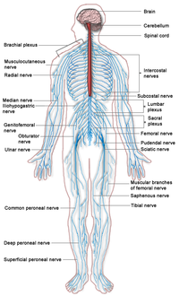
Changes for: enteric nervous system
- Deleted
- - enteric nervous system database cross reference UMLS:C0206111 { source=NIF_GrossAnatomy:birnlex_1166 }
- Added
- + enteric nervous system database cross reference UMLS:C0206111 { source=NIFSTD:birnlex_1166 }
Changes for: placenta metrial gland
- Deleted
Changes for: epithelium of mammary gland
- Deleted
- - epithelium of mammary gland definition An epithelium that is part of a mammary gland [Automatically generated definition]. { database cross reference=OBOL:automatic }
- Added
- + epithelium of mammary gland definition the epithelial layer of the luminal surfaces of the mammary gland { database cross reference=MP:0009504,MGI:csmith }
Changes for: right putamen
- Added
- + right putamen database cross reference HBA:4289
Changes for: pulmonary vascular system
- Added
- + pulmonary vascular system SubClassOf has part some pulmonary artery
- + pulmonary vascular system SubClassOf has part some pulmonary vein
- + pulmonary vascular system SubClassOf structure with developmental contribution from neural crest
Changes for: left putamen
- Added
- + left putamen database cross reference HBA:4288
Changes for: rostral migratory stream
- Deleted
- - rostral migratory stream SubClassOf regional part of telencephalon
- - rostral migratory stream database cross reference DHBA:RMS
- - rostral migratory stream database cross reference PBA:RMS
- - rostral migratory stream definition Unique telencephalic subventricular zones that extend from the lateral ventricles into the olfactory bulbs. Newly produced GABAergic interneurons migrate along the RMS and settle in the bulb. { database cross reference=http://www.ncbi.nlm.nih.gov/pubmed/12453055 , database cross reference=NIF_GrossAnatomy:birnlex_1702 }
- Added
- + rostral migratory stream SubClassOf regional part of brain
- + rostral migratory stream SubClassOf structure with developmental contribution from neural crest
- + rostral migratory stream database cross reference DHBA:10556
- + rostral migratory stream database cross reference PBA:128012584
- + rostral migratory stream definition Unique telencephalic subventricular zones that extend from the lateral ventricles into the olfactory bulbs. Newly produced GABAergic interneurons migrate along the RMS and settle in the bulb. { database cross reference=http://www.ncbi.nlm.nih.gov/pubmed/12453055 , database cross reference=NIFSTD:birnlex_1702 }
Changes for: ligament of pinna
- Deleted
- - ligament of pinna SubClassOf head connective tissue
Changes for: obsolete nerve of central nervous system
- Deleted
- - obsolete nerve of central nervous system comment obsoleted pending formal definition of nerve, with explicit reference to PNS vs CNS
- Added
- + obsolete nerve of central nervous system comment obsoleted pending formal definition of nerve, with explicit reference to PNS vs CNS. See https://github.com/obophenotype/mouse-anatomy-ontology/issues/6
Changes for: neural lobe of neurohypophysis
- Deleted
- - neural lobe of neurohypophysis SubClassOf regional part of diencephalon
- - neural lobe of neurohypophysis database cross reference DHBA:PCb
- - neural lobe of neurohypophysis database cross reference UMLS:C0447640 { source=NIF_GrossAnatomy:birnlex_941 }
- - neural lobe of neurohypophysis database cross reference http://braininfo.rprc.washington.edu/centraldirectory.aspx?ID=402 { source=NIF_GrossAnatomy:birnlex_941 }
- - neural lobe of neurohypophysis has broad synonym PNHP { database cross reference=NIF_GrossAnatomy:NeuroNames_abbrevSource,NIF_GrossAnatomy:birnlex_941 , has synonym type=abbreviation }
- - neural lobe of neurohypophysis has exact synonym pars nervosa pituitary gland { database cross reference=NIF_GrossAnatomy:birnlex_941 }
- Added
- + neural lobe of neurohypophysis SubClassOf regional part of brain
- + neural lobe of neurohypophysis database cross reference DHBA:12844
- + neural lobe of neurohypophysis database cross reference UMLS:C0447640 { source=NIFSTD:birnlex_941 }
- + neural lobe of neurohypophysis database cross reference http://braininfo.rprc.washington.edu/centraldirectory.aspx?ID=402 { source=NIFSTD:birnlex_941 }
- + neural lobe of neurohypophysis has broad synonym PNHP { database cross reference=http://uri.neuinfo.org/nif/nifstd/birnlex_941 , database cross reference=NIFSTD:NeuroNames_abbrevSource , has synonym type=abbreviation }
- + neural lobe of neurohypophysis has exact synonym pars nervosa pituitary gland { database cross reference=NIFSTD:birnlex_941 }
Changes for: taenia tectum of brain
- Deleted
- - taenia tectum of brain database cross reference DMBA:TTe
- Added
- + taenia tectum of brain database cross reference DMBA:16154
- + taenia tectum of brain database cross reference MBA:589
Changes for: gubernaculum (male or female)
- Deleted
- - gubernaculum (male or female) SubClassOf abdomen connective tissue
- - gubernaculum (male or female) SubClassOf embryonic tissue
- - gubernaculum (male or female) SubClassOf vestigial embryonic structure
- Added
- + gubernaculum (male or female) SubClassOf abdomen element
- + gubernaculum (male or female) SubClassOf embryonic structure
Changes for: allantois
- Deleted
- - allantois SubClassOf develops from some endoderm
- - allantois SubClassOf develops from some splanchnic layer of lateral plate mesoderm
- - allantois SubClassOf develops from some yolk sac
- - allantois SubClassOf mixed endoderm/mesoderm-derived structure
- Added
- + allantois SubClassOf develops from some cloaca
- + allantois SubClassOf has developmental contribution from some endoderm
- + allantois SubClassOf has developmental contribution from some splanchnic layer of lateral plate mesoderm
- + allantois SubClassOf mesoderm-derived structure
Changes for: dorsal premammillary nucleus
- Deleted
- Added
- + dorsal premammillary nucleus database cross reference DMBA:15719
- + dorsal premammillary nucleus database cross reference HBA:4677
- + dorsal premammillary nucleus database cross reference MBA:980
Changes for: ventral premammillary nucleus
- Deleted
- Added
Changes for: medial preoptic region
- Deleted
- - medial preoptic region SubClassOf regional part of hypothalamus
- - medial preoptic region database cross reference ABA:MPO
- - medial preoptic region database cross reference DHBA:MPA
- - medial preoptic region database cross reference HBA:MPA
- Added
- + medial preoptic region SubClassOf regional part of brain
- + medial preoptic region database cross reference DHBA:266441547
- + medial preoptic region database cross reference HBA:4543
- + medial preoptic region database cross reference MBA:523
Changes for: cerebellum culmen
- Deleted
- - cerebellum culmen database cross reference ABA:CUL
- Added
- + cerebellum culmen SubClassOf regional part of a lobe of the cerebellum
- + cerebellum culmen database cross reference MBA:928
Changes for: paramedian reticular nucleus
- Deleted
- Added
- + paramedian reticular nucleus database cross reference DHBA:12496
- + paramedian reticular nucleus database cross reference MBA:995
Changes for: gyrus
- Deleted
- Added
Changes for: vitelline blood vessel
- Deleted
- - vitelline blood vessel EquivalentTo blood vessel and has part some part of some yolk sac
- - vitelline blood vessel SubClassOf has part some part of some yolk sac
- - vitelline blood vessel definition blood vesself of the yolk sac. { database cross reference=MP:0001719 }
- Added
- + vitelline blood vessel EquivalentTo blood vessel and connected to some yolk sac
- + vitelline blood vessel SubClassOf connected to some yolk sac
- + vitelline blood vessel definition blood vessel that is connected to the yolk sac. { database cross reference=UBERON:cjm }
Changes for: pallium
- Deleted
- - pallium SubClassOf regional part of telencephalon
- - pallium database cross reference DMBA:Pall
- Added
- + pallium SubClassOf regional part of brain
- + pallium SubClassOf structure with developmental contribution from neural crest
- + pallium database cross reference DMBA:15903
Changes for: ventral part of telencephalon
- Deleted
- - ventral part of telencephalon SubClassOf regional part of telencephalon
- - ventral part of telencephalon database cross reference DMBA:SPall
- Added
Changes for: pharyngeal arch 1
- Deleted
- - pharyngeal arch 1 definition The first pharyngeal arch is the most anteriorly positioned arch and forms the jaw. It is located between the stomodeum and the first pharyngeal groove. The neural crest cells that contribute to pharyngeal arch 1 arise from the caudal midbrain and rhombomeres 1 and 2 of the hindbrain. { database cross reference=http://en.wikipedia.org/wiki/First_pharyngeal_arch }
- Added
- + pharyngeal arch 1 definition The first of the series of pharyngeal arches that develop into jaw bones or their homologs, and their associated nerves and arteries
- + pharyngeal arch 1 external definition the first arch which contributes to development of mastication muscles, maxilla, mandible, incus, malleus, Meckel’s cartilage, trigeminal nerve, and maxillary artery { source=MP:0006337,MGI:smb }
Changes for: tetrapod frontal bone
- Deleted
- - tetrapod frontal bone definition In humans: A bone resembling a cockleshell in two portions the squama frontalis, corresponding with the region of the forehead, and an orbital or horizontal portion, the pars orbitalis, which enters into the formation of the roofs of the orbital and nasal cavities. { database cross reference=http://en.wikipedia.org/wiki/Frontal_bone }
- Added
- + tetrapod frontal bone definition the bone forming the forehead and roof of the eye orbit { database cross reference=http://en.wikipedia.org/wiki/Frontal_bone , database cross reference=MP:0000107,ISBN:0-683-40008-8,PMID:7914451 }
Changes for: vitelline vasculature
- Deleted
- - vitelline vasculature EquivalentTo vasculature and has part some part of some yolk sac
- - vitelline vasculature SubClassOf has part some part of some yolk sac
- - vitelline vasculature definition vasculature of the yolk sac. { database cross reference=MP:0001719 }
- Added
- + vitelline vasculature EquivalentTo vasculature and connected to some yolk sac
- + vitelline vasculature SubClassOf connected to some yolk sac
- + vitelline vasculature database cross reference http://en.wikipedia.org/wiki/Vitelline_circulation
- + vitelline vasculature definition vascular network that is connected to the yolk sac. { database cross reference=UBERON:cjm }
Changes for: induseum griseum
- Deleted
- - induseum griseum database cross reference DMBA:IG
- - induseum griseum definition The supracallosal gyrus (indusium griseum; gyrus epicallosus) consists of a thin layer of grey matter in contact with the dorsal surface of the corpus callosum and continuous laterally with the grey matter of the cingulate gyrus. { database cross reference=http://en.wikipedia.org/wiki/Indusium_griseum }
- Added
- + induseum griseum database cross reference DMBA:16147
- + induseum griseum database cross reference MBA:19
- + induseum griseum definition Thin layer of cortex lying on the dorsal surface of the corpus callosum. It has been debated whether it is more related to the hippocampus or the olfactory bulb (Shipley et al., Olfactory System In Paxinos The Rat Nervous System, 2nd ed) { database cross reference=NIFSTD:nlx_49940 }
Changes for: lateral nucleus of trapezoid body
- Deleted
- - lateral nucleus of trapezoid body SubClassOf located in some trapezoid body
- - lateral nucleus of trapezoid body database cross reference DHBA:TrZl
- - lateral nucleus of trapezoid body database cross reference DMBA:LTz
- Added
- + lateral nucleus of trapezoid body SubClassOf part of some superior olivary complex
- + lateral nucleus of trapezoid body SubClassOf part of some trapezoid body
- + lateral nucleus of trapezoid body SubClassOf ventral to some lateral superior olivary nucleus
- + lateral nucleus of trapezoid body database cross reference DHBA:12459
- + lateral nucleus of trapezoid body database cross reference DMBA:17274
- + lateral nucleus of trapezoid body has exact synonym LNTB { has synonym type=abbreviation }
Changes for: retroambiguus nucleus
- Deleted
- - retroambiguus nucleus database cross reference HBA:Ramb
- Added
- + retroambiguus nucleus database cross reference HBA:9650
Changes for: paratrigeminal nucleus
- Deleted
- - paratrigeminal nucleus database cross reference DHBA:Pa5
- - paratrigeminal nucleus database cross reference HBA:Pa5
- Added
- + paratrigeminal nucleus database cross reference DHBA:12656
- + paratrigeminal nucleus database cross reference HBA:9630
Changes for: lung connective tissue
- Deleted
- - lung connective tissue definition A portion of connective tissue that is part of a lung [Automatically generated definition]. { database cross reference=OBOL:automatic }
- Added
- + lung connective tissue definition the connective tissue located between the respiratory (airway and alveolar) epithelium, the capillary endothelium and pleural mesothelium; it contains basement membrane composed of collagen, elastin, proteoglycans, and fibronectin; it functions in the mechanical behavior of the lung, and forms a barrier to regulate the flow of plasma constituents from the capillaries to the airway and alveolar spaces { database cross reference=MP:0002276,ISBN:0-683-40008-8,MGI:anna,PMID:14635660 }
Changes for: facial nucleus
- Deleted
- - facial nucleus database cross reference DHBA:7N
- Added
- + facial nucleus EquivalentTo cranial nerve nucleus and extends fibers into some facial nerve
- + facial nucleus SubClassOf extends fibers into some facial nerve
- + facial nucleus database cross reference DHBA:12420
- + facial nucleus definition the group of motor neurons residing in the pons that innervate the muscles of facial expression { database cross reference=MP:0000909,ISBN:0838580343 }
Changes for: cranial nerve nucleus
- Deleted
- - cranial nerve nucleus EquivalentTo nucleus of brain and has part some part of some cranial nerve
- - cranial nerve nucleus SubClassOf has part some part of some cranial nerve
- - cranial nerve nucleus definition collection of neurons (gray matter) in the brain stem that is associated with one or more cranial nerves. { database cross reference=http://en.wikipedia.org/wiki/Cranial_nerve_nucleus }
- Added
- + cranial nerve nucleus EquivalentTo nucleus of brain and extends fibers into some cranial nerve
- + cranial nerve nucleus SubClassOf extends fibers into some cranial nerve
- + cranial nerve nucleus definition Nucleus that receives projections from or contains neurons that send projections through one of the cranial nerves { database cross reference=NIFSTD:nlx_28532 }
Changes for: epineurium
- Deleted
- - epineurium SubClassOf bounding layer of some nerve trunk
- - epineurium SubClassOf surrounds some peripheral nerve
- - epineurium definition A loose connective tissue sheath surrounding each peripheral nerve. Composed mainly of colagen and fibroblasts, it forms a substantial covering over nerve trunks, then thins to an incomplete layer around smaller branches near their terminations. The epineurium is continuous centrally with the dura. Peripherally, it usually ends near the termination of a nerve fiber, but it may continue as the capsule of Meissner corpuscles and a few other encapsulated endings. Nolte, J. The Human Brain 3rd edition { database cross reference=NIF_GrossAnatomy:nlx_anat_090203 }
- Added
- + epineurium SubClassOf part of some peripheral nervous system
- + epineurium SubClassOf surrounds some nerve
- + epineurium definition A loose connective tissue sheath surrounding each peripheral nerve. Composed mainly of colagen and fibroblasts, it forms a substantial covering over nerve trunks, then thins to an incomplete layer around smaller branches near their terminations. The epineurium is continuous centrally with the dura. Peripherally, it usually ends near the termination of a nerve fiber, but it may continue as the capsule of Meissner corpuscles and a few other encapsulated endings. Nolte, J. The Human Brain 3rd edition { database cross reference=NIFSTD:nlx_anat_090203 }
Changes for: dorsal anterior lateral line ganglion
- Deleted
Changes for: neural nucleus
- Deleted
- - neural nucleus definition A spatially aggregated collection of nerve cell bodies in the CNS, consisting of one or more subpopulations that share cell type, chemical phenotype, and connections, and including nearby cells that share the same cell type, chemical phenotype, and connections. (CUMBO) { database cross reference=NIF_GrossAnatomy:nlx_28443 }
- - neural nucleus has exact synonym nucleus of CNS { database cross reference=NIF_GrossAnatomy:nlx_28443 }
- Added
- + neural nucleus definition A spatially aggregated collection of nerve cell bodies in the CNS, consisting of one or more subpopulations that share cell type, chemical phenotype, and connections, and including nearby cells that share the same cell type, chemical phenotype, and connections. (CUMBO) { database cross reference=NIFSTD:nlx_28443 }
- + neural nucleus has exact synonym nucleus of CNS { database cross reference=NIFSTD:nlx_28443 }
Changes for: ventral anterior lateral line ganglion
- Deleted
Changes for: posterior lateral line ganglion
- Deleted
Changes for: endoneurium
- Deleted
- - endoneurium SubClassOf part of some nervous system
- - endoneurium definition The loose connective tissue within the perineurium that continues into nerve fascicles and surrounds individual fibers. (Nolte, J. The Human Brain 3rd edition) { database cross reference=NIF_GrossAnatomy:nlx_anat_090202 }
- Added
- + endoneurium SubClassOf part of some peripheral nervous system
- + endoneurium definition The loose connective tissue within the perineurium that continues into nerve fascicles and surrounds individual fibers. (Nolte, J. The Human Brain 3rd edition) { database cross reference=NIFSTD:nlx_anat_090202 }
Changes for: perineurium
- Deleted
- - perineurium definition A layer of thin, concentrically arranged cells with interspersed collagen that lies within the epineurium. (Nolte, J., The Human Brain 3rd edition) { database cross reference=NIF_GrossAnatomy:nlx_anat_090205 }
- Added
- + perineurium definition A layer of thin, concentrically arranged cells with interspersed collagen that lies within the epineurium. (Nolte, J., The Human Brain 3rd edition) { database cross reference=NIFSTD:nlx_anat_090205 }
Changes for: spinothalamic tract
- Deleted
- - spinothalamic tract database cross reference DHBA:spth
- - spinothalamic tract definition A tract that originates from neurons in the spinal central gray and projects to various somatosensory nuclei of the thalamus. It is formed in the medulla by merger of the anterior spinothalamic tract and lateral spinothalamic tract (adapted from Braininfo.org) { database cross reference=NIF_GrossAnatomy:nlx_anat_1010017 }
- Added
- + spinothalamic tract database cross reference DHBA:12789
- + spinothalamic tract database cross reference MBA:871
- + spinothalamic tract definition A tract that originates from neurons in the spinal central gray and projects to various somatosensory nuclei of the thalamus. It is formed in the medulla by merger of the anterior spinothalamic tract and lateral spinothalamic tract (adapted from Braininfo.org) { database cross reference=NIFSTD:nlx_anat_1010017 }
Changes for: theca cell layer
- Deleted
- - theca cell layer comment Function notes: The theca folliculi are responsible for the production of testosterone from androstenedione in females, and indirectly the production of estrogens by supplying the neighboring granulosa cells with androstenedione that can then be used as a substrate for aromatase. [Wikipedia:Theca_of_follicle]
- Added
- + theca cell layer function notes The theca folliculi are responsible for the production of testosterone from androstenedione in females, and indirectly the production of estrogens by supplying the neighboring granulosa cells with androstenedione that can then be used as a substrate for aromatase. [Wikipedia:Theca_of_follicle]
Changes for: superior cerebellar peduncle of pons
- Deleted
- - superior cerebellar peduncle of pons SubClassOf regional part of metencephalon
- - superior cerebellar peduncle of pons database cross reference http://braininfo.rprc.washington.edu/centraldirectory.aspx?ID=833 { source=NIF_GrossAnatomy:birnlex_1726 }
- - superior cerebellar peduncle of pons has broad synonym scpp { database cross reference=NIF_GrossAnatomy:NeuroNames_abbrevSource,NIF_GrossAnatomy:birnlex_1726 , has synonym type=abbreviation }
- - superior cerebellar peduncle of pons has exact synonym SCPP { database cross reference=NIF_GrossAnatomy:birnlex_1726 }
- - superior cerebellar peduncle of pons has exact synonym superior cerebellar peduncle of pons { database cross reference=NIF_GrossAnatomy:birnlex_1726 }
- Added
- + superior cerebellar peduncle of pons SubClassOf regional part of cerebellar white matter
- + superior cerebellar peduncle of pons SubClassOf structure with developmental contribution from neural crest
- + superior cerebellar peduncle of pons database cross reference http://braininfo.rprc.washington.edu/centraldirectory.aspx?ID=833 { source=NIFSTD:birnlex_1726 }
- + superior cerebellar peduncle of pons has broad synonym SCPP { database cross reference=http://uri.neuinfo.org/nif/nifstd/birnlex_1726 , database cross reference=NIFSTD:NeuroNames_abbrevSource , has synonym type=abbreviation }
- + superior cerebellar peduncle of pons has exact synonym SCPP { database cross reference=NIFSTD:birnlex_1726 }
- + superior cerebellar peduncle of pons has exact synonym superior cerebellar peduncle of pons { database cross reference=NIFSTD:birnlex_1726 }
Changes for: superior cerebellar peduncle of midbrain
- Deleted
- - superior cerebellar peduncle of midbrain SubClassOf regional part of midbrain tegmentum
- - superior cerebellar peduncle of midbrain database cross reference http://braininfo.rprc.washington.edu/centraldirectory.aspx?ID=820 { source=NIF_GrossAnatomy:birnlex_1162 }
- - superior cerebellar peduncle of midbrain definition Regional part of superior cerebellar peduncle, located in the midbrain, consisting of a large bundle of fibers largely projecting from the deep cerebellar nuclei to the midbrain and thalamus. It is continuous with the superior cerebellar peduncle of the pons. (MM). { database cross reference=NIF_GrossAnatomy:birnlex_1162 }
- - superior cerebellar peduncle of midbrain has broad synonym scpmb { database cross reference=NIF_GrossAnatomy:NeuroNames_abbrevSource,NIF_GrossAnatomy:birnlex_1162 , has synonym type=abbreviation }
- - superior cerebellar peduncle of midbrain has exact synonym SCPMB { database cross reference=NIF_GrossAnatomy:birnlex_1162 }
- - superior cerebellar peduncle of midbrain has exact synonym superior cerebellar peduncle of midbrain { database cross reference=NIF_GrossAnatomy:birnlex_1162 }
- Added
- + superior cerebellar peduncle of midbrain SubClassOf regional part of cerebellar white matter
- + superior cerebellar peduncle of midbrain database cross reference http://braininfo.rprc.washington.edu/centraldirectory.aspx?ID=820 { source=NIFSTD:birnlex_1162 }
- + superior cerebellar peduncle of midbrain definition Regional part of superior cerebellar peduncle, located in the midbrain, consisting of a large bundle of fibers largely projecting from the deep cerebellar nuclei to the midbrain and thalamus. It is continuous with the superior cerebellar peduncle of the pons. (MM). { database cross reference=NIFSTD:birnlex_1162 }
- + superior cerebellar peduncle of midbrain has broad synonym SCPMB { database cross reference=http://uri.neuinfo.org/nif/nifstd/birnlex_1162 , database cross reference=NIFSTD:NeuroNames_abbrevSource , has synonym type=abbreviation }
- + superior cerebellar peduncle of midbrain has exact synonym SCPMB { database cross reference=NIFSTD:birnlex_1162 }
- + superior cerebellar peduncle of midbrain has exact synonym superior cerebellar peduncle of midbrain { database cross reference=NIFSTD:birnlex_1162 }
Changes for: lateral paragigantocellular nucleus
- Deleted
- Added
Changes for: dorsal paragigantocellular nucleus
- Deleted
- Added
Changes for: dorsal paramedian reticular nucleus
- Deleted
- Added
- + dorsal paramedian reticular nucleus database cross reference DHBA:12597
- + dorsal paramedian reticular nucleus definition Small nucleus in the brainstem of human, located adjacent and medial to the nucleus prepositus in the dorsal medulla, described in several atlases of human brain stem. In transverse sections, it is oval with its long axis aligned with the dorsal border of the brainstem. It begins at about the same A-P level as the nucleus prepositus, just rostral to the hypoglossal nucleus. It includes calretinin immunoreactive large cells with oval or polygonal cell bodies. Cells are not immunoreactive for either calbindin or parvalbumin, but a few fibers immunoreactive to each protein are found within its central region. Cells in PMD are also immunoreactive to nNOS, and immunoreactivity to a neurofilament protein shows many labeled cells and fibers. According to Baizer et al., no equivalent structure was observed in the cat, rat, mouse or monkey atlas although they did not look at the brains of the great apes. { database cross reference=NIFSTD:nlx_143549 }
Changes for: fourth thoracic spinal ganglion
- Deleted
- - fourth thoracic spinal ganglion database cross reference UMLS:C0501702 { source=NIF_GrossAnatomy:birnlex_2614 }
- - fourth thoracic spinal ganglion has exact synonym forth thoracic dorsal root ganglion { database cross reference=NIF_GrossAnatomy:birnlex_2614 }
- Added
- + fourth thoracic spinal ganglion database cross reference UMLS:C0501702 { source=NIFSTD:birnlex_2614 }
- + fourth thoracic spinal ganglion has exact synonym forth thoracic dorsal root ganglion { database cross reference=NIFSTD:birnlex_2614 }
Changes for: fourth sacral spinal ganglion
- Deleted
- - fourth sacral spinal ganglion database cross reference UMLS:C0501723 { source=NIF_GrossAnatomy:birnlex_2631 }
- - fourth sacral spinal ganglion has exact synonym forth sacral dorsal root ganglion { database cross reference=NIF_GrossAnatomy:birnlex_2631 }
- Added
- + fourth sacral spinal ganglion database cross reference UMLS:C0501723 { source=NIFSTD:birnlex_2631 }
- + fourth sacral spinal ganglion has exact synonym forth sacral dorsal root ganglion { database cross reference=NIFSTD:birnlex_2631 }
Changes for: cervical subsegment of spinal cord
- Deleted
- - cervical subsegment of spinal cord SubClassOf spinal cord segment
- - cervical subsegment of spinal cord has exact synonym segment part of cervical spinal cord { database cross reference=NIF_GrossAnatomy:birnlex_1688 }
- Added
- + cervical subsegment of spinal cord SubClassOf regional part of cervical spinal cord
- + cervical subsegment of spinal cord has exact synonym segment part of cervical spinal cord { database cross reference=NIFSTD:birnlex_1688 }
Changes for: thoracic subsegment of spinal cord
- Deleted
- - thoracic subsegment of spinal cord SubClassOf thoracic spinal cord
- - thoracic subsegment of spinal cord has exact synonym segment part of thoracic spinal cord { database cross reference=NIF_GrossAnatomy:birnlex_892 }
- Added
- + thoracic subsegment of spinal cord SubClassOf regional part of thoracic spinal cord
- + thoracic subsegment of spinal cord has exact synonym segment part of thoracic spinal cord { database cross reference=NIFSTD:birnlex_892 }
Changes for: intermediate nucleus of lateral lemniscus
- Deleted
- Added
Changes for: sixth cervical dorsal root ganglion
- Deleted
- - sixth cervical dorsal root ganglion has exact synonym sixth cervical dorsal root ganglion { database cross reference=NIF_GrossAnatomy:birnlex_2608 }
- Added
- + sixth cervical dorsal root ganglion has exact synonym sixth cervical dorsal root ganglion { database cross reference=NIFSTD:birnlex_2608 }
Changes for: sacral subsegment of spinal cord
- Deleted
- - sacral subsegment of spinal cord SubClassOf spinal cord segment
- - sacral subsegment of spinal cord has exact synonym segment part of sacral spinal cord { database cross reference=NIF_GrossAnatomy:birnlex_1512 }
- Added
- + sacral subsegment of spinal cord SubClassOf regional part of sacral spinal cord
- + sacral subsegment of spinal cord has exact synonym segment part of sacral spinal cord { database cross reference=NIFSTD:birnlex_1512 }
Changes for: lumbar subsegment of spinal cord
- Deleted
- - lumbar subsegment of spinal cord SubClassOf lumbar spinal cord
- - lumbar subsegment of spinal cord has exact synonym segment part of lumbar spinal cord { database cross reference=NIF_GrossAnatomy:birnlex_1652 }
- Added
- + lumbar subsegment of spinal cord SubClassOf regional part of lumbar spinal cord
- + lumbar subsegment of spinal cord has exact synonym segment part of lumbar spinal cord { database cross reference=NIFSTD:birnlex_1652 }
Changes for: primitive urogenital sinus
- Deleted
- - primitive urogenital sinus definition Ventral part of the cloaca remaining after septation of the rectum, which further develops into part of the bladder, part of the prostatic part of the male urethra and the urethra and vestibule in females[MP]. { database cross reference=http://en.wikipedia.org/wiki/Definitive_urogenital_sinus }
- Added
- + primitive urogenital sinus definition the ventral part of the cloaca remaining after septation of the rectum, which further develops into part of the bladder, part of the prostatic part of the male urethra and the urethra and vestibule in females { database cross reference=MP:0010116,ISBN:0-683-40008-8 }
Changes for: rostrum of corpus callosum
- Deleted
- - rostrum of corpus callosum SubClassOf regional part of telencephalon
- - rostrum of corpus callosum database cross reference DHBA:ccr
- - rostrum of corpus callosum database cross reference HBA:rcc
- Added
- + rostrum of corpus callosum SubClassOf cerebral hemisphere white matter
- + rostrum of corpus callosum database cross reference DHBA:10562
- + rostrum of corpus callosum database cross reference HBA:9226
- + rostrum of corpus callosum database cross reference MBA:979
- + rostrum of corpus callosum definition The part of the corpus callosum that projects posteriorly and inferiorly from the anteriormost genu. { database cross reference=http://en.wikipedia.org/wiki/Corpus_callosum }
Changes for: splenium of the corpus callosum
- Deleted
- - splenium of the corpus callosum SubClassOf regional part of telencephalon
- - splenium of the corpus callosum database cross reference DHBA:ccs
- - splenium of the corpus callosum database cross reference HBA:scc
- - splenium of the corpus callosum has related synonym splenium { database cross reference=NeuroNames:196 }
- Added
- + splenium of the corpus callosum SubClassOf cerebral hemisphere white matter
- + splenium of the corpus callosum SubClassOf in posterior side of some corpus callosum
- + splenium of the corpus callosum database cross reference DHBA:10565
- + splenium of the corpus callosum database cross reference HBA:9225
- + splenium of the corpus callosum database cross reference MBA:986
- + splenium of the corpus callosum database cross reference http://en.wikipedia.org/wiki/Splenium
- + splenium of the corpus callosum definition The posterior end of the corpus callosum. It overlaps the tela chorioidea of the third ventricle and the mid-brain, and ends in a thick, convex, free border. { database cross reference=http://en.wikipedia.org/wiki/Splenium }
- + splenium of the corpus callosum has broad synonym splenium { database cross reference=NeuroNames:196 }
Changes for: prostate gland dorsolateral lobe
- Added
- + prostate gland dorsolateral lobe definition the rodent prostate lobe that lies in the triangular area between urethra and seminal vesicle { database cross reference=MP:0009381,PMID:12645922,PMID:3308446 }
Changes for: nasopalatine nerve
- Deleted
- - nasopalatine nerve SubClassOf nerve
- Added
- + nasopalatine nerve SubClassOf nerve of head region
Changes for: forestomach
- Added
- + forestomach definition the dilated area of tissue where the stomach and esophagus meet { database cross reference=MP:0010784,MGI:csmith }
Changes for: cerebellum external granule cell layer
- Deleted
- - cerebellum external granule cell layer database cross reference UMLS:C0228473 { source=NIF_GrossAnatomy:birnlex_797 }
- - cerebellum external granule cell layer database cross reference UMLS:C1289480 { source=NIF_GrossAnatomy:birnlex_797 }
- - cerebellum external granule cell layer has exact synonym outer granular layer of cerebellar cortex { database cross reference=NIF_GrossAnatomy:birnlex_797 }
- Added
- + cerebellum external granule cell layer database cross reference UMLS:C0228473 { source=NIFSTD:birnlex_797 }
- + cerebellum external granule cell layer database cross reference UMLS:C1289480 { source=NIFSTD:birnlex_797 }
- + cerebellum external granule cell layer has exact synonym outer granular layer of cerebellar cortex { database cross reference=NIFSTD:birnlex_797 }
Changes for: roof plate of midbrain
- Deleted
- - roof plate of midbrain database cross reference DHBA:IZMro
- - roof plate of midbrain definition A roof plate that is part of a midbrain [Automatically generated definition]. { database cross reference=OBOL:automatic }
- Added
- + roof plate of midbrain database cross reference DHBA:12320
- + roof plate of midbrain definition the mesencephalic roof plate, including the caudal and rostral part of the midbrain roof { database cross reference=MP:0006103,MGI:anna,MGI:smb }
Changes for: transitional epithelium of prostatic urethra
- Deleted
Changes for: great auricular nerve
- Deleted
- - great auricular nerve SubClassOf peripheral nerve
- Added
- + great auricular nerve SubClassOf nerve
Changes for: cerebellum internal granule cell layer
- Deleted
- - cerebellum internal granule cell layer database cross reference UMLS:C0228471 { source=NIF_GrossAnatomy:birnlex_790 }
- - cerebellum internal granule cell layer database cross reference UMLS:C1289476 { source=NIF_GrossAnatomy:birnlex_790 }
- - cerebellum internal granule cell layer has exact synonym inner granular layer of cerebellar cortex { database cross reference=NIF_GrossAnatomy:birnlex_790 }
- Added
- + cerebellum internal granule cell layer database cross reference UMLS:C0228471 { source=NIFSTD:birnlex_790 }
- + cerebellum internal granule cell layer database cross reference UMLS:C1289476 { source=NIFSTD:birnlex_790 }
- + cerebellum internal granule cell layer has exact synonym inner granular layer of cerebellar cortex { database cross reference=NIFSTD:birnlex_790 }
Changes for: skeletal ligament
- Added
- + skeletal ligament SubClassOf composed primarily of some dense regular connective tissue
- + skeletal ligament database cross reference VSAO:0000072
Changes for: nonskeletal ligament
- Added
- + nonskeletal ligament external ontology notes this class is used more generally than the FMA class, to include for example folds of mesentery such as the broad ligament of the uterus. { external ontology=FMA }
Changes for: ovarian ligament
- Deleted
- - ovarian ligament SubClassOf mesoderm-derived structure
- Added
- + ovarian ligament SubClassOf part of some female reproductive system
- + ovarian ligament SubClassOf reproductive structure
Changes for: suspensory ligament of testis
- Deleted
- - suspensory ligament of testis SubClassOf embryonic tissue
- Added
Changes for: suspensory ligament
- Deleted
- - suspensory ligament database cross reference http://en.wikipedia.org/wiki/Cooper%27s_ligaments_of_penis
- - suspensory ligament definition A ligament that supports a body part, especially an organ. { database cross reference=http://en.wikipedia.org/wiki/Cooper%27s_ligaments_of_penis }
- Added
- + suspensory ligament definition A ligament that supports a body part, especially an organ. { database cross reference=UBERON:cjm }
Changes for: gubernaculum testis
- Deleted
- - gubernaculum testis comment The upper part of the gubernaculum degenerates; The lower part persists as the gubernaculum testis (“scrotal ligament”). This ligament secures the testis to the most inferior portion of the scrotum, tethering it in place and limiting the degree to which the testis can move within the scrotum[WP]
- - gubernaculum testis has related synonym scrotal ligament { database cross reference=http://en.wikipedia.org/wiki/Gubernaculum }
- Added
- + gubernaculum testis development notes The upper part of the gubernaculum degenerates; The lower part persists as the gubernaculum testis (‘scrotal ligament’). This ligament secures the testis to the most inferior portion of the scrotum, tethering it in place and limiting the degree to which the testis can move within the scrotum[WP]
- + gubernaculum testis has exact synonym male gubernaculum
Changes for: smooth muscle of eye
- Deleted
- - smooth muscle of eye definition A portion of smooth muscle tissue that is part of a eye [Automatically generated definition]. { database cross reference=OBOL:automatic }
- - smooth muscle of eye editor note 331. Includes ciliary and iris smooth muscle
- Added
- + smooth muscle of eye definition any of the striated muscles that move the eye and include: superior rectus, inferior rectus, medial rectus, lateral rectus, superior oblique, inferior oblique, retractor bulbi { database cross reference=MP:0005247,MGI:cwg }
- + smooth muscle of eye editor note 331. MA includes ciliary and iris smooth muscle
Changes for: epithelium of vomeronasal organ
- Added
- + epithelium of vomeronasal organ editor note TODO - add cell types
Changes for: ganglion of central nervous system
- Deleted
- - ganglion of central nervous system comment this has no subclasses in FMA. In EHDAA2 it has only the cranial ganglia
- Added
- + ganglion of central nervous system comment this has no subclasses in FMA. In EHDAA2 and EMAPA it has only the cranial ganglia
Changes for: ganglion of peripheral nervous system
- Deleted
- - ganglion of peripheral nervous system database cross reference birnlex_2548
- - ganglion of peripheral nervous system definition A spatially aggregated collection of nerve cell bodies in the PNS, consisting of one or more subpopulations that share cell type, chemical phenotype, and connections. (CUMBO). { database cross reference=NIF_GrossAnatomy:birnlex_2548 }
- Added
- + ganglion of peripheral nervous system database cross reference http://uri.neuinfo.org/nif/nifstd/birnlex_2548
- + ganglion of peripheral nervous system definition A spatially aggregated collection of nerve cell bodies in the PNS, consisting of one or more subpopulations that share cell type, chemical phenotype, and connections. (CUMBO). { database cross reference=NIFSTD:birnlex_2548 }
Changes for: epaxial musclulature
- Deleted
- - epaxial musclulature definition Musculature that is derived from the epaxial myotome region, ventral to the horizontal septum of the vertebrae. Epaxial muscles include other (dorsal) muscles associated with the vertebrae, ribs, and base of the skull. { database cross reference=http://en.wikipedia.org/wiki/Epaxial_and_hypaxial_muscles }
- Added
- + epaxial musclulature definition Musculature that is derived from the epaxial myotome region, ventral to the horizontal septum of the vertebrae. Epaxial muscles include other (dorsal) muscles associated with the vertebrae, ribs, and base of the skull. { database cross reference=http://en.wikipedia.org/wiki/Epaxial_and_hypaxial_muscles , database cross reference=UBERON:cjm }
- + epaxial musclulature external definition the muscles derived from the medial myotome; includes the intrinsic back muscles { source=MP:0004248,PMID:1591996 }
Changes for: floor plate of medulla oblongata
- Deleted
- Added
Changes for: floor plate of metencephalon
- Deleted
- Added
Changes for: floor plate of diencephalon
- Deleted
- - floor plate of diencephalon SubClassOf regional part of diencephalon
- Added
- + floor plate of diencephalon SubClassOf regional part of brain
Changes for: floor plate of midbrain
- Deleted
- - floor plate of midbrain SubClassOf regional part of midbrain
- - floor plate of midbrain database cross reference DHBA:IZMfl
- Added
- + floor plate of midbrain SubClassOf regional part of brain
- + floor plate of midbrain database cross reference DHBA:12325
Changes for: floor plate of telencephalon
- Deleted
- Added
Changes for: anular ligament of radius
- Deleted
- - anular ligament of radius SubClassOf elbow connective tissue
- - anular ligament of radius SubClassOf lower arm connective tissue
Changes for: lateral recess of fourth ventricle
- Deleted
- Added
Changes for: amnion
- Deleted
- - amnion definition A membrane building the amniotic sac that surrounds and protects an embryo. It is developed in reptiles, birds, and mammals, which are hence called ‘Amniota’; but not in amphibians and fish (Ichthyopsida), which are consequently termed ‘Anamniota’. The primary function of this is the protection of the embryo for its development. It stems from parts of the mesoderm on the outer side and the ectoderm on the inner side[WP]. { database cross reference=http://en.wikipedia.org/wiki/Amnion }
- Added
- + amnion definition the thin innermost layer of the extraembryonic membranes that contains the amniotic fluid; the membrane forms a closed sac in which the embryo and later, the fetus, is suspended and protected { database cross reference=MP:0005029,MGI:csmith }
Changes for: hippocampus stratum lacunosum moleculare
- Deleted
- - hippocampus stratum lacunosum moleculare database cross reference http://braininfo.rprc.washington.edu/centraldirectory.aspx?ID=155 { source=NIF_GrossAnatomy:birnlex_1359 }
- - hippocampus stratum lacunosum moleculare has exact synonym stratum lacunosum moleculare { database cross reference=NIF_GrossAnatomy:birnlex_1359 }
- Added
- + hippocampus stratum lacunosum moleculare database cross reference http://braininfo.rprc.washington.edu/centraldirectory.aspx?ID=155 { source=NIFSTD:birnlex_1359 }
- + hippocampus stratum lacunosum moleculare has exact synonym stratum lacunosum moleculare { database cross reference=NIFSTD:birnlex_1359 }
Changes for: trigeminal nuclear complex
- Deleted
- - trigeminal nuclear complex SubClassOf continuous with some trigeminal nerve
- - trigeminal nuclear complex database cross reference BAMS:5
- - trigeminal nuclear complex database cross reference DHBA:5N
- - trigeminal nuclear complex database cross reference HBA:5
- - trigeminal nuclear complex database cross reference UMLS:C0040999 { source=NIF_GrossAnatomy:birnlex_4096 }
- Added
- + trigeminal nuclear complex SubClassOf extends fibers into some trigeminal nerve
- + trigeminal nuclear complex database cross reference DHBA:266441713
- + trigeminal nuclear complex database cross reference HBA:9203
- + trigeminal nuclear complex database cross reference UMLS:C0040999 { source=NIFSTD:birnlex_4096 }
Changes for: magnocellular nucleus of stria terminalis
- Deleted
- - magnocellular nucleus of stria terminalis database cross reference ABA:BSTmg
- - magnocellular nucleus of stria terminalis has related synonym anterior mamillary nucleus { database cross reference=BAMS:MA }
- Added
Changes for: subparaventricular zone
- Deleted
- - subparaventricular zone SubClassOf regional part of hypothalamus
- - subparaventricular zone database cross reference ABA:SBPV
- - subparaventricular zone database cross reference DHBA:SPZ
- Added
- + subparaventricular zone SubClassOf regional part of brain
- + subparaventricular zone database cross reference DHBA:266441555
- + subparaventricular zone database cross reference HBA:4574
- + subparaventricular zone database cross reference MBA:347
Changes for: lateral septal complex
- Deleted
- Added
Changes for: medial septal complex
- Deleted
- Added
Changes for: septohippocampal nucleus
- Deleted
- Added
- + septohippocampal nucleus database cross reference DMBA:16204
- + septohippocampal nucleus database cross reference MBA:333
- + septohippocampal nucleus definition Part of septal nuclear complex. In the human, it refers to the most caudal of scattered clusters of neurons in the septum pellucidum that extend, on the underside of the corpus callosum, from the level of the medial septal nucleus to the CA fields ( Crosby-1962 ). In the rat it is associated with the lateral septal nucleus throughout its rostro-caudal course toward the CA fields ( Swanson-1998. Adapted from Brain Info. { database cross reference=NIFSTD:nlx_60880 }
Changes for: hippocampus stratum lucidum
- Deleted
- - hippocampus stratum lucidum database cross reference OldNeuroNames:-975815906 { source=NIF_GrossAnatomy:birnlex_4108 }
- - hippocampus stratum lucidum has broad synonym stratum lucidum { database cross reference=NIF_GrossAnatomy:birnlex_4108 }
- Added
- + hippocampus stratum lucidum database cross reference OldNeuroNames:-975815906 { source=NIFSTD:birnlex_4108 }
- + hippocampus stratum lucidum has broad synonym stratum lucidum { database cross reference=NIFSTD:birnlex_4108 }
Changes for: Barrington’s nucleus
- Deleted
- - Barrington’s nucleus database cross reference ABA:B
- - Barrington’s nucleus database cross reference DHBA:Bar
- - Barrington’s nucleus database cross reference DMBA:Bar
- - Barrington’s nucleus has related synonym basal nucleus (Meynert) { database cross reference=BAMS:B }
- Added
- + Barrington’s nucleus database cross reference DHBA:12506
- + Barrington’s nucleus database cross reference DMBA:16973
- + Barrington’s nucleus database cross reference MBA:280
Changes for: accessory olfactory bulb glomerular layer
- Deleted
- Added
Changes for: parabrachial nucleus
- Deleted
- - parabrachial nucleus database cross reference ABA:PB
- - parabrachial nucleus database cross reference DHBA:PB
- - parabrachial nucleus database cross reference DMBA:PBC
- Added
- + parabrachial nucleus database cross reference DHBA:12481
- + parabrachial nucleus database cross reference DMBA:16844
- + parabrachial nucleus database cross reference MBA:867
Changes for: nucleus of trapezoid body
- Deleted
- - nucleus of trapezoid body database cross reference ABA:NTB
- - nucleus of trapezoid body database cross reference DHBA:TrZ
- - nucleus of trapezoid body database cross reference HBA:Tz
- - nucleus of trapezoid body database cross reference UMLS:C0228436 { source=NIF_GrossAnatomy:birnlex_745 }
- - nucleus of trapezoid body database cross reference UMLS:C1289449 { source=NIF_GrossAnatomy:birnlex_745 }
- - nucleus of trapezoid body database cross reference http://braininfo.rprc.washington.edu/centraldirectory.aspx?ID=576 { source=NIF_GrossAnatomy:birnlex_745 }
- - nucleus of trapezoid body editor note consider splitting into nucleus and complex
- - nucleus of trapezoid body has broad synonym Tz { database cross reference=NIF_GrossAnatomy:NeuroNames_abbrevSource,NIF_GrossAnatomy:birnlex_745 , has synonym type=abbreviation }
- - nucleus of trapezoid body has exact synonym trapezoid nuclear complex { database cross reference=NIF_GrossAnatomy:birnlex_745 }
- - nucleus of trapezoid body has related synonym trapezoid gray { database cross reference=NIF_GrossAnatomy:birnlex_745 }
- - nucleus of trapezoid body has related synonym trapezoid nuclei { database cross reference=NIF_GrossAnatomy:birnlex_745 }
- Added
- + nucleus of trapezoid body SubClassOf located in some trapezoid body
- + nucleus of trapezoid body database cross reference DHBA:12458
- + nucleus of trapezoid body database cross reference HBA:9185
- + nucleus of trapezoid body database cross reference MBA:642
- + nucleus of trapezoid body database cross reference UMLS:C0228436 { source=NIFSTD:birnlex_745 }
- + nucleus of trapezoid body database cross reference UMLS:C1289449 { source=NIFSTD:birnlex_745 }
- + nucleus of trapezoid body database cross reference http://braininfo.rprc.washington.edu/centraldirectory.aspx?ID=576 { source=NIFSTD:birnlex_745 }
- + nucleus of trapezoid body definition A nucleus embedded within the fibers of the trapezoid body. { database cross reference=https://en.wikipedia.org/wiki/Trapezoid_body , database cross reference=UBERON:cjm }
- + nucleus of trapezoid body has broad synonym Tz { database cross reference=http://uri.neuinfo.org/nif/nifstd/birnlex_745 , database cross reference=NIFSTD:NeuroNames_abbrevSource , has synonym type=abbreviation }
- + nucleus of trapezoid body has exact synonym trapezoid nuclear complex { database cross reference=NIFSTD:birnlex_745 }
- + nucleus of trapezoid body has related synonym trapezoid gray { database cross reference=NIFSTD:birnlex_745 }
- + nucleus of trapezoid body has related synonym trapezoid nuclei { database cross reference=NIFSTD:birnlex_745 }
Changes for: hippocampus alveus
- Deleted
- - hippocampus alveus database cross reference DHBA:alv
- - hippocampus alveus database cross reference HBA:alv
- - hippocampus alveus database cross reference UMLS:C0228247 { source=NIF_GrossAnatomy:birnlex_1510 }
- - hippocampus alveus database cross reference http://braininfo.rprc.washington.edu/centraldirectory.aspx?ID=186 { source=NIF_GrossAnatomy:birnlex_1510 }
- - hippocampus alveus definition Regional part of fornix consisting of a thin layer of white matter on the surface of the hippocampal formation, bordering the wall of the lateral ventricle and composed of white, myelinated fibers. The alveus arises from cell bodies in the subiculum and hippocampus, and eventually merges with the fimbria of the hippocampus. The fimbria goes on to become the fornix (MM: 2006-10-26). { database cross reference=NIF_GrossAnatomy:birnlex_1510 }
- - hippocampus alveus has exact synonym alveus { database cross reference=NIF_GrossAnatomy:birnlex_1510 }
- Added
- + hippocampus alveus database cross reference DHBA:10570
- + hippocampus alveus database cross reference HBA:9234
- + hippocampus alveus database cross reference MBA:466
- + hippocampus alveus database cross reference UMLS:C0228247 { source=NIFSTD:birnlex_1510 }
- + hippocampus alveus database cross reference http://braininfo.rprc.washington.edu/centraldirectory.aspx?ID=186 { source=NIFSTD:birnlex_1510 }
- + hippocampus alveus definition Regional part of fornix consisting of a thin layer of white matter on the surface of the hippocampal formation, bordering the wall of the lateral ventricle and composed of white, myelinated fibers. The alveus arises from cell bodies in the subiculum and hippocampus, and eventually merges with the fimbria of the hippocampus. The fimbria goes on to become the fornix (MM: 2006-10-26). { database cross reference=NIFSTD:birnlex_1510 }
- + hippocampus alveus has exact synonym alveus { database cross reference=NIFSTD:birnlex_1510 }
Changes for: ligament
- Deleted
- - ligament SubClassOf dense regular connective tissue
- - ligament SubClassOf has part some dense regular connective tissue
- - ligament database cross reference VSAO:0000072
- Added
- + ligament SubClassOf has part some connective tissue
- + ligament SubClassOf organ
Changes for: oral part of ventral posterolateral nucleus
- Deleted
- - oral part of ventral posterolateral nucleus database cross reference UMLS:C0175286 { source=NIF_GrossAnatomy:birnlex_918 }
- - oral part of ventral posterolateral nucleus database cross reference http://braininfo.rprc.washington.edu/centraldirectory.aspx?ID=345 { source=NIF_GrossAnatomy:birnlex_918 }
- - oral part of ventral posterolateral nucleus has broad synonym VPLO { database cross reference=NIF_GrossAnatomy:NeuroNames_abbrevSource,NIF_GrossAnatomy:birnlex_918 , has synonym type=abbreviation }
- - oral part of ventral posterolateral nucleus has exact synonym nucleus lateralis intermedius lateralis { database cross reference=NIF_GrossAnatomy:birnlex_918 }
- - oral part of ventral posterolateral nucleus has exact synonym nucleus posteroventralis oralis { database cross reference=NIF_GrossAnatomy:birnlex_918 }
- - oral part of ventral posterolateral nucleus has exact synonym nucleus ventralis intermedius (dewulf) { database cross reference=NIF_GrossAnatomy:birnlex_918 }
- - oral part of ventral posterolateral nucleus has exact synonym nucleus ventralis intermedius (walker) { database cross reference=NIF_GrossAnatomy:birnlex_918 }
- - oral part of ventral posterolateral nucleus has exact synonym nucleus ventralis intermedius thalami { database cross reference=NIF_GrossAnatomy:birnlex_918 }
- - oral part of ventral posterolateral nucleus has exact synonym nucleus ventralis posterior lateralis, pars oralis { database cross reference=NIF_GrossAnatomy:birnlex_918 }
- - oral part of ventral posterolateral nucleus has exact synonym nucleus ventrointermedius { database cross reference=NIF_GrossAnatomy:birnlex_918 }
- Added
- + oral part of ventral posterolateral nucleus database cross reference UMLS:C0175286 { source=NIFSTD:birnlex_918 }
- + oral part of ventral posterolateral nucleus database cross reference http://braininfo.rprc.washington.edu/centraldirectory.aspx?ID=345 { source=NIFSTD:birnlex_918 }
- + oral part of ventral posterolateral nucleus has broad synonym VPLO { database cross reference=http://uri.neuinfo.org/nif/nifstd/birnlex_918 , database cross reference=NIFSTD:NeuroNames_abbrevSource , has synonym type=abbreviation }
- + oral part of ventral posterolateral nucleus has exact synonym nucleus lateralis intermedius lateralis { database cross reference=NIFSTD:birnlex_918 }
- + oral part of ventral posterolateral nucleus has exact synonym nucleus posteroventralis oralis { database cross reference=NIFSTD:birnlex_918 }
- + oral part of ventral posterolateral nucleus has exact synonym nucleus ventralis intermedius (dewulf) { database cross reference=NIFSTD:birnlex_918 }
- + oral part of ventral posterolateral nucleus has exact synonym nucleus ventralis intermedius (walker) { database cross reference=NIFSTD:birnlex_918 }
- + oral part of ventral posterolateral nucleus has exact synonym nucleus ventralis intermedius thalami { database cross reference=NIFSTD:birnlex_918 }
- + oral part of ventral posterolateral nucleus has exact synonym nucleus ventralis posterior lateralis, pars oralis { database cross reference=NIFSTD:birnlex_918 }
- + oral part of ventral posterolateral nucleus has exact synonym nucleus ventrointermedius { database cross reference=NIFSTD:birnlex_918 }
Changes for: lateral pes lemniscus
- Deleted
- - lateral pes lemniscus SubClassOf structure with developmental contribution from neural crest
- - lateral pes lemniscus database cross reference UMLS:C0175409 { source=NIF_GrossAnatomy:birnlex_902 }
- - lateral pes lemniscus database cross reference http://braininfo.rprc.washington.edu/centraldirectory.aspx?ID=531 { source=NIF_GrossAnatomy:birnlex_902 }
- - lateral pes lemniscus has broad synonym lpl { database cross reference=NIF_GrossAnatomy:NeuroNames_abbrevSource,NIF_GrossAnatomy:birnlex_902 , has synonym type=abbreviation }
- Added
- + lateral pes lemniscus database cross reference UMLS:C0175409 { source=NIFSTD:birnlex_902 }
- + lateral pes lemniscus database cross reference http://braininfo.rprc.washington.edu/centraldirectory.aspx?ID=531 { source=NIFSTD:birnlex_902 }
- + lateral pes lemniscus has broad synonym LPL { database cross reference=http://uri.neuinfo.org/nif/nifstd/birnlex_902 , database cross reference=NIFSTD:NeuroNames_abbrevSource , has synonym type=abbreviation }
Changes for: flocculonodular lobe
- Deleted
- - flocculonodular lobe database cross reference DHBA:FNCb
- - flocculonodular lobe database cross reference HBA:Cb-FNL
- Added
- + flocculonodular lobe database cross reference DHBA:12852
- + flocculonodular lobe database cross reference HBA:12944
- + flocculonodular lobe function notes receives input from the inferior and medial vestibular nuclei and sends fibers back to the vestibular nuclei, and processes and integrates these signals to allow for the constant maintenance of balance [MP:0009961]
Changes for: facial motor nucleus
- Deleted
- - facial motor nucleus database cross reference ABA:VII
- - facial motor nucleus database cross reference DMBA:7M
- - facial motor nucleus database cross reference HBA:7
- - facial motor nucleus database cross reference UMLS:C0175442 { source=NIF_GrossAnatomy:birnlex_903 }
- - facial motor nucleus database cross reference http://braininfo.rprc.washington.edu/centraldirectory.aspx?ID=586 { source=NIF_GrossAnatomy:birnlex_903 }
- - facial motor nucleus definition The facial motor nucleus is a collection of neurons in the brainstem that belong to the facial nerve. These lower motor neurons innervate the muscles of facial expression and the stapedius. [WP,unvetted]. { database cross reference=http://en.wikipedia.org/wiki/Facial_motor_nucleus }
- Added
- + facial motor nucleus database cross reference DMBA:17317
- + facial motor nucleus database cross reference HBA:9142
- + facial motor nucleus database cross reference MBA:661
- + facial motor nucleus database cross reference UMLS:C0175442 { source=NIFSTD:birnlex_903 }
- + facial motor nucleus database cross reference http://braininfo.rprc.washington.edu/centraldirectory.aspx?ID=586 { source=NIFSTD:birnlex_903 }
- + facial motor nucleus definition Nucleus located in the pontine tegmentum containing motor neurons that innervate the muscles of the face. Some neurons that project to the cerebellum have also been identified in some species. { database cross reference=NIFSTD:birnlex_903 }
Changes for: postcommissural fornix of brain
- Deleted
- - postcommissural fornix of brain database cross reference UMLS:C0175340 { source=NIF_GrossAnatomy:birnlex_914 }
- - postcommissural fornix of brain database cross reference http://braininfo.rprc.washington.edu/centraldirectory.aspx?ID=431 { source=NIF_GrossAnatomy:birnlex_914 }
- - postcommissural fornix of brain has broad synonym pofx { database cross reference=NIF_GrossAnatomy:NeuroNames_abbrevSource,NIF_GrossAnatomy:birnlex_914 , has synonym type=abbreviation }
- Added
- + postcommissural fornix of brain database cross reference MBA:737
- + postcommissural fornix of brain database cross reference UMLS:C0175340 { source=NIFSTD:birnlex_914 }
- + postcommissural fornix of brain database cross reference http://braininfo.rprc.washington.edu/centraldirectory.aspx?ID=431 { source=NIFSTD:birnlex_914 }
- + postcommissural fornix of brain has broad synonym POFX { database cross reference=http://uri.neuinfo.org/nif/nifstd/birnlex_914 , database cross reference=NIFSTD:NeuroNames_abbrevSource , has synonym type=abbreviation }
Changes for: parvicellular part of ventral posteromedial nucleus
- Deleted
- - parvicellular part of ventral posteromedial nucleus database cross reference ABA:VPMpc
- - parvicellular part of ventral posteromedial nucleus database cross reference DHBA:VPMpc
- - parvicellular part of ventral posteromedial nucleus database cross reference UMLS:C0175290 { source=NIF_GrossAnatomy:birnlex_917 }
- - parvicellular part of ventral posteromedial nucleus database cross reference http://braininfo.rprc.washington.edu/centraldirectory.aspx?ID=349 { source=NIF_GrossAnatomy:birnlex_917 }
- - parvicellular part of ventral posteromedial nucleus has broad synonym VPMPC { database cross reference=NIF_GrossAnatomy:NeuroNames_abbrevSource,NIF_GrossAnatomy:birnlex_917 , has synonym type=abbreviation }
- - parvicellular part of ventral posteromedial nucleus has exact synonym gustatory nucleus (thalamus) { database cross reference=NIF_GrossAnatomy:birnlex_917 }
- - parvicellular part of ventral posteromedial nucleus has exact synonym nucleus ventralis posterior medialis thalami, pars parvicellularis { database cross reference=NIF_GrossAnatomy:birnlex_917 }
- - parvicellular part of ventral posteromedial nucleus has exact synonym ventroposteromedial nucleus of the thalamus, parvicellular part { database cross reference=NIF_GrossAnatomy:birnlex_917 }
- Added
- + parvicellular part of ventral posteromedial nucleus database cross reference DHBA:146034766
- + parvicellular part of ventral posteromedial nucleus database cross reference MBA:741
- + parvicellular part of ventral posteromedial nucleus database cross reference UMLS:C0175290 { source=NIFSTD:birnlex_917 }
- + parvicellular part of ventral posteromedial nucleus database cross reference http://braininfo.rprc.washington.edu/centraldirectory.aspx?ID=349 { source=NIFSTD:birnlex_917 }
- + parvicellular part of ventral posteromedial nucleus has broad synonym VPMPC { database cross reference=http://uri.neuinfo.org/nif/nifstd/birnlex_917 , database cross reference=NIFSTD:NeuroNames_abbrevSource , has synonym type=abbreviation }
- + parvicellular part of ventral posteromedial nucleus has exact synonym gustatory nucleus (thalamus) { database cross reference=NIFSTD:birnlex_917 }
- + parvicellular part of ventral posteromedial nucleus has exact synonym nucleus ventralis posterior medialis thalami, pars parvicellularis { database cross reference=NIFSTD:birnlex_917 }
- + parvicellular part of ventral posteromedial nucleus has exact synonym ventroposteromedial nucleus of the thalamus, parvicellular part { database cross reference=NIFSTD:birnlex_917 }
Changes for: substantia innominata
- Deleted
- - substantia innominata database cross reference ABA:SI
- - substantia innominata database cross reference DHBA:SI
- - substantia innominata database cross reference HBA:SI
- - substantia innominata database cross reference UMLS:C0038589 { source=NIF_GrossAnatomy:birnlex_915 }
- - substantia innominata database cross reference http://braininfo.rprc.washington.edu/centraldirectory.aspx?ID=274 { source=NIF_GrossAnatomy:birnlex_915 }
- - substantia innominata definition The substantia innominata (literally ‘unnamed substance’) of Meynert is a stratum consisting partly of gray and partly of white substance, which lies below the anterior part of the thalamus and lentiform nucleus. The gross anatomical structure is called anterior perforated substance because to the naked eye looks perforated by many holes. Holes are due to many blood vessels that pass through this area. It is part of the basal forebrain. [WP,unvetted]. { database cross reference=http://en.wikipedia.org/wiki/Substantia_innominata }
- Added
- + substantia innominata database cross reference DHBA:13034
- + substantia innominata database cross reference HBA:13003
- + substantia innominata database cross reference MBA:342
- + substantia innominata database cross reference UMLS:C0038589 { source=NIFSTD:birnlex_915 }
- + substantia innominata database cross reference http://braininfo.rprc.washington.edu/centraldirectory.aspx?ID=274 { source=NIFSTD:birnlex_915 }
- + substantia innominata definition A predominantly gray matter structure of the basal telencephalon defined on the basis of Nissl stain. Caudal to the anterior commissure it lies deep to the globus pallidus and dorsal to the amygdala. Rostral to the anterior commissure it lies deep to the striatum. The more rostral portion contains the basal forebrain nucleus ( Anthoney-1994 ). In the late twentieth century the region containing the substantia innominata was resegmented on the basis of neurochemistry and connectivity to constitute the striatopallidal system ( Heimer-1995 ). In this scheme the caudal portion of substantia innominata is located largely in the ventral pallidum and the rostral portion in the ventral striatum. (from Brain Info) { database cross reference=NIFSTD:birnlex_915 }
Changes for: dorsal longitudinal fasciculus of hypothalamus
- Deleted
- - dorsal longitudinal fasciculus of hypothalamus database cross reference UMLS:C0262221 { source=NIF_GrossAnatomy:birnlex_898 }
- - dorsal longitudinal fasciculus of hypothalamus database cross reference http://braininfo.rprc.washington.edu/centraldirectory.aspx?ID=432 { source=NIF_GrossAnatomy:birnlex_898 }
- - dorsal longitudinal fasciculus of hypothalamus definition A dorsal longitudinal fasciculus that is part of a hypothalamus [Automatically generated definition]. { database cross reference=OBOL:automatic }
- - dorsal longitudinal fasciculus of hypothalamus has broad synonym dlfh { database cross reference=NIF_GrossAnatomy:NeuroNames_abbrevSource,NIF_GrossAnatomy:birnlex_898 , has synonym type=abbreviation }
- - dorsal longitudinal fasciculus of hypothalamus has exact synonym bundle of Schutz of hypothalamus { database cross reference=OBOL:automatic }
- - dorsal longitudinal fasciculus of hypothalamus has exact synonym bundle of Schutz of preoptico-hypothalamic area { database cross reference=OBOL:automatic }
- - dorsal longitudinal fasciculus of hypothalamus has exact synonym bundle of Schutz of preoptico-hypothalamic region { database cross reference=OBOL:automatic }
- - dorsal longitudinal fasciculus of hypothalamus has exact synonym dorsal longitudinal fasciculus of preoptico-hypothalamic area { database cross reference=OBOL:automatic }
- - dorsal longitudinal fasciculus of hypothalamus has exact synonym dorsal longitudinal fasciculus of preoptico-hypothalamic region { database cross reference=OBOL:automatic }
- - dorsal longitudinal fasciculus of hypothalamus has exact synonym fasciculus of Schutz of hypothalamus { database cross reference=OBOL:automatic }
- - dorsal longitudinal fasciculus of hypothalamus has exact synonym fasciculus of Schutz of preoptico-hypothalamic area { database cross reference=OBOL:automatic }
- - dorsal longitudinal fasciculus of hypothalamus has exact synonym fasciculus of Schutz of preoptico-hypothalamic region { database cross reference=OBOL:automatic }
- - dorsal longitudinal fasciculus of hypothalamus has exact synonym hypothalamus bundle of Schutz { database cross reference=OBOL:automatic }
- - dorsal longitudinal fasciculus of hypothalamus has exact synonym hypothalamus dorsal longitudinal fasciculus { database cross reference=OBOL:automatic }
- - dorsal longitudinal fasciculus of hypothalamus has exact synonym hypothalamus fasciculus of Schutz { database cross reference=OBOL:automatic }
- - dorsal longitudinal fasciculus of hypothalamus has exact synonym hypothalamus posterior longitudinal fasciculus { database cross reference=OBOL:automatic }
- - dorsal longitudinal fasciculus of hypothalamus has exact synonym posterior longitudinal fasciculus of hypothalamus { database cross reference=OBOL:automatic }
- - dorsal longitudinal fasciculus of hypothalamus has exact synonym posterior longitudinal fasciculus of preoptico-hypothalamic area { database cross reference=OBOL:automatic }
- - dorsal longitudinal fasciculus of hypothalamus has exact synonym posterior longitudinal fasciculus of preoptico-hypothalamic region { database cross reference=OBOL:automatic }
- - dorsal longitudinal fasciculus of hypothalamus has exact synonym preoptico-hypothalamic area bundle of Schutz { database cross reference=OBOL:automatic }
- - dorsal longitudinal fasciculus of hypothalamus has exact synonym preoptico-hypothalamic area dorsal longitudinal fasciculus { database cross reference=OBOL:automatic }
- - dorsal longitudinal fasciculus of hypothalamus has exact synonym preoptico-hypothalamic area fasciculus of Schutz { database cross reference=OBOL:automatic }
- - dorsal longitudinal fasciculus of hypothalamus has exact synonym preoptico-hypothalamic area posterior longitudinal fasciculus { database cross reference=OBOL:automatic }
- - dorsal longitudinal fasciculus of hypothalamus has exact synonym preoptico-hypothalamic region bundle of Schutz { database cross reference=OBOL:automatic }
- - dorsal longitudinal fasciculus of hypothalamus has exact synonym preoptico-hypothalamic region dorsal longitudinal fasciculus { database cross reference=OBOL:automatic }
- - dorsal longitudinal fasciculus of hypothalamus has exact synonym preoptico-hypothalamic region fasciculus of Schutz { database cross reference=OBOL:automatic }
- - dorsal longitudinal fasciculus of hypothalamus has exact synonym preoptico-hypothalamic region posterior longitudinal fasciculus { database cross reference=OBOL:automatic }
- Added
- + dorsal longitudinal fasciculus of hypothalamus database cross reference UMLS:C0262221 { source=NIFSTD:birnlex_898 }
- + dorsal longitudinal fasciculus of hypothalamus database cross reference http://braininfo.rprc.washington.edu/centraldirectory.aspx?ID=432 { source=NIFSTD:birnlex_898 }
- + dorsal longitudinal fasciculus of hypothalamus definition Part of dorsal longitudinal fasciculus located within the hypothalamus { database cross reference=NIFSTD:birnlex_898 }
- + dorsal longitudinal fasciculus of hypothalamus has broad synonym DLFH { database cross reference=http://uri.neuinfo.org/nif/nifstd/birnlex_898 , database cross reference=NIFSTD:NeuroNames_abbrevSource , has synonym type=abbreviation }
Changes for: dorsal tegmental decussation
- Deleted
- - dorsal tegmental decussation SubClassOf structure with developmental contribution from neural crest
- - dorsal tegmental decussation database cross reference DHBA:xdtg
- - dorsal tegmental decussation database cross reference UMLS:C0152370 { source=NIF_GrossAnatomy:birnlex_901 }
- - dorsal tegmental decussation database cross reference http://braininfo.rprc.washington.edu/centraldirectory.aspx?ID=523 { source=NIF_GrossAnatomy:birnlex_901 }
- - dorsal tegmental decussation has broad synonym dtgx { database cross reference=NIF_GrossAnatomy:NeuroNames_abbrevSource,NIF_GrossAnatomy:birnlex_901 , has synonym type=abbreviation }
- Added
- + dorsal tegmental decussation database cross reference DHBA:12340
- + dorsal tegmental decussation database cross reference UMLS:C0152370 { source=NIFSTD:birnlex_901 }
- + dorsal tegmental decussation database cross reference http://braininfo.rprc.washington.edu/centraldirectory.aspx?ID=523 { source=NIFSTD:birnlex_901 }
- + dorsal tegmental decussation has broad synonym DTGX { database cross reference=http://uri.neuinfo.org/nif/nifstd/birnlex_901 , database cross reference=NIFSTD:NeuroNames_abbrevSource , has synonym type=abbreviation }
Changes for: medial lemniscus
- Deleted
- - medial lemniscus database cross reference DHBA:ml
- - medial lemniscus database cross reference DMBA:ml
- - medial lemniscus database cross reference HBA:ml
- - medial lemniscus definition The medial lemniscus, also known as Reil’s band or Reil’s ribbon, is a pathway in the brainstem that carries sensory information from the gracile and cuneate nuclei to the thalamus. [WP,unvetted]. { database cross reference=http://en.wikipedia.org/wiki/Medial_lemniscus }
- Added
- + medial lemniscus database cross reference DHBA:12763
- + medial lemniscus database cross reference DMBA:17776
- + medial lemniscus database cross reference HBA:12961
- + medial lemniscus database cross reference MBA:697
- + medial lemniscus definition Fiber tract arising predominantly from neurons in the nucleus gracilis and nucleus cuneatus in the medulla, projecting to thalamus. Fibers cross in the decussation of the medial lemniscus. { database cross reference=NIFSTD:birnlex_887 }
Changes for: lateral parabrachial nucleus
- Deleted
- - lateral parabrachial nucleus database cross reference DHBA:LPB
- - lateral parabrachial nucleus database cross reference DMBA:LPB
- - lateral parabrachial nucleus database cross reference HBA:LPB
- - lateral parabrachial nucleus database cross reference UMLS:C0175445 { source=NIF_GrossAnatomy:birnlex_897 }
- - lateral parabrachial nucleus database cross reference http://braininfo.rprc.washington.edu/centraldirectory.aspx?ID=588 { source=NIF_GrossAnatomy:birnlex_897 }
- Added
- + lateral parabrachial nucleus database cross reference DHBA:12482
- + lateral parabrachial nucleus database cross reference DMBA:16852
- + lateral parabrachial nucleus database cross reference HBA:9145
- + lateral parabrachial nucleus database cross reference MBA:881
- + lateral parabrachial nucleus database cross reference UMLS:C0175445 { source=NIFSTD:birnlex_897 }
- + lateral parabrachial nucleus database cross reference http://braininfo.rprc.washington.edu/centraldirectory.aspx?ID=588 { source=NIFSTD:birnlex_897 }
Changes for: dorsal nucleus of lateral lemniscus
- Deleted
- - dorsal nucleus of lateral lemniscus database cross reference ABA:NLLd
- - dorsal nucleus of lateral lemniscus database cross reference DHBA:DLL
- - dorsal nucleus of lateral lemniscus database cross reference DMBA:DLL
- - dorsal nucleus of lateral lemniscus database cross reference HBA:DLL
- - dorsal nucleus of lateral lemniscus database cross reference UMLS:C0175443 { source=NIF_GrossAnatomy:birnlex_894 }
- - dorsal nucleus of lateral lemniscus database cross reference http://braininfo.rprc.washington.edu/centraldirectory.aspx?ID=591 { source=NIF_GrossAnatomy:birnlex_894 }
- Added
- + dorsal nucleus of lateral lemniscus database cross reference DHBA:12455
- + dorsal nucleus of lateral lemniscus database cross reference DMBA:16860
- + dorsal nucleus of lateral lemniscus database cross reference HBA:9139
- + dorsal nucleus of lateral lemniscus database cross reference MBA:82
- + dorsal nucleus of lateral lemniscus database cross reference UMLS:C0175443 { source=NIFSTD:birnlex_894 }
- + dorsal nucleus of lateral lemniscus database cross reference http://braininfo.rprc.washington.edu/centraldirectory.aspx?ID=591 { source=NIFSTD:birnlex_894 }
Changes for: dorsal longitudinal fasciculus of midbrain
- Deleted
- - dorsal longitudinal fasciculus of midbrain SubClassOf structure with developmental contribution from neural crest
- - dorsal longitudinal fasciculus of midbrain database cross reference UMLS:C0262222 { source=NIF_GrossAnatomy:birnlex_893 }
- - dorsal longitudinal fasciculus of midbrain database cross reference http://braininfo.rprc.washington.edu/centraldirectory.aspx?ID=525 { source=NIF_GrossAnatomy:birnlex_893 }
- - dorsal longitudinal fasciculus of midbrain definition A dorsal longitudinal fasciculus that is part of a midbrain [Automatically generated definition]. { database cross reference=OBOL:automatic }
- - dorsal longitudinal fasciculus of midbrain has broad synonym dlfmb { database cross reference=NIF_GrossAnatomy:NeuroNames_abbrevSource,NIF_GrossAnatomy:birnlex_893 , has synonym type=abbreviation }
- Added
- + dorsal longitudinal fasciculus of midbrain database cross reference UMLS:C0262222 { source=NIFSTD:birnlex_893 }
- + dorsal longitudinal fasciculus of midbrain database cross reference http://braininfo.rprc.washington.edu/centraldirectory.aspx?ID=525 { source=NIFSTD:birnlex_893 }
- + dorsal longitudinal fasciculus of midbrain definition Part of dorsal longitudinal fasciculus located in the midbrain tegmentum { database cross reference=NIFSTD:birnlex_893 }
- + dorsal longitudinal fasciculus of midbrain has broad synonym DLFMB { database cross reference=http://uri.neuinfo.org/nif/nifstd/birnlex_893 , database cross reference=NIFSTD:NeuroNames_abbrevSource , has synonym type=abbreviation }
Changes for: median raphe nucleus
- Deleted
- - median raphe nucleus database cross reference ABA:CS
- - median raphe nucleus database cross reference BAMS:CD
- - median raphe nucleus database cross reference BAMS:CS
- - median raphe nucleus database cross reference BAMS:SuC
- - median raphe nucleus database cross reference DHBA:MnR
- - median raphe nucleus has related synonym central superior nucleus of the raphe { database cross reference=BAMS:CS }
- - median raphe nucleus has related synonym superior central nucleus raphC) { database cross reference=BAMS:CS }
- Added
- + median raphe nucleus database cross reference DHBA:12235
- + median raphe nucleus database cross reference MBA:679
Changes for: posterior nucleus of thalamus
- Deleted
- - posterior nucleus of thalamus database cross reference DHBA:Po
- - posterior nucleus of thalamus database cross reference UMLS:C0262315 { source=NIF_GrossAnatomy:birnlex_939 }
- - posterior nucleus of thalamus database cross reference http://braininfo.rprc.washington.edu/centraldirectory.aspx?ID=362 { source=NIF_GrossAnatomy:birnlex_939 }
- - posterior nucleus of thalamus has broad synonym PTh { database cross reference=NIF_GrossAnatomy:NeuroNames_abbrevSource,NIF_GrossAnatomy:birnlex_939 , has synonym type=abbreviation }
- - posterior nucleus of thalamus has exact synonym nucleus posterior thalami { database cross reference=NIF_GrossAnatomy:birnlex_939 }
- Added
- + posterior nucleus of thalamus database cross reference DHBA:10440
- + posterior nucleus of thalamus database cross reference HBA:4431
- + posterior nucleus of thalamus database cross reference UMLS:C0262315 { source=NIFSTD:birnlex_939 }
- + posterior nucleus of thalamus database cross reference http://braininfo.rprc.washington.edu/centraldirectory.aspx?ID=362 { source=NIFSTD:birnlex_939 }
- + posterior nucleus of thalamus has broad synonym PTh { database cross reference=http://uri.neuinfo.org/nif/nifstd/birnlex_939 , database cross reference=NIFSTD:NeuroNames_abbrevSource , has synonym type=abbreviation }
- + posterior nucleus of thalamus has exact synonym nucleus posterior thalami { database cross reference=NIFSTD:birnlex_939 }
Changes for: submedial nucleus of thalamus
- Deleted
- - submedial nucleus of thalamus database cross reference ABA:SMT
- - submedial nucleus of thalamus database cross reference BAMS:SUM
- - submedial nucleus of thalamus database cross reference DMBA:SubM
- - submedial nucleus of thalamus database cross reference HBA:Sm
- - submedial nucleus of thalamus database cross reference UMLS:C0175301 { source=NIF_GrossAnatomy:birnlex_946 }
- - submedial nucleus of thalamus database cross reference http://braininfo.rprc.washington.edu/centraldirectory.aspx?ID=364 { source=NIF_GrossAnatomy:birnlex_946 }
- - submedial nucleus of thalamus definition Thalamic nucleus separated from the ventromedial part of the mediodorsal nucleus of the thalamus by the internal medullary lamina (Paxinos The rat nervous system 2nd ed, Academic Press, 1995). { database cross reference=NIF_GrossAnatomy:birnlex_946 }
- - submedial nucleus of thalamus has broad synonym SM { database cross reference=NIF_GrossAnatomy:NeuroNames_abbrevSource,NIF_GrossAnatomy:birnlex_946 , has synonym type=abbreviation }
- - submedial nucleus of thalamus has exact synonym nucleus submedius thalami { database cross reference=NIF_GrossAnatomy:birnlex_946 }
- - submedial nucleus of thalamus has exact synonym submedial nucleus thalamus { database cross reference=NIF_GrossAnatomy:birnlex_946 }
- - submedial nucleus of thalamus has exact synonym submedial thalamic nucleus { database cross reference=NIF_GrossAnatomy:birnlex_946 }
- - submedial nucleus of thalamus has related synonym nucleus of the stria medullaris { database cross reference=BAMS:SM }
- - submedial nucleus of thalamus has related synonym submammillothalamic nucleus { database cross reference=BAMS:SMT }
- Added
- + submedial nucleus of thalamus database cross reference DMBA:16426
- + submedial nucleus of thalamus database cross reference HBA:4406
- + submedial nucleus of thalamus database cross reference MBA:366
- + submedial nucleus of thalamus database cross reference UMLS:C0175301 { source=NIFSTD:birnlex_946 }
- + submedial nucleus of thalamus database cross reference http://braininfo.rprc.washington.edu/centraldirectory.aspx?ID=364 { source=NIFSTD:birnlex_946 }
- + submedial nucleus of thalamus definition Thalamic nucleus separated from the ventromedial part of the mediodorsal nucleus of the thalamus by the internal medullary lamina (Paxinos The rat nervous system 2nd ed, Academic Press, 1995). { database cross reference=NIFSTD:birnlex_946 }
- + submedial nucleus of thalamus has broad synonym SM { database cross reference=http://uri.neuinfo.org/nif/nifstd/birnlex_946 , database cross reference=NIFSTD:NeuroNames_abbrevSource , has synonym type=abbreviation }
- + submedial nucleus of thalamus has exact synonym nucleus submedius thalami { database cross reference=NIFSTD:birnlex_946 }
- + submedial nucleus of thalamus has exact synonym submedial nucleus thalamus { database cross reference=NIFSTD:birnlex_946 }
- + submedial nucleus of thalamus has exact synonym submedial thalamic nucleus { database cross reference=NIFSTD:birnlex_946 }
Changes for: thoracic spinal cord
- Deleted
- - thoracic spinal cord database cross reference UMLS:C0581620 { source=NIF_GrossAnatomy:birnlex_966 }
- Added
- + thoracic spinal cord database cross reference UMLS:C0581620 { source=NIFSTD:birnlex_966 }
- + thoracic spinal cord database cross reference http://braininfo.rprc.washington.edu/centraldirectory.aspx?ID=1718
Changes for: anterior commissure anterior part
- Added
Changes for: central dorsal nucleus
- Deleted
- - central dorsal nucleus database cross reference ABA:NC
- - central dorsal nucleus database cross reference BAMS:NC
- - central dorsal nucleus database cross reference BAMS:SuC
- - central dorsal nucleus database cross reference DHBA:CD
- - central dorsal nucleus database cross reference UMLS:C0752053 { source=NIF_GrossAnatomy:birnlex_956 }
- - central dorsal nucleus database cross reference http://braininfo.rprc.washington.edu/centraldirectory.aspx?ID=319 { source=NIF_GrossAnatomy:birnlex_956 }
- - central dorsal nucleus has broad synonym CD { database cross reference=NIF_GrossAnatomy:NeuroNames_abbrevSource,NIF_GrossAnatomy:birnlex_956 , has synonym type=abbreviation }
- - central dorsal nucleus has exact synonym nucleus centralis dorsalis thalami { database cross reference=NIF_GrossAnatomy:birnlex_956 }
- - central dorsal nucleus has exact synonym nucleus centralis superior lateralis thalami { database cross reference=NIF_GrossAnatomy:birnlex_956 }
- - central dorsal nucleus has exact synonym nucleus centralis superior lateralis { database cross reference=NIF_GrossAnatomy:birnlex_956 }
- - central dorsal nucleus has exact synonym nucleus circularis { database cross reference=NIF_GrossAnatomy:birnlex_956 }
- - central dorsal nucleus has related synonym supraoptic nucleus, accessory supraoptic group, nucleus circularis { database cross reference=BAMS:NC }
- Added
- + central dorsal nucleus database cross reference DHBA:13331
- + central dorsal nucleus database cross reference MBA:432
- + central dorsal nucleus database cross reference UMLS:C0752053 { source=NIFSTD:birnlex_956 }
- + central dorsal nucleus database cross reference http://braininfo.rprc.washington.edu/centraldirectory.aspx?ID=319 { source=NIFSTD:birnlex_956 }
- + central dorsal nucleus has broad synonym CD { database cross reference=http://uri.neuinfo.org/nif/nifstd/birnlex_956 , database cross reference=NIFSTD:NeuroNames_abbrevSource , has synonym type=abbreviation }
- + central dorsal nucleus has exact synonym central dorsal nucleus of thalamus { database cross reference=DHBA:13331 }
- + central dorsal nucleus has exact synonym nucleus centralis dorsalis thalami { database cross reference=NIFSTD:birnlex_956 }
- + central dorsal nucleus has exact synonym nucleus centralis superior lateralis thalami { database cross reference=NIFSTD:birnlex_956 }
- + central dorsal nucleus has exact synonym nucleus centralis superior lateralis { database cross reference=NIFSTD:birnlex_956 }
- + central dorsal nucleus has exact synonym nucleus circularis { database cross reference=NIFSTD:birnlex_956 }
Changes for: suprageniculate nucleus of thalamus
- Deleted
- - suprageniculate nucleus of thalamus database cross reference ABA:SGN
- - suprageniculate nucleus of thalamus database cross reference BAMS:SG
- - suprageniculate nucleus of thalamus database cross reference BAMS:SGe
- - suprageniculate nucleus of thalamus database cross reference DHBA:SGN
- - suprageniculate nucleus of thalamus database cross reference DMBA:SG
- - suprageniculate nucleus of thalamus database cross reference HBA:SG
- - suprageniculate nucleus of thalamus database cross reference PBA:SG
- - suprageniculate nucleus of thalamus database cross reference UMLS:C0152346 { source=NIF_GrossAnatomy:birnlex_953 }
- - suprageniculate nucleus of thalamus database cross reference http://braininfo.rprc.washington.edu/centraldirectory.aspx?ID=363 { source=NIF_GrossAnatomy:birnlex_953 }
- - suprageniculate nucleus of thalamus has broad synonym SG { database cross reference=NIF_GrossAnatomy:NeuroNames_abbrevSource,NIF_GrossAnatomy:birnlex_953 , has synonym type=abbreviation }
- - suprageniculate nucleus of thalamus has exact synonym nucleus suprageniculatus { database cross reference=NIF_GrossAnatomy:birnlex_953 }
- - suprageniculate nucleus of thalamus has related synonym subgeniculate nucleus { database cross reference=BAMS:SGN }
- - suprageniculate nucleus of thalamus has related synonym supargenual nucleus { database cross reference=BAMS:SG }
- - suprageniculate nucleus of thalamus has related synonym supragenual nucleus { database cross reference=BAMS:SG }
- - suprageniculate nucleus of thalamus has related synonym supragenual nucleus { database cross reference=BAMS:SGe }
- Added
- + suprageniculate nucleus of thalamus database cross reference DHBA:13044
- + suprageniculate nucleus of thalamus database cross reference DMBA:16452
- + suprageniculate nucleus of thalamus database cross reference HBA:4430
- + suprageniculate nucleus of thalamus database cross reference MBA:325
- + suprageniculate nucleus of thalamus database cross reference UMLS:C0152346 { source=NIFSTD:birnlex_953 }
- + suprageniculate nucleus of thalamus database cross reference http://braininfo.rprc.washington.edu/centraldirectory.aspx?ID=363 { source=NIFSTD:birnlex_953 }
- + suprageniculate nucleus of thalamus has broad synonym SG { database cross reference=http://uri.neuinfo.org/nif/nifstd/birnlex_953 , database cross reference=NIFSTD:NeuroNames_abbrevSource , has synonym type=abbreviation }
- + suprageniculate nucleus of thalamus has exact synonym nucleus suprageniculatus { database cross reference=NIFSTD:birnlex_953 }
Changes for: central lateral nucleus
- Deleted
- - central lateral nucleus database cross reference ABA:CL
- - central lateral nucleus database cross reference DHBA:CL
- - central lateral nucleus database cross reference DMBA:CL
- - central lateral nucleus database cross reference HBA:CL
- - central lateral nucleus database cross reference UMLS:C0228355 { source=NIF_GrossAnatomy:birnlex_961 }
- - central lateral nucleus database cross reference http://braininfo.rprc.washington.edu/centraldirectory.aspx?ID=320 { source=NIF_GrossAnatomy:birnlex_961 }
- - central lateral nucleus has broad synonym CL { database cross reference=NIF_GrossAnatomy:NeuroNames_abbrevSource,NIF_GrossAnatomy:birnlex_961 , has synonym type=abbreviation }
- - central lateral nucleus has exact synonym nucleus centralis lateralis of thalamus { database cross reference=NIF_GrossAnatomy:birnlex_961 }
- - central lateral nucleus has exact synonym nucleus centralis lateralis thalami { database cross reference=NIF_GrossAnatomy:birnlex_961 }
- Added
- + central lateral nucleus database cross reference DHBA:10444
- + central lateral nucleus database cross reference DMBA:16418
- + central lateral nucleus database cross reference HBA:4434
- + central lateral nucleus database cross reference MBA:575
- + central lateral nucleus database cross reference UMLS:C0228355 { source=NIFSTD:birnlex_961 }
- + central lateral nucleus database cross reference http://braininfo.rprc.washington.edu/centraldirectory.aspx?ID=320 { source=NIFSTD:birnlex_961 }
- + central lateral nucleus has broad synonym CL { database cross reference=http://uri.neuinfo.org/nif/nifstd/birnlex_961 , database cross reference=NIFSTD:NeuroNames_abbrevSource , has synonym type=abbreviation }
- + central lateral nucleus has exact synonym nucleus centralis lateralis of thalamus { database cross reference=NIFSTD:birnlex_961 }
- + central lateral nucleus has exact synonym nucleus centralis lateralis thalami { database cross reference=NIFSTD:birnlex_961 }
Changes for: regional part of cerebellar white matter
- Deleted
- - regional part of cerebellar white matter has exact synonym segment of cerebellar white matter { database cross reference=NIF_GrossAnatomy:birnlex_959 }
- Added
- + regional part of cerebellar white matter has exact synonym segment of cerebellar white matter { database cross reference=NIFSTD:birnlex_959 }
Changes for: central lobule
- Deleted
- - central lobule database cross reference ABA:CENT
- Added
- + central lobule SubClassOf regional part of a lobe of the cerebellum
- + central lobule database cross reference MBA:920
Changes for: subcallosal area
- Deleted
- - subcallosal area database cross reference UMLS:C0228282 { source=NIF_GrossAnatomy:birnlex_919 }
- - subcallosal area database cross reference http://braininfo.rprc.washington.edu/centraldirectory.aspx?ID=278 { source=NIF_GrossAnatomy:birnlex_919 }
- Added
- + subcallosal area database cross reference UMLS:C0228282 { source=NIFSTD:birnlex_919 }
- + subcallosal area database cross reference http://braininfo.rprc.washington.edu/centraldirectory.aspx?ID=278 { source=NIFSTD:birnlex_919 }
Changes for: stria terminalis
- Deleted
- - stria terminalis database cross reference DHBA:st
- - stria terminalis database cross reference HBA:st
- - stria terminalis database cross reference UMLS:C0175243 { source=NIF_GrossAnatomy:birnlex_937 }
- - stria terminalis database cross reference http://braininfo.rprc.washington.edu/centraldirectory.aspx?ID=286 { source=NIF_GrossAnatomy:birnlex_937 }
- - stria terminalis definition The stria terminalis (terminal stria) is a structure in the brain consisting of a band of fibers running along the lateral margin of the ventricular surface of the thalamus. Serving as a major output pathway of the amygdala, the stria terminalis runs from its corticomedial division to the ventral medial nucleus of the hypothalamus. [WP,unvetted]. { database cross reference=http://en.wikipedia.org/wiki/Stria_terminalis }
- Added
- + stria terminalis database cross reference DHBA:10591
- + stria terminalis database cross reference HBA:9278
- + stria terminalis database cross reference MBA:301
- + stria terminalis database cross reference UMLS:C0175243 { source=NIFSTD:birnlex_937 }
- + stria terminalis database cross reference http://braininfo.rprc.washington.edu/centraldirectory.aspx?ID=286 { source=NIFSTD:birnlex_937 }
- + stria terminalis definition White matter structure in the brain consisting of fibers running along the lateral margin of the ventricular surface of the thalamus. The stria terminalis covers the thalamostriate vein, marking a line of separation between the thalamus and the caudate nucleus as seen upon gross dissection of the ventricles of the brain, viewed from the superior aspect. The stria terminalis extends from the region of the interventricular foramen to the temporal horn of the lateral ventricle, carrying fibers from the amygdala to the septal, hypothalamic, and thalamic areas of the brain. It also carries fibers projecting from these areas back to the amygdala. (http://en.wikipedia.org/wiki/Stria_terminalis) { database cross reference=NIFSTD:birnlex_937 }
Changes for: commissure of inferior colliculus
- Deleted
- - commissure of inferior colliculus database cross reference DHBA:cmic
- - commissure of inferior colliculus database cross reference HBA:cmic
- - commissure of inferior colliculus database cross reference UMLS:C0152367 { source=NIF_GrossAnatomy:birnlex_935 }
- - commissure of inferior colliculus database cross reference http://braininfo.rprc.washington.edu/centraldirectory.aspx?ID=481 { source=NIF_GrossAnatomy:birnlex_935 }
- Added
- + commissure of inferior colliculus database cross reference DHBA:12333
- + commissure of inferior colliculus database cross reference HBA:265505434
- + commissure of inferior colliculus database cross reference MBA:633
- + commissure of inferior colliculus database cross reference UMLS:C0152367 { source=NIFSTD:birnlex_935 }
- + commissure of inferior colliculus database cross reference http://braininfo.rprc.washington.edu/centraldirectory.aspx?ID=481 { source=NIFSTD:birnlex_935 }
Changes for: cingulate cortex
- Deleted
- - cingulate cortex database cross reference DMBA:CCx
- - cingulate cortex database cross reference UMLS:C0598179 { source=NIF_GrossAnatomy:birnlex_934 }
- Added
- + cingulate cortex database cross reference DMBA:16072
- + cingulate cortex database cross reference UMLS:C0598179 { source=NIFSTD:birnlex_934 }
Changes for: limitans nucleus
- Deleted
- - limitans nucleus database cross reference DHBA:Lim
- - limitans nucleus database cross reference UMLS:C0175299 { source=NIF_GrossAnatomy:birnlex_931 }
- - limitans nucleus database cross reference http://braininfo.rprc.washington.edu/centraldirectory.aspx?ID=361 { source=NIF_GrossAnatomy:birnlex_931 }
- - limitans nucleus has broad synonym Lim { database cross reference=NIF_GrossAnatomy:NeuroNames_abbrevSource,NIF_GrossAnatomy:birnlex_931 , has synonym type=abbreviation }
- - limitans nucleus has exact synonym limitans thalamic nucleus { database cross reference=NIF_GrossAnatomy:birnlex_931 }
- - limitans nucleus has exact synonym nucleus limitans opticus (hassler) { database cross reference=NIF_GrossAnatomy:birnlex_931 }
- - limitans nucleus has exact synonym nucleus limitans thalami { database cross reference=NIF_GrossAnatomy:birnlex_931 }
- - limitans nucleus has exact synonym nucleus limitans { database cross reference=NIF_GrossAnatomy:birnlex_931 }
- Added
- + limitans nucleus database cross reference DHBA:13043
- + limitans nucleus database cross reference HBA:4429
- + limitans nucleus database cross reference UMLS:C0175299 { source=NIFSTD:birnlex_931 }
- + limitans nucleus database cross reference http://braininfo.rprc.washington.edu/centraldirectory.aspx?ID=361 { source=NIFSTD:birnlex_931 }
- + limitans nucleus has broad synonym Lim { database cross reference=http://uri.neuinfo.org/nif/nifstd/birnlex_931 , database cross reference=NIFSTD:NeuroNames_abbrevSource , has synonym type=abbreviation }
- + limitans nucleus has exact synonym limitans thalamic nucleus { database cross reference=NIFSTD:birnlex_931 }
- + limitans nucleus has exact synonym nucleus limitans opticus (hassler) { database cross reference=NIFSTD:birnlex_931 }
- + limitans nucleus has exact synonym nucleus limitans thalami { database cross reference=NIFSTD:birnlex_931 }
- + limitans nucleus has exact synonym nucleus limitans { database cross reference=NIFSTD:birnlex_931 }
Changes for: brachium of inferior colliculus
- Deleted
- - brachium of inferior colliculus database cross reference DHBA:bic
- - brachium of inferior colliculus database cross reference DMBA:bic
- - brachium of inferior colliculus database cross reference HBA:bic
- - brachium of inferior colliculus database cross reference UMLS:C0175372 { source=NIF_GrossAnatomy:birnlex_929 }
- - brachium of inferior colliculus database cross reference http://braininfo.rprc.washington.edu/centraldirectory.aspx?ID=480 { source=NIF_GrossAnatomy:birnlex_929 }
- Added
- + brachium of inferior colliculus database cross reference DHBA:12327
- + brachium of inferior colliculus database cross reference DMBA:17753
- + brachium of inferior colliculus database cross reference HBA:265505386
- + brachium of inferior colliculus database cross reference MBA:482
- + brachium of inferior colliculus database cross reference UMLS:C0175372 { source=NIFSTD:birnlex_929 }
- + brachium of inferior colliculus database cross reference http://braininfo.rprc.washington.edu/centraldirectory.aspx?ID=480 { source=NIFSTD:birnlex_929 }
Changes for: principal part of ventral posteromedial nucleus
- Deleted
- - principal part of ventral posteromedial nucleus database cross reference UMLS:C0175289 { source=NIF_GrossAnatomy:birnlex_927 }
- - principal part of ventral posteromedial nucleus database cross reference http://braininfo.rprc.washington.edu/centraldirectory.aspx?ID=348 { source=NIF_GrossAnatomy:birnlex_927 }
- - principal part of ventral posteromedial nucleus has broad synonym VPMPr { database cross reference=NIF_GrossAnatomy:NeuroNames_abbrevSource,NIF_GrossAnatomy:birnlex_927 , has synonym type=abbreviation }
- - principal part of ventral posteromedial nucleus has exact synonym nucleus ventralis posteromedialis, pars prinicipalis { database cross reference=NIF_GrossAnatomy:birnlex_927 }
- Added
- + principal part of ventral posteromedial nucleus database cross reference UMLS:C0175289 { source=NIFSTD:birnlex_927 }
- + principal part of ventral posteromedial nucleus database cross reference http://braininfo.rprc.washington.edu/centraldirectory.aspx?ID=348 { source=NIFSTD:birnlex_927 }
- + principal part of ventral posteromedial nucleus has broad synonym VPMPr { database cross reference=http://uri.neuinfo.org/nif/nifstd/birnlex_927 , database cross reference=NIFSTD:NeuroNames_abbrevSource , has synonym type=abbreviation }
- + principal part of ventral posteromedial nucleus has exact synonym nucleus ventralis posteromedialis, pars prinicipalis { database cross reference=NIFSTD:birnlex_927 }
Changes for: pontine tegmentum
- Deleted
- - pontine tegmentum database cross reference DHBA:PnTg
- - pontine tegmentum database cross reference HBA:PTg
- - pontine tegmentum database cross reference UMLS:C0228426 { source=NIF_GrossAnatomy:birnlex_923 }
- - pontine tegmentum database cross reference http://braininfo.rprc.washington.edu/centraldirectory.aspx?ID=557 { source=NIF_GrossAnatomy:birnlex_923 }
- - pontine tegmentum definition The pontine tegmentum is a part of the pons of the brain involved in the initiation of REM sleep. It includes the pedunculopontine nucleus and the laterodorsal tegmental nucleus, among others, and is located near the raphe nucleus and the locus ceruleus. The trapezoid body is part of the pontine tegmentum. [WP,unvetted]. { database cross reference=http://en.wikipedia.org/wiki/Pontine_tegmentum }
- Added
- + pontine tegmentum database cross reference DHBA:12416
- + pontine tegmentum database cross reference HBA:9135
- + pontine tegmentum database cross reference UMLS:C0228426 { source=NIFSTD:birnlex_923 }
- + pontine tegmentum database cross reference http://braininfo.rprc.washington.edu/centraldirectory.aspx?ID=557 { source=NIFSTD:birnlex_923 }
- + pontine tegmentum definition Dorsal portion of the pons, containing cranial nervee nuclei, ascending and descending tracts and reticular nuclei. It is continuous with the reticular formation of the medulla (Carpenter, A Core Text of Neuroanatomy, 3rd ed, 1985, pg 133). { database cross reference=NIFSTD:birnlex_923 }
- + pontine tegmentum function notes involved in the initiation of REM sleep [Wikipedia:Pontine_tegmentum]
Changes for: systemic artery
- Deleted
- - systemic artery definition Systemic arteries are the arteries of the systemic circulation, which is the part of the cardiovascular system which carries oxygenated blood away from the heart, to the body, and returns deoxygenated blood back to the heart. { database cross reference=Systemic arteries }
- Added
- + systemic artery definition An artery of the systemic circulation, which is the part of the cardiovascular system which carries oxygenated blood away from the heart, to the body, and returns deoxygenated blood back to the heart. { database cross reference=Systemic arteries , database cross reference=UBERON:cjm }
Changes for: arterial system
- Added
- + arterial system SubClassOf has member some artery
Changes for: systemic arterial system
- Deleted
- - systemic arterial system definition The part of the cardiovascular system which carries oxygenated blood away from the heart to the body, and returns deoxygenated blood back to the heart. { database cross reference=Arteries }
- Added
- + systemic arterial system definition The part of the arterial system which carries oxygenated blood away from the heart to the body, and returns deoxygenated blood back to the heart. { database cross reference=Arteries , database cross reference=UBERON:cjm }
Changes for: epididymis epithelium
- Deleted
- - epididymis epithelium definition A simple columnar epithelium that is part of a epididymis [Automatically generated definition]. { database cross reference=OBOL:automatic }
- Added
- + epididymis epithelium definition the layer of secretory cells which lines the epididymis { database cross reference=MP:0004933,MGI:csmith }
Changes for: external capsule of telencephalon
- Deleted
- - external capsule database cross reference DHBA:extC
- - external capsule database cross reference DMBA:ec
- - external capsule database cross reference HBA:ec
- - external capsule label external capsule
- Added
- + external capsule of telencephalon database cross reference DHBA:10573
- + external capsule of telencephalon database cross reference DMBA:17765
- + external capsule of telencephalon database cross reference HBA:9243
- + external capsule of telencephalon database cross reference MBA:579
- + external capsule of telencephalon has exact synonym external capsule { database cross reference=MA:0002896 }
- + external capsule of telencephalon label external capsule of telencephalon
Changes for: decidua capsularis
- Deleted
Changes for: gastroesophageal sphincter
- Deleted
- - gastroesophageal sphincter definition A sphincter muscle that is part of a cardia of stomach [Automatically generated definition]. { database cross reference=OBOL:automatic }
- - gastroesophageal sphincter has related synonym LES { database cross reference=http://en.wikipedia.org/wiki/Cardia }
- - gastroesophageal sphincter has related synonym cardiac sphincter { database cross reference=MA:0002951 }
- Added
- + gastroesophageal sphincter definition the thick circular layer of smooth gastric musculature encircling the gastroesophageal junction that functions to reduce backflow into the esophagus from the stomach { database cross reference=MP:0010153,ISBN:0-683-40008-8 }
- + gastroesophageal sphincter has exact synonym cardiac sphincter { database cross reference=MA:0002951 , database cross reference=MP:0010153 }
- + gastroesophageal sphincter has related synonym LES { database cross reference=http://en.wikipedia.org/wiki/Cardia , has synonym type=abbreviation }
- + gastroesophageal sphincter has related synonym constrictor cardiae { database cross reference=MP:0010153 , has synonym type=latin term }
Changes for: genu of corpus callosum
- Deleted
- - genu of corpus callosum SubClassOf regional part of telencephalon
- - genu of corpus callosum database cross reference DHBA:ccg
- - genu of corpus callosum database cross reference HBA:gcc
- - genu of corpus callosum definition The anterior end of the corpus callosum is named the genu, and is bent downward and backward in front of the septum pellucidum; diminishing rapidly in thickness, it is prolonged backward under the name of the rostrum, which is connected below with the lamina terminalis. { database cross reference=http://en.wikipedia.org/wiki/Genu_of_the_corpus_callosum }
- Added
- + genu of corpus callosum SubClassOf in anterior side of some corpus callosum
- + genu of corpus callosum SubClassOf part of some brain
- + genu of corpus callosum SubClassOf regional part of brain
- + genu of corpus callosum SubClassOf structure with developmental contribution from neural crest
- + genu of corpus callosum database cross reference DHBA:10563
- + genu of corpus callosum database cross reference HBA:9223
- + genu of corpus callosum database cross reference MBA:1108
- + genu of corpus callosum definition Part of corpus callosum comprising the “kneelike” anterior bend (adapted from Nolte, The Human Brain, 6th ed., 2009 pg 664) { database cross reference=NIFSTD:nlx_144462 }
Changes for: glossopharyngeal nerve fiber bundle
- Deleted
- - glossopharyngeal nerve fiber bundle SubClassOf regional part of medulla oblongata
- - glossopharyngeal nerve fiber bundle database cross reference UMLS:C0175550 { source=NIF_GrossAnatomy:birnlex_1274 }
- - glossopharyngeal nerve fiber bundle database cross reference http://braininfo.rprc.washington.edu/centraldirectory.aspx?ID=793 { source=NIF_GrossAnatomy:birnlex_1274 }
- - glossopharyngeal nerve fiber bundle has exact synonym central part of glossopharyngeal nerve { database cross reference=NIF_GrossAnatomy:birnlex_1274 }
- - glossopharyngeal nerve fiber bundle has exact synonym glossopharyngeal nerve fiber bundle { database cross reference=NIF_GrossAnatomy:birnlex_1274 }
- - glossopharyngeal nerve fiber bundle has exact synonym glossopharyngeal nerve fibers { database cross reference=NIF_GrossAnatomy:birnlex_1274 }
- - glossopharyngeal nerve fiber bundle has exact synonym ninth cranial nerve fibers { database cross reference=NIF_GrossAnatomy:birnlex_1274 }
- Added
- + glossopharyngeal nerve fiber bundle SubClassOf regional part of brain
- + glossopharyngeal nerve fiber bundle SubClassOf structure with developmental contribution from neural crest
- + glossopharyngeal nerve fiber bundle database cross reference UMLS:C0175550 { source=NIFSTD:birnlex_1274 }
- + glossopharyngeal nerve fiber bundle database cross reference http://braininfo.rprc.washington.edu/centraldirectory.aspx?ID=793 { source=NIFSTD:birnlex_1274 }
- + glossopharyngeal nerve fiber bundle has exact synonym central part of glossopharyngeal nerve { database cross reference=NIFSTD:birnlex_1274 }
- + glossopharyngeal nerve fiber bundle has exact synonym glossopharyngeal nerve fiber bundle { database cross reference=NIFSTD:birnlex_1274 }
- + glossopharyngeal nerve fiber bundle has exact synonym glossopharyngeal nerve fibers { database cross reference=NIFSTD:birnlex_1274 }
- + glossopharyngeal nerve fiber bundle has exact synonym ninth cranial nerve fibers { database cross reference=NIFSTD:birnlex_1274 }
Changes for: inferior horn of the lateral ventricle
- Deleted
- - inferior horn of the lateral ventricle database cross reference DHBA:iLV
- - inferior horn of the lateral ventricle database cross reference UMLS:C0152283 { source=NIF_GrossAnatomy:birnlex_1292 }
- - inferior horn of the lateral ventricle definition The part of the lateral ventricle extending downward and anteriorly in the temporal lobe. { database cross reference=NIF_GrossAnatomy:birnlex_1292 }
- - inferior horn of the lateral ventricle has exact synonym inferior horn of the lateral ventricle { database cross reference=NIF_GrossAnatomy:birnlex_1292 }
- - inferior horn of the lateral ventricle has exact synonym temporal horn of lateral ventricle { database cross reference=NIF_GrossAnatomy:birnlex_1292 }
- Added
- + inferior horn of the lateral ventricle database cross reference DHBA:10600
- + inferior horn of the lateral ventricle database cross reference UMLS:C0152283 { source=NIFSTD:birnlex_1292 }
- + inferior horn of the lateral ventricle definition The part of the lateral ventricle extending downward and anteriorly in the temporal lobe. { database cross reference=NIFSTD:birnlex_1292 }
- + inferior horn of the lateral ventricle has exact synonym inferior horn of the lateral ventricle { database cross reference=NIFSTD:birnlex_1292 }
- + inferior horn of the lateral ventricle has exact synonym temporal horn of lateral ventricle { database cross reference=NIFSTD:birnlex_1292 }
Changes for: cuneus cortex
- Deleted
- - cuneus cortex database cross reference DHBA:Cun
- - cuneus cortex database cross reference HBA:Cun
- - cuneus cortex database cross reference UMLS:C0152307 { source=NIF_GrossAnatomy:birnlex_1396 }
- - cuneus cortex database cross reference http://braininfo.rprc.washington.edu/centraldirectory.aspx?ID=157 { source=NIF_GrossAnatomy:birnlex_1396 }
- - cuneus cortex definition Component of the occipital lobe. The rostral boundary was the first coronal slice above the calcarine sulcus where the cuneus cortex becomes visible whereas the caudal boundary was the last slice where the calcarine sulcus was visualized. The medial boundary was the most medial portion of the occipital and parietal cortices. The superio-lateral boundary was the parieto-occipital fissure whereas the inferolateral boundary was the pericalcarine cortex (Christine Fennema-Notestine). { database cross reference=NIF_GrossAnatomy:birnlex_1396 }
- - cuneus cortex has exact synonym cuneate lobule { database cross reference=NIF_GrossAnatomy:birnlex_1396 }
- - cuneus cortex has exact synonym cuneus cortex { database cross reference=NIF_GrossAnatomy:birnlex_1396 }
- - cuneus cortex has exact synonym cuneus gyrus { database cross reference=NIF_GrossAnatomy:birnlex_1396 }
- - cuneus cortex has exact synonym cuneus of hemisphere { database cross reference=NIF_GrossAnatomy:birnlex_1396 }
- - cuneus cortex has exact synonym cuneus { database cross reference=NIF_GrossAnatomy:birnlex_1396 }
- Added
- + cuneus cortex database cross reference DHBA:12150
- + cuneus cortex database cross reference HBA:4184
- + cuneus cortex database cross reference UMLS:C0152307 { source=NIFSTD:birnlex_1396 }
- + cuneus cortex database cross reference http://braininfo.rprc.washington.edu/centraldirectory.aspx?ID=157 { source=NIFSTD:birnlex_1396 }
- + cuneus cortex definition Component of the occipital lobe. The rostral boundary was the first coronal slice above the calcarine sulcus where the cuneus cortex becomes visible whereas the caudal boundary was the last slice where the calcarine sulcus was visualized. The medial boundary was the most medial portion of the occipital and parietal cortices. The superio-lateral boundary was the parieto-occipital fissure whereas the inferolateral boundary was the pericalcarine cortex (Christine Fennema-Notestine). { database cross reference=NIFSTD:birnlex_1396 }
- + cuneus cortex has exact synonym cuneate lobule { database cross reference=NIFSTD:birnlex_1396 }
- + cuneus cortex has exact synonym cuneus cortex { database cross reference=NIFSTD:birnlex_1396 }
- + cuneus cortex has exact synonym cuneus gyrus { database cross reference=NIFSTD:birnlex_1396 }
- + cuneus cortex has exact synonym cuneus of hemisphere { database cross reference=NIFSTD:birnlex_1396 }
- + cuneus cortex has exact synonym cuneus { database cross reference=NIFSTD:birnlex_1396 }
Changes for: precuneus cortex
- Deleted
- - precuneus cortex database cross reference DHBA:PrCun
- - precuneus cortex database cross reference HBA:PCu
- - precuneus cortex database cross reference UMLS:C0152306 { source=NIF_GrossAnatomy:birnlex_1446 }
- - precuneus cortex database cross reference http://braininfo.rprc.washington.edu/centraldirectory.aspx?ID=110 { source=NIF_GrossAnatomy:birnlex_1446 }
- - precuneus cortex definition Component of the pareital lobe. The rostral boundary was the posterior extent of the paracentral lobule whereas the caudal boundary was the lingual gyrus. The medial and lateral boundaries were the parieto-occipital fissure and the superior parietal gyrus respectively (Christine Fennema-Notestine). { database cross reference=NIF_GrossAnatomy:birnlex_1446 }
- - precuneus cortex has exact synonym precuneate lobule { database cross reference=NIF_GrossAnatomy:birnlex_1446 }
- - precuneus cortex has exact synonym precuneus cortex { database cross reference=NIF_GrossAnatomy:birnlex_1446 }
- - precuneus cortex has exact synonym precuneus { database cross reference=NIF_GrossAnatomy:birnlex_1446 }
- - precuneus cortex has exact synonym quadrate lobule { database cross reference=NIF_GrossAnatomy:birnlex_1446 }
- Added
- + precuneus cortex database cross reference DHBA:12137
- + precuneus cortex database cross reference HBA:4118
- + precuneus cortex database cross reference UMLS:C0152306 { source=NIFSTD:birnlex_1446 }
- + precuneus cortex database cross reference http://braininfo.rprc.washington.edu/centraldirectory.aspx?ID=110 { source=NIFSTD:birnlex_1446 }
- + precuneus cortex definition Component of the pareital lobe. The rostral boundary was the posterior extent of the paracentral lobule whereas the caudal boundary was the lingual gyrus. The medial and lateral boundaries were the parieto-occipital fissure and the superior parietal gyrus respectively (Christine Fennema-Notestine). { database cross reference=NIFSTD:birnlex_1446 }
- + precuneus cortex has exact synonym precuneate lobule { database cross reference=NIFSTD:birnlex_1446 }
- + precuneus cortex has exact synonym precuneus cortex { database cross reference=NIFSTD:birnlex_1446 }
- + precuneus cortex has exact synonym precuneus { database cross reference=NIFSTD:birnlex_1446 }
- + precuneus cortex has exact synonym quadrate lobule { database cross reference=NIFSTD:birnlex_1446 }
Changes for: inferior parietal cortex
- Deleted
- - inferior parietal cortex database cross reference BAMS:IPL
- - inferior parietal cortex database cross reference DHBA:IPL
- - inferior parietal cortex database cross reference HBA:IPL
- - inferior parietal cortex database cross reference UMLS:C0152304 { source=NIF_GrossAnatomy:birnlex_1194 }
- - inferior parietal cortex database cross reference http://braininfo.rprc.washington.edu/centraldirectory.aspx?ID=107 { source=NIF_GrossAnatomy:birnlex_1194 }
- - inferior parietal cortex definition Component of the parietal lobe. The inferior parietal cortex label includes the inferior parietal gyrus and the angular gyrus and lies inferior to the superior parietal gyrus. The rostral and caudal boundaries were the supramarginal gyrus and the parieto-occipital incisure respectively. The medial and lateral boundaries were the superior parietal gyrus and the middle temporal gyrus respectively (Christine Fennema-Notestine). { database cross reference=NIF_GrossAnatomy:birnlex_1194 }
- - inferior parietal cortex has exact synonym inferior parietal cortex { database cross reference=NIF_GrossAnatomy:birnlex_1194 }
- - inferior parietal cortex has exact synonym inferior parietal lobule { database cross reference=NIF_GrossAnatomy:birnlex_1194 }
- - inferior parietal cortex has related synonym interpeduncular nucleus lateral subnucleus { database cross reference=BAMS:IPL }
- - inferior parietal cortex has related synonym interpeduncular nucleus, lateral subnucleus { database cross reference=BAMS:IPL }
- Added
- + inferior parietal cortex database cross reference DHBA:12134
- + inferior parietal cortex database cross reference HBA:4103
- + inferior parietal cortex database cross reference UMLS:C0152304 { source=NIFSTD:birnlex_1194 }
- + inferior parietal cortex database cross reference http://braininfo.rprc.washington.edu/centraldirectory.aspx?ID=107 { source=NIFSTD:birnlex_1194 }
- + inferior parietal cortex definition Component of the parietal lobe. The inferior parietal cortex label includes the inferior parietal gyrus and the angular gyrus and lies inferior to the superior parietal gyrus. The rostral and caudal boundaries were the supramarginal gyrus and the parieto-occipital incisure respectively. The medial and lateral boundaries were the superior parietal gyrus and the middle temporal gyrus respectively (Christine Fennema-Notestine). { database cross reference=NIFSTD:birnlex_1194 }
- + inferior parietal cortex has exact synonym inferior parietal cortex { database cross reference=NIFSTD:birnlex_1194 }
- + inferior parietal cortex has exact synonym inferior parietal lobule { database cross reference=NIFSTD:birnlex_1194 }
Changes for: internal arcuate fiber bundle
- Deleted
- - internal arcuate fiber bundle database cross reference DHBA:iaf
- - internal arcuate fiber bundle database cross reference HBA:iaf
- - internal arcuate fiber bundle database cross reference UMLS:C0228533 { source=NIF_GrossAnatomy:birnlex_1091 }
- - internal arcuate fiber bundle database cross reference http://braininfo.rprc.washington.edu/centraldirectory.aspx?ID=795 { source=NIF_GrossAnatomy:birnlex_1091 }
- - internal arcuate fiber bundle has exact synonym arcuate fibers medial lemniscus { database cross reference=NIF_GrossAnatomy:birnlex_1091 }
- - internal arcuate fiber bundle has exact synonym internal arcuate fibers { database cross reference=NIF_GrossAnatomy:birnlex_1091 }
- Added
- + internal arcuate fiber bundle database cross reference DHBA:12756
- + internal arcuate fiber bundle database cross reference HBA:265505610
- + internal arcuate fiber bundle database cross reference MBA:396
- + internal arcuate fiber bundle database cross reference UMLS:C0228533 { source=NIFSTD:birnlex_1091 }
- + internal arcuate fiber bundle database cross reference http://braininfo.rprc.washington.edu/centraldirectory.aspx?ID=795 { source=NIFSTD:birnlex_1091 }
- + internal arcuate fiber bundle has exact synonym arcuate fibers medial lemniscus { database cross reference=NIFSTD:birnlex_1091 }
- + internal arcuate fiber bundle has exact synonym internal arcuate fibers { database cross reference=NIFSTD:birnlex_1091 }
Changes for: dorsal external arcuate fiber bundle
- Deleted
- - dorsal external arcuate fiber bundle database cross reference DHBA:eaf
- - dorsal external arcuate fiber bundle database cross reference UMLS:C0175552 { source=NIF_GrossAnatomy:birnlex_1250 }
- - dorsal external arcuate fiber bundle database cross reference http://braininfo.rprc.washington.edu/centraldirectory.aspx?ID=796 { source=NIF_GrossAnatomy:birnlex_1250 }
- - dorsal external arcuate fiber bundle has exact synonym dorsal external arcuate fiber bundle { database cross reference=NIF_GrossAnatomy:birnlex_1250 }
- - dorsal external arcuate fiber bundle has exact synonym dorsal external arcuate fibers { database cross reference=NIF_GrossAnatomy:birnlex_1250 }
- - dorsal external arcuate fiber bundle has exact synonym dorsal superficial arcuate fibers { database cross reference=NIF_GrossAnatomy:birnlex_1250 }
- - dorsal external arcuate fiber bundle has exact synonym external arcuate fibers { database cross reference=NIF_GrossAnatomy:birnlex_1250 }
- Added
- + dorsal external arcuate fiber bundle database cross reference DHBA:12737
- + dorsal external arcuate fiber bundle database cross reference UMLS:C0175552 { source=NIFSTD:birnlex_1250 }
- + dorsal external arcuate fiber bundle database cross reference http://braininfo.rprc.washington.edu/centraldirectory.aspx?ID=796 { source=NIFSTD:birnlex_1250 }
- + dorsal external arcuate fiber bundle has exact synonym dorsal external arcuate fiber bundle { database cross reference=NIFSTD:birnlex_1250 }
- + dorsal external arcuate fiber bundle has exact synonym dorsal external arcuate fibers { database cross reference=NIFSTD:birnlex_1250 }
- + dorsal external arcuate fiber bundle has exact synonym dorsal superficial arcuate fibers { database cross reference=NIFSTD:birnlex_1250 }
- + dorsal external arcuate fiber bundle has exact synonym external arcuate fibers { database cross reference=NIFSTD:birnlex_1250 }
Changes for: perirhinal cortex
- Deleted
- - perirhinal cortex SubClassOf regional part of cerebral cortex
- - perirhinal cortex database cross reference ABA:PERI
- - perirhinal cortex database cross reference BAMS:PRC
- - perirhinal cortex database cross reference DHBA:PrC
- - perirhinal cortex database cross reference PBA:PC
- - perirhinal cortex definition Region of cortex near to the rhinal sulcus. It is defined differently in different nomenclatures, due in part to the lack of agreement on the definition of the border of this region. In the rat, the perirhinal cortex occupies only caudal levels of the rhinal sulcus while in the monkey, it occurpies the full rostrocaudal extent of the rhinal sulcus (Burwell et al., Hippocampus, 5: 390-408, 1995). { database cross reference=NIF_GrossAnatomy:nlx_anat_1005006 }
- - perirhinal cortex has exact synonym perirhinal area { database cross reference=NIF_GrossAnatomy:nlx_anat_1005006 }
- - perirhinal cortex has exact synonym perirhinal cortex { database cross reference=NIF_GrossAnatomy:nlx_anat_1005006 }
- - perirhinal cortex has related synonym precommissural nucleus { database cross reference=BAMS:PRC }
- Added
- + perirhinal cortex SubClassOf part of some telencephalon
- + perirhinal cortex SubClassOf regional part of brain
- + perirhinal cortex SubClassOf structure with developmental contribution from neural crest
- + perirhinal cortex database cross reference MBA:922
- + perirhinal cortex database cross reference PBA:128012080
- + perirhinal cortex definition Region of cortex near to the rhinal sulcus. It is defined differently in different nomenclatures, due in part to the lack of agreement on the definition of the border of this region. In the rat, the perirhinal cortex occupies only caudal levels of the rhinal sulcus while in the monkey, it occurpies the full rostrocaudal extent of the rhinal sulcus (Burwell et al., Hippocampus, 5: 390-408, 1995). { database cross reference=NIFSTD:nlx_anat_1005006 }
- + perirhinal cortex has exact synonym perirhinal area { database cross reference=NIFSTD:nlx_anat_1005006 }
- + perirhinal cortex has exact synonym perirhinal cortex { database cross reference=NIFSTD:nlx_anat_1005006 }
Changes for: stria medullaris
- Deleted
- - stria medullaris database cross reference BAMS:SMT
- - stria medullaris database cross reference DHBA:smt
- - stria medullaris database cross reference DMBA:sm
- - stria medullaris database cross reference HBA:sm
- - stria medullaris database cross reference UMLS:C0228321 { source=NIF_GrossAnatomy:birnlex_1066 }
- - stria medullaris database cross reference http://braininfo.rprc.washington.edu/centraldirectory.aspx?ID=298 { source=NIF_GrossAnatomy:birnlex_1066 }
- - stria medullaris has broad synonym sm { database cross reference=NIF_GrossAnatomy:NeuroNames_abbrevSource,NIF_GrossAnatomy:birnlex_1066 , has synonym type=abbreviation }
- - stria medullaris has exact synonym stria habenularis { database cross reference=NIF_GrossAnatomy:birnlex_1066 }
- - stria medullaris has exact synonym stria medullaris (wenzel - wenzel) { database cross reference=NIF_GrossAnatomy:birnlex_1066 }
- - stria medullaris has exact synonym stria medullaris of thalamus { database cross reference=FMA:62080 , database cross reference=NIF_GrossAnatomy:birnlex_1066 }
- - stria medullaris has exact synonym stria medullaris thalami { database cross reference=NIF_GrossAnatomy:birnlex_1066 , has synonym type=plural term }
- - stria medullaris has exact synonym stria medullaris thalamica { database cross reference=NIF_GrossAnatomy:birnlex_1066 }
- - stria medullaris has related synonym submammillothalamic nucleus { database cross reference=BAMS:SMT }
- Added
- + stria medullaris database cross reference DHBA:10590
- + stria medullaris database cross reference DMBA:17794
- + stria medullaris database cross reference HBA:9275
- + stria medullaris database cross reference MBA:802
- + stria medullaris database cross reference UMLS:C0228321 { source=NIFSTD:birnlex_1066 }
- + stria medullaris database cross reference http://braininfo.rprc.washington.edu/centraldirectory.aspx?ID=298 { source=NIFSTD:birnlex_1066 }
- + stria medullaris has broad synonym SM { database cross reference=http://uri.neuinfo.org/nif/nifstd/birnlex_1066 , database cross reference=NIFSTD:NeuroNames_abbrevSource , has synonym type=abbreviation }
- + stria medullaris has exact synonym stria habenularis { database cross reference=NIFSTD:birnlex_1066 }
- + stria medullaris has exact synonym stria medullaris (wenzel - wenzel) { database cross reference=NIFSTD:birnlex_1066 }
- + stria medullaris has exact synonym stria medullaris of thalamus { database cross reference=FMA:62080 , database cross reference=NIFSTD:birnlex_1066 }
- + stria medullaris has exact synonym stria medullaris thalami { database cross reference=NIFSTD:birnlex_1066 , has synonym type=plural term }
- + stria medullaris has exact synonym stria medullaris thalamica { database cross reference=NIFSTD:birnlex_1066 }
Changes for: ventral external arcuate fiber bundle
- Deleted
- - ventral external arcuate fiber bundle SubClassOf regional part of medulla oblongata
- - ventral external arcuate fiber bundle database cross reference UMLS:C0175553 { source=NIF_GrossAnatomy:birnlex_1628 }
- - ventral external arcuate fiber bundle database cross reference http://braininfo.rprc.washington.edu/centraldirectory.aspx?ID=797 { source=NIF_GrossAnatomy:birnlex_1628 }
- - ventral external arcuate fiber bundle has exact synonym ventral external arcuate fiber bundle { database cross reference=NIF_GrossAnatomy:birnlex_1628 }
- - ventral external arcuate fiber bundle has exact synonym ventral external arcuate fibers { database cross reference=NIF_GrossAnatomy:birnlex_1628 }
- Added
- + ventral external arcuate fiber bundle SubClassOf regional part of brain
- + ventral external arcuate fiber bundle SubClassOf structure with developmental contribution from neural crest
- + ventral external arcuate fiber bundle database cross reference UMLS:C0175553 { source=NIFSTD:birnlex_1628 }
- + ventral external arcuate fiber bundle database cross reference http://braininfo.rprc.washington.edu/centraldirectory.aspx?ID=797 { source=NIFSTD:birnlex_1628 }
- + ventral external arcuate fiber bundle has exact synonym ventral external arcuate fiber bundle { database cross reference=NIFSTD:birnlex_1628 }
- + ventral external arcuate fiber bundle has exact synonym ventral external arcuate fibers { database cross reference=NIFSTD:birnlex_1628 }
Changes for: posterior transverse temporal area 42
- Deleted
- - posterior transverse temporal area 42 database cross reference UMLS:C1272506 { source=NIF_GrossAnatomy:birnlex_1775 }
- - posterior transverse temporal area 42 database cross reference http://braininfo.rprc.washington.edu/centraldirectory.aspx?ID=96 { source=NIF_GrossAnatomy:birnlex_1589 }
- - posterior transverse temporal area 42 has broad synonym B09-42 { database cross reference=NIF_GrossAnatomy:SumsDB_abbrevSource,NIF_GrossAnatomy:birnlex_1775 , has synonym type=abbreviation }
- - posterior transverse temporal area 42 has exact synonym B09-42 { database cross reference=NIF_GrossAnatomy:birnlex_1775 }
- - posterior transverse temporal area 42 has exact synonym Brodmann (1909) area 42 { database cross reference=NIF_GrossAnatomy:birnlex_1775 }
- - posterior transverse temporal area 42 has exact synonym Brodmann area 42, posterior transverse temporal { database cross reference=NIF_GrossAnatomy:birnlex_1775 }
- - posterior transverse temporal area 42 has exact synonym Brodmann’s area 42 { database cross reference=NIF_GrossAnatomy:birnlex_1589 }
- - posterior transverse temporal area 42 has exact synonym area 42 of Brodmann { database cross reference=NIF_GrossAnatomy:birnlex_1589 }
- - posterior transverse temporal area 42 has exact synonym area 42 of Brodmann-1909 { database cross reference=NIF_GrossAnatomy:birnlex_1775 }
- - posterior transverse temporal area 42 has exact synonym area temporalis transversa posterior { database cross reference=NIF_GrossAnatomy:birnlex_1775 }
- - posterior transverse temporal area 42 has exact synonym auditory association area { database cross reference=NIF_GrossAnatomy:birnlex_1589 }
- - posterior transverse temporal area 42 has exact synonym posterior transverse termporal area 42 { database cross reference=NIF_GrossAnatomy:birnlex_1589 , has synonym type=dubious or contested synonym }
- Added
- + posterior transverse temporal area 42 database cross reference UMLS:C1272506 { source=NIFSTD:birnlex_1775 }
- + posterior transverse temporal area 42 database cross reference http://braininfo.rprc.washington.edu/centraldirectory.aspx?ID=96 { source=NIFSTD:birnlex_1589 }
- + posterior transverse temporal area 42 definition A subdivision of the cytoarchitecturally defined temporal region of cerebral cortex. It is located in the bank of the lateral sulcus on the dorsal surface of the temporal lobe. Cytoarchitecturally it is bounded medially by the anterior transverse temporal area 41(H) and laterally by the superior temporal area 22 (Brodmann-1909). Adapted from Brain Info { database cross reference=NIFSTD:birnlex_1589 }
- + posterior transverse temporal area 42 has broad synonym B09-42 { database cross reference=NIFSTD:SumsDB_abbrevSource,NIFSTD:birnlex_1775 , has synonym type=abbreviation }
- + posterior transverse temporal area 42 has exact synonym B09-42 { database cross reference=NIFSTD:birnlex_1775 }
- + posterior transverse temporal area 42 has exact synonym Brodmann (1909) area 42 { database cross reference=NIFSTD:birnlex_1775 }
- + posterior transverse temporal area 42 has exact synonym Brodmann area 42, posterior transverse temporal { database cross reference=NIFSTD:birnlex_1775 }
- + posterior transverse temporal area 42 has exact synonym Brodmann’s area 42 { database cross reference=NIFSTD:birnlex_1589 }
- + posterior transverse temporal area 42 has exact synonym area 42 of Brodmann { database cross reference=NIFSTD:birnlex_1589 }
- + posterior transverse temporal area 42 has exact synonym area 42 of Brodmann-1909 { database cross reference=NIFSTD:birnlex_1775 }
- + posterior transverse temporal area 42 has exact synonym area temporalis transversa posterior { database cross reference=NIFSTD:birnlex_1775 }
- + posterior transverse temporal area 42 has exact synonym auditory association area { database cross reference=NIFSTD:birnlex_1589 }
- + posterior transverse temporal area 42 has exact synonym posterior transverse termporal area 42 { database cross reference=NIFSTD:birnlex_1589 , has synonym type=dubious or contested synonym }
Changes for: anterior transverse temporal area 41
- Deleted
- - anterior transverse temporal area 41 database cross reference UMLS:C1272502 { source=NIF_GrossAnatomy:birnlex_1774 }
- - anterior transverse temporal area 41 database cross reference http://braininfo.rprc.washington.edu/centraldirectory.aspx?ID=95 { source=NIF_GrossAnatomy:birnlex_1582 }
- - anterior transverse temporal area 41 has broad synonym B09-41 { database cross reference=NIF_GrossAnatomy:SumsDB_abbrevSource,NIF_GrossAnatomy:birnlex_1774 , has synonym type=abbreviation }
- - anterior transverse temporal area 41 has exact synonym B09-41 { database cross reference=NIF_GrossAnatomy:birnlex_1774 }
- - anterior transverse temporal area 41 has exact synonym Brodmann (1909) area 41 { database cross reference=NIF_GrossAnatomy:birnlex_1774 }
- - anterior transverse temporal area 41 has exact synonym Brodmann area 41, anterior transverse temporal { database cross reference=NIF_GrossAnatomy:birnlex_1774 }
- - anterior transverse temporal area 41 has exact synonym Brodmann’s area 41 { database cross reference=NIF_GrossAnatomy:birnlex_1582 }
- - anterior transverse temporal area 41 has exact synonym anterior transverse termporal area 41 { database cross reference=NIF_GrossAnatomy:birnlex_1582 , has synonym type=dubious or contested synonym }
- - anterior transverse temporal area 41 has exact synonym area 41 of Brodmann { database cross reference=NIF_GrossAnatomy:birnlex_1582 }
- - anterior transverse temporal area 41 has exact synonym area 41 of Brodmann-1909 { database cross reference=NIF_GrossAnatomy:birnlex_1774 }
- - anterior transverse temporal area 41 has exact synonym area temporalis transversa anterior { database cross reference=NIF_GrossAnatomy:birnlex_1774 }
- - anterior transverse temporal area 41 has exact synonym principle auditory receptive areas { database cross reference=NIF_GrossAnatomy:birnlex_1582 }
- Added
- + anterior transverse temporal area 41 database cross reference UMLS:C1272502 { source=NIFSTD:birnlex_1774 }
- + anterior transverse temporal area 41 database cross reference http://braininfo.rprc.washington.edu/centraldirectory.aspx?ID=95 { source=NIFSTD:birnlex_1582 }
- + anterior transverse temporal area 41 definition A subdivision of the cytoarchitecturally defined temporal region of cerebral cortex. It occupies the anterior transverse temporal gyrus (H) in the bank of the lateral sulcus on the dorsal surface of the temporal lobe. Cytoarchitecturally it is bounded medially by the parainsular area 52 (H) and laterally by the posterior transverse temporal area 42 (H) (Brodmann-1909). Adapted from Brain Info { database cross reference=NIFSTD:birnlex_1582 }
- + anterior transverse temporal area 41 has broad synonym B09-41 { database cross reference=NIFSTD:SumsDB_abbrevSource,NIFSTD:birnlex_1774 , has synonym type=abbreviation }
- + anterior transverse temporal area 41 has exact synonym B09-41 { database cross reference=NIFSTD:birnlex_1774 }
- + anterior transverse temporal area 41 has exact synonym Brodmann (1909) area 41 { database cross reference=NIFSTD:birnlex_1774 }
- + anterior transverse temporal area 41 has exact synonym Brodmann area 41, anterior transverse temporal { database cross reference=NIFSTD:birnlex_1774 }
- + anterior transverse temporal area 41 has exact synonym Brodmann’s area 41 { database cross reference=NIFSTD:birnlex_1582 }
- + anterior transverse temporal area 41 has exact synonym anterior transverse termporal area 41 { database cross reference=NIFSTD:birnlex_1582 , has synonym type=dubious or contested synonym }
- + anterior transverse temporal area 41 has exact synonym area 41 of Brodmann { database cross reference=NIFSTD:birnlex_1582 }
- + anterior transverse temporal area 41 has exact synonym area 41 of Brodmann-1909 { database cross reference=NIFSTD:birnlex_1774 }
- + anterior transverse temporal area 41 has exact synonym area temporalis transversa anterior { database cross reference=NIFSTD:birnlex_1774 }
- + anterior transverse temporal area 41 has exact synonym principle auditory receptive areas { database cross reference=NIFSTD:birnlex_1582 }
Changes for: superior parietal cortex
- Deleted
- - superior parietal cortex database cross reference HBA:SPL
- - superior parietal cortex database cross reference UMLS:C0152303 { source=NIF_GrossAnatomy:birnlex_1450 }
- - superior parietal cortex database cross reference http://braininfo.rprc.washington.edu/centraldirectory.aspx?ID=106 { source=NIF_GrossAnatomy:birnlex_1450 }
- - superior parietal cortex definition Copmonent of the parietal lobe. The rostral and caudal boundaries of the superior parietal cortex were the precentral gyrus and lateral occipital cortex respectively. The medial and lateral boundaries were the precuneus and/or cuneus cortex and the infererior parietal cortex respectively (Christine Fennema-Notestine). { database cross reference=NIF_GrossAnatomy:birnlex_1450 }
- - superior parietal cortex has exact synonym superior parietal cortex { database cross reference=NIF_GrossAnatomy:birnlex_1450 }
- - superior parietal cortex has exact synonym superior parietal gyrus { database cross reference=NIF_GrossAnatomy:birnlex_1450 }
- - superior parietal cortex has exact synonym superior parietal lobule { database cross reference=NIF_GrossAnatomy:birnlex_1450 }
- - superior parietal cortex has exact synonym superior portion of parietal gyrus { database cross reference=NIF_GrossAnatomy:birnlex_1450 }
- Added
- + superior parietal cortex database cross reference HBA:4096
- + superior parietal cortex database cross reference UMLS:C0152303 { source=NIFSTD:birnlex_1450 }
- + superior parietal cortex database cross reference http://braininfo.rprc.washington.edu/centraldirectory.aspx?ID=106 { source=NIFSTD:birnlex_1450 }
- + superior parietal cortex definition Copmonent of the parietal lobe. The rostral and caudal boundaries of the superior parietal cortex were the precentral gyrus and lateral occipital cortex respectively. The medial and lateral boundaries were the precuneus and/or cuneus cortex and the infererior parietal cortex respectively (Christine Fennema-Notestine). { database cross reference=NIFSTD:birnlex_1450 }
- + superior parietal cortex has exact synonym superior parietal cortex { database cross reference=NIFSTD:birnlex_1450 }
- + superior parietal cortex has exact synonym superior parietal gyrus { database cross reference=NIFSTD:birnlex_1450 }
- + superior parietal cortex has exact synonym superior parietal lobule { database cross reference=NIFSTD:birnlex_1450 }
- + superior parietal cortex has exact synonym superior portion of parietal gyrus { database cross reference=NIFSTD:birnlex_1450 }
Changes for: Brodmann (1909) area 1
- Deleted
- - Brodmann (1909) area 1 database cross reference UMLS:C1272523 { source=NIF_GrossAnatomy:birnlex_1732 }
- - Brodmann (1909) area 1 database cross reference http://braininfo.rprc.washington.edu/centraldirectory.aspx?ID=1022 { source=NIF_GrossAnatomy:birnlex_1732 }
- - Brodmann (1909) area 1 definition . { database cross reference=NIF_GrossAnatomy:birnlex_1732 }
- - Brodmann (1909) area 1 has broad synonym B09-1 { database cross reference=NIF_GrossAnatomy:SumsDB_abbrevSource,NIF_GrossAnatomy:birnlex_1732 , has synonym type=abbreviation }
- - Brodmann (1909) area 1 has exact synonym B09-1 { database cross reference=NIF_GrossAnatomy:birnlex_1732 }
- - Brodmann (1909) area 1 has exact synonym Brodmann (1909) area 1 { database cross reference=NIF_GrossAnatomy:birnlex_1732 }
- - Brodmann (1909) area 1 has exact synonym Brodmann area 1 { database cross reference=NIF_GrossAnatomy:birnlex_1732 }
- - Brodmann (1909) area 1 has exact synonym area 1 of Brodmann-1909 { database cross reference=NIF_GrossAnatomy:birnlex_1732 }
- - Brodmann (1909) area 1 has exact synonym area postcentralis intermedia { database cross reference=NIF_GrossAnatomy:birnlex_1732 }
- - Brodmann (1909) area 1 has exact synonym intermediate postcentral { database cross reference=NIF_GrossAnatomy:birnlex_1732 }
- Added
- + Brodmann (1909) area 1 database cross reference UMLS:C1272523 { source=NIFSTD:birnlex_1732 }
- + Brodmann (1909) area 1 database cross reference http://braininfo.rprc.washington.edu/centraldirectory.aspx?ID=1022 { source=NIFSTD:birnlex_1732 }
- + Brodmann (1909) area 1 definition . { database cross reference=NIFSTD:birnlex_1732 }
- + Brodmann (1909) area 1 has broad synonym B09-1 { database cross reference=NIFSTD:SumsDB_abbrevSource,NIFSTD:birnlex_1732 , has synonym type=abbreviation }
- + Brodmann (1909) area 1 has exact synonym B09-1 { database cross reference=NIFSTD:birnlex_1732 }
- + Brodmann (1909) area 1 has exact synonym Brodmann (1909) area 1 { database cross reference=NIFSTD:birnlex_1732 }
- + Brodmann (1909) area 1 has exact synonym Brodmann area 1 { database cross reference=NIFSTD:birnlex_1732 }
- + Brodmann (1909) area 1 has exact synonym area 1 of Brodmann-1909 { database cross reference=NIFSTD:birnlex_1732 }
- + Brodmann (1909) area 1 has exact synonym area postcentralis intermedia { database cross reference=NIFSTD:birnlex_1732 }
- + Brodmann (1909) area 1 has exact synonym intermediate postcentral { database cross reference=NIFSTD:birnlex_1732 }
Changes for: basal nuclear complex
- Deleted
- - basal nuclear complex SubClassOf regional part of forebrain
- - basal nuclear complex database cross reference UMLS:C0004781 { source=NIF_GrossAnatomy:birnlex_1629 }
- - basal nuclear complex database cross reference http://braininfo.rprc.washington.edu/centraldirectory.aspx?ID=224 { source=NIF_GrossAnatomy:birnlex_1629 }
- - basal nuclear complex definition The deep nuclei of telencephalic origin found in the basal region of the forebrain { database cross reference=NIF_GrossAnatomy:birnlex_1629 }
- - basal nuclear complex has exact synonym basal nuclei of the forebrain { database cross reference=NIF_GrossAnatomy:birnlex_1629 }
- - basal nuclear complex has exact synonym basal nuclei { database cross reference=NIF_GrossAnatomy:birnlex_1629 }
- - basal nuclear complex has related synonym basal ganglia { database cross reference=NIF_GrossAnatomy:birnlex_1629 , has synonym type=indicates that a synonym is used in an inconsistent or confusing way, typically between species }
- Added
- + basal nuclear complex SubClassOf part of some forebrain
- + basal nuclear complex SubClassOf regional part of brain
- + basal nuclear complex database cross reference UMLS:C0004781 { source=NIFSTD:birnlex_1629 }
- + basal nuclear complex database cross reference http://braininfo.rprc.washington.edu/centraldirectory.aspx?ID=224 { source=NIFSTD:birnlex_1629 }
- + basal nuclear complex definition The deep nuclei of telencephalic origin found in the basal region of the forebrain { database cross reference=NIFSTD:birnlex_1629 }
- + basal nuclear complex has exact synonym basal nuclei of the forebrain { database cross reference=NIFSTD:birnlex_1629 }
- + basal nuclear complex has exact synonym basal nuclei { database cross reference=NIFSTD:birnlex_1629 }
- + basal nuclear complex has related synonym basal ganglia { database cross reference=NIFSTD:birnlex_1629 , has synonym type=indicates that a synonym is used in an inconsistent or confusing way, typically between species }
Changes for: cartilaginous neural arch
- Added
- + cartilaginous neural arch has related synonym neural arch primordium
Changes for: hemal arch
- Deleted
- - hemal arch SubClassOf bone of dorsum
- - hemal arch SubClassOf endochondral bone
- - hemal arch definition Bony canal attached to the ventral surface or the vertebral centrum that encloses and protects the caudal artery and vein ventrally. The arch consists of right and left halves, which are fused ventrally to give a V- or Y-shape to the ventral aspect of the vertebra[AAO]. Hemal arches extend ventrally from the centra of caudal and transitional vertebrae to enclose the caudal artery and vein. In cyprinids, the hemal arch forms directly in membrane bone[ZFA]. { database cross reference=http://en.wikipedia.org/wiki/Haemal_arch , database cross reference=AAO:0000726 , database cross reference=ZFIN:curator }
- Added
- + hemal arch SubClassOf dorsal region element
- + hemal arch definition A vertebral arch that is attached to the ventral surface or the vertebral centrum and encloses and protects the caudal artery and vein ventrally. { database cross reference=http://en.wikipedia.org/wiki/Haemal_arch , database cross reference=AAO:0000726 , database cross reference=UBERON:cjm }
- + hemal arch taxon notes In cyprinids, the hemal arch forms directly in membrane bone[ZFA]
Changes for: obsolete regional part of hypothalamus
- Deleted
- - regional part of hypothalamus EquivalentTo multi-tissue structure and part of some hypothalamus
- - regional part of hypothalamus SubClassOf part of some hypothalamus
- - regional part of hypothalamus SubClassOf regional part of diencephalon
- - regional part of hypothalamus database cross reference FMA:67706
- - regional part of hypothalamus database cross reference http://uri.neuinfo.org/nif/nifstd/birnlex_995
- - regional part of hypothalamus database cross reference http://www.snomedbrowser.com/Codes/Details/360454003
- - regional part of hypothalamus has exact synonym segment of hypothalamus { database cross reference=NIF_GrossAnatomy:birnlex_995 }
- - regional part of hypothalamus label regional part of hypothalamus
- Added
- + obsolete regional part of hypothalamus consider FMA:67706
- + obsolete regional part of hypothalamus consider SCTID:360454003
- + obsolete regional part of hypothalamus consider http://uri.neuinfo.org/nif/nifstd/birnlex_995
- + obsolete regional part of hypothalamus deprecated true
- + obsolete regional part of hypothalamus has exact synonym segment of hypothalamus { database cross reference=NIFSTD:birnlex_995 }
- + obsolete regional part of hypothalamus label obsolete regional part of hypothalamus
Changes for: body of corpus callosum
- Deleted
- - body of corpus callosum database cross reference DHBA:ccb
- - body of corpus callosum database cross reference HBA:bcc
- Added
- + body of corpus callosum database cross reference DHBA:10564
- + body of corpus callosum database cross reference HBA:9224
Changes for: uncinate fasciculus
- Deleted
- - uncinate fasciculus database cross reference DHBA:unf
- - uncinate fasciculus database cross reference UMLS:C0228271 { source=NIF_GrossAnatomy:birnlex_983 }
- - uncinate fasciculus database cross reference http://braininfo.rprc.washington.edu/centraldirectory.aspx?ID=597 { source=NIF_GrossAnatomy:birnlex_983 }
- - uncinate fasciculus definition The uncinate fasciculus is a white matter tract in the human brain that connects parts of the limbic system such as the hippocampus and amygdala in the temporal lobe with frontal ones such as the orbitofrontal cortex. Its function is unknown though it is affected in several psychiatric conditions. It is the last white matter tract to mature in the human brain. [WP,unvetted]. { database cross reference=http://en.wikipedia.org/wiki/Uncinate_fasciculus }
- Added
- + uncinate fasciculus database cross reference DHBA:10594
- + uncinate fasciculus database cross reference UMLS:C0228271 { source=NIFSTD:birnlex_983 }
- + uncinate fasciculus database cross reference http://braininfo.rprc.washington.edu/centraldirectory.aspx?ID=597 { source=NIFSTD:birnlex_983 }
- + uncinate fasciculus definition Pathway arising from the fastigial nucleus to the vestibular nuclei, reticular formation, motor neurons of brainstem and cervical spinal cord (Butler and Hodos, Comparative Vertebrate Neuroanatomy, 2nd ed., 2005, pg 261). { database cross reference=NIFSTD:birnlex_983 }
Changes for: dorsal longitudinal fasciculus
- Deleted
- - dorsal longitudinal fasciculus database cross reference DHBA:dlf
- - dorsal longitudinal fasciculus database cross reference HBA:dlf
- - dorsal longitudinal fasciculus database cross reference http://braininfo.rprc.washington.edu/centraldirectory.aspx?ID=722 { source=NIF_GrossAnatomy:birnlex_986 }
- - dorsal longitudinal fasciculus definition The dorsal longitudinal fasciculus (DLF) (not to be confused with the medial longitudinal fasciculus) is a white matter fiber tract located within the brain stem, specifically in the dorsal brainstem tegmentum. The DLF travels through the periaqueductal gray matter. The tract is comprised of a diffuse brainstem pathway located in the periventricular gray matter comprising ascending visceral sensory axons and descending hypothalamic axons. As with all white matter tracts, the DLF consists of myelinated axons carrying information between neurons. The DLF, carries both ascending and descending fibers, conveys visceral motor and sensory signals. [WP,unvetted]. { database cross reference=http://en.wikipedia.org/wiki/Dorsal_longitudinal_fasciculus }
- Added
- + dorsal longitudinal fasciculus database cross reference DHBA:12035
- + dorsal longitudinal fasciculus database cross reference HBA:9348
- + dorsal longitudinal fasciculus database cross reference MBA:547
- + dorsal longitudinal fasciculus database cross reference http://braininfo.rprc.washington.edu/centraldirectory.aspx?ID=722 { source=NIFSTD:birnlex_986 }
- + dorsal longitudinal fasciculus definition A fiber bundle containing fibers from nuclei of the hypothalamus that project to the dorsal tegmental nucleus and other regions of the midbrain and the pons, as well as fibers originating in the pontine reticular formation and the medullary reticular formation that project to the thalamus and other regions of the forebrain (Carpenter-83). In NeuroNames it is a composite structure consisting of the dorsal longitudinal fasciculus of hypothalamus, the dorsal longitudinal fasciculus of midbrain, the dorsal longitudinal fasciculus of pons and the dorsal longitudinal fasciculus of medulla. (NeuroNames) { database cross reference=NIFSTD:birnlex_986 }
Changes for: ventral acoustic stria
- Deleted
- - ventral acoustic stria database cross reference UMLS:C0175456 { source=NIF_GrossAnatomy:birnlex_991 }
- - ventral acoustic stria database cross reference http://braininfo.rprc.washington.edu/centraldirectory.aspx?ID=604 { source=NIF_GrossAnatomy:birnlex_991 }
- - ventral acoustic stria definition White matter structure containing fibers arising from neurons in the cochlear nuclear complex { database cross reference=NIF_GrossAnatomy:birnlex_991 }
- Added
- + ventral acoustic stria database cross reference UMLS:C0175456 { source=NIFSTD:birnlex_991 }
- + ventral acoustic stria database cross reference http://braininfo.rprc.washington.edu/centraldirectory.aspx?ID=604 { source=NIFSTD:birnlex_991 }
- + ventral acoustic stria definition White matter structure containing fibers arising from neurons in the cochlear nuclear complex { database cross reference=NIFSTD:birnlex_991 }
Changes for: obsolete regional part of pretectal region
- Deleted
- - regional part of pretectal region EquivalentTo multi-tissue structure and part of some pretectal region
- - regional part of pretectal region SubClassOf part of some pretectal region
- - regional part of pretectal region SubClassOf regional part of brain
- - regional part of pretectal region database cross reference FMA:74519
- - regional part of pretectal region database cross reference http://uri.neuinfo.org/nif/nifstd/birnlex_993
- - regional part of pretectal region has exact synonym segment of pretectal region { database cross reference=NIF_GrossAnatomy:birnlex_993 }
- - regional part of pretectal region label regional part of pretectal region
- Added
- + obsolete regional part of pretectal region consider FMA:74519
- + obsolete regional part of pretectal region consider http://uri.neuinfo.org/nif/nifstd/birnlex_993
- + obsolete regional part of pretectal region deprecated true
- + obsolete regional part of pretectal region has exact synonym segment of pretectal region { database cross reference=NIFSTD:birnlex_993 }
- + obsolete regional part of pretectal region label obsolete regional part of pretectal region
Changes for: central gray substance of midbrain
- Deleted
- - central gray substance of midbrain database cross reference ABA:PAG
- - central gray substance of midbrain database cross reference DHBA:PAG
- - central gray substance of midbrain database cross reference HBA:CGMB
- - central gray substance of midbrain database cross reference UMLS:C0228398 { source=NIF_GrossAnatomy:birnlex_973 }
- - central gray substance of midbrain database cross reference http://braininfo.rprc.washington.edu/centraldirectory.aspx?ID=510 { source=NIF_GrossAnatomy:birnlex_973 }
- - central gray substance of midbrain has broad synonym CGMB { database cross reference=NIF_GrossAnatomy:NeuroNames_abbrevSource,NIF_GrossAnatomy:birnlex_973 , has synonym type=abbreviation }
- - central gray substance of midbrain has exact synonym griseum centrale { database cross reference=NIF_GrossAnatomy:birnlex_973 }
- - central gray substance of midbrain has exact synonym substantia grisea centralis { database cross reference=NIF_GrossAnatomy:birnlex_973 }
- Added
- + central gray substance of midbrain database cross reference DHBA:12209
- + central gray substance of midbrain database cross reference HBA:9003
- + central gray substance of midbrain database cross reference MBA:795
- + central gray substance of midbrain database cross reference UMLS:C0228398 { source=NIFSTD:birnlex_973 }
- + central gray substance of midbrain database cross reference http://braininfo.rprc.washington.edu/centraldirectory.aspx?ID=510 { source=NIFSTD:birnlex_973 }
- + central gray substance of midbrain has broad synonym CGMB { database cross reference=http://uri.neuinfo.org/nif/nifstd/birnlex_973 , database cross reference=NIFSTD:NeuroNames_abbrevSource , has synonym type=abbreviation }
- + central gray substance of midbrain has exact synonym griseum centrale { database cross reference=NIFSTD:birnlex_973 }
- + central gray substance of midbrain has exact synonym substantia grisea centralis { database cross reference=NIFSTD:birnlex_973 }
Changes for: trigeminal nerve fibers
- Deleted
- - trigeminal nerve fibers database cross reference UMLS:C0175452 { source=NIF_GrossAnatomy:birnlex_974 }
- - trigeminal nerve fibers database cross reference http://braininfo.rprc.washington.edu/centraldirectory.aspx?ID=607 { source=NIF_GrossAnatomy:birnlex_974 }
- Added
- + trigeminal nerve fibers database cross reference UMLS:C0175452 { source=NIFSTD:birnlex_974 }
- + trigeminal nerve fibers database cross reference http://braininfo.rprc.washington.edu/centraldirectory.aspx?ID=607 { source=NIFSTD:birnlex_974 }
Changes for: posterior part of anterior commissure
- Deleted
- - posterior part of anterior commissure database cross reference UMLS:C0175210 { source=NIF_GrossAnatomy:birnlex_977 }
- - posterior part of anterior commissure has related synonym acoustic tubercle { database cross reference=BAMS:act }
- Added
- + posterior part of anterior commissure database cross reference MBA:908
- + posterior part of anterior commissure database cross reference UMLS:C0175210 { source=NIFSTD:birnlex_977 }
Changes for: vitelline artery
- Deleted
- - vitelline artery EquivalentTo artery and continuous with some yolk sac and continuous with some dorsal aorta
- - vitelline artery SubClassOf artery
- - vitelline artery SubClassOf continuous with some dorsal aorta
- - vitelline artery SubClassOf continuous with some yolk sac
- - vitelline artery definition The paired arteries that carry blood to the yolk sac / gut tube from the dorsal aorta; the vitelline arteries give rise to the celiac artery, superior mesenteric artery, and inferior mesenteric artery in the adult { database cross reference=http://en.wikipedia.org/wiki/Vitelline_arteries , database cross reference=MP:0010664 }
- Added
- + vitelline artery EquivalentTo artery and supplies some yolk sac
- + vitelline artery SubClassOf branching part of some dorsal aorta
- + vitelline artery SubClassOf supplies some yolk sac
- + vitelline artery SubClassOf systemic artery
- + vitelline artery SubClassOf trunk blood vessel
- + vitelline artery SubClassOf vitelline blood vessel
- + vitelline artery comment Development notes:; the vitelline arteries give rise to the celiac artery, superior mesenteric artery, and inferior mesenteric artery in the adult[MP:0010664]
- + vitelline artery definition The paired arteries that carry blood to the yolk sac from the dorsal aorta { database cross reference=http://en.wikipedia.org/wiki/Vitelline_arteries , database cross reference=MP:0010664 }
Changes for: ventricular zone
- Deleted
- - ventricular zone database cross reference DHBA:VZ
- - ventricular zone database cross reference PBA:VZ
- - ventricular zone external ontology notes in BTO this is restricted to the 4th ventricle - however this class also represents the early ventricular zone as well as its post-natal remnants. The MA class is named ‘ventricular zone of brain’ and is presumably restricted to the post-natal zone { external ontology=BTO }
- Added
- + ventricular zone database cross reference DHBA:10542
- + ventricular zone database cross reference PBA:294022030
- + ventricular zone external ontology notes in BTO this is restricted to the 4th ventricle - however this class also represents the early ventricular zone as well as its post-natal remnants. The MA class is named ‘ventricular zone of brain’ and is presumably restricted to the post-natal zone; DHBA class is forebrain; PBA class is neocortex; TAO/ZFA is neocortex { external ontology=BTO }
Changes for: ear vesicle
- Deleted
- - ear vesicle SubClassOf develops from some otic cup
- Added
- + ear vesicle SubClassOf develops from some otic pit
Changes for: pre-Botzinger complex
- Deleted
- - pre-Botzinger complex database cross reference DHBA:PrBo
- Added
- + pre-Botzinger complex database cross reference DHBA:12659
Changes for: midbrain-hindbrain boundary
- Deleted
- Added
- + midbrain-hindbrain boundary database cross reference DMBA:16810
Changes for: neural plate
- Deleted
- - neural plate database cross reference DHBA:NP
- - neural plate database cross reference DMBA:NP
- Added
- + neural plate database cross reference DHBA:10153
- + neural plate database cross reference DMBA:15565
Changes for: optic stalk
- Deleted
- - optic stalk SubClassOf embryonic neuroepithelium
- - optic stalk SubClassOf immature eye
- - optic stalk definition The optic vesicles project toward the sides of the head, and the peripheral part of each expands to form a hollow bulb, while the proximal part remains narrow and constitutes the optic stalk. [WP,unvetted]. { database cross reference=http://en.wikipedia.org/wiki/Optic_stalk }
- Added
- + optic stalk SubClassOf developing neuroepithelium
- + optic stalk SubClassOf ecto-epithelium
- + optic stalk definition the narrow, proximal portion of the optic vesicle which connects the embryonic eye and forebrain { database cross reference=MP:0004268,ISBN:0-914294-08-3 }
Changes for: sphenoid bone pterygoid process
- Deleted
- - sphenoid bone pterygoid process definition The pterygoid processes of the sphenoid, one on either side, descend perpendicularly from the regions where the body and great wings unite. Each process consists of a medial pterygoid plate and a lateral pterygoid plate, the upper parts of which are fused anteriorly; a vertical sulcus, the pterygopalatine groove, descends on the front of the line of fusion. The plates are separated below by an angular cleft, the pterygoid fissure (or pterygoid notch), the margins of which are rough for articulation with the pyramidal process of the palatine bone. The two plates diverge behind and enclose between them a V-shaped fossa, the pterygoid fossa, which contains pterygoideus internus and tensor veli palatini. Above this fossa is a small, oval, shallow depression, the scaphoid fossa, which gives origin to the tensor veli palatini. The anterior surface of the pterygoid process is broad and triangular near its root, where it forms the posterior wall of the pterygopalatine fossa and presents the anterior orifice of the pterygoid canal. { database cross reference=http://en.wikipedia.org/wiki/Pterygoid_processes_of_the_sphenoid }
- Added
- + sphenoid bone pterygoid process definition the process projecting downward from either side of the sphenoid bone, in vertebrates divided into two plates, an inner and an outer (sometimes called the lateral and medial); the posterior nares pass through the space, called the pterygoid fossa, between the processes { database cross reference=MP:0004452,ISBN:0-683-40008-8 }
Changes for: liver lobule
- Deleted
- - liver lobule comment should not be confused with the anatomic lobes of the liver (caudate lobe, quadrate lobe, left lobe, and right lobe), or any of the functional lobe classification systems.[WP]
- - liver lobule definition A small division of the liver defined at the histological scale, the functional unit of which is the hepatic acinus. { database cross reference=http://en.wikipedia.org/wiki/Lobules_of_liver }
- Added
- + liver lobule curator notes should not be confused with the anatomic lobes of the liver (caudate lobe, quadrate lobe, left lobe, and right lobe), or any of the functional lobe classification systems.[WP]
- + liver lobule definition the polygonal structure of the liver that consists of hepatocytes radiating outward from a hepatic vein { database cross reference=MP:0008987,ISBN:0-683-40008-8 }
Changes for: spleen capsule
- Deleted
- - spleen capsule definition A capsule that is part of a spleen [Automatically generated definition]. { database cross reference=OBOL:automatic }
- Added
- + spleen capsule definition the connective tissue that surrounds the spleen { database cross reference=MP:0002227,MGI:cwg }
Changes for: renal efferent arteriole
- Deleted
- - efferent arteriole definition The efferent arterioles are blood vessels that are part of the urinary tract of organisms. The efferent arterioles form from a convergence of the capillaries of the glomerulus. They play an important role in maintaining the glomerular filtration rate despite fluctuations in blood pressure. In the mammalian kidney they follow two markedly different courses, depending on the location of the glomeruli from which they arise. In the mammalian kidney about 15% of glomeruli lie close to the boundary between the renal cortex and renal medulla and are known as juxtamedullary glomeruli. The rest are simply undifferentiated cortical glomeruli. [WP,unvetted]. { database cross reference=http://en.wikipedia.org/wiki/Efferent_arteriole }
- - efferent arteriole label efferent arteriole
- Added
- + renal efferent arteriole database cross reference FMA:77043
- + renal efferent arteriole definition the blood vessels that convey blood from the glomerulocapillary network to the capillary bed of the proximal convoluted tubule { database cross reference=MP:0011313,ISBN:0-683-40008-8,MGI:csmith }
- + renal efferent arteriole has exact synonym efferent arteriole { database cross reference=MA:0002585 }
- + renal efferent arteriole has exact synonym efferent glomerular arteriole of kidney { database cross reference=FMA:77043 }
- + renal efferent arteriole label renal efferent arteriole
Changes for: renal afferent arteriole
- Deleted
- - afferent arteriole definition The afferent arterioles are a group of blood vessels that supply the nephrons in many excretory systems. They play an important role in the regulation of blood pressure. The afferent arterioles branch from the renal artery which supplies blood to the kidneys. The afferent arterioles later diverge into the capillaries of the glomerulus. When renal blood flow is reduced, the macula densa releases prostaglandins, which cause the juxtaglomerular cells lining the afferent arterioles to release renin, activating the renin-angiotensin-aldosterone system, to increase blood pressure. [WP,unvetted]. { database cross reference=http://en.wikipedia.org/wiki/Afferent_arterioles }
- - afferent arteriole label afferent arteriole
- Added
- + renal afferent arteriole SubClassOf branching part of some interlobular artery
- + renal afferent arteriole SubClassOf supplies some nephron
- + renal afferent arteriole database cross reference FMA:77042
- + renal afferent arteriole definition the blood vessels that branch from the kidney interlobular artery, convey blood to the glomerular capillaries, and play an important role in the regulation of blood pressure as a part of the tubuloglomerular feedback mechanism { database cross reference=MP:0011312,ISBN:0-683-40008-8,MGI:csmith }
- + renal afferent arteriole has exact synonym afferent arteriole { database cross reference=MA:0002579 }
- + renal afferent arteriole has exact synonym afferent glomerular arteriole of kidney { database cross reference=FMA:77042 }
- + renal afferent arteriole label renal afferent arteriole
Changes for: habenular nucleus
- Deleted
- - habenular nucleus database cross reference DHBA:HN
- - habenular nucleus database cross reference HBA:Hb
- Added
- + habenular nucleus database cross reference DHBA:10452
- + habenular nucleus database cross reference HBA:4521
Changes for: chorion
- Deleted
- - chorion definition The chorion is one of the membranes that exists during pregnancy between the developing fetus and mother. It is formed by extraembryonic mesoderm and the two layers of trophoblast and surrounds the embryo and other membranes. The chorionic villi emerge from the chorion, invade the endometrium, and allow transfer of nutrients from maternal blood to fetal blood. { database cross reference=http://en.wikipedia.org/wiki/Chorion }
- - chorion taxon notes The chorion of placentals is bilaminar as in reptiles and birds, but forms from the trophoblast and includes the ajdacent mesodermal layer [ISBN10:0073040584 ‘Vertebrates, Kardong’]
- Added
- + chorion definition the outermost extraembryonic membrane { database cross reference=MP:0002836,MESH:A10.615.284.473 }
- + chorion taxon notes The chorion of placentals is bilaminar as in reptiles and birds, but forms from the trophoblast and includes the ajdacent mesodermal layer [ISBN10:0073040584]
Changes for: cranium
- Deleted
- - cranium definition Upper portion of the skull that excludes the mandible (when present in the organism). { database cross reference=http://en.wikipedia.org/wiki/Cranium_(anatomy) }
- Added
- + cranium definition Upper portion of the skull that excludes the mandible (when present in the organism). { database cross reference=http://en.wikipedia.org/wiki/Cranium_(anatomy) , database cross reference=UBERON:cjm }
Changes for: oviduct shell gland
- Deleted
- - shell gland definition Any of various structures that secrete covering material for eggs or egg masses { database cross reference=http://www.answers.com/topic/nidamental-gland }
- - shell gland label shell gland
- Added
- + oviduct shell gland SubClassOf part of some oviduct
- + oviduct shell gland definition A gland in the posterior expansion of the oviduct that secretes the calcareous surroundings of the egg shell. { database cross reference=ISBN-13:978-0226870137 }
- + oviduct shell gland has exact synonym shell gland { database cross reference=BTO:0001241 }
- + oviduct shell gland label oviduct shell gland
Changes for: decussation of diencephalon
- Deleted
- - decussation of diencephalon definition A decussation of neuraxis that is part of a diencephalon. { database cross reference=OBOL:automatic }
- Added
- + decussation of diencephalon definition A neural decussation that is part of a diencephalon. { database cross reference=OBOL:automatic }
Changes for: neural decussation
- Deleted
- - decussation of neuraxis definition Nerve fibers crossing the midline of the neuraxis relative to their point of origin, on a course to from one level of the nervous system to another. (CUMBO, Heimer, L. The Human Brain, 2nd ed., 1995, pg 6) { database cross reference=NIF_GrossAnatomy:nlx_144064 }
- - decussation of neuraxis label decussation of neuraxis
- Added
- + neural decussation definition Nerve fibers crossing the midline of the neuraxis relative to their point of origin, on a course to from one level of the nervous system to another. (CUMBO, Heimer, L. The Human Brain, 2nd ed., 1995, pg 6) { database cross reference=NIFSTD:nlx_144064 }
- + neural decussation has broad synonym chiasm
- + neural decussation has broad synonym chiasma
- + neural decussation has broad synonym decussation
- + neural decussation has exact synonym decussation of neuraxis { database cross reference=FMA:83858 }
- + neural decussation has exact synonym neuraxis chiasm
- + neural decussation has exact synonym neuraxis chiasma
- + neural decussation label neural decussation
Changes for: nucleus of midbrain reticular formation
- Deleted
- Added
Changes for: cerebellar peduncle
- Added
- + cerebellar peduncle database cross reference MBA:752
- + cerebellar peduncle definition any of the three large paired bundles of nerve fibers that connect the cerebellum to the brain stem { database cross reference=MP:0009976,MGI:csmith }
Changes for: nucleus of pontine reticular formation
- Deleted
- Added
Changes for: nucleus of midbrain tegmentum
- Deleted
Changes for: midbrain raphe nuclei
- Deleted
- - midbrain raphe nuclei SubClassOf regional part of midbrain tegmentum
- - midbrain raphe nuclei database cross reference ABA:RAmb
- - midbrain raphe nuclei database cross reference DHBA:MRa
- - midbrain raphe nuclei database cross reference HBA:MBRa
- - midbrain raphe nuclei definition Midline nuclei found in the midbrain, includes the dorsal raphe nucleus and the rostral linear nucleus of raphe. { database cross reference=NIF_GrossAnatomy:birnlex_1676 }
- - midbrain raphe nuclei has exact synonym midbrain raphe nuclei { database cross reference=NIF_GrossAnatomy:birnlex_1676 }
- - midbrain raphe nuclei has exact synonym midbrain raphe { database cross reference=NIF_GrossAnatomy:birnlex_1676 }
- Added
- + midbrain raphe nuclei SubClassOf regional part of brain
- + midbrain raphe nuclei database cross reference DHBA:12222
- + midbrain raphe nuclei database cross reference HBA:9455
- + midbrain raphe nuclei database cross reference MBA:165
- + midbrain raphe nuclei definition Midline nuclei found in the midbrain, includes the dorsal raphe nucleus and the rostral linear nucleus of raphe. { database cross reference=NIFSTD:birnlex_1676 }
- + midbrain raphe nuclei has exact synonym midbrain raphe nuclei { database cross reference=NIFSTD:birnlex_1676 }
- + midbrain raphe nuclei has exact synonym midbrain raphe { database cross reference=NIFSTD:birnlex_1676 }
Changes for: Peyer’s patch epithelium
- Deleted
- - Peyer’s patch epithelium definition An epithelium that is part of a Peyer’ s patch [Automatically generated definition]. { database cross reference=OBOL:automatic }
- Added
- + Peyer’s patch epithelium definition the specialized epithelium of the Peyer’s patch { database cross reference=MP:0002388,ISBN:0-8153-1691-7,MGI:cwg }
Changes for: lieno-renal ligament
- Deleted
- - lieno-renal ligament SubClassOf abdomen connective tissue
- Added
- + lieno-renal ligament SubClassOf abdomen element
Changes for: parasubiculum
- Deleted
- - parasubiculum SubClassOf regional part of telencephalon
- - parasubiculum database cross reference ABA:PAR
- - parasubiculum database cross reference DMBA:PaS
- - parasubiculum database cross reference OldNeuroNames:2304 { source=NIF_GrossAnatomy:nlx_anat_091002 }
- - parasubiculum definition A transitional zone between the presubiculum and the entorhinal area in the mouse (Paxinos-2001), the rat (Swanson-1998) and the primate (Zilles-1990). Defined on the basis of cytoarchitecture, it is more similar to the presubiculum than to the entorhinal area (Zilles-1990). (from Brain Info) { database cross reference=NIF_GrossAnatomy:nlx_anat_091002 }
- - parasubiculum has exact synonym parasubicular area { database cross reference=NIF_GrossAnatomy:nlx_anat_091002 }
- - parasubiculum has exact synonym parasubiculum { database cross reference=NIF_GrossAnatomy:nlx_anat_091002 }
- Added
- + parasubiculum SubClassOf regional part of brain
- + parasubiculum SubClassOf structure with developmental contribution from neural crest
- + parasubiculum database cross reference DMBA:16186
- + parasubiculum database cross reference HBA:4253
- + parasubiculum database cross reference MBA:843
- + parasubiculum database cross reference OldNeuroNames:2304 { source=NIFSTD:nlx_anat_091002 }
- + parasubiculum definition A transitional zone between the presubiculum and the entorhinal area in the mouse (Paxinos-2001), the rat (Swanson-1998) and the primate (Zilles-1990). Defined on the basis of cytoarchitecture, it is more similar to the presubiculum than to the entorhinal area (Zilles-1990). (from Brain Info) { database cross reference=NIFSTD:nlx_anat_091002 }
- + parasubiculum has exact synonym parasubicular area { database cross reference=NIFSTD:nlx_anat_091002 }
- + parasubiculum has exact synonym parasubiculum { database cross reference=NIFSTD:nlx_anat_091002 }
Changes for: raphe nuclei
- Deleted
- - raphe nuclei definition The raphe nuclei are thin plates of cells in and immediately adjacent to the sagittal plane. { database cross reference=https://sourceforge.net/tracker/?func=detail&aid=3248146&group_id=36855&atid=440764 , database cross reference=NIF_GrossAnatomy:nlx_anat_20090205 }
- - raphe nuclei has exact synonym raphe nuclei { database cross reference=NIF_GrossAnatomy:nlx_anat_20090205 }
- Added
- + raphe nuclei definition The raphe nuclei are thin plates of cells in and immediately adjacent to the sagittal plane. { database cross reference=https://sourceforge.net/tracker/?func=detail&aid=3248146&group_id=36855&atid=440764 , database cross reference=NIFSTD:nlx_anat_20090205 }
- + raphe nuclei has exact synonym raphe nuclei { database cross reference=NIFSTD:nlx_anat_20090205 }
Changes for: middle temporal area
- Deleted
- - middle temporal area SubClassOf regional part of cerebral cortex
- - middle temporal area comment “The term “middle temporal visual area” was first used by Allman and Kaas (1971) for a region of the owl monkey brain located on the “caudal third of the middle temporal gyrus.” Functionally, it contains “a complete representation of the contralateral half of the visual field… This representation of the visual field (MT) corresponds to a histologically distinct area adjacent and rostral to area 19… The horizontal meridian divides MT into a lateral portion representing the upper visual quadrant and a medial portion representing the lower quadrant. The center of gaze is represented in the caudal portion of MT bordering area 19.”” [NIF_GrossAnatomy:nlx_anat_20090308]
- - middle temporal area has exact synonym middle temporal area { database cross reference=NIF_GrossAnatomy:nlx_anat_20090308 }
- Added
- + middle temporal area SubClassOf regional part of brain
- + middle temporal area SubClassOf structure with developmental contribution from neural crest
- + middle temporal area definition The term “middle temporal visual area” was first used by Allman and Kaas (1971) for a region of the owl monkey brain located on the “caudal third of the middle temporal gyrus.” Functionally, it contains “a complete representation of the contralateral half of the visual field… This representation of the visual field (MT) corresponds to a histologically distinct area adjacent and rostral to area 19… The horizontal meridian divides MT into a lateral portion representing the upper visual quadrant and a medial portion representing the lower quadrant. The center of gaze is represented in the caudal portion of MT bordering area 19.” { database cross reference=NIFSTD:nlx_anat_20090308 }
- + middle temporal area has exact synonym middle temporal area { database cross reference=NIFSTD:nlx_anat_20090308 }
Changes for: gastro-splenic ligament
- Deleted
- - gastro-splenic ligament SubClassOf abdomen connective tissue
- Added
- + gastro-splenic ligament SubClassOf abdomen element
Changes for: body of fornix
- Deleted
- - body of fornix database cross reference DHBA:fx-b
- - body of fornix database cross reference DHBA:fx-co
- - body of fornix database cross reference UMLS:C0228289 { source=NIF_GrossAnatomy:birnlex_739 }
- - body of fornix definition Part of fornix adjacent to the crura where they join together at the midline underneath the corpus callosum { database cross reference=NIF_GrossAnatomy:birnlex_739 }
- - body of fornix has exact synonym body of fornix { database cross reference=NIF_GrossAnatomy:birnlex_739 }
- Added
- + body of fornix database cross reference DHBA:10577
- + body of fornix database cross reference DHBA:10578
- + body of fornix database cross reference HBA:9252
- + body of fornix database cross reference MBA:436
- + body of fornix database cross reference UMLS:C0228289 { source=NIFSTD:birnlex_739 }
- + body of fornix definition Part of fornix adjacent to the crura where they join together at the midline underneath the corpus callosum { database cross reference=NIFSTD:birnlex_739 }
- + body of fornix has exact synonym body of fornix { database cross reference=NIFSTD:birnlex_739 }
Changes for: vestibular system
- Deleted
- - vestibular system definition The sensory system for the sense of balance. { database cross reference=NIF_GrossAnatomy:nlx_anat_090819 }
- - vestibular system has exact synonym vestibular system { database cross reference=NIF_GrossAnatomy:nlx_anat_090819 }
- Added
- + vestibular system definition The sensory system for the sense of balance. { database cross reference=NIFSTD:nlx_anat_090819 }
- + vestibular system has exact synonym vestibular system { database cross reference=NIFSTD:nlx_anat_090819 }
Changes for: corona radiata of neuraxis
- Deleted
- - corona radiata of neuraxis database cross reference DHBA:cor
- - corona radiata of neuraxis database cross reference HBA:cor
- - corona radiata of neuraxis definition For the structure in embryology, see Corona radiata (embryology) In neuroanatomy, the corona radiata is a white matter sheet that continues caudally as the internal capsule and rostrally as the centrum semiovale. This sheet of axons contains both descending and ascending axons that carry nearly all of the neural traffic from and to the cerebral cortex. The corona radiata is associated with the corticospinal tract, the corticopontine tract, and the corticobulbar tract. [WP,unvetted]. { database cross reference=http://en.wikipedia.org/wiki/Corona_radiata }
- - corona radiata of neuraxis has exact synonym corona radiata { database cross reference=NIF_GrossAnatomy:nlx_anat_090903 }
- Added
- + corona radiata of neuraxis database cross reference DHBA:12030
- + corona radiata of neuraxis database cross reference HBA:265505046
- + corona radiata of neuraxis definition Fan shaped white matter mass comprised of afferent and efferent fibers from the cerebral cortex arranged in radially arrayed bundles, converging towards the internal capsule (Carpenter, Core Text of Neuroanatomy, 3rd ed, 1985, pg. 30; Heimer, The Human Brain and Spinal Cord, 2nd ed., 1995, pg 84). { database cross reference=NIFSTD:nlx_anat_090903 }
- + corona radiata of neuraxis has exact synonym corona radiata { database cross reference=NIFSTD:nlx_anat_090903 }
Changes for: apex of spinal cord dorsal horn
- Deleted
- - apex of spinal cord dorsal horn has exact synonym apex of spinal cord dorsal horn { database cross reference=NIF_GrossAnatomy:birnlex_2670 }
- - apex of spinal cord dorsal horn has exact synonym apex of spinal cord posterior horn { database cross reference=NIF_GrossAnatomy:birnlex_2670 }
- Added
- + apex of spinal cord dorsal horn has exact synonym apex of spinal cord dorsal horn { database cross reference=NIFSTD:birnlex_2670 }
- + apex of spinal cord dorsal horn has exact synonym apex of spinal cord posterior horn { database cross reference=NIFSTD:birnlex_2670 }
Changes for: dentate gyrus molecular layer
- Deleted
- - dentate gyrus molecular layer definition The molecular layer of the dentate gyrus that is bounded by the pial surface superficially and the dentate gyrus granule cell layer deep. It is divided into 2 or more sublayers. { database cross reference=NIF_GrossAnatomy:birnlex_4127 }
- - dentate gyrus molecular layer has exact synonym dentate gyrus molecular layer { database cross reference=NIF_GrossAnatomy:birnlex_4127 }
- Added
- + dentate gyrus molecular layer database cross reference MBA:10703
- + dentate gyrus molecular layer definition The molecular layer of the dentate gyrus that is bounded by the pial surface superficially and the dentate gyrus granule cell layer deep. It is divided into 2 or more sublayers. { database cross reference=NIFSTD:birnlex_4127 }
- + dentate gyrus molecular layer has exact synonym dentate gyrus molecular layer { database cross reference=NIFSTD:birnlex_4127 }
Changes for: spinal cord lateral horn
- Deleted
- - spinal cord lateral horn database cross reference UMLS:C0228567 { source=NIF_GrossAnatomy:birnlex_2666 }
- - spinal cord lateral horn has exact synonym lateral gray horn { database cross reference=NIF_GrossAnatomy:birnlex_2666 }
- - spinal cord lateral horn has exact synonym spinal cord intermediate horn { database cross reference=NIF_GrossAnatomy:birnlex_2666 }
- - spinal cord lateral horn has exact synonym spinal cord lateral horn { database cross reference=NIF_GrossAnatomy:birnlex_2666 }
- Added
- + spinal cord lateral horn database cross reference UMLS:C0228567 { source=NIFSTD:birnlex_2666 }
- + spinal cord lateral horn has exact synonym lateral gray horn { database cross reference=NIFSTD:birnlex_2666 }
- + spinal cord lateral horn has exact synonym spinal cord intermediate horn { database cross reference=NIFSTD:birnlex_2666 }
- + spinal cord lateral horn has exact synonym spinal cord lateral horn { database cross reference=NIFSTD:birnlex_2666 }
Changes for: spinal cord gray commissure
- Deleted
- - spinal cord gray commissure database cross reference DHBA:CGL
- - spinal cord gray commissure database cross reference HBA:CGS
- - spinal cord gray commissure database cross reference UMLS:C1512274 { source=NIF_GrossAnatomy:birnlex_2669 }
- - spinal cord gray commissure definition The gray commissure is a thin strip of gray matter that surrounds the central canal of the spinal cord and, along with the anterior white commissure, connects the two halves of the cord. It comprises lamina X in the Rexed classification. { database cross reference=http://en.wikipedia.org/wiki/Gray_commissure }
- - spinal cord gray commissure has exact synonym spinal cord gray commissure { database cross reference=NIF_GrossAnatomy:birnlex_2669 }
- Added
- + spinal cord gray commissure database cross reference DHBA:12647
- + spinal cord gray commissure database cross reference HBA:9525
- + spinal cord gray commissure database cross reference UMLS:C1512274 { source=NIFSTD:birnlex_2669 }
- + spinal cord gray commissure definition the band of grey substance spanning the midline of the spinal cord that surrounds the central canal { database cross reference=MP:0009696,ISBN:0-683-40008-8 }
- + spinal cord gray commissure has exact synonym spinal cord gray commissure { database cross reference=NIFSTD:birnlex_2669 }
Changes for: facial nerve root
- Deleted
- - facial nerve root EquivalentTo nerve root and continuous with some facial nerve
- - facial nerve root SubClassOf central nervous system cell part cluster
- - facial nerve root SubClassOf continuous with some facial nerve
- - facial nerve root database cross reference DHBA:r7
- - facial nerve root database cross reference DMBA:7n
- - facial nerve root definition A nerve root that continuous_with a facial nerve. { database cross reference=OBOL:automatic }
- - facial nerve root has exact synonym facial nerve root { database cross reference=NIF_GrossAnatomy:birnlex_1513 }
- Added
- + facial nerve root EquivalentTo nerve root and extends fibers into some facial nerve
- + facial nerve root SubClassOf extends fibers into some facial nerve
- + facial nerve root database cross reference DHBA:12862
- + facial nerve root database cross reference DMBA:17742
- + facial nerve root definition A nerve root that extends_fibers_into a facial nerve. { database cross reference=OBOL:automatic }
- + facial nerve root has exact synonym facial nerve root { database cross reference=NIFSTD:birnlex_1513 }
Changes for: hypoglossal nerve root
- Deleted
- - hypoglossal nerve root EquivalentTo nerve root and continuous with some hypoglossal nerve
- - hypoglossal nerve root SubClassOf central nervous system cell part cluster
- - hypoglossal nerve root SubClassOf continuous with some hypoglossal nerve
- - hypoglossal nerve root database cross reference DHBA:r12
- - hypoglossal nerve root database cross reference DMBA:12n
- - hypoglossal nerve root definition A nerve root that continuous_with a hypoglossal nerve. { database cross reference=OBOL:automatic }
- - hypoglossal nerve root has exact synonym hypoglossal nerve root { database cross reference=NIF_GrossAnatomy:birnlex_1588 }
- Added
- + hypoglossal nerve root EquivalentTo nerve root and extends fibers into some hypoglossal nerve
- + hypoglossal nerve root SubClassOf extends fibers into some hypoglossal nerve
- + hypoglossal nerve root database cross reference DHBA:12886
- + hypoglossal nerve root database cross reference DMBA:17750
- + hypoglossal nerve root database cross reference http://linkedlifedata.com/resource/umls/id/C0175555
- + hypoglossal nerve root database cross reference http://uri.neuinfo.org/nif/nifstd/birnlex_1288
- + hypoglossal nerve root definition A nerve root that extends_fibers_into a hypoglossal nerve. { database cross reference=OBOL:automatic }
- + hypoglossal nerve root has alternative id UBERON:0022760
- + hypoglossal nerve root has exact synonym hypoglossal nerve fiber bundle { database cross reference=NIFSTD:birnlex_1288 , has synonym type=preferred term when talking about an instance of this class in Homo sapiens }
- + hypoglossal nerve root has exact synonym hypoglossal nerve fiber bundle { database cross reference=NIFSTD:birnlex_1288 }
- + hypoglossal nerve root has exact synonym hypoglossal nerve root { database cross reference=NIFSTD:birnlex_1588 }
- + hypoglossal nerve root has related synonym central part of hypoglossal nerve { database cross reference=NIFSTD:birnlex_1288 }
- + hypoglossal nerve root has related synonym hypoglossal nerve fibers { database cross reference=NIFSTD:birnlex_1288 }
- + hypoglossal nerve root has related synonym root of hypoglossal nerve { database cross reference=NIFSTD:birnlex_1288 }
Changes for: posterior horn lateral ventricle
- Deleted
- - posterior horn lateral ventricle database cross reference DHBA:pLV
- - posterior horn lateral ventricle database cross reference UMLS:C0152282 { source=NIF_GrossAnatomy:birnlex_1297 }
- - posterior horn lateral ventricle definition Part of the lateral ventricle that extends posteriorly into the occipital lobe. { database cross reference=NIF_GrossAnatomy:birnlex_1297 }
- - posterior horn lateral ventricle has exact synonym occipital horn { database cross reference=NIF_GrossAnatomy:birnlex_1297 }
- - posterior horn lateral ventricle has exact synonym posterior horn lateral ventricle { database cross reference=NIF_GrossAnatomy:birnlex_1297 }
- - posterior horn lateral ventricle has exact synonym posterior horn of the lateral ventricle { database cross reference=NIF_GrossAnatomy:birnlex_1297 }
- - posterior horn lateral ventricle has related synonym olfactory ventricle { database cross reference=BAMS:OLV }
- Added
- + posterior horn lateral ventricle database cross reference DHBA:10599
- + posterior horn lateral ventricle database cross reference UMLS:C0152282 { source=NIFSTD:birnlex_1297 }
- + posterior horn lateral ventricle definition Part of the lateral ventricle that extends posteriorly into the occipital lobe. { database cross reference=NIFSTD:birnlex_1297 }
- + posterior horn lateral ventricle has exact synonym occipital horn { database cross reference=NIFSTD:birnlex_1297 }
- + posterior horn lateral ventricle has exact synonym posterior horn lateral ventricle { database cross reference=NIFSTD:birnlex_1297 }
- + posterior horn lateral ventricle has exact synonym posterior horn of the lateral ventricle { database cross reference=NIFSTD:birnlex_1297 }
Changes for: trigeminal nerve root
- Deleted
- - trigeminal nerve root EquivalentTo nerve root and continuous with some trigeminal nerve
- - trigeminal nerve root SubClassOf central nervous system cell part cluster
- - trigeminal nerve root SubClassOf continuous with some trigeminal nerve
- - trigeminal nerve root database cross reference DHBA:r5
- - trigeminal nerve root database cross reference DMBA:5n
- - trigeminal nerve root definition A nerve root that continuous_with a trigeminal nerve. { database cross reference=OBOL:automatic }
- - trigeminal nerve root has exact synonym trigeminal nerve root { database cross reference=NIF_GrossAnatomy:birnlex_1346 }
- Added
- + trigeminal nerve root EquivalentTo nerve root and extends fibers into some trigeminal nerve
- + trigeminal nerve root SubClassOf extends fibers into some trigeminal nerve
- + trigeminal nerve root database cross reference DHBA:12865
- + trigeminal nerve root database cross reference DMBA:17739
- + trigeminal nerve root definition A nerve root that extends_fibers_into a trigeminal nerve. { database cross reference=OBOL:automatic }
- + trigeminal nerve root has exact synonym trigeminal nerve root { database cross reference=NIFSTD:birnlex_1346 }
Changes for: ependyma
- Deleted
- - ependyma EquivalentTo epithelium and part of some ventricle of nervous system
- - ependyma definition The thin epithelial membrane lining the ventricular system of the brain and the spinal cord. Ependyma is one of the four types of neuroglia in the central nervous system. It is involved in the production of cerebrospinal fluid (CSF). It is simple, columnar or cuboidal. { database cross reference=http://en.wikipedia.org/wiki/Ependyma , database cross reference=BTO:0000401 }
- Added
- + ependyma SubClassOf develops from some neural tube ventricular layer
- + ependyma SubClassOf ecto-epithelium
- + ependyma definition The thin epithelium-like membrane composed of ependymal cells that lines the ventricular system of the brain and the spinal cord. { database cross reference=http://en.wikipedia.org/wiki/Ependyma , database cross reference=ISBN-10:0387949542 , database cross reference=UBERON:cjm }
- + ependyma structure notes unlike other epithelia, there is no basement membrane under the ependymal epithelium[ISBN-10:0387949542]
Changes for: gyrus rectus
- Deleted
- - gyrus rectus database cross reference HBA:GRe
- - gyrus rectus database cross reference UMLS:C0152300 { source=NIF_GrossAnatomy:birnlex_1103 }
- - gyrus rectus database cross reference http://braininfo.rprc.washington.edu/centraldirectory.aspx?ID=94 { source=NIF_GrossAnatomy:birnlex_1103 }
- - gyrus rectus has exact synonym gyrus rectus { database cross reference=NIF_GrossAnatomy:birnlex_1103 }
- Added
- + gyrus rectus database cross reference HBA:4047
- + gyrus rectus database cross reference UMLS:C0152300 { source=NIFSTD:birnlex_1103 }
- + gyrus rectus database cross reference http://braininfo.rprc.washington.edu/centraldirectory.aspx?ID=94 { source=NIFSTD:birnlex_1103 }
- + gyrus rectus has exact synonym gyrus rectus { database cross reference=NIFSTD:birnlex_1103 }
Changes for: vertebra lamina
- Added
- + vertebra lamina definition two broad plates directed dorsomedially from the pedicles; these fuse at the dorsal midline, and complete the dorsal wall of the vertebral foramen { database cross reference=MP:0004605,ISBN:0-683-40008-8,MGI:csmith }
Changes for: aorta wall
- Deleted
- - aorta wall definition An anatomical wall that is part of an aorta [Automatically generated definition]. { database cross reference=OBOL:automatic }
- Added
- + aorta wall definition An anatomical wall that is part of an aorta, enclosing the luminal space. { database cross reference=UBERON:cjm }
Changes for: aorta tunica adventitia
- Deleted
- - aorta tunica adventitia definition A tunica adventitia that is part of a aorta. { database cross reference=OBOL:automatic }
- Added
- + aorta tunica adventitia definition the outermost layer of the aorta wall, containing connective tissue and collagen and elastic fibers { database cross reference=MP:0009871,ISBN:0-683-40008-8 }
Changes for: scapula spine
- Deleted
- - scapula spine definition A prominent plate of bone, which crosses obliquely the medial four-fifths of the scapula at its upper part, and separates the supra- from the infraspinatous fossa. { database cross reference=http://en.wikipedia.org/wiki/Spine_of_scapula }
- Added
- + scapula spine definition the triangular ridge on the dorsal aspect of the scapula which separates the supra- from the infraspinatous fossa and where the trapezius and deltoid muscles are attached { database cross reference=MP:0004347,ISBN:0-683-40008-8 }
Changes for: mandible condylar process
- Deleted
- - mandible condylar process definition The posterior process on the ramus of the mandible composed of two parts: a superior part, the articular portion, and an inferior part, the condylar neck. { database cross reference=http://en.wikipedia.org/wiki/Condyloid_process , database cross reference=BTO:0001750 }
- Added
- + mandible condylar process definition articular part of the mandible bone { database cross reference=MFMO:0000056 }
Changes for: head
- Deleted
- - head database cross reference UMLS:C0018670 { source=NIF_GrossAnatomy:birnlex_1230 }
- Added
- + head database cross reference UMLS:C0018670 { source=NIFSTD:birnlex_1230 }
Changes for: ganglion
- Deleted
- - ganglion external definition Subdivision of nervous system which primarily consists of cell bodies of neurons located outside the neuraxis (brain and spinal cord. Examples: spinal ganglion, trigeminal ganglion, superior cervical ganglion, celiac ganglion, inferior hypogastric (pelvic) ganglion (adapted from Foundational Model of Anatomy)[NIF_GrossAnatomy:nlx_anat_100302]. { source=NIF_GrossAnatomy:nlx_anat_100302 }
- Added
- + ganglion external definition A ganglion is a cluster of ➞neurons. It is part of a ➞nervous system. It may include ➞glial cells. The neurons are arranged in a specific constellation: neuronal somata are concentrated at the surface, thus forming a cell cortex, and ➞neurites are concentrated in the centre of the ganglion to form the ➞neuropil. A ganglion is a distinct unit but several ganglia may be anterio-posteriorly joined by ➞connectives or transversally by ➞commissures { source=http://www.ncbi.nlm.nih.gov/pubmed/21062451 }
- + ganglion external definition Subdivision of nervous system which primarily consists of cell bodies of neurons located outside the neuraxis (brain and spinal cord. Examples: spinal ganglion, trigeminal ganglion, superior cervical ganglion, celiac ganglion, inferior hypogastric (pelvic) ganglion (adapted from Foundational Model of Anatomy)[NIFSTD:nlx_anat_100302]. { source=NIFSTD:nlx_anat_100302 }
Changes for: dorsal root ganglion
- Deleted
- - dorsal root ganglion EquivalentTo ganglion and part of some dorsal root of spinal cord
- - dorsal root ganglion SubClassOf ganglion of peripheral nervous system
- - dorsal root ganglion SubClassOf part of some dorsal root of spinal cord
- - dorsal root ganglion database cross reference UMLS:C0017070 { source=NIF_GrossAnatomy:birnlex_2596 }
- - dorsal root ganglion definition ganglion on the dorsal root of each spinal nerve that is one of a series of ganglia lodging cell bodies of sensory neurons[BTO]. Trunk ganglion which is located adjacent to the spine on a dorsal root and contains the cell bodies of afferent sensory nerves[..]. one on the posterior root of each spinal nerve, composed of unipolar nerve cell bodies of the sensory neurons of the nerve[TFD]. { database cross reference=http://en.wikipedia.org/wiki/Dorsal_root_ganglion }
- - dorsal root ganglion development notes The sensory ganglia of spinal nerves arise only from the neural crest, whereas many sensory ganglia of the ‘dorsal’ cranial nerves arise from epibranchial placodes as well as the neural crest[doi: 10.1093/icb/icn065]
- - dorsal root ganglion external ontology notes FMA treats dorsal root ganglion and spinal ganglion as equivalent labels. MA is the only ontology to introduce two classes here (also EHDAA2) { external ontology=FMA }
- Added
- + dorsal root ganglion EquivalentTo ganglion and extends fibers into some spinal nerve and extends fibers into some dorsal root of spinal cord
- + dorsal root ganglion SubClassOf extends fibers into some dorsal root of spinal cord
- + dorsal root ganglion SubClassOf extends fibers into some spinal nerve
- + dorsal root ganglion SubClassOf located in some vertebral canal
- + dorsal root ganglion database cross reference UMLS:C0017070 { source=NIFSTD:birnlex_2596 }
- + dorsal root ganglion definition * Sensory ganglia located on the dorsal spinal roots within the vertebral column. The spinal ganglion cells are pseudounipolar. The single primary branch bifurcates sending a peripheral process to carry sensory information from the periphery and a central branch which relays that information to the spinal cord or brain. (MSH) * ganglion found on the posterior root of each spinal nerve, composed of the unipolar nerve cell bodies of the sensory neurons of the nerve. (CSP) { database cross reference=NIFSTD:birnlex_2596 }
- + dorsal root ganglion development notes The sensory ganglia of spinal nerves arise only from the neural crest, whereas many sensory ganglia of the ‘dorsal’ cranial nerves arise from epibranchial placodes as well as the neural crest[doi:10.1093/icb/icn065]
- + dorsal root ganglion external ontology notes MA and EMAPA introduce distinct classes for what appears to be the same thing { external ontology=FMA }
Changes for: central pretectum
- Deleted
- - central pretectum SubClassOf regional part of pretectal region
- Added
- + central pretectum SubClassOf regional part of brain
Changes for: sural nerve
- Deleted
- - sural nerve SubClassOf peripheral nerve
Changes for: parasympathetic nervous system
- Deleted
- - parasympathetic nervous system database cross reference UMLS:C0030510 { source=NIF_GrossAnatomy:birnlex_2517 }
- - parasympathetic nervous system database cross reference UMLS:C1305770 { source=NIF_GrossAnatomy:birnlex_2517 }
- Added
- + parasympathetic nervous system database cross reference UMLS:C0030510 { source=NIFSTD:birnlex_2517 }
- + parasympathetic nervous system database cross reference UMLS:C1305770 { source=NIFSTD:birnlex_2517 }
Changes for: peripheral nervous system
- Deleted
- - peripheral nervous system database cross reference UMLS:C0206417 { source=NIF_GrossAnatomy:birnlex_1111 }
- - peripheral nervous system database cross reference UMLS:C1305921 { source=NIF_GrossAnatomy:birnlex_1111 }
- - peripheral nervous system database cross reference http://braininfo.rprc.washington.edu/centraldirectory.aspx?ID=247 { source=NIF_GrossAnatomy:birnlex_1111 }
- Added
- + peripheral nervous system database cross reference UMLS:C0206417 { source=NIFSTD:birnlex_1111 }
- + peripheral nervous system database cross reference UMLS:C1305921 { source=NIFSTD:birnlex_1111 }
- + peripheral nervous system database cross reference http://braininfo.rprc.washington.edu/centraldirectory.aspx?ID=247 { source=NIFSTD:birnlex_1111 }
Changes for: sympathetic nervous system
- Deleted
- - sympathetic nervous system database cross reference UMLS:C0039044 { source=NIF_GrossAnatomy:birnlex_2516 }
- - sympathetic nervous system database cross reference UMLS:C1269646 { source=NIF_GrossAnatomy:birnlex_2516 }
- Added
- + sympathetic nervous system database cross reference UMLS:C0039044 { source=NIFSTD:birnlex_2516 }
- + sympathetic nervous system database cross reference UMLS:C1269646 { source=NIFSTD:birnlex_2516 }
Changes for: somatic nervous system
- Deleted
- - somatic nervous system definition Part of peripheral nervous system that includes the somatic parts of the cranial and spinal nerves and their ganglia and the peripheral sensory receptors. { database cross reference=NIF_GrossAnatomy:nlx_anat_100301 }
- - somatic nervous system has exact synonym somatic nervous system, somatic division { database cross reference=NIF_GrossAnatomy:nlx_anat_100301 }
- - somatic nervous system has exact synonym somatic peripheral nervous system { database cross reference=NIF_GrossAnatomy:nlx_anat_100301 }
- Added
- + somatic nervous system definition Part of peripheral nervous system that includes the somatic parts of the cranial and spinal nerves and their ganglia and the peripheral sensory receptors. { database cross reference=NIFSTD:nlx_anat_100301 }
- + somatic nervous system has exact synonym somatic nervous system, somatic division { database cross reference=NIFSTD:nlx_anat_100301 }
- + somatic nervous system has exact synonym somatic peripheral nervous system { database cross reference=NIFSTD:nlx_anat_100301 }
Changes for: camera-type eye
- Deleted
- - camera-type eye database cross reference UMLS:C0015392 { source=NIF_GrossAnatomy:birnlex_1169 }
- - camera-type eye database cross reference UMLS:C1280202 { source=NIF_GrossAnatomy:birnlex_1169 }
- Added
- + camera-type eye database cross reference UMLS:C0015392 { source=NIFSTD:birnlex_1169 }
- + camera-type eye database cross reference UMLS:C1280202 { source=NIFSTD:birnlex_1169 }
Changes for: dorsal periventricular hypothalamus
- Deleted
- Added
Changes for: olfactory apparatus
- Added
- + olfactory apparatus definition the organ that is specialized for smell and is part of the respiratory system { database cross reference=MP:0002233,MESH:A01.456.505.733,MGI:cwg }
Changes for: pituitary gland
- Deleted
- - pituitary gland database cross reference DHBA:Pit
- - pituitary gland database cross reference HBA:Pit
- - pituitary gland database cross reference UMLS:C0032005 { source=NIF_GrossAnatomy:birnlex_1353 }
- - pituitary gland database cross reference http://braininfo.rprc.washington.edu/centraldirectory.aspx?ID=399 { source=NIF_GrossAnatomy:birnlex_1353 }
- - pituitary gland has broad synonym Hp { database cross reference=NIF_GrossAnatomy:NeuroNames_abbrevSource,NIF_GrossAnatomy:birnlex_1353 , has synonym type=abbreviation }
- - pituitary gland has related synonym posterior inferotemporal cortex { database cross reference=BAMS:PIT }
- Added
- + pituitary gland database cross reference DHBA:10505
- + pituitary gland database cross reference HBA:4634
- + pituitary gland database cross reference UMLS:C0032005 { source=NIFSTD:birnlex_1353 }
- + pituitary gland database cross reference http://braininfo.rprc.washington.edu/centraldirectory.aspx?ID=399 { source=NIFSTD:birnlex_1353 }
- + pituitary gland has broad synonym Hp { database cross reference=http://uri.neuinfo.org/nif/nifstd/birnlex_1353 , database cross reference=NIFSTD:NeuroNames_abbrevSource , has synonym type=abbreviation }
Changes for: islet of Langerhans
- Deleted
- - islet of Langerhans definition Regions of the pancreas that contain its endocrine (i.e., hormone-producing) cells. { database cross reference=http://en.wikipedia.org/wiki/Islets_of_Langerhans }
- - islet of Langerhans fma set term http://purl.obolibrary.org/obo/FMA_76489
- Added
- + islet of Langerhans definition the clusters of hormone-producing cells that are scattered throughout the pancreas { database cross reference=MP:0005215,MA:0000127,MESH:A03.734.414 }
- + islet of Langerhans fma set term http://purl.obolibrary.org/obo/FMA_76489
Changes for: accessory olfactory bulb mitral cell layer
- Deleted
- Added
Changes for: inferior lobe
- Deleted
- - inferior lobe SubClassOf regional part of hypothalamus
- Added
- + inferior lobe SubClassOf regional part of brain
Changes for: somatosensory cortex
- Deleted
- - somatosensory cortex SubClassOf regional part of telencephalon
- Added
Changes for: gastropod albumen gland
- Deleted
- - albumen gland label albumen gland
- Added
- + gastropod albumen gland label gastropod albumen gland
Changes for: secondary somatosensory cortex
- Added
- + secondary somatosensory cortex database cross reference MBA:378
- + secondary somatosensory cortex definition the area of the upper bank of the lateral sulcus that is involved in somatic sensation { database cross reference=MP:0000863,ISBN:0838580343 }
Changes for: primary somatosensory cortex
- Deleted
- Added
- + primary somatosensory cortex database cross reference MBA:322
- + primary somatosensory cortex definition (Chapin & Lin, 1984, rat): the region considered as the SI cortex is not a cytoarchitecturally homogeneous structure but consists instead of a patchwork array of areas containing dense aggregations of layer IV granule cells, surrounded by granule-cell-sparse regions. As was shown by Welker (b71,b76), and in our own mapping studies (see Fig. 3), this discontinuous pattern of granular, or koniocortical, zones contains within itself a map of the ratbs cutaneous periphery. There are clear subtypes within this cytoarchitectural subregion, notably including the bgranular aggregateb type of cytoarchitecture characteristic of the paw, limb, and mystacial vibrissae areas, and the bbarrel-fieldb type (originally described by Woolsey and Van der Loos, b70) seen in the nose and perioral regions. In the mouse, but not the rat, such barrels also cover the whole whisker representation (Welker and Woolsey, b74). { database cross reference=NIFSTD:nlx_143551 }
Changes for: lateral line ganglion
- Deleted
- - lateral line ganglion EquivalentTo cranial ganglion and part of some lateral line system
- Added
- + lateral line ganglion EquivalentTo cranial ganglion and extends fibers into some lateral line nerve
- + lateral line ganglion SubClassOf develops from some lateral line placode
- + lateral line ganglion SubClassOf ectoderm-derived structure
- + lateral line ganglion SubClassOf extends fibers into some lateral line nerve
Changes for: regional part of nervous system
- Deleted
- - regional part of nervous system definition An anatomical structure that is part of a nervous system [Automatically generated definition]. { database cross reference=OBOL:automatic }
- - regional part of nervous system has exact synonym part of nervous system { database cross reference=NIF_GrossAnatomy:birnlex_1157 }
- Added
- + regional part of nervous system definition Any part or collection of parts of the central or peripheral nervous system. Parts may span both CNS and PNS. { database cross reference=NIFSTD:birnlex_1157 }
- + regional part of nervous system has exact synonym part of nervous system { database cross reference=NIFSTD:birnlex_1157 }
Changes for: hypoblast (generic)
- Deleted
- - hypoblast (generic) definition . { database cross reference=http://en.wikipedia.org/wiki/Hypoblast , database cross reference=https://sourceforge.net/tracker/?func=detail&aid=3560500&group_id=36855&atid=440764 }
- - hypoblast (generic) has related synonym endomesoderm { database cross reference=VHOG:0001222 }
- Added
- + hypoblast (generic) definition . { database cross reference=http://en.wikipedia.org/wiki/Hypoblast , database cross reference=https://sourceforge.net/p/geneontology/ontology-requests/9676/ , database cross reference=GO:dph , database cross reference=ZFIN:cvs }
- + hypoblast (generic) has related synonym endomesoderm { database cross reference=ISBN10:0073040584 , database cross reference=VHOG:0001222 }
- + hypoblast (generic) has related synonym future endoderm and mesoderm { database cross reference=ISBN10:0073040584 }
Changes for: fornix of brain
- Deleted
- - fornix of brain database cross reference DHBA:fx
- - fornix of brain database cross reference DMBA:fx
- - fornix of brain database cross reference HBA:fx
- - fornix of brain database cross reference UMLS:C0152334 { source=NIF_GrossAnatomy:birnlex_705 }
- - fornix of brain database cross reference http://braininfo.rprc.washington.edu/centraldirectory.aspx?ID=268 { source=NIF_GrossAnatomy:birnlex_705 }
- - fornix of brain definition A C-shaped bundle of fibres (axons) in the brain, and carries signals from the hippocampus to the mammillary bodies and septal nuclei. It is typically divided into the columns (crus), body, commissure and the pre-commissural and post-commissural fornix (MM). { database cross reference=NIF_GrossAnatomy:birnlex_705 }
- Added
- + fornix of brain database cross reference DHBA:10576
- + fornix of brain database cross reference DMBA:17767
- + fornix of brain database cross reference HBA:9249
- + fornix of brain database cross reference UMLS:C0152334 { source=NIFSTD:birnlex_705 }
- + fornix of brain database cross reference http://braininfo.rprc.washington.edu/centraldirectory.aspx?ID=268 { source=NIFSTD:birnlex_705 }
- + fornix of brain definition A C-shaped bundle of fibres (axons) in the brain, and carries signals from the hippocampus to the mammillary bodies and septal nuclei. It is typically divided into the columns (crus), body, commissure and the pre-commissural and post-commissural fornix (MM). { database cross reference=NIFSTD:birnlex_705 }
Changes for: macula lutea
- Deleted
- - macula lutea database cross reference UMLS:C0450295 { source=NIF_GrossAnatomy:birnlex_2540 }
- - macula lutea database cross reference UMLS:C1284755 { source=NIF_GrossAnatomy:birnlex_2540 }
- Added
- + macula lutea database cross reference UMLS:C0450295 { source=NIFSTD:birnlex_2540 }
- + macula lutea database cross reference UMLS:C1284755 { source=NIFSTD:birnlex_2540 }
Changes for: urethra
- Deleted
- - urethra definition A tube which connects the urinary bladder to the outside of the body. { database cross reference=http://en.wikipedia.org/wiki/Urethra }
- Added
- + urethra definition the fibromuscular tubular canal through which urine is discharged from the bladder to the exterior via the external urinary meatus; in males, the urethra is joined by the ejaculatory ducts and serves as a passageway for semen during ejaculation, as well as a canal for urine during voiding; in females, the urethra is shorter and emerges above the vaginal opening { database cross reference=MP:0000537,MGI:anna }
Changes for: lateral recess of third vetricle
- Added
Changes for: organ part
- Deleted
- - organ part has related synonym regional part of organ { database cross reference=NIF_GrossAnatomy:birnlex_16 }
- Added
- + organ part has related synonym regional part of organ { database cross reference=NIFSTD:birnlex_16 }
Changes for: lateral line nerve
- Deleted
- - lateral line nerve EquivalentTo nerve and part of some lateral line system
- Added
- + lateral line nerve EquivalentTo cranial nerve and extends fibers into some neuromast and extends fibers into some lateral line nucleus
- + lateral line nerve SubClassOf extends fibers into some lateral line nucleus
- + lateral line nerve SubClassOf extends fibers into some neuromast
Changes for: ansiform lobule crus I
- Deleted
- - ansiform lobule crus I database cross reference ABA:ANcr1
- - ansiform lobule crus I has exact synonym crus II of the ansiform lobule (HVII) { database cross reference=NIF_GrossAnatomy:nlx_anat_20081209 }
- - ansiform lobule crus I has exact synonym hemispheric lobule viibi { database cross reference=NIF_GrossAnatomy:nlx_anat_20081209 }
- - ansiform lobule crus I has exact synonym lobulus ansiform crus II { database cross reference=NIF_GrossAnatomy:nlx_anat_20081209 }
- - ansiform lobule crus I has exact synonym lobulus semilunaris inferior { database cross reference=NIF_GrossAnatomy:nlx_anat_20081209 }
- Added
- + ansiform lobule crus I database cross reference MBA:1056
- + ansiform lobule crus I has exact synonym crus II of the ansiform lobule (HVII) { database cross reference=NIFSTD:nlx_anat_20081209 }
- + ansiform lobule crus I has exact synonym hemispheric lobule viibi { database cross reference=NIFSTD:nlx_anat_20081209 }
- + ansiform lobule crus I has exact synonym lobulus ansiform crus II { database cross reference=NIFSTD:nlx_anat_20081209 }
- + ansiform lobule crus I has exact synonym lobulus semilunaris inferior { database cross reference=NIFSTD:nlx_anat_20081209 }
Changes for: ansiform lobule crus II
- Deleted
- - ansiform lobule crus II database cross reference ABA:ANcr2
- - ansiform lobule crus II has exact synonym crus I of the ansiform lobule (HVII) { database cross reference=NIF_GrossAnatomy:nlx_anat_20081208 }
- - ansiform lobule crus II has exact synonym hemispheric lobule viia { database cross reference=NIF_GrossAnatomy:nlx_anat_20081208 }
- - ansiform lobule crus II has exact synonym lobulus ansiform crus I { database cross reference=NIF_GrossAnatomy:nlx_anat_20081208 }
- - ansiform lobule crus II has exact synonym lobulus semilunaris superior { database cross reference=NIF_GrossAnatomy:nlx_anat_20081208 }
- - ansiform lobule crus II has related synonym insulae of Calleja { database cross reference=BAMS:ISL }
- Added
- + ansiform lobule crus II database cross reference MBA:1064
- + ansiform lobule crus II has exact synonym crus I of the ansiform lobule (HVII) { database cross reference=NIFSTD:nlx_anat_20081208 }
- + ansiform lobule crus II has exact synonym hemispheric lobule viia { database cross reference=NIFSTD:nlx_anat_20081208 }
- + ansiform lobule crus II has exact synonym lobulus ansiform crus I { database cross reference=NIFSTD:nlx_anat_20081208 }
- + ansiform lobule crus II has exact synonym lobulus semilunaris superior { database cross reference=NIFSTD:nlx_anat_20081208 }
Changes for: brain commissure
- Deleted
- - brain commissure definition A commissure that is part of a brain. { database cross reference=OBOL:automatic }
- Added
- + brain commissure definition any of the nerve fiber tracts that span the longitudinal fissure between the cerebral and/or cerebellar hemispheres of the brain { database cross reference=MP:0002199,ISBN:0-683-40008-8,MGI:csmith }
Changes for: olfactory bulb outer nerve layer
- Deleted
- - olfactory bulb outer nerve layer database cross reference DHBA:OBonl
- - olfactory bulb outer nerve layer definition Superficial layer of the main olfactory bulb containing axons from the olfactory nerve and glial cells { database cross reference=NIF_GrossAnatomy:nlx_anat_1005010 }
- - olfactory bulb outer nerve layer has exact synonym olfactory bulb main olfactory nerve layer { database cross reference=NIF_GrossAnatomy:nlx_anat_1005010 }
- Added
- + olfactory bulb outer nerve layer database cross reference DHBA:11326
- + olfactory bulb outer nerve layer database cross reference MBA:1016
- + olfactory bulb outer nerve layer definition Superficial layer of the main olfactory bulb containing axons from the olfactory nerve and glial cells { database cross reference=NIFSTD:nlx_anat_1005010 }
- + olfactory bulb outer nerve layer has exact synonym olfactory bulb main olfactory nerve layer { database cross reference=NIFSTD:nlx_anat_1005010 }
Changes for: maxillary prominence
- Deleted
- - maxillary prominence definition Continuous with the dorsal end of the mandibular arch, and growing forward from its cephalic border, is a triangular process, the maxillary process (or maxillary prominence), the ventral extremity of which is separated from the mandibular arch by a ‘>’ shaped notch. The maxillary process forms the lateral wall and floor of the orbit, and in it are ossified the zygomatic bone and the greater part of the maxilla; it meets with the lateral nasal process, from which, however, it is separated for a time by a groove, the naso-optic furrow, that extends from the furrow encircling the eyeball to the olfactory pit. The maxillary processes ultimately fuse with the lateral nasal and globular processes, and form the lateral parts of the upper lip and the posterior boundaries of the nares. It is innervated by the maxillary nerve. { database cross reference=http://en.wikipedia.org/wiki/Maxillary_prominence }
- Added
- + maxillary prominence definition the paired dorsal prominences formed by bifurcation of the first pharyngeal arches in the embryo that unite with the ipsilateral medial nasal process to form the upper jaw { database cross reference=MP:0010940,ISBN:0-683-40008-8 }
Changes for: mandibular prominence
- Deleted
- - mandibular prominence definition The mandibular prominence is an embryological structure which gives rise to the lower portion of the face. The mandible and lower lip derive from it. It is innervated by the mandibular nerve. { database cross reference=http://en.wikipedia.org/wiki/Mandibular_prominence }
- Added
- + mandibular prominence definition the paired ventral prominences formed by bifurcation of the first pharyngeal arches in the embryo; the two prominences unite ventrally and fuse to form the mandible and lower lip { database cross reference=MP:0010939,ISBN:0-683-40008-8 }
Changes for: undifferentiated genital tubercle
- Deleted
- - undifferentiated genital tubercle definition A genital tubercle is a body of tissue present in the development of the urinary and reproductive organs. It forms in the ventral, caudal region of mammalian embryos of both sexes, and eventually develops into a phallus. In the human fetus the genital tubercle develops around week 4 of gestation, and by week 9 becomes recognizably either a clitoris or penis. This should not be confused with the sinus tubercle which is a proliferation of endoderm induced by paramesonephic ducts. Even after the phallus is developed, the term genital tubercle remains, but only as the terminal end of it[2], which develops into either the glans penis or the glans clitoridis. The genital tubercle is sensitive to dihydrotestosterone and rich in 5-alpha-reductase, so that the amount of fetal testosterone present after the second month is a major determinant of phallus size at birth.[WP]. { database cross reference=http://en.wikipedia.org/wiki/Genital_tubercle }
- Added
- + undifferentiated genital tubercle definition the embryonic anlage of the external genitalia, which emerges as paired swellings ventral to the cloacal membrane, is proposed to be derived from all three germ layers, and gives rise to the penis, clitoris, scrotum, labia, and foreskin { database cross reference=MP:0011771,MGI:anna }
- + undifferentiated genital tubercle taxon notes GT development is indistinguishable in male and female mouse embryos until approximately E16, when the urethral plate begins to be masculinized to form the penile urethra, marking the beginning of an androgen-dependent phase of sexual differentiation[MP:0011771]
Changes for: inner ear epithelium
- Deleted
- - inner ear epithelium definition An epithelium that is part of a internal ear. { database cross reference=OBOL:automatic }
- Added
- + inner ear epithelium definition the epithelial cell layer containing the sensory hair cells and their associated sensory nerve terminals { database cross reference=MP:0003308,MGI:csmith }
Changes for: thymus subcapsular epithelium
- Deleted
- - thymus subcapsular epithelium definition An epithelium that is part of a capsule of thymus. { database cross reference=OBOL:automatic }
- Added
- + thymus subcapsular epithelium definition the epithelial cells lining the capsule and trabeculae of the thymus { database cross reference=MP:0002369,ISBN:0-8153-1691-7,MGI:cwg }
Changes for: neural tube alar plate
- Deleted
- - neural tube alar plate definition The mantle layer of the neural tube that lies dorsal to the sulcus limitans and contains primarily sensory neurons and interneurons involved in communication of sensory impulses. { database cross reference=http://en.wikipedia.org/wiki/Alar_plate , database cross reference=MP:0009692 }
- Added
- + neural tube alar plate definition The mantle layer of the neural tube that lies dorsal to the sulcus limitans and contains primarily sensory neurons and interneurons involved in communication of sensory impulses. { database cross reference=http://en.wikipedia.org/wiki/Alar_plate , database cross reference=http://www.ncbi.nlm.nih.gov/books/NBK10047 , database cross reference=MP:0009692 }
- + neural tube alar plate has related synonym sensory part of neural tube
Changes for: gracile fasciculus
- Deleted
- - gracile fasciculus database cross reference DHBA:gr
- - gracile fasciculus database cross reference HBA:gr
- Added
- + gracile fasciculus database cross reference DHBA:12739
- + gracile fasciculus database cross reference HBA:265505586
- + gracile fasciculus database cross reference MBA:388
Changes for: submandibular gland primordium epithelium
- Deleted
- - submandibular gland primordium epithelium SubClassOf epithelium
- Added
Changes for: cuneate fasciculus
- Deleted
- - cuneate fasciculus database cross reference DHBA:cuf
- - cuneate fasciculus database cross reference HBA:cuf
- Added
- + cuneate fasciculus database cross reference DHBA:12731
- + cuneate fasciculus database cross reference HBA:265505550
- + cuneate fasciculus database cross reference MBA:380
Changes for: pars distalis of adenohypophysis
- Deleted
- - pars distalis of adenohypophysis database cross reference DHBA:ACb
- - pars distalis of adenohypophysis database cross reference UMLS:C0175322 { source=NIF_GrossAnatomy:birnlex_1022 }
- - pars distalis of adenohypophysis database cross reference http://braininfo.rprc.washington.edu/centraldirectory.aspx?ID=410 { source=NIF_GrossAnatomy:birnlex_1022 }
- - pars distalis of adenohypophysis has broad synonym DHP { database cross reference=NIF_GrossAnatomy:NeuroNames_abbrevSource,NIF_GrossAnatomy:birnlex_1022 , has synonym type=abbreviation }
- - pars distalis of adenohypophysis has exact synonym distal part of hypophysis { database cross reference=NIF_GrossAnatomy:birnlex_1022 }
- Added
- + pars distalis of adenohypophysis database cross reference DHBA:12838
- + pars distalis of adenohypophysis database cross reference UMLS:C0175322 { source=NIFSTD:birnlex_1022 }
- + pars distalis of adenohypophysis database cross reference http://braininfo.rprc.washington.edu/centraldirectory.aspx?ID=410 { source=NIFSTD:birnlex_1022 }
- + pars distalis of adenohypophysis has broad synonym DHP { database cross reference=http://uri.neuinfo.org/nif/nifstd/birnlex_1022 , database cross reference=NIFSTD:NeuroNames_abbrevSource , has synonym type=abbreviation }
- + pars distalis of adenohypophysis has exact synonym distal part of hypophysis { database cross reference=NIFSTD:birnlex_1022 }
Changes for: thoracic spinal cord dorsal column
- Deleted
- - thoracic spinal cord dorsal column database cross reference UMLS:C0228634 { source=NIF_GrossAnatomy:birnlex_1442 }
- - thoracic spinal cord dorsal column has exact synonym thoracic spinal cord posterior column { database cross reference=NIF_GrossAnatomy:birnlex_1442 }
- Added
- + thoracic spinal cord dorsal column database cross reference UMLS:C0228634 { source=NIFSTD:birnlex_1442 }
- + thoracic spinal cord dorsal column has exact synonym thoracic spinal cord posterior column { database cross reference=NIFSTD:birnlex_1442 }
Changes for: spinal cord segment
- Deleted
- - spinal cord segment has exact synonym axial part of spinal cord { database cross reference=NIF_GrossAnatomy:birnlex_2665 }
- - spinal cord segment has exact synonym axial regional part of spinal cord { database cross reference=NIF_GrossAnatomy:birnlex_2665 }
- Added
- + spinal cord segment SubClassOf part of some spinal cord
- + spinal cord segment has exact synonym axial part of spinal cord { database cross reference=NIFSTD:birnlex_2665 }
- + spinal cord segment has exact synonym axial regional part of spinal cord { database cross reference=NIFSTD:birnlex_2665 }
Changes for: lumbar spinal cord dorsal column
- Deleted
- - lumbar spinal cord dorsal column has exact synonym lumbar spinal cord posterior column { database cross reference=NIF_GrossAnatomy:birnlex_834 }
- Added
- + lumbar spinal cord dorsal column has exact synonym lumbar spinal cord posterior column { database cross reference=NIFSTD:birnlex_834 }
Changes for: sacral spinal cord
- Deleted
- - sacral spinal cord database cross reference UMLS:C0581669 { source=NIF_GrossAnatomy:birnlex_1275 }
- - sacral spinal cord database cross reference UMLS:C1280196 { source=NIF_GrossAnatomy:birnlex_1275 }
- Added
- + sacral spinal cord database cross reference UMLS:C0581669 { source=NIFSTD:birnlex_1275 }
- + sacral spinal cord database cross reference UMLS:C1280196 { source=NIFSTD:birnlex_1275 }
Changes for: sacral spinal cord dorsal column
- Deleted
- - sacral spinal cord dorsal column has exact synonym sacral spinal cord posterior column { database cross reference=NIF_GrossAnatomy:birnlex_1514 }
- Added
- + sacral spinal cord dorsal column has exact synonym sacral spinal cord posterior column { database cross reference=NIFSTD:birnlex_1514 }
Changes for: cervical spinal cord dorsal column
- Deleted
- - cervical spinal cord dorsal column database cross reference UMLS:C0228617 { source=NIF_GrossAnatomy:birnlex_736 }
- - cervical spinal cord dorsal column has exact synonym cervical spinal cord posterior column { database cross reference=NIF_GrossAnatomy:birnlex_736 }
- Added
- + cervical spinal cord dorsal column database cross reference UMLS:C0228617 { source=NIFSTD:birnlex_736 }
- + cervical spinal cord dorsal column has exact synonym cervical spinal cord posterior column { database cross reference=NIFSTD:birnlex_736 }
Changes for: cervical spinal cord lateral column
- Deleted
- - cervical spinal cord lateral column database cross reference UMLS:C0228618 { source=NIF_GrossAnatomy:birnlex_729 }
- Added
- + cervical spinal cord lateral column database cross reference UMLS:C0228618 { source=NIFSTD:birnlex_729 }
Changes for: thoracic spinal cord lateral column
- Deleted
- - thoracic spinal cord lateral column database cross reference UMLS:C0228635 { source=NIF_GrossAnatomy:birnlex_1439 }
- Added
- + thoracic spinal cord lateral column database cross reference UMLS:C0228635 { source=NIFSTD:birnlex_1439 }
Changes for: cervical spinal cord ventral column
- Deleted
- - cervical spinal cord ventral column database cross reference UMLS:C0228619 { source=NIF_GrossAnatomy:birnlex_1660 }
- - cervical spinal cord ventral column database cross reference UMLS:C1278746 { source=NIF_GrossAnatomy:birnlex_1660 }
- - cervical spinal cord ventral column has exact synonym cervical spinal cord anterior column { database cross reference=NIF_GrossAnatomy:birnlex_1660 }
- Added
- + cervical spinal cord ventral column database cross reference UMLS:C0228619 { source=NIFSTD:birnlex_1660 }
- + cervical spinal cord ventral column database cross reference UMLS:C1278746 { source=NIFSTD:birnlex_1660 }
- + cervical spinal cord ventral column has exact synonym cervical spinal cord anterior column { database cross reference=NIFSTD:birnlex_1660 }
Changes for: thoracic spinal cord ventral column
- Deleted
- - thoracic spinal cord ventral column database cross reference UMLS:C0228636 { source=NIF_GrossAnatomy:birnlex_1448 }
- - thoracic spinal cord ventral column database cross reference UMLS:C1288556 { source=NIF_GrossAnatomy:birnlex_1448 }
- Added
- + thoracic spinal cord ventral column database cross reference UMLS:C0228636 { source=NIFSTD:birnlex_1448 }
- + thoracic spinal cord ventral column database cross reference UMLS:C1288556 { source=NIFSTD:birnlex_1448 }
Changes for: rostral ventrolateral medulla
- Deleted
- Added
Changes for: cochlear ganglion
- Deleted
- - cochlear ganglion EquivalentTo cranial ganglion and part of some vestibulocochlear ganglion and part of some auditory system
- Added
- + cochlear ganglion EquivalentTo cranial ganglion and extends fibers into some cochlear nerve
- + cochlear ganglion SubClassOf extends fibers into some cochlear nerve
Changes for: vallate papilla
- Deleted
- - vallate papilla database cross reference UMLS:C0226965 { source=NIF_GrossAnatomy:birnlex_4106_2 }
- - vallate papilla database cross reference UMLS:C1182961 { source=NIF_GrossAnatomy:birnlex_4106_2 }
- - vallate papilla database cross reference UMLS:C1267555 { source=NIF_GrossAnatomy:birnlex_4106_2 }
- Added
- + vallate papilla database cross reference UMLS:C0226965 { source=NIFSTD:birnlex_4106_2 }
- + vallate papilla database cross reference UMLS:C1182961 { source=NIFSTD:birnlex_4106_2 }
- + vallate papilla database cross reference UMLS:C1267555 { source=NIFSTD:birnlex_4106_2 }
Changes for: leptomeninx
- Deleted
- - leptomeninx has exact synonym leptomeninges { database cross reference=NIF_GrossAnatomy:nlx_anat_090207 , has synonym type=plural term }
- Added
- + leptomeninx has exact synonym leptomeninges { database cross reference=NIFSTD:nlx_anat_090207 , has synonym type=plural term }
Changes for: epiglottis
- Deleted
- - epiglottis definition A subdivision of the pharynx that consists of a flap of elastic cartilage tissue plus a mucous membrane covering, attached to the entrance of the larynx[WP,modified]. { database cross reference=http://en.wikipedia.org/wiki/Epiglottis }
- Added
- + epiglottis definition A part of the pharynx that consists of a flap of elastic cartilage tissue plus a mucous membrane covering, attached to the entrance of the larynx. The epiglottis is found at the root of the tongue and folds back over the entrance to the larynx during swallowing, preventing inhalation of food or drink[MP,modified] { database cross reference=http://en.wikipedia.org/wiki/Epiglottis , database cross reference=MP:0010371,MGI:llw2,UBERON:cjm }
Changes for: tapetum of corpus callosum
- Deleted
- Added
- + tapetum of corpus callosum database cross reference DHBA:12027
- + tapetum of corpus callosum database cross reference HBA:265504966
Changes for: maxillary nerve
- Deleted
- - maxillary nerve SubClassOf nerve
- Added
- + maxillary nerve SubClassOf nerve of head region
Changes for: mandibular nerve
- Deleted
- - mandibular nerve SubClassOf nerve
- - mandibular nerve SubClassOf segmental subdivision of nervous system
- Added
- + mandibular nerve SubClassOf nerve of head region
Changes for: renal medulla
- Deleted
- - renal medulla definition The renal medulla is the innermost part of the kidney. [WP,unvetted]. { database cross reference=http://en.wikipedia.org/wiki/Renal_medulla }
- Added
- + renal medulla definition the inner portion of the kidney consisting of the renal pyramids { database cross reference=MP:0003014,ISBN:0-683-40008-8,MGI:smb }
Changes for: nuchal ligament
- Deleted
- - nuchal ligament SubClassOf back connective tissue
- - nuchal ligament SubClassOf neck connective tissue
- Added
- + nuchal ligament SubClassOf dorsal region element
Changes for: preputial gland
- Deleted
- Added
Changes for: ophthalmic nerve
- Deleted
- - ophthalmic nerve SubClassOf peripheral nerve
- Added
- + ophthalmic nerve SubClassOf innervates some skin of face
- + ophthalmic nerve SubClassOf nerve of head region
- + ophthalmic nerve has exact synonym rostral branch of trigeminal nerve { database cross reference=UBERON:cjm }
Changes for: intestinal gland
- Deleted
- - intestinal gland has related synonym Lieberkuehn`s gland { database cross reference=BTO:0000640 }
- - intestinal gland has related synonym crypt of Lieberkuehn { database cross reference=BTO:0000640 }
- - intestinal gland has related synonym gland of Lieberkuehn { database cross reference=BTO:0000640 }
Changes for: lateral tuberal nucleus
- Deleted
- - lateral tuberal nucleus database cross reference ABA:TU
- - lateral tuberal nucleus database cross reference DHBA:LT
- - lateral tuberal nucleus database cross reference DMBA:LTu
- - lateral tuberal nucleus database cross reference HBA:LTu
- - lateral tuberal nucleus database cross reference UMLS:C0086527 { source=NIF_GrossAnatomy:birnlex_1206 }
- - lateral tuberal nucleus database cross reference http://braininfo.rprc.washington.edu/centraldirectory.aspx?ID=428 { source=NIF_GrossAnatomy:birnlex_1206 }
- - lateral tuberal nucleus has broad synonym LTu { database cross reference=NIF_GrossAnatomy:NeuroNames_abbrevSource,NIF_GrossAnatomy:birnlex_1206 , has synonym type=abbreviation }
- - lateral tuberal nucleus has exact synonym lateral tuberal nuclear complex { database cross reference=NIF_GrossAnatomy:birnlex_1206 }
- Added
- + lateral tuberal nucleus database cross reference DHBA:10493
- + lateral tuberal nucleus database cross reference DMBA:16271
- + lateral tuberal nucleus database cross reference HBA:12915
- + lateral tuberal nucleus database cross reference MBA:614
- + lateral tuberal nucleus database cross reference UMLS:C0086527 { source=NIFSTD:birnlex_1206 }
- + lateral tuberal nucleus database cross reference http://braininfo.rprc.washington.edu/centraldirectory.aspx?ID=428 { source=NIFSTD:birnlex_1206 }
- + lateral tuberal nucleus has broad synonym LTu { database cross reference=http://uri.neuinfo.org/nif/nifstd/birnlex_1206 , database cross reference=NIFSTD:NeuroNames_abbrevSource , has synonym type=abbreviation }
- + lateral tuberal nucleus has exact synonym lateral tuberal nuclear complex { database cross reference=NIFSTD:birnlex_1206 }
Changes for: anterior paraventricular nucleus of thalamus
- Deleted
- Added
- + anterior paraventricular nucleus of thalamus database cross reference DMBA:16400
- + anterior paraventricular nucleus of thalamus database cross reference HBA:12906
- + anterior paraventricular nucleus of thalamus database cross reference MBA:30
- + anterior paraventricular nucleus of thalamus database cross reference http://braininfo.rprc.washington.edu/centraldirectory.aspx?ID=2928
Changes for: ventral medial complex of thalamus
- Deleted
Changes for: endopeduncular nucleus
- Deleted
- - endopeduncular nucleus database cross reference DHBA:EnP
- Added
- + endopeduncular nucleus database cross reference DHBA:13057
Changes for: posterior paraventricular nucleus of thalamus
- Added
Changes for: glomerular basement membrane
- Deleted
- - glomerular basement membrane definition The glomerular basement membrane is the basal laminal portion of the glomerulus which performs the actual filtration through the filtration slits between the podocytes, separating the blood on the inside from the filtrate on the outside. It is a fusion of the endothelial cell and podocyte basal laminas. { database cross reference=http://en.wikipedia.org/wiki/Glomerular_basement_membrane }
- Added
- + glomerular basement membrane definition the layer of extracellular matrix that lies between the endothelium of the glomerular capillaries and the podocytes of the inner or visceral layer of the Bowman capsule; it is a fusion of the endothelial cell and podocyte basal laminas and acts as a physical barrier and an ion-selective filter { database cross reference=MP:0011348,MGI:anna }
Changes for: septum of telencephalon
- Deleted
- - septum of telencephalon database cross reference UMLS:C0752060 { source=NIF_GrossAnatomy:birnlex_963 }
- - septum of telencephalon database cross reference http://braininfo.rprc.washington.edu/centraldirectory.aspx?ID=255 { source=NIF_GrossAnatomy:birnlex_963 }
- - septum of telencephalon definition Gray matter structure located on the midline of the forebrain consisting of the septum pellucidum (in some species) and the septal nuclei (Heimer, 1996). { database cross reference=NIF_GrossAnatomy:birnlex_963 }
- - septum of telencephalon has broad synonym septum { database cross reference=NIF_GrossAnatomy:birnlex_963 }
- Added
- + septum of telencephalon database cross reference UMLS:C0752060 { source=NIFSTD:birnlex_963 }
- + septum of telencephalon database cross reference http://braininfo.rprc.washington.edu/centraldirectory.aspx?ID=255 { source=NIFSTD:birnlex_963 }
- + septum of telencephalon definition Gray matter structure located on the midline of the forebrain consisting of the septum pellucidum (in some species) and the septal nuclei (Heimer, 1996). { database cross reference=NIFSTD:birnlex_963 }
- + septum of telencephalon has broad synonym septum { database cross reference=NIFSTD:birnlex_963 }
Changes for: habenular trigone
- Deleted
- - habenular trigone SubClassOf regional part of epithalamus
- Added
- + habenular trigone SubClassOf regional part of brain
Changes for: visual cortex
- Deleted
- - visual cortex SubClassOf regional part of cerebral cortex
- Added
- + visual cortex SubClassOf part of some cerebral cortex
- + visual cortex SubClassOf regional part of brain
- + visual cortex SubClassOf structure with developmental contribution from neural crest
- + visual cortex database cross reference MBA:669
- + visual cortex database cross reference http://braininfo.rprc.washington.edu/centraldirectory.aspx?ID=1911
Changes for: mandibular ramus
- Deleted
- - mandibular ramus definition The ramus of the mandible (perpendicular portion) is quadrilateral in shape, and has two surfaces, four borders, and two processes[WP]. { database cross reference=http://en.wikipedia.org/wiki/Ramus_of_the_mandible }
- Added
- + mandibular ramus definition the upturned perpendicular extremity of the mandible { database cross reference=MP:0010150,ISBN:0-683-40008-8 }
Changes for: sympathetic trunk
- Deleted
- - sympathetic trunk definition One of a a paired bundle of nerve fibers that run from the base of the skull to the coccyx. { database cross reference=http://en.wikipedia.org/wiki/Sympathetic_trunk }
- Added
- + sympathetic trunk definition One of a a paired bundle of nerve fibers plus ganglia that run from the base of the skull to the coccyx. { database cross reference=http://en.wikipedia.org/wiki/Sympathetic_trunk , database cross reference=UBERON:cjm }
Changes for: vertebral ganglion
- Deleted
- - vertebral ganglion SubClassOf sympathetic ganglion
- - vertebral ganglion SubClassOf trunk ganglion
- Added
- + vertebral ganglion SubClassOf paravertebral ganglion
Changes for: thoracic spinal cord ventral commissure
- Deleted
- - thoracic spinal cord ventral commissure has exact synonym thoracic spinal cord anterior commissure { database cross reference=NIF_GrossAnatomy:birnlex_1556 }
- Added
- + thoracic spinal cord ventral commissure has exact synonym thoracic spinal cord anterior commissure { database cross reference=NIFSTD:birnlex_1556 }
Changes for: cervical spinal cord ventral commissure
- Deleted
- - cervical spinal cord ventral commissure has exact synonym cervical spinal cord anterior commissure { database cross reference=NIF_GrossAnatomy:birnlex_1552 }
- Added
- + cervical spinal cord ventral commissure has exact synonym cervical spinal cord anterior commissure { database cross reference=NIFSTD:birnlex_1552 }
Changes for: sacral spinal cord ventral commissure
- Deleted
- - sacral spinal cord ventral commissure has exact synonym sacral spinal cord anterior commissure { database cross reference=NIF_GrossAnatomy:birnlex_1528 }
- Added
- + sacral spinal cord ventral commissure has exact synonym sacral spinal cord anterior commissure { database cross reference=NIFSTD:birnlex_1528 }
Changes for: lumbar spinal cord ventral commissure
- Deleted
- - lumbar spinal cord ventral commissure has exact synonym lumbar spinal cord anterior commissure { database cross reference=NIF_GrossAnatomy:birnlex_1326 }
- Added
- + lumbar spinal cord ventral commissure has exact synonym lumbar spinal cord anterior commissure { database cross reference=NIFSTD:birnlex_1326 }
Changes for: osseus semicircular canal
- Added
- + osseus semicircular canal definition the organ of balance composed of three long bony tubes of the labyrinth { database cross reference=MP:0002428,MESH:A09.246.631.663,MGI:cwg }
Changes for: major vestibular gland
- Deleted
- - major vestibular gland definition Gland found in pairs located slightly below and to the left and right of the opening of the vagina in women. Bartholin’s glands are homologous to bulbourethral glands in males. However, while Bartholin’s glands are located in the superficial perineal pouch, bulbourethral glands are located in the deep perineal pouch. They secrete mucus to provide vaginal lubrication. { database cross reference=http://en.wikipedia.org/wiki/Bartholin’s_gland }
- Added
- + major vestibular gland definition the paired glands located slightly posterior and to the left and right of the opening of the vagina in the superficial perineal pouch in females; they secrete mucus to lubricate the vagina and are homologous to bulbourethral (Cowper’s) glands in males { database cross reference=MP:0011787,MGI:anna }
- + major vestibular gland editor note resolve difference in WP vs MP, superficial vs deep perineal pouch
Changes for: anatomical system
- Deleted
- - anatomical system has exact synonym body system { database cross reference=NIF_GrossAnatomy:birnlex_14 }
- Added
- + anatomical system has exact synonym body system { database cross reference=NIFSTD:birnlex_14 }
Changes for: prefrontal cortex
- Deleted
- - prefrontal cortex SubClassOf regional part of cerebral cortex
- - prefrontal cortex database cross reference DHBA:PFC
- - prefrontal cortex definition The anterior part of the frontal lobes of the brain, lying in front of the motor and premotor areas.nnThis brain region has been implicated in planning complex cognitive behaviors, personality expression, decision making and moderating correct social behavior. The basic activity of this brain region is considered to be orchestration of thoughts and actions in accordance with internal goals.nnThe most typical psychological term for functions carried out by the pre-frontal cortex area is executive function. Executive function relates to abilities to differentiate among conflicting thoughts, determine good and bad, better and best, same and different, future consequences of current activities, working toward a defined goal, prediction of outcomes, expectation based on actions, and social ‘control’ (the ability to suppress urges that, if not suppressed, could lead to socially-unacceptable outcomes).nnMany authors have indicated an integral link between a person’s personality and the functions of the prefrontal cortex. - definition adapted from Wikipedia { database cross reference=NIF_GrossAnatomy:nlx_anat_090801 }
- Added
- + prefrontal cortex SubClassOf part of some cerebral cortex
- + prefrontal cortex SubClassOf regional part of brain
- + prefrontal cortex SubClassOf structure with developmental contribution from neural crest
- + prefrontal cortex database cross reference DHBA:10172
- + prefrontal cortex definition The anterior part of the frontal lobes of the brain, lying in front of the motor and premotor areas.nnThis brain region has been implicated in planning complex cognitive behaviors, personality expression, decision making and moderating correct social behavior. The basic activity of this brain region is considered to be orchestration of thoughts and actions in accordance with internal goals.nnThe most typical psychological term for functions carried out by the pre-frontal cortex area is executive function. Executive function relates to abilities to differentiate among conflicting thoughts, determine good and bad, better and best, same and different, future consequences of current activities, working toward a defined goal, prediction of outcomes, expectation based on actions, and social ‘control’ (the ability to suppress urges that, if not suppressed, could lead to socially-unacceptable outcomes).nnMany authors have indicated an integral link between a person’s personality and the functions of the prefrontal cortex. - definition adapted from Wikipedia { database cross reference=NIFSTD:nlx_anat_090801 }
Changes for: decidua basalis
- Deleted
- - decidua basalis SubClassOf mixed endoderm/mesoderm-derived structure
Changes for: female reproductive system
- Deleted
- - female reproductive system definition The organs of the female reproductive system. { database cross reference=http://en.wikipedia.org/wiki/Female_genitalia }
- Added
- + female reproductive system definition The organs and associated structures associated with bearing offspring in a female animal. { database cross reference=http://en.wikipedia.org/wiki/Female_genitalia , database cross reference=MP:0001119 , database cross reference=UBERON:cjm }
Changes for: testis
- Deleted
- - testis definition gonad of a male animal, produces and releases sperm. { database cross reference=http://en.wikipedia.org/wiki/Testis }
- - testis external definition Either of paired male reproductive organs which are involved in the production of sperm and male sex hormones.[AAO] { date retrieved=2012-06-20 , external class=AAO:0000606 , ontology=AAO , source=AAO:BJB }
- - testis has exact synonym genitalia of male organism gonad { database cross reference=OBOL:automatic }
- - testis has exact synonym genitalia of male organism gonada { database cross reference=OBOL:automatic }
- - testis has exact synonym gonad of genitalia of male organism { database cross reference=OBOL:automatic }
- - testis has exact synonym gonad of male genitals { database cross reference=OBOL:automatic }
- - testis has exact synonym gonad of male organism genitalia { database cross reference=OBOL:automatic }
- - testis has exact synonym gonad of male organism reproductive system { database cross reference=OBOL:automatic }
- - testis has exact synonym gonad of reproductive system of male organism { database cross reference=OBOL:automatic }
- - testis has exact synonym gonada of genitalia of male organism { database cross reference=OBOL:automatic }
- - testis has exact synonym gonada of male genitalia { database cross reference=OBOL:automatic }
- - testis has exact synonym gonada of male genitals { database cross reference=OBOL:automatic }
- - testis has exact synonym gonada of male organism genitalia { database cross reference=OBOL:automatic }
- - testis has exact synonym gonada of male organism reproductive system { database cross reference=OBOL:automatic }
- - testis has exact synonym gonada of male reproductive system { database cross reference=OBOL:automatic }
- - testis has exact synonym gonada of reproductive system of male organism { database cross reference=OBOL:automatic }
- - testis has exact synonym male genitalia gonad { database cross reference=OBOL:automatic }
- - testis has exact synonym male genitalia gonada { database cross reference=OBOL:automatic }
- - testis has exact synonym male genitals gonad { database cross reference=OBOL:automatic }
- - testis has exact synonym male organism genitalia gonad { database cross reference=OBOL:automatic }
- - testis has exact synonym male organism genitalia gonada { database cross reference=OBOL:automatic }
- - testis has exact synonym male organism reproductive system gonad { database cross reference=OBOL:automatic }
- - testis has exact synonym male organism reproductive system gonada { database cross reference=OBOL:automatic }
- - testis has exact synonym male reproductive system gonad { database cross reference=OBOL:automatic }
- - testis has exact synonym male reproductive system gonada { database cross reference=OBOL:automatic }
- - testis has exact synonym reproductive system of male organism gonad { database cross reference=OBOL:automatic }
- - testis has exact synonym reproductive system of male organism gonada { database cross reference=OBOL:automatic }
- - testis has related synonym testiculus { database cross reference=BTO:0001363 }
- - testis taxon notes The testes are descended in metatherian and eutherian mammals. { source=cjm , taxon=NCBITaxon:40674 }
- Added
- + testis definition A gonad of a male animal. A gonad produces and releases sperm. { database cross reference=UBERON:cjm }
- + testis has related synonym testiculus { database cross reference=BTO:0001363 , has synonym type=latin term }
- + testis taxon notes The testes are descended in metatherian and eutherian mammals, first transabdominally, then inguinoscrotally { source=ISBN-10:0123971756 , taxon=NCBITaxon:40674 }
Changes for: semi-lunar valve
- Added
- + semi-lunar valve definition the valves that gate the flow of blood from the ventricles into the aorta and pulmonary trunk { database cross reference=MP:0002746,MGI:pvb }
Changes for: rhombomere 6 roof plate
- Deleted
- - rhombomere 6 roof plate database cross reference DMBA:r6R
- Added
- + rhombomere 6 roof plate database cross reference DMBA:17291
Changes for: rhombomere 5 roof plate
- Deleted
- - rhombomere 5 roof plate database cross reference DMBA:r5R
- Added
- + rhombomere 5 roof plate database cross reference DMBA:17222
Changes for: rhombomere 6 floor plate
- Deleted
- - rhombomere 6 floor plate database cross reference DMBA:r6F
- Added
- + rhombomere 6 floor plate database cross reference DMBA:17351
Changes for: rhombomere 7 roof plate
- Deleted
- - rhombomere 7 roof plate database cross reference DMBA:r7R
- Added
- + rhombomere 7 roof plate database cross reference DMBA:17354
Changes for: rhombomere 8 floor plate
- Deleted
- - rhombomere 8 floor plate database cross reference DMBA:r8F
- Added
- + rhombomere 8 floor plate database cross reference DMBA:17475
Changes for: rhombomere 7 floor plate
- Deleted
- - rhombomere 7 floor plate database cross reference DMBA:r7F
- Added
- + rhombomere 7 floor plate database cross reference DMBA:17415
Changes for: rhombomere 8 roof plate
- Deleted
- - rhombomere 8 roof plate database cross reference DMBA:r8R
- Added
- + rhombomere 8 roof plate database cross reference DMBA:17417
Changes for: mammillotegmental axonal tract
- Deleted
- - mammillotegmental axonal tract SubClassOf continuous with some midbrain tegmentum
- - mammillotegmental axonal tract SubClassOf continuous with some pons
- - mammillotegmental axonal tract database cross reference DHBA:mtg
- - mammillotegmental axonal tract database cross reference DMBA:mtg
- - mammillotegmental axonal tract database cross reference HBA:mtg
- - mammillotegmental axonal tract database cross reference UMLS:C0228376 { source=NIF_GrossAnatomy:birnlex_1497 }
- - mammillotegmental axonal tract database cross reference http://braininfo.rprc.washington.edu/centraldirectory.aspx?ID=423 { source=NIF_GrossAnatomy:birnlex_1497 }
- - mammillotegmental axonal tract has broad synonym mtg { database cross reference=NIF_GrossAnatomy:NeuroNames_abbrevSource,NIF_GrossAnatomy:birnlex_1497 , has synonym type=abbreviation }
- - mammillotegmental axonal tract has exact synonym mammillotegmental tract of hypothalamus { database cross reference=NIF_GrossAnatomy:birnlex_1497 }
- Added
- + mammillotegmental axonal tract SubClassOf extends fibers into some midbrain tegmentum
- + mammillotegmental axonal tract SubClassOf extends fibers into some pons
- + mammillotegmental axonal tract database cross reference DHBA:12065
- + mammillotegmental axonal tract database cross reference DMBA:17781
- + mammillotegmental axonal tract database cross reference HBA:265505178
- + mammillotegmental axonal tract database cross reference MBA:681
- + mammillotegmental axonal tract database cross reference UMLS:C0228376 { source=NIFSTD:birnlex_1497 }
- + mammillotegmental axonal tract database cross reference http://braininfo.rprc.washington.edu/centraldirectory.aspx?ID=423 { source=NIFSTD:birnlex_1497 }
- + mammillotegmental axonal tract has broad synonym MTG { database cross reference=http://uri.neuinfo.org/nif/nifstd/birnlex_1497 , database cross reference=NIFSTD:NeuroNames_abbrevSource , has synonym type=abbreviation }
- + mammillotegmental axonal tract has exact synonym mammillotegmental tract of hypothalamus { database cross reference=NIFSTD:birnlex_1497 }
Changes for: mammillothalamic axonal tract
- Deleted
- - mammillothalamic axonal tract database cross reference DHBA:mtt
- - mammillothalamic axonal tract database cross reference DMBA:mth
- - mammillothalamic axonal tract database cross reference HBA:mtt
- - mammillothalamic axonal tract definition A fiber pathway that originates from neurons in the posterior hypothalamic region and projects to various nuclei of the anterior nuclear group of the thalamus. It is a composite structure that consists of the mammillothalamic tract of the hypothalamus and the mammillothalamic tract of the thalamus (Carpenter-1983). (from Brain Info.org) { database cross reference=NIF_GrossAnatomy:nlx_anat_1010016 }
- Added
- + mammillothalamic axonal tract database cross reference DHBA:10586
- + mammillothalamic axonal tract database cross reference DMBA:17782
- + mammillothalamic axonal tract database cross reference HBA:9266
- + mammillothalamic axonal tract definition A fiber pathway that originates from neurons in the posterior hypothalamic region and projects to various nuclei of the anterior nuclear group of the thalamus. It is a composite structure that consists of the mammillothalamic tract of the hypothalamus and the mammillothalamic tract of the thalamus (Carpenter-1983). (from Brain Info.org) { database cross reference=NIFSTD:nlx_anat_1010016 }
Changes for: mammillotectal axonal tract
- Deleted
- - mammillotectal axonal tract SubClassOf continuous with some diencephalon
- - mammillotectal axonal tract SubClassOf continuous with some superior colliculus
- Added
- + mammillotectal axonal tract SubClassOf extends fibers into some diencephalon
- + mammillotectal axonal tract SubClassOf extends fibers into some superior colliculus
Changes for: central tendon of diaphragm
- Deleted
- - central tendon of diaphragm definition The central tendon of the diaphragm is a thin but strong aponeurosis situated near the center of the vault formed by the muscle, but somewhat closer to the front than to the back of the thorax, so that the posterior muscular fibers are longer. It is situated immediately below the pericardium(fibrous part), with which it is partially blended. The caval opening(T8) passes through the central tendon. This transmits the inferior vena cava & right phrenic nerve. { database cross reference=http://en.wikipedia.org/wiki/Central_tendon_of_diaphragm }
- Added
- + central tendon of diaphragm definition the three-lobed cloverleaf-shaped aponeurosis situated at the center of the diaphragm; the central tendon is fused with the fibrous pericardium that provides attachment for the muscle fibers { database cross reference=MP:0012061,MGI:csmith }
Changes for: superficial pretectum
- Deleted
- - superficial pretectum SubClassOf regional part of pretectal region
- Added
- + superficial pretectum SubClassOf regional part of brain
Changes for: tunica vaginalis testis
- Added
Changes for: crus of clitoris
- Added
Changes for: remnant of urachus
- Deleted
- Added
- + remnant of urachus SubClassOf mesoderm-derived structure
Changes for: caudal tuberculum
- Deleted
- - caudal tuberculum SubClassOf regional part of diencephalon
- Added
- + caudal tuberculum SubClassOf regional part of brain
Changes for: caudal zone of median tuberal portion of hypothalamus
- Deleted
- Added
Changes for: rhombomere 5 floor plate
- Deleted
- - rhombomere 5 floor plate database cross reference DMBA:r5F
- Added
- + rhombomere 5 floor plate database cross reference DMBA:17289
Changes for: rhombomere 4 roof plate
- Deleted
- - rhombomere 4 roof plate database cross reference DMBA:r4R
- Added
- + rhombomere 4 roof plate database cross reference DMBA:17157
Changes for: rhombomere 4 floor plate
- Deleted
- - rhombomere 4 floor plate database cross reference DMBA:r4F
- Added
- + rhombomere 4 floor plate database cross reference DMBA:17219
Changes for: rhombomere 3 roof plate
- Deleted
- - rhombomere 3 roof plate database cross reference DMBA:r3R
- Added
- + rhombomere 3 roof plate database cross reference DMBA:17094
Changes for: rhombomere 3 floor plate
- Deleted
- - rhombomere 3 floor plate database cross reference DMBA:r3F
- Added
- + rhombomere 3 floor plate database cross reference DMBA:17155
Changes for: rhombomere 2 roof plate
- Deleted
- - rhombomere 2 roof plate database cross reference DMBA:r2R
- Added
- + rhombomere 2 roof plate database cross reference DMBA:17024
Changes for: rhombomere 2 floor plate
- Deleted
- - rhombomere 2 floor plate database cross reference DMBA:r2F
- Added
- + rhombomere 2 floor plate database cross reference DMBA:17091
Changes for: rhombomere 1 roof plate
- Deleted
- - rhombomere 1 roof plate database cross reference DMBA:r1R
- Added
- + rhombomere 1 roof plate database cross reference DMBA:16916
Changes for: rhombomere 2
- Deleted
- - rhombomere 2 database cross reference DHBA:Rh2
- - rhombomere 2 database cross reference DMBA:r2
- Added
- + rhombomere 2 database cross reference DHBA:12668
- + rhombomere 2 database cross reference DMBA:17023
- + rhombomere 2 definition the second transiently divided segment of the developing rhombencephalon; rhombomeres are lineage restricted, express different genes from one another, and adopt different developmental fates; rhombomeres are numbered in caudal to rostral order { database cross reference=MP:0012788,MGI:anna,MGI:csmith,UBERON:0005569 }
Changes for: rhombomere 1 floor plate
- Deleted
- - rhombomere 1 floor plate database cross reference DMBA:r1F
- Added
- + rhombomere 1 floor plate database cross reference DMBA:17022
Changes for: rhombomere 8
- Deleted
- - rhombomere 8 database cross reference DHBA:Rh8
- - rhombomere 8 database cross reference DHBA:r8
- - rhombomere 8 database cross reference DMBA:r8
- Added
- + rhombomere 8 database cross reference DHBA:12677
- + rhombomere 8 database cross reference DHBA:12868
- + rhombomere 8 database cross reference DMBA:17416
- + rhombomere 8 definition the eighth transiently divided segment of the developing rhombencephalon; rhombomeres are lineage restricted, express different genes from one another, and adopt different developmental fates; rhombomeres are numbered in caudal to rostral order { database cross reference=MP:0012812,MGI:anna,MGI:csmith,UBERON:0005527 }
Changes for: rhombomere 7
- Deleted
- - rhombomere 7 database cross reference DHBA:Rh7
- - rhombomere 7 database cross reference DMBA:r7
- Added
- + rhombomere 7 database cross reference DHBA:12675
- + rhombomere 7 database cross reference DMBA:17353
- + rhombomere 7 definition the seventh transiently divided segment of the developing rhombencephalon; rhombomeres are lineage restricted, express different genes from one another, and adopt different developmental fates; rhombomeres are numbered in caudal to rostral order { database cross reference=MP:0012808,MGI:anna,MGI:csmith,UBERON:0005523 }
Changes for: rhombomere 6
- Deleted
- - rhombomere 6 database cross reference DHBA:Rh6
- - rhombomere 6 database cross reference DMBA:r6
- Added
- + rhombomere 6 database cross reference DHBA:12674
- + rhombomere 6 database cross reference DMBA:17290
- + rhombomere 6 definition the sixth transiently divided segment of the developing rhombencephalon; rhombomeres are lineage restricted, express different genes from one another, and adopt different developmental fates; rhombomeres are numbered in caudal to rostral order { database cross reference=MP:0012804,MGI:anna,MGI:csmith,UBERON:0005519 }
Changes for: rhombomere 4
- Deleted
- - rhombomere 4 database cross reference DHBA:Rh4
- - rhombomere 4 database cross reference DMBA:r4
- Added
- + rhombomere 4 database cross reference DHBA:12671
- + rhombomere 4 database cross reference DMBA:17156
- + rhombomere 4 definition the fourth transiently divided segment of the developing rhombencephalon; rhombomeres are lineage restricted, express different genes from one another, and adopt different developmental fates; rhombomeres are numbered in caudal to rostral order { database cross reference=MP:0012796,MGI:anna,MGI:csmith,UBERON:0005511 }
Changes for: rhombomere 5
- Deleted
- - rhombomere 5 database cross reference DHBA:Rh5
- - rhombomere 5 database cross reference DMBA:r5
- Added
- + rhombomere 5 database cross reference DHBA:12673
- + rhombomere 5 database cross reference DMBA:17221
- + rhombomere 5 definition the fifth transiently divided segment of the developing rhombencephalon; rhombomeres are lineage restricted, express different genes from one another, and adopt different developmental fates; rhombomeres are numbered in caudal to rostral order { database cross reference=MP:0012800,MGI:anna,MGI:csmith,UBERON:0005515 }
Changes for: rhombomere 3
- Deleted
- - rhombomere 3 database cross reference DHBA:Rh3
- - rhombomere 3 database cross reference DMBA:r3
- Added
- + rhombomere 3 database cross reference DHBA:12669
- + rhombomere 3 database cross reference DMBA:17093
- + rhombomere 3 definition the third transiently divided segment of the developing rhombencephalon; rhombomeres are lineage restricted, express different genes from one another, and adopt different developmental fates; rhombomeres are numbered in caudal to rostral order { database cross reference=MP:0012792,MGI:anna,MGI:csmith,UBERON:0005507 }
Changes for: platysma
- Deleted
- - platysma definition The platysma is a superficial muscle that overlaps the sternocleidomastoid. It is a broad sheet arising from the fascia covering the upper parts of the pectoralis major and deltoid; its fibers cross the clavicle, and proceed obliquely upward and medially along the side of the neck. The anterior fibers interlace, below and behind the symphysis menti, with the fibers of the muscle of the opposite side; the posterior fibers cross the mandible, some being inserted into the bone below the oblique line, others into the skin and subcutaneous tissue of the lower part of the face. Many of these fibers blend with the muscles about the angle and lower part of the mouth. Sometimes fibers can be traced to the zygomaticus, or to the margin of the orbicularis oculi. Beneath the platysma, the external jugular vein descends from the angle of the mandible to the clavicle. { database cross reference=http://en.wikipedia.org/wiki/Platysma_muscle }
- Added
- + platysma definition The platysma is a pharyngeal arch 2 muscle and a superficial facial muscle that participates in oral/pharyngeal behaviors and is innervated by the cervical branch of the facial nerve. It attaches to the mandible and it attaches to the skin of the head, neck, and thoracic region where it is found superficial to the clavicle. Burrows et al. (2011) argue that in hylobatids (and, presumably, other primates) the platysma in the region of the modiolus represents both the cervicale and myoides muscles. { database cross reference=MFMO:0000059 }
Changes for: spinal nerve trunk
- Deleted
- - spinal nerve trunk definition A nerve trunk that is part of a spinal nerve. { database cross reference=OBOL:automatic }
- Added
- + spinal nerve trunk definition trunk part of spinal nerve, where dorsal and ventral roots meet to form the spinal nerve, before branching off to dorsal and ventral rami. { database cross reference=UBERON:cjm }
Changes for: sulcus limitans of neural tube
- Deleted
- - sulcus limitans of neural tube definition A longitudinal groove in the neural tube wall of the embryo; stretches from the mesencephalon caudad[TMD]. { database cross reference=http://medical-dictionary.thefreedictionary.com/Sulcus+limitans }
- Added
- + sulcus limitans of neural tube definition A longitudinal groove in the neural tube wall that divides the neural tube into dorsal (alar) and ventral (basal) halves (dorsal receives input from sensory neurons, ventral effects motor functions); stretches from the mesencephalon caudad[TMD]. { database cross reference=http://medical-dictionary.thefreedictionary.com/Sulcus+limitans , database cross reference=http://www.ncbi.nlm.nih.gov/books/NBK10047 }
Changes for: vitelline vein
- Deleted
- - vitelline vein SubClassOf embryonic structure
- - vitelline vein definition One of two veins which drain blood from the yolk sac. They run upward at first in front, and subsequently on either side of the intestinal canal. They unite on the ventral aspect of the canal, and beyond this are connected to one another by two anastomotic branches, one on the dorsal, and the other on the ventral aspect of the duodenal portion of the intestine, which is thus encircled by two venous rings; into the middle or dorsal anastomosis the superior mesenteric vein opens. The portions of the veins above the upper ring become interrupted by the developing liver and broken up by it into a plexus of small capillary-like vessels termed sinusoids. { database cross reference=http://en.wikipedia.org/wiki/Vitelline_veins }
- Added
- + vitelline vein EquivalentTo vein and drains some yolk sac
- + vitelline vein SubClassOf drains some yolk sac
- + vitelline vein SubClassOf vitelline blood vessel
- + vitelline vein definition the paired veins that carry blood from the yolk sac back to the embryo { database cross reference=MP:0010666,MGI:csmith }
Changes for: valve of inferior vena cava
- Deleted
- - valve of inferior vena cava definition A cardial valve that lies at the junction of the inferior vena cava and right atrium. During embryonic development the Eustachian valve helps direct the flow of oxygen-rich blood through the right atrium into the left cardiac atrium via the foramen ovale (preventing blood flowing into the right ventricle). { database cross reference=http://en.wikipedia.org/wiki/Valve_of_inferior_vena_cava }
- Added
- + valve of inferior vena cava definition the semicircular fold of endocardium located on the right atrium at the point of attachment of the inferior vena cava; during development this valve helps direct the flow of oxygen-rich blood through the right atrium into the left cardiac atrium via the foramen ovale (preventing blood flowing into the right ventricle) { database cross reference=MP:0010490,ISBN:0-683-40008-8,Wikipedia:Valve_of_inferior_vena_cava }
Changes for: rhombomere 1
- Deleted
- - rhombomere 1 database cross reference DHBA:Rh1
- - rhombomere 1 database cross reference DMBA:r1
- Added
- + rhombomere 1 database cross reference DHBA:12667
- + rhombomere 1 database cross reference DMBA:16915
Changes for: female urethral meatus
- Deleted
- - female urethral meatus external definition he external urethral orifice (urinary meatus) is the external opening of the urethra, from which urine is ejected during urination. It is placed about 2.5 cm behind the glans clitoridis and immediately in front of that of the vagina; it usually assumes the form of a short, sagittal cleft with slightly raised margins. To its left and right are the openings of the Skene’s ducts.. { source=http://en.wikipedia.org/wiki/External_urethral_orifice_%28male%29 }
- Added
- + female urethral meatus external definition The external urethral orifice (urinary meatus) is the external opening of the urethra, from which urine is ejected during urination. It is placed about 2.5 cm behind the glans clitoridis and immediately in front of that of the vagina; it usually assumes the form of a short, sagittal cleft with slightly raised margins. To its left and right are the openings of the Skene’s ducts. { source=http://en.wikipedia.org/wiki/External_urethral_orifice_%28female%29 }
Changes for: vestibular organ
- Deleted
- - vestibular organ database cross reference UMLS:C0682674 { source=NIF_GrossAnatomy:birnlex_2519 }
- Added
- + vestibular organ database cross reference UMLS:C0682674 { source=NIFSTD:birnlex_2519 }
Changes for: gubernacular cord
- Deleted
- - chorda gubernaculum SubClassOf vestigial embryonic structure
- - chorda gubernaculum definition A portion of the gubernaculum testis and round ligament that develops in the body wall of the embryo. { database cross reference=Dorlands_Medical_Dictionary:MerckSource }
- - chorda gubernaculum label chorda gubernaculum
- Added
- + gubernacular cord SubClassOf organ part
- + gubernacular cord comment Attaches to mesonephric-gonadal complex[doi:10.1046/j.1469-7580.2002.00087.x]
- + gubernacular cord definition The cranial, thin portion of the gubernaculum. { database cross reference=ISBN-10:0123971756 , database cross reference=UBERON:cjm }
- + gubernacular cord has exact synonym chorda gubernaculum { database cross reference=BTO:0002814 }
- + gubernacular cord label gubernacular cord
Changes for: ventral zone
- Deleted
- - ventral zone SubClassOf regional part of hypothalamus
- Added
- + ventral zone SubClassOf regional part of brain
Changes for: pallidum
- Deleted
- - pallidum database cross reference ABA:PAL
- - pallidum database cross reference BAMS:P
- Added
- + pallidum database cross reference MBA:803
Changes for: dorsal pallidum
- Deleted
- - dorsal pallidum database cross reference ABA:PALd
- - dorsal pallidum database cross reference BAMS:DP
- - dorsal pallidum has related synonym Pallidum dorsal region { database cross reference=BAMS:PALd }
- - dorsal pallidum has related synonym dorsal peduncular area { database cross reference=BAMS:DP }
- - dorsal pallidum has related synonym dorsal peduncular cortex { database cross reference=BAMS:DP }
- - dorsal pallidum has related synonym dorsal prelunate { database cross reference=BAMS:DP }
- Added
- + dorsal pallidum database cross reference MBA:818
- + dorsal pallidum has related synonym pallidum dorsal region { database cross reference=BAMS:PALd }
Changes for: ansa cervicalis
- Deleted
- - ansa cervicalis SubClassOf trunk of peripheral nerve
- - ansa cervicalis has related synonym ansa hypoglossi { database cross reference=http://en.wikipedia.org/wiki/Ansa_cervicalis }
- Added
- + ansa cervicalis SubClassOf develops from some hypoglossal nerve
- + ansa cervicalis SubClassOf innervates some infrahyoid muscle
- + ansa cervicalis SubClassOf nerve
- + ansa cervicalis SubClassOf part of some cervical plexus
- + ansa cervicalis SubClassOf structure with developmental contribution from neural crest
- + ansa cervicalis has exact synonym ansa cervicalis { database cross reference=http://en.wikipedia.org/wiki/Ansa_cervicalis , has synonym type=latin term }
- + ansa cervicalis has related synonym ansa hypoglossi { database cross reference=http://en.wikipedia.org/wiki/Ansa_cervicalis , has synonym type=latin term }
Changes for: coronary sinus
- Deleted
- - coronary sinus definition The coronary sinus is a collection of veins joined together to form a large vessel that collects blood from the myocardium of the heart. The coronary sinus opens into the right atrium, between the inferior vena cava and the auriculo-ventricular opening. It returns the blood from the substance of the heart, and is protected by a semicircular fold of the lining membrane of the auricle, the coronary valve { database cross reference=http://en.wikipedia.org/wiki/Coronary_sinus }
- Added
- + coronary sinus definition the short trunk that recieves most of the cardiac veins carrying the blood from the myocardium and delivers it to the right atrium, with the sinoatrial connection occurring between the inferior vena cava and the atrioventricular orifice { database cross reference=MP:0010436,ISBN:0-683-40008-8 }
Changes for: greater sac
- Deleted
- - greater sac SubClassOf multi-tissue structure
- Added
- + greater sac SubClassOf organ part
Changes for: abdominal external oblique muscle
- Deleted
- - external oblique label external oblique
- Added
- + abdominal external oblique muscle has exact synonym external oblique { database cross reference=FMA:13335 }
- + abdominal external oblique muscle label abdominal external oblique muscle
Changes for: hindlimb bud
- Deleted
- - hindlimb bud definition A limb bud that develops into a hindlimb. { database cross reference=OBOL:automatic }
- Added
- + hindlimb bud definition A limb bud that develops into a hindlimb. { database cross reference=UBERON:cjm }
Changes for: forelimb bud
- Deleted
- - forelimb bud definition A limb bud that develops into a forelimb. { database cross reference=OBOL:automatic }
- Added
- + forelimb bud definition A limb bud that develops into a forelimb. { database cross reference=UBERON:cjm }
Changes for: spinocerebellar tract
- Deleted
- - spinocerebellar tract database cross reference DHBA:sct
- - spinocerebellar tract definition This tract conveys interoceptive, proprioceptive, and exteroceptive information from the body’s internal organs, and from the trunk, extremities, and neck region to the Cerebellum. { database cross reference=NIF_GrossAnatomy:nlx_anat_1005036 }
- Added
- + spinocerebellar tract database cross reference DHBA:12749
- + spinocerebellar tract database cross reference MBA:85
- + spinocerebellar tract definition This tract conveys interoceptive, proprioceptive, and exteroceptive information from the body’s internal organs, and from the trunk, extremities, and neck region to the Cerebellum. { database cross reference=NIFSTD:nlx_anat_1005036 }
Changes for: zone of polarizing activity
- Deleted
- - zone of polarizing activity comment WP definition generalized to cover limb/fin
- - zone of polarizing activity definition An area of mesenchyme that contains signals which instruct the developing limb or fin bud to form along the anterior/posterior axis[WP,cjm]. { database cross reference=http://en.wikipedia.org/wiki/Zone_of_polarizing_activity }
- Added
- + zone of polarizing activity definition An area of mesenchymal cells that contains signals which instruct the developing limb or fin bud to form along the anterior/posterior axis. { database cross reference=http://en.wikipedia.org/wiki/Zone_of_polarizing_activity , database cross reference=UBERON:cjm }
- + zone of polarizing activity development notes Sonic hedgehog (Shh) produced by ZPA represents the key mediator of the polarizing activity that regulates patterning of the limb/fin along the anterio-posterior (A-P) axis[MP:0012528]
Changes for: bony otic capsule
- Added
- + bony otic capsule definition the cartilage or bony capsule surrounding the inner ear mechanism { database cross reference=MP:0000039,ISBN:0-683-40008-8 }
Changes for: optic fissure
- Added
- + optic fissure database cross reference MBA:116
Changes for: sublingual ganglion
- Added
- + sublingual ganglion definition the small parasympathetic ganglion found anterior to the submandibular gland that provides postsynaptic fibers to the sublingual gland { database cross reference=MP:0010105,ISBN:0-683-40008-8 }
Changes for: circumventricular organ
- Deleted
- - circumventricular organ definition Brain region located around or in relation to the ventricular system that is highly vascularized and distinguished by the lack of a blood brain barrier. { database cross reference=http://en.wikipedia.org/wiki/Circumventricular_organs , database cross reference=NIF_GrossAnatomy:nlx_anat_20090312 }
- - circumventricular organ has exact synonym CVO { database cross reference=NIF_GrossAnatomy:nlx_anat_20090312 , has synonym type=abbreviation }
- - circumventricular organ has exact synonym circumventricular organ { database cross reference=NIF_GrossAnatomy:nlx_anat_20090312 }
- Added
- + circumventricular organ definition Brain region located around or in relation to the ventricular system that is highly vascularized and distinguished by the lack of a blood brain barrier. { database cross reference=http://en.wikipedia.org/wiki/Circumventricular_organs , database cross reference=NIFSTD:nlx_anat_20090312 }
- + circumventricular organ has exact synonym CVO { database cross reference=NIFSTD:nlx_anat_20090312 , has synonym type=abbreviation }
- + circumventricular organ has exact synonym circumventricular organ { database cross reference=NIFSTD:nlx_anat_20090312 }
Changes for: philtrum
- Deleted
- - philtrum definition medial cleft extending from the nose to the upper lip { database cross reference=http://en.wikipedia.org/wiki/Philtrum }
- Added
- + philtrum definition the vertical groove found on the median line of the upper lip, extending to the nose { database cross reference=MP:0012256,MGI:anna,Wikipedia:Philtrum }
Changes for: ventral striatum
- Deleted
- - ventral striatum database cross reference ABA:STRv
- - ventral striatum database cross reference DMBA:VStr
- - ventral striatum database cross reference PBA:vSTR
- - ventral striatum definition The ventral striatum is a portion of the striatum. It consists of the nucleus accumbens and the olfactory tubercle. Some sources also include the ventromedial parts of the caudate nucleus and putamen. It is considered a reward center. { database cross reference=http://en.wikipedia.org/wiki/Ventral_striatum }
- Added
- + ventral striatum database cross reference DMBA:15858
- + ventral striatum database cross reference MBA:493
- + ventral striatum database cross reference PBA:10091
- + ventral striatum definition A composite structure of the telencephalon that is defined in the striatopallidal system by connectivity and neurochemical staining. It includes the nucleus accumbens, the most ventral portions of the caudate nucleus and the putamen, the rostrolateral portion of the anterior perforated substance, the islands of Calleja and a rostral subcommissural portion of substantia innominata. The boundary between the ventral striatum and dorsal striatum is indistinct in sections stained for Nissl substance (Heimer-1995; adapted from Brain Info). { database cross reference=NIFSTD:nlx_57107 }
Changes for: inferior vagus X ganglion
- Added
- + inferior vagus X ganglion definition the large group of sensory neuron cell bodies, anterior to the jugular vein, associated with the vagus nerve (tenth cranial nerve) { database cross reference=MP:0001087,ISBN:0-683-40008-8,PMID:9362461 }
Changes for: superior vagus X ganglion
- Added
- + superior vagus X ganglion definition the upper ganglion of the vagus nerve located at the jugular foramen { database cross reference=MP:0001101,ISBN:0-683-40008-8 }
Changes for: vagus X ganglion
- Deleted
- - vagus X ganglion SubClassOf has part some part of some vagus nerve
- - vagus X ganglion SubClassOf structure with developmental contribution from neural crest
- - vagus X ganglion definition A small, elongated ganglion located between the esophagus and aorta, marking the terminus of the recurrent nerve[WP] { database cross reference=http://en.wikipedia.org/wiki/Vagus_ganglion }
- Added
- + vagus X ganglion EquivalentTo ganglion and extends fibers into some vagus nerve
- + vagus X ganglion SubClassOf extends fibers into some vagus nerve
- + vagus X ganglion definition the group of sensory neuron cell bodies associated with the vagus nerve { database cross reference=MP:0001100,ISBN:0-683-40008-8 }
Changes for: hippocampus molecular layer
- Deleted
- - hippocampus molecular layer has exact synonym hippocampal molecular layer { database cross reference=NIF_GrossAnatomy:birnlex_1211 }
- - hippocampus molecular layer has exact synonym molecular layer of hippocampus { database cross reference=NIF_GrossAnatomy:birnlex_1211 }
- Added
- + hippocampus molecular layer has exact synonym hippocampal molecular layer { database cross reference=NIFSTD:birnlex_1211 }
- + hippocampus molecular layer has exact synonym molecular layer of hippocampus { database cross reference=NIFSTD:birnlex_1211 }
Changes for: olfactory lobe
- Deleted
- - olfactory lobe SubClassOf regional part of telencephalon
- Added
- + olfactory lobe SubClassOf regional part of brain
- + olfactory lobe SubClassOf structure with developmental contribution from neural crest
Changes for: hippocampus stratum oriens
- Deleted
- - hippocampus stratum oriens database cross reference http://braininfo.rprc.washington.edu/centraldirectory.aspx?ID=157 { source=NIF_GrossAnatomy:birnlex_4113 }
- - hippocampus stratum oriens has exact synonym oriens layer of hippocampus { database cross reference=NIF_GrossAnatomy:birnlex_4113 }
- - hippocampus stratum oriens has exact synonym oriens layer of the hippocampus { database cross reference=NIF_GrossAnatomy:birnlex_4113 }
- - hippocampus stratum oriens has exact synonym polymorphic layer of hippocampus { database cross reference=NIF_GrossAnatomy:birnlex_4113 }
- - hippocampus stratum oriens has exact synonym polymorphic layer of the hippocampus { database cross reference=NIF_GrossAnatomy:birnlex_4113 }
- - hippocampus stratum oriens has exact synonym stratum oriens { database cross reference=NIF_GrossAnatomy:birnlex_4113 }
- Added
- + hippocampus stratum oriens database cross reference http://braininfo.rprc.washington.edu/centraldirectory.aspx?ID=157 { source=NIFSTD:birnlex_4113 }
- + hippocampus stratum oriens has exact synonym oriens layer of hippocampus { database cross reference=NIFSTD:birnlex_4113 }
- + hippocampus stratum oriens has exact synonym oriens layer of the hippocampus { database cross reference=NIFSTD:birnlex_4113 }
- + hippocampus stratum oriens has exact synonym polymorphic layer of hippocampus { database cross reference=NIFSTD:birnlex_4113 }
- + hippocampus stratum oriens has exact synonym polymorphic layer of the hippocampus { database cross reference=NIFSTD:birnlex_4113 }
- + hippocampus stratum oriens has exact synonym stratum oriens { database cross reference=NIFSTD:birnlex_4113 }
Changes for: hippocampus stratum radiatum
- Deleted
- - hippocampus stratum radiatum database cross reference http://braininfo.rprc.washington.edu/centraldirectory.aspx?ID=158 { source=NIF_GrossAnatomy:birnlex_1298 }
- - hippocampus stratum radiatum definition The layer located immediately above the pyramidal cell layer in CA2 and CA1 and superficial to the stratum lucidum in CA3. Suprapyramidal region in which CA3 to CA3 associational connections and CA3 to CA1 Schaffer collateral connections are located. { database cross reference=NIF_GrossAnatomy:birnlex_1298 }
- - hippocampus stratum radiatum has exact synonym stratum radiatum { database cross reference=NIF_GrossAnatomy:birnlex_1298 }
- Added
- + hippocampus stratum radiatum database cross reference http://braininfo.rprc.washington.edu/centraldirectory.aspx?ID=158 { source=NIFSTD:birnlex_1298 }
- + hippocampus stratum radiatum definition The layer located immediately above the pyramidal cell layer in CA2 and CA1 and superficial to the stratum lucidum in CA3. Suprapyramidal region in which CA3 to CA3 associational connections and CA3 to CA1 Schaffer collateral connections are located. { database cross reference=NIFSTD:birnlex_1298 }
- + hippocampus stratum radiatum has exact synonym stratum radiatum { database cross reference=NIFSTD:birnlex_1298 }
Changes for: spinal cord dorsal column
- Deleted
- - spinal cord dorsal column definition Area of white matter in the dorsomedial side of the spinal cord. It is made up of the fasciculus gracilis and fasciculus cuneatus and itself is part of the dorsal funiculus. It is part of an ascending pathway that is important for well-localized fine touch and conscious proprioception called the posterior column-medial lemniscus pathway[WP]. { database cross reference=http://en.wikipedia.org/wiki/Posterior_column }
- Added
- + spinal cord dorsal column database cross reference MBA:514
- + spinal cord dorsal column definition the wedge-shaped fiber bundle of white matter in the dorsomedial side of the spinal cord that is made up of the fasciculus gracilis and fasciculus cuneatus; it is part of the ascending posterior column-medial lemniscus pathway that is important for well-localized fine touch and conscious proprioception { database cross reference=MP:0009677,ISBN-10:0940780577,ISBN:0-683-40008-8 }
Changes for: spinal cord lateral column
- Added
- + spinal cord lateral column definition the region of white matter of the spinal cord that is located between the dorsal and ventral spinal roots { database cross reference=MP:0009678,ISBN:0-683-40008-8 }
Changes for: spinal cord ventral column
- Added
- + spinal cord ventral column definition the area of white matter of the spinal cord located on either side of the ventral (anterior) medial fissure { database cross reference=MP:0009679,ISBN:0-683-40008-8 }
Changes for: olfactory bulb external plexiform layer
- Deleted
- - olfactory bulb external plexiform layer database cross reference DMBA:OB-Opl
- - olfactory bulb external plexiform layer definition Cytoarchitectural part of the olfactory bulb, lying superior to the mitral cell layer and inferior to the glomerular layer. The EPL is mostly neuropil composed almost entirely of mitral and tulfted cell dendrites and granule cell dendrites and their synaptic inputs. { database cross reference=NIF_GrossAnatomy:nlx_anat_20090403 }
- - olfactory bulb external plexiform layer has exact synonym olfactory bulb main external plexiform layer { database cross reference=NIF_GrossAnatomy:nlx_anat_20090403 }
- Added
- + olfactory bulb external plexiform layer database cross reference DMBA:15913
- + olfactory bulb external plexiform layer definition Cytoarchitectural part of the olfactory bulb, lying superior to the mitral cell layer and inferior to the glomerular layer. The EPL is mostly neuropil composed almost entirely of mitral and tulfted cell dendrites and granule cell dendrites and their synaptic inputs. { database cross reference=NIFSTD:nlx_anat_20090403 }
- + olfactory bulb external plexiform layer has exact synonym olfactory bulb main external plexiform layer { database cross reference=NIFSTD:nlx_anat_20090403 }
Changes for: olfactory bulb glomerular layer
- Deleted
- - olfactory bulb glomerular layer database cross reference ABA:MOBgl
- - olfactory bulb glomerular layer database cross reference DHBA:OBglo
- - olfactory bulb glomerular layer has exact synonym olfactory bulb main glomerulus { database cross reference=NIF_GrossAnatomy:nlx_anat_1005007 }
- Added
- + olfactory bulb glomerular layer database cross reference DHBA:11327
- + olfactory bulb glomerular layer database cross reference MBA:212
- + olfactory bulb glomerular layer has exact synonym olfactory bulb main glomerulus { database cross reference=NIFSTD:nlx_anat_1005007 }
Changes for: olfactory bulb granule cell layer
- Deleted
- - olfactory bulb granule cell layer database cross reference DMBA:OB-Gr
- - olfactory bulb granule cell layer has exact synonym olfactory bulb main granule cell layer { database cross reference=NIF_GrossAnatomy:nlx_anat_20090404 }
- - olfactory bulb granule cell layer has related synonym granule layer of main olfactory bulb { database cross reference=NIF_GrossAnatomy:nlx_anat_20090404 }
- Added
- + olfactory bulb granule cell layer database cross reference DMBA:15909
- + olfactory bulb granule cell layer database cross reference MBA:220
- + olfactory bulb granule cell layer has exact synonym olfactory bulb main granule cell layer { database cross reference=NIFSTD:nlx_anat_20090404 }
- + olfactory bulb granule cell layer has related synonym granule layer of main olfactory bulb { database cross reference=NIFSTD:nlx_anat_20090404 }
Changes for: olfactory bulb internal plexiform layer
- Deleted
- Added
Changes for: paramedian lobule
- Deleted
- - paramedian lobule database cross reference ABA:PRM
- - paramedian lobule database cross reference DHBA:CbVIIIb
- - paramedian lobule database cross reference DMBA:PM
- - paramedian lobule database cross reference HBA:Cb-VIIIB
- - paramedian lobule has exact synonym hemispheric lobule VIIBii { database cross reference=NIF_GrossAnatomy:nlx_anat_20081210 }
- - paramedian lobule has exact synonym lobulus gracilis { database cross reference=NIF_GrossAnatomy:nlx_anat_20081210 , has synonym type=latin term }
- - paramedian lobule has exact synonym lobulus paramedianus { database cross reference=NIF_GrossAnatomy:nlx_anat_20081210 }
- - paramedian lobule has exact synonym paramedian 1 (hVII) { database cross reference=NIF_GrossAnatomy:nlx_anat_20081210 }
- Added
- + paramedian lobule SubClassOf regional part of a lobe of the cerebellum
- + paramedian lobule database cross reference DHBA:12850
- + paramedian lobule database cross reference DMBA:16943
- + paramedian lobule database cross reference HBA:12942
- + paramedian lobule database cross reference MBA:1025
- + paramedian lobule has exact synonym hemispheric lobule VIIBii { database cross reference=NIFSTD:nlx_anat_20081210 }
- + paramedian lobule has exact synonym lobulus gracilis { database cross reference=NIFSTD:nlx_anat_20081210 , has synonym type=latin term }
- + paramedian lobule has exact synonym lobulus paramedianus { database cross reference=NIFSTD:nlx_anat_20081210 }
- + paramedian lobule has exact synonym paramedian 1 (hVII) { database cross reference=NIFSTD:nlx_anat_20081210 }
Changes for: copula pyramidis
- Deleted
- - copula pyramidis database cross reference ABA:COPY
- Added
- + copula pyramidis database cross reference MBA:1033
Changes for: ansiform lobule
- Deleted
- - ansiform lobule database cross reference ABA:AN
- Added
- + ansiform lobule database cross reference MBA:1017
Changes for: cerebellum vermis lobule VIIa
- Deleted
- - cerebellum vermis lobule VIIa has exact synonym vermic lobule VIIA { database cross reference=NIF_GrossAnatomy:nlx_anat_20081231 }
- Added
- + cerebellum vermis lobule VIIa has exact synonym vermic lobule VIIA { database cross reference=NIFSTD:nlx_anat_20081231 }
Changes for: cerebellum vermis lobule VIIb
- Deleted
- - cerebellum vermis lobule VIIb database cross reference DHBA:CbVIIb
- - cerebellum vermis lobule VIIb database cross reference HBA:Cb-VIIB
- - cerebellum vermis lobule VIIb has exact synonym vermic lobule VIIb { database cross reference=NIF_GrossAnatomy:nlx_anat_20081232 }
- - cerebellum vermis lobule VIIb has related synonym tuber { database cross reference=NIF_GrossAnatomy:nlx_anat_20081232 }
- Added
- + cerebellum vermis lobule VIIb database cross reference DHBA:12848
- + cerebellum vermis lobule VIIb database cross reference HBA:12940
- + cerebellum vermis lobule VIIb has exact synonym vermic lobule VIIb { database cross reference=NIFSTD:nlx_anat_20081232 }
- + cerebellum vermis lobule VIIb has related synonym tuber { database cross reference=NIFSTD:nlx_anat_20081232 }
Changes for: cortical plate
- Deleted
- - cortical plate SubClassOf part of some neural tube
- - cortical plate database cross reference ABA:CTXpl
- - cortical plate database cross reference DHBA:CP
- - cortical plate database cross reference PBA:CP
- - cortical plate definition The outer neural tube region in which post-mitotic neuroblasts migrate along radial glia to form the adult cortical layers[MP]. { database cross reference=MP:0008439 }
- Added
- + cortical plate SubClassOf has potential to develop into some neocortex
- + cortical plate database cross reference DHBA:10515
- + cortical plate database cross reference MBA:695
- + cortical plate database cross reference PBA:294021914
- + cortical plate definition The outer neural tube region in which post-mitotic neuroblasts migrate along radial glia to form the adult cortical layers[MP]. { database cross reference=http://www.ncbi.nlm.nih.gov/books/NBK10047 , database cross reference=MP:0008439 }
- + cortical plate development notes A single stem cell in the ventricular layer can give rise to neurons (and glial cells) in any of the cortical layers (Walsh and Cepko 1988)[NCBIBook:NBK10047]
- + cortical plate has exact synonym future cortical layers II-VI { database cross reference=UBERON:cjm }
- + cortical plate has exact synonym neocortex cortical plate
- + cortical plate has related synonym future neocortex { database cross reference=UBERON:cjm }
Changes for: peritoneal vaginal process
- Deleted
- Added
- + peritoneal vaginal process SubClassOf embryonic structure
- + peritoneal vaginal process SubClassOf fold of peritoneum
- + peritoneal vaginal process SubClassOf part of some abdominal wall
- + peritoneal vaginal process has broad synonym processus vaginalis
- + peritoneal vaginal process has exact synonym saccus vaginalis { database cross reference=http://en.wikipedia.org/wiki/Processus_vaginalis , has synonym type=latin term }
Changes for: lobule simplex
- Deleted
- - lobule simplex database cross reference ABA:SIM
- - lobule simplex database cross reference DMBA:Sim
- - lobule simplex has exact synonym hemispheric lobule VI { database cross reference=NIF_GrossAnatomy:nlx_anat_20081206 }
- - lobule simplex has exact synonym lobulus quadrangularis posterior { database cross reference=NIF_GrossAnatomy:nlx_anat_20081206 }
- - lobule simplex has exact synonym lobulus simplex { database cross reference=NIF_GrossAnatomy:nlx_anat_20081206 }
- - lobule simplex has exact synonym simplex (hvi) { database cross reference=NIF_GrossAnatomy:nlx_anat_20081206 }
- - lobule simplex has exact synonym simplex lobule { database cross reference=NIF_GrossAnatomy:nlx_anat_20081206 }
- Added
- + lobule simplex database cross reference DMBA:16940
- + lobule simplex database cross reference MBA:1007
- + lobule simplex has exact synonym hemispheric lobule VI { database cross reference=NIFSTD:nlx_anat_20081206 }
- + lobule simplex has exact synonym lobulus quadrangularis posterior { database cross reference=NIFSTD:nlx_anat_20081206 }
- + lobule simplex has exact synonym lobulus simplex { database cross reference=NIFSTD:nlx_anat_20081206 }
- + lobule simplex has exact synonym simplex (hvi) { database cross reference=NIFSTD:nlx_anat_20081206 }
- + lobule simplex has exact synonym simplex lobule { database cross reference=NIFSTD:nlx_anat_20081206 }
Changes for: paraflocculus
- Deleted
- - paraflocculus database cross reference ABA:PFL
- - paraflocculus database cross reference DMBA:PFl
- - paraflocculus has exact synonym cerebellar tonsil { database cross reference=NIF_GrossAnatomy:nlx_anat_20081212 }
- - paraflocculus has exact synonym hemispheric lobule IX { database cross reference=NIF_GrossAnatomy:nlx_anat_20081212 }
- - paraflocculus has exact synonym tonsil (HXI) { database cross reference=NIF_GrossAnatomy:nlx_anat_20081212 }
- - paraflocculus has exact synonym tonsilla { database cross reference=NIF_GrossAnatomy:nlx_anat_20081212 }
- Added
- + paraflocculus SubClassOf regional part of a lobe of the cerebellum
- + paraflocculus database cross reference DMBA:16945
- + paraflocculus database cross reference MBA:1041
- + paraflocculus has exact synonym cerebellar tonsil { database cross reference=NIFSTD:nlx_anat_20081212 }
- + paraflocculus has exact synonym hemispheric lobule IX { database cross reference=NIFSTD:nlx_anat_20081212 }
- + paraflocculus has exact synonym tonsil (HXI) { database cross reference=NIFSTD:nlx_anat_20081212 }
- + paraflocculus has exact synonym tonsilla { database cross reference=NIFSTD:nlx_anat_20081212 }
Changes for: spinal cord ependyma
- Deleted
- - spinal cord ependyma SubClassOf ecto-epithelium
- - spinal cord ependyma definition An ependyma that is part of a spinal cord. { database cross reference=OBOL:automatic }
- Added
- + spinal cord ependyma SubClassOf develops from some spinal cord ventricular layer
- + spinal cord ependyma definition the ependymal cell layer that lines the spinal cord { database cross reference=ISBN-10:0387949542 , database cross reference=UBERON:cjm }
Changes for: brain ependyma
- Deleted
- - brain ependyma SubClassOf ecto-epithelium
- - brain ependyma definition An ependyma that is part of a brain ventricle. { database cross reference=OBOL:automatic }
- Added
- + brain ependyma definition the ependymal cell layer that lines the brain ventricles { database cross reference=ISBN-10:0387949542 , database cross reference=UBERON:cjm }
Changes for: nasal cavity epithelium
- Deleted
- - nasal cavity epithelium definition An epithelium that is part of a nasal cavity [Automatically generated definition]. { database cross reference=OBOL:automatic }
- Added
- + nasal cavity epithelium definition An epithelium that lines the nasal cavity. { database cross reference=UBERON:cjm }
Changes for: caudate-putamen
- Deleted
- - caudate-putamen database cross reference ABA:CP
- - caudate-putamen definition Regional part of telencephalon in some species, e.g., rodent, equivalent to the dorsal striatum (caudate nucleus and putamen). Unlike the dorsal striatum of primates, for example, the caudoputamen is not split into separate nuclei by the fibers of the internal capsule. Rather, the internal capsule splits into fiber bundles which course through the structure. { database cross reference=NIF_GrossAnatomy:nlx_anat_100312 }
- - caudate-putamen has exact synonym caudateputamen { database cross reference=NIF_GrossAnatomy:nlx_anat_100312 }
- - caudate-putamen has exact synonym caudoputamen { database cross reference=NIF_GrossAnatomy:nlx_anat_100312 }
- - caudate-putamen has related synonym caudate putamen { database cross reference=NIF_GrossAnatomy:nlx_anat_100312 }
- - caudate-putamen has related synonym caudate-putamen { database cross reference=NIF_GrossAnatomy:nlx_anat_100312 }
- - caudate-putamen has related synonym caudateputamen { database cross reference=NIF_GrossAnatomy:nlx_anat_100312 }
- - caudate-putamen has related synonym dorsal striatum { database cross reference=NIF_GrossAnatomy:nlx_anat_100312 }
- - caudate-putamen has related synonym neostriatum { database cross reference=NIF_GrossAnatomy:nlx_anat_100312 }
- - caudate-putamen has related synonym striatum { database cross reference=NIF_GrossAnatomy:nlx_anat_100312 }
- Added
- + caudate-putamen database cross reference MBA:672
- + caudate-putamen definition Regional part of telencephalon in some species, e.g., rodent, equivalent to the dorsal striatum (caudate nucleus and putamen). Unlike the dorsal striatum of primates, for example, the caudoputamen is not split into separate nuclei by the fibers of the internal capsule. Rather, the internal capsule splits into fiber bundles which course through the structure. { database cross reference=NIFSTD:nlx_anat_100312 }
- + caudate-putamen has exact synonym caudateputamen { database cross reference=NIFSTD:nlx_anat_100312 }
- + caudate-putamen has exact synonym caudoputamen { database cross reference=NIFSTD:nlx_anat_100312 }
- + caudate-putamen has related synonym caudate putamen { database cross reference=NIFSTD:nlx_anat_100312 }
- + caudate-putamen has related synonym caudate-putamen { database cross reference=NIFSTD:nlx_anat_100312 }
- + caudate-putamen has related synonym caudateputamen { database cross reference=NIFSTD:nlx_anat_100312 }
- + caudate-putamen has related synonym dorsal striatum { database cross reference=NIFSTD:nlx_anat_100312 }
- + caudate-putamen has related synonym neostriatum { database cross reference=NIFSTD:nlx_anat_100312 }
- + caudate-putamen has related synonym striatum { database cross reference=NIFSTD:nlx_anat_100312 }
Changes for: dorsal striatum
- Deleted
- - dorsal striatum database cross reference ABA:STRd
- - dorsal striatum database cross reference PBA:dSTR
- - dorsal striatum definition Dorsal components of the striatum comprising the caudate nucleus and the putamen, in species that have distinct nuclei; the caudoputamen in species which don’t. { database cross reference=NIF_GrossAnatomy:nlx_anat_1010001 }
- - dorsal striatum has related synonym caudoputamen { database cross reference=NIF_GrossAnatomy:nlx_anat_100312 }
- Added
- + dorsal striatum database cross reference MBA:485
- + dorsal striatum database cross reference PBA:10081
- + dorsal striatum definition Dorsal components of the striatum comprising the caudate nucleus and the putamen, in species that have distinct nuclei; the caudoputamen in species which don’t. { database cross reference=NIFSTD:nlx_anat_1010001 }
- + dorsal striatum has related synonym caudoputamen { database cross reference=NIFSTD:nlx_anat_100312 }
Changes for: dentate gyrus granule cell layer
- Deleted
- - dentate gyrus granule cell layer database cross reference ABA:DG-sg
- - dentate gyrus granule cell layer database cross reference DMBA:DGGran
- - dentate gyrus granule cell layer definition Granule cell layer is also called the DG principal cell layer. This one of the three layers of dentate gyrus that lies deep to the molecular layer and is made of densely packed layer that is four to eight granule cells thick. { database cross reference=NIF_GrossAnatomy:birnlex_4129 }
- - dentate gyrus granule cell layer has exact synonym DG granule cell layer { database cross reference=NIF_GrossAnatomy:birnlex_4129 }
- Added
- + dentate gyrus granule cell layer database cross reference DMBA:16122
- + dentate gyrus granule cell layer database cross reference MBA:632
- + dentate gyrus granule cell layer definition Granule cell layer is also called the DG principal cell layer. This one of the three layers of dentate gyrus that lies deep to the molecular layer and is made of densely packed layer that is four to eight granule cells thick. { database cross reference=NIFSTD:birnlex_4129 }
- + dentate gyrus granule cell layer has exact synonym DG granule cell layer { database cross reference=NIFSTD:birnlex_4129 }
Changes for: olfactory bulb subependymal zone
- Added
- + olfactory bulb subependymal zone definition the region of mitotically active layer of cells surrounding the lateral brain ventricles that consists of migrating neuroblasts, astrocytes and transitory amplifying progenitor cells, that produce neurons that migrate to the olfactory bulb { database cross reference=MP:0009952,MGI:csmith }
Changes for: cortical layer VI
- Deleted
- - cortical layer VI database cross reference PBA:L6
- - cortical layer VI definition Innermost layer of neocortex lying deep to the internal pyramidal cell layer defined cytoarchitecturally by cells of varying size. { database cross reference=NIF_GrossAnatomy:nlx_anat_090812 }
- - cortical layer VI has exact synonym layer VI of neocortex { database cross reference=NIF_GrossAnatomy:nlx_anat_090812 }
- - cortical layer VI has exact synonym multiform layer of neocortex { database cross reference=NIF_GrossAnatomy:nlx_anat_090812 }
- - cortical layer VI has exact synonym neocortex layer 6 { database cross reference=NIF_GrossAnatomy:nlx_anat_090812 }
- - cortical layer VI has exact synonym neocortex layer VI { database cross reference=NIF_GrossAnatomy:nlx_anat_090812 }
- - cortical layer VI has exact synonym neocortex multiform layer { database cross reference=NIF_GrossAnatomy:nlx_anat_090812 }
- - cortical layer VI has exact synonym neocortical layer 6 { database cross reference=NIF_GrossAnatomy:nlx_anat_090812 }
- - cortical layer VI has exact synonym neocortical layer VI { database cross reference=NIF_GrossAnatomy:nlx_anat_090812 }
- - cortical layer VI has exact synonym pleiomorphic layer of neocortex { database cross reference=NIF_GrossAnatomy:nlx_anat_090812 }
- Added
- + cortical layer VI database cross reference PBA:294021896
- + cortical layer VI definition Innermost layer of neocortex lying deep to the internal pyramidal cell layer defined cytoarchitecturally by cells of varying size. { database cross reference=NIFSTD:nlx_anat_090812 }
- + cortical layer VI has exact synonym layer VI of neocortex { database cross reference=NIFSTD:nlx_anat_090812 }
- + cortical layer VI has exact synonym multiform layer of neocortex { database cross reference=NIFSTD:nlx_anat_090812 }
- + cortical layer VI has exact synonym neocortex layer 6 { database cross reference=NIFSTD:nlx_anat_090812 }
- + cortical layer VI has exact synonym neocortex layer VI { database cross reference=NIFSTD:nlx_anat_090812 }
- + cortical layer VI has exact synonym neocortex multiform layer { database cross reference=NIFSTD:nlx_anat_090812 }
- + cortical layer VI has exact synonym neocortical layer 6 { database cross reference=NIFSTD:nlx_anat_090812 }
- + cortical layer VI has exact synonym neocortical layer VI { database cross reference=NIFSTD:nlx_anat_090812 }
- + cortical layer VI has exact synonym pleiomorphic layer of neocortex { database cross reference=NIFSTD:nlx_anat_090812 }
Changes for: cortical layer V
- Deleted
- - cortical layer V database cross reference PBA:L5
- - cortical layer V definition Layer of neocortex lying deep to the internal granule cell layer (layer 4) defined cytoarchitecturally by the predominance of large pyramidal cell bodies { database cross reference=NIF_GrossAnatomy:nlx_anat_090811 }
- - cortical layer V has exact synonym internal pyramidal cell layer of neocortex { database cross reference=NIF_GrossAnatomy:nlx_anat_090811 }
- - cortical layer V has exact synonym layer 5 of neocortex { database cross reference=NIF_GrossAnatomy:nlx_anat_090811 }
- - cortical layer V has exact synonym layer V of neocortex { database cross reference=NIF_GrossAnatomy:nlx_anat_090811 }
- - cortical layer V has exact synonym neocortex internal pyramidal cell layer { database cross reference=NIF_GrossAnatomy:nlx_anat_090811 }
- - cortical layer V has exact synonym neocortex layer 5 { database cross reference=NIF_GrossAnatomy:nlx_anat_090811 }
- - cortical layer V has exact synonym neocortex layer V { database cross reference=NIF_GrossAnatomy:nlx_anat_090811 }
- - cortical layer V has exact synonym neocortical layer 5 { database cross reference=NIF_GrossAnatomy:nlx_anat_090811 }
- - cortical layer V has exact synonym neocortical layer V { database cross reference=NIF_GrossAnatomy:nlx_anat_090811 }
- Added
- + cortical layer V database cross reference PBA:294021878
- + cortical layer V definition Layer of neocortex lying deep to the internal granule cell layer (layer 4) defined cytoarchitecturally by the predominance of large pyramidal cell bodies { database cross reference=NIFSTD:nlx_anat_090811 }
- + cortical layer V has exact synonym internal pyramidal cell layer of neocortex { database cross reference=NIFSTD:nlx_anat_090811 }
- + cortical layer V has exact synonym layer 5 of neocortex { database cross reference=NIFSTD:nlx_anat_090811 }
- + cortical layer V has exact synonym layer V of neocortex { database cross reference=NIFSTD:nlx_anat_090811 }
- + cortical layer V has exact synonym neocortex internal pyramidal cell layer { database cross reference=NIFSTD:nlx_anat_090811 }
- + cortical layer V has exact synonym neocortex layer 5 { database cross reference=NIFSTD:nlx_anat_090811 }
- + cortical layer V has exact synonym neocortex layer V { database cross reference=NIFSTD:nlx_anat_090811 }
- + cortical layer V has exact synonym neocortical layer 5 { database cross reference=NIFSTD:nlx_anat_090811 }
- + cortical layer V has exact synonym neocortical layer V { database cross reference=NIFSTD:nlx_anat_090811 }
Changes for: cortical layer II
- Deleted
- - cortical layer II database cross reference PBA:L2
- - cortical layer II definition Layer of neocortex lying just deep to molecular layer characterized by numerous small cell bodies. { database cross reference=NIF_GrossAnatomy:nlx_anat_090808 }
- - cortical layer II has exact synonym external granule cell layer of neocortex { database cross reference=NIF_GrossAnatomy:nlx_anat_090808 }
- - cortical layer II has exact synonym layer II of neocortex { database cross reference=NIF_GrossAnatomy:nlx_anat_090808 }
- - cortical layer II has exact synonym neocortex layer 2 { database cross reference=NIF_GrossAnatomy:nlx_anat_090808 }
- - cortical layer II has exact synonym neocortex layer II { database cross reference=NIF_GrossAnatomy:nlx_anat_090808 }
- Added
- + cortical layer II database cross reference PBA:294021800
- + cortical layer II definition Layer of neocortex lying just deep to molecular layer characterized by numerous small cell bodies. { database cross reference=NIFSTD:nlx_anat_090808 }
- + cortical layer II has exact synonym external granule cell layer of neocortex { database cross reference=NIFSTD:nlx_anat_090808 }
- + cortical layer II has exact synonym layer II of neocortex { database cross reference=NIFSTD:nlx_anat_090808 }
- + cortical layer II has exact synonym neocortex layer 2 { database cross reference=NIFSTD:nlx_anat_090808 }
- + cortical layer II has exact synonym neocortex layer II { database cross reference=NIFSTD:nlx_anat_090808 }
Changes for: cortical layer I
- Deleted
- - cortical layer I database cross reference PBA:L1
- - cortical layer I definition The most superficial layer of the neocortex characterized by a paucity of cell bodies. { database cross reference=NIF_GrossAnatomy:nlx_anat_090807 }
- - cortical layer I has exact synonym lamina molecularis isocorticis [lamina i] { database cross reference=FMA:83900 , database cross reference=FMA:TA , has synonym type=latin term }
- - cortical layer I has exact synonym layer 1 of neocortex { database cross reference=NIF_GrossAnatomy:nlx_anat_090807 }
- - cortical layer I has exact synonym layer I of neocortex { database cross reference=NIF_GrossAnatomy:nlx_anat_090807 }
- - cortical layer I has exact synonym molecular layer of neocortex { database cross reference=NIF_GrossAnatomy:nlx_anat_090807 }
- - cortical layer I has exact synonym neocortex layer 1 { database cross reference=NIF_GrossAnatomy:nlx_anat_090807 }
- - cortical layer I has exact synonym neocortex layer I { database cross reference=NIF_GrossAnatomy:nlx_anat_090807 }
- - cortical layer I has exact synonym neocortex molecular layer { database cross reference=NIF_GrossAnatomy:nlx_anat_090807 }
- - cortical layer I has exact synonym neocortex plexiform layer { database cross reference=NIF_GrossAnatomy:nlx_anat_090807 }
- - cortical layer I has exact synonym plexiform layer of neocortex { database cross reference=NIF_GrossAnatomy:nlx_anat_090807 }
- Added
- + cortical layer I database cross reference PBA:294021786
- + cortical layer I definition The most superficial layer of the neocortex characterized by a paucity of cell bodies. { database cross reference=NIFSTD:nlx_anat_090807 }
- + cortical layer I has exact synonym lamina molecularis isocorticis [lamina I] { database cross reference=FMA:83900 , database cross reference=FMA:TA , has synonym type=latin term }
- + cortical layer I has exact synonym layer 1 of neocortex { database cross reference=NIFSTD:nlx_anat_090807 }
- + cortical layer I has exact synonym layer I of neocortex { database cross reference=NIFSTD:nlx_anat_090807 }
- + cortical layer I has exact synonym molecular layer of neocortex { database cross reference=NIFSTD:nlx_anat_090807 }
- + cortical layer I has exact synonym neocortex layer 1 { database cross reference=NIFSTD:nlx_anat_090807 }
- + cortical layer I has exact synonym neocortex layer I { database cross reference=NIFSTD:nlx_anat_090807 }
- + cortical layer I has exact synonym neocortex molecular layer { database cross reference=NIFSTD:nlx_anat_090807 }
- + cortical layer I has exact synonym neocortex plexiform layer { database cross reference=NIFSTD:nlx_anat_090807 }
- + cortical layer I has exact synonym plexiform layer of neocortex { database cross reference=NIFSTD:nlx_anat_090807 }
Changes for: cortical layer IV
- Deleted
- - cortical layer IV database cross reference PBA:L4
- - cortical layer IV definition Layer of neocortex lying deep to the external pyramidal cell layer defined cytoarchitecturally by the presence of numerous small cells { database cross reference=NIF_GrossAnatomy:nlx_anat_090810 }
- - cortical layer IV has exact synonym internal granule cell layer of neocortex { database cross reference=NIF_GrossAnatomy:nlx_anat_090810 }
- - cortical layer IV has exact synonym layer 4 of neocortex { database cross reference=NIF_GrossAnatomy:nlx_anat_090810 }
- - cortical layer IV has exact synonym layer IV of neocortex { database cross reference=NIF_GrossAnatomy:nlx_anat_090810 }
- - cortical layer IV has exact synonym neocortex internal granule cell layer { database cross reference=NIF_GrossAnatomy:nlx_anat_090810 }
- - cortical layer IV has exact synonym neocortex layer 4 { database cross reference=NIF_GrossAnatomy:nlx_anat_090810 }
- - cortical layer IV has exact synonym neocortex layer IV { database cross reference=NIF_GrossAnatomy:nlx_anat_090810 }
- - cortical layer IV has exact synonym neocortical internal granule cell layer { database cross reference=NIF_GrossAnatomy:nlx_anat_090810 }
- - cortical layer IV has exact synonym neocortical layer IV { database cross reference=NIF_GrossAnatomy:nlx_anat_090810 }
- Added
- + cortical layer IV database cross reference PBA:294021852
- + cortical layer IV definition Layer of neocortex lying deep to the external pyramidal cell layer defined cytoarchitecturally by the presence of numerous small cells { database cross reference=NIFSTD:nlx_anat_090810 }
- + cortical layer IV has exact synonym internal granule cell layer of neocortex { database cross reference=NIFSTD:nlx_anat_090810 }
- + cortical layer IV has exact synonym layer 4 of neocortex { database cross reference=NIFSTD:nlx_anat_090810 }
- + cortical layer IV has exact synonym layer IV of neocortex { database cross reference=NIFSTD:nlx_anat_090810 }
- + cortical layer IV has exact synonym neocortex internal granule cell layer { database cross reference=NIFSTD:nlx_anat_090810 }
- + cortical layer IV has exact synonym neocortex layer 4 { database cross reference=NIFSTD:nlx_anat_090810 }
- + cortical layer IV has exact synonym neocortex layer IV { database cross reference=NIFSTD:nlx_anat_090810 }
- + cortical layer IV has exact synonym neocortical internal granule cell layer { database cross reference=NIFSTD:nlx_anat_090810 }
- + cortical layer IV has exact synonym neocortical layer IV { database cross reference=NIFSTD:nlx_anat_090810 }
Changes for: cortical layer III
- Deleted
- - cortical layer III database cross reference PBA:L3
- - cortical layer III definition Layer of neocortex lying deep to the external granule cell layer defined cytoarchitecturally by numerous small pyramidal neurons. { database cross reference=NIF_GrossAnatomy:nlx_anat_090809 }
- - cortical layer III has exact synonym external pyramidal cell layer of neocortex { database cross reference=NIF_GrossAnatomy:nlx_anat_090809 }
- - cortical layer III has exact synonym layer 3 of neocortex { database cross reference=NIF_GrossAnatomy:nlx_anat_090809 }
- - cortical layer III has exact synonym layer III of neocortex { database cross reference=NIF_GrossAnatomy:nlx_anat_090809 }
- - cortical layer III has exact synonym neocortex external pyramidal cell layer { database cross reference=NIF_GrossAnatomy:nlx_anat_090809 }
- - cortical layer III has exact synonym neocortex layer 3 { database cross reference=NIF_GrossAnatomy:nlx_anat_090809 }
- - cortical layer III has exact synonym neocortex layer III { database cross reference=NIF_GrossAnatomy:nlx_anat_090809 }
- Added
- + cortical layer III database cross reference PBA:294021824
- + cortical layer III definition Layer of neocortex lying deep to the external granule cell layer defined cytoarchitecturally by numerous small pyramidal neurons. { database cross reference=NIFSTD:nlx_anat_090809 }
- + cortical layer III has exact synonym external pyramidal cell layer of neocortex { database cross reference=NIFSTD:nlx_anat_090809 }
- + cortical layer III has exact synonym layer 3 of neocortex { database cross reference=NIFSTD:nlx_anat_090809 }
- + cortical layer III has exact synonym layer III of neocortex { database cross reference=NIFSTD:nlx_anat_090809 }
- + cortical layer III has exact synonym neocortex external pyramidal cell layer { database cross reference=NIFSTD:nlx_anat_090809 }
- + cortical layer III has exact synonym neocortex layer 3 { database cross reference=NIFSTD:nlx_anat_090809 }
- + cortical layer III has exact synonym neocortex layer III { database cross reference=NIFSTD:nlx_anat_090809 }
Changes for: footplate
- Deleted
- - footplate definition An autopod plate that has_potential_to_develop_into a pes. { database cross reference=OBOL:automatic }
- Added
- + footplate definition the distal elements of the developing limb of vertebrates that will give rise to the pedal appendages (e.g. hand, foot, paw) { database cross reference=MP:0004576,MGI:csmith }
Changes for: decidua parietalis
- Deleted
Changes for: posterior pretectal nucleus
- Deleted
- Added
Changes for: cerebellar commissure
- Deleted
- - cerebellar commissure database cross reference DMBA:cbc
- Added
- + cerebellar commissure database cross reference DMBA:17754
- + cerebellar commissure database cross reference MBA:744
Changes for: nucleus of lateral lemniscus
- Deleted
- Added
- + nucleus of lateral lemniscus database cross reference DHBA:12454
- + nucleus of lateral lemniscus database cross reference MBA:612
- + nucleus of lateral lemniscus definition Groups of cells situated largely within the fiber bundles of the lateral lemniscus. Two nuclei are usually distinguished: a ventral group and a dorsal group (Brodal, Neurological Anatomy, 3rd ed., 1981, pg. 619). { database cross reference=NIFSTD:nlx_69745 }
Changes for: root of cranial nerve
- Deleted
- - root of cranial nerve EquivalentTo nerve root and continuous with some cranial nerve
- - root of cranial nerve SubClassOf continuous with some cranial nerve
- Added
- + root of cranial nerve EquivalentTo nerve root and part of some central nervous system and extends fibers into some cranial nerve
- + root of cranial nerve SubClassOf central nervous system cell part cluster
- + root of cranial nerve SubClassOf extends fibers into some cranial nerve
- + root of cranial nerve SubClassOf part of some central nervous system
Changes for: arthropod oviduct
- Deleted
- - arthropod oviduct editor note to be ceded to ATHRRO
- Added
- + arthropod oviduct editor note to be ceded to ARTHRO
Changes for: outflow tract pulmonary component
- Added
- + outflow tract pulmonary component editor note merge with RVOT?
Changes for: outflow tract aortic component
- Added
- + outflow tract aortic component editor note merge with LVOT?
Changes for: chorioallantoic membrane
- Deleted
- Added
- + chorioallantoic membrane SubClassOf mesoderm-derived structure
Changes for: mammary bud
- Added
- + mammary bud has related synonym mammary primordium
Changes for: mammary placode
- Added
- + mammary placode has related synonym mammary anlage
Changes for: submucous nerve plexus
- Deleted
- - submucous nerve plexus definition The nerves of the small intestines are derived from the plexuses of parasympathetic nerves around the superior mesenteric artery. From this source they run to the myenteric plexus of nerves and ganglia situated between the circular muscular fibers and the mucosal cells. From this a secondary plexus, the plexus of the submucosa (Meissner’s plexus, Submucous plexus, submucosal plexus) is derived, and it is formed by branches that have perforated the circular muscular fibers. This plexus lies in the submucous coat of the intestine; it also contains ganglia from which nerve fibers pass to the muscularis mucosae and to the mucous membrane. The nerve bundles of the submucous plexus are finer than those of the myenteric plexus. Its function is to innervate cells in the epithelial layer and the smooth muscle of the[WP]. { database cross reference=http://en.wikipedia.org/wiki/Meissner’s_plexus }
- Added
- + submucous nerve plexus definition the gangliated plexus of unmyelinated nerve fibers that ramify the stomach and intestinal submucosa { database cross reference=MP:0010800,ISBN:0-683-40008-8 }
Changes for: preganglionic parasympathetic fiber
- Deleted
- - preganglionic parasympathetic fiber EquivalentTo preganglionic autonomic fiber and part of some parasympathetic nervous system
- - preganglionic parasympathetic fiber SubClassOf part of some parasympathetic nervous system
- Added
- + preganglionic parasympathetic fiber EquivalentTo nerve fiber and extends fibers into some central nervous system and extends fibers into some parasympathetic ganglion
- + preganglionic parasympathetic fiber SubClassOf extends fibers into some central nervous system
- + preganglionic parasympathetic fiber SubClassOf extends fibers into some parasympathetic ganglion
- + preganglionic parasympathetic fiber SubClassOf part of some parasympathetic nerve
Changes for: postganglionic autonomic fiber
- Deleted
- - postganglionic autonomic fiber SubClassOf part of some autonomic nervous system
- Added
- + postganglionic autonomic fiber EquivalentTo nerve fiber and extends fibers into some autonomic ganglion and extends fibers into some nerve ending
- + postganglionic autonomic fiber SubClassOf extends fibers into some autonomic ganglion
- + postganglionic autonomic fiber SubClassOf extends fibers into some nerve ending
- + postganglionic autonomic fiber SubClassOf part of some autonomic nerve
Changes for: preganglionic autonomic fiber
- Deleted
- - preganglionic autonomic fiber SubClassOf part of some autonomic nervous system
- Added
- + preganglionic autonomic fiber EquivalentTo nerve fiber and extends fibers into some central nervous system and extends fibers into some autonomic ganglion
- + preganglionic autonomic fiber SubClassOf extends fibers into some autonomic ganglion
- + preganglionic autonomic fiber SubClassOf extends fibers into some central nervous system
- + preganglionic autonomic fiber SubClassOf part of some autonomic nerve
Changes for: postganglionic sympathetic fiber
- Deleted
- - postganglionic sympathetic fiber EquivalentTo postganglionic autonomic fiber and part of some sympathetic nervous system
- - postganglionic sympathetic fiber SubClassOf part of some sympathetic nervous system
- Added
- + postganglionic sympathetic fiber EquivalentTo nerve fiber and extends fibers into some sympathetic ganglion and extends fibers into some nerve ending
- + postganglionic sympathetic fiber SubClassOf extends fibers into some nerve ending
- + postganglionic sympathetic fiber SubClassOf extends fibers into some sympathetic ganglion
- + postganglionic sympathetic fiber SubClassOf part of some sympathetic nerve
Changes for: preganglionic sympathetic fiber
- Deleted
- - preganglionic sympathetic fiber EquivalentTo preganglionic autonomic fiber and part of some sympathetic nervous system
- - preganglionic sympathetic fiber SubClassOf part of some sympathetic nervous system
- Added
- + preganglionic sympathetic fiber EquivalentTo nerve fiber and extends fibers into some central nervous system and extends fibers into some sympathetic ganglion
- + preganglionic sympathetic fiber SubClassOf extends fibers into some central nervous system
- + preganglionic sympathetic fiber SubClassOf extends fibers into some sympathetic ganglion
- + preganglionic sympathetic fiber SubClassOf part of some sympathetic nerve
Changes for: postganglionic parasympathetic fiber
- Deleted
- - postganglionic parasympathetic fiber EquivalentTo postganglionic autonomic fiber and part of some parasympathetic nervous system
- - postganglionic parasympathetic fiber SubClassOf part of some parasympathetic nervous system
- Added
- + postganglionic parasympathetic fiber EquivalentTo nerve fiber and extends fibers into some parasympathetic ganglion and extends fibers into some nerve ending
- + postganglionic parasympathetic fiber SubClassOf extends fibers into some nerve ending
- + postganglionic parasympathetic fiber SubClassOf extends fibers into some parasympathetic ganglion
- + postganglionic parasympathetic fiber SubClassOf part of some parasympathetic nerve
Changes for: lesser sac cavity
- Deleted
- - omental bursa cavity EquivalentTo anatomical cavity and part of some lesser sac
- - omental bursa cavity SubClassOf part of some lesser sac
- - omental bursa cavity label omental bursa cavity
- Added
- + lesser sac cavity EquivalentTo anatomical cavity and luminal space of some lesser sac
- + lesser sac cavity SubClassOf luminal space of some lesser sac
- + lesser sac cavity has exact synonym omental bursa cavity { database cross reference=EMAPA:18461 }
- + lesser sac cavity label lesser sac cavity
Changes for: renal cortex vein
- Deleted
- - renal cortex vein EquivalentTo vein and part of some cortex of kidney
- Added
- + renal cortex vein EquivalentTo vein and drains some cortex of kidney
- + renal cortex vein SubClassOf drains some cortex of kidney
Changes for: renal cortex artery
- Deleted
- - renal cortex artery definition Artery that supplies renal cortex. { database cross reference=OBOL:automatic }
- Added
- + renal cortex artery definition An artery that supplies the renal cortex. { database cross reference=UBERON:cjm }
Changes for: peritubular capillary
- Deleted
- - peritubular capillary definition In the renal system, peritubular capillaries are tiny blood vessels that travel alongside nephrons allowing reabsorption and secretion between blood and the inner lumen of the nephron. Ions and minerals that need to be saved in the body are reabsorbed into the peritubular capillaries through active transport, secondary active transport, or transcytosis. The ions that need to be excreted as waste are secreted from the capillaries into the nephron to be sent towards the bladder and out of the body. The majority of exchange through the peritubular capillaries occurs because of chemical gradients, osmosis and Na+ pumps[WP]. { database cross reference=http://en.wikipedia.org/wiki/Peritubular_capillaries }
- Added
- + peritubular capillary definition the tiny blood vessels that receive blood from the efferent arterioles of the glomerulus, and interact with superficial cortical nephrons allowing reabsorption and secretion between blood and the inner lumen of the nephron; peritubular capillaries are situated around the tubule and are at low pressure { database cross reference=MP:0011321,MGI:csmith }
Changes for: medial ligament of ankle joint
- Deleted
- - medial ligament of ankle joint SubClassOf hindlimb connective tissue
Changes for: talofibular ligament
- Deleted
- - talofibular ligament SubClassOf hindlimb connective tissue
Changes for: myelencephalon
- Deleted
- - myelencephalon database cross reference HBA:MY
- Added
- + myelencephalon database cross reference HBA:9512
Changes for: ventricular system of central nervous system
- Deleted
- - ventricular system of central nervous system database cross reference BAMS:VS
- - ventricular system of central nervous system has related synonym ventral septal nucleus { database cross reference=BAMS:VS }
- Added
Changes for: genital ridge
- Deleted
- - genital ridge definition The precursor to the gonads. { database cross reference=http://en.wikipedia.org/wiki/Gonadal_ridge }
- Added
- + genital ridge database cross reference BTO:0001402
- + genital ridge database cross reference http://www.snomedbrowser.com/Codes/Details/308801004
- + genital ridge definition the elevation of thickened mesothelium and underlying mesenchyme found on the ventromedial border of the embryonic mesonephros in which the primordial germ cells become embedded, establishing it as the primordium of the testis or ovary { database cross reference=MP:0011411,MGI:anna }
- + genital ridge development notes in eutherians, SRY transforms the indifferent gonad into testes
- + genital ridge has exact synonym indifferent gonadal ridge { database cross reference=UBERON:cjm }
Changes for: cerebellum lobe
- Deleted
- - cerebellum lobe has exact synonym lobe parts of the cerebellar cortex { database cross reference=NIF_GrossAnatomy:birnlex_1076 }
- Added
- + cerebellum lobe has exact synonym lobe parts of the cerebellar cortex { database cross reference=NIFSTD:birnlex_1076 }
Changes for: deep layer of superior colliculus
- Deleted
- Added
Changes for: accessory XI nerve nucleus
- Added
- + accessory XI nerve nucleus comment See also: http://en.wikipedia.org/wiki/Spinal_accessory_nucleus
- + accessory XI nerve nucleus has related synonym spinal accessory nucleus { database cross reference=UBERON:cjm }
Changes for: superficial layer of superior colliculus
- Deleted
- - superficial layer of superior colliculus database cross reference BAMS:SCS
- - superficial layer of superior colliculus database cross reference DHBA:SCS
- - superficial layer of superior colliculus has related synonym subcallosal stratum { database cross reference=BAMS:SCS }
- Added
Changes for: intermediate layer of superior colliculus
- Deleted
- Added
Changes for: deep white layer of superior colliculus
- Deleted
- Added
Changes for: cardial valve
- Deleted
- - cardial valve definition Flap of tissue that prevents regurgitation of blood from the ventricles to the atria or from the pulmonary arteries or aorta to the ventricles. { database cross reference=http://en.wikipedia.org/wiki/Cardiac_valve }
- Added
- + cardial valve definition the membranous folds of the heart that prevent reflux of fluid { database cross reference=MP:0000285,MESH:A07.541.510,PMID:7926784 }
Changes for: suspensory ligament of lens
- Deleted
- - suspensory ligament of lens SubClassOf head connective tissue
Changes for: aorta
- Deleted
- - aorta SubClassOf has part some part of some thoracic cavity
- - aorta definition Artery carrying blood from the heart to all the organs and other structures of the body, bringing oxygenated blood to all parts of the body in the systemic circulation { database cross reference=http://en.wikipedia.org/wiki/Aorta }
- Added
- + aorta SubClassOf located in some thoracic cavity
- + aorta SubClassOf thoracic cavity blood vessel
- + aorta definition The main trunk of the systemic arterial system that carries blood from the heart to all the organs and other structures of the body, bringing oxygenated blood to all parts of the body in the systemic circulation { database cross reference=http://en.wikipedia.org/wiki/Aorta , database cross reference=MP:0000272,ISBN:0-683-40008-8,MESH:A07.231.114.056 , database cross reference=UBERON:cjm }
Changes for: cranial nerve II
- Deleted
- - cranial nerve II EquivalentTo cranial nerve and continuous with some optic chiasma and continuous with some retina
- - cranial nerve II SubClassOf continuous with some optic chiasma
- - cranial nerve II SubClassOf continuous with some optic stalk
- - cranial nerve II SubClassOf continuous with some retina
- - cranial nerve II SubClassOf cranial nerve
- - cranial nerve II SubClassOf ectoderm-derived structure
- - cranial nerve II SubClassOf nerve connecting eye with brain
- - cranial nerve II SubClassOf nerve trunk
- - cranial nerve II database cross reference BAMS:ON
- - cranial nerve II database cross reference DHBA:on
- - cranial nerve II database cross reference HBA:2n
- - cranial nerve II database cross reference UMLS:C0029130 { source=NIF_GrossAnatomy:birnlex_1640 }
- - cranial nerve II database cross reference http://braininfo.rprc.washington.edu/centraldirectory.aspx?ID=289 { source=NIF_GrossAnatomy:birnlex_1640 }
- - cranial nerve II definition Cranial nerve which is comprised of retinal ganglion cell axons running posterior medially towards the optic chiasm, at which some of the axons cross the midline and after which the structure is termed the optic tract. Transmits visual information from the retina to the brain[ZFA]. { database cross reference=http://en.wikipedia.org/wiki/Optic_nerve , database cross reference=ISBN:0471209627 , database cross reference=ZFA:0000435 }
- - cranial nerve II editor note - determine the precise relationship between CN II and the CNS
- - cranial nerve II editor note A sensory tract that develops as an outpocketing of the brain - we classify as a cranial nerve as is traditional, but not strictly a nerve.
- - cranial nerve II has broad synonym 2n { database cross reference=NIF_GrossAnatomy:NeuroNames_abbrevSource,NIF_GrossAnatomy:birnlex_1640 , has synonym type=abbreviation }
- - cranial nerve II has related synonym Onuf nucleus { database cross reference=BAMS:ON }
- - cranial nerve II has related synonym olfactory nerve layer { database cross reference=BAMS:ON }
- - cranial nerve II structure notes We are consistent here with the FMA in considering CN-II continuous with the retina
- Added
- + cranial nerve II EquivalentTo cranial neuron projection bundle and innervates some retina and extends fibers into some optic chiasma
- + cranial nerve II SubClassOf central nervous system cell part cluster
- + cranial nerve II SubClassOf cranial neuron projection bundle
- + cranial nerve II SubClassOf extends fibers into some optic chiasma
- + cranial nerve II SubClassOf extends fibers into some optic stalk
- + cranial nerve II SubClassOf innervates some retina
- + cranial nerve II SubClassOf part of some central nervous system
- + cranial nerve II SubClassOf structure with developmental contribution from neural crest
- + cranial nerve II database cross reference DHBA:15544
- + cranial nerve II database cross reference HBA:9307
- + cranial nerve II database cross reference MBA:848
- + cranial nerve II database cross reference UMLS:C0029130 { source=NIFSTD:birnlex_1640 }
- + cranial nerve II database cross reference http://braininfo.rprc.washington.edu/centraldirectory.aspx?ID=289 { source=NIFSTD:birnlex_1640 }
- + cranial nerve II definition Cranial nerve fiber tract which is comprised of retinal ganglion cell axons running posterior medially towards the optic chiasm, at which some of the axons cross the midline and after which the structure is termed the optic tract. Transmits visual information from the retina to the brain[ZFA]. { database cross reference=ISBN10:0471888893 , database cross reference=ISBN:0471209627 }
- + cranial nerve II editor note Do not classify under ‘cranial nerve’, as this is not a true nerve - should be classified as evaginated sensory afferent[ISBN10:0471888893]
- + cranial nerve II has broad synonym 2n { database cross reference=http://uri.neuinfo.org/nif/nifstd/birnlex_1640 , database cross reference=NIFSTD:NeuroNames_abbrevSource , has synonym type=abbreviation }
Changes for: frontal nerve (branch of ophthalmic)
- Deleted
- - frontal nerve (branch of ophthalmic) SubClassOf nerve
- Added
- + frontal nerve (branch of ophthalmic) SubClassOf nerve of head region
Changes for: anterior commissure
- Deleted
- - anterior commissure database cross reference DHBA:ac
- - anterior commissure database cross reference DMBA:ac
- - anterior commissure database cross reference HBA:ac
- - anterior commissure database cross reference UMLS:C0152335 { source=NIF_GrossAnatomy:birnlex_1557 }
- - anterior commissure has related synonym ambiens gyrus { database cross reference=BAMS:AG }
- - anterior commissure has related synonym annectant gyrus { database cross reference=BAMS:AG }
- Added
- + anterior commissure database cross reference DHBA:10559
- + anterior commissure database cross reference DMBA:17751
- + anterior commissure database cross reference HBA:9221
- + anterior commissure database cross reference UMLS:C0152335 { source=NIFSTD:birnlex_1557 }
- + anterior commissure database cross reference http://uri.neuinfo.org/nif/nifstd/nlx_144088
- + anterior commissure has related synonym AC { database cross reference=NIFSTD:nlx_144088 , has synonym type=abbreviation }
Changes for: posterior commissure
- Deleted
- - posterior commissure database cross reference DHBA:poc
- - posterior commissure database cross reference DMBA:pc
- - posterior commissure database cross reference HBA:pc
- - posterior commissure database cross reference UMLS:C0152327 { source=NIF_GrossAnatomy:birnlex_1026 }
- - posterior commissure database cross reference http://braininfo.rprc.washington.edu/centraldirectory.aspx?ID=484 { source=NIF_GrossAnatomy:birnlex_1026 }
- Added
- + posterior commissure database cross reference DHBA:10567
- + posterior commissure database cross reference DMBA:17787
- + posterior commissure database cross reference HBA:9229
- + posterior commissure database cross reference MBA:158
- + posterior commissure database cross reference UMLS:C0152327 { source=NIFSTD:birnlex_1026 }
- + posterior commissure database cross reference http://braininfo.rprc.washington.edu/centraldirectory.aspx?ID=484 { source=NIFSTD:birnlex_1026 }
Changes for: pharyngeal muscle
- Deleted
- Added
Changes for: stomodeum
- Deleted
- - stomodeum definition Anterior part of the embryonic digestive tract that develops into a mouth. The stomodeum includes as parts an invagination of the ectoderm and the stomodeal cavity. { database cross reference=http://en.wikipedia.org/wiki/Stomodeum }
- Added
- + stomodeum definition Anterior part of the embryonic digestive tract that develops into a mouth. The stomodeum includes as parts an invagination of the ectoderm and the stomodeal cavity. { database cross reference=http://en.wikipedia.org/wiki/Stomodeum , database cross reference=UBERON:cjm }
Changes for: nerve of cervical vertebra
- Added
- + nerve of cervical vertebra EquivalentTo spinal nerve and extends fibers into some cervical spinal cord
- + nerve of cervical vertebra SubClassOf extends fibers into some cervical spinal cord
Changes for: deep gray layer of superior colliculus
- Deleted
- Added
Changes for: cornea
- Deleted
- - cornea definition transparent front part of the eye that covers the iris, pupil, and anterior chamber. Together with the lens, the cornea refracts light, accounting for approximately two-thirds of the eye’s total optical power. { database cross reference=http://en.wikipedia.org/wiki/Cornea }
- Added
- + cornea definition the transparent anterior portion of the fibrous coat of the eye that serves as the chief refractory structure { database cross reference=MP:0001312,ISBN:0-683-40008-8,MESH:A09.371.060.217 }
Changes for: middle gray layer of superior colliculus
- Deleted
- Added
Changes for: lens of camera-type eye
- Deleted
- - lens of camera-type eye definition Transparent part of camera-type eye that helps to refract light to be focused on the retina. { database cross reference=http://en.wikipedia.org/wiki/Lens_(anatomy) }
- Added
- + lens of camera-type eye definition Transparent part of camera-type eye that helps to refract light to be focused on the retina. { database cross reference=http://en.wikipedia.org/wiki/Lens_(anatomy) , database cross reference=UBERON:cjm }
Changes for: middle white layer of superior colliculus
- Deleted
- Added
Changes for: retina
- Deleted
- - retina database cross reference NIF_GrossAnatomy_RETIRED:birnlex_1156
- - retina database cross reference UMLS:C0035298 { source=NIF_GrossAnatomy:birnlex_1153 }
- - retina database cross reference UMLS:C1278894 { source=NIF_GrossAnatomy:birnlex_1153 }
- Added
- + retina database cross reference MBA:304325711
- + retina database cross reference NIFSTD_RETIRED:birnlex_1156
- + retina database cross reference UMLS:C0035298 { source=NIFSTD:birnlex_1153 }
- + retina database cross reference UMLS:C1278894 { source=NIFSTD:birnlex_1153 }
Changes for: white matter of superior colliculus
- Deleted
- - white matter of superior colliculus has exact synonym predominantly white regional part of superior colliculus { database cross reference=NIF_GrossAnatomy:birnlex_1023 }
- Added
- + white matter of superior colliculus has exact synonym predominantly white regional part of superior colliculus { database cross reference=NIFSTD:birnlex_1023 }
Changes for: layer of superior colliculus
- Deleted
- - layer of superior colliculus has exact synonym cytoarchitectural part of superior colliculus { database cross reference=NIF_GrossAnatomy:nlx_anat_1005033 }
- Added
- + layer of superior colliculus has exact synonym cytoarchitectural part of superior colliculus { database cross reference=NIFSTD:nlx_anat_1005033 }
Changes for: stratum lemnisci of superior colliculus
- Added
Changes for: zonal layer of superior colliculus
- Deleted
- - zonal layer of superior colliculus database cross reference ABA:SCzo
- - zonal layer of superior colliculus database cross reference BAMS:GI
- - zonal layer of superior colliculus database cross reference DHBA:SCSz
- - zonal layer of superior colliculus has related synonym granular insular cortex { database cross reference=BAMS:GI }
- - zonal layer of superior colliculus has related synonym zonal layer of the superior colliculus { database cross reference=BAMS:GI }
- Added
Changes for: superficial white layer of superior colliculus
- Deleted
- Added
Changes for: brain
- Deleted
- - brain database cross reference DHBA:Br
- - brain database cross reference HBA:Br
- - brain database cross reference PBA:Br
- - brain database cross reference UMLS:C0006104 { source=NIF_GrossAnatomy:birnlex_796 }
- - brain database cross reference UMLS:C1269537 { source=NIF_GrossAnatomy:birnlex_796 }
- - brain database cross reference http://braininfo.rprc.washington.edu/centraldirectory.aspx?ID=21 { source=NIF_GrossAnatomy:birnlex_796 }
- Added
- + brain database cross reference DHBA:10155
- + brain database cross reference HBA:4005
- + brain database cross reference PBA:3999
- + brain database cross reference UMLS:C0006104 { source=NIFSTD:birnlex_796 }
- + brain database cross reference UMLS:C1269537 { source=NIFSTD:birnlex_796 }
- + brain database cross reference http://braininfo.rprc.washington.edu/centraldirectory.aspx?ID=21 { source=NIFSTD:birnlex_796 }
Changes for: cerebral cortex
- Deleted
- - cerebral cortex database cross reference ABA:CTX
- - cerebral cortex database cross reference DHBA:Cx
- - cerebral cortex database cross reference HBA:Cx
- - cerebral cortex database cross reference PBA:Cx
- - cerebral cortex database cross reference UMLS:C0007776 { source=NIF_GrossAnatomy:birnlex_1494 }
- - cerebral cortex database cross reference http://braininfo.rprc.washington.edu/centraldirectory.aspx?ID=39 { source=NIF_GrossAnatomy:birnlex_1494 }
- - cerebral cortex definition The thin layer of gray matter on the surface of the cerebral hemisphere that develops from the telencephalon. It consists of the neocortex (6 layered cortex or isocortex), the hippocampal formation and the olfactory cortex. { database cross reference=NIF_GrossAnatomy:birnlex_1494 }
- - cerebral cortex has related synonym central canal, spinal cord/medulla { database cross reference=BAMS:C }
- Added
- + cerebral cortex database cross reference DHBA:10159
- + cerebral cortex database cross reference HBA:4008
- + cerebral cortex database cross reference MBA:688
- + cerebral cortex database cross reference PBA:128011354
- + cerebral cortex database cross reference UMLS:C0007776 { source=NIFSTD:birnlex_1494 }
- + cerebral cortex database cross reference http://braininfo.rprc.washington.edu/centraldirectory.aspx?ID=39 { source=NIFSTD:birnlex_1494 }
- + cerebral cortex definition The thin layer of gray matter on the surface of the cerebral hemisphere that develops from the telencephalon. It consists of the neocortex (6 layered cortex or isocortex), the hippocampal formation and the olfactory cortex. { database cross reference=NIFSTD:birnlex_1494 }
Changes for: optic chiasma
- Deleted
- - optic chiasma SubClassOf continuous with some cranial nerve II
- - optic chiasma SubClassOf continuous with some optic tract
- - optic chiasma SubClassOf ectoderm-derived structure
- - optic chiasma SubClassOf nerve trunk
- - optic chiasma database cross reference DHBA:ox
- - optic chiasma database cross reference DMBA:och
- - optic chiasma database cross reference UMLS:C0029126 { source=NIF_GrossAnatomy:birnlex_1416 }
- - optic chiasma database cross reference http://braininfo.rprc.washington.edu/centraldirectory.aspx?ID=459 { source=NIF_GrossAnatomy:birnlex_1416 }
- - optic chiasma definition Nerve trunk which is continuous with right and left optic nerves and right and left optic tracts.[FMA] { database cross reference=http://en.wikipedia.org/wiki/Optic_chiasm , database cross reference=FMA:FMA , database cross reference=ZFIN:curator }
- - optic chiasma has exact synonym chiasma opticum { database cross reference=FMA:62045 , database cross reference=FMA:TA , has synonym type=latin term }
- Added
- + optic chiasma EquivalentTo neural decussation and part of some diencephalon and extends fibers into some cranial nerve II and extends fibers into some optic tract
- + optic chiasma SubClassOf decussation of diencephalon
- + optic chiasma SubClassOf extends fibers into some cranial nerve II
- + optic chiasma SubClassOf extends fibers into some optic tract
- + optic chiasma database cross reference DHBA:10644
- + optic chiasma database cross reference DMBA:17783
- + optic chiasma database cross reference HBA:9310
- + optic chiasma database cross reference MBA:117
- + optic chiasma database cross reference UMLS:C0029126 { source=NIFSTD:birnlex_1416 }
- + optic chiasma database cross reference http://braininfo.rprc.washington.edu/centraldirectory.aspx?ID=459 { source=NIFSTD:birnlex_1416 }
- + optic chiasma definition A decussation of the diencephalon where the fibers of the optic nerve cross { database cross reference=http://en.wikipedia.org/wiki/Optic_chiasm , database cross reference=MGI:llw2 , database cross reference=MP:0009770 , database cross reference=UBERON:cjm }
Changes for: liver perisinusoidal space
- Deleted
- - liver perisinusoidal space definition The space of Disse (or perisinusoidal space) is a location in the liver between a hepatocyte and a sinusoid. It contains the blood plasma. Microvilli of hepatocytes extend into this space, allowing proteins and other plasma components from the sinusoids to be taken up by the hepatocytes. This space may be obliterated in liver disease, leading to decreased uptake by hepatocytes of nutrients and wastes (like bilirubin, for example). The Space of Disse also contains Ito cells, also called hepatic stellate cells, which store fat or fat soluble vitamins. Excessive intake of fat soluble vitamins can lead to pathologically enlarged Ito cells, which can form collagen and lead to fibrosis and cirrhosis. The Space of Disse was named after German anatomist, Joseph Disse (1852b1912). { database cross reference=http://en.wikipedia.org/wiki/Space_of_Disse }
- Added
- + liver perisinusoidal space definition the extravascular space in the liver between the parenchymal cells and a sinusoid that contains the blood plasma { database cross reference=MP:0008988,ISBN:0-683-40008-8 }
Changes for: hippocampal commissure
- Deleted
- - hippocampal commissure database cross reference DHBA:hic
- - hippocampal commissure database cross reference DMBA:hc
- - hippocampal commissure database cross reference HBA:hc
- - hippocampal commissure database cross reference UMLS:C0228287 { source=NIF_GrossAnatomy:birnlex_746 }
- - hippocampal commissure definition A commissure that connects the two hippocampi across the middle line. The fibers emanate from the lateral portions of the body of the fornix[WP,modified]. { database cross reference=http://en.wikipedia.org/wiki/Commissure_of_fornix }
- Added
- + hippocampal commissure database cross reference DHBA:10560
- + hippocampal commissure database cross reference DMBA:17769
- + hippocampal commissure database cross reference HBA:9228
- + hippocampal commissure database cross reference MBA:618
- + hippocampal commissure database cross reference UMLS:C0228287 { source=NIFSTD:birnlex_746 }
- + hippocampal commissure definition the triangular subcallosal plate of commissural fibers resulting from the converging of the right and left fornix bundles which exchange numerous fibers and which curve back in the contralateral fornix to end in the hippocampus of the opposite side { database cross reference=MP:0008221,ISBN:0-683-40008-8 }
Changes for: alisphenoid bone
- Deleted
- - alisphenoid bone definition The greater wing of the sphenoid bone, or alisphenoid, is a bony process of the sphenoid bone; there is one on each side, extending from the side of the body of the sphenoid and curving upward, laterally, and backwards[WP]. { database cross reference=http://en.wikipedia.org/wiki/Greater_wing_of_sphenoid_bone }
- Added
- + alisphenoid bone definition either of the broad curved wing like expanses on each side of the sphenoid bone in adults; may exist independently in the young { database cross reference=MP:0003235,ISBN:0-683-40008-8,MGI:smb }
Changes for: spiral ligament
- Deleted
- - spiral ligament SubClassOf head connective tissue
Changes for: cochlear modiolus
- Deleted
- - cochlear modiolus definition The modiolus is a conical shaped central axis in the cochlea. The Modiolus consists of spongy bone and the cochlea turns approximately 2.75 times around the modiolus The spiral ganglion is situated inside the bony modiolus. { database cross reference=http://en.wikipedia.org/wiki/Modiolus_%28cochlea%29 }
- Added
- + cochlear modiolus definition the central cone shaped core of spongy bone about which turns the spiral canal { database cross reference=MP:0003309,ISBN:0-683-40008-8,MGI:anna }
Changes for: pharyngeal branch of vagus nerve
- Deleted
- Added
- + pharyngeal branch of vagus nerve SubClassOf nerve of head region
Changes for: paleodentate of dentate nucleus
- Deleted
- - paleodentate of dentate nucleus database cross reference UMLS:C0175486 { source=NIF_GrossAnatomy:birnlex_1490 }
- - paleodentate of dentate nucleus database cross reference http://braininfo.rprc.washington.edu/centraldirectory.aspx?ID=685 { source=NIF_GrossAnatomy:birnlex_1490 }
- - paleodentate of dentate nucleus has exact synonym PDT { database cross reference=NIF_GrossAnatomy:birnlex_1490 }
- - paleodentate of dentate nucleus has exact synonym paleodentate of dentate nucleus { database cross reference=NIF_GrossAnatomy:birnlex_1490 }
- Added
- + paleodentate of dentate nucleus database cross reference UMLS:C0175486 { source=NIFSTD:birnlex_1490 }
- + paleodentate of dentate nucleus database cross reference http://braininfo.rprc.washington.edu/centraldirectory.aspx?ID=685 { source=NIFSTD:birnlex_1490 }
- + paleodentate of dentate nucleus has exact synonym PDT { database cross reference=NIFSTD:birnlex_1490 }
- + paleodentate of dentate nucleus has exact synonym paleodentate of dentate nucleus { database cross reference=NIFSTD:birnlex_1490 }
Changes for: penis
- Deleted
- - penis definition The penis is an external sexual organ of certain biologically male organisms. The penis is a reproductive organ, technically an intromittent organ, and for placental mammals, additionally serves as the external organ of urination. The penis is generally found on mammals and reptiles. [WP,unvetted]. { database cross reference=http://en.wikipedia.org/wiki/Penis }
- Added
- + penis definition A intromittent organ in certain biologically male organisms. In placental mammals, this also serves as the organ of urination { database cross reference=http://en.wikipedia.org/wiki/Penis , database cross reference=MP:0005187,ISBN:0-683-40008-8 , database cross reference=UBERON:cjm }
Changes for: pons
- Deleted
- - pons SubClassOf regional part of metencephalon
- - pons database cross reference ABA:P
- - pons database cross reference BAMS:P
- - pons database cross reference DHBA:Pn
- - pons database cross reference HBA:Pons
- - pons database cross reference UMLS:C0032639 { source=NIF_GrossAnatomy:birnlex_733 }
- - pons database cross reference UMLS:C1280999 { source=NIF_GrossAnatomy:birnlex_733 }
- - pons database cross reference http://braininfo.rprc.washington.edu/centraldirectory.aspx?ID=547 { source=NIF_GrossAnatomy:birnlex_733 }
- Added
- + pons SubClassOf regional part of brain
- + pons SubClassOf structure with developmental contribution from neural crest
- + pons database cross reference DHBA:10661
- + pons database cross reference HBA:9131
- + pons database cross reference MBA:771
- + pons database cross reference UMLS:C0032639 { source=NIFSTD:birnlex_733 }
- + pons database cross reference UMLS:C1280999 { source=NIFSTD:birnlex_733 }
- + pons database cross reference http://braininfo.rprc.washington.edu/centraldirectory.aspx?ID=547 { source=NIFSTD:birnlex_733 }
Changes for: pleura
- Deleted
- - pleura definition The delicate serous membrane that lines each half of the thorax of mammals and is folded back over the surface of the lung of the same side[BTO]. { database cross reference=http://en.wikipedia.org/wiki/Pleura }
- Added
- + pleura definition the serous membrane surrounding the lungs and lining the walls of the pleural cavity { database cross reference=MP:0010820,ISBN:0-683-40008-8 }
Changes for: vagina
- Deleted
- - vagina definition A fibromuscular tubular tract leading from the uterus to the exterior of the body in female placental mammals and marsupials, or to the cloaca in female birds, monotremes, and some reptiles[WP]. { database cross reference=http://en.wikipedia.org/wiki/Vagina , database cross reference=FMA:19949 }
- Added
- + vagina definition A fibromuscular tubular tract leading from the uterus to the exterior of the body in female placental mammals and marsupials, or to the cloaca in female birds, monotremes, and some reptiles[WP]. { database cross reference=http://en.wikipedia.org/wiki/Vagina , database cross reference=UBERON:cjm }
Changes for: uterus
- Deleted
- - uterus definition A major female hormone-responsive reproductive sex organ of most mammals including humans. One end, the cervix, opens into the vagina, while the other is connected to one or both fallopian tubes, depending on the species. It is within the uterus that the fetus develops during gestation, usually developing completely in placental mammals such as humans and partially in marsupials such as kangaroos and opossums. Two uteruses usually form initially in a female fetus, and in placental mammals they may partially or completely fuse into a single uterus depending on the species. In many species with two uteruses, only one is functional. Humans and other higher primates such as chimpanzees, along with horses, usually have a single completely fused uterus, although in some individuals the uteruses may not have completely fused[WP]. { database cross reference=http://en.wikipedia.org/wiki/Uterus }
- Added
- + uterus definition the female muscular organ of gestation in which the developing embryo or fetus is nourished until birth { database cross reference=MP:0001120,MGI:csmith }
- + uterus development notes Two uteruses usually form initially in a female fetus, and in placental mammals they may partially or completely fuse into a single uterus depending on the species. In many species with two uteruses, only one is functional. Humans and other higher primates such as chimpanzees, along with horses, usually have a single completely fused uterus, although in some individuals the uteruses may not have completely fused [Wikipedia:Uterus]
Changes for: thoracic mammary gland
- Deleted
- Added
Changes for: reproductive system
- Deleted
- Added
Changes for: female gonad
- Deleted
- - female gonad definition Ovum-producing female reproductive organ. { database cross reference=http://en.wikipedia.org/wiki/Ovary }
- Added
- + female gonad definition the gonad of a female organism which contains germ cells { database cross reference=http://en.wikipedia.org/wiki/Ovary , database cross reference=MP:0001126,ISBN:0-683-40008-8 }
Changes for: oviduct
- Deleted
- - oviduct curator notes sometimes oviduct is used interchangeably with follaopian tube. Here they are different - the oviduct connects the gonad to the outside in a variety of animals. The mammal-class fallopian tube is derived from the mullerian duct and connects the gonads to the uterus
- - oviduct definition passage from the ovaries to the outside of the body[Wikipedia:Oviduct]. { database cross reference=http://en.wikipedia.org/wiki/Oviduct }
- Added
- + oviduct curator notes sometimes oviduct is used interchangeably with follaopian tube. Here they are different - the oviduct connects the gonad to the outside in a variety of animals. The mammal-class fallopian tube is derived from the mullerian duct and connects the gonads/infubdibulum to the uterus
- + oviduct definition A tube or collection of tubes in an animal from the ovaries to the outside of the body. { database cross reference=UBERON:cjm }
- + oviduct has exact synonym ovarian duct { database cross reference=UBERON:cjm }
- + oviduct has exact synonym ovarian tube { database cross reference=UBERON:cjm }
- + oviduct has related synonym female reproductive tracts { database cross reference=UBERON:cjm }
- + oviduct has related synonym tubular parts of female reproductive system { database cross reference=UBERON:cjm }
- + oviduct taxon notes The only female vertebrates to lack oviducts are the jawless fishes. In these species, the single fused ovary releases eggs directly into the body cavity. The fish eventually extrudes the eggs through small genital pores towards the rear of the body[WP]
Report for properties
ObjectProperty objects lost from source: 0
ObjectProperty objects new in target: 2
New ObjectProperty : extends_fibers_into
- SubObjectPropertyOf( extends fibers into connected to )
- extends fibers into id extends_fibers_into
- extends fibers into has obo namespace uberon
- SymmetricObjectProperty( extends fibers into )
- extends fibers into label extends_fibers_into
New ObjectProperty : trunk_part_of
- trunk part of id trunk_part_of
- trunk part of has obo namespace uberon
- trunk part of label trunk_part_of
- SubObjectPropertyOf( trunk part of part of )
Changed ObjectProperty objects: 0
January 1, 2015
|
Tweet
Comments Section
Feel free to comment on the post but keep it clean and on topic.
comments powered by Disqus