2016-09-07 release
This release of uberon has an interesting new class, for the glymphatic system. Comments welcome on this ticket
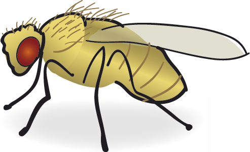
One of the major changes in this release was the ‘budding off’ of uberon to make a new insect module. This is analogous to the module created for phenoscape editors to edit highly specific vertebrate skeletal terms, described in:
Haendel, M. a, Balhoff, J. P., Bastian, F. B., Blackburn, D. C., Blake, J. a, Bradford, Y., … Mungall, C. J. (2014). Unification of multi-species vertebrate anatomy ontologies for comparative biology in Uberon. Journal of Biomedical Semantics, 5(1), 21. doi:10.1186/2041-1480-5-21
This means that ontology editors can more easily add insect-specific terms, by editing this module independently. See: obophenotype/insect-anatomy-ontology
These ontologies are merged at the time of release. So for uberon users, this results in this release having an additional 235 insect-specific terms. But note these are filtered out in species-specific views, e.g. composite-metazoan.
For more details, see: issue 1262
Ontology Diff Report
- Neuroanatomy
- Insect refactoring
- splitting insect terms into new edit module 1262
- obsoleting core uberon visceral musculature as it was not clear what was meant https://github.com/obophenotype/insect_neuroanatomy_ontology/issues/8
- disambiguating cardiogenic mesoderm. See https://github.com/obophenotype/insect_neuroanatomy_ontology/issues/7
- disambiguating spiracle
- disambiguating egg
- Adding missing xrefs for FBbts. See 1262
- GO
- taxon constraints for gallbladder, GOC:mr, https://github.com/geneontology/go-ontology/issues/12590
- aligned GO with uberon
- Design patterns
Original Ontology
- IRI: http://purl.obolibrary.org/obo/uberon.owl
- VersionIRI: http://purl.obolibrary.org/obo/uberon/releases/2016-08-12/uberon.owl
New Ontology
- IRI: http://purl.obolibrary.org/obo/uberon.owl
- VersionIRI: http://purl.obolibrary.org/obo/uberon/releases/2016-09-07/uberon.owl
Report for classes
Class objects lost from source: 0
Class objects new in target: 239
New Class : wing margin
- wing margin definition The border or edge of the wing. It separates the dorsal and ventral compartments. { database cross reference=FBC:gg }
- wing margin database cross reference FBbt:00005378
- wing margin has related synonym margin
- wing margin label wing margin
- wing margin id UBERON:6005378
- wing margin SubClassOf insect region of integument
New Class : synaptic neuropil block
- synaptic neuropil block definition A closely associated group of synaptic neuropil domains. { database cross reference=FBC:DOS , database cross reference=FBC:KI }
- synaptic neuropil block SubClassOf anatomical group
- synaptic neuropil block id UBERON:6041000
- synaptic neuropil block label synaptic neuropil block
- synaptic neuropil block database cross reference FBbt:00041000
- synaptic neuropil block has exact synonym level 1 neuropil { database cross reference=FlyBase:FBrf0224194 }
New Class : embryonic/larval integumentary system
- embryonic/larval integumentary system has exact synonym larval integumentary system
- embryonic/larval integumentary system id UBERON:6005393
- embryonic/larval integumentary system label embryonic/larval integumentary system
- embryonic/larval integumentary system SubClassOf integumental system
- embryonic/larval integumentary system database cross reference FBbt:00005393
New Class : adult integumentary system
- adult integumentary system database cross reference FBbt:00005396
- adult integumentary system has related synonym adult external
- adult integumentary system SubClassOf integumental system
- adult integumentary system id UBERON:6005396
- adult integumentary system has exact synonym adult integument
- adult integumentary system label adult integumentary system
New Class : abdominal histoblast primordium
- abdominal histoblast primordium label abdominal histoblast primordium
- abdominal histoblast primordium database cross reference FBbt:00016022
- abdominal histoblast primordium SubClassOf embryonic structure
- abdominal histoblast primordium definition . { database cross reference=FBC:VH }
- abdominal histoblast primordium SubClassOf primordium
- abdominal histoblast primordium has related synonym abdominal histoblast specific anlage
- abdominal histoblast primordium id UBERON:6016022
New Class : lymph gland primordium
- lymph gland primordium id UBERON:6005467
- lymph gland primordium SubClassOf circulatory system primordium
- lymph gland primordium definition . { database cross reference=FBC:VH }
- lymph gland primordium has related synonym lymph gland specific anlage
- lymph gland primordium database cross reference FBbt:00005467
- lymph gland primordium SubClassOf embryonic structure
- lymph gland primordium label lymph gland primordium
New Class : circulatory system primordium
- circulatory system primordium has related synonym embryonic/larval circulatory system specific anlage
- circulatory system primordium SubClassOf primordium
- circulatory system primordium id UBERON:6005461
- circulatory system primordium database cross reference FBbt:00005461
- circulatory system primordium label circulatory system primordium
New Class : trunk mesoderm anlage
- trunk mesoderm anlage database cross reference FBbt:00005436
- trunk mesoderm anlage label trunk mesoderm anlage
- trunk mesoderm anlage SubClassOf anlage
- trunk mesoderm anlage has related synonym Asn/A TrMes { database cross reference=FBC:DOS }
- trunk mesoderm anlage SubClassOf embryonic tissue
- trunk mesoderm anlage id UBERON:6005436
New Class : visual anlage
- visual anlage has related synonym AVis
- visual anlage has related synonym A VisSys { database cross reference=FBC:DOS }
- visual anlage SubClassOf anlage
- visual anlage database cross reference FBbt:00005434
- visual anlage label visual anlage
- visual anlage id UBERON:6005434
New Class : clypeo-labral anlage
- clypeo-labral anlage definition . { database cross reference=FBC:DOS , database cross reference=FBC:VH }
- clypeo-labral anlage has related synonym A Cly { database cross reference=FBC:DOS }
- clypeo-labral anlage id UBERON:6005439
- clypeo-labral anlage label clypeo-labral anlage
- clypeo-labral anlage has related synonym clypeo-labral specific anlage
- clypeo-labral anlage database cross reference FBbt:00005439
- clypeo-labral anlage SubClassOf anlage
New Class : visual anlage in statu nascendi
- visual anlage in statu nascendi id UBERON:6005425
- visual anlage in statu nascendi SubClassOf anlage in statu nascendi
- visual anlage in statu nascendi has related synonym A0Vis
- visual anlage in statu nascendi has related synonym Asn VisSys { database cross reference=FBC:DOS }
- visual anlage in statu nascendi label visual anlage in statu nascendi
- visual anlage in statu nascendi database cross reference FBbt:00005425
New Class : dorsal ectoderm anlage
- dorsal ectoderm anlage SubClassOf anlage
- dorsal ectoderm anlage database cross reference FBbt:00005428
- dorsal ectoderm anlage label dorsal ectoderm anlage
- dorsal ectoderm anlage id UBERON:6005428
New Class : ectoderm anlage
- ectoderm anlage id UBERON:6005427
- ectoderm anlage database cross reference FBbt:00005427
- ectoderm anlage SubClassOf anlage
- ectoderm anlage label ectoderm anlage
New Class : anlage in statu nascendi
- anlage in statu nascendi has related synonym A0
- anlage in statu nascendi label anlage in statu nascendi
- anlage in statu nascendi id UBERON:6005413
- anlage in statu nascendi definition “anlagen in statu nascendi” are domains that do not yet coincide 1:1 with a later organ. Anlagen in statu nascendi are typically defined for the early blastoderm by the expression domains of genes which, in the late blastoderm or later, are expressed in specific anlagen, but initially come on in larger domains. { database cross reference=FlyBase:FBrf0178740 }
- anlage in statu nascendi SubClassOf embryonic structure
- anlage in statu nascendi database cross reference FBbt:00005413
New Class : chaeta
- chaeta has exact synonym sensillum chaeticum { database cross reference=FlyBase:FBrf0111704 }
- chaeta database cross reference FBbt:00005177
- chaeta SubClassOf eo-type sensillum
- chaeta definition A sensillum with a long, unicellular, setiform outgrowth that is strongly chitinized. { database cross reference=FlyBase:FBrf0111704 , database cross reference=FlyBase:FBrf0166419 }
- chaeta label chaeta
- chaeta id UBERON:6005177
New Class : external sensory organ
- external sensory organ database cross reference FBbt:00005168
- external sensory organ SubClassOf sense organ
- external sensory organ has related synonym es
- external sensory organ has related synonym sensillum
- external sensory organ has related synonym eo
- external sensory organ label external sensory organ
- external sensory organ id UBERON:6005168
New Class : adult external head
- adult external head label adult external head
- adult external head database cross reference FBbt:00004481
- adult external head SubClassOf tagmatic subdivision of integument
- adult external head id UBERON:6004481
New Class : sternite
- sternite SubClassOf sclerite
- sternite database cross reference FBbt:00004477
- sternite definition An adult ventral sclerite. { database cross reference=FlyBase:FBrf0007734 , database cross reference=FlyBase:FBrf0111704 }
- sternite id UBERON:6004477
- sternite label sternite
- sternite has exact synonym abdominal sternite
New Class : tergite
- tergite definition The dorsal plate (sclerite) of an adult abdominal segment. { database cross reference=FlyBase:FBrf0007734 , database cross reference=FlyBase:FBrf0111704 }
- tergite id UBERON:6004476
- tergite SubClassOf sclerite
- tergite has exact synonym abdominal tergite
- tergite database cross reference FBbt:00004476
- tergite label tergite
New Class : sclerite
- sclerite definition A region of integument whose external cuticular part is a hard, sclerotized plate. { database cross reference=FBC:gg }
- sclerite label sclerite
- sclerite has related synonym plate
- sclerite id UBERON:6004475
- sclerite has exact synonym integumentary plate
- sclerite database cross reference FBbt:00004475
- sclerite SubClassOf insect region of integument
New Class : Bolwig organ
- Bolwig organ has exact synonym embryonic Bolwig’s organ
- Bolwig organ has exact synonym Bolwig’s organ
- Bolwig organ SubClassOf eye
- Bolwig organ SubClassOf internal sensillum
- Bolwig organ label Bolwig organ
- Bolwig organ SubClassOf embryonic/larval head sensillum
- Bolwig organ has related synonym dorsocaudal pharyngeal sense organ
- Bolwig organ id UBERON:6005805
- Bolwig organ has exact synonym embryonic visual system
- Bolwig organ definition Visual organ of the larva. It consists of a dense cluster of 12 ciliated photoreceptor neurons. { database cross reference=FlyBase:FBrf0089570 }
- Bolwig organ SubClassOf embryonic/larval ocular segment sensillum
- Bolwig organ has exact synonym larval Bolwig’s organ
- Bolwig organ database cross reference FBbt:00005805
New Class : adult somatic muscle
- adult somatic muscle id UBERON:6003259
- adult somatic muscle SubClassOf muscle structure
- adult somatic muscle database cross reference FBbt:00003259
- adult somatic muscle definition Muscle attached to the inner surface of the adult body wall, extending between articulated or flexibly joined areas, that serves to move the parts of the exoskeleton. { database cross reference=FlyBase:FBrf0007735 }
- adult somatic muscle label adult somatic muscle
New Class : mesothoracic tergum
- mesothoracic tergum has exact synonym mesonotum { database cross reference=FlyBase:FBrf0166419 }
- mesothoracic tergum database cross reference FBbt:00004580
- mesothoracic tergum SubClassOf tergum
- mesothoracic tergum has broad synonym notum
- mesothoracic tergum label mesothoracic tergum
- mesothoracic tergum has exact synonym alinotum { database cross reference=FlyBase:FBrf0111704 }
- mesothoracic tergum definition The dorsal sclerites (tergites) of the mesothorax. { database cross reference=FBC:DOS }
- mesothoracic tergum id UBERON:6004580
New Class : adult external mesothorax
- adult external mesothorax SubClassOf segmental subdivision of integument
- adult external mesothorax id UBERON:6004578
- adult external mesothorax label adult external mesothorax
- adult external mesothorax database cross reference FBbt:00004578
New Class : dorsal mesothorax
- dorsal mesothorax database cross reference FBbt:00004579
- dorsal mesothorax id UBERON:6004579
- dorsal mesothorax SubClassOf insect region of integument
- dorsal mesothorax label dorsal mesothorax
New Class : tergum
- tergum database cross reference FBbt:00004552
- tergum definition The upper, originally unpaired plate of the segmented insect body, which either independently or fused with a ventral plate covers the body dorsally, specifically that of the thorax. { database cross reference=FlyBase:FBrf0166419 }
- tergum label tergum
- tergum id UBERON:6004552
- tergum SubClassOf insect region of integument
New Class : adult external thorax
- adult external thorax id UBERON:6004551
- adult external thorax label adult external thorax
- adult external thorax SubClassOf tagmatic subdivision of integument
- adult external thorax definition . { database cross reference=FlyBase:FBrf0006943 }
- adult external thorax database cross reference FBbt:00004551
New Class : adult muscle system
- adult muscle system SubClassOf musculature of body
- adult muscle system label adult muscle system
- adult muscle system database cross reference FBbt:00003218
- adult muscle system id UBERON:6003218
- adult muscle system definition Muscle system of the adult. { database cross reference=FlyBase:FBrf0007735 }
New Class : basiproboscis
- basiproboscis database cross reference FBbt:00004540
- basiproboscis SubClassOf multicellular anatomical structure
- basiproboscis label basiproboscis
- basiproboscis id UBERON:6004540
New Class : proboscis
- proboscis SubClassOf organism subdivision
- proboscis definition The trunk-like extension of adult head, forming a protrusible sucking proboscis for the ingestion of liquid food (pg 425 Miller et al., 1950; pg 383, Ferris et al., 1950). { database cross reference=FlyBase:FBrf0007734 , database cross reference=FlyBase:FBrf0007735 }
- proboscis id UBERON:6004535
- proboscis label proboscis
- proboscis database cross reference FBbt:00004535
New Class : clypeus
- clypeus id UBERON:6004521
- clypeus SubClassOf sclerite
- clypeus label clypeus
- clypeus database cross reference FBbt:00004521
- clypeus definition The anterior part of the head between the frons and the labrum, which occasionally may fuse with either one to form the frontoclypeus and the clypeolabrum. In the context of Drosophila refers to a round structure on the center of the frontal adult external head. { database cross reference=FBC:gg , database cross reference=FlyBase:FBrf0166419 }
New Class : mouthpart
- mouthpart database cross reference FBbt:00004520
- mouthpart label mouthpart
- mouthpart SubClassOf anatomical entity
- mouthpart id UBERON:6004520
New Class : arista
- arista database cross reference FBbt:00004519
- arista SubClassOf segment of antenna
- arista has exact synonym segment 6
- arista definition Highly specialised, slender sixth segment of the antenna covered in bristle-like outgrowths known as laterals. It extends laterally from the dorsal region of the third segment of the antenna. { database cross reference=FlyBase:FBrf0007734 , database cross reference=FlyBase:FBrf0031004 , database cross reference=FlyBase:FBrf0137012 }
- arista id UBERON:6004519
- arista label arista
New Class : meningeal branch of mandibular nerve
- meningeal branch of mandibular nerve label meningeal branch of mandibular nerve
- meningeal branch of mandibular nerve has exact synonym ramus meningeus (Nervus mandibularis) { database cross reference=FMA:53047 }
- meningeal branch of mandibular nerve database cross reference http://en.wikipedia.org/wiki/Meningeal_branch_of_the_mandibular_nerve
- meningeal branch of mandibular nerve database cross reference http://www.snomedbrowser.com/Codes/Details/280249002
- meningeal branch of mandibular nerve has exact synonym nervus spinosus { database cross reference=FMA:TA , has synonym type=latin term }
- meningeal branch of mandibular nerve id UBERON:0036143
- meningeal branch of mandibular nerve database cross reference FMA:53047
- meningeal branch of mandibular nerve SubClassOf branching part of some mandibular nerve { source=FMA }
- meningeal branch of mandibular nerve has related synonym nervus spinosus { database cross reference=http://en.wikipedia.org/wiki/Meningeal_branch_of_the_mandibular_nerve }
- meningeal branch of mandibular nerve definition The meningeal branch of the mandibular nerve (recurrent branch, nervus spinosus) is a branch of the mandibular nerve that supplies the dura mater. { database cross reference=http://en.wikipedia.org/wiki/Meningeal_branch_of_the_mandibular_nerve }
- meningeal branch of mandibular nerve SubClassOf nerve of head region
- meningeal branch of mandibular nerve has obo namespace uberon
- meningeal branch of mandibular nerve has exact synonym ramus meningeus nervus mandibularis { database cross reference=FMA:TA , has synonym type=latin term }
New Class : incisive duct
- incisive duct database cross reference FMA:77282
- incisive duct label incisive duct
- incisive duct id UBERON:0036144
- incisive duct SubClassOf anatomical conduit space { source=FMA }
- incisive duct SubClassOf part of some incisive canal
- incisive duct definition an infrequent rudimentary duct, or protrusion of the mucous membrane into the incisive canal, on either side of the anterior extremity of the nasal crest { database cross reference=http://medical-dictionary.thefreedictionary.com/incisive+duct }
- incisive duct has obo namespace uberon
- incisive duct taxon notes horses exhibit Flehmen response but do not have an incisive duct communication between the nasal and the oral cavity because they do not breathe through their mouths, instead, the VNOs connect to the nasal passages by the nasopalatine duct
- incisive duct has exact synonym ductus incisivus { database cross reference=FMA:TA , has synonym type=latin term }
- incisive duct comment Taxon notes notes: In domestic mammals the Ductus incisivus is considerably better developed than in man. It is not accommodated in a Canalis incisivus, as in man, but in the Fissura palatina[NOMINA ANATOMICA VETERINARIA (2005)]
- incisive duct has exact synonym conduit incisif@fr { database cross reference=FMA:77282 }
New Class : glymphatic system
- glymphatic system label glymphatic system
- glymphatic system has obo namespace uberon
- glymphatic system SubClassOf part of some nervous system
- glymphatic system id UBERON:0036145
- glymphatic system definition macroscopic waste clearance system that utilizes a unique system of perivascular tunnels, formed by astroglial cells, to promote efficient elimination of soluble proteins and metabolites from the central nervous system. { database cross reference=http://dx.doi.org/10.1007/s11064-015-1581-6 , database cross reference=https://github.com/obophenotype/uberon/issues/1260 , database cross reference=MGI:sbello }
- glymphatic system SubClassOf anatomical system
- glymphatic system function notes hypothesized to be have roles such as waste clearance, distribution of compounds
New Class : dorsal imaginal precursor
- dorsal imaginal precursor database cross reference FBbt:00005831
- dorsal imaginal precursor label dorsal imaginal precursor
- dorsal imaginal precursor SubClassOf imaginal precursor
- dorsal imaginal precursor id UBERON:6005831
New Class : Bolwig organ primordium
- Bolwig organ primordium SubClassOf anterior ectoderm derivative
- Bolwig organ primordium has related synonym P1 BolOrg { database cross reference=FBC:DOS }
- Bolwig organ primordium has exact synonym Bolwig’s organ primordium
- Bolwig organ primordium database cross reference FBbt:00005830
- Bolwig organ primordium label Bolwig organ primordium
- Bolwig organ primordium definition . { database cross reference=FBC:DOS , database cross reference=FBC:VH }
- Bolwig organ primordium id UBERON:6005830
- Bolwig organ primordium SubClassOf primordium
New Class : arista lateral
- arista lateral label arista lateral
- arista lateral SubClassOf trichome
- arista lateral id UBERON:6005913
- arista lateral definition A bristle-like, non-innervated outgrowth of the arista. Each lateral is formed from the outgrowth of a single polyploid central core cell. { database cross reference=FlyBase:FBrf0137012 }
- arista lateral database cross reference FBbt:00005913
New Class : presumptive embryonic/larval tracheal system
- presumptive embryonic/larval tracheal system id UBERON:6005569
- presumptive embryonic/larval tracheal system SubClassOf presumptive embryonic/larval system
- presumptive embryonic/larval tracheal system label presumptive embryonic/larval tracheal system
- presumptive embryonic/larval tracheal system has related synonym embryonic tracheal system { database cross reference=FBC:DOS }
- presumptive embryonic/larval tracheal system database cross reference FBbt:00005569
- presumptive embryonic/larval tracheal system definition The sum of all the structures in the embryo that will develop into the embryonic/larval tracheal system. This corresponds to the tracheal primordia and their component tracheal branch primordia along with primordia of the spiracles. { database cross reference=FBC:DOS }
New Class : ventral ectoderm
- ventral ectoderm SubClassOf developing anatomical structure
- ventral ectoderm database cross reference FBbt:00005558
- ventral ectoderm has related synonym P2 VenEc { database cross reference=FBC:DOS }
- ventral ectoderm has related synonym venEc
- ventral ectoderm label ventral ectoderm
- ventral ectoderm id UBERON:6005558
- ventral ectoderm definition . { database cross reference=FlyBase:FBrf0089570 }
New Class : cardiogenic mesoderm
- cardiogenic mesoderm has exact synonym CardMes
- cardiogenic mesoderm label cardiogenic mesoderm
- cardiogenic mesoderm database cross reference FBbt:00005541
- cardiogenic mesoderm has exact synonym CardMesP2
- cardiogenic mesoderm SubClassOf visceral mesoderm derivative
- cardiogenic mesoderm has related synonym cardiac mesoderm primordium
- cardiogenic mesoderm id UBERON:6005541
- cardiogenic mesoderm has exact synonym cardiac mesoderm
- cardiogenic mesoderm has exact synonym P2 CardMes { database cross reference=FBC:DOS }
New Class : adult clypeo-labral anlage
- adult clypeo-labral anlage label adult clypeo-labral anlage
- adult clypeo-labral anlage database cross reference FBbt:00004203
- adult clypeo-labral anlage has related synonym A AdCly { database cross reference=FBC:DOS }
- adult clypeo-labral anlage id UBERON:6004203
- adult clypeo-labral anlage SubClassOf anlage
- adult clypeo-labral anlage SubClassOf embryonic tissue
- adult clypeo-labral anlage definition . { database cross reference=FBC:VH }
New Class : ventral epidermis primordium
- ventral epidermis primordium has related synonym venEpiP2
- ventral epidermis primordium database cross reference FBbt:00005533
- ventral epidermis primordium SubClassOf primordium
- ventral epidermis primordium SubClassOf ventral ectoderm derivative
- ventral epidermis primordium label ventral epidermis primordium
- ventral epidermis primordium has related synonym P2 iVenEp { database cross reference=FBC:DOS }
- ventral epidermis primordium id UBERON:6005533
New Class : clypeo-labral primordium
- clypeo-labral primordium database cross reference FBbt:00005538
- clypeo-labral primordium SubClassOf anterior ectoderm derivative
- clypeo-labral primordium id UBERON:6005538
- clypeo-labral primordium has related synonym ClyP2
- clypeo-labral primordium label clypeo-labral primordium
- clypeo-labral primordium SubClassOf primordium
New Class : dorsal epidermis primordium
- dorsal epidermis primordium SubClassOf primordium
- dorsal epidermis primordium database cross reference FBbt:00005526
- dorsal epidermis primordium SubClassOf embryonic structure
- dorsal epidermis primordium SubClassOf dorsal ectoderm derivative
- dorsal epidermis primordium has related synonym dorsal epidermis specific anlage
- dorsal epidermis primordium has related synonym dorEpiP2
- dorsal epidermis primordium label dorsal epidermis primordium
- dorsal epidermis primordium id UBERON:6005526
New Class : adult clypeo-labral primordium
- adult clypeo-labral primordium label adult clypeo-labral primordium
- adult clypeo-labral primordium id UBERON:6005514
- adult clypeo-labral primordium SubClassOf embryonic imaginal precursor
- adult clypeo-labral primordium SubClassOf primordium
- adult clypeo-labral primordium has related synonym A AdCly { database cross reference=FBC:DOS }
- adult clypeo-labral primordium database cross reference FBbt:00005514
New Class : vein of vestibular aqueduct
- vein of vestibular aqueduct has exact synonym vestibular aqueduct vein { database cross reference=FMA:52367 }
- vein of vestibular aqueduct id UBERON:0036074
- vein of vestibular aqueduct definition A vein that leaves the vestibule through an individual bone canal running parallel to the vestibular aqueduct up to the dura of the posterior side of the petrosa in the area of the endolymphatic sac. It then opens in the inferior petrosal sinus or the jugular bulb. The vein receives other branches from the bone, dura and sac. { database cross reference=http://www.ncbi.nlm.nih.gov/pubmed/42339 }
- vein of vestibular aqueduct SubClassOf craniocervical region vein
- vein of vestibular aqueduct SubClassOf tributary of some transverse sinus { source=FMA }
- vein of vestibular aqueduct has obo namespace uberon
- vein of vestibular aqueduct SubClassOf tributary of some inferior petrosal sinus { source=FMA }
- vein of vestibular aqueduct SubClassOf neck blood vessel
- vein of vestibular aqueduct label vein of vestibular aqueduct
- vein of vestibular aqueduct database cross reference FMA:52367
- vein of vestibular aqueduct SubClassOf ectoderm-derived structure
- vein of vestibular aqueduct SubClassOf tributary of some sigmoid sinus { source=FMA }
- vein of vestibular aqueduct SubClassOf head blood vessel
- vein of vestibular aqueduct has exact synonym vena aqueductus vestibuli { database cross reference=FMA:TA , has synonym type=latin term }
New Class : sex comb
- sex comb id UBERON:6004296
- sex comb SubClassOf sensilla row
- sex comb label sex comb
- sex comb definition A row of darkly pigmented, male specific chaetae located on the prothoracic metatarsal segment of the prothoracic leg. The number of chaetae (teeth) per sex comb varies between individuals, e.g.- Hannah-Alava and Stern report 10-13. { database cross reference=FlyBase:FBrf0011482 , database cross reference=FlyBase:FBrf0012178 }
- sex comb has exact synonym ctenidium { database cross reference=FlyBase:FBrf0012178 }
- sex comb database cross reference FBbt:00004296
- sex comb has exact synonym metatarsal comb
New Class : adult abdominal segment
- adult abdominal segment definition Any abdominal segment (UBERON:6000021) that is part of some adult abdomen (UBERON:6003023). { database cross reference=FBC:auto_generated_definition }
- adult abdominal segment database cross reference FBbt:00003024
- adult abdominal segment label adult abdominal segment
- adult abdominal segment SubClassOf insect abdominal segment
- adult abdominal segment SubClassOf insect adult segment
- adult abdominal segment id UBERON:6003024
New Class : adult abdomen
- adult abdomen definition Abdomen of the adult. { database cross reference=FlyBase:FBim0000809 , database cross reference=FlyBase:FBim0000810 }
- adult abdomen database cross reference FBbt:00003023
- adult abdomen SubClassOf insect abdomen
- adult abdomen label adult abdomen
- adult abdomen SubClassOf adult tagma
- adult abdomen id UBERON:6003023
New Class : adult prothoracic segment
- adult prothoracic segment has exact synonym t1
- adult prothoracic segment SubClassOf adult thoracic segment
- adult prothoracic segment has exact synonym thoracic segment 1
- adult prothoracic segment definition Any prothoracic segment (UBERON:6000017) that is part of some adult thorax (UBERON:6003018). { database cross reference=FBC:auto_generated_definition }
- adult prothoracic segment id UBERON:6003020
- adult prothoracic segment SubClassOf prothoracic segment
- adult prothoracic segment has exact synonym prothorax
- adult prothoracic segment database cross reference FBbt:00003020
- adult prothoracic segment label adult prothoracic segment
New Class : adult mesothoracic segment
- adult mesothoracic segment label adult mesothoracic segment
- adult mesothoracic segment SubClassOf mesothoracic segment
- adult mesothoracic segment definition Any mesothoracic segment (UBERON:6000018) that is part of some adult thorax (UBERON:6003018). { database cross reference=FBC:auto_generated_definition }
- adult mesothoracic segment id UBERON:6003021
- adult mesothoracic segment has exact synonym t2
- adult mesothoracic segment SubClassOf adult thoracic segment
- adult mesothoracic segment has exact synonym thoracic segment 2
- adult mesothoracic segment database cross reference FBbt:00003021
- adult mesothoracic segment has exact synonym mesothorax
New Class : adult thoracic segment
- adult thoracic segment SubClassOf insect adult segment
- adult thoracic segment definition Any segment (UBERON:6000003) that is part of some adult thorax (UBERON:6003018). { database cross reference=FBC:auto_generated_definition }
- adult thoracic segment id UBERON:6003019
- adult thoracic segment SubClassOf thoracic segment
- adult thoracic segment label adult thoracic segment
- adult thoracic segment database cross reference FBbt:00003019
New Class : adult thorax
- adult thorax SubClassOf adult tagma
- adult thorax id UBERON:6003018
- adult thorax definition Thorax of the adult. { database cross reference=FlyBase:FBim0000792 }
- adult thorax label adult thorax
- adult thorax SubClassOf insect thorax
- adult thorax database cross reference FBbt:00003018
New Class : adult labral segment
- adult labral segment SubClassOf labral segment
- adult labral segment id UBERON:6003011
- adult labral segment SubClassOf adult procephalic segment
- adult labral segment label adult labral segment
- adult labral segment definition Any labral segment (UBERON:6000008) that is part of some adult head (UBERON:6003007). { database cross reference=FBC:auto_generated_definition }
- adult labral segment database cross reference FBbt:00003011
New Class : adult antennal segment
- adult antennal segment SubClassOf antennal segment
- adult antennal segment id UBERON:6003012
- adult antennal segment label adult antennal segment
- adult antennal segment definition Any antennal segment (UBERON:6000009) that is part of some adult head (UBERON:6003007). { database cross reference=FBC:auto_generated_definition }
- adult antennal segment SubClassOf adult procephalic segment
- adult antennal segment has exact synonym adult antennal structure
- adult antennal segment database cross reference FBbt:00003012
New Class : adult procephalic segment
- adult procephalic segment SubClassOf procephalic segment
- adult procephalic segment database cross reference FBbt:00003010
- adult procephalic segment definition Any procephalic segment (UBERON:6000007) that is part of some adult head (UBERON:6003007). { database cross reference=FBC:auto_generated_definition }
- adult procephalic segment id UBERON:6003010
- adult procephalic segment label adult procephalic segment
- adult procephalic segment SubClassOf adult head segment
New Class : wing hair
- wing hair database cross reference FBbt:00004340
- wing hair label wing hair
- wing hair id UBERON:6004340
- wing hair has related synonym wing trichome
- wing hair SubClassOf trichome
- wing hair definition The posteriorly oriented trichome of a cell of the wing blade. Each cell makes one of these trichomes at its posterior vertex. { database cross reference=FBC:DOS }
New Class : adult tagma
- adult tagma id UBERON:6003005
- adult tagma label adult tagma
- adult tagma definition Any tagma (UBERON:6000002) that is part of some adult (UBERON:6003004). { database cross reference=FBC:auto_generated_definition }
- adult tagma database cross reference FBbt:00003005
- adult tagma SubClassOf tagma
New Class : adult head segment
- adult head segment label adult head segment
- adult head segment id UBERON:6003009
- adult head segment SubClassOf head segment
- adult head segment SubClassOf insect adult segment
- adult head segment definition Segment of the adult head. { database cross reference=FlyBase:FBrf0064788 }
- adult head segment database cross reference FBbt:00003009
New Class : insect adult segment
- insect adult segment label insect adult segment
- insect adult segment database cross reference FBbt:00003006
- insect adult segment SubClassOf organismal segment
- insect adult segment id UBERON:6003006
- insect adult segment definition Segment of the adult. { database cross reference=FBC:SPR }
New Class : insect adult head
- insect adult head SubClassOf adult tagma
- insect adult head SubClassOf head
- insect adult head definition Head of the adult organism. { database cross reference=FBC:SPR }
- insect adult head database cross reference FBbt:00003007
- insect adult head id UBERON:6003007
- insect adult head label insect adult head
New Class : sensilla row
- sensilla row label sensilla row
- sensilla row SubClassOf anatomical row
- sensilla row database cross reference FBbt:00100153
- sensilla row definition A row of sensilla. { database cross reference=FBC:DOS }
- sensilla row id UBERON:6100153
New Class : non-connected developing system
- non-connected developing system label non-connected developing system
- non-connected developing system SubClassOf developing anatomical structure
- non-connected developing system database cross reference FBbt:00040003
- non-connected developing system definition The sum of all the anlagen and primordia that will develop into a single system. { database cross reference=FBC:DOS }
- non-connected developing system SubClassOf anatomical group
- non-connected developing system id UBERON:6040003
New Class : synaptic neuropil
- synaptic neuropil database cross reference FBbt:00040005
- synaptic neuropil label synaptic neuropil
- synaptic neuropil definition The part of the neuropil consisting largely of synapsing neuron projections. { database cross reference=FlyBase:FBrf0224194 }
- synaptic neuropil SubClassOf anatomical group
- synaptic neuropil id UBERON:6040005
New Class : synaptic neuropil domain
- synaptic neuropil domain database cross reference FBbt:00040007
- synaptic neuropil domain SubClassOf multi cell part structure
- synaptic neuropil domain has related synonym synaptic neuropil
- synaptic neuropil domain has related synonym fibrous neuropil
- synaptic neuropil domain definition Region of synaptic neuropil whose boundaries are largely discontinuities in the density of presynaptic terminals: high density in the synaptic neuropil domain, lower or zero density outside this region. These boundaries often correspond to a boundary with glial sheath, tract neuropil or cortex. { database cross reference=FlyBase:FBrf0155900 , database cross reference=FlyBase:FBrf0224194 , database cross reference=http://flybrain.neurobio.arizona.edu/Flybrain/html/terms/terms.html }
- synaptic neuropil domain id UBERON:6040007
- synaptic neuropil domain label synaptic neuropil domain
New Class : adult nervous system
- adult nervous system SubClassOf nervous system
- adult nervous system definition . { database cross reference=FlyBase:FBrf0002482 , database cross reference=FlyBase:FBrf0007196 , database cross reference=FlyBase:FBrf0007735 , database cross reference=FlyBase:FBrf0034076 }
- adult nervous system database cross reference FBbt:00003559
- adult nervous system label adult nervous system
- adult nervous system id UBERON:6003559
New Class : adult central complex
- adult central complex has exact synonym CX { database cross reference=FlyBase:FBrf0224194 , has synonym type= }
- adult central complex has related synonym central body complex
- adult central complex has related synonym central body
- adult central complex SubClassOf synaptic neuropil block
- adult central complex id UBERON:6003632
- adult central complex label adult central complex
- adult central complex definition A midline crossing complex of 4 synaptic neuropil domains of the adult brain: the ellipsoid body, the fan-shaped body, the paired noduli, and the protocerebral bridge. It is closely associated with another paired synaptic neuropil domain, the lateral complex. It lies in the middle of the brain between the pedunculi of the mushroom bodies and is bounded ventrally by the esophagus, dorsally by the pars intercerebralis and laterally by the antenno-glomerular tracts. { database cross reference=FlyBase:FBrf0049409 , database cross reference=http://flybrain.neurobio.arizona.edu/Flybrain/html/terms/terms.html }
- adult central complex has exact synonym adult central body complex
- adult central complex database cross reference FBbt:00003632
New Class : supraesophageal ganglion
- supraesophageal ganglion label supraesophageal ganglion
- supraesophageal ganglion definition The pre-oral neuropils of the brain located above and some of it below the esophagus, comprising three fused ganglia (protocerebrum, deutocerebrum, and tritocerebrum) in the head. { database cross reference=FlyBase:FBrf0224194 , database cross reference=http://flybrain.neurobio.arizona.edu/Flybrain/html/terms/terms.html }
- supraesophageal ganglion id UBERON:6003626
- supraesophageal ganglion has related synonym SPG
- supraesophageal ganglion SubClassOf ganglion
- supraesophageal ganglion database cross reference FBbt:00003626
New Class : protocerebrum
- protocerebrum SubClassOf segmental subdivision of nervous system
- protocerebrum database cross reference FBbt:00003627
- protocerebrum has exact synonym protocerebral neuromere { database cross reference=FlyBase:FBrf0092819 }
- protocerebrum definition The most anterior of the segmental subdivisions of the insect CNS; thought to represent the first pre-oral segment of the brain. The protocerebrum comprises many discrete neuropil regions including the central body complex and mushroom bodies. { database cross reference=http://flybrain.neurobio.arizona.edu/Flybrain/html/terms/terms.html }
- protocerebrum id UBERON:6003627
- protocerebrum label protocerebrum
New Class : adult brain
- adult brain id UBERON:6003624
- adult brain SubClassOf brain
- adult brain database cross reference FBbt:00003624
- adult brain label adult brain
- adult brain definition Brain of the adult. { database cross reference=FBC:SPR }
New Class : adult central nervous system
- adult central nervous system database cross reference FBbt:00003623
- adult central nervous system SubClassOf central nervous system
- adult central nervous system label adult central nervous system
- adult central nervous system definition Central nervous system of the adult. { database cross reference=FBC:SPR }
- adult central nervous system id UBERON:6003623
New Class : embryonic brain
- embryonic brain SubClassOf brain
- embryonic brain definition Brain of the embryo. { database cross reference=FBC:SPR }
- embryonic brain database cross reference FBbt:00001060
- embryonic brain label embryonic brain
- embryonic brain id UBERON:6001060
New Class : presumptive embryonic/larval central nervous system
- presumptive embryonic/larval central nervous system database cross reference FBbt:00001056
- presumptive embryonic/larval central nervous system id UBERON:6001056
- presumptive embryonic/larval central nervous system definition The sum of all the structures in the embryo that will develop into the embryonic/larval central nervous system. { database cross reference=FlyBase:FBrf0031012 }
- presumptive embryonic/larval central nervous system has related synonym embryonic central nervous system { database cross reference=FBC:DOS }
- presumptive embryonic/larval central nervous system SubClassOf presumptive embryonic/larval system
- presumptive embryonic/larval central nervous system label presumptive embryonic/larval central nervous system
New Class : presumptive embryonic/larval nervous system
- presumptive embryonic/larval nervous system label presumptive embryonic/larval nervous system
- presumptive embryonic/larval nervous system id UBERON:6001055
- presumptive embryonic/larval nervous system definition The sum of all the structures in the embryo that will develop into the embryonic/larval nervous system. { database cross reference=FlyBase:FBrf0031012 }
- presumptive embryonic/larval nervous system SubClassOf presumptive embryonic/larval system
- presumptive embryonic/larval nervous system has related synonym embryonic nervous system { database cross reference=FBC:DOS }
- presumptive embryonic/larval nervous system database cross reference FBbt:00001055
New Class : neurogenic region
- neurogenic region id UBERON:6001057
- neurogenic region SubClassOf developing anatomical structure
- neurogenic region database cross reference FBbt:00001057
- neurogenic region label neurogenic region
New Class : visual primordium
- visual primordium has related synonym optic lobe primordium
- visual primordium id UBERON:6001059
- visual primordium SubClassOf anterior ectoderm derivative
- visual primordium has related synonym optic lobe placode
- visual primordium has related synonym P2 VisSys { database cross reference=FBC:DOS }
- visual primordium database cross reference FBbt:00001059
- visual primordium SubClassOf primordium
- visual primordium label visual primordium
New Class : embryonic/larval cuticle
- embryonic/larval cuticle has exact synonym larval cuticle
- embryonic/larval cuticle database cross reference FBbt:00004983
- embryonic/larval cuticle SubClassOf chitin-based cuticle
- embryonic/larval cuticle label embryonic/larval cuticle
- embryonic/larval cuticle id UBERON:6004983
New Class : third instar larval cuticle
- third instar larval cuticle SubClassOf embryonic/larval cuticle
- third instar larval cuticle database cross reference FBbt:00004986
- third instar larval cuticle label third instar larval cuticle
- third instar larval cuticle id UBERON:6004986
New Class : trichome
- trichome label trichome
- trichome id UBERON:6004979
- trichome has related synonym cell hair
- trichome definition Non-innervated projection of the cuticle around an actin-rich cellular process of an underlying epidermal cell. { database cross reference=FBC:DOS }
- trichome database cross reference FBbt:00004979
- trichome SubClassOf cuticular specialization
New Class : prothoracic metatarsus
- prothoracic metatarsus has exact synonym T1 tarsal segment 1
- prothoracic metatarsus database cross reference FBbt:00004670
- prothoracic metatarsus has exact synonym prothoracic tarsal segment 1
- prothoracic metatarsus has exact synonym T1 Ts1 { database cross reference=FlyBase:FBrf0219028 }
- prothoracic metatarsus label prothoracic metatarsus
- prothoracic metatarsus id UBERON:6004670
- prothoracic metatarsus SubClassOf metatarsus
- prothoracic metatarsus definition First tarsal segment of the prothoracic leg. Proximally, it articulates with the tibia and distally with the second tarsal segment. Its surface is covered with eight longitudinal rows of bristles and with transverse rows of bristles (TR) in the anterior compartment, between proximo-distal rows 7 and 8. There is sexual dimorphism in the number of TRs (transverse row bristles, TR). In males, 2 TRs are replaced by the sex comb and one central bristle. { database cross reference=FlyBase:FBrf0031004 , database cross reference=FlyBase:FBrf0219028 }
- prothoracic metatarsus has exact synonym prothoracic basitarsus { database cross reference=FlyBase:FBrf0111704 }
- prothoracic metatarsus SubClassOf prothoracic tarsal segment
New Class : prothoracic leg
- prothoracic leg definition Leg of the prothoracic segment. { database cross reference=FBC:DOS }
- prothoracic leg SubClassOf insect leg
- prothoracic leg id UBERON:6004663
- prothoracic leg label prothoracic leg
- prothoracic leg database cross reference FBbt:00004663
- prothoracic leg has exact synonym first leg
- prothoracic leg has exact synonym T1 leg
New Class : prothoracic tarsal segment
- prothoracic tarsal segment id UBERON:6004668
- prothoracic tarsal segment SubClassOf tarsal segment
- prothoracic tarsal segment database cross reference FBbt:00004668
- prothoracic tarsal segment definition Tarsal segment of the adult prothoracic leg. { database cross reference=FBC:MMC }
- prothoracic tarsal segment label prothoracic tarsal segment
New Class : metatarsus
- metatarsus SubClassOf tarsal segment
- metatarsus label metatarsus
- metatarsus database cross reference FBbt:00004648
- metatarsus has exact synonym first segment of the tarsus
- metatarsus has exact synonym 1st tarsal segment
- metatarsus has exact synonym tarsal segment 1
- metatarsus id UBERON:6004648
- metatarsus has exact synonym first tarsal segment
- metatarsus has exact synonym basitarsus { database cross reference=FlyBase:FBrf0111704 }
- metatarsus definition The first of the tarsal segments of the adult leg. It is located between the tibia and the second tarsal segment and is longer than the other tarsal segments. It has 8 longitudinal rows of bristles around the circumference. { database cross reference=FlyBase:FBim0000836 , database cross reference=FlyBase:FBrf0007734 , database cross reference=FlyBase:FBrf0012178 , database cross reference=FlyBase:FBrf0111704 }
New Class : tarsal segment
- tarsal segment definition A segment of the tarsus of the adult leg. The first tarsal segment, the metatarsus, is proximally connected to the tibia. Tarsal segments are covered in 8 longitudinal rows of bristles, with row 1 being the most ventral. { database cross reference=FlyBase:FBim0000836.html , database cross reference=FlyBase:FBrf0007734 , database cross reference=FlyBase:FBrf0012178 }
- tarsal segment id UBERON:6004646
- tarsal segment SubClassOf segment of leg
- tarsal segment has exact synonym tarsomere { database cross reference=FlyBase:FBrf0111704 }
- tarsal segment label tarsal segment
- tarsal segment database cross reference FBbt:00004646
New Class : trunk mesoderm derivative
- trunk mesoderm derivative definition . { database cross reference=FBC:DOS }
- trunk mesoderm derivative SubClassOf mesoderm-derived structure
- trunk mesoderm derivative label trunk mesoderm derivative
- trunk mesoderm derivative database cross reference FBbt:00026000
- trunk mesoderm derivative id UBERON:6026000
New Class : visceral mesoderm derivative
- visceral mesoderm derivative id UBERON:6026002
- visceral mesoderm derivative SubClassOf trunk mesoderm derivative
- visceral mesoderm derivative label visceral mesoderm derivative
- visceral mesoderm derivative definition . { database cross reference=FBC:DOS }
- visceral mesoderm derivative database cross reference FBbt:00026002
New Class : adult external abdomen
- adult external abdomen label adult external abdomen
- adult external abdomen database cross reference FBbt:00004788
- adult external abdomen SubClassOf tagmatic subdivision of integument
- adult external abdomen id UBERON:6004788
New Class : male analia
- male analia id UBERON:6004825
- male analia label male analia
- male analia database cross reference FBbt:00004825
- male analia SubClassOf analia
New Class : female analia
- female analia database cross reference FBbt:00004824
- female analia SubClassOf analia
- female analia id UBERON:6004824
- female analia label female analia
New Class : analia
- analia SubClassOf anatomical cluster
- analia id UBERON:6004823
- analia label analia
- analia database cross reference FBbt:00004823
New Class : clypeo-labral anlage in statu nascendi
- clypeo-labral anlage in statu nascendi has related synonym Asn Cly { database cross reference=FBC:DOS }
- clypeo-labral anlage in statu nascendi database cross reference FBbt:00000094
- clypeo-labral anlage in statu nascendi definition . { database cross reference=FBC:VH }
- clypeo-labral anlage in statu nascendi id UBERON:6000094
- clypeo-labral anlage in statu nascendi label clypeo-labral anlage in statu nascendi
- clypeo-labral anlage in statu nascendi SubClassOf anlage in statu nascendi
New Class : cephalic furrow
- cephalic furrow id UBERON:6000097
- cephalic furrow label cephalic furrow
- cephalic furrow SubClassOf epithelial furrow
- cephalic furrow database cross reference FBbt:00000097
New Class : ventral furrow
- ventral furrow database cross reference FBbt:00000096
- ventral furrow SubClassOf epithelial furrow
- ventral furrow label ventral furrow
- ventral furrow id UBERON:6000096
New Class : dorsal appendage
- dorsal appendage id UBERON:6000046
- dorsal appendage has exact synonym chorionic appendage { database cross reference=FlyBase:FBrf0047871 }
- dorsal appendage label dorsal appendage
- dorsal appendage SubClassOf chorionic specialization
- dorsal appendage definition Paired appendage of the eggshell, located on the anterior-dorsal side of the egg, just posterior to the operculum. Functions as a respiratory structures. { database cross reference=FBC:DOS }
- dorsal appendage database cross reference FBbt:00000046
New Class : abdominal segment 9
- abdominal segment 9 database cross reference FBbt:00000030
- abdominal segment 9 SubClassOf insect abdominal segment
- abdominal segment 9 id UBERON:6000030
- abdominal segment 9 label abdominal segment 9
- abdominal segment 9 has related synonym a9
New Class : abdominal segment 8
- abdominal segment 8 database cross reference FBbt:00000029
- abdominal segment 8 label abdominal segment 8
- abdominal segment 8 SubClassOf insect abdominal segment
- abdominal segment 8 id UBERON:6000029
- abdominal segment 8 has related synonym a8
New Class : insect abdominal segment
- insect abdominal segment id UBERON:6000021
- insect abdominal segment label insect abdominal segment
- insect abdominal segment definition Metameric subdivision of the abdomen. { database cross reference=FBC:SPR }
- insect abdominal segment SubClassOf organismal segment
- insect abdominal segment database cross reference FBbt:00000021
New Class : insect abdomen
- insect abdomen database cross reference FBbt:00000020
- insect abdomen SubClassOf tagma
- insect abdomen id UBERON:6000020
- insect abdomen definition The most posterior of the three tagma (UBERON:6000002). { database cross reference=FlyBase:FBrf0166419 }
- insect abdomen label insect abdomen
New Class : metathoracic segment
- metathoracic segment SubClassOf thoracic segment
- metathoracic segment database cross reference FBbt:00000019
- metathoracic segment label metathoracic segment
- metathoracic segment has related synonym t3
- metathoracic segment definition The third (most posterior) segment of the thorax. { database cross reference=FBC:DOS }
- metathoracic segment has related synonym thoracic segment 3
- metathoracic segment id UBERON:6000019
- metathoracic segment has related synonym metathorax
New Class : mesothoracic segment
- mesothoracic segment has related synonym thoracic segment 2
- mesothoracic segment has related synonym mesothorax
- mesothoracic segment label mesothoracic segment
- mesothoracic segment id UBERON:6000018
- mesothoracic segment has related synonym t2
- mesothoracic segment SubClassOf thoracic segment
- mesothoracic segment definition The second (middle) segment of the thorax. { database cross reference=FBC:DOS }
- mesothoracic segment database cross reference FBbt:00000018
New Class : gnathal segment
- gnathal segment id UBERON:6000011
- gnathal segment has related synonym postoral segment
- gnathal segment SubClassOf head segment
- gnathal segment definition The head segments that develop posterior to the embryonic/larval mouth. { database cross reference=FBC:gg }
- gnathal segment database cross reference FBbt:00000011
- gnathal segment label gnathal segment
New Class : insect thorax
- insect thorax database cross reference FBbt:00000015
- insect thorax label insect thorax
- insect thorax definition The main middle section of the insect body comprising three thoracic rings: the pro-, the meso- and the metathoraces which are more or less well fused and cask-like sometimes having on the upper lateral part one of two pairs of wings, while on the ventrolateral part each thoracic ring bears a pair of legs. { database cross reference=FlyBase:FBrf0166419 }
- insect thorax SubClassOf tagma
- insect thorax id UBERON:6000015
New Class : labial segment
- labial segment SubClassOf gnathal segment
- labial segment definition Most posterior of the gnathal segments. In the adult it bears a pair of fused appendages, the labia, and its ectodermal invaginations give rise to the salivary glands. { database cross reference=FlyBase:FBrf0007734 }
- labial segment database cross reference FBbt:00000014
- labial segment id UBERON:6000014
- labial segment label labial segment
New Class : prothoracic segment
- prothoracic segment SubClassOf thoracic segment
- prothoracic segment definition The first (most anterior) segment of the thorax. { database cross reference=FBC:DOS }
- prothoracic segment has related synonym prothorax
- prothoracic segment id UBERON:6000017
- prothoracic segment label prothoracic segment
- prothoracic segment has related synonym thoracic segment 1
- prothoracic segment has related synonym t1
- prothoracic segment database cross reference FBbt:00000017
New Class : thoracic segment
- thoracic segment label thoracic segment
- thoracic segment SubClassOf organismal segment
- thoracic segment definition Any segment (UBERON:6000003) that is part of some thorax (UBERON:6000015). { database cross reference=FBC:auto_generated_definition }
- thoracic segment database cross reference FBbt:00000016
- thoracic segment id UBERON:6000016
New Class : trunk mesoderm
- trunk mesoderm SubClassOf mesoderm
- trunk mesoderm has related synonym AtrunkMes
- trunk mesoderm definition The mesoderm of segments T1-A9. It first becomes morphologically distinct during ventral furrow formation. Following invagination during stages 6 and 7, it remains a coherent structure with no morphologically apparent subdivisions, apart from transient metamery during stage 9, until stage 11. It undergoes a number of morphological changes during this period: as its cells divide following invagination, it loses its epithelial integrity and by stage 9 has rearranged into a monolayer of cuboidal cells. A further division happens during stage 10 leading to two distinct layers by stage 11. { database cross reference=FlyBase:FBrf0089570 }
- trunk mesoderm id UBERON:6000128
- trunk mesoderm has related synonym TrMes
- trunk mesoderm label trunk mesoderm
- trunk mesoderm has related synonym P2 TrMes { database cross reference=FBC:DOS }
- trunk mesoderm database cross reference FBbt:00000128
New Class : anterior ectoderm
- anterior ectoderm database cross reference FBbt:00000119
- anterior ectoderm SubClassOf multicellular anatomical structure
- anterior ectoderm label anterior ectoderm
- anterior ectoderm definition The region of the ectoderm anterior to the cephalic furrow. { database cross reference=FBC:DOS }
- anterior ectoderm id UBERON:6000119
New Class : dorsal ectoderm
- dorsal ectoderm has related synonym AdorEc
- dorsal ectoderm database cross reference FBbt:00000112
- dorsal ectoderm label dorsal ectoderm
- dorsal ectoderm SubClassOf multicellular anatomical structure
- dorsal ectoderm id UBERON:6000112
- dorsal ectoderm has related synonym dorEc
New Class : mesoderm anlage
- mesoderm anlage label mesoderm anlage
- mesoderm anlage SubClassOf anlage
- mesoderm anlage has related synonym ventral plate
- mesoderm anlage database cross reference FBbt:00000104
- mesoderm anlage SubClassOf embryonic tissue
- mesoderm anlage id UBERON:6000104
New Class : adult cerebral ganglion
- adult cerebral ganglion database cross reference FBbt:00110636
- adult cerebral ganglion SubClassOf supraesophageal ganglion
- adult cerebral ganglion has related synonym supraesophageal ganglion
- adult cerebral ganglion has related synonym SPG
- adult cerebral ganglion has exact synonym CRG { database cross reference=FlyBase:FBrf0224194 , has synonym type= }
- adult cerebral ganglion id UBERON:6110636
- adult cerebral ganglion has related synonym cerebrum
- adult cerebral ganglion has related synonym brain
- adult cerebral ganglion label adult cerebral ganglion
- adult cerebral ganglion definition The pre-oral neuropils of the adult brain located above, around and partially below the esophagus, including the optic lobes. It excludes the gnathal ganglion. Developmentally, it comprises three fused neuromeres: protocerebrum, deutocerebrum, and tritocerebrum. { database cross reference=FlyBase:FBrf0224194 , database cross reference=http://flybrain.neurobio.arizona.edu/Flybrain/html/terms/terms.html }
New Class : embryonic optic lobe primordium
- embryonic optic lobe primordium label embryonic optic lobe primordium
- embryonic optic lobe primordium id UBERON:6000186
- embryonic optic lobe primordium has related synonym optic lobe primordium
- embryonic optic lobe primordium definition . { database cross reference=FBC:DOS , database cross reference=FBC:VH }
- embryonic optic lobe primordium SubClassOf primordium
- embryonic optic lobe primordium database cross reference FBbt:00000186
- embryonic optic lobe primordium has related synonym optic lobe placode
New Class : embryonic abdominal segment 9
- embryonic abdominal segment 9 definition Any abdominal segment 9 (UBERON:6000030) that is part of some embryonic abdomen (UBERON:6000171). { database cross reference=FBC:auto_generated_definition }
- embryonic abdominal segment 9 SubClassOf insect embryonic abdominal segment
- embryonic abdominal segment 9 has related synonym a9
- embryonic abdominal segment 9 id UBERON:6000181
- embryonic abdominal segment 9 label embryonic abdominal segment 9
- embryonic abdominal segment 9 SubClassOf abdominal segment 9
- embryonic abdominal segment 9 database cross reference FBbt:00000181
New Class : embryonic abdominal segment 8
- embryonic abdominal segment 8 database cross reference FBbt:00000180
- embryonic abdominal segment 8 definition Any abdominal segment 8 (UBERON:6000029) that is part of some embryonic abdomen (UBERON:6000171). { database cross reference=FBC:auto_generated_definition }
- embryonic abdominal segment 8 SubClassOf abdominal segment 8
- embryonic abdominal segment 8 has related synonym a8
- embryonic abdominal segment 8 label embryonic abdominal segment 8
- embryonic abdominal segment 8 id UBERON:6000180
- embryonic abdominal segment 8 SubClassOf insect embryonic abdominal segment
New Class : embryonic metathoracic segment
- embryonic metathoracic segment has related synonym thoracic segment 3
- embryonic metathoracic segment has related synonym t3
- embryonic metathoracic segment SubClassOf embryonic thoracic segment
- embryonic metathoracic segment database cross reference FBbt:00000170
- embryonic metathoracic segment has related synonym metathorax
- embryonic metathoracic segment label embryonic metathoracic segment
- embryonic metathoracic segment definition Metathoracic segment of the embryo. { database cross reference=FBC:DOS }
- embryonic metathoracic segment SubClassOf metathoracic segment
- embryonic metathoracic segment id UBERON:6000170
New Class : insect embryonic abdominal segment
- insect embryonic abdominal segment database cross reference FBbt:00000172
- insect embryonic abdominal segment SubClassOf insect abdominal segment
- insect embryonic abdominal segment definition Metameric subdivision of the embryonic abdomen. { database cross reference=FBC:SPR }
- insect embryonic abdominal segment id UBERON:6000172
- insect embryonic abdominal segment label insect embryonic abdominal segment
- insect embryonic abdominal segment SubClassOf embryonic segment
New Class : insect embryonic abdomen
- insect embryonic abdomen id UBERON:6000171
- insect embryonic abdomen definition Abdomen of the embryo. { database cross reference=FBC:DOS }
- insect embryonic abdomen SubClassOf insect abdomen
- insect embryonic abdomen label insect embryonic abdomen
- insect embryonic abdomen database cross reference FBbt:00000171
- insect embryonic abdomen SubClassOf embryonic tagma
New Class : embryonic labial segment
- embryonic labial segment label embryonic labial segment
- embryonic labial segment id UBERON:6000165
- embryonic labial segment SubClassOf labial segment
- embryonic labial segment SubClassOf embryonic gnathal segment
- embryonic labial segment definition Any labial segment (UBERON:6000014) that is part of some embryonic head (UBERON:6000155). { database cross reference=FBC:auto_generated_definition }
- embryonic labial segment database cross reference FBbt:00000165
New Class : embryonic thoracic segment
- embryonic thoracic segment database cross reference FBbt:00000167
- embryonic thoracic segment SubClassOf embryonic segment
- embryonic thoracic segment SubClassOf thoracic segment
- embryonic thoracic segment id UBERON:6000167
- embryonic thoracic segment label embryonic thoracic segment
- embryonic thoracic segment definition Any segment (UBERON:6000003) that is part of some embryonic thorax (UBERON:6000166). { database cross reference=FBC:auto_generated_definition }
New Class : insect embryonic thorax
- insect embryonic thorax id UBERON:6000166
- insect embryonic thorax definition Any thorax (UBERON:6000015) that is part of some embryo (UBERON:6000052). { database cross reference=FBC:auto_generated_definition }
- insect embryonic thorax SubClassOf embryonic tagma
- insect embryonic thorax database cross reference FBbt:00000166
- insect embryonic thorax SubClassOf insect thorax
- insect embryonic thorax label insect embryonic thorax
New Class : embryonic mesothoracic segment
- embryonic mesothoracic segment SubClassOf mesothoracic segment
- embryonic mesothoracic segment label embryonic mesothoracic segment
- embryonic mesothoracic segment database cross reference FBbt:00000169
- embryonic mesothoracic segment has related synonym thoracic segment 2
- embryonic mesothoracic segment has related synonym mesothorax
- embryonic mesothoracic segment id UBERON:6000169
- embryonic mesothoracic segment SubClassOf embryonic thoracic segment
- embryonic mesothoracic segment has related synonym t2
- embryonic mesothoracic segment definition Mesothoracic segment of the embryo. { database cross reference=FBC:DOS }
New Class : embryonic prothoracic segment
- embryonic prothoracic segment SubClassOf prothoracic segment
- embryonic prothoracic segment has related synonym prothorax
- embryonic prothoracic segment definition Prothoracic segment of the embryo. { database cross reference=FBC:DOS }
- embryonic prothoracic segment SubClassOf embryonic thoracic segment
- embryonic prothoracic segment database cross reference FBbt:00000168
- embryonic prothoracic segment id UBERON:6000168
- embryonic prothoracic segment has related synonym t1
- embryonic prothoracic segment label embryonic prothoracic segment
- embryonic prothoracic segment has related synonym thoracic segment 1
New Class : embryonic antennal segment
- embryonic antennal segment SubClassOf embryonic procephalic segment
- embryonic antennal segment database cross reference FBbt:00000160
- embryonic antennal segment id UBERON:6000160
- embryonic antennal segment label embryonic antennal segment
- embryonic antennal segment SubClassOf antennal segment
- embryonic antennal segment definition Any antennal segment (UBERON:6000009) that is part of some embryonic head (UBERON:6000155). { database cross reference=FBC:auto_generated_definition }
New Class : embryonic gnathal segment
- embryonic gnathal segment database cross reference FBbt:00000162
- embryonic gnathal segment SubClassOf embryonic head segment
- embryonic gnathal segment label embryonic gnathal segment
- embryonic gnathal segment SubClassOf gnathal segment
- embryonic gnathal segment id UBERON:6000162
- embryonic gnathal segment definition Any gnathal segment (UBERON:6000011) that is part of some embryonic head (UBERON:6000155). { database cross reference=FBC:auto_generated_definition }
New Class : embryonic segment
- embryonic segment database cross reference FBbt:00000154
- embryonic segment definition Any segment (UBERON:6000003) that is part of some embryo (UBERON:6000052). { database cross reference=FBC:auto_generated_definition }
- embryonic segment label embryonic segment
- embryonic segment id UBERON:6000154
- embryonic segment SubClassOf organismal segment
New Class : embryonic procephalic segment
- embryonic procephalic segment SubClassOf procephalic segment
- embryonic procephalic segment SubClassOf embryonic head segment
- embryonic procephalic segment id UBERON:6000158
- embryonic procephalic segment definition Any procephalic segment (UBERON:6000007) that is part of some embryonic head (UBERON:6000155). { database cross reference=FBC:auto_generated_definition }
- embryonic procephalic segment label embryonic procephalic segment
- embryonic procephalic segment database cross reference FBbt:00000158
New Class : embryonic head segment
- embryonic head segment SubClassOf head segment
- embryonic head segment label embryonic head segment
- embryonic head segment id UBERON:6000157
- embryonic head segment SubClassOf embryonic segment
- embryonic head segment database cross reference FBbt:00000157
- embryonic head segment definition Any segment (UBERON:6000003) that is part of some embryonic head (UBERON:6000155). { database cross reference=FBC:auto_generated_definition , database cross reference=FlyBase:FBrf0075072 }
New Class : mesodermal crest of segment T3
- mesodermal crest of segment T3 database cross reference FBbt:00000132
- mesodermal crest of segment T3 id UBERON:6000132
- mesodermal crest of segment T3 SubClassOf visceral mesoderm derivative
- mesodermal crest of segment T3 label mesodermal crest of segment T3
New Class : mesodermal crest
- mesodermal crest SubClassOf visceral mesoderm derivative
- mesodermal crest id UBERON:6000131
- mesodermal crest label mesodermal crest
- mesodermal crest database cross reference FBbt:00000131
New Class : embryonic tagma
- embryonic tagma id UBERON:6000137
- embryonic tagma SubClassOf tagma
- embryonic tagma definition Any tagma (UBERON:6000002) that is part of some embryo (UBERON:6000052). { database cross reference=FBC:auto_generated_definition }
- embryonic tagma label embryonic tagma
- embryonic tagma database cross reference FBbt:00000137
New Class : visceral mesoderm
- visceral mesoderm id UBERON:6000130
- visceral mesoderm SubClassOf embryonic structure
- visceral mesoderm label visceral mesoderm
- visceral mesoderm database cross reference FBbt:00000130
New Class : proneural cluster
- proneural cluster id UBERON:6001135
- proneural cluster label proneural cluster
- proneural cluster definition An equivalence group of the neurectoderm where all cells can, although only one will, become a neural progenitor cell. { database cross reference=FlyBase:FBrf0074477 , database cross reference=FlyBase:FBrf0089570 }
- proneural cluster database cross reference FBbt:00001135
- proneural cluster SubClassOf anlage
New Class : labral segment
- labral segment id UBERON:6000008
- labral segment SubClassOf procephalic segment
- labral segment has related synonym clypeo-labrum
- labral segment label labral segment
- labral segment definition Procephalic segment anterior to the antennal segment. In the adult it bears the clypeo-labrum. { database cross reference=FlyBase:FBrf0075072 , database cross reference=FlyBase:FBrf0089570 }
- labral segment database cross reference FBbt:00000008
New Class : procephalic segment
- procephalic segment has exact synonym pregnathal segment
- procephalic segment label procephalic segment
- procephalic segment SubClassOf head segment
- procephalic segment database cross reference FBbt:00000007
- procephalic segment has exact synonym preoral segment
- procephalic segment has related synonym cephalic segment
- procephalic segment has related synonym procephalon
- procephalic segment definition A segment that is anterior to the gnathal segments. { database cross reference=FBC:gg }
- procephalic segment id UBERON:6000007
New Class : antennal segment
- antennal segment database cross reference FBbt:00000009
- antennal segment definition Segment anterior to the intercalary segment. In the adult it bears the antennae. { database cross reference=FBC:DOS }
- antennal segment id UBERON:6000009
- antennal segment label antennal segment
- antennal segment SubClassOf procephalic segment
New Class : germ layer derivative
- germ layer derivative database cross reference FBbt:00000000
- germ layer derivative label germ layer derivative
- germ layer derivative id UBERON:6000000
- germ layer derivative SubClassOf embryonic structure
New Class : tagma
- tagma database cross reference FBbt:00000002
- tagma definition The three main divisions of the whole organism formed from groups of segments. { database cross reference=FBC:gg }
- tagma id UBERON:6000002
- tagma SubClassOf anterior-posterior subdivision of organism
- tagma label tagma
New Class : head segment
- head segment database cross reference FBbt:00000006
- head segment definition Any segment (UBERON:6000003) that is part of some head (UBERON:6000004). { database cross reference=FBC:auto_generated_definition , database cross reference=FlyBase:FBrf0075072 }
- head segment SubClassOf organismal segment
- head segment id UBERON:6000006
- head segment label head segment
New Class : ocular segment
- ocular segment SubClassOf procephalic segment
- ocular segment has related synonym acron
- ocular segment label ocular segment
- ocular segment definition Head segment derived from the second embryonic segment (between the labral and antennal segments). In the larva, this segment includes Bolwig’s organ. { database cross reference=FlyBase:FBrf0055841 , database cross reference=FlyBase:FBrf0075072 }
- ocular segment id UBERON:6000005
- ocular segment database cross reference FBbt:00000005
New Class : embryonic/larval ocular segment sensillum
- embryonic/larval ocular segment sensillum definition Any embryonic/larval sense organ (UBERON:6002639) that is part of some larval ocular segment (UBERON:6001731). { database cross reference=FBC:auto_generated_definition }
- embryonic/larval ocular segment sensillum SubClassOf embryonic/larval head sense organ
- embryonic/larval ocular segment sensillum database cross reference FBbt:00002642
- embryonic/larval ocular segment sensillum label embryonic/larval ocular segment sensillum
- embryonic/larval ocular segment sensillum has exact synonym larval ocular segment sensillum
- embryonic/larval ocular segment sensillum id UBERON:6002642
New Class : embryonic/larval sense organ
- embryonic/larval sense organ has exact synonym larval sense organ
- embryonic/larval sense organ definition Any sense organ (UBERON:6005155) that is part of some larva (UBERON:6001727). { database cross reference=FBC:auto_generated_definition }
- embryonic/larval sense organ id UBERON:6002639
- embryonic/larval sense organ SubClassOf sense organ
- embryonic/larval sense organ database cross reference FBbt:00002639
- embryonic/larval sense organ label embryonic/larval sense organ
New Class : cephalopharyngeal skeleton
- cephalopharyngeal skeleton has exact synonym mouth parts
- cephalopharyngeal skeleton SubClassOf cuticular specialization
- cephalopharyngeal skeleton has exact synonym cephalo-pharyngeal skeleton { database cross reference=FlyBase:FBrf0045366 }
- cephalopharyngeal skeleton id UBERON:6001845
- cephalopharyngeal skeleton label cephalopharyngeal skeleton
- cephalopharyngeal skeleton has exact synonym larval head skeleton { database cross reference=FlyBase:FBrf0205317 }
- cephalopharyngeal skeleton definition A group of connected sclerites of the anterior embryonic/larval digestive system (atrium, pharynx and dorsal pouch). The cibarial dilator muscles connect to the cephalopharyngeal skeleton to lift the roof of the pharynx and thereby widen its lumen (Jurgens, 1986). { database cross reference=FlyBase:FBrf0007734 , database cross reference=FlyBase:FBrf0045366 }
- cephalopharyngeal skeleton database cross reference FBbt:00001845
New Class : epipharyngeal sclerite
- epipharyngeal sclerite label epipharyngeal sclerite
- epipharyngeal sclerite has exact synonym epistomal sclerite { database cross reference=FlyBase:FBrf0045366 }
- epipharyngeal sclerite definition Unpaired sclerite to which the median tooth (labrum) posteriorly merges, and bears the cuticular pores of the labral sense organ. { database cross reference=FlyBase:FBrf0045366 }
- epipharyngeal sclerite database cross reference FBbt:00001848
- epipharyngeal sclerite SubClassOf cuticular specialization
- epipharyngeal sclerite id UBERON:6001848
New Class : insect embryonic/larval digestive system
- insect embryonic/larval digestive system database cross reference FBbt:00001842
- insect embryonic/larval digestive system label insect embryonic/larval digestive system
- insect embryonic/larval digestive system definition . { database cross reference=FlyBase:FBrf0007733 , database cross reference=FlyBase:FBrf0064792 }
- insect embryonic/larval digestive system has exact synonym larval digestive system
- insect embryonic/larval digestive system SubClassOf digestive system
- insect embryonic/larval digestive system id UBERON:6001842
New Class : posterior dorsal histoblast nest abdominal
- posterior dorsal histoblast nest abdominal database cross reference FBbt:00001809
- posterior dorsal histoblast nest abdominal label posterior dorsal histoblast nest abdominal
- posterior dorsal histoblast nest abdominal id UBERON:6001809
- posterior dorsal histoblast nest abdominal SubClassOf histoblast nest
New Class : ventral ectoderm derivative
- ventral ectoderm derivative database cross reference FBbt:00025993
- ventral ectoderm derivative label ventral ectoderm derivative
- ventral ectoderm derivative SubClassOf ectoderm-derived structure
- ventral ectoderm derivative id UBERON:6025993
- ventral ectoderm derivative definition . { database cross reference=FBC:DOS }
New Class : anterior ectoderm derivative
- anterior ectoderm derivative label anterior ectoderm derivative
- anterior ectoderm derivative id UBERON:6025991
- anterior ectoderm derivative definition . { database cross reference=FBC:DOS }
- anterior ectoderm derivative SubClassOf ectoderm-derived structure
- anterior ectoderm derivative database cross reference FBbt:00025991
New Class : embryonic/larval protocerebrum
- embryonic/larval protocerebrum label embryonic/larval protocerebrum
- embryonic/larval protocerebrum id UBERON:6001925
- embryonic/larval protocerebrum definition Protocerebrum of the embryo/larva. { database cross reference=FBC:SPR }
- embryonic/larval protocerebrum has related synonym larval protocerebrum
- embryonic/larval protocerebrum database cross reference FBbt:00001925
- embryonic/larval protocerebrum SubClassOf protocerebrum
New Class : insect embryonic/larval brain
- insect embryonic/larval brain has narrow synonym larval supraesophageal ganglion
- insect embryonic/larval brain id UBERON:6001920
- insect embryonic/larval brain label insect embryonic/larval brain
- insect embryonic/larval brain has narrow synonym larval brain
- insect embryonic/larval brain SubClassOf brain
- insect embryonic/larval brain SubClassOf ganglion of central nervous system
- insect embryonic/larval brain definition Brain of the embryo/larva. { database cross reference=FBC:SPR }
- insect embryonic/larval brain database cross reference FBbt:00001920
New Class : embryonic/larval central nervous system
- embryonic/larval central nervous system SubClassOf central nervous system
- embryonic/larval central nervous system database cross reference FBbt:00001919
- embryonic/larval central nervous system label embryonic/larval central nervous system
- embryonic/larval central nervous system id UBERON:6001919
- embryonic/larval central nervous system definition Central nervous system of the embryo/larva. { database cross reference=FBC:SPR }
- embryonic/larval central nervous system has exact synonym larval central nervous system
New Class : insect embryonic/larval nervous system
- insect embryonic/larval nervous system SubClassOf nervous system
- insect embryonic/larval nervous system id UBERON:6001911
- insect embryonic/larval nervous system definition Nervous system of the embryo/larva. { database cross reference=FlyBase:FBrf0045359 , database cross reference=FlyBase:FBrf0075072 , database cross reference=FlyBase:FBrf0105282 }
- insect embryonic/larval nervous system label insect embryonic/larval nervous system
- insect embryonic/larval nervous system database cross reference FBbt:00001911
- insect embryonic/larval nervous system has exact synonym larval nervous system
New Class : presumptive prothoracic metatarsus
- presumptive prothoracic metatarsus label presumptive prothoracic metatarsus
- presumptive prothoracic metatarsus database cross reference FBbt:00110746
- presumptive prothoracic metatarsus definition Region of the prothoracic leg disc that will develop into the prothoracic metatarsus. { database cross reference=FlyBase:FBrf0219028 }
- presumptive prothoracic metatarsus SubClassOf anlage
- presumptive prothoracic metatarsus id UBERON:6110746
New Class : embryonic/larval lymph gland
- embryonic/larval lymph gland has exact synonym embryonic/larval hematopoietic organ { database cross reference=FlyBase:FBrf0074444 }
- embryonic/larval lymph gland id UBERON:6001668
- embryonic/larval lymph gland label embryonic/larval lymph gland
- embryonic/larval lymph gland has exact synonym hematopoietic organ
- embryonic/larval lymph gland database cross reference FBbt:00001668
- embryonic/larval lymph gland definition Hematopoietic organ of the larva, located along the dorsal vessel, behind the brain. It produces the plasmatocytes, crystal cells and lamellocytes which are released at the end of the larval instar and onset of metamorphosis. It is composed of 3 pairs of bilateral lobes: an anterior primary lobe, a posterior secondary lobe and the most posterior lobe (or multiple lobes), the tertiary lobe. Each lobe is separated by pericardial cells. In the late embryo, the lymph gland consists of a single pair of lobes (primary lobes) containing around 20 cells each that flank the dorsal vessel. By the second larval instar, the lymph gland is composed of two or three new pairs of posterior lobes, and the primary lobes have increased in size to contain around 200 cells. During the first and early second larval instars, the lymph gland comprises only the hematopoietic progenitor population. The lymph gland increases in size ten-fold by the late third instar, and at this stage the progenitor cells (prohemocytes) become restricted to the medial region (medullary zone) of the primary lobe and become quiescent. The differentiated hemocytes are found in the periphery (cortical zone) of the primary lobe and these cells proliferate extensively. The posterior signalling center is a group of about 30 cells at the posterior tip of the primary lobe that secretes several signalling molecules and functions as a stem-cell niche. The secondary and tertiary lobes contain prohemocytes, and rarely a differentiated cell. Lymph glands are eliminated at metamorphosis. { database cross reference=FlyBase:FBrf0031001 , database cross reference=FlyBase:FBrf0032406 , database cross reference=FlyBase:FBrf0074444 , database cross reference=FlyBase:FBrf0155787 , database cross reference=FlyBase:FBrf0162082 , database cross reference=FlyBase:FBrf0187467 }
- embryonic/larval lymph gland SubClassOf multi-tissue structure
New Class : genital disc primordium
- genital disc primordium has related synonym genital disc specific anlage
- genital disc primordium SubClassOf imaginal disc primordium
- genital disc primordium label genital disc primordium
- genital disc primordium definition Primordium from which the genital disc develops. { database cross reference=FBC:SPR }
- genital disc primordium id UBERON:6001661
- genital disc primordium database cross reference FBbt:00001661
New Class : female genital disc primordium
- female genital disc primordium has related synonym female genital disc specific anlage
- female genital disc primordium SubClassOf genital disc primordium
- female genital disc primordium database cross reference FBbt:00001663
- female genital disc primordium label female genital disc primordium
- female genital disc primordium id UBERON:6001663
- female genital disc primordium definition Primordium from which the female genital disc derives. { database cross reference=FBC:SPR }
New Class : male genital disc primordium
- male genital disc primordium definition Primordium from which the male genital disc derives. { database cross reference=FBC:SPR }
- male genital disc primordium id UBERON:6001662
- male genital disc primordium label male genital disc primordium
- male genital disc primordium has related synonym male genital disc specific anlage
- male genital disc primordium database cross reference FBbt:00001662
- male genital disc primordium SubClassOf genital disc primordium
New Class : embryonic/larval circulatory system
- embryonic/larval circulatory system SubClassOf circulatory system
- embryonic/larval circulatory system label embryonic/larval circulatory system
- embryonic/larval circulatory system has exact synonym larval circulatory system
- embryonic/larval circulatory system database cross reference FBbt:00001664
- embryonic/larval circulatory system definition Tubular system of the embryo/larva through which hemolymph flows. { database cross reference=FlyBase:FBrf0031012 }
- embryonic/larval circulatory system id UBERON:6001664
New Class : ventral prothoracic disc primordium
- ventral prothoracic disc primordium id UBERON:6001658
- ventral prothoracic disc primordium SubClassOf ventral thoracic disc primordium
- ventral prothoracic disc primordium label ventral prothoracic disc primordium
- ventral prothoracic disc primordium database cross reference FBbt:00001658
- ventral prothoracic disc primordium has related synonym ventral prothoracic disc specific anlage
New Class : ventral thoracic disc primordium
- ventral thoracic disc primordium database cross reference FBbt:00001657
- ventral thoracic disc primordium label ventral thoracic disc primordium
- ventral thoracic disc primordium SubClassOf imaginal disc primordium
- ventral thoracic disc primordium id UBERON:6001657
- ventral thoracic disc primordium has related synonym ventral thoracic disc specific anlage
New Class : labial disc primordium
- labial disc primordium label labial disc primordium
- labial disc primordium has related synonym labial disc specific anlage
- labial disc primordium SubClassOf imaginal disc primordium
- labial disc primordium database cross reference FBbt:00001650
- labial disc primordium definition Primordium that will form the labial disc. { database cross reference=FBC:SPR }
- labial disc primordium id UBERON:6001650
New Class : eye-antennal disc primordium
- eye-antennal disc primordium definition Primordium from which the eye-antennal disc develops. { database cross reference=FBC:SPR }
- eye-antennal disc primordium label eye-antennal disc primordium
- eye-antennal disc primordium SubClassOf imaginal disc primordium
- eye-antennal disc primordium has related synonym eye-antennal disc specific anlage
- eye-antennal disc primordium id UBERON:6001652
- eye-antennal disc primordium database cross reference FBbt:00001652
New Class : dorsal thoracic disc primordium
- dorsal thoracic disc primordium SubClassOf imaginal disc primordium
- dorsal thoracic disc primordium id UBERON:6001653
- dorsal thoracic disc primordium has related synonym dorsal thoracic disc specific anlage
- dorsal thoracic disc primordium label dorsal thoracic disc primordium
- dorsal thoracic disc primordium database cross reference FBbt:00001653
New Class : dorsal metathoracic disc primordium
- dorsal metathoracic disc primordium database cross reference FBbt:00001656
- dorsal metathoracic disc primordium SubClassOf dorsal thoracic disc primordium
- dorsal metathoracic disc primordium has related synonym dorsal metathoracic disc specific anlage
- dorsal metathoracic disc primordium label dorsal metathoracic disc primordium
- dorsal metathoracic disc primordium id UBERON:6001656
New Class : dorsal mesothoracic disc primordium
- dorsal mesothoracic disc primordium label dorsal mesothoracic disc primordium
- dorsal mesothoracic disc primordium database cross reference FBbt:00001655
- dorsal mesothoracic disc primordium SubClassOf dorsal thoracic disc primordium
- dorsal mesothoracic disc primordium id UBERON:6001655
- dorsal mesothoracic disc primordium has related synonym dorsal mesothoracic disc specific anlage
New Class : imaginal disc primordium
- imaginal disc primordium database cross reference FBbt:00001649
- imaginal disc primordium definition Population of contiguous, morphologically distinct cells that will form an imaginal disc. { database cross reference=FBC:SPR }
- imaginal disc primordium label imaginal disc primordium
- imaginal disc primordium has related synonym imaginal disc specific anlage
- imaginal disc primordium SubClassOf primordium
- imaginal disc primordium id UBERON:6001649
- imaginal disc primordium SubClassOf embryonic imaginal precursor
New Class : embryonic imaginal precursor
- embryonic imaginal precursor SubClassOf imaginal precursor
- embryonic imaginal precursor id UBERON:6001648
- embryonic imaginal precursor label embryonic imaginal precursor
- embryonic imaginal precursor database cross reference FBbt:00001648
- embryonic imaginal precursor SubClassOf embryonic structure
- embryonic imaginal precursor definition Embryonic structure that will proliferate during larval development into a larval imaginal disc. During early embryogenesis the precursor is part of the epithelial sheet before segregating from the epithelial sheet by late embryogenesis (Bate and Martinez Arias, 1991). { database cross reference=FlyBase:FBrf0053815 , database cross reference=FlyBase:FBrf0064789 }
New Class : presumptive arista
- presumptive arista database cross reference FBbt:00110811
- presumptive arista SubClassOf anlage
- presumptive arista definition Region of the antennal disc that will develop into the arista. { database cross reference=FlyBase:FBrf0031004 }
- presumptive arista id UBERON:6110811
- presumptive arista label presumptive arista
New Class : anterior dorsal histoblast nest abdominal
- anterior dorsal histoblast nest abdominal label anterior dorsal histoblast nest abdominal
- anterior dorsal histoblast nest abdominal database cross reference FBbt:00001792
- anterior dorsal histoblast nest abdominal SubClassOf dorsal histoblast nest abdominal
- anterior dorsal histoblast nest abdominal id UBERON:6001792
New Class : dorsal histoblast nest abdominal
- dorsal histoblast nest abdominal id UBERON:6001791
- dorsal histoblast nest abdominal label dorsal histoblast nest abdominal
- dorsal histoblast nest abdominal SubClassOf dorsal imaginal precursor
- dorsal histoblast nest abdominal database cross reference FBbt:00001791
- dorsal histoblast nest abdominal SubClassOf histoblast nest
New Class : histoblast nest
- histoblast nest SubClassOf histoblast
- histoblast nest database cross reference FBbt:00001790
- histoblast nest id UBERON:6001790
- histoblast nest definition One of several groups of cells in the larval abdomen that gives rise to the adult external abdomen. { database cross reference=FBC:gg }
- histoblast nest label histoblast nest
New Class : histoblast
- histoblast database cross reference FBbt:00001789
- histoblast definition A progenitor of the adult external abdomen. { database cross reference=FBC:DOS , database cross reference=FBC:gg }
- histoblast id UBERON:6001789
- histoblast label histoblast
- histoblast SubClassOf embryonic structure
- histoblast SubClassOf embryonic/larval imaginal precursor
New Class : prothoracic leg disc
- prothoracic leg disc has exact synonym 1st leg disc
- prothoracic leg disc definition Imaginal disc of the ventral prothoracic segment. Precursor of structures of the adult ventral prothorax including the prothoracic (1st) leg. { database cross reference=FlyBase:FBrf0064789 }
- prothoracic leg disc id UBERON:6001781
- prothoracic leg disc has exact synonym ventral prothoracic disc
- prothoracic leg disc label prothoracic leg disc
- prothoracic leg disc has exact synonym T1 leg disc
- prothoracic leg disc database cross reference FBbt:00001781
- prothoracic leg disc SubClassOf ventral thoracic disc
New Class : genital disc
- genital disc definition Imaginal disc that gives rise to the internal and external genitalia, analia and adult hind gut (Cohen, 1993). { database cross reference=FlyBase:FBrf0064789 }
- genital disc SubClassOf imaginal disc
- genital disc id UBERON:6001784
- genital disc label genital disc
- genital disc database cross reference FBbt:00001784
New Class : male genital disc
- male genital disc definition Genital disc that gives rise to the penis and external genitalia, sperm pump, ejaculatory duct, paragonia and vas deferens, anal plates and adult hindgut (Cohen, 1993). { database cross reference=FlyBase:FBrf0064789 }
- male genital disc SubClassOf genital disc
- male genital disc id UBERON:6001785
- male genital disc label male genital disc
- male genital disc database cross reference FBbt:00001785
New Class : female genital disc
- female genital disc definition Genital disc that gives rise to the vagina and vaginal plates, uterus, parovaria, spermatheca, seminal recepticle, oviducts, female analia and hindgut. (Cohen, 1993). { database cross reference=FlyBase:FBrf0064789 }
- female genital disc database cross reference FBbt:00001787
- female genital disc SubClassOf genital disc
- female genital disc id UBERON:6001787
- female genital disc label female genital disc
New Class : ventral thoracic disc
- ventral thoracic disc database cross reference FBbt:00001780
- ventral thoracic disc SubClassOf imaginal disc
- ventral thoracic disc has exact synonym ventral thoracic disk
- ventral thoracic disc label ventral thoracic disc
- ventral thoracic disc id UBERON:6001780
- ventral thoracic disc definition Imaginal disc that is a precursor of ventral thoracic structures of the adult. { database cross reference=FlyBase:FBrf0064789 , database cross reference=FlyBase:FBrf0064803 }
- ventral thoracic disc has related synonym leg disc
New Class : haltere disc
- haltere disc database cross reference FBbt:00001779
- haltere disc label haltere disc
- haltere disc id UBERON:6001779
- haltere disc has exact synonym dorsal metathoracic disc
- haltere disc SubClassOf dorsal thoracic disc
- haltere disc definition Dorsal imaginal disc of the metathorax. Precursor of structure of the adult dorsal metathorax including the haltere. { database cross reference=FlyBase:FBrf0064789 }
New Class : wing disc
- wing disc label wing disc
- wing disc SubClassOf dorsal thoracic disc
- wing disc id UBERON:6001778
- wing disc has exact synonym dorsal mesothoracic disc
- wing disc database cross reference FBbt:00001778
- wing disc definition Dorsal imaginal disc of the mesothorax. Precursor of dorsal mesothoracic structures of the adult including the postnotum, scutum, scutellum, wing, wing hinge and part of the notal pleura. { database cross reference=FlyBase:FBrf0064789 }
New Class : dorsal thoracic disc
- dorsal thoracic disc label dorsal thoracic disc
- dorsal thoracic disc id UBERON:6001776
- dorsal thoracic disc has exact synonym dorsal thoracic disk
- dorsal thoracic disc definition Imaginal disc that is a precursor of dorsal thoracic structures of the adult. { database cross reference=FlyBase:FBrf0064789 , database cross reference=FlyBase:FBrf0064803 }
- dorsal thoracic disc database cross reference FBbt:00001776
- dorsal thoracic disc SubClassOf imaginal disc
New Class : antennal disc
- antennal disc SubClassOf imaginal disc
- antennal disc definition Posterior portion of the eye-antennal disc. It gives rise to the adult antennal segments and the maxillary palp, as well as contributing to the head capsule. { database cross reference=FlyBase:FBrf0064789 }
- antennal disc label antennal disc
- antennal disc database cross reference FBbt:00001767
- antennal disc id UBERON:6001767
New Class : embryonic/larval imaginal precursor
- embryonic/larval imaginal precursor database cross reference FBbt:00001760
- embryonic/larval imaginal precursor SubClassOf imaginal precursor
- embryonic/larval imaginal precursor id UBERON:6001760
- embryonic/larval imaginal precursor label embryonic/larval imaginal precursor
- embryonic/larval imaginal precursor definition Imaginal precursor that is part of the embryo/larva. { database cross reference=FBC:SPR }
- embryonic/larval imaginal precursor has exact synonym larval imaginal precursor
New Class : labial disc
- labial disc definition Imaginal disc that, in the adult, gives rise to the proboscis, including the prementum, prestomal cavity, labellum, labellar cap and pseudotrachea (Cohen, 1993). { database cross reference=FlyBase:FBrf0033866 , database cross reference=FlyBase:FBrf0064789 }
- labial disc label labial disc
- labial disc SubClassOf imaginal disc
- labial disc id UBERON:6001764
- labial disc database cross reference FBbt:00001764
New Class : eye-antennal disc
- eye-antennal disc database cross reference FBbt:00001766
- eye-antennal disc SubClassOf imaginal disc
- eye-antennal disc definition Imaginal disc that, in the adults, gives rise to the eye, antenna, head capsule (including all bristles and external membranes) and the maxillary palps (Cohen, 1993). { database cross reference=FlyBase:FBrf0064789 }
- eye-antennal disc id UBERON:6001766
- eye-antennal disc label eye-antennal disc
New Class : clypeo-labral disc
- clypeo-labral disc database cross reference FBbt:00001765
- clypeo-labral disc label clypeo-labral disc
- clypeo-labral disc id UBERON:6001765
- clypeo-labral disc definition Imaginal disc that in the adults gives rise to the labrum, anterior and posterior cibarial plates, fish trap bristles, epipharyngeal sclerite and clypeus (Cohen, 1993). { database cross reference=FlyBase:FBrf0064789 }
- clypeo-labral disc SubClassOf imaginal disc
- clypeo-labral disc has exact synonym clypeolabral disc
New Class : larval abdominal segment 9
- larval abdominal segment 9 has related synonym a9
- larval abdominal segment 9 SubClassOf larval abdominal segment
- larval abdominal segment 9 label larval abdominal segment 9
- larval abdominal segment 9 SubClassOf abdominal segment 9
- larval abdominal segment 9 id UBERON:6001756
- larval abdominal segment 9 database cross reference FBbt:00001756
- larval abdominal segment 9 definition Any abdominal segment 9 (UBERON:6000030) that is part of some larval abdomen (UBERON:6001746). { database cross reference=FBC:auto_generated_definition }
New Class : larval abdominal segment 8
- larval abdominal segment 8 id UBERON:6001755
- larval abdominal segment 8 label larval abdominal segment 8
- larval abdominal segment 8 definition Any abdominal segment 8 (UBERON:6000029) that is part of some larval abdomen (UBERON:6001746). { database cross reference=FBC:auto_generated_definition }
- larval abdominal segment 8 has related synonym a8
- larval abdominal segment 8 SubClassOf abdominal segment 8
- larval abdominal segment 8 database cross reference FBbt:00001755
- larval abdominal segment 8 SubClassOf larval abdominal segment
New Class : larval abdomen
- larval abdomen SubClassOf insect abdomen
- larval abdomen database cross reference FBbt:00001746
- larval abdomen definition Any abdomen (UBERON:6000020) that is part of some larva (UBERON:6001727). { database cross reference=FBC:auto_generated_definition }
- larval abdomen id UBERON:6001746
- larval abdomen label larval abdomen
- larval abdomen SubClassOf larval tagma
New Class : larval metathoracic segment
- larval metathoracic segment has related synonym thoracic segment 3
- larval metathoracic segment definition Any metathoracic segment (UBERON:6000019) that is part of some larval thorax (UBERON:6001741). { database cross reference=FBC:auto_generated_definition }
- larval metathoracic segment has related synonym metathorax
- larval metathoracic segment database cross reference FBbt:00001745
- larval metathoracic segment has related synonym t3
- larval metathoracic segment id UBERON:6001745
- larval metathoracic segment label larval metathoracic segment
- larval metathoracic segment SubClassOf larval thoracic segment
- larval metathoracic segment SubClassOf metathoracic segment
New Class : larval abdominal segment
- larval abdominal segment label larval abdominal segment
- larval abdominal segment SubClassOf larval segment
- larval abdominal segment database cross reference FBbt:00001747
- larval abdominal segment SubClassOf insect abdominal segment
- larval abdominal segment definition Any segment (UBERON:6000003) that is part of some larval abdomen (UBERON:6001746). { database cross reference=FBC:auto_generated_definition }
- larval abdominal segment id UBERON:6001747
New Class : larval labial segment
- larval labial segment database cross reference FBbt:00001740
- larval labial segment SubClassOf labial segment
- larval labial segment id UBERON:6001740
- larval labial segment label larval labial segment
- larval labial segment definition Any labial segment (UBERON:6000014) that is part of some larval head (UBERON:6001730). { database cross reference=FBC:auto_generated_definition }
- larval labial segment SubClassOf larval gnathal segment
New Class : larval thoracic segment
- larval thoracic segment SubClassOf thoracic segment
- larval thoracic segment definition Any segment (UBERON:6000003) that is part of some larval thorax (UBERON:6001741). { database cross reference=FBC:auto_generated_definition }
- larval thoracic segment label larval thoracic segment
- larval thoracic segment id UBERON:6001742
- larval thoracic segment SubClassOf larval segment
- larval thoracic segment database cross reference FBbt:00001742
New Class : larval thorax
- larval thorax id UBERON:6001741
- larval thorax label larval thorax
- larval thorax SubClassOf insect thorax
- larval thorax definition Any thorax (UBERON:6000015) that is part of some larva (UBERON:6001727). { database cross reference=FBC:auto_generated_definition }
- larval thorax SubClassOf larval tagma
- larval thorax database cross reference FBbt:00001741
New Class : larval mesothoracic segment
- larval mesothoracic segment database cross reference FBbt:00001744
- larval mesothoracic segment SubClassOf larval thoracic segment
- larval mesothoracic segment label larval mesothoracic segment
- larval mesothoracic segment has related synonym mesothorax
- larval mesothoracic segment id UBERON:6001744
- larval mesothoracic segment SubClassOf mesothoracic segment
- larval mesothoracic segment has related synonym thoracic segment 2
- larval mesothoracic segment has related synonym t2
- larval mesothoracic segment definition Any mesothoracic segment (UBERON:6000018) that is part of some larval thorax (UBERON:6001741). { database cross reference=FBC:auto_generated_definition }
New Class : larval prothoracic segment
- larval prothoracic segment SubClassOf larval thoracic segment
- larval prothoracic segment SubClassOf prothoracic segment
- larval prothoracic segment has related synonym prothorax
- larval prothoracic segment id UBERON:6001743
- larval prothoracic segment has related synonym t1
- larval prothoracic segment label larval prothoracic segment
- larval prothoracic segment database cross reference FBbt:00001743
- larval prothoracic segment definition Any prothoracic segment (UBERON:6000017) that is part of some larval thorax (UBERON:6001741). { database cross reference=FBC:auto_generated_definition }
- larval prothoracic segment has related synonym thoracic segment 1
New Class : larval antennal segment
- larval antennal segment id UBERON:6001735
- larval antennal segment label larval antennal segment
- larval antennal segment SubClassOf antennal segment
- larval antennal segment SubClassOf larval procephalic segment
- larval antennal segment database cross reference FBbt:00001735
- larval antennal segment definition Any antennal segment (UBERON:6000009) that is part of some larval head (UBERON:6001730). { database cross reference=FBC:auto_generated_definition }
New Class : larval labral segment
- larval labral segment label larval labral segment
- larval labral segment SubClassOf larval procephalic segment
- larval labral segment id UBERON:6001734
- larval labral segment database cross reference FBbt:00001734
- larval labral segment SubClassOf labral segment
- larval labral segment definition Any labral segment (UBERON:6000008) that is part of some larval head (UBERON:6001730). { database cross reference=FBC:auto_generated_definition }
New Class : larval gnathal segment
- larval gnathal segment label larval gnathal segment
- larval gnathal segment definition Any gnathal segment (UBERON:6000011) that is part of some larval head (UBERON:6001730). { database cross reference=FBC:auto_generated_definition }
- larval gnathal segment database cross reference FBbt:00001737
- larval gnathal segment SubClassOf gnathal segment
- larval gnathal segment SubClassOf larval head segment
- larval gnathal segment id UBERON:6001737
New Class : larval ocular segment
- larval ocular segment SubClassOf larval procephalic segment
- larval ocular segment id UBERON:6001731
- larval ocular segment SubClassOf ocular segment
- larval ocular segment definition Any ocular segment (UBERON:6000005) that is part of some larval head (UBERON:6001730). { database cross reference=FBC:auto_generated_definition }
- larval ocular segment database cross reference FBbt:00001731
- larval ocular segment has related synonym larval acron
- larval ocular segment label larval ocular segment
New Class : larval head
- larval head definition Any head (UBERON:6000004) that is part of some larva (UBERON:6001727). { database cross reference=FBC:auto_generated_definition }
- larval head database cross reference FBbt:00001730
- larval head label larval head
- larval head SubClassOf head
- larval head id UBERON:6001730
- larval head SubClassOf larval tagma
New Class : larval procephalic segment
- larval procephalic segment SubClassOf larval head segment
- larval procephalic segment label larval procephalic segment
- larval procephalic segment id UBERON:6001733
- larval procephalic segment definition Any procephalic segment (UBERON:6000007) that is part of some larval head (UBERON:6001730). { database cross reference=FBC:auto_generated_definition }
- larval procephalic segment database cross reference FBbt:00001733
- larval procephalic segment SubClassOf procephalic segment
New Class : larval head segment
- larval head segment SubClassOf head segment
- larval head segment SubClassOf larval segment
- larval head segment database cross reference FBbt:00001732
- larval head segment definition Any segment (UBERON:6000003) that is part of some larval head (UBERON:6001730). { database cross reference=FBC:auto_generated_definition , database cross reference=FlyBase:FBrf0075072 }
- larval head segment label larval head segment
- larval head segment id UBERON:6001732
New Class : larval tagma
- larval tagma id UBERON:6001728
- larval tagma label larval tagma
- larval tagma SubClassOf tagma
- larval tagma database cross reference FBbt:00001728
- larval tagma definition Any tagma (UBERON:6000002) that is part of some larva (UBERON:6001727). { database cross reference=FBC:auto_generated_definition }
New Class : larval segment
- larval segment database cross reference FBbt:00001729
- larval segment definition Any segment (UBERON:6000003) that is part of some larva (UBERON:6001727). { database cross reference=FBC:auto_generated_definition }
- larval segment SubClassOf organismal segment
- larval segment label larval segment
- larval segment id UBERON:6001729
New Class : ring gland
- ring gland database cross reference FBbt:00001722
- ring gland definition A closely associated cluster of three neuroendocrine glands located anterior to the aorta and dorsal to the esophagus. At embryonic stage 17 and in larva it is found anterior to the brain and is formed by the corpus cardiacum ventrally, the prothoracic gland laterally and the corpus allatum dorsally. The adult ring gland is located anterior to the proventriculus and is formed by the corpus cardiacum ventrally, the corpus allatum dorsally and the hypocerebral ganglion posterior to the corpus cardiacum. It is innervated by neurons of the nervus corporis cardiaci (NccI-III). { database cross reference=FlyBase:FBrf0064803 , database cross reference=FlyBase:FBrf0089570 , database cross reference=FlyBase:FBrf0179777 , database cross reference=PMID:12966498 }
- ring gland label ring gland
- ring gland SubClassOf anatomical cluster
- ring gland id UBERON:6001722
- ring gland SubClassOf endocrine system component
New Class : anterior-posterior subdivision of organism
- anterior-posterior subdivision of organism label anterior-posterior subdivision of organism
- anterior-posterior subdivision of organism SubClassOf organism subdivision
- anterior-posterior subdivision of organism id UBERON:6057001
- anterior-posterior subdivision of organism database cross reference FBbt:00057001
New Class : segment of leg
- segment of leg definition Any appendage segment (UBERON:6007018) that is part of some leg (UBERON:6004640). { database cross reference=FBC:auto_generated_definition }
- segment of leg database cross reference FBbt:00007150
- segment of leg SubClassOf subdivision of organism along appendicular axis
- segment of leg id UBERON:6007150
- segment of leg label segment of leg
New Class : segment of antenna
- segment of antenna SubClassOf subdivision of organism along appendicular axis
- segment of antenna label segment of antenna
- segment of antenna database cross reference FBbt:00007149
- segment of antenna definition Any appendage segment (UBERON:6007018) that is part of some antenna (UBERON:6004511). { database cross reference=FBC:auto_generated_definition }
- segment of antenna id UBERON:6007149
New Class : adult protocerebrum
- adult protocerebrum SubClassOf protocerebrum
- adult protocerebrum id UBERON:6007145
- adult protocerebrum label adult protocerebrum
- adult protocerebrum definition Protocerebrum of the adult brain. { database cross reference=FBC:SPR }
- adult protocerebrum database cross reference FBbt:00007145
New Class : presumptive embryonic/larval system
- presumptive embryonic/larval system SubClassOf embryonic structure
- presumptive embryonic/larval system label presumptive embryonic/larval system
- presumptive embryonic/larval system id UBERON:6007116
- presumptive embryonic/larval system database cross reference FBbt:00007116
- presumptive embryonic/larval system definition The sum of all the developing presumptive components of a specific embryonic/larval system in an embryo. { database cross reference=FBC:DOS }
- presumptive embryonic/larval system SubClassOf non-connected developing system
New Class : insect integumentary specialisation
- insect integumentary specialisation SubClassOf insect region of integument
- insect integumentary specialisation database cross reference FBbt:00007288
- insect integumentary specialisation label insect integumentary specialisation
- insect integumentary specialisation id UBERON:6007288
New Class : tagmatic subdivision of integument
- tagmatic subdivision of integument database cross reference FBbt:00007289
- tagmatic subdivision of integument SubClassOf insect region of integument
- tagmatic subdivision of integument id UBERON:6007289
- tagmatic subdivision of integument label tagmatic subdivision of integument
New Class : insect region of integument
- insect region of integument SubClassOf organ system subdivision
- insect region of integument id UBERON:6007284
- insect region of integument definition A region of cuticle and its underlying epidermis. { database cross reference=FBC:DOS }
- insect region of integument label insect region of integument
- insect region of integument database cross reference FBbt:00007284
New Class : segmental subdivision of integument
- segmental subdivision of integument id UBERON:6007285
- segmental subdivision of integument label segmental subdivision of integument
- segmental subdivision of integument definition A segmental subdivision of the integument of the whole animal. { database cross reference=FBC:DOS }
- segmental subdivision of integument database cross reference FBbt:00007285
- segmental subdivision of integument SubClassOf insect region of integument
New Class : embryonic/larval head sense organ
- embryonic/larval head sense organ database cross reference FBbt:00007280
- embryonic/larval head sense organ has exact synonym larval head sense organ
- embryonic/larval head sense organ label embryonic/larval head sense organ
- embryonic/larval head sense organ id UBERON:6007280
- embryonic/larval head sense organ SubClassOf embryonic/larval sense organ
- embryonic/larval head sense organ definition Any sense organ (UBERON:6005155) that is part of some larval head (UBERON:6001730). { database cross reference=FBC:auto_generated_definition }
New Class : chorionic specialization
- chorionic specialization SubClassOf acellular anatomical structure
- chorionic specialization definition Specialized structure of the chorion of the egg. { database cross reference=FBC:MMC }
- chorionic specialization id UBERON:6007248
- chorionic specialization label chorionic specialization
- chorionic specialization database cross reference FBbt:00007248
New Class : cuticular specialization
- cuticular specialization database cross reference FBbt:00007245
- cuticular specialization label cuticular specialization
- cuticular specialization SubClassOf acellular anatomical structure
- cuticular specialization id UBERON:6007245
New Class : embryonic/larval sensillum
- embryonic/larval sensillum database cross reference FBbt:00007240
- embryonic/larval sensillum SubClassOf arthropod sensillum
- embryonic/larval sensillum definition Any sensillum (UBERON:6007152) that is part of some larva (UBERON:6001727). { database cross reference=FBC:auto_generated_definition }
- embryonic/larval sensillum id UBERON:6007240
- embryonic/larval sensillum has exact synonym larval sensillum
- embryonic/larval sensillum label embryonic/larval sensillum
- embryonic/larval sensillum SubClassOf embryonic/larval sense organ
New Class : embryonic/larval head sensillum
- embryonic/larval head sensillum label embryonic/larval head sensillum
- embryonic/larval head sensillum SubClassOf embryonic/larval sensillum
- embryonic/larval head sensillum has narrow synonym larval head sensillum
- embryonic/larval head sensillum SubClassOf head sensillum
- embryonic/larval head sensillum id UBERON:6007242
- embryonic/larval head sensillum definition Any sensillum (UBERON:6007152) that is part of some larval head (UBERON:6001730). { database cross reference=FBC:auto_generated_definition }
- embryonic/larval head sensillum database cross reference FBbt:00007242
- embryonic/larval head sensillum SubClassOf embryonic/larval head sense organ
New Class : internal sensillum
- internal sensillum id UBERON:6007233
- internal sensillum SubClassOf arthropod sensillum
- internal sensillum definition Sensillum that is internal to the animal - i.e. having no part that is part of the cuticle. { database cross reference=FBC:DOS }
- internal sensillum SubClassOf internal sense organ
- internal sensillum database cross reference FBbt:00007233
- internal sensillum label internal sensillum
New Class : external sensillum
- external sensillum definition Sensillum which is embedded in the body wall and has a specific cuticular structure involved in the transduction of some sensory signal as a part. { database cross reference=FBC:DOS }
- external sensillum label external sensillum
- external sensillum database cross reference FBbt:00007231
- external sensillum SubClassOf arthropod sensillum
- external sensillum SubClassOf external sensory organ
- external sensillum id UBERON:6007231
New Class : eo-type sensillum
- eo-type sensillum database cross reference FBbt:00007232
- eo-type sensillum SubClassOf external sensillum
- eo-type sensillum definition Sensillum with innervated external cuticular sensory structure consisting of one or more bipolar sensory neurons, and 3 support cells: a trichogen cell which makes the external cuticular sensory structure, a thecogen cell which makes the socket that holds the base of the external sensory structure and a tormogen cell which makes the cuticular matrix (cap) at the tip of the innervating dendrite(s). { database cross reference=FlyBase:FBrf0089570 }
- eo-type sensillum id UBERON:6007232
- eo-type sensillum label eo-type sensillum
- eo-type sensillum has related synonym eso
New Class : centro-posterior medial synaptic neuropil domain
- centro-posterior medial synaptic neuropil domain has related synonym CPM
- centro-posterior medial synaptic neuropil domain has exact synonym larval central complex
- centro-posterior medial synaptic neuropil domain database cross reference FBbt:00007070
- centro-posterior medial synaptic neuropil domain has exact synonym centro-posterior medial synaptic neuropil domain
- centro-posterior medial synaptic neuropil domain definition A synaptic neuropil domain of the larval brain located posterior to the medial lobe of the mushroom body. It is separated from the centro-lateral compartment by the peduncle, and from the centro-posterior intermediate compartment by the antenno-cerebral tract to which it is connected via a branch. { database cross reference=FlyBase:FBrf0155900 }
- centro-posterior medial synaptic neuropil domain label centro-posterior medial synaptic neuropil domain
- centro-posterior medial synaptic neuropil domain id UBERON:6007070
- centro-posterior medial synaptic neuropil domain has exact synonym centro-posterior medial neuropil
- centro-posterior medial synaptic neuropil domain SubClassOf synaptic neuropil domain
New Class : dorsal ectoderm derivative
- dorsal ectoderm derivative id UBERON:6007046
- dorsal ectoderm derivative SubClassOf multicellular anatomical structure
- dorsal ectoderm derivative label dorsal ectoderm derivative
- dorsal ectoderm derivative database cross reference FBbt:00007046
New Class : trunk ectoderm
- trunk ectoderm definition The region of the ectoderm posterior to the cephalic furrow. { database cross reference=FBC:DOS }
- trunk ectoderm database cross reference FBbt:00007045
- trunk ectoderm label trunk ectoderm
- trunk ectoderm SubClassOf developing anatomical structure
- trunk ectoderm id UBERON:6007045
New Class : metatarsus of male prothoracic leg
- metatarsus of male prothoracic leg label metatarsus of male prothoracic leg
- metatarsus of male prothoracic leg id UBERON:6007020
- metatarsus of male prothoracic leg SubClassOf prothoracic metatarsus
- metatarsus of male prothoracic leg definition First tarsal segment of the male prothoracic leg. In contrast to females, it has a sex comb, central bristle and 5 to 6 transverse rows of bristles (TR). { database cross reference=FlyBase:FBrf0012178 , database cross reference=FlyBase:FBrf0031004 , database cross reference=FlyBase:FBrf0219028 }
- metatarsus of male prothoracic leg database cross reference FBbt:00007020
New Class : abdominal histoblast anlage
- abdominal histoblast anlage SubClassOf embryonic tissue
- abdominal histoblast anlage SubClassOf anlage
- abdominal histoblast anlage id UBERON:6017021
- abdominal histoblast anlage label abdominal histoblast anlage
- abdominal histoblast anlage database cross reference FBbt:00017021
- abdominal histoblast anlage definition . { database cross reference=FBC:VH }
New Class : stomatogastric nervous system
- stomatogastric nervous system label stomatogastric nervous system
- stomatogastric nervous system definition A group of small, interconnected ganglia situated posterior to and between the two brain hemispheres and associated with the pharynx, esophagus and aorta. { database cross reference=FlyBase:FBrf0089570 , database cross reference=PMID:12966498 }
- stomatogastric nervous system SubClassOf organ system subdivision
- stomatogastric nervous system database cross reference FBbt:00005096
- stomatogastric nervous system has related synonym stomodaeal nervous system { database cross reference=FlyBase:FBrf0111704 }
- stomatogastric nervous system id UBERON:6005096
- stomatogastric nervous system has related synonym stomodeal nervous system { database cross reference=FlyBase:FBrf0111704 }
New Class : obsolete visceral muscle
- obsolete visceral muscle label obsolete visceral muscle
- obsolete visceral muscle deprecated true
- obsolete visceral muscle consider FBbt:00005070
- obsolete visceral muscle id UBERON:6005070
- obsolete visceral muscle consider CL:0008007
- obsolete visceral muscle definition The visceral musculature comprises circular and longitudinal fibers which surround the entire intestinal tract, with the exception of the recurrent layer of the proventriculus. The circular fibers derive from a bilaterally symmetrical band of mesodermal cells extending continuously throughout most of the germ band. The longitudinal fibers derive from clusters of mesodermal cells which appear during stage 12 at the posterior end of the embryo and migrate anteriorly. { database cross reference=FlyBase:FBrf0064793 , database cross reference=FlyBase:FBrf0089570 , database cross reference=https://github.com/obophenotype/insect_neuroanatomy_ontology/issues/8 }
New Class : spiracle
- spiracle SubClassOf insect integumentary specialisation
- spiracle definition Opening of the tracheal system on the surface of the body. { database cross reference=FlyBase:FBrf0166419 }
- spiracle database cross reference FBbt:00005054
- spiracle label spiracle
- spiracle id UBERON:6005054
New Class : tracheal primordium
- tracheal primordium label tracheal primordium
- tracheal primordium SubClassOf embryonic structure
- tracheal primordium definition Primordium of a single tracheal metamere of the embryonic/larval tracheal system from its appearance as a placode during stage 11 to the completion of fusion with adjacent tracheal metameres in stage 15/16. There are 10 pairs of tracheal primordia - one pair in each segment from T2 to A8. Each tracheal primordium originates as a slight depression in the lateral ectoderm during stage 10 known as a tracheal placode. During stage 11, these placodes invaginate to form tracheal pits that elongate and branch. These pits close over during stage 13. Fusion of tracheal primordia begins at stage 14 with fusion of the dorsal trunk primordia and is complete by early stage 16. { database cross reference=FlyBase:FBrf0089570 }
- tracheal primordium SubClassOf dorsal ectoderm derivative
- tracheal primordium database cross reference FBbt:00005037
- tracheal primordium has related synonym TrachP3
- tracheal primordium SubClassOf primordium
- tracheal primordium has related synonym tracheal placode
- tracheal primordium has exact synonym P2 TrachP { database cross reference=FBC:DOS }
- tracheal primordium id UBERON:6005037
New Class : tracheal pit
- tracheal pit has exact synonym tracheal sac { database cross reference=FlyBase:FBrf0147036 }
- tracheal pit label tracheal pit
- tracheal pit has exact synonym embryonic tracheal pit
- tracheal pit database cross reference FBbt:00005036
- tracheal pit SubClassOf tracheal primordium
- tracheal pit id UBERON:6005036
- tracheal pit definition Deep and narrow pit formed by the invagination of a tracheal placode in a late extended germ band embryo. Invagination of the placode begins during stage 11. Branches begin to bud from the pit during stage 11, with a dorsal and ventral stem being apparent by mid-stage 12. The pit closes over during stage 13. There are 10 pairs of tracheal pits - one pair in each tracheal metamere (each segment from T2 to A8). Pits are located in the central one third of each parasegment, with the most anterior in parasegment 4 and the most posterior in parasegment 14. { database cross reference=FlyBase:FBrf0064787 , database cross reference=FlyBase:FBrf0089570 }
New Class : imaginal precursor
- imaginal precursor database cross reference FBbt:00005023
- imaginal precursor definition Structure that proliferates to produce a larval imaginal disc and which gives rise to adult tissues (Bate and Martinez Arias, 1991). { database cross reference=FlyBase:FBrf0053815 , database cross reference=FlyBase:FBrf0064789 }
- imaginal precursor label imaginal precursor
- imaginal precursor id UBERON:6005023
- imaginal precursor SubClassOf developing anatomical structure
New Class : internal sense organ
- internal sense organ id UBERON:6007373
- internal sense organ label internal sense organ
- internal sense organ definition Sense organ that is internal to the animal - i.e. having no part that is part of the cuticle. { database cross reference=FBC:DOS }
- internal sense organ SubClassOf sense organ
- internal sense organ database cross reference FBbt:00007373
New Class : mesothoracic tergum primordium
- mesothoracic tergum primordium label mesothoracic tergum primordium
- mesothoracic tergum primordium database cross reference FBbt:00006032
- mesothoracic tergum primordium definition Region of the dorsal mesothoracic disc that will develop into the mesothoracic tergum. { database cross reference=FBC:DOS , database cross reference=FlyBase:FBrf0027793 }
- mesothoracic tergum primordium SubClassOf simple columnar epithelium
- mesothoracic tergum primordium id UBERON:6006032
New Class : pharate adult
- pharate adult database cross reference FBbt:00006011
- pharate adult definition The developing adult after pupal-adult apolysis, i.e. from stage P8 (when yellow eye color first becomes visible through the pupal case), to eclosion. { database cross reference=FlyBase:FBrf0049147 }
- pharate adult SubClassOf multicellular organism
- pharate adult id UBERON:6006011
- pharate adult label pharate adult
New Class : segmental subdivision of organ system
- segmental subdivision of organ system id UBERON:6007331
- segmental subdivision of organ system definition The part of some organism that is part of some specified segment. { database cross reference=FBC:DOS }
- segmental subdivision of organ system database cross reference FBbt:00007331
- segmental subdivision of organ system SubClassOf organ system subdivision
- segmental subdivision of organ system label segmental subdivision of organ system
New Class : endocrine system component
- endocrine system component label endocrine system component
- endocrine system component id UBERON:6007435
- endocrine system component database cross reference FBbt:00007435
- endocrine system component SubClassOf material anatomical entity
- endocrine system component definition Any material anatomical entity (UBERON:6007016) that capable of some endocrine hormone secretion (GO:0060986). { database cross reference=FBC:auto_generated_definition }
- endocrine system component has exact synonym endocrine organ
New Class : epithelial furrow
- epithelial furrow definition A region of an epithelium that folds inwards from the epithelial plane to form a furrow. { database cross reference=FBC:DOS }
- epithelial furrow database cross reference FBbt:00007424
- epithelial furrow id UBERON:6007424
- epithelial furrow label epithelial furrow
- epithelial furrow SubClassOf epithelium
Changed Class objects: 108
Changes for: foramen magnum
- Deleted
- - foramen magnum SubClassOf bone foramen
- Added
- + foramen magnum EquivalentTo foramen of skull and conduit for some medulla oblongata
- + foramen magnum SubClassOf foramen of skull
- + foramen magnum comment Design pattern notes: check whether this fits the foramen design pattern
Changes for: meninx
- Deleted
- - meninx external ontology notes Not consider part of the CNS/neuraxis in FMA { external ontology=FMA }
- Added
- + meninx external ontology notes Not consider part of the CNS/neuraxis in FMA { external ontology=FMA , seeAlso=https://github.com/obophenotype/uberon/issues/1261 }
Changes for: sensory system
- Deleted
- - sensory system database cross reference FBbt:00005827
- Added
- + sensory system database cross reference FBbt:00007692
Changes for: dura mater
- Added
- + dura mater external ontology notes some ontologies consider dura mater part of the CNS, others dispute this and claim it surrounds it (e.g. http://dx.doi.org/10.1111/joa.12381)[https://github.com/obophenotype/uberon/issues/1261]
Changes for: vestibular aqueduct
- Added
- + vestibular aqueduct EquivalentTo foramen of skull and conduit for some vein of vestibular aqueduct
- + vestibular aqueduct SubClassOf conduit for some vein of vestibular aqueduct
Changes for: fused tarsal bones 2 and 3
- Deleted
- - fused tarsals 2 and 3 has exact synonym fused tarsal bones 2 and 3
- - fused tarsals 2 and 3 label fused tarsals 2 and 3
- Added
- + fused tarsal bones 2 and 3 has exact synonym fused tarsals 2 and 3
- + fused tarsal bones 2 and 3 label fused tarsal bones 2 and 3
Changes for: perirhinal cortex of primate of Burwell et al 1995
- Deleted
- - perirhinal cortex of primate of Burwell et al 1995 has related synonym perirhinal cortex of rodent { database cross reference=NIFSTD:nlx_anat_1005014 }
- - perirhinal cortex of primate of Burwell et al 1995 has related synonym rodent perirhinal cortex { database cross reference=NIFSTD:nlx_anat_1005014 }
- Added
- + perirhinal cortex of primate of Burwell et al 1995 has related synonym perirhinal cortex of primate { database cross reference=UBERON:cjm }
- + perirhinal cortex of primate of Burwell et al 1995 has related synonym primate perirhinal cortex { database cross reference=UBERON:cjm }
Changes for: fused carpal bones 2 and 3
- Deleted
- - fused carpals 2 and 3 label fused carpals 2 and 3
- Added
- + fused carpal bones 2 and 3 has exact synonym fused carpals 2 and 3
- + fused carpal bones 2 and 3 has related synonym carpals 2+3
- + fused carpal bones 2 and 3 label fused carpal bones 2 and 3
Changes for: perivascular space
- Deleted
- - perivascular space definition The space between a blood vessel and the pia mater. { database cross reference=ncithesaurus:Perivascular_Space }
- Added
- + perivascular space SubClassOf contains some cerebrospinal fluid
- + perivascular space definition The space between a blood vessel and the pia mater. { database cross reference=http://dx.doi.org/10.1007/s11064-015-1581-6 , database cross reference=ncithesaurus:Perivascular_Space }
- + perivascular space http://xmlns.com/foaf/0.1/depicted_by https://static-content.springer.com/image/art%3A10.1007%2Fs11064-015-1581-6/MediaObjects/11064_2015_1581_Fig3_HTML.gif
Changes for: intertrochanteric crest
- Deleted
- - intertrochanteric crest definition The intertrochanteric crest is a bony ridge located on the posterior side of the head of the femur, stretching obliquely downward and medially from the summit of the greater trochanter to the lesser trochanter. Together with the intertrochanteric line on the anterior side of the head, the crest mark the transition between of the neck of femur into the shaft of femur. An elevation between the middle and proximal third of the crest is known as the quadrate tubercle. The upper half of the crest forms the posterior border of the greater trochanter. The distal capsular attachment on the femur follows the shape of the irregular rim between the head and the neck. As a consequence, the capsule of the hip joint attaches in the region of the intertrochanteric line on the anterior side, but a finger away from the intertrochanteric crest on the posterior side of the head. { database cross reference=http://en.wikipedia.org/wiki/Intertrochanteric_crest }
- Added
- + intertrochanteric crest comment Together with the intertrochanteric line on the anterior side of the head, the crest mark the transition between of the neck of femur into the shaft of femur. An elevation between the middle and proximal third of the crest is known as the quadrate tubercle. The upper half of the crest forms the posterior border of the greater trochanter. The distal capsular attachment on the femur follows the shape of the irregular rim between the head and the neck. As a consequence, the capsule of the hip joint attaches in the region of the intertrochanteric line on the anterior side, but a finger away from the intertrochanteric crest on the posterior side of the head
- + intertrochanteric crest definition The intertrochanteric crest is a bony ridge located on the posterior side of the head of the femur, stretching obliquely downward and medially from the summit of the greater trochanter to the lesser trochanter. { database cross reference=http://en.wikipedia.org/wiki/Intertrochanteric_crest }
Changes for: sesamoid cartilage
- Deleted
- - sesamoid cartilage definition A sesamoid element that is composed primarily of a cartilage tissue. { database cross reference=OBOL:automatic }
- Added
- + sesamoid cartilage definition A sesamoid element that is composed primarily of cartilage tissue. { database cross reference=OBOL:automatic }
Changes for: intertrochanteric line
- Deleted
- - intertrochanteric line definition The intertrochanteric line (or spiral line of the femur) is a line located on the anterior side of the proximal end of the femur. The rough, variable ridge stretches between the lesser trochanter and the greater trochanter forming the base of the neck of the femur, roughly following the direction of the shaft of the femur. The iliofemoral ligament — the largest ligament of the human body — attaches above the line which also strengthen the capsule of the hip joint. The lower half, less prominent than the upper half, gives origin to the upper part of the Vastus medialis. Just like the intertrochanteric crest on the posterior side of the femural head, the intertrochanteric line mark the transition between the femural neck and shaft. The distal capsular attachment on the femur follows the shape of the irregular rim between the head and the neck. As a consequence, the capsule of the hip joint attaches in the region of the intertrochanteric line on the anterior side, but a finger away from the intertrochanteric crest on the posterior side of the head. The fibers of the ischiocapsular ligament attaches both on the joint capsule and to the intertrochanteric line. { database cross reference=http://en.wikipedia.org/wiki/Intertrochanteric_line }
- Added
- + intertrochanteric line definition The intertrochanteric line (or spiral line of the femur) is a line located on the anterior side of the proximal end of the femur that stretches between the lesser trochanter and the greater trochanter forming the base of the neck of the femur, roughly following the direction of the shaft of the femur. { database cross reference=http://en.wikipedia.org/wiki/Intertrochanteric_line }
Changes for: distal tarsal bone 2
- Deleted
- - distal tarsal bone 2 has exact synonym distal tarsal 2 { database cross reference=VSAO:0005053 }
- Added
- + distal tarsal bone 2 has exact synonym distal tarsal 2 bone { database cross reference=UBERON:cjm , has synonym type=a systematic synonym used as the base name for design patterns }
- + distal tarsal bone 2 has related synonym distal tarsal 2 { database cross reference=VSAO:0005053 }
Changes for: distal tarsal bone 3
- Deleted
- - distal tarsal bone 3 has exact synonym distal tarsal 3 { database cross reference=VSAO:0005054 }
- Added
- + distal tarsal bone 3 has exact synonym distal tarsal 3 bone { database cross reference=UBERON:cjm , has synonym type=a systematic synonym used as the base name for design patterns }
- + distal tarsal bone 3 has related synonym distal tarsal 3 { database cross reference=VSAO:0005054 }
Changes for: calcaneus
- Deleted
- - calcaneus editor note endochondral pattern not implemented
- Added
- + calcaneus has exact synonym calcaneus bone { database cross reference=UBERON:cjm }
Changes for: distal tarsal bone 1
- Deleted
- - distal tarsal bone 1 has exact synonym distal tarsal 1 { database cross reference=VSAO:0005052 }
- Added
- + distal tarsal bone 1 has exact synonym distal tarsal 1 bone { database cross reference=UBERON:cjm , has synonym type=a systematic synonym used as the base name for design patterns }
- + distal tarsal bone 1 has related synonym distal tarsal 1 { database cross reference=VSAO:0005052 }
Changes for: cervical vertebra cartilage element
- Deleted
- - cervical vertebra cartilage element definition A cervical vertebra endochondral element that is composed primarily of a cartilage tissue. { database cross reference=OBOL:automatic }
- Added
- + cervical vertebra cartilage element definition A cervical vertebra endochondral element that is composed primarily of cartilage tissue. { database cross reference=OBOL:automatic }
Changes for: caudal vertebra cartilage element
- Deleted
- - caudal vertebra cartilage element definition A caudal vertebra endochondral element that is composed primarily of a cartilage tissue. { database cross reference=OBOL:automatic }
- Added
- + caudal vertebra cartilage element definition A caudal vertebra endochondral element that is composed primarily of cartilage tissue. { database cross reference=OBOL:automatic }
Changes for: lumbar vertebra cartilage element
- Deleted
- - lumbar vertebra cartilage element definition A lumbar vertebra endochondral element that is composed primarily of a cartilage tissue. { database cross reference=OBOL:automatic }
- Added
- + lumbar vertebra cartilage element definition A lumbar vertebra endochondral element that is composed primarily of cartilage tissue. { database cross reference=OBOL:automatic }
Changes for: thoracic vertebra cartilage element
- Deleted
- - thoracic vertebra cartilage element definition A thoracic vertebra endochondral element that is composed primarily of a cartilage tissue. { database cross reference=OBOL:automatic }
- Added
- + thoracic vertebra cartilage element definition A thoracic vertebra endochondral element that is composed primarily of cartilage tissue. { database cross reference=OBOL:automatic }
Changes for: cerebrospinal fluid
- Deleted
- - cerebrospinal fluid SubClassOf develops from some choroid plexus of lateral ventricle { source=EHDAA2 }
- Added
- + cerebrospinal fluid SubClassOf develops from some choroid plexus of lateral ventricle { notes=see notes , source=EHDAA2 }
- + cerebrospinal fluid editor note many sources state the CP as sole producer of CSF, but this is disputed [doi:10.1007/s11064-015-1581-6]
- + cerebrospinal fluid external ontology notes EHDAA2 models this as developing from CP, which is wrong { external ontology=EHDAA2 }
Changes for: malleus cartilage element
- Deleted
- - malleus cartilage element definition A malleus endochondral element that is composed primarily of a cartilage tissue. { database cross reference=OBOL:automatic }
- Added
- + malleus cartilage element definition A malleus endochondral element that is composed primarily of cartilage tissue. { database cross reference=OBOL:automatic }
Changes for: stapes cartilage element
- Deleted
- - stapes cartilage element definition A stapes endochondral element that is composed primarily of a cartilage tissue. { database cross reference=OBOL:automatic }
- Added
- + stapes cartilage element definition A stapes endochondral element that is composed primarily of cartilage tissue. { database cross reference=OBOL:automatic }
Changes for: spiracle (sensu Vertebrata)
- Deleted
- - spiracle (sensu Vertebrata) has exact synonym spiracle { database cross reference=NCBITaxon:7742 , has synonym type=taxonomic disambiguation }
- Added
- + spiracle (sensu Vertebrata) has broad synonym spiracle { database cross reference=NCBITaxon:7742 , has synonym type=taxonomic disambiguation }
Changes for: femur cartilage element
- Deleted
- - femur cartilage element definition A femur endochondral element that is composed primarily of a cartilage tissue. { database cross reference=OBOL:automatic }
- Added
- + femur cartilage element definition A femur endochondral element that is composed primarily of cartilage tissue. { database cross reference=OBOL:automatic }
Changes for: infra-orbital canal of maxilla
- Deleted
- - infra-orbital canal of maxilla EquivalentTo bone foramen and part of some maxilla and continuous with some infra-orbital foramen of maxilla and continuous with some infra-orbital groove of maxilla and conduit for some infraorbital artery and conduit for some infra-orbital nerve
- Added
- + infra-orbital canal of maxilla EquivalentTo foramen of skull and part of some maxilla and continuous with some infra-orbital foramen of maxilla and continuous with some infra-orbital groove of maxilla and conduit for some infraorbital artery and conduit for some infra-orbital nerve
Changes for: anatomical group
- Added
- + anatomical group database cross reference FBbt:00007276
Changes for: incisive foramen
- Deleted
- - incisive foramen SubClassOf anatomical space
- - incisive foramen SubClassOf part of some skull
- Added
- + incisive foramen SubClassOf foramen of skull
- + incisive foramen has exact synonym nasopalatine foramen { database cross reference=FMA:75305 }
Changes for: fused tarsal bones 1 and 2
- Deleted
- - fused tarsals 1 and 2 has exact synonym fused tarsal bones 1 and 2
- - fused tarsals 1 and 2 label fused tarsals 1 and 2
- Added
- + fused tarsal bones 1 and 2 has exact synonym fused tarsals 1 and 2
- + fused tarsal bones 1 and 2 label fused tarsal bones 1 and 2
Changes for: margin of tongue
- Deleted
- - margin of tongue SubClassOf anatomical line { source=FMA }
- Added
- + margin of tongue EquivalentTo anatomical line and connects some superior surface of tongue and connects some inferior surface of tongue
- + margin of tongue SubClassOf anatomical line
- + margin of tongue SubClassOf connects some inferior surface of tongue
- + margin of tongue SubClassOf connects some superior surface of tongue
Changes for: hindlimb intermedium bone
- Deleted
- - hindlimb intermedium label hindlimb intermedium
- Added
- + hindlimb intermedium bone has exact synonym hidlimb intermedium
- + hindlimb intermedium bone label hindlimb intermedium bone
Changes for: dura mater lymph vessel
- Deleted
- - dura mater lymph vessel definition Any lymph vessel that is located in the dura mater of the brain . { database cross reference=http://dx.doi.org/10.1038/nature14432 }
- Added
- + dura mater lymph vessel comment Louveau et al (doi:10.1038/nature14432) claim the discovery of lymphatics in the cerebral dura mater, but this is disputed by Bucchieri et al (doi:10.1111/joa.12381)
- + dura mater lymph vessel definition Any lymph vessel that is located in the dura mater of the brain. { database cross reference=http://dx.doi.org/10.1038/nature14432 , database cross reference=http://dx.doi.org/10.1111/joa.12381 }
- + dura mater lymph vessel structure notes while Louveau et al (doi:10.1038/nature14432) state this is within the CNS (thus overturning dogma about the presence of lymphatics in the CNS), in this ontology the dura
Changes for: sphenoid cartilage element
- Deleted
- - sphenoid cartilage element definition A sphenoid endochondral element that is composed primarily of a cartilage tissue. { database cross reference=OBOL:automatic }
- Added
- + sphenoid cartilage element definition A sphenoid endochondral element that is composed primarily of cartilage tissue. { database cross reference=OBOL:automatic }
Changes for: anatomical row
- Added
- + anatomical row database cross reference FBbt:00100152
Changes for: incus cartilage element
- Deleted
- - incus cartilage element definition A incus endochondral element that is composed primarily of a cartilage tissue. { database cross reference=OBOL:automatic }
- Added
- + incus cartilage element definition A incus endochondral element that is composed primarily of cartilage tissue. { database cross reference=OBOL:automatic }
Changes for: shelled egg
- Deleted
- Added
- + shelled egg has related synonym egg { database cross reference=BTO:0000369 }
- + shelled egg label shelled egg
Changes for: calcaneum cartilage element
- Deleted
- - calcaneum cartilage element definition A calcaneum endochondral element that is composed primarily of a cartilage tissue. { database cross reference=OBOL:automatic }
- Added
- + calcaneum cartilage element definition A calcaneum endochondral element that is composed primarily of cartilage tissue. { database cross reference=OBOL:automatic }
Changes for: radius-ulna cartilage element
- Deleted
- - radius-ulna cartilage element definition A radius-ulna endochondral element that is composed primarily of a cartilage tissue. { database cross reference=OBOL:automatic }
- Added
- + radius-ulna cartilage element definition A radius-ulna endochondral element that is composed primarily of cartilage tissue. { database cross reference=OBOL:automatic }
Changes for: obsolete visceral musculature
- Deleted
- - visceral musculature SubClassOf musculature of body
- - visceral musculature database cross reference BILA:0000132
- - visceral musculature label visceral musculature
- Added
- + obsolete visceral musculature comment obsolete as it was not clear whether the class was intended in the insect or vertebrate sense of the term. For vertebrates consider the term smooth muscle [https://github.com/obophenotype/insect_neuroanatomy_ontology/issues/8]
- + obsolete visceral musculature consider BILA:0000132
- + obsolete visceral musculature consider UBERON:0001135
- + obsolete visceral musculature deprecated true
- + obsolete visceral musculature label obsolete visceral musculature
Changes for: sacral vertebra cartilage element
- Deleted
- - sacral vertebra cartilage element definition A sacral vertebra endochondral element that is composed primarily of a cartilage tissue. { database cross reference=OBOL:automatic }
- Added
- + sacral vertebra cartilage element definition A sacral vertebra endochondral element that is composed primarily of cartilage tissue. { database cross reference=OBOL:automatic }
Changes for: alisphenoid cartilage element
- Deleted
- - alisphenoid cartilage element definition A alisphenoid endochondral element that is composed primarily of a cartilage tissue. { database cross reference=OBOL:automatic }
- Added
- + alisphenoid cartilage element definition A alisphenoid endochondral element that is composed primarily of cartilage tissue. { database cross reference=OBOL:automatic }
Changes for: distal tarsal bone 4
- Deleted
- - distal tarsal bone 4 has exact synonym distal tarsal 4 { database cross reference=VSAO:0005055 }
- - distal tarsal bone 4 has exact synonym tarsal 4 { database cross reference=AAO:0000932 }
- Added
- + distal tarsal bone 4 has broad synonym distal tarsal 4 { database cross reference=VSAO:0005055 }
- + distal tarsal bone 4 has broad synonym tarsal 4 { database cross reference=AAO:0000932 }
Changes for: exoccipital cartilage element
- Deleted
- - exoccipital cartilage element definition A exoccipital endochondral element that is composed primarily of a cartilage tissue. { database cross reference=OBOL:automatic }
- Added
- + exoccipital cartilage element definition A exoccipital endochondral element that is composed primarily of cartilage tissue. { database cross reference=OBOL:automatic }
Changes for: phalanx cartilage element
- Added
- + phalanx cartilage element has exact synonym phalanx cartilage condensation { database cross reference=EMAPA:32721 }
Changes for: iliac cartilage element
- Deleted
- - iliac cartilage element definition A iliac endochondral element that is composed primarily of a cartilage tissue. { database cross reference=OBOL:automatic }
- Added
- + iliac cartilage element definition A iliac endochondral element that is composed primarily of cartilage tissue. { database cross reference=OBOL:automatic }
Changes for: pubic cartilage element
- Deleted
- - pubic cartilage element definition A pubic endochondral element that is composed primarily of a cartilage tissue. { database cross reference=OBOL:automatic }
- Added
- + pubic cartilage element definition A pubic endochondral element that is composed primarily of cartilage tissue. { database cross reference=OBOL:automatic }
Changes for: obsolete brain of rodent
- Added
- + obsolete brain of rodent comment Made obsolete as we do not add taxon-specific classes, consider using a combination of the uberon class and NCBITaxon class
- + obsolete brain of rodent consider UBERON:0000955
- + obsolete brain of rodent seeAlso http://purl.obolibrary.org/obo/NCBITaxon_9989
Changes for: obsolete telencephalon of rodent
- Added
- + obsolete telencephalon of rodent comment Made obsolete as we do not add taxon-specific classes, consider using a combination of the uberon class and NCBITaxon class
- + obsolete telencephalon of rodent consider UBERON:0001893
- + obsolete telencephalon of rodent seeAlso http://purl.obolibrary.org/obo/NCBITaxon_9989
Changes for: obsolete olfactory bulb main of rodent
- Added
- + obsolete olfactory bulb main of rodent comment Made obsolete as we do not add taxon-specific classes, consider using a combination of the uberon class and NCBITaxon class
- + obsolete olfactory bulb main of rodent consider UBERON:0009951
- + obsolete olfactory bulb main of rodent seeAlso http://purl.obolibrary.org/obo/NCBITaxon_9989
Changes for: left lung associated mesenchyme
- Added
- + left lung associated mesenchyme has exact synonym left lung mesenchyme { database cross reference=EMAPA:17654 }
Changes for: right lung associated mesenchyme
- Added
- + right lung associated mesenchyme has exact synonym right lung mesenchyme { database cross reference=EMAPA:17662 }
Changes for: cardiogenic splanchnic mesoderm
- Deleted
- - cardiac mesoderm definition The splanchnic mesoderm in the cardiogenic region where the heart develops; it gives rise to endocardial heart tubes that fuse to form the primordial cardiac tube, the heart primordium[web]. Two migratory heart primordia that move ventrally during the course of neurulation, and then fuse[XAO]. { database cross reference=http://www.drugs.com/dict/cardiogenic-mesoderm.html , database cross reference=XAO:0000235 }
- - cardiac mesoderm label cardiac mesoderm
- Added
- + cardiogenic splanchnic mesoderm definition The splanchnic mesoderm in the cardiogenic region where the heart develops; it gives rise to endocardial heart tubes that fuse to form the primordial cardiac tube, the heart primordium[web]. Two migratory heart primordia that move ventrally during the course of neurulation, and then fuse[XAO]. { database cross reference=http://www.drugs.com/dict/cardiogenic-mesoderm.html , database cross reference=https://github.com/obophenotype/insect_neuroanatomy_ontology/issues/7 , database cross reference=XAO:0000235 }
- + cardiogenic splanchnic mesoderm has exact synonym cardiogenic splanchnopleure { database cross reference=EHDAA2:0000214 }
- + cardiogenic splanchnic mesoderm has related synonym cardiac mesoderm { database cross reference=XAO:0000235 }
- + cardiogenic splanchnic mesoderm label cardiogenic splanchnic mesoderm
Changes for: infra-orbital foramen of maxilla
- Deleted
- - infra-orbital foramen of maxilla EquivalentTo bone foramen and part of some maxilla and continuous with some infra-orbital canal of maxilla and conduit for some infraorbital artery and conduit for some infra-orbital nerve
- Added
- + infra-orbital foramen of maxilla EquivalentTo foramen of skull and part of some maxilla and continuous with some infra-orbital canal of maxilla and conduit for some infraorbital artery and conduit for some infra-orbital nerve
Changes for: osseus cochlear canal
- Added
- + osseus cochlear canal comment Design pattern note: check if conforms to foramen pattern
Changes for: facial nerve canal
- Deleted
- - facial nerve canal EquivalentTo bone foramen and conduit for some facial nerve
- - facial nerve canal definition A Z-shaped canal running through the temporal bone from the internal acoustic meatus to the stylomastoid foramen . It is located within the middle ear region, according to its shape it is divided into three main segments: the labyrinthine, the tympanic, and the mastoidal segment . { database cross reference=http://en.wikipedia.org/wiki/Facial_canal }
- Added
- + facial nerve canal EquivalentTo foramen of skull and conduit for some facial nerve
- + facial nerve canal definition A Z-shaped canal running through the temporal bone from the internal acoustic meatus to the stylomastoid foramen. It is located within the middle ear region, according to its shape it is divided into three main segments: the labyrinthine, the tympanic, and the mastoidal segment . { database cross reference=http://en.wikipedia.org/wiki/Facial_canal }
Changes for: foramen for glossopharyngeal nerve
- Deleted
- - foramen for glossopharyngeal nerve EquivalentTo bone foramen and conduit for some glossopharyngeal nerve
- Added
- + foramen for glossopharyngeal nerve EquivalentTo foramen of skull and conduit for some glossopharyngeal nerve
Changes for: humerus cartilage element
- Deleted
- - humerus cartilage element definition A humerus endochondral element that is composed primarily of a cartilage tissue. { database cross reference=OBOL:automatic }
- Added
- + humerus cartilage element definition A humerus endochondral element that is composed primarily of cartilage tissue. { database cross reference=OBOL:automatic }
Changes for: ischial cartilage element
- Deleted
- - ischial cartilage element definition A ischial endochondral element that is composed primarily of a cartilage tissue. { database cross reference=OBOL:automatic }
- Added
- + ischial cartilage element definition A ischial endochondral element that is composed primarily of cartilage tissue. { database cross reference=OBOL:automatic }
Changes for: basioccipital cartilage element
- Deleted
- - basioccipital cartilage element definition A basioccipital endochondral element that is composed primarily of a cartilage tissue. { database cross reference=OBOL:automatic }
- Added
- + basioccipital cartilage element definition A basioccipital endochondral element that is composed primarily of cartilage tissue. { database cross reference=OBOL:automatic }
Changes for: scapula cartilage element
- Deleted
- - scapula cartilage element definition A scapula endochondral element that is composed primarily of a cartilage tissue. { database cross reference=OBOL:automatic }
- Added
- + scapula cartilage element definition A scapula endochondral element that is composed primarily of cartilage tissue. { database cross reference=OBOL:automatic }
Changes for: rib cartilage element
- Deleted
- - rib cartilage element definition A rib endochondral element that is composed primarily of a cartilage tissue. { database cross reference=OBOL:automatic }
- Added
- + rib cartilage element definition A rib endochondral element that is composed primarily of cartilage tissue. { database cross reference=OBOL:automatic }
Changes for: distal mesopodial endochondral element
- Added
- + distal mesopodial endochondral element has exact synonym distal mesopodial { database cross reference=UBERON:cjm , has synonym type=a systematic synonym used as the base name for design patterns }
Changes for: distal mesopodial cartilage element
- Deleted
- - distal mesopodial cartilage element definition A distal mesopodial endochondral element that is composed primarily of a cartilage tissue. { database cross reference=OBOL:automatic }
- Added
- + distal mesopodial cartilage element definition A distal mesopodial endochondral element that is composed primarily of cartilage tissue. { database cross reference=OBOL:automatic }
Changes for: mental foramen
- Deleted
- - mental foramen EquivalentTo bone foramen and conduit for some mental nerve
- Added
- + mental foramen EquivalentTo foramen of skull and conduit for some mental nerve
Changes for: foramen rotundum
- Deleted
- - foramen rotundum SubClassOf part of some chondrocranium
- - foramen rotundum SubClassOf part of some neurocranium
- - foramen rotundum SubClassOf surrounded by some sphenoid bone
- Added
- + foramen rotundum EquivalentTo foramen of skull and part of some sphenoid bone and conduit for some maxillary nerve
- + foramen rotundum SubClassOf conduit for some maxillary nerve
- + foramen rotundum SubClassOf neural crest-derived structure
- + foramen rotundum SubClassOf part of some sphenoid bone
Changes for: ectoderm-derived structure
- Added
- + ectoderm-derived structure database cross reference FBbt:00025990
- + ectoderm-derived structure has exact synonym ectodermal deriviative { database cross reference=FBbt:00025990 }
Changes for: mesoderm-derived structure
- Added
- + mesoderm-derived structure database cross reference FBbt:00025998
- + mesoderm-derived structure has exact synonym mesodermal derivative { database cross reference=FBbt:00025998 }
Changes for: jugular foramen
- Deleted
- - jugular foramen SubClassOf foramen of skull
- - jugular foramen SubClassOf mesoderm-derived structure
- - jugular foramen SubClassOf part of some chondrocranium
- - jugular foramen SubClassOf part of some neurocranium
- Added
- + jugular foramen EquivalentTo foramen of skull and part of some temporal bone and conduit for some internal jugular vein
- + jugular foramen SubClassOf conduit for some accessory XI nerve
- + jugular foramen SubClassOf conduit for some internal jugular vein
- + jugular foramen SubClassOf conduit for some vagus nerve
- + jugular foramen SubClassOf foramen for glossopharyngeal nerve
- + jugular foramen SubClassOf neural crest-derived structure
- + jugular foramen SubClassOf part of some temporal bone
Changes for: cartilaginous otic capsule
- Deleted
- - cartilaginous otic capsule definition A otic capsule endochondral element that is composed primarily of a cartilage tissue. { database cross reference=OBOL:automatic }
- Added
- + cartilaginous otic capsule definition A otic capsule endochondral element that is composed primarily of cartilage tissue. { database cross reference=OBOL:automatic }
Changes for: alveolar canal
- Added
- + alveolar canal comment Desig pattern note: check design pattern
Changes for: carotid canal
- Deleted
- - carotid canal EquivalentTo bone foramen and conduit for some internal carotid artery
- Added
- + carotid canal EquivalentTo foramen of skull and conduit for some internal carotid artery
Changes for: incisive canal
- Deleted
- - incisive canal comment Taxon notes notes: In domestic mammals the Ductus incisivus is considerably better developed than in man. It is not accommodated in a Canalis incisivus, as in man, but in the Fissura palatina[NOMINA ANATOMICA VETERINARIA (2005)]
- Added
- + incisive canal SubClassOf conduit for some nasopalatine nerve
- + incisive canal comment Design pattern notes: see also incisive duct and foramen
- + incisive canal database cross reference http://en.wikipedia.org/wiki/Incisive_canals
- + incisive canal definition One of several bony canals leading from the floor of the nasal cavity into the incisive fossa on the palatal surface of the maxilla; they convey the nasopalatine nerves and branches of the greater palatine arteries that anastomose with the septal branch of the sphenopalatine artery. { database cross reference=http://medical-dictionary.thefreedictionary.com/incisive+canal }
- + incisive canal latin term canales incisivi { database cross reference=http://en.wikipedia.org/wiki/Incisive_canals , has synonym type=EXACT }
Changes for: mandibular canal
- Added
- + mandibular canal EquivalentTo bone foramen and part of some mandible and conduit for some inferior alveolar nerve
Changes for: distal tarsal bone 5 cartilage
- Deleted
- - distal tarsal bone 5 cartilage definition A distal tarsal bone 5 endochondral element that is composed primarily of a cartilage tissue. { database cross reference=OBOL:automatic }
- Added
- + distal tarsal bone 5 cartilage definition A distal tarsal bone 5 endochondral element that is composed primarily of cartilage tissue. { database cross reference=OBOL:automatic }
Changes for: distal tarsal bone 4 endochondral element
- Added
Changes for: distal tarsal bone 4 cartilage
- Deleted
- - distal tarsal bone 4 cartilage definition A distal tarsal bone 4 endochondral element that is composed primarily of a cartilage tissue. { database cross reference=OBOL:automatic }
- Added
- + distal tarsal bone 4 cartilage definition A distal tarsal bone 4 endochondral element that is composed primarily of cartilage tissue. { database cross reference=OBOL:automatic }
Changes for: distal tarsal bone 5 endochondral element
- Added
Changes for: distal tarsal bone 2 endochondral element
- Added
Changes for: distal tarsal bone 2 cartilage
- Deleted
- - distal tarsal bone 2 cartilage definition A distal tarsal bone 2 endochondral element that is composed primarily of a cartilage tissue. { database cross reference=OBOL:automatic }
- Added
- + distal tarsal bone 2 cartilage definition A distal tarsal bone 2 endochondral element that is composed primarily of cartilage tissue. { database cross reference=OBOL:automatic }
- + distal tarsal bone 2 cartilage has exact synonym distal tarsal 2 cartilage element { database cross reference=UBERON:cjm , has synonym type=a systematic synonym used as the base name for design patterns }
- + distal tarsal bone 2 cartilage has exact synonym distal tarsal 2 cartilage { database cross reference=UBERON:cjm }
Changes for: distal tarsal cartilage
- Deleted
- - distal tarsal cartilage definition A distal tarsal endochondral element that is composed primarily of a cartilage tissue. { database cross reference=OBOL:automatic }
- Added
- + distal tarsal cartilage definition A distal tarsal endochondral element that is composed primarily of cartilage tissue. { database cross reference=OBOL:automatic }
Changes for: distal tarsal bone 1 endochondral element
- Added
Changes for: distal tarsal bone 1 cartilage
- Deleted
- - distal tarsal bone 1 cartilage definition A distal tarsal bone 1 endochondral element that is composed primarily of a cartilage tissue. { database cross reference=OBOL:automatic }
- Added
- + distal tarsal bone 1 cartilage definition A distal tarsal bone 1 endochondral element that is composed primarily of cartilage tissue. { database cross reference=OBOL:automatic }
- + distal tarsal bone 1 cartilage has exact synonym distal tarsal 1 cartilage element { database cross reference=UBERON:cjm , has synonym type=a systematic synonym used as the base name for design patterns }
- + distal tarsal bone 1 cartilage has exact synonym distal tarsal 1 cartilage { database cross reference=UBERON:cjm }
Changes for: distal tarsal bone 3 endochondral element
- Added
Changes for: distal tarsal bone 3 cartilage
- Deleted
- - distal tarsal bone 3 cartilage definition A distal tarsal bone 3 endochondral element that is composed primarily of a cartilage tissue. { database cross reference=OBOL:automatic }
- Added
- + distal tarsal bone 3 cartilage definition A distal tarsal bone 3 endochondral element that is composed primarily of cartilage tissue. { database cross reference=OBOL:automatic }
Changes for: optic foramen
- Deleted
- - optic foramen EquivalentTo bone foramen and conduit for some cranial nerve II
- Added
- + optic foramen EquivalentTo foramen of skull and conduit for some cranial nerve II
Changes for: proximal mesopodial cartilage element
- Deleted
- - proximal mesopodial cartilage element definition A proximal mesopodial endochondral element that is composed primarily of a cartilage tissue. { database cross reference=OBOL:automatic }
- Added
- + proximal mesopodial cartilage element definition A proximal mesopodial endochondral element that is composed primarily of cartilage tissue. { database cross reference=OBOL:automatic }
Changes for: foramen spinosum of sphenoid bone
- Deleted
- - foramen spinosum of sphenoid bone SubClassOf conduit for some mandibular nerve
- Added
- + foramen spinosum of sphenoid bone EquivalentTo foramen of skull and part of some sphenoid bone and conduit for some meningeal branch of mandibular nerve
- + foramen spinosum of sphenoid bone SubClassOf conduit for some meningeal branch of mandibular nerve
Changes for: orbitosphenoid cartilage element
- Deleted
- - orbitosphenoid cartilage element definition A orbitosphenoid endochondral element that is composed primarily of a cartilage tissue. { database cross reference=OBOL:automatic }
- Added
- + orbitosphenoid cartilage element definition A orbitosphenoid endochondral element that is composed primarily of cartilage tissue. { database cross reference=OBOL:automatic }
Changes for: neural plate proneural cluster
- Deleted
- - proneural cluster has exact synonym proneural clusters { database cross reference=TAO:0000068 , has synonym type=PLURAL }
- - proneural cluster label proneural cluster
- Added
- + neural plate proneural cluster has exact synonym neural plate proneural clusters { database cross reference=TAO:0000068 , has synonym type=PLURAL }
- + neural plate proneural cluster has related synonym proneural cluster { database cross reference=TAO:0000068 }
- + neural plate proneural cluster label neural plate proneural cluster
Changes for: centrale cartilage
- Deleted
- - centrale cartilage definition A centrale endochondral element that is composed primarily of a cartilage tissue. { database cross reference=OBOL:automatic }
- Added
- + centrale cartilage definition A centrale endochondral element that is composed primarily of cartilage tissue. { database cross reference=OBOL:automatic }
Changes for: proximal carpal endochondral element
- Added
- + proximal carpal endochondral element has exact synonym proximal carpal element { database cross reference=UBERON:cjm }
- + proximal carpal endochondral element has related synonym proximal carpal { database cross reference=UBERON:cjm , has synonym type=a systematic synonym used as the base name for design patterns }
Changes for: proximal carpal cartilage
- Deleted
- - proximal carpal cartilage definition A proximal carpal endochondral element that is composed primarily of a cartilage tissue. { database cross reference=OBOL:automatic }
- Added
- + proximal carpal cartilage definition A proximal carpal endochondral element that is composed primarily of cartilage tissue. { database cross reference=OBOL:automatic }
Changes for: autopod cartilage
- Deleted
- - autopod cartilage definition A autopod endochondral element that is composed primarily of a cartilage tissue. { database cross reference=OBOL:automatic }
- Added
- + autopod cartilage definition A autopod endochondral element that is composed primarily of cartilage tissue. { database cross reference=OBOL:automatic }
Changes for: distal carpal endochondral element
- Added
Changes for: distal carpal cartilage element
- Deleted
- - distal carpal cartilage element definition A distal carpal endochondral element that is composed primarily of a cartilage tissue. { database cross reference=OBOL:automatic }
- Added
- + distal carpal cartilage element definition A distal carpal endochondral element that is composed primarily of cartilage tissue. { database cross reference=OBOL:automatic }
Changes for: distal carpal bone 5 endochondral element
- Added
Changes for: distal carpal bone 5 cartilage
- Deleted
- - distal carpal bone 5 cartilage definition A distal carpal bone 5 endochondral element that is composed primarily of a cartilage tissue. { database cross reference=OBOL:automatic }
- Added
- + distal carpal bone 5 cartilage definition A distal carpal bone 5 endochondral element that is composed primarily of cartilage tissue. { database cross reference=OBOL:automatic }
Changes for: distal tarsal endochondral element
- Added
Changes for: distal carpal bone 4 endochondral element
- Added
Changes for: distal carpal bone 4 cartilage
- Deleted
- - distal carpal bone 4 cartilage definition A distal carpal bone 4 endochondral element that is composed primarily of a cartilage tissue. { database cross reference=OBOL:automatic }
- Added
- + distal carpal bone 4 cartilage definition A distal carpal bone 4 endochondral element that is composed primarily of cartilage tissue. { database cross reference=OBOL:automatic }
Changes for: distal carpal bone 1 cartilage
- Deleted
- - distal carpal bone 1 cartilage definition A distal carpal bone 1 endochondral element that is composed primarily of a cartilage tissue. { database cross reference=OBOL:automatic }
- Added
- + distal carpal bone 1 cartilage definition A distal carpal bone 1 endochondral element that is composed primarily of cartilage tissue. { database cross reference=OBOL:automatic }
Changes for: distal carpal bone 2 endochondral element
- Added
Changes for: distal carpal bone 2 cartilage
- Deleted
- - distal carpal bone 2 cartilage definition A distal carpal bone 2 endochondral element that is composed primarily of a cartilage tissue. { database cross reference=OBOL:automatic }
- Added
- + distal carpal bone 2 cartilage definition A distal carpal bone 2 endochondral element that is composed primarily of cartilage tissue. { database cross reference=OBOL:automatic }
Changes for: proximal tarsal endochondral element
- Added
Changes for: proximal tarsal cartilage
- Deleted
- - proximal tarsal cartilage definition A proximal tarsal endochondral element that is composed primarily of a cartilage tissue. { database cross reference=OBOL:automatic }
- Added
- + proximal tarsal cartilage definition A proximal tarsal endochondral element that is composed primarily of cartilage tissue. { database cross reference=OBOL:automatic }
Changes for: distal carpal bone 1 endochondral element
- Added
Changes for: distal carpal bone 3 endochondral element
- Added
Changes for: distal carpal bone 3 cartilage
- Deleted
- - distal carpal bone 3 cartilage definition A distal carpal bone 3 endochondral element that is composed primarily of a cartilage tissue. { database cross reference=OBOL:automatic }
- Added
- + distal carpal bone 3 cartilage definition A distal carpal bone 3 endochondral element that is composed primarily of cartilage tissue. { database cross reference=OBOL:automatic }
Report for properties
ObjectProperty objects lost from source: 0
ObjectProperty objects new in target: 0
Changed ObjectProperty objects: 0
September 7, 2016
|
Tweet
Comments Section
Feel free to comment on the post but keep it clean and on topic.
comments powered by Disqus